the synthesis of glucosamine-based derivatives with
TRANSCRIPT

The Synthesis of Glucosamine-Based
Derivatives with Potential as
Immunomodulators, Kinase Inhibitors,
and Prodrugs
Carol Elizabeth Buggy, B.Sc.
A thesis submitted to the University of Ireland Maynooth in accordance with
the requirements for the Degree of Doctor of Philosophy in the Faculty of
Science
March 2012
Department of Chemistry
NUI Maynooth
Supervisor: Dr. Trinidad Velasco-Torrijos
Head of Dept.: Prof. John Lowry

Page | i
Table of Contents Memorandum ......................................................................................................................... vi
Abstract .................................................................................................................................. vii
Acknowledgements ............................................................................................................... viii
Abbreviations .......................................................................................................................... ix
Chapter 1: An Introduction to N-Acetyl Glucosamine ............................................................. 1
1.1 Introduction: Carbohydrates in the Development of Novel Therapeutics .................... 1
1.2 N-Acetyl-D-Glucosamine ................................................................................................ 2
1.3 The Role of O-GlcNAcylation in the Regulation of Protein Function.............................. 5
Chapter 2: The Synthesis of Glycosylated-Serine Glycolipid Derivatives ............................... 11
2.1 Introduction: Glycolipids .............................................................................................. 11
2.1.1 Sphingolipids ......................................................................................................... 11
2.1.2 Glycosphingolipids ................................................................................................. 12
2.1.3 The Role of Glycosphingolipids in Disease ............................................................ 13
2.2 Non-Ceramide Containing Glycolipids as Therapeutics ............................................... 14
2.3 Glycolipids as Immunomodulators ............................................................................... 15
2.3.1 The Immune Response .......................................................................................... 15
2.3.2 Natural Killer T Cells .............................................................................................. 15
2.3.3 α-Galactosylceramide ............................................................................................ 18
2.3.4 Synthetic Analogues; The Nature of the Lipid Chains ........................................... 19
2.3.5 Synthetic Analogues; Glycosidic Bond Derivatives ................................................ 21
2.3.6 Synthetic Analogues; β-Glucosylceramides .......................................................... 23
2.3.7 Synthetic Analogues; Modification of the Carbohydrate Moiety ......................... 24
2.3.8 A Multitude of Analogues...................................................................................... 26
2.4 L-Serine ......................................................................................................................... 26
2.5 Aim ............................................................................................................................... 28
2.6 Synthesis of Glycosyl Building Block 2.22 ..................................................................... 29
2.7 Synthesis of Glycolipids 2.71 and 2.74 ......................................................................... 34
2.7.1 Route A .................................................................................................................. 34
2.7.2 Route B .................................................................................................................. 37
2.7.3 Various Coupling Conditions Investigated From Free Acid 2.54 ........................... 38
2.7.4 Route C .................................................................................................................. 45
2.8 Biological Evaluation of Glycolipids 2.71 and 2.74 as Immunomodulators ................. 49

Page | ii
2.8.1 Materials and Methods used for Testing[106] ......................................................... 50
2.8.2 Ability of Glycolipids to Induce Cytotoxicity .......................................................... 50
2.8.3 Analysis of the Cytokine Release Profile Induced by Glycolipids .......................... 52
2.8.4 Summary of Immunological Results ...................................................................... 55
2.9 Chapter Conclusion ...................................................................................................... 56
Chapter 3: The Synthesis of a Series of Glycopeptides .......................................................... 57
3.1 Introduction: Glycopeptides ......................................................................................... 57
3.2 Alzheimer’s Disease ...................................................................................................... 57
3.3 Tau Protein ................................................................................................................... 58
3.4 Tau and AD ................................................................................................................... 59
3.5 Current Tau–Focused Strategies for the Treatment of AD .......................................... 62
3.5.1 Microtubule Stabilising Strategies ......................................................................... 62
3.5.2 Reduction of the Level of Hyperphosphorylated Tau in the Brain ........................ 63
3.5.3 Inhibition of Tau Oligomer Assembly .................................................................... 64
3.5.4 Inhibition of Tau Hyperphosphorylation ............................................................... 65
3.6 Glycogen-Synthase Kinase-3 ........................................................................................ 69
3.7 Solid-Phase Peptide Synthesis ...................................................................................... 72
3.8 Aim ............................................................................................................................... 74
3.9 Synthesis of Protected Glycosyl-Serine Building Block 3.5........................................... 75
3.9.1 Synthesis of N-Fmoc Protected Glycosyl Acceptor 3.2 ......................................... 75
3.9.2 Synthesis of Building Block 3.5 .............................................................................. 75
3.10 Synthesis of Glycopeptides ........................................................................................ 76
3.10.1 Synthesis of Decapeptide 3.6 .............................................................................. 76
3.10.2 Synthesis of a Library of Glycopeptides, 3.8, 3.9 and 3.10 ................................. 83
3.11 Chapter Conclusion .................................................................................................... 88
Chapter 4: Functionalisation at the C-6 Position of Glucosamine-Containing Building Blocks
............................................................................................................................................... 89
4.1 Aim ............................................................................................................................... 89
4.2 Functionalisation at the C-6 Position of the Sugar Moiety .......................................... 90
4.2.1 Differential Protection of the C-6 Position of Glycosyl Derivatives 4.1 and 4.7.... 90
4.3 Differential Protection of the C-6 Position of 3.4 ......................................................... 96
4.3.1 Optimisation of Deprotection Conditions of 3.4 ................................................... 96
4.3.2 Enzymatic Deacetylation Methods ........................................................................ 97
4.3.3 Chemical Deacetylation Methods ......................................................................... 99

Page | iii
4.3.4 Introduction of Azide Functionality at C-6 of Glycosylated Serine Building Block
......................................................................................................................................... 102
4.4 Chapter Conclusion .................................................................................................... 107
Chapter 5: The Synthesis of a Glycosylated-Dopamine Prodrug ......................................... 108
5.1 Introduction: Prodrugs ............................................................................................... 108
5.1.1 What is a Prodrug ................................................................................................ 108
5.1.2 Improving the Pharmacokinetic Properties of a Drug ......................................... 108
5.2 Glycosylation can Enhance Pharmacological Profile of Drugs ................................... 109
5.2.1 Glycosyl Prodrugs ................................................................................................ 109
5.2.2 Barriers to the Treatment of CNS Disorders ....................................................... 111
5.2.3 GLUT Transporter ................................................................................................ 112
5.2.4 Glycosylated Prodrugs for CNS Targeting............................................................ 113
5.3 Dopamine ................................................................................................................... 114
5.3.1 The Neurotransmitter Dopamine ........................................................................ 114
5.3.2 Glycosylation as a Transport Mechanism for Dopamine Delivery ...................... 115
5.3.3 Non-Glycosidic Dopamine Prodrug Approaches ................................................. 118
5.4 Aim ............................................................................................................................. 119
5.5 The Synthesis of Glucosamine-Based Dopamine Prodrug 5.9 ................................... 120
5.5.1 The Synthesis of Linker-Attached Dopamine ...................................................... 120
5.5.2 The Synthesis of Sugar Moiety 5.18 .................................................................... 121
5.5.3 Synthesis of Glucosyl-Dopamine Prodrug 5.9 ..................................................... 122
5.6 Chapter Conclusions ................................................................................................... 131
Chapter 6: Experimental Details .......................................................................................... 133
6.1 General Procedures .................................................................................................... 133
6.1.1 Reagents .............................................................................................................. 133
6.1.2 Equipment ........................................................................................................... 133
6.2 Experimental Details .................................................................................................. 135
6.3 General Procedures for Solid Phase Peptide Synthesis (SPPS)[172] .............................. 187
6.3.1 Swelling of the Resin ........................................................................................... 187
6.3.2 Introduction of the First Amino Acid (AA) ........................................................... 187
6.3.3 Capping of the First AA ........................................................................................ 187
6.3.4 Estimation of Loading of the First AA[180] ............................................................ 187
6.3.5 Removal of Fmoc Protecting Group .................................................................... 188
6.3.6 Monitoring Fmoc Removal/ Coupling of AA – The Kaiser Test: .......................... 188

Page | iv
6.3.7 Coupling of the Second, and Each Consecutive AA ............................................. 189
6.3.8 Capping of the Second, and Consecutive AA After Coupling .............................. 189
6.3.9 Cleavage from the Resin for Mass Spectrometry and for Final Peptide ............. 189
Chapter 7: Appendices ......................................................................................................... 192
7.1 Appendix 1: Assignation of 1H NMR data for Glycoside 5.37 ..................................... 192
7.2 Appendix 2: Experimental Data for Attempted Reactions ......................................... 195
Chapter 8: Bibliography ....................................................................................................... 199

Page | v
To my parents and to Colm
Page | vi
Memorandum
I declare that the work in this thesis dissertation was carried out in accordance with
the regulations of NUI Maynooth. The work is original, except where indicated by
reference, and has not been submitted for any other degree. Any views expressed in
the dissertation are those of the author and in no way represent those of NUI
Maynooth.
Signed: Date:
Carol Buggy

Page | vii
Abstract
Chapter 1 gives a brief outline of the relevance of carbohydrates in biological
systems. Special attention has been given to the roles of glucosamine and N-acetyl
glucosamine.
Chapter 2 details the synthesis of novel glucosamine-based serine glycolipids, which
were tested for their immunomodulatory ability. The experimental conditions
investigated are discussed in detail.
Chapter 3 describes the synthesis of a glucosamine-based serine building block, and
its incorporation into a series of glycopeptides, via SPPS techniques, with potential
as a kinase inhibitor. The experimental conditions for the synthesis and purification
of these peptides are discussed in detail.
Chapter 4 describes an investigation into the introduction of functionalities at the C-
6 position of the sugar moiety, with the aim of synthesising an azido-functionalised
glucosamine derivative. The experimental details of this investigation are described.
Chapter 5 describes the synthesis of a glucosamine-based dopamine derivative,
which has potential as a dopamine prodrug. The various approaches taken in this
synthesis, and the experimental conditions are discussed in detail.
Chapter 6 gives the experimental details and characterisations for the compounds
reported throughout this thesis.
Chapter 7 contains appendices describing 1H NMR assignation, and also
experimental details of attempted experiments.
Chapter 8 contains the bibliographic information used as references in this thesis.

Page | viii
Acknowledgements
I would like to thank Prof. John Lowry for the opportunity to carry out this research.
I would sincerely like to thank my supervisor, Dr. Trinidad Velasco-Torrijos, for her
dedicated support, advice, kind words, and friendship over the past four years.
I would like to thank John O’ Brien, at Trinity College, Dublin, for the beautiful
NMRs which he carried out on my glycolipids. I also want to thank Dr. Derek
Doherty, and his students, Andrea Petrasca and Niamh Murphy at St. James’
Hospital, Dublin, for the immunological evaluation of these glycolipids. Thanks to
Martain Feaney for mass spec analysis of my glycolipids.
My parents, for their continued support and love, it is forever appreciated. Your
encouragement and love mean so much to me.
My family and friends, Kim, Liam, Carrie, Claire, Sarah, Zara, and the O’Mearas,
for their friendship, support, and forgiveness for my absences!
To Colm, for his constant love and support, the too frequent counselling sessions,
and for being the brightness in my days. Without your strength to borrow I couldn’t
have made it through.
To Niamh, Trish, Alanna, and Louise a special thank you. Thank you for listening,
for your advice, and the bucketloads of encouragement. Without your friendship and
smiles the last four years would surely have made me insane!
To Noel, for the life-saving operations carried out on computers, and for the chats.
To the technicians Ollie, Orla, Ria and Ken, for patience analysing our difficult
samples, and for all the advice.
A special thank you to Barbara, for your patience, advice, and friendship.
To the members of my research group, Roisín, Lorna and Gama. To the postgrads
and postdocs, past and present, who have helped, advised, and lent an ear to listen
over the past four years, thank you.

Page | ix
Abbreviations
[α]: Specific rotation [expressed without units, the units are (deg.mL)/(g
.dm)]
AA: Amino acid
Ac: Acetyl
AcOH: Acetic acid
ACN: Acetonitrile
AD: Alzheimer’s disease
Ala: Alanine
aq: Aqueous
Ar: Aryl
Asn: Asparagine
Asp: Aspartic acid
ATP: Adenosine 5’-triphosphate
BBB: Blood brain barrier
Bn: Benzyl
Boc: tert-Butoxycarbonyl
bs: Broad singlet
t-Bu: tert-Butyl
°C: Degrees Celsius
Cbz: Benzyloxycarbonyl
cm-1
: Wavenumber
CDCl3: Deuterated chloroform
COSY: Correlation spectroscopy

Page | x
CTC: Chlorotrityl chloride
Cys: Cysteine
δ: Chemical shift in parts per million downfield from tetramethylsilane
d: doublet
DBTO: Dibutyltin oxide
DBU: 1,8-diazabicyclo(5.4.0)undec-7-ene
DCC: N,N’-dicyclohexylcarbodiimide
DIPEA: Diisopropylethylamine
DCM : Dichloromethane
DEPT: Distortionless enhancement by polarisation transfer
DMAP: 4-(N,N-dimethylamino)pyridine
DMF: Dimethylformamide
DMSO: Dimethyl sulfoxide
Equiv: Equivalent
ESI: Electrospray ionisation
Et: Ethyl
Et3N: Triethylamine
EtOAc: Ethyl acetate
Fmoc: 9-Fluorenylmethoxycarbonyl
g: Grams
GlcNAc: N-Acetylglucosamine
GlcN: Glucosamine
Glc: Glucose

Page | xi
Gln: Glutamine
Gly: Glycine
Glu: Glutaminc acid
h: Hour(s)
H2: Hydrogen gas
H2O: Water
HBr/AcOH: Hydrobromic acid in acetic acid
HCl: Hydrochloric acid
Hex: Hexane
HOBt: Hydroxybenzotriazole
HPLC: High-performance liquid chromatography
HSQC: Heteronuclear single quantum correlation
Hz: Hertz
Ile: Isoleucine
IR: Infrared
J: Coupling constant
KBr: Potassium bromide
L: Litre(s)
Leu: Leucine
Lit: Literature value
Lys: Lysine
m: multiplet (spectral), milli
MgSO4: Magnesium sulphate

Page | xii
M: Molar (moles per litre)
M+: Parent molecular ion
Me: Methyl
MeOH: Methanol
MHz: Megahertz
min: minute
mol: moles
mmol: Milimoles
MOM: Methoxymethyl
m.p.: Melting point
MS: Mass Spectrometry
N2: Nitrogen gas
NaHCO3: Sodium hydrogen carbonate
NaH: Sodium hydride
NaCl: Sodium chloride
NBS: N-Bromosuccinimide
NMR: Nuclear Magnetic Resonance
NOESY: Nuclear Overhauser enhancement spectroscopy
Pd/C: Palladium on carbon
Pet. Ether: Petroleum ether
Ph: Phenyl
ppm: parts per million
ppt: Precipitate
Page | xiii
Pro: Proline
pyr: Pyridine
q: Quartet
Rf: Retention factor
RNA: Ribonucleic acid
rt: Room temperature
s: Singlet
SAR: Structure activity relationship
Sat.: Saturated
Ser: Serine
t: triplet
TBDMS: tert-Butyl dimethylsilyl
TBTU: O-(Benzotriazol-1, yl) N,N,N’,N’-tetramethyluronium tetrafluoroborate
TFA: Trifluoroacetic acid
THF: Tetrahydrofuran
Thr: Threonine
TLC: Thin-layer chromatography
TMS: Trimethyl silyl
TMSOTf: Trimethylsilyl trifluoromethane sulfonate
TOF: Time of flight
pTSOH: para- Toluene sulphonic acid
Tr: Trityl (Triphenylmethyl)
Troc: Trichloroethoxy carbonyl
Page | xiv
Ts: tosyl (para-Toluenesulfonyl)
UV: Ultraviolet
v: Volts
Val: Valine
WT: Wild type

Chapter 1: An Introduction to N-Acetyl Glucosamine
Page | 1
Chapter 1: An Introduction to N-Acetyl Glucosamine
1.1 Introduction: Carbohydrates in the Development of Novel
Therapeutics
Sugars are ubiquitous in the human body, and in nature. They have been found to be
integral to many different processes in living organisms such as cell recognition,
development, growth, function and survival.[1, 2]
For these reasons, oligosaccharides
have become interesting in the search for new therapeutics.
Glycosylation is the process of attachment of a sugar moiety to the hydroxy, or other
group, of another molecule. In the body, the sugar moiety is attached enzymatically
to lipids (glycolipids), other sugars (oligosaccharides), or proteins (glycoproteins).
Glycosylation of proteins is one of the most important post translational
modifications (PTM) in the human body. The resulting linkage can lead to either the
α or β stereoisomer. This linkage can be one of several different kinds when attached
to a protein. One type is an N-glycosidic linkage, in which the oligosaccharide
moiety is bound to the nitrogen of an asparagine amino acid residue. A second is an
O-glycosidic linkage, where the sugar is attached to the hydroxy group of an amino
acid, usually serine or threonine.[3]
Each of these glycoproteins serve different
purposes in the body, such as forming a protective biofilm on epithelial cells, or as
anchors for proteins in cell membranes. For the purposes of this discussion only the
O-glycoproteins will be addressed.
Glycoproteins derived from natural sources are a complex mixture, due to the
process of PTM. The glycoproteins in this mixture are composed of the same peptide
backbone, but have slight variations in the nature and site of glycosylation. These are
known as glycoforms. A pure glycoform is, therefore, hard to obtain from natural
sources, and a synthetic approach is one of the ways in which to achieve a pure,
homogenous glycan. These pure forms are necessary for investigation of structure-
activity relationships, and also due to the different properties which these glycoforms
exhibit.[4]
New synthetic techniques and methods have made this feat more
achievable.[5-7]

Chapter 1: An Introduction to N-Acetyl Glucosamine
Page | 2
Changes in the glycosylation pattern of cell surface proteins can be a marker for
disease. Cancer cells have been found to have altered cell surface glycosylation
patterns compared to normal cells. A high abundance of abnormal glycans have been
detected on these cells, such as sialyl-Lex, Globo-H and TF.
[8] These antigens are
known as tumor-associated carbohydrate antigens (TACA), and are associated with
adhesion and invasion of cancer cells, contributing to the metastasis process.
Although normal cells do express some of these antigens in low levels, they are
much more abundant in abnormal cells. These facts make TACAs a good target for
diagnostic purposes, and also as a therapeutic template. Globo-H has been evaluated
as an anticancer vaccine in the treatment of breast and prostate cancer (Fig. 1.1).[9]
Figure 1.1 Globo-H
Cell-surface carbohydrates play a major role in recognition of antibodies, as
receptors for bacterial adhesion, and in cell-cell interactions. HNK-1 glycan is a
glucuronylated and sulphated carbohydrate, which is expressed in a cell-type specific
manner. Studies have been carried out into the tumor suppressant activity of HNK-1
glycan. This glycan has been shown to play an important role in cell-cell interaction
and in cell migration. Studies have found that, in patients suffering from astrocytic
tumors, higher HNK-1 expression was associated with increased survival of patients.
C6 glioma cells were engineered with HNK-1 positive cells, and these were injected
into mouse brains in a controlled study. It was observed that, in the HNK-1 positive
mice, the size of tumors were 60 % smaller than in the control mice, which had just
been injected with the glioma cells. These results all indicate that HNK-1 glycan
functions as a tumor suppressant.[10]
1.2 N-Acetyl-D-Glucosamine
The 2-deoxy-2-amino pyranoses are widely found in nature. These sugars have been
found to play important roles in vital processes. D-Glucosamine (1.1) is the most

Chapter 1: An Introduction to N-Acetyl Glucosamine
Page | 3
abundant of these amino sugars, and it exists as the N-acetyl derivative (1.2) in most
cases (Fig. 1.2). The β-glycosidic linkage has been found to be prevalent in many
glycoproteins (1.3).[11]
Figure 1.2 D-Glucosamine, N-aceytl-D-glucosamine and β-glycoprotein
D-Glucosamine (GlcN) has become of interest to the pharmaceutical and
neutraceutical industries over the past few decades.[12]
One of the areas of high
interest is in the treatment of osteoporosis and osteoarthritis (OA). OA is a disease
which affects the joints of the elderly. The cartilage matrix of the joints is mainly
composed of collagen and proteoglycan, which GlcN is a large constituent of. This
disease is characterized by inflammation, pain and swelling of cartilage. In-vitro
studies have shown GlcN to suppress mediators which cause these effects. GlcN has
been classified as a neutraceutical supplement in many countries, although some
controversy exists as to the beneficial effects touted by manufacturers of these
supplements.[13]
GlcN has also been investigated for its anti-oxidative and immunostimulating
properties. Studies have found that GlcN exhibited strong chelating effects on
ferrous ions, and inferred protection onto some macromolecules, such as proteins,
from oxidative damage by hydroxyl radicals. These antioxidative activities make
GlcN an attractive candidate for therapeutic development. N-Acetyl glucosamine
(GlcNAc) was investigated, but showed almost no chelating effects. GlcN
demonstrated impressive immunostimulating properties also, by enhancement of
serum antibody levels in mice.[12]
Glycoproteins possessing the O-linked β-N-acetylglucosamine (O-GlcNAc)
modification were discovered relatively recently by Torres and Hart in 1984.[14]
They
described it as a new PTM which is found abundantly on cytoplasmic and nuclear
proteins. There are several reasons for this late discovery of such an integral player
in most biological systems. Firstly, the O-GlcNAc moiety is small and uncharged,

Chapter 1: An Introduction to N-Acetyl Glucosamine
Page | 4
and so is not picked up by many detection systems. It does not usually alter the
migration of proteins in gel electrophoresis. The second reason is that all cells
contain hydrolases which cleave O-GlcNAc from intracellular proteins once the cells
are damaged. Yet another reason is that the degree of protein glycosylation is
substoichiometric at each site, and so is difficult to detect by means such as mass
spectrometry. Finally, the glycosidic linkage of O-GlcNAc is very labile, especially
upon ionisation. This means that the sugar is often lost at the ionisation source, and
not detected.[15]
Developments in detection methods have made this task more
manageable.
O-GlcNAc is part of some structural polysaccharides, such as chitin (Fig. 1.3).
Chitin forms part of the exoskeleton of invertebrates such as insects and arthropods,
and also is a constituent of fungal cell walls.
Figure 1.3 The polysaccharide chitin
Enzymes which can hydrolyse chitin are known as chitinases, but humans are
deficient in these enzymes. These chitinases could be candidates in the development
of insecticides and drugs against pathogens such as Candida albicans or
Pseudomonas falciparum.[16]
Chitinase inhibitors have also been investigated for
their anti-fungal properties. Chitinases play an essential role in the life cycle of
pathogenic fungi, degrading the chitin found in the cell wall. They can also cleave
random β-(1,4)-glycosidic bonds within the oligosaccharide, resulting in fragments
of varying lengths. Introduction of a chitinase inhibitor could potentially disrupt the
natural processes in fungal cells.[16]
Human milk contains many soluble oligosaccharides, such as lacto-N-neotstraose
(Fig. 1.4). These are known as human milk oligosaccharides (HMO). This collection
of sugars is not found in the milk of other mammals. HMOs mainly consist of a
lactose moiety as the reducing terminal, and a lactosamine/ isolactosamine at the
non-reducing terminal. They are a mixture of linear and branched structures. These
oligosaccharides are of major interest due to the properties they infer on the milk.

Chapter 1: An Introduction to N-Acetyl Glucosamine
Page | 5
They can reach the large intestine, where they are easily absorbed into the
bloodstream, as they are resistant to hydrolysis. They play roles as prebiotics, and as
competitive ligands to pathogenic microorganisms.[11]
Adherence is the first step in
the pathogenic cycle of invading bacteria. Many of these HMOs have been shown to
act as soluble receptor analogues of epithelial cell-surface carbohydrates, and so act
as receptor decoys, preventing bacterial adhesion.[17]
Figure 1.4 Human milk oligosaccharide lacto-N-neotstraose
1.3 The Role of O-GlcNAcylation in the Regulation of Protein
Function
To date more than 1000 proteins have been found to be glycosylated with O-GlcNAc
residues.[18]
This dynamic process is carried out by O-GlcNAc transferase (OGT),
which glycosylates the hydroxy side chain of serine and threonine residues of
proteins (Fig. 1.5). The gene which encodes for OGT is located near the centromere
Xq13, which is a chromosomal region linked to several neurological diseases, such
as Parkinson’s dystonia.[15]
The substrate for OGT attachment of O-GlcNAc is
known as UDP-O-GlcNAc (uridine-5’-diphosphate O-GlcNAc). Cleavage of O-
GlcNAc residues from an amino acid is facilitated by a glycosidase enzyme, known
as O-GlcNAcase. The interplay between these two enzymes allows regulation of
many different processes in the body.
Figure 1.5 Dynamic action of O-GlcNAc transferase and O-GlcNAcase
O-GlcNAcylation plays an essential role in cell signalling pathways which regulate
nuclear and cytoplasmic proteins. Demonstration of the vital nature of O-
GlcNAcylation is noted in studies where O-GlcNAc cycling has been blocked. In

Chapter 1: An Introduction to N-Acetyl Glucosamine
Page | 6
one investigation, galactosyl transferase was used to cap the O-GlcNAc moieties
with a galactose residue, thereby blocking recognition by enzymes like OGT and O-
GlcNAcase. This modification to the cell resulted in apoptosis.[19]
In another study
carried out by O’Donnell et al, OGT knockout mice were engineered. Embryonic
cells were modified to be deficient in the gene encoding for OGT. This alteration
resulted in mice lacking in O-GlcNAc, with the outcome of T-cell apoptosis,
neuronal tau hyperphosphorylation, and fibroblast growth arrest. Another interesting
part of this study was found within the female mouse population. OGT is an X-
linked enzyme, and, in female mice, there was no observed alteration in OGT
production. This suggests that cells preferentially deactivate the OGT gene deficient
chromosome in early development, further demonstrating the necessity of O-
GlcNAcylation.[20]
Numerous proteins from many functional classes have been found to be O-
GlcNAcylated, including kinases, transcriptional factors, and even some viral
proteins (Fig. 1.6).
Figure 1.6 Different functional classes of proteins found to be O-GlcNAcylated[15]
Regulation of protein function is controlled by several different mechanisms. These
include mediating protein phosphorylation, protein degradation, protein-protein
interactions, adjusting localisation of proteins, and mediation of transcription.[21]

Chapter 1: An Introduction to N-Acetyl Glucosamine
Page | 7
O-GlcNAcylation has a reciprocal nature with phosphorylation (Fig. 1.6). Both are
important PTM, and occur on serine/threonine residues of proteins. A complex
relationship exists between these two processes, and each is seen as a regulatory
mechanism for the other.
Figure 1.6 Dynamic interplay between glycosylation and phosphorylation
There are at least four different types of interplay which have been identified
between O-GlcNAcylation and phosphorylation. These include competitive
occupancy, where phosphorylation occurs at the same residue as glycosylation (Fig.
1.4), and alternative occupancy, where different residues are targeted by each
process.[15]
Numerous studies have been carried out to investigate this relationship,
with the observation that activation of phosphate kinases resulted in a decrease in O-
GlcNAcylation. Conversely, when those same kinases were inhibited, an increase in
O-GlcNAcylation was observed.[22]
This relationship can be seen in patients who
suffer with Alzheimer’s disease (AD). Hyperphosphorylation of the microtubule
bundling protein tau is a hallmark of AD, while tau is known to be heavily O-
GlcNAcylated in the normal human brain.[23]
Protein degradation is required to regulate cell function. O-GlcNAcylation can alter
the rate of protein degradation in two ways. The first is by altering the targeting of
certain proteins to the proteasome, and the second is by altering the activity of the
proteasome. Many sites of O-GlcNAcylation are located in sequences possessing a
high PEST score. These sequences are rich in proline, glutamic acid, serine and
threonine residues, which has been shown to mediate protein degradation.[24, 25]
A
study carried out into murine estrogen receptor-β (mER-β) demonstrated the
importance of O-GlcNAcylation and phosphorylation in protein degradation. It was
shown that Ser16
of mER-β is the site for both PTMs to occur. Mutant strains of
mER-β, in which the hydroxyl amino acid had been removed at this locus, were
glycosylation and phosphorylation deficient. The result of this was a slower rate of

Chapter 1: An Introduction to N-Acetyl Glucosamine
Page | 8
degradation of mER-β, showing the importance of glycosylation and
phosphorylation to protein degradation and activity.[26]
Many transcription factors have been found to be O-GlcNAc modified. When
extracellular concentrations of glucose/glucosamine are altered, the transcription of
multiple genes is up- and down-regulated.[21]
β-Catenin is a protein which has a vital
role in intracellular adhesion and regulating gene expression. Increased
transcriptional activity of β-catenin has been associated with the progression of
many cancers. Sayat et al have shown that O-GlcNAcylation of β-catenin negatively
regulates its levels in the nucleus. They found that normal prostate cells display
higher amounts of O-GlcNAcylated β-catenin than prostate cancer cells. O-
GlcNAcylation of β-catenin significantly reduced its transcriptional activity, making
this modification interesting in the development of cancer treatments.[27]
UDP-O-GlcNAc is a terminal by-product of the hexosamine biosynthetic pathway
(HBP), and is a donor substrate for O-GlcNAcylation, as mentioned earlier (Scheme
1.1).[23]

Chapter 1: An Introduction to N-Acetyl Glucosamine
Page | 9
Scheme 1.1 Hexosamine biosynthetic pathway[15]
When the rate of HBP flux varies, this causes changes in the level of UDP-O-
GlcNAc, which in turn alters the extent of O-GlcNAcylation of numerous proteins.
HBP is an important pathway in the regulation of insulin levels in the body, acting as
a “fuel sensor”. It senses the levels of glucose and free fatty acids, and breaks them
down into substrates which can be stored within the body. Glucose is rapidly
converted to glucose-6-phosphate, and then to fructose-6-phosphate. This is followed
by conversion to glucosamine-6-phosphate, which is catalysed by glutamine:
fructose-6-phosphate amidotransferase (GFAT). Glucosamine-6-phosphate is next
converted to UDP-O-GlcNAc, which is the endpoint of the HBP pathway (Scheme
1.1).[28]
UDP-O-GlcNAc is then used as the donor substrate for O-GlcNAcylation of
proteins. If there is an abnormal increase in the degree of O-GlcNAcylation, this

Chapter 1: An Introduction to N-Acetyl Glucosamine
Page | 10
disrupts the normal dynamic between O-GlcNAcylation and O-phosphorylation.
This, in turn, disturbs signalling, transcription, and other cellular functions, leading
to the toxicity associated with type II diabetes, and other diseases.[15]
When the HBP
flux is increased for prolonged periods of time, over-glycosylation has been shown
to occur. This may contribute to impaired cardiac contractile function during the
onset of insulin resistance. High circulating free fatty acids observed in obese
individuals may chronically activate the HBP, leading to development of insulin
resistance. In a study carried out by Rajamani et al, it was observed that increasing
the HBP flux in a rat model of diet-induced hyperglycaemia/insulin resistance
elicited myocardial apoptosis.[28]
The presence of O-GlcNAc has been shown to play many vital roles in the
functioning of the cell, and regulation of numerous processes. Its integral part in
such diverse and important areas make it interesting as a therapeutic target, or as a
template for drug design. The prevalence of the labile β-glycosidic bond in nature,
and also the added complication of four hydroxy groups of similar reactivity, mean
that each step of any synthesis must be carefully planned and carried out. New
developments in synthetic carbohydrate chemistry are paving the way for this task,[7]
for example, the introduction of less labile, non-native linkages, such as C-
glycans.[29, 30]
In any case, the synthesis of GlcNAc derivatives is of interest in order
to study biological processes, and in the search for new therapeutics.

Chapter 2: The Synthesis of Glycosylated-Serine Glycolipid Derivatives
Page | 11
Chapter 2: The Synthesis of Glycosylated-Serine Glycolipid
Derivatives
2.1 Introduction: Glycolipids
In biological systems, lipids exist as amphiphilic molecules, which consist of a
hydrophilic and a lipophilic portion. The hydrophilic part can be either a phosphate
group, in the case of phospholipids, or a carbohydrate moiety in the case of
glycoglycerolipids or glycosphingolipids. The lipophilic part is composed of either
1,2-di-O-diacylglycerol or N-acylsphingosine (Fig. 2.1).[3]
Figure 2.1 Structures of some phospholipids and glycolipids
2.1.1 Sphingolipids
Sphingolipids contain an amine functionality, and when this is acylated it results in
the hydrophobic family of compounds, called ceramides, which are a component of
most glycosphingolipids (GSLs). The majority of ceramides contain a trans C4-C5
double bond in the lipid chain. Sphingolipids which lack this double bond are known
as sphinganines, and those which are hydroxylated on C-4 are known as
phytosphingosines (Fig. 2.2).[31]

Chapter 2: The Synthesis of Glycosylated-Serine Glycolipid Derivatives
Page | 12
Figure 2.2 Structures of the different sphingolipids
Sphingolipids are produced in the body by either of two different mechanisms. The
first is by de novo biosynthesis from L-serine and fatty acids, the second is through a
salvage pathway, which involves the recycling of building blocks (sphingosine from
lysosomal degradation).[32]
2.1.2 Glycosphingolipids
Glycosylated sphingolipids are known as glycosphingolipids (GSL). These are
bioactive molecules which are ubiquitous in eukaryotic cell membranes. They are
essential to many biological processes such as cellular trafficking, signalling,[33]
proliferation, apoptosis,[34]
and cell recognition.
Hundreds of different GSLs have been characterised, which vary greatly in respect to
the nature of the hydrophobic chains in the ceramide moiety, and also in regard to
the carbohydrate portion. GSLs have been loosely divided into several
classifications. Cerebrosides contain one sugar residue per ceramide, and are widely
distributed in the plasma membranes of neuronal cells. Sulphatides, or
sulphoglycosphingosines, consist of a sugar residue which has been sulphated.
Gangliosides are complex GSLs, which contain sialic acid residues. And finally,
neutral GSLs, or globosides, which contain only neutral oligosaccharides, two or
more sugar residues in length.[3]
The composition of GSLs is cell-type specific. For example, gangliosides are a large
component of the nervous system, making up 10-12 % of the lipid content, and
playing an important part in its crucial processes.[35]
The lipid chains of the
gangliosides are embedded into the outer leaflet of the plasma membrane, with the

Chapter 2: The Synthesis of Glycosylated-Serine Glycolipid Derivatives
Page | 13
complex glycan moiety extending out into the extracellular space.[36]
Gangliosides
are involved in regulation of the immune system, metabolic regulation, and in cancer
progression. They play an important role in cell-cell recognition, due to binding
interactions between the extended glycan and complementary glycan binding
proteins (lectins) on other cells. An example of this is the interaction between the
sialic acid residues of a ganglioside, and the inhibitory Siglec-7 receptor on natural
killer cells, which regulates their cytotoxicity. They can also regulate signalling
proteins in their own membranes, as in the case of epidermal growth factor, by
interacting laterally.[37]
Brain gangliosides GD1a and GT1b bear a sequence
(NeuAcα-2-3Galβ1-3GalNAc) which is recognised by myelin-associated
glycoprotein (MAG). This cell-cell interaction is required for axon-myelin stability
and axon regeneration. Further study of this interaction may provide a way to aid
recovery after nerve damage.[38]
2.1.3 The Role of Glycosphingolipids in Disease
The enzymatic degradation of glycosphingolipids by lysosomal hydrolases takes
place in endosomes and lysosomes. This is a process which involves several steps,
and the failure of any one step can cause accumulation of the lipid substrates in the
endosomal compartment. Many symptoms of different diseases have been attributed
to this build-up of material, such as Krabbe disease and Fabry disease, and these
diseases are collectively known as sphingolipidoses.[39]
Krabbe disease is an inherited autosomal disorder which is caused by a deficiency of
galactosylcerebrosidase. This enzyme catalyses hydrolysis of galactose from
galactosylceramide and galactosylsphingosine, and its absence causes an
accumulation of these glycolipids. An excess of galactosylsphingosine results in the
loss of myelin and oligodendrites in the brains of Krabbe disease sufferers. A study
was carried out by Pannuzzo et al into a galactose free diet for twitcher mice (the
mouse model for Krabbe disease). The dietary regime resulted in delayed onset of
symptoms in the mice, and may show promise for management of certain forms of
the disease.[40]
While being implicated in various disorders, glycosphingolipids have also been
investigated as a potential treatment for disease. Sulfatide (Fig. 2.3) and its precursor
β-galactosylceramide (β-GalCer) are native GSLs found in pancreatic β-cells. It has

Chapter 2: The Synthesis of Glycosylated-Serine Glycolipid Derivatives
Page | 14
been shown that sulfatide decreases, and β-GalCer increases, the production of
proinflammatory cytokines, such as IL-1 β and TNF-α.[41]
Figure 2.3 Sulfatide
2.2 Non-Ceramide Containing Glycolipids as Therapeutics
Synthetic non-ceramide analogues have been made which display anti-human
immunodeficiency virus (HIV) activity. These derivatives appear to be mainly
sulfated glycolipids. Dextran sulphate, heparin, and other sulphated polysaccharides
have been reported to be highly selective inhibitors of HIV replication.[42]
Achiwa and co-workers have synthesised a number of non-ceramide analogues,
which were tested for their anti-HIV activity. They made several different sugar
derivatives, which displayed varying amounts of activity. The activity of these
compounds was determined by measuring the concentration of the compound
(µg/mL) at which 50 % of MT-4 cells expressed HIV-1 antigens. Glycolipids shown
in Fig. 2.4 were the most active in this study, and were non-cytotoxic. The glucose
derivative 2.1 was the most potent. In the same study, sulphated galactose and
glucose serine-based glycolipids were also tested, but showed no anti-HIV
activity.[43]
Figure 2.4 Glycolipid derivatives which display anti-HIV activity
Several glycolipid derivatives have been described so far, with potential for use in
the treatment of disease. The area of immune stimulation has attracted huge interest

Chapter 2: The Synthesis of Glycosylated-Serine Glycolipid Derivatives
Page | 15
in recent years, with work being carried out into the development of glycolipid
therapeutics.
2.3 Glycolipids as Immunomodulators
2.3.1 The Immune Response
There are two elements to the human immune response; the innate and the adaptive
immune systems. The innate immune system deals with many of the pathogens and
foreign molecules which the body encounters. It includes physical barriers such as
the skin, or epithelia covering vital organs, and also cells like macrophages, dendritic
cells, neutrophils, and natural killer (NK) cells. These lymphocytes have the ability
to recognise an invading molecule as foreign, and to mount a non-specific attack in
order to remove the pathogen from the body. It is a fast response to an unknown
pathogen, and is a sufficient barrier in most cases. If the innate immune system
becomes overwhelmed, or circumvented, the adaptive immune response is activated.
The adaptive immune system is a more specific response, it recognises almost any
pathogen. The response is a slower one, as the adaptive system needs to produce
lymphocytes with specific antigen receptors, such as T-cell receptors (TCR) and
immunoglobulins. These lymphocytes, such as memory T-cells, retain a memory of
the invading pathogen. This response provides the host with the ability to recognise
these pathogens in the case of reinfection, and so a faster response can be
mounted.[44]
2.3.2 Natural Killer T Cells
Natural killer T (NKT) cells are a class of T-lymphocytes which can be considered a
hybrid of natural killer (NK) cells and memory T-cells. T-Cells are produced in the
thymus, and usually detect peptide antigens which are presented by conventional
major histocompatibility (MHC) molecules. NKT cells possess a T-cell receptor
(TCR), but differ from traditional T-cells, in that they recognise lipid antigens
presented by the non-traditional MHC molecule, CD1d.[45]
CD1d is carried by
antigen presenting cells, such as dendritic cells or macrophages. NKT cells play an
important role in the regulation of the immune response. They have been shown to
be involved in host defence against microbial infections, in maintaining immune
tolerance,[46]
and in the elimination or control of malignant cancers.[45]
These

Chapter 2: The Synthesis of Glycosylated-Serine Glycolipid Derivatives
Page | 16
regulatory properties are due to the release of a variety of different cytokines, and
activation of cells in the innate and adaptive immune systems. NKT cells are CD1d
reactive. The production of cytokines is stimulated by the presentation of specific
classes of glycolipids, by the CD1d receptor, to the NKT cells (Fig. 2.5).[47]
Figure 2.5 A simplified illustration of NKT cell stimulation by binding to CD1d/Glycolipid
complex
The CD1d molecule is composed of an α1 helix and an α2 helix, which flank a β-
sheet floor. There are two hydrophobic pockets, or channels, in the molecule.
Binding of specific glycolipid antigens to CD1d is facilitated by the insertion of the
lipophilic glycolipid tails into these pockets. The binding is further strengthened by
hydrogen bonds formed between the external polar sugar moiety and amino acid
residues at the mouth of the binding pocket. It is this protruding glycan which acts as
the TCR binding region. Several features contribute to the specificity of CD1d
towards the glycolipid ligand. The lipid chains of the ligand must be of a certain
length to fit snugly into the hydrophobic channels; too short and the binding will not
be tight enough, too long and the lipid tail will not fit into the pocket. The second
factor is the amino acid residues at the binding pocket; these infer specificity for the
sugar functionality.[48, 49]
Once NKT cells are activated by binding to this CD1d/glycolipid complex, they can
activate the release of different cytokines. The pattern of cytokine release is
associated with different immune responses, for example the type 1 and 2 helper T
cell based (Th1 and Th2) pathways. It is these two pathways which have received the
main focus in the literature. Stimulation of the Th1 response results in the release of
Chapter 2: The Synthesis of Glycosylated-Serine Glycolipid Derivatives
Page | 17
cytokines such as Interferon-γ (IFN-γ) and Interleukin-12 (IL-12), which have been
shown to have positive effects on mouse models of malaria and tumor metastasis.[50]
Activation of the Th2 pathway induces the release of cytokines such as IL-4 and IL-
5, which are regulators of inflammation.[51]
As the effects of these two pathways are
opposing, the search for a glycolipid which could induce a biased response has
become important. Selective activation of either the Th1 or Th2 pathway could be
vital for the treatment of many diseases, such as type 1 diabetes or multiple
sclerosis.[52]
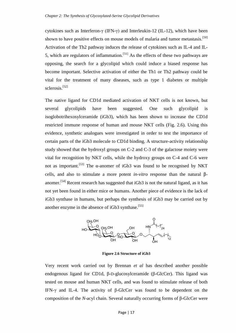
The native ligand for CD1d mediated activation of NKT cells is not known, but
several glycolipids have been suggested. One such glycolipid is
isoglobotrihexosylceramide (iGb3), which has been shown to increase the CD1d
restricted immune response of human and mouse NKT cells (Fig. 2.6). Using this
evidence, synthetic analogues were investigated in order to test the importance of
certain parts of the iGb3 molecule to CD1d binding. A structure-activity relationship
study showed that the hydroxyl groups on C-2 and C-3 of the galactose moiety were
vital for recognition by NKT cells, while the hydroxy groups on C-4 and C-6 were
not as important.[53]
The α-anomer of iGb3 was found to be recognised by NKT
cells, and also to stimulate a more potent in-vitro response than the natural β-
anomer.[54]
Recent research has suggested that iGb3 is not the natural ligand, as it has
not yet been found in either mice or humans. Another piece of evidence is the lack of
iGb3 synthase in humans, but perhaps the synthesis of iGb3 may be carried out by
another enzyme in the absence of iGb3 synthase.[55]
Figure 2.6 Structure of iGb3
Very recent work carried out by Brennan et al has described another possible
endogenous ligand for CD1d, β-D-glucosylceramide (β-GlcCer). This ligand was
tested on mouse and human NKT cells, and was found to stimulate release of both
IFN-γ and IL-4. The activity of β-GlcCer was found to be dependent on the
composition of the N-acyl chain. Several naturally occurring forms of β-GlcCer were

Chapter 2: The Synthesis of Glycosylated-Serine Glycolipid Derivatives
Page | 18
tested, with some derivatives, such as 2.4, displaying potent stimulatory activity, and
2.5 showing no activity (Fig. 2.7).[56]
Some other studies by Stanic et al have
reported β-GlcCer as being non-antigenic, but the length and degree of saturation of
the hydrocarbon chains was different to those tested here.[57]
Figure 2.7 Endogenous β-GlcCer derivatives
While the endogenous ligand for CD1d is almost certainly a β-linked glycolipid,
much evidence in the literature points to the potent NKT stimulatory abilities of α-
glycolipids. The glycoside which has received the most attention in recent years is α-
galactosylceramide.
2.3.3 α-Galactosylceramide
α-Galactosylceramide (α-GalCer) is an α-anomeric glycolipid, which is not present
in the mammalian body (Fig. 2.8). It was originally isolated from the marine sponge
Agelas mauritianus, but α-GalCer used in most biological studies is now synthesised
in the lab instead, and is known as KRN7000. KRN7000 is usually referred to as α-
GalCer, and this convention will be used for the purposes of this discussion. It has
been demonstrated that α-GalCer has potent immunomodulatory activity, and much
investigation has gone into the use of this glycolipid for the treatment of diseases
such as tuberculosis, cancer, and malaria.[58, 59]
Figure 2.8 Structure of α-GalCer
The stimulation of NKT cells by CD1d molecule presentation of α-GalCer rapidly
produces various cytokines, such as IL-4, IL-10, IFN-γ, and tumor-necrosis factor
(TNF). In-vivo administration of α-GalCer to mice has shown a characteristic

Chapter 2: The Synthesis of Glycosylated-Serine Glycolipid Derivatives
Page | 19
pattern. IL-4 tends to be the main cytokine secreted in the first 24 h, changing to an
IFN-γ dominated population after this time.[60]
While α-GalCer has shown promise as a potent immunomodulator, studies have
shown that administration can stimulate both the Th1 and Th2 immune responses.
This is a problem in the treatment of many diseases, such as autoimmune conditions,
where Th1 cytokines are thought to exacerbate symptoms.[53]
Other unwanted side-
effects such as liver toxicity and aggravation of atherogenesis in mice have also been
recorded.[61]
Copious studies have been carried out into the response generated by α-GalCer
administration to mouse and human subjects. The potential of α-GalCer is widely
acknowledged, but a biased immune response is required for therapeutic
applications. This has spurred work into the development of analogues of α-GalCer
and other glycolipids.
2.3.4 Synthetic Analogues; The Nature of the Lipid Chains
One point of structural variation is the length of the lipid chains. The hydrophobic
pockets of the CD1d molecule are of a specific size, and, by altering the length of the
lipid chain, the “fit” of the glycolipid tails can be modified. The length of the
hydrocarbon chain may also cause changes in the conformation of CD1d, which
could result in a different immune response upon NKT cell activation. Small
structural changes could help to generate a biased Th1 or Th2 response. One of the
first truncated glycolipid analogues which was tested is known as OCH (Fig. 2.9).
This glycolipid contains a shorter sphingosine chain; C5H11 compared to that of α-
GalCer, which is C14H29.
Figure 2.9 Structure of the truncated analogue OCH[62]
OCH has been shown to skew the release of cytokines towards a Th2 profile, with
IL-4 being the main cytokine produced. This kind of bias has been beneficial in
suppressing the myelin-antigen specific Th1 response in the mouse model of

Chapter 2: The Synthesis of Glycosylated-Serine Glycolipid Derivatives
Page | 20
multiple sclerosis. There is an obvious difference in cytokine stimulation by OCH,
compared to the unbiased response of α-GalCer administration.[62]
This is thought to
be because the shorter sphingosine chain of OCH is not as tightly bound into the
hydrophobic pocket of the CD1d molecule. The sugar moiety is then angled
differently, lessening interactions with amino acid residues of CD1d, resulting in a
shorter association time for the glycolipid/CD1d complex. The release of Th1
cytokines appears to require a longer period of NKT stimulation.[63, 64]
Other truncated analogues of α-GalCer have been synthesised, with several showing
a biased Th2 response. Shortening of the acyl chain, for example to C7H15, also
stimulates an increase in the release of Th2 cytokines.[65]
Care must be taken when considering much of the results of these tests. Often a
skewed response is noted when working with mouse cell lines, but when the testing
is carried out on human cell lines this skewing is not observed. An example of this
can be seen in a study carried out by Veerapen et al. This group synthesised several
truncated OCH glycolipid analogues (Fig. 2.10). They found that several were
active, and displayed a biased Th2 response with mouse iNKT (invariant NKT) cells.
The same compounds were active when tested with human iNKT cells, but a
nonbiased response was stimulated, with secretion of both IL-4 and IFN-γ.[66]
It has
been found that mouse NKT cells are phenotypically different to those expressed by
humans, with a different subset of NKT cells being present. This is especially noted
among human iNKT cells, which can be CD4+, CD8
+, or CD4
-CD8
-. Mouse iNKT
cells lack the CD8+ subtype. Healthy mice also possess more iNKT cells than
healthy humans. These differences can account for the variation in activity between
mouse and human NKT cells.[67]
Figure 2.10 Truncated OCH analogues synthesised by Veerapen et al[66]
Changes in the nature of the lipid chains described so far have mostly elicited a
biased Th2 response, but analogues have also been synthesised which encourage a

Chapter 2: The Synthesis of Glycosylated-Serine Glycolipid Derivatives
Page | 21
Th1, or IFN-γ, based response. Work carried out by Wong and co-workers has
produced compounds which stimulate a biased Th1 response in human iNKT cells.
They have carried out modifications to introduce a more polar phenyl ring onto the
end of the hydrophobic chain, with several compounds displaying the desired
activity (Fig. 2.11). These glycolipids were the first of such compounds to stimulate
a more potent release of IFN-γ than α-GalCer, and could have potential as adjuvants,
or antiviral agents. They also synthesised shorter chain derivatives, but these did not
show a biased response, indicating the need for the spacer chain.[68]
Figure 2.11 Glycolipid derivatives synthesised by Wong and co-workers[68]
Serine-based analogues have been investigated, such as that synthesised by Fan et al
(Fig. 2.12). This glycolipid was found to activate mouse NKT cells, but the response
was non-biased, and mild, with release of small amounts of both IL-4 and IFN-γ.
The fucosyl ceramide, and glucosyl-serine derivatives were also synthesised, but
showed no immune-activity.[69]
Figure 2.12 Serine-based glycolipid synthesised by Fan et al[69]
2.3.5 Synthetic Analogues; Glycosidic Bond Derivatives
Another modification which has been carried out is the replacement of the glycosidic
oxygen with a different atom, such as carbon, nitrogen, or sulphur.
One of the first such analogues was α-C-GalCer, 2.6, synthesised by Schmieg et al
(Fig. 2.13). In this derivative carbon replaces the more electrophilic oxygen atom. In
mouse studies α-C-GalCer was found to be 1000-fold more potent than its oxygen

Chapter 2: The Synthesis of Glycosylated-Serine Glycolipid Derivatives
Page | 22
counterpart, stimulating a biased Th1 cytokine response. The efficacy of this
analogue has been attributed to the resistance of the non-native carbon linkage to
enzymatic hydrolysis.[70]
When α-C-GalCer was tested with human iNKT cells the
results were a lot less promising. Further modifications involving C-glycosides
include the introduction of an alkene at the glycosidic linkage as in 2.7 (Fig. 2.13).
This derivative was shown to have potent human NKT stimulating effects, with a
bias towards a Th1 response.[71]
Figure 2.13 C-Glycosidic derivatives
Thio-derivatives have also been of interest. Thioglycoside analogues are those in
which the glycosidic oxygen has been replaced by a sulphur atom. An α-S-GalCer
derivative, synthesised by Zhu and co-workers, was found to stimulate the
production of cytokines by human NKT cells, with a comparable potency to α-
GalCer (Fig. 2.14). It was found that the thioglycoside did not stimulate mouse NKT
cells. This analogue shows promise as a positive control in biological studies, and
may be a more suitable derivative than α-GalCer, which also stimulates mouse NKT
cells. Although this derivative did stimulate cytokine release, a biased response was
not obtained.[72]
Figure 2.14 Thioglycoside derivative synthesised by Zhu and co-workers[72]
Another popular modification of the glycosidic linkage is to synthesise β-glycolipid
analogues. These are of great importance, as, although α-glycolipids have been
shown to be potent immunostimmulators, there are no α-glycosphingolipids known
in the human body, to date. The native ligand for CD1d must, therefore, be a β-

Chapter 2: The Synthesis of Glycosylated-Serine Glycolipid Derivatives
Page | 23
glycolipid, as discussed earlier, for example β-GlcCer.[56, 73]
Much investigation has
been carried out into the stimulation of NKT cells by β-glycolipids, and it has been
found that they are capable of this activation, albeit in a lower potency than their α-
linked analogues. In the binding cleft of the CD1d molecule α- and β-linked sugars
adopt different orientations. Observations by Pellicci et al show that, even though
the sugars orientation at the binding cleft is different, the TCR of the NKT cell is
capable of flattening the glycan so that both anomers possess a similar conformation.
This is also true for oligosaccharides, like iGb3. The less potent activity of β-
glycolipids is attributed to the energetic penalty which is incurred in order to distort
them into the shape of an α-linked glycoside.[74]
These results were strengthened by
Yu et al who carried out a similar study.[75]
2.3.6 Synthetic Analogues; β-Glucosylceramides
β-Glucosylceramides (β-GlcCer) are a naturally occurring family of glycolipids
found in the human body. β-GlcCer occurs in many different, closely related forms,
with variations on the hydrocarbon chains. Only certain forms are found in the body,
and, as discussed earlier, endogenous β-GlcCer derivatives have been found to have
NKT stimulatory properties.[56]
Kobayashi et al examined the effects of synthetic β-GalCer and β-GlcCer derivatives
on murine NKT cells. They found that the β-glycolipids did stimulate NKT cells, but
at a much lower level than their α-equivalents.[76]
Work carried out by Oku et al has shown that β-GlcCer (2.8) displays specific
cytotoxicity to tumor cells in-vivo. They also found that β-GalCer (2.9) has the same
effect, suggesting that the mode of action is not dependent on the specific sugar
structure (Fig. 2.15). These glycolipids were derived from malt feed. The effect of
the ceramide portion alone was investigated, and these compounds showed no
cytotoxicity towards cancer cells, indicating the need for the glycan component. This
may also be due to the hydrophobicity of the ceramide, which would alter its
solubility. Glycosylation of the ceramide would introduce a more hydrophilic nature
to the compound. This cytotoxicity is not attributed to the production of IFN-γ, as is
the case with some other glycolipid derivatives.[77]

Chapter 2: The Synthesis of Glycosylated-Serine Glycolipid Derivatives
Page | 24
Figure 2.15 β-Glycolipid derivatives which display cytotoxicity to tumor cells
Ya’acov et al have synthesised a thio-derivative of β-GlcCer, and have tested both
the O- and S- glycosides for their immunoregulatory activity (Fig. 2.16). They found
that both β-GlcCer and β-S-GlcCer led to a decrease in STAT-1 phosphorylation,
and a decrease in IFN-γ levels. This result is of importance in the treatment of auto-
immune diseases, for example in the alleviation of ConA immune-mediated
hepatitis. The thio-analogue was less susceptible to enzymatic degradation, and was
shown to increase the suppression of intra-hepatic NKT lymphocytes.[73]
Figure 2.16 The structures of β-GlcCer 2.10 and β-S-GlcCer 2.11
Several β-linked glycolipid derivatives have been tested for their immunostimulatory
ability, but, for the main part, they have shown weaker activity than their α-linked
counterparts. This should not be seen as a deterrent, as, even with weak activity, as
long as the immune response generated is biased, there may still be a use for β-
glycosides as immunomodulators.
2.3.7 Synthetic Analogues; Modification of the Carbohydrate Moiety
Modification of the sugar moiety is another area which has been investigated.
Several 6’-modified α-GalCer derivatives have been shown to stimulate cytokine
release from mouse NKT cells. The analogue synthesised by Trappeniers et al,
shown in Fig. 2.17, was found to induce a strong Th1 biased response.[78]
This
compound was later tested for activation of human NKT cells, and was found to
stimulate the release of cytokines.[79]

Chapter 2: The Synthesis of Glycosylated-Serine Glycolipid Derivatives
Page | 25
Figure 2.17 6’-Modified analogue synthesised by Trappeniers et al[78]
Substitution of the galactose sugar moiety for a different sugar has been carried out.
An example of this is β-mannosylceramide (β-ManCer), which has been shown to
activate both mice and human NKT cells (Fig. 2.18). Unusually, β-ManCer infers
some tumor immunity in mouse studies, but does not stimulate the release of IFN-γ,
which is thought to be the source of most analogues anti-tumor properties. It also
does not induce release of IL-4, or other commonly stimulated cytokines. The mode
of action of β-ManCer was investigated, and found to be due to the stimulation of
NOS (Nitric Oxide Synthase) and TNF-α. Mouse models were given an NOS
inhibitor called N-nitro-D-arginine-methyl ester. This NOS inhibition had no effect
on the tumor formation in mice treated with α-GalCer, showing that the immunity
given by α-GalCer is not dependent on NOS production. Conversely, the protection
brought about by β-ManCer was completely eradicated in these mice, showing the
NOS-dependent manner of protection by this analogue. NOS can be induced by
TNF-α. Blocking of TNF-α was also found to reverse tumor immunity.[80]
Figure 2.18 The structure of β-mannosylceramide
A non-glycosidic analogue, known as threitolceramide, has also been synthesised
(Fig 2.19). This derivative demonstrates stimulation of NKT cells, even without the
sugar moiety. The levels of cytokine release were lower than that of α-GalCer, but
the levels of NKT cell-mediated lysis of the antigen bearing cells (dendritic cells)
was also lowered. This is due to the lower affinity of the NKT cell TCR for
threitolceramide than for a glycan.[81]

Chapter 2: The Synthesis of Glycosylated-Serine Glycolipid Derivatives
Page | 26
Figure 2.19 Non-glycosidic analogue threitolceramide
2.3.8 A Multitude of Analogues
The glycolipid analogues discussed above are just a small selection of the work
which has been carried out in this area. Several reviews exist which detail many
more derivatives, some with promising effects.[53, 82]
It is a field which holds great
potential in the treatment of disease, and work will continue in the effort to
synthesise, or discover, a glycolipid which induces a strong biased response in
human models.
2.4 L-Serine
Serine is a naturally occurring amino acid, and is found in many proteins (Fig. 2.20).
Glycosylated serine is not often found in glycolipids, but is very common in
glycoproteins.[8]
Figure 2.20 The structure of L-Serine
Serine provides an attractive option to develop ceramide analogues, as it allows the
use of simple, well established peptide coupling methodology to introduce different
functionalities. Several serine-based glycolipids have emerged, which have been
demonstrated to be bioactive.
Achiwa and co-workers synthesised galactosamine and glucosamine serine-based
glycolipids (Fig. 2.21). The non-sulphated derivatives, 2.12 and 2.13, displayed no
anti-HIV activity, but the sulphated analogues, 2.14 and 2.15, showed good anti-HIV
activity, and were non-cytotoxic.[83]

Chapter 2: The Synthesis of Glycosylated-Serine Glycolipid Derivatives
Page | 27
Figure 2.21 Serine-based glycolipid derivatives
Huang et al have synthesised some serine-based glycolipids, and tested their activity
as Toll-like receptor 4 (TLR4) activators (Fig. 2.22). Lipopolysaccharides (LPS) are
amphiphilic glycolipid component of the outer membrane of Gram-negative bacteria.
When these bacteria enter the body, they are recognised by TLR4, which stimulates
the innate and adaptive immune systems. Compounds 2.16, 2.17 and 2.18 were all
shown to activate TLR4. Changes in the sugar moiety, and conversion to the β-
linkage in the glucosyl derivative 2.18 were tolerated. The β-anomer of the
galactosyl derivative resulted in a loss of activity.[84]
Figure 2.22 Serine-based glycolipids showing TLR4 activation
Anionic β-glycolipids 2.19 and 2.20 were synthesised by Faroux-Corlay et al, and
were found to display anti-HIV activity (Fig. 2.23). Binding studies carried out on
these glycolipids found that, on addition of gp120 (the envelope glycoprotein of
HIV), the gp120 inserted into monolayers of the galactolipids. This binding may be
responsible for the anti-HIV properties of these analogues.[85]

Chapter 2: The Synthesis of Glycosylated-Serine Glycolipid Derivatives
Page | 28
Figure 2.23 Anti-HIV glycolipids synthesised by Faroux-Corlay et al[85]
Serine-based glycolipid derivatives have also been used for a slightly different
purpose, with the β-GalCer analogue shown in Fig. 2.24 displaying potential for
inclusion in cosmetics and medicines, to improve skin barrier function. This
derivative increases β-glucocerebrosidase activity in-vitro, which is associated with
the skin barrier function. β-Glucocerebrosidase produces acylceramide, which is an
important lipid in the prevention of various skin diseases, such as dermatitis.[86]
Figure 2.24 β-GalCer derivative synthesised by Fukunaga et al[86]
2.5 Aim
The aim of this section was to synthesise some glucosamine-based serine glycolipid
derivatives (Fig. 2.25), and to have them tested on human NKT cell lines to assess
their potential as immunomodulators.
Figure 2.25 Glucosamine based serine glycolipid derivatives (n = 8 or 22)
As mentioned earlier, there are several advantages to incorporating serine into
glycolipids. The hydrocarbon chains can be introduced using peptide coupling
methodology of TBTU/HOBt, which is a well-established procedure. Serine is

Chapter 2: The Synthesis of Glycosylated-Serine Glycolipid Derivatives
Page | 29
structurally similar to ceramide, and it was postulated that small changes in this part
of the molecule may induce favorable differences in activation of NKT cells.
Glucosamine has been demonstrated to be involved with many regulatory processes
in the body.[18]
We wanted to synthesise glucosamine-based glycolipids in order to
investigate the effect that the acetamide group, at the C-2 position of GlcNAc, would
have on the glycolipids affinity for the CD1d molecule. It is thought that the
acetamide functionality may change the binding affinity, and may also alter the
profile of cytokine release by the activated NKT cell, as compared to that of
galactose or glucose-based analogues.
As discussed previously, it has recently emerged that the endogenous ligand for
CD1d cells could be β-GlcCer, and so it is important for new, synthetic β-glycolipid
analogues to be made.[56]
2.6 Synthesis of Glycosyl Building Block 2.22
Scheme 2.1 Retrosynthetic approach to the synthesis of glycosyl building block
The retrosynthetic scheme for the synthesis of a suitable building block is shown in
Scheme 2.1. These glycosides, 2.21 and 2.22, have been synthesised by multistep
procedures in the literature, but it was thought that a more succinct synthesis could
be possible.[87]
The protected serine acceptor, 2.24, was synthesised following
literature procedures, by alkylation of the carboxylic acid functionality of
commercially available L-Boc serine, with benzyl bromide, to produce the benzyl
ester 2.24 (Scheme 2.2).[88]
The free acid is protected to prevent undesired
glycosylation from occurring at the carboxylic acid position.

Chapter 2: The Synthesis of Glycosylated-Serine Glycolipid Derivatives
Page | 30
Scheme 2.2 Synthesis of glycosyl acceptor 2.24. Reagents and conditions:
i) BnBr, K2CO3, DMF, overnight, rt, 68 %
N-Troc glucosamine derivative 2.26, the first glycosyl donor prepared for the
synthesis of 2.21, was obtained following literature procedures.[89]
The N-Troc
protecting group was chosen for its stability to mildly acidic and basic conditions. It
is removed under very specific conditions, and is orthogonal to the other protecting
groups used. D-Glucosamine hydrochloride was first N-Troc protected at the amine
functionality, by reacting with trichloroethoxycarbonyl chloride (TrocCl), in the
presence of NaHCO3. This was followed by acetylation of the hydroxy groups with
acetic anhydride, in the presence of pyridine, to give the anomeric mixture of 2.26
(Scheme 2.3). This protection is necessary, as D-glucosamine contains four hydroxy
groups, each of which could act as the glycosyl acceptor, although the hydroxy
group at the C-1 position is the most reactive.
Peracetylated donors are well documented in the literature,[90]
and glycosylation of
2.24 directly from 2.26 was attempted, using BF3.OEt2 as the promoter. This was
unsuccessful from our donor, with full recovery of starting materials (Scheme 2.3).
Scheme 2.3 Glycosylation directly from N-Troc protected donor 2.26. Reagents and conditions: i)
TrocCl, H2O, NaHCO3, 2.5 h, rt; ii) Ac2O, pyr, overnight, rt, 87 % over two steps; iii) BF3.OEt2,
DCM, rt, overnight

Chapter 2: The Synthesis of Glycosylated-Serine Glycolipid Derivatives
Page | 31
As the above route was unsuccessful, an alternative donor was investigated.
Glycosyl halides are popular donors for the Köenigs-Knorr reaction.[91]
The glycosyl
halide is activated with metal salts, and the halide (Br or Cl) is substituted by an
alcohol. If there is neighboring group participation with the functionality at C-2, the
new glycosidic bond will be in the β configuration. Peracetylated glucosamine 2.30
was synthesised by reacting D-glucosamine hydrochloride with acetic anhydride, in
the presence of pyridine. 2.30 was treated with HBr/AcOH in an attempt to form
glycosyl donor 2.31 (Scheme 2.4). Some glycosyl halides, such as glycosyl iodides,
are very reactive and unstable, they are generally reacted in-situ.[92]
In contrast,
glycosyl bromides are usually relatively stable, and, for most sugars, can be prepared
in advance.[93]
In this case, using N-acetyl glucosamine, the bromide was never
isolated. It was found that, instead of detecting the bromine containing compound
2.31 as expected, the oxazoline derivative 2.32 was formed. This was confirmed by
MS, and also 1H NMR data was in agreement with reported values.
[94] It has been
reported in the literature that this oxazoline ring can be opened, in the presence of a
Lewis acid, to react with an alcohol acceptor.[95]
This reaction was attempted using
two different Lewis acid promoters, TMSOTf or BF3.OEt2. These reactions were
unsuccessful, and the starting material was recovered (Scheme 2.4).
Scheme 2.4 Attempted synthesis of glycosyl building block 2.21. Reagents and conditions:i) Ac2O,
pyr, rt, overnight, 34 %; ii) 33 % HBr/HOAc, DCM, 0 ˚C → rt, overnight, 44 % of 2.32; iii) TMSOTf
or BF3.OEt2, DCM, rt, overnight

Chapter 2: The Synthesis of Glycosylated-Serine Glycolipid Derivatives
Page | 32
As these syntheses were unsuccessful, it was decided to follow the well-established
trichloroacetimidate donor route. This synthesis involves several more steps than
those previously attempted, but the use of the imidate donor, by Schmidt and
coworkers, lends itself well to glucosamine.[87]
Although the reactions to synthesise glucosyl building block 2.22 have been reported
in literature,[87, 89]
some optimization was required. N-Troc protected 2.26 was
synthesised as described above. Selective deacetylation at the anomeric position
resulted in an anomeric mixture of hemiacetal 2.33. This reaction was initially
carried out in the presence of benzylamine. Dimethylamine later replaced
benzylamine, as an alternative nucleophile. This improved the purification of
compound 2.33. Synthesis of hemiacetal 2.33 was also carried out using hydrazine
acetate, which reduced the reaction time from overnight to 4 h. The free hydroxyl
group of 2.33 was treated with trichloroacetonitrile and sodium hydride to give
exclusively the α-imidate 2.34, which would serve as the glycosyl donor (Scheme
2.5). The α-anomer is the expected product of this reaction, due to the influence of
the anomeric effect on an electronegative substituent, and also encouraged by the use
of a strong base for deprotonation of the anomeric hydroxyl group. Imidate 2.34 was
also synthesized using DBU as an alternative base to NaH. The yields obtained were
comparable to the synthesis carried out with NaH (86 % with NaH, compared to 72
% with DBU), but DBU would be preferable to work with, as NaH is highly
flammable when exposed to water. Glycosylation of Boc protected serine acceptor
2.24 by glycosyl donor 2.34 was then carried out, using a 0.04 N solution of
TMSOTf in DCM as the promoter (Scheme 2.5).

Chapter 2: The Synthesis of Glycosylated-Serine Glycolipid Derivatives
Page | 33
Scheme 2.5 Synthesis of glycosyl building block 2.22. Reagents and conditions: i) TrocCl, H2O,
NaHCO3, 2.5 h, rt; ii) Ac2O, pyr, overnight, rt, 87 % over two steps; iii) NH(CH3)2, THF,
overnight, reflux, 81 %; iv) Cl3CCN, DBU or NaH, 2h, rt, 86 %; v) 0.04 N TMSOTf, DCM,
overnight, rt, 49 %
It is important that the glycosylation reaction is carried out under anhydrous
conditions. The presence of water can severely reduce the yield of the desired
product by attacking the activated glycosyl donor, in place of the glycosyl acceptor,
and decomposing it to hemiacetal 2.33. To minimise the amount of moisture in the
reaction several steps were taken; the glassware was dried in an oven overnight, the
reaction was carried out under a nitrogen atmosphere, and the reactants 2.34 and 2.24
were dried under vacuum for several hours. The glycosylation mechanism is shown
in Scheme 2.6, and examination of this mechanism illustrates how an unwanted side
reaction could occur in the presence of water. The water molecule could also attack
the glycosyl cation which is formed during the reaction, resulting in the synthesis of
hemiacetal 2.33.

Chapter 2: The Synthesis of Glycosylated-Serine Glycolipid Derivatives
Page | 34
Scheme 2.6 Mechanism of TMSOTf promoted glycosylation
The first step in the reaction involves attack on the silicon by the anomeric oxygen.
This places a positive charge onto the oxygen, in turn making the anomeric center
more electrophilic. The lone pair on the ring oxygen aids the departure of the leaving
group, and stabilises the oxocarbenium ion, 2.36, which is formed. The reaction
proceeds in an SN1 type fashion. The presence of the N-Troc protected neighbouring
group on C-2 of the sugar ensures that a β-glycosidic linkage is formed. The oxygen
lone pair of the N-Troc group attacks the anomeric center, forming an oxazoline-type
intermediate, 2.37. This neighbouring group participation only allows the alcohol
acceptor 2.24 to attack at the upper face of C-1, resulting in the β-glycoside 2.22
(Scheme 2.6).
2.7 Synthesis of Glycolipids 2.71 and 2.74
2.7.1 Route A
Orthogonal protecting groups are those which can be selectively removed, without
interfering with other protecting groups present in the molecule. This allows for
selective adaption of a specific functional group. The next stage in the synthesis

Chapter 2: The Synthesis of Glycosylated-Serine Glycolipid Derivatives
Page | 35
involved removal of these orthogonal protecting groups, from the glucosyl building
block, 2.22, to allow introduction of the hydrocarbon chains.
Selective removal of the benzyl ester protecting group from 2.22 was carried out by
hydrogenolysis, using a Pd/C catalyst, under H2, to give acid 2.38. This was
followed by introduction of the tetradecylamide chain, using standard coupling
conditions of TBTU and HOBt, in the presence of Et3N, resulting in 2.39 (Scheme
2.7).
Scheme 2.7 Synthesis of hydrocarbon chain amide derivative 2.39. Reagents and conditions:
i) Pd/C, H2,EtOAc, 2 h, rt, 75 %; ii) HOBt, TBTU, NH2C14H29, DCM, overnight, rt, 77 %
TBTU is often used in peptide synthesis methodologies as it greatly reduces the risk
of racemisation, and is useful for hindered couplings. The rate of reaction is quite
fast, preventing unwanted side reactions from occurring.[96]
The use of HOBt also
helps to prevent racemisation, and speeds up the rate of the reaction.[97]
TBTU is known as a “uronium” based coupling reagent, but it has been
demonstrated that, in the crystalline state, TBTU exists as the guanidinium N-oxide
derivatives 2.41 and 2.40.[98]
Another closely related reagent, HATU, also exhibits
this property. In solution both forms of the TBTU molecule are thought to be
present, and either can react with a carboxylate to form an activated ester.[99]
The
mechanism proceeds with attack of the carboxylate nucleophile 2.40 on the TBTU
carbocation to form the acyloxyuronium species 2.43. This intermediate immediately
reacts with the released BtO- 2.44, at the carbonyl carbon, to form the activated ester
2.45. The amino containing compound 2.46 can now attack the carbonyl carbon of

Chapter 2: The Synthesis of Glycosylated-Serine Glycolipid Derivatives
Page | 36
2.45 to form the amide bond, resulting in 2.48 (Scheme 2.8). The acid is generally
activated by stirring with base and the activating agent, i.e. TBTU, for 5-10 mins, to
ensure formation of the activated ester before addition of the amine.
Scheme 2.8 Mechanism of amide formation using the TBTU coupling reagent
Initially, N-Boc deprotection of 2.39 was attempted using the standard reaction
conditions of TFA in DCM (Scheme 2.9). The N-Boc group is an acid labile
protecting group, and should be easily removed under these conditions. The reaction
was carried out by adding a solution of 50 % TFA in DCM to a solution of 2.39, in
DCM, on ice, then allowing the reaction mixture to reach rt and stir for 2 h. Free
amine 2.49 was not produced in this reaction, with full recovery of the starting
material. The reaction was repeated, adding neat TFA to a solution of 2.39 in DCM

Chapter 2: The Synthesis of Glycosylated-Serine Glycolipid Derivatives
Page | 37
at rt, and left to stir overnight at rt. Starting material was still present after this
treatment, and none of the deprotected 2.49 was observed.
Scheme 2.9 Attempted deprotection of the N-Boc protecting group of 2.39. Reagents and
conditions: i) 50 % TFA, DCM, 1-24 h
It was postulated that the orientation of the long hydrocarbon chain may have been
presenting a steric hindrance, and so preventing removal of the N-Boc group. In
order to confirm this, a shorter chain propyl derivative, 2.50, was synthesised. The
N-Boc group was easily removed from this derivative, under the standard conditions
of TFA at rt overnight, to give 2.51 (Scheme 2.10). This was confirmed by MS
analysis, and also by the disappearance of the characteristic N-Boc CH3 peak at 1.4
ppm in the 1H NMR spectrum.
Scheme 2.10 Removal of N-Boc protecting group from propyl derivative 2.50. Reagents and
conditions: i) 50 % TFA, DCM, overnight, rt, 97 %
2.7.2 Route B
As the presence of the long hydrocarbon chain appeared to have prevented N-Boc
deprotection, the order of introduction of the hydrocarbon chains was modified.
From building block 2.22, N-Boc deprotection was carried out, with the addition of
TFA in DCM carried out on ice, and the reaction mixture allowed to stir at 0 ˚C for 1
h. The reaction was successful, resulting in compound 2.52 in good yield. This was
followed by coupling of decanoic acid to the amine 2.52, using the TBTU/ HOBt
methodology to give 2.53. Hydrogenolysis of the benzyl ester 2.53 was then carried
out, using Pd/C and H2, resulting in acid 2.54. Introduction of the second
hydrocarbon chain was attempted using the standard TBTU/HOBt coupling
conditions, but this reaction was unsuccessful (Scheme 2.11).

Chapter 2: The Synthesis of Glycosylated-Serine Glycolipid Derivatives
Page | 38
Scheme 2.11 Synthesis of protected glycolipid derivative 2.55. Reagents and conditions: i) 50 %
TFA, DCM, 0 ˚C, 1 h, 90 %; ii) HOBt, TBTU, Et3N, C9H19C(O)OH, DCM, overnight, rt, 88 %; iii)
Pd/C, H2, EtOAc, 2 h, rt, 98 %; iv) HOBt, TBTU, Et3N, NH2C14H29, DCM, overnight, rt
2.7.3 Various Coupling Conditions Investigated From Free Acid 2.54
The initial attempts to introduce a second hydrocarbon chain at the free acid, using
the standard coupling conditions shown above, were unsuccessful (Scheme 2.11).
The attempted coupling reaction resulted in degradation of compound 2.54. On
examination of the 1H NMR spectral data, and also by TLC analysis, hemiacetal 2.33
was identified. This was isolated by column chromatography, and was confirmed by
MS. The presence of 2.33 indicated that degradation had occurred during the
reaction. It was postulated that this degradation could have resulted from either
cleavage of the glycosidic bond by hydrolysis at the anomeric centre, or by α-H
elimination via a retro-Michael-type mechanism. Hydrolysis of 2.54 would result in
hemiacetal 2.33 and the functionalised serine 2.56 (Scheme 2.12). However,
hydrolysis of an acetal is normally observed under strongly acidic conditions, while

Chapter 2: The Synthesis of Glycosylated-Serine Glycolipid Derivatives
Page | 39
this reaction was carried out in a basic environment. Compound 2.56 was not
isolated, and was not detected in MS analysis of the crude reaction material.
Scheme 2.12 Degradation mediated by hydrolysis at the anomeric center
On the other hand, the retro-Michael reaction would yield a dehydroalanine type
product, 2.57, as well as hemiacetal, 2.33. The presence of the dehydroalanine
compound 2.57 was confirmed by MS analysis, which indicated that this degradation
was due to α-elimination (Scheme 2.13). On further examination of the literature, it
was found that these types of glycosylated serine compounds are indeed base
sensitive, with degradation occurring over ~ pH 10.5.[100, 101]
In fact, some groups
carry out this elimination on purpose, as the dehydroalanine acts as a protected form
of serine, which can be used in peptide synthesis.[102]
2.54 was stirred overnight in
DCM, in the presence of Et3N, without addition of the coupling reagents
(HOBt/TBTU). Degradation to the hemiacetal did not occur, with recovery of
starting material. This experiment demonstrated that the α-proton of 2.54 was not
acidic enough to be removed before being converted into the activated ester by
TBTU. The mechanism of the elimination observed with 2.54 is known as E1cB.
E1cB elimination is usually catalysed by a strong base, like KOH or Et3N, and is
made possible by an anion-stabilising group, like a carbonyl. The proton which is
adjacent to the activated carbonyl is very acidic. The base can remove this proton,
without a leaving group departing at the same time, due to delocalisation of the
negative charge onto the carbonyl group. The carbonyl stabilises the anion which is
formed, but elimination of a leaving group will still occur. The electrons flow back
down from the carbonyl oxygen, and the result is formation of an alkene containing
compound, and an elimination of the adjacent group (Scheme 2.13). This elimination
is promoted by the formation of the activated ester during the first stages of the
coupling reaction. The α-proton is made more acidic, and is easily deprotonated,
even in the presence of a mild base like pyridine (pKaH ~5.2). The activated ester
will quickly hydrolyse to the corresponding carboxylic acid, 2.57.

Chapter 2: The Synthesis of Glycosylated-Serine Glycolipid Derivatives
Page | 40
Scheme 2.13 Mechanism of E1cB elimination
In order to further confirm the presence of the dehydroalanine degradation
compound 2.57, a trapping experiment was performed, which involved carrying out
the standard coupling reaction in the presence of 2.2 equivalents of NBS (Scheme
2.14). The dehydroalanine compound, 2.57, can be unstable, due to addition
reactions at the double bond. The NBS acted as a source of electrophilic bromine,
which could “trap” the dehydroalanine as it is formed, by addition of bromine across
the alkene bond to give compound 2.58.[103]
The presence of this compound was
confirmed by MS.

Chapter 2: The Synthesis of Glycosylated-Serine Glycolipid Derivatives
Page | 41
Scheme 2.14 NBS trapping of dehydroalanine 2.57. Reagents and conditions: i) HOBt, TBTU,
Et3N, DMF, NBS, 2 h, rt
Various different conditions to avoid the undesired elimination, as outlined in Table
2.1, were investigated for this coupling reaction. Entry 1 shows the standard
conditions which were initially investigated. Entry 2 was carried out in the absence
of HOBt, to investigate if this additive may be enhancing the degradation, but
hemiacetal 2.33 was still detected after the reaction. Entry 3 shows the reaction
carried out with less equivalents of the base Et3N, which made no difference to the
reaction, with MS showing the presence of hemiacetal 2.33, and also some
remaining starting acid 2.54. Entry 4 was carried out in the presence of a more
hindered base, DIPEA, to see if this would prevent the elimination, but hemiacetal
was again detected in this experiment. Entries 5 and 6 show the reaction carried out
in less basic conditions, using pyridine and N-methyl morpholine respectively, but
hemiacetal 2.33 was again produced in both reactions. From this data it is clear that
the nature of the base is not important. Deprotonation of the α-H occurred in all
cases where the activated carboxylic acid, 2.58, described earlier, was being
generated using the TBTU and HOBt coupling conditions. It was then decided to
change the method of coupling, and the acid chloride of 2.54 was synthesised. This
was carried out by reaction of the acid 2.54 with oxalyl chloride, in DMF, followed
by addition of the long chain tetradecylamide, in the presence of pyridine (entry 7).
These alternative conditions did not yield the desired product, 2.55, but, again, the
presence of starting acid 2.54 and dehydroalanine 2.57 were confirmed by MS.

Chapter 2: The Synthesis of Glycosylated-Serine Glycolipid Derivatives
Page | 42
Entry Coupling reagent HOBt Base (equiv.)
1 TBTU Y NEt3 (2)
2 TBTU N NEt3 (2)
3 TBTU Y NEt3 (1.2)
4 TBTU N DIPEA (2)
5 TBTU N Pyridine (2)
6 TBTU N N-methyl-morpholine(2)
7 (COCl)2 N Pyridine (2)
Table 2.1 Investigation of the coupling conditions using compound 2.54
In order to investigate if the observed E1cB elimination would occur with any
functionality in the amide position, lysine derivative 2.59 was synthesised.
Introduction of the tetradecylamine chain was attempted using the standard
TBTU/HOBt conditions (Scheme 2.15). This was also unsuccessful, with hemiacetal
2.33 again being isolated. This suggested that activation of any amide containing
derivative of 2.54 would result in degradation.

Chapter 2: The Synthesis of Glycosylated-Serine Glycolipid Derivatives
Page | 43
Scheme 2.15 Attempted coupling of second chain to lysine derivative 2.59. Reagents and
conditions: i) HOBt, TBTU, Et3N, NH2C14H29, DCM, overnight, rt
Another approach which was taken in the synthesis of 2.55 was to carry out the
glycosylation on an acceptor which had already been functionalised with both
hydrocarbon chains, 2.63 (Scheme 2.17). The synthesis of this acceptor is shown in
Scheme 2.16, below. The tetradecylamine chain was introduced using TBTU and
Et3N, to give 2.61. This was followed by deprotection of the N-Boc group, using a
50 % solution of TFA (v/v) in DCM, to give the free amine, 2.62. Interestingly, this
deprotection did not present the difficulty which had been encountered in the
presence of the sugar moiety in 2.39. The second hydrocarbon chain was introduced
into the molecule, again using TBTU and Et3N, resulting in 2.63 (Scheme 2.16).
Scheme 2.16 Synthesis of lipid 2.63. Reagents and conditions: i) TBTU, Et3N, DCM,
NH2C14H29, rt, overnight, 75 %; ii) 50 % TFA, DCM, 5 h, rt, 91 %; iii) TBTU, C9H19C(O)OH,
DMF, Et3N, overnight, rt, 32 %

Chapter 2: The Synthesis of Glycosylated-Serine Glycolipid Derivatives
Page | 44
Scheme 2.17 Attempted glycosylation of acceptor 2.63. Reagents and conditions: i) 0.04 N
TMSOTf or BF3.OEt2, DCM, overnight, rt
Direct glycosylation of a serine acceptor, which had been functionalised with one
hydrocarbon chain, has shown success in the literature,[43]
using BF3.OEt2 as the
promoter. For our acceptor the reaction did not yield any of the desired product, with
recovery of hemiacetal 2.33, and the functionalised serine. It was postulated that this
reaction did not progress due to a steric hindrance caused by the presence of the
second hydrocarbon chain.
In view that degradation was observed, even in the presence of a very mild base such
as pyridine, introduction of the second chain to the free acid was carried out in the
absence of base (Scheme 2.18). This reaction produced the desired product 2.55,
which was confirmed by MS, but in low yield of 13 %.
Scheme 2.18 Coupling of tetradecyl chain in the absence of base. Reagents and conditions: i)
TBTU, HOBt, NH2C14H29, DCM, overnight, rt, 13 %
From glycolipid 2.55 the removal of the N-Troc protecting group was attempted,
using the standard conditions of Zn, Ac2O and Et3N described by Zhu and Schmidt
(Scheme 2.19).[104]
This reaction was unsuccessful, and starting material was
recovered. It was thought that, again, the presence of the long hydrocarbon chains
was posing a steric hindrance.

Chapter 2: The Synthesis of Glycosylated-Serine Glycolipid Derivatives
Page | 45
Scheme 2.19 Attempted deprotection of N-Troc from 2.55. Reagents and conditions: i) Zn, Ac2O,
Et3N, overnight, rt
2.7.4 Route C
It was observed in route B that degradation occurred when activating the carboxylic
acid of 2.54 to introduce the second hydrocarbon chain. It had also been observed, as
discussed in route A, using the N-Boc protected building block 2.22, that
functionalisation could not be carried out at the acid functionality first, due to
problems with N-Boc deprotection. For this reason N-Cbz protected glycosyl
acceptor 2.65 was synthesised. Only one hydrocarbon chain was introduced into this
acceptor, and so the problem encountered with acceptor 2.63 could be avoided. The
N-Cbz protecting group is removed by hydrogenolysis, which would not be
orthogonal to the benzyl protecting group of the serine acceptor, 2.24, utilised in
earlier syntheses. For this reason, introduction of the tetradecyl chain was carried out
before glycosylation, removing the need for benzyl ester protection (Scheme 2.20).
Scheme 2.20 Coupling of the tetradecyl chain to N-Cbz serine. Reagents and conditions: i)
TBTU, HOBt, Et3N, NH2C14H29, DCM, overnight, rt, 55 %
Imidate glycosyl donor 2.34 was used for the glycosylation reaction. TMSOTf was
again used as the promoter for the reaction, resulting in 2.67. This was followed by
deprotection of the N-Troc protecting group using standard conditions of freshly
activated zinc, and acetic anhydride to give 2.68. Hydrogenolysis of the N-Cbz
protecting group was then carried out, using Pd/C under a H2 atmosphere, to give
amine 2.69 (Scheme 2.21). The order of deprotection is crucial to this synthesis.
When hydrogenolysis of the N-Cbz group was attempted from 2.67, prior to

Chapter 2: The Synthesis of Glycosylated-Serine Glycolipid Derivatives
Page | 46
reduction of the N-Troc group, this step was unsuccessful, with return of starting
material (Scheme 2.22). Also, it was previously noted in compound 2.55 (Route B)
that reduction of the N-Troc protecting group could not be carried out once the
compound was functionalised with both lipid chains.
Coupling of decanoic acid to the free amine 2.69 was carried out using TBTU and
HOBt, in the presence of Et3N, to give the protected glycolipid 2.70. This was
followed by selective hydrolysis of the O-acetyl protecting groups, leaving the N-
acetyl group intact, using MeOH/H2O/Et3N (Scheme 2.21). Standard Zemplén
conditions of NaOMe/MeOH for deacetylation were not used, as these were
anticipated to be too harsh, and may also remove the N-acetyl protecting group. Fig.
2.26 shows the 1H NMR spectrum of the deprotected glycolipid 2.71. The doublet at
approx. 8.8 ppm is attributed to the NHAc amide proton, with the signal at 8.5 ppm
being attributed to the amide protons of both hydrocarbon chains. The resonance of
the α-anomeric H-1 is observed at 5.3 ppm, while the β-anomeric H-1 is seen at a
lower ppm of 5.1, which is characteristic of each of these anomers. The singlet at 2.1
ppm represents the methyl protons of the NHAc group, and integrates for three
protons. Most of the long hydrocarbon chain CH2 protons are observed as a multiplet
at 1.2 ppm. The analysis was carried out in deuterated pyridine, as residual solvent
peaks from deuterated DMSO would have obscured the sugar peaks. Solubility was
also poor in other solvents, and a sharp, clear spectrum was obtained in pyridine.

Chapter 2: The Synthesis of Glycosylated-Serine Glycolipid Derivatives
Page | 47
Scheme 2.21 Synthesis of glycolipid 2.71. Reagents and conditions: i) 0.04 N TMSOTf, DCM, rt,
overnight, 53 %; ii) Zn, Et3N, Ac2O, rt, 5 h, 70 %; iii) H2, Pd/C, EtOAc, 2 h, rt, 95 %; iv) HOBt ,
TBTU, Et3N, C9H19C(O)OH, DMF, overnight, rt, 40 %; v) Et3N/MeOH/H2O, 40 °C, overnight, 41 %
Scheme 2.22 N-Cbz deprotection from 2.67. Reagents and conditions: i) H2, Pd/C, EtOAc,
overnight, rt

Chapter 2: The Synthesis of Glycosylated-Serine Glycolipid Derivatives
Page | 48
Figure 2.26 1H NMR spectrum of deprotected glycolipid 2.71 in C5D5N
A longer hydrocarbon chain derivative, 2.74, was synthesised, which would be more
comparable to other glycolipids which have been tested for their immunomodulatory
activities, like α-GalCer. Tetracosanoic acid was coupled to 2.69, using TBTU and
HOBt, in the presence of Et3N, to give glycolipid 2.73. The low yield of this reaction
is due to the poor solubility of tetracosanoic acid in either DCM or DMF. This
glycolipid analogue was then subjected to the same deprotection conditions as the
shorter chain derivative, resulting in deprotected glycolipid 2.74 (Scheme 2.23).

Chapter 2: The Synthesis of Glycosylated-Serine Glycolipid Derivatives
Page | 49
Scheme 2.23 Synthesis of tetracosonoyl glycolipid derivative 2.74. Reagents and conditions: i)
TBTU, HOBt, Et3N, C23H57C(O)OH, DMF, overnight, rt, 17 %; ii) Et3N/MeOH/H2O, 45 °C,
overnight, 57 %
2.8 Biological Evaluation of Glycolipids 2.71 and 2.74 as
Immunomodulators
Deprotected glycolipids 2.71 and 2.74, and also lipid 2.63 were tested by Dr. Derek
Doherty, Andreea Petrasca, and Niamh Murphy at St. James’ Hospital, Dublin for
their potential as immunostimulators (Fig. 2.27). Glycolipids 2.71 and 2.74 are novel
glycolipids and differ only in the length of one hydrocarbon chain, while 2.63 has
been recorded in the literature,[105]
and does not contain the sugar moiety. 2.63 was
included in the testing to investigate if the sugar moiety was required for iNKT
stimulation and cytokine release, or if a different cytokine profile would be induced
by the glycosides.

Chapter 2: The Synthesis of Glycosylated-Serine Glycolipid Derivatives
Page | 50
Figure 2.27 Lipids tested for immunostimulatory activity
The aim of this study was to investigate the potential of these glycolipids to
stimulate iNKT cells to mediate cytotoxicity, T helper function and cytokine
secretion.
2.8.1 Materials and Methods used for Testing[106]
iNKT cell lines were co-cultured with cancer cells in the presence of the above
glycolipids, and assessed using flow cytometry and ELISAs (enzyme-linked
immunosorbent assays). Two different types of cell line were transfected with CD1d;
HeLa cells were derived from cervical cancer cells, and C1R cells are a mutant
lymphoblastoid cell line. Testing was carried out using literature procedures,
described by Hogan et al.[72]
α-GalCer was used as the positive control, and gave a
positive result for all ELISAs. The negative control was HeLa-CD1d cells with
iNKT cells but no compound added (vehicle). This gave a baseline result with which
to compare the results of the novel glycolipids.
2.8.2 Ability of Glycolipids to Induce Cytotoxicity
iNKT cells have the ability to induce an immune response and also to mediate
cytotoxicity, which accounts for their antitumor activity. In this study, cytotoxicity
was evaluated by flow cytometric analysis of CD107a positive cells. CD107a is a
lysosomal protein which lines the surface of cytolytic granules and lysosomes, and it
becomes exposed to the surface during degranulation. This makes it an ideal marker
for assessing cytotoxic activity.[107]
Glycolipids 2.71 and 2.74, and lipid 2.63 were
tested for their ability to prime iNKT cell lines for cytotoxicity, using iNKT cells

Chapter 2: The Synthesis of Glycosylated-Serine Glycolipid Derivatives
Page | 51
from four different donors. In the presence of C1R cells only α-GalCer was able to
cause CD107a externalisation, the other lipids did not induce any cytotoxicity
against this cell line. In the presence of CD1d-HeLa cells, 2.71 and 2.74 were able to
externalise CD107a in just one iNKT cell line, while 2.64 induced CD107a
expression by two cell lines (Fig. 2.28). These results indicate that the cells are
undergoing degranulation, and so are mediating cytotoxicity.
Figure 2.28 iNKT cell cytotoxicity in four different donors (BC1-4) after exposure to HeLa
cells presenting various lipids
These lipids were also tested for induction of CD40L expression by iNKT cells,
which is located on B-cells, dendritic cells and macrophages. This protein is
important in T-cell activation and also plays other roles in immune stimulation.[108]
2.71, 2.74 and 2.63 did not induce expression of CD40L, indicating that these
compounds would not play a part in induction of T-helper function (data not shown).
The iNKT cell lines which gave a CD107a+ result were examined to find out what
subsets they contained. It has been reported that therapeutic outcomes can vary in
patients expressing different subset compositions. iNKT cells can be CD4+, CD8
+ or
CD4-CD8
- (Double negative, DN); CD4
+ iNKT cells expand more readily than the
others upon mitogen stimulation, while CD8+ and CD4
-CD8
- iNKT cells most
potently killed CD1d+ cell lines and primary leukaemia cells.
[109] In this study the
CD107a+ cells were found to be mostly CD8
+, and CD107a expression for each of
the subsets is illustrated in Fig. 2.29 below. CD40L+ cells were found to be mainly

Chapter 2: The Synthesis of Glycosylated-Serine Glycolipid Derivatives
Page | 52
CD4+, which could account for the lack of CD40L expression by the glycolipids
tested.
Figure 2.29 Expression of CD107a by iNKT subsets in two cell lines
2.8.3 Analysis of the Cytokine Release Profile Induced by Glycolipids
The extracellular cytokine expression induced by the different lipids was determined
using ELISAs to measure cytokine concentrations in the supernatant, 24 h after lipid
stimulation of iNKT cells. α-GalCer was the only lipid tested that showed evidence
of IL-4, IFN-γ, IL-13 and GM-CSF secretion (Fig. 2.30).

Chapter 2: The Synthesis of Glycosylated-Serine Glycolipid Derivatives
Page | 53
Figure 2.30 Effect of glycolipid stimulation on extracellular cytokine secretion
The intracellular cytokine profile was also examined by flow cytometry. This
intracellular cytokine assay only measures the percentage of cytokine-secreting cells,
and so does not actually quantify how much cytokine secretion is induced.
Glycolipid 2.71, which is the shorter chain derivative tested, was shown to weakly
induce secretion of Th2 cytokine IL-4 in each of the four cell lines (BC1-4), and also
IFN-γ and TNF-α secretion in two cell lines (BC1-2). The percentage of IFN-γ
positive cells induced by 2.71 was higher than that induced by α-GalCer in cell lines
BC1-2. The longer chain derivative 2.74 and the non-glycosidic 2.63 failed to induce
an intracellular secretion of any of these cytokines (Fig. 2.31).

Chapter 2: The Synthesis of Glycosylated-Serine Glycolipid Derivatives
Page | 54
Figure 2.31 Intracellular cytokine profile
As our glycolipids did not illicit a strong release of the typical cytokines examined
for immunostimulation, they were tested for their ability to produce IL-10. This
cytokine has become of interest in the area of immunoregulation, and is known as a
suppressor cytokine.[110]
IL-10 has been found to display immunosuppressive
properties, including inhibition of inflammatory cytokine production (such as IL-4),
which could be used in the treatment of certain diseases, such as allergic responses.
Immunostimulatory abilities of IL-10 were also noted, for example proliferation and
differentiation of B cells.[111]
All of our glycolipids tested induced secretion of IL-10,
with 2.74 showing the highest release of IL-10, followed by 2.71, and a lesser
stimulation was noted with the non-glycosidic derivative 2.63 (Fig. 2.32). This again
shows the importance of the sugar moiety in cytokine release. 2.71 induced a more

Chapter 2: The Synthesis of Glycosylated-Serine Glycolipid Derivatives
Page | 55
potent response than α-GalCer at a concentration of 100 ng/mL. 2.73 also induced a
strong response, but, as the 100 ng/mL measurement was not carried out, it cannot be
said with certainty that the level of IL-10 released was higher than α-GalCer. The
suppressive nature of IL-10 could account for the failure of these lipids to stimulate
the release of IL-4 in the earlier studies. The testing was carried out at different
concentrations of the lipids to investigate if the response was dose dependant. Some
measurements were not carried out due to the amount of sample available. From Fig.
2.32 it can be observed that the response did not increase with higher dosage levels.
In this study medium alone was used as the negative control, and phorbol myristate
acetate (PMA) with ionomycin was used alongside α-GalCer as the positive control.
Figure 2.32 Stimulation of IL-10 production by novel glycolipids
2.8.4 Summary of Immunological Results
2.71, the decanoic acid derivative, was shown to illicit a non-biased release of
cytokines, with induction of both Th1 (IFN-γ and TNF-α) and Th2 (IL-4) responses.
2.71 was found to lack strong cytotoxicity. This lack of cytotoxicity may be due to a
structural change induced when 2.71 binds to the CD1d molecule, which stimulates
cytokine release, but makes it non-cytotoxic. The tetracosanoic acid derivative, 2.74,
and the non-glycosidic 2.63 both failed to activate the iNKT cells in this study. As
2.63 is structurally similar to 2.71, with equivalent lipid length and character, the
lack of iNKT cell activation by this compound demonstrates the necessity of the
glycan moiety.

Chapter 2: The Synthesis of Glycosylated-Serine Glycolipid Derivatives
Page | 56
All of the lipids 2.63, 2.71 and 2.74 induced release of IL-10, the suppressor
cytokine. The non-glycosidic 2.63 did not illicit as potent a response as the glycosyl
derivatives. This result indicates a possible use in the treatment of Th2 aggravated
diseases.
2.9 Chapter Conclusion
To summarise, two glycolipid derivatives, 2.71 and 2.74, were successfully
synthesised, and showed some interesting biological results, such as the stimulation
of the release of Il-10. Several barriers were overcome throughout the synthesis,
including the problems presented by the lipid chains in the removal of protecting
groups. Another issue which was encountered during the synthesis of these
derivatives was the elimination of the sugar moiety, under basic coupling conditions.
This synthesis demonstrated the importance of the order of reactions in the synthetic
scheme.
Future work could include the synthesis of a derivative in which the length of the
other hydrocarbon chain, tetradecyl in each of our glycolipids, is modified. The α-
anomeric analogue could also be synthesised, in order to compare the biological
activity of this glycolipid.
As will be discussed in chapter 4, functionalisation at the C-6 position of the sugar
could be carried out. The introduction of other biologically relevant groups at this
position may enhance the immunomodulatory activity of the glycolipid.

Chapter 3: The Synthesis of a Series of Glycopeptides
Page | 57
Chapter 3: The Synthesis of a Series of Glycopeptides
3.1 Introduction: Glycopeptides
A large amount of peptides in the body have been found to be glycosylated.
Glycosylation involving N-acetyl glucosamine (GlcNAc) plays an important part in
the regulation of protein function, for example in the stability or targeting of a
glycoprotein. This has already been discussed in detail in chapter 1. The protein
which we will be focusing on is the tau protein, which has been implicated in the
development of neurodegenerative disorders, such as Alzheimer’s disease.
3.2 Alzheimer’s Disease
Alzheimer’s disease (AD) is the most common form of dementia, accounting for 50-
60 % of all cases. It is a progressive neurological condition, which is manifested by
debilitating effects. It can become more and more difficult for sufferers of AD to
remember, reason and use language. The loss of memory of recent events is often
one of the first difficulties to be noticed. The person may also become disorientated,
be at a loss for a word when speaking, and have increasing difficulty with simple
daily tasks, such as using the phone. The risk of developing AD increases with age,
with more than 25 % of those over the age of 80 suffering from the disease.
Although it is rare, AD can also occur in people under 65.[112]
AD is a complicated disease, with multiple factors influencing its onset. The brains
of people that suffer from AD display pathological hallmarks of the disease:
Neuronal and synaptic loss, neurofibrillary tangles, and β-amyloid (Aβ) plaques.
These plaques were first reported in 1907 by Alois Alzheimer.[113]
Neurofibrillary
tangles (NFT) are twisted, stringy fibres of tau protein, which accumulate inside
brain cells, and disrupt normal brain cell function. These will be discussed in more
detail later. Aβ plaques are insoluble deposits of β-amyloid peptide, and are thought
to accumulate due to mutations in the amyloid precursor protein. Studies carried out
by Shankar et al have shown that the plaques did not impair cognitive function, but
that soluble Aβ dimers which were released from the plaques could cause AD-like
symptoms.[114]
Misfolding of Aβ may trigger events which result in aggregation of
Chapter 3: The Synthesis of a Series of Glycopeptides
Page | 58
the tau protein.[115]
A third abnormality, degeneration of cholinergic neurons, has
also been reported. This neuronal death leads to the cognitive impairment associated
with AD. Therapies targeting this pathological marker of AD aim to promote the
activity of surviving cholinergic neurons.[116]
For the purposes of this discussion, the molecular target which is focused on will be
the hyperphosphorylation of the tau protein, and development of NFT.
The treatment of AD is complicated for several reasons. The first is the contribution
of different factors to the development and progression of the disease, as mentioned
above. The second reason is the lack of diagnostic techniques. A person suffering
from AD is often not diagnosed until the later stages, when the disease has
progressed, and symptoms, such as memory loss, have become pronounced. New
techniques are being pioneered for the diagnosis of AD. The use of neuroimaging-
biomarkers will allow for earlier diagnosis, although the pathological markers of AD
mentioned above need to have developed. Techniques such as high-field MRI of
brain atrophy, and in-vivo amyloid plaque imaging, using radio-labelled ligands, may
allow for diagnosis of AD at a stage where its progression could be curbed.[117]
3.3 Tau Protein
Tau is a microtubule-associated protein (MAP) which is expressed throughout the
central nervous system (CNS), but is mostly found in neuronal axons. Tau plays an
important part in the organisation of microtubules in these axons. Microtubules are
involved in cell shape maintenance and serve as channels for axonal transport. Tau
association reduces the probability that a microtubule will shift from the growing
phase to the shrinking phase. Partial phosphorylation of serine and threonine
residues of tau is important to regulate the growth of microtubules by decreasing the
affinity that tau has for the microtubule lattice. It has been shown that
hyperphosphorylation of the tau protein causes destabilisation of microtubules by
preventing binding.[118]
Phosphorylation of tau is carried out by phosphate kinases,
such as glycogen synthase kinase 3 (GSK-3), extracellular signal-regulated kinase 2
(ERK-2), and cyclin-dependant kinase 5 (CDK-5) (Scheme 3.1). Phosphate kinases
use adenosine-5’-triphosphate (ATP) as their source of phosphate.[119]

Chapter 3: The Synthesis of a Series of Glycopeptides
Page | 59
The tau protein is also modified by addition of O-GlcNAc to specific serine or
threonine residues in the sequence.[120]
As mentioned in chapter 1, glycosylation is
carried out by the enzyme O-GlcNAc transferase (OGT). Glycosylation has been
shown to occur at many of the same sites as phosphorylation, and also to regulate the
phosphorylation of adjacent sites. Studies have demonstrated that inhibition of tau
phosphorylation results in increased glycosylation, and that targeted deletion of OGT
causes hyperphosphorylation of tau.[121]
This is yet another example of the dynamic
interplay observed between glycosylation and phosphorylation of proteins, which
was discussed in chapter 1 (Scheme 3.1).
Scheme 3.1 The dynamic relationship between tau phosphorylation and glycosylation
Tau protein is translated from a single gene located on chromosome 17. Six different
isoforms of tau have been found, each generated from alternative mRNA splicing at
chromosome 17. These isoforms range from 352-441 amino acids in length. A
highly conserved repeat region of 31 or 32 amino acids has been determined in all of
these isoforms. This region also repeats itself several times within a single
isoform.[122]
Further study into the repeat region has revealed a strong Pro-Gly-Gly-
Gly motif, which has been shown to represent the tubulin binding domain. The
longest isoforms of tau contain up to 79 serine and threonine residues at which
phosphorylation can occur. Only about 30 of these residues are actually
phosphorylated at one time under normal physiological conditions.[123]
3.4 Tau and AD
In patients with AD, tau shows abnormal hyperphosphorylation, which causes it to
aggregate into paired helical filaments (PHF) and form neurofibrillary tangles
(NFT). When this happens, the microtubules disintegrate, collapsing the neuron's
transport system. This may result first in communication malfunctions between
neurons, and later in cell death (Fig. 3.1).[115]
Tau has also been implicated in other

Chapter 3: The Synthesis of a Series of Glycopeptides
Page | 60
neurodegenerative diseases, such as Parkinson’s disease and Pick’s disease. These
diseases are collectively known as tauopathies.[124]
Figure 3.1 a) The stabilising effect of the tau protein on a healthy microtubule, and b) Collapse
of a diseased neurons transport system[115]
Studies have shown that NFT contain all six hyperphosphorylated isoforms of the
tau protein. A total of 25 serine/threonine sites of abnormal phosphorylation have
been identified in AD.[125]
It has been observed that almost all of these sites of
phosphorylation are also found in non-aggregated tau, but that the tau protein which
is found in NFT is phosphorylated to a higher extent.[126]
While there are many factors which contribute to the progression of AD, the loss of
function of the tau protein is the main element. Initially, it was thought that the cause
of AD progression was either Aβ plaques or NFT formation. After many years of
investigation, it appears that there is a definite interplay between these two
pathological markers, and that neither is independent of the other. In a study carried
out by Roberson et al, reduction of endogenous levels of tau protein has prevented
the behavioural deficits originally associated with Aβ plaques, but the extent of Aβ
plaque formation is not affected.[127]
This study, and other evidence, point to a
synergistic relationship between tau and Aβ, but also show the central role played by
the tau protein in AD.[128, 129]
This makes tau an interesting molecular target for
treatment of AD, and other tauopathies.
Han et al have carried out work which shows the importance of the site of
phosphorylation of tau. They found that, by encouraging phosphorylation in-vitro at

Chapter 3: The Synthesis of a Series of Glycopeptides
Page | 61
ser202
, of wild-type (WT) tau and four tau mutants, the microtubule assembly-
promoting ability of tau was inhibited. This phosphorylated species has been isolated
from the brains of AD sufferers. It is suggested that phosphorylation at Ser202
is a
major event which leads on to NFT development in AD, and also in other
tauopathies. Phosphorylation was carried out by the cyclin-dependant protein kinase
5 (CDK-5).[130]
Bramblett et al have shown that tau protein isolated from PHF is
hyperphosphorylated, it does not bind to microtubules or promote microtubule
assembly in-vitro. Once this tau protein has been dephosphorylated, it recovers the
ability to bind to, and promote, microtubule assembly. They also demonstrate the
inhibitory effect that hyperphosphorylation at Ser396
of tau has on binding
microtubules.[131]
Several other sites of phosphorylation have been implicated in the development of
the pathological markers of AD, such as Ser262
, Ser356
, and Thr231
. It is thought that a
combination of phosphorylated residues, and not just a specific one, is responsible
for the neurotoxic effects of tau.[132]
Mutation of 14 sites of phosphorylation in the
tau protein to alanine, in place of serine and threonine, resulted in a lack of tau-
induced toxicity. When individual residues were mutated to alanine, toxicity was still
observed. This study indicated that no particular site was responsible for tau
toxicity.[133]
Transgenic mice which have been made to express human tau have shown
neurological deficits and neuronal loss, accompanied by the appearance of NFT.[134]
It had been assumed that these NFT were responsible for the cytotoxic effects of tau
protein, but recent findings, using a mouse model of AD, appear to show that this is
not the case. Santacruz et al have engineered mice to express a repressible human tau
variant. These mice developed progressive age-related NFT, neuronal loss,
behavioural impairments and decreased cognitive function. When this human tau
was suppressed, the mice recovered cognitive function and neuron numbers
stabilised. In older mice models NFT continued to accumulate, but behavioural
impairments did not return. This study showed that NFT alone are not enough to
cause neuronal loss or behavioural impairments. It is more likely that the loss of
normal function of the tau protein, due to hyperphosphorylation, and its aggregation

Chapter 3: The Synthesis of a Series of Glycopeptides
Page | 62
into soluble oligomers, are the causes of neuronal and cognitive function loss. It also
raises the question as to whether the NFT are markers of the disease, or if they are a
protective mechanism of the body’s, allowing transformation of a neurotoxic species
(soluble, hyperphosphorylated tau) into a less toxic form, as seen for the Huntington
protein.[135]
In some neurodegenerative disease models this aggregation into a less
toxic species, followed by elimination from the body by proteasomal activity, or
autophagy, has been observed, but this has yet to be proven in AD.[136]
3.5 Current Tau–Focused Strategies for the Treatment of AD
While much work is being carried out which targets Aβ plaques and cholinergic
neurons, this discussion will focus on tau-based strategies for the treatment of AD.
3.5.1 Microtubule Stabilising Strategies
The role of the tau protein in the CNS is the stabilisation of microtubules, as
mentioned above. When tau becomes hyperphosphorylated and aggregates into NFT
this function is lost. Several therapeutics have been investigated to fill this role, and
stabilise the neuronal transport system.
The microtubule-stabilising drug, Paclitaxel, was administered to transgenic mice
that displayed microtubule and motor deficits. Palitaxel is an antimitotic agent used
in the treatment of cancer. An improvement in microtubule density and motor
function was noted upon administration of this drug, demonstrating how small-
molecule microtubule stabilisers could be a possible treatment in AD. Palitaxel does
not readily cross the blood brain barrier, but the effects observed were attributed to
uptake at peripheral neuromuscular junctions.[137]
Taxane based derivatives which
display microtubule-stabilising abilities have been investigated, but, as for Palitaxel,
they are often not able to cross the BBB, and would require high doses to illicit the
desired effect. It would be desirable to keep the peripheral levels of microtubule-
stabilising drugs as low as possible, as they are antimitotic agents and can cause
adverse effects in the body. For these reasons, Taxanes are not ideal drug candidates
for the treatment of neurological diseases.[115]
An octapeptide, known as NAP, with a sequence of Asn-Ala-Pro-Val-Ser-Ile-Pro-
Gln, has been shown to promote microtubule assembly. It has the ability to cross the
BBB, and may be a possible candidate for the treatment of neurodegenerative

Chapter 3: The Synthesis of a Series of Glycopeptides
Page | 63
disorders. In mouse models of AD, a reduction in tau hyperphosphorylation and
levels of Aβ plaques was also observed. NAP administration reduced
hyperphosphorylation at Thr231
, which is a site known to influence microtubule
binding.[138]
3.5.2 Reduction of the Level of Hyperphosphorylated Tau in the Brain
Utilising autophagy, one of the mechanisms used by the body to degrade misfolded
protein, is another possible tau-based strategy for the treatment of neurodegenerative
disease (Fig. 3.2). Misfolded protein is prone to formation of the aggregates
associated with diseases like AD. Autophagy is carried out by encapsulation of these
proteins by an autophagosome, followed by fusion with a lysosome to degrade the
protein aggregates.[139]
Chemical inducers of autophagy are of interest in the
treatment of neurodegenerative diseases. As mentioned earlier, in the case of AD, the
aggregated NFT are not thought to be the toxic species, while non-aggregated,
soluble forms of hyperphosphorylated tau may be the cause of tau-toxicity.[132]
This
approach may enhance elimination of hyperphosphorylated tau.
Figure 3.2 Autophagy of aggregated proteins[139]
Several chemical inducers of autophagy are being investigated for the treatment of
neurodegenerative diseases. The naturally occurring disaccharide, trehalose, has
been shown to increase the removal of abnormal proteins by promotion of autophagy
in mouse models (Fig. 3.3). After treatment with trehalose the levels of
phosphorylated tau and Aβ plaques were reduced. Trehalose is free of toxic effects at
high concentrations, so may be a potential candidate for human trials.[140]

Chapter 3: The Synthesis of a Series of Glycopeptides
Page | 64
Figure 3.3 Naturally occurring disaccharide Trehalose
Another method of reducing the levels of hyperphosphorylated tau in the brain is by
tau immunotherapy. Mice which had been engineered to express tau pathology were
immunised with a small, phospho-tau peptide. Three different sequences, each based
around known phosphorylated sites of the tau protein, of between 13-18 amino acids,
which are phosphorylated at one site, were used in this study. It was observed that
the mice produced antibodies that entered the brain, and could reduce tau pathology,
as well as improving cognitive impairments.[141]
3.5.3 Inhibition of Tau Oligomer Assembly
When tau becomes hyperphosphorylated it begins to bundle together, and form
soluble oligomers. These oligomers aggregate together further, to form the insoluble
NFT associated with AD. As discussed above, it is thought that the soluble
aggregates of hyperphosphorylated tau are the toxic species (Fig. 3.4). For this
reason, inhibiting the formation of these oligomers is an area of interest in the
treatment of AD. Decreasing the levels of oligomeric, hyperphosphorylated tau
protein could also contribute to microtubule stabilisation by increasing the levels of
monomeric tau.[115]
Figure 3.4 The aggregation of hyperphosphorylated tau and the postulated toxicity of each stage
Methylene blue has been used to treat many different diseases, varying from malaria
to depression (Fig 3.5). Recently there has been evidence that methylene blue could
have potential as a treatment for AD. It has been observed that methylene blue
inhibits the aggregation of tau protein, and also Aβ species. Methylene blue has been
used for decades for the treatment of other diseases, and so its safety in humans has
been well assessed. The drug has undergone some clinical trials, under the trade

Chapter 3: The Synthesis of a Series of Glycopeptides
Page | 65
name Rember™, and has shown beneficial effects, with reduction of aggregation of
both tau and Aβ, along with other aggregation-prone proteins in the body. Rember™
may be available as a registered drug as soon as this year.[142]
Figure 3.5 The structure of methylene blue
3.5.4 Inhibition of Tau Hyperphosphorylation
A major area of tau-focused strategies in the treatment of AD and other tauopathies
is the inhibition or reduction of tau hyperphosphorylation. This is essentially the
reason that tau loses the ability to stabilise microtubules, leading to cell death.
The main method which has been investigated is the administration of kinase
inhibitors. Kinases are enzymes which catalyse the transfer of a phosphate group
(PO3-) from a donor molecule, such as the terminal PO3
- of adenosine-5’-triphosphate
(ATP), onto a receiving molecule, such as a protein. Often, the phosphorylated
products of these reactions play important roles in cellular processes, and their
deregulation contributes to a wide range of pathological conditions.[143]
Many different phosphate kinases are present in the body, but the main kinases
which have been associated with AD and tau phosphorylation are the proline
directed kinases GSK-3, CDK-5 and ERK-2, and also the non-proline directed
kinase microtubule affinity-regulating kinase (MARK). It has been observed that 13
of the phosphorylated sites of the tau protein are followed by proline, and so are
potential targets for the proline directed kinases.[119]
The microtubule binding region
of tau is associated with the Pro-Gly-Gly-Gly motif, as mentioned earlier, and so this
region of the tau protein is important for maintaining microtubule integrity. Many
known sites of O-GlcNAcylation in proteins are also sites which are phosphorylated
by proline-directed kinases.[144]
For these reasons, inhibition of the proline-directed
kinases is interesting in the search for treatments of AD.
Extracellular signal-regulated kinase 2 (ERK-2) is also known as mitogen-activated
protein kinase 1 (MAPK-1), and is a serine/threonine kinase. ERK-2 has been found
to be upregulated in AD, and in other neurodegenerative diseases, even some that do

Chapter 3: The Synthesis of a Series of Glycopeptides
Page | 66
not involve tau hyperphosphorylation. Some studies have suggested that ERK-2 may
be used as a marker for these diseases, as the levels of this kinase in cerebrospinal-
fluid were elevated in patients with neurodegenerative disorders, such as CJD.[145]
Inhibition of ERK-2 in mouse models of AD has been shown to have a beneficial
effect. Caffeine has an inhibitory effect on ERK-2, which, in turn, has been observed
to decrease the levels of Aβ plaques and tau hyperphosphorylation. Caffeine also
inhibits other kinases, and so improvements in neuronal survival may be due to a
contributory effect.[146]
Inhibitors of cyclin-dependant kinase 5 (CDK-5) have also been investigated. This
kinase requires association with a regulatory subunit, either p35 or p39 proteins, to
activate it. CDK-5 is essential in neuronal development, but deregulation of this
kinase has been observed in AD.[147]
The neurodegenerative effects associated with
deregulation of CDK-5 appear to be observed when it is associated with a shorter
form of p35, which is known as p25. The cleavage of p35 to the truncated form, p25,
is mediated by the calcium dependant protease, calpain. The p25 fragment is more
stable than p35, and is generally formed under conditions of cell stress or injury. The
CDK-5/p25 complex displays aberrant activity, and carries out abnormal
phosphorylation of substrates, including the tau protein.[148]
Inhibitors of CDK-5
must target the CDK-5/p25 complex. Jain et al have synthesised a series of
inhibitors, but only one compound displayed acceptable inhibition of the CDK-5/p25
complex (Fig. 3.6). Structural modifications were carried out to investigate the SAR
between the inhibitor and CDK-5. It was found that the terminal hydroxyl group, and
also the ethyl group, are required for activity. The inhibitor did not show an
increased affinity for CDK-5 over other kinases, which may be expected, due to
exploitation of binding interactions within the ATP binding pocket, as will be
discussed later.[149]

Chapter 3: The Synthesis of a Series of Glycopeptides
Page | 67
Figure 3.6 Binding interactions between the inhibitor and amino acid residues of CDK-5[149]
One CDK inhibitor which has shown promising results is R-roscovitine, which is
involved in clinical trials for the treatment of cancer (Fig. 3.7). This compound
shows potent inhibition of CDK-5, as well as other kinases in the CDK family.
Again, interactions within the ATP binding pocket are utilised.[150]
Figure 3.7 The structure of CDK-5/p25 inhibitor, R-roscovitine
While inhibition of CDK-5 has shown some success, it may not be an effective target
in the treatment of AD. When CDK-5 was inhibited in mouse models of AD, there
was a noted decrease in the production of Aβ, but an increase in tau phosphorylation.
It was demonstrated that, upon inhibition of CDK-5, an up-regulation of GSK-3
occurs. This indicates that CDK-5 may not be suitable for the treatment of AD, as,
even if selective inhibition can be achieved, GSK-3 will carry out phosphorylation in
place of CDK-5.[151]
Another issue with inhibiting CDK-5 is the central role it plays
in neuronal development, as mentioned earlier. Blocking of this function could
produce unwanted side-effects, interfering with cell cycle progression.[152]

Chapter 3: The Synthesis of a Series of Glycopeptides
Page | 68
The evidence leads to the postulation that GSK-3 is the ideal phosphate kinase to
inhibit, in order to decrease hyperphosphorylation of the tau protein. In a study
carried out by Wang et al, the relationship between GSK-3 and O-GlcNAcylation of
tau was demonstrated. Inhibition of GSK-3 by lithium resulted in an increase in O-
GlcNAcylation of several sites on tau, while a decrease in O-GlcNAcylation has
been noted in the brains of people with AD.[144]
Most in-vivo studies which have
been carried using phosphate kinase inhibitors have been targeted at GSK-3 activity,
highlighting the importance of this kinase in tau hyperphosphorylation.[115]
These
results make GSK-3 an interesting target in the treatment of AD. GSK-3, and current
inhibitors of this kinase, will be discussed in detail below.
Many different kinase inhibitors have been investigated. These inhibitors have been
both ATP competitive and non-competitive. As phosphate kinases use ATP as their
source of phosphate, they all possess an ATP binding pocket, and some inhibitors
exploit this binding site in their design (Fig. 3.8). These types of inhibitor are known
as ATP competitive, such as those discussed above for CDK-5 inhibition (Figs. 3.6
and 3.7).
Figure 3.8 The structure of adenosine-5’-triphosphate
In order to make an inhibitor more selective, these molecules may take advantage of
residues surrounding the ATP pocket, which are unique to each kinase, to increase
their binding affinity.[153]
ATP-competitive inhibitors often display poor activity and
selectivity in-vivo. The high sequence homology among kinase ATP pockets reduces
specificity, sometimes producing unwanted side effects. Competition with
intracellular levels of ATP also contributes to reduced activity when compared to in-
vitro studies.[154]
Non-ATP competitive inhibitors have shown some specificity for certain kinases.
These compounds do not depend on the ATP binding pocket to increase their

Chapter 3: The Synthesis of a Series of Glycopeptides
Page | 69
affinity, but use other residues to make binding interactions. Although elucidation of
the SAR between other sites on the kinase and the potential inhibitor may be more
difficult, this approach is more attractive. It will infer greater kinase specificity on
the inhibitor, which, in turn, means that there will be less interference with other
ATP mediated processes in the body.
Non-ATP competitive inhibitors generally work by inducing a conformational shift
in the kinase, which prevents it from functioning. There are three different
approaches to non-ATP competitive inhibitor design. The first involves compounds
which bind to the inactive form of the kinase, stopping it from activating, and so
preventing it from binding ATP. The second is binding to sites on the kinase other
than those surrounding ATP binding cleft, inducing a conformational change in the
kinase. The third involves binding to the cysteine residue at the ATP binding cleft,
preventing ATP from binding, and so making the kinase inactive. The last approach
does not make the inhibitor as selective as the other approaches, unless other sites on
the kinase are also utilised for binding.[155]
3.6 Glycogen-Synthase Kinase-3
GSK-3 is a serine/threonine kinase which is involved in many processes in the body,
including glucose metabolism, gene expression, structural regulation (microtubules),
cell growth, cell survival, cardiac hypertrophy, oncogenesis and apoptosis. GSK-3 is
found as two isoforms, GSK-3α and GSK-3β, which are similar in their structure,
substrate specificity, and mechanisms of regulation. They share a 98 % sequence
homology in their catalytic domains.[156]
Deregulation of GSK-3 has been associated
with several disorders such as diabetes,[157]
some forms of cancer, schizophrenia, and
in the development of neurodegenerative diseases like AD. It is highly expressed in
the brain, and it has been proven that GSK-3 phosphorylates specific amino acid side
chains of the tau protein.[158]
This kinase mediates tau hyperphosphorylation and Aβ-
induced toxicity.[159]
Studies have shown that GSK-3 activity may increase with age,
which is in agreement with the age-dependant development of AD. Inhibition of
GSK-3 in-vivo has resulted in reversal of both tau hyperphosphorylation and
behavioural defects in mouse models of AD.[160]
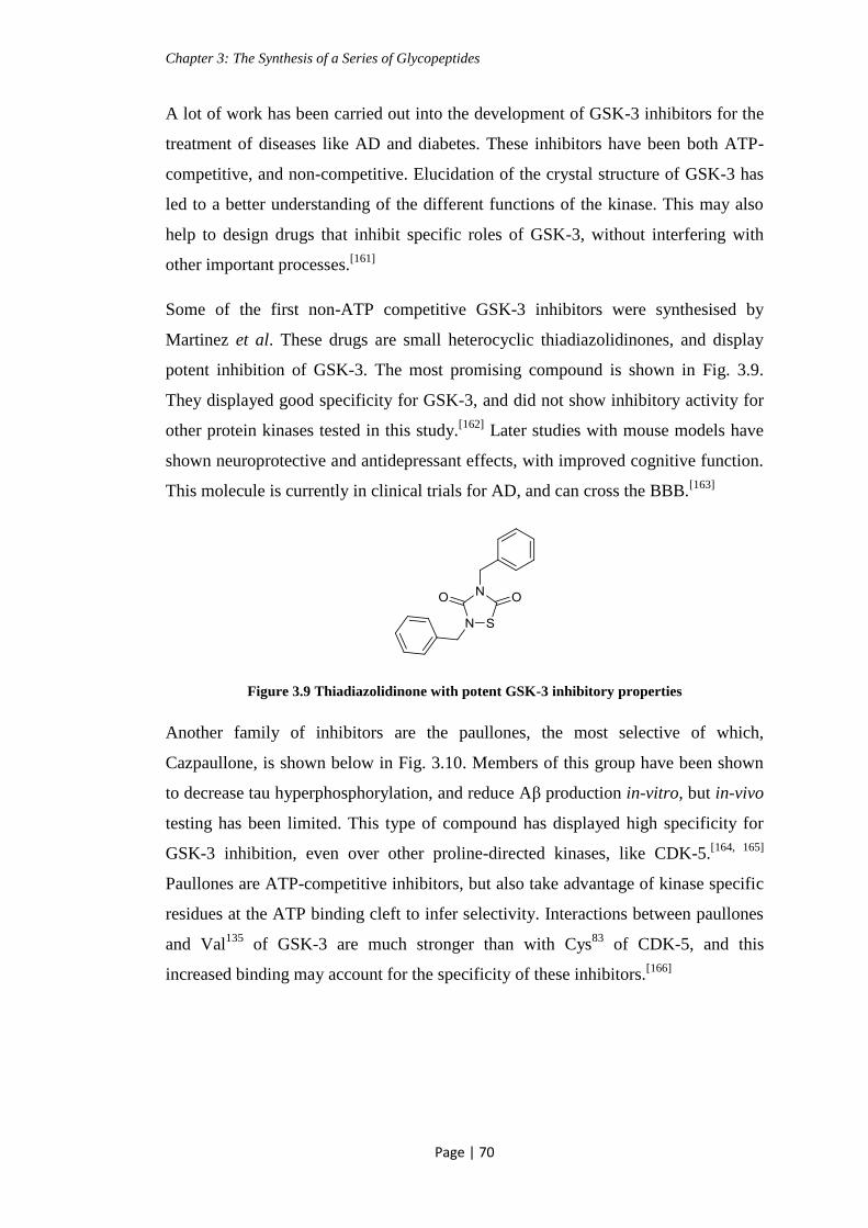
Chapter 3: The Synthesis of a Series of Glycopeptides
Page | 70
A lot of work has been carried out into the development of GSK-3 inhibitors for the
treatment of diseases like AD and diabetes. These inhibitors have been both ATP-
competitive, and non-competitive. Elucidation of the crystal structure of GSK-3 has
led to a better understanding of the different functions of the kinase. This may also
help to design drugs that inhibit specific roles of GSK-3, without interfering with
other important processes.[161]
Some of the first non-ATP competitive GSK-3 inhibitors were synthesised by
Martinez et al. These drugs are small heterocyclic thiadiazolidinones, and display
potent inhibition of GSK-3. The most promising compound is shown in Fig. 3.9.
They displayed good specificity for GSK-3, and did not show inhibitory activity for
other protein kinases tested in this study.[162]
Later studies with mouse models have
shown neuroprotective and antidepressant effects, with improved cognitive function.
This molecule is currently in clinical trials for AD, and can cross the BBB.[163]
Figure 3.9 Thiadiazolidinone with potent GSK-3 inhibitory properties
Another family of inhibitors are the paullones, the most selective of which,
Cazpaullone, is shown below in Fig. 3.10. Members of this group have been shown
to decrease tau hyperphosphorylation, and reduce Aβ production in-vitro, but in-vivo
testing has been limited. This type of compound has displayed high specificity for
GSK-3 inhibition, even over other proline-directed kinases, like CDK-5.[164, 165]
Paullones are ATP-competitive inhibitors, but also take advantage of kinase specific
residues at the ATP binding cleft to infer selectivity. Interactions between paullones
and Val135
of GSK-3 are much stronger than with Cys83
of CDK-5, and this
increased binding may account for the specificity of these inhibitors.[166]

Chapter 3: The Synthesis of a Series of Glycopeptides
Page | 71
Figure 3.10 Cazpaullone, a selective GSK-3 inhibitor
Lithium has been investigated as a GSK-3 inhibitor. Lithium has been used for
decades as a treatment for bipolar disorder, and so has been shown to be safe to
administer for prolonged periods. Lithium is a potent GSK-3 inhibitor, acting
through competition with magnesium ions, and binding to the kinase. Lithium
treatment in-vivo, in mice, has displayed a reduction in tau hyperphosphorylation,
and decreased Aβ toxicity. These results are promising, but one consideration is that
lithium inhibits other enzymes apart from GSK-3, and can be neurotoxic due to
alteration of neurotransmitter levels.[167]
Lithium treatment of mouse models of AD
has demonstrated pathological improvements, but memory deficits were not
recovered. Human trials were carried out on a small scale, for 10 weeks, but these
trials did not show encouraging results, with no pathological or behavioural
improvements.[168]
While many inhibitors of GSK-3 contain aromatic groups in their structure, there has
been evidence to suggest that short peptides can selectively inhibit protein kinases.
The structure of these peptides can be based on “natural” sequences which are
known to be targeted by the kinase, and so make the inhibitor selective.[169]
As GSK-
3 is essential in many processes in the body, a weak-to-moderate inhibition is
desirable. By synthesising a peptide sequence which mimics the natural substrate of
GSK-3, there will be competitive binding, resulting in partial inhibition of the
kinase. Substrate-competitive inhibitors are more selective than ATP-competitive
inhibitors, and so this strategy is attractive for clinical use.[163]
As mentioned earlier,
the crystal structure of GSK-3 has been obtained. Examination of this structure
indicates the presence of a substrate peptide binding site, which is specific for this
kinase.[161]
Plotkin et al have synthesised a phosphorylated peptide inhibitor of GSK-3, for use
in the treatment of diabetes. These peptides are substrate-competitive, and in-vivo
mouse models of diabetes have shown a marked improvement. The membrane

Chapter 3: The Synthesis of a Series of Glycopeptides
Page | 72
permeable inhibitor showing the most potential is known as L803-mts. The sequence
was derived from a GSK-3 substrate, heat shock factor-1, and contains the GSK-3
recognition motif S1XXXS
2. S
1 is the site which is known to be phosphorylated by
GSK-3, S2 is the priming site, and X is any amino acid. The sequence of the
phospho-peptide, L803-mts, is shown in Fig. 3.11, with S1 and S
2 underlined. The
site of phosphorylation in the natural sequence has been substituted with alanine.[157]
Figure 3.11 Sequence of L803-mts
Structural variations in the peptide revealed that elimination of the phosphate group
prevented inhibition, and also that shortening the sequence to just S1XXXS
2
eliminated the inhibitory capacity.[157]
This peptide was found to have
neuroprotective effects in-vitro.[170]
Closer examination revealed that the substrate
and inhibitor binding sites were not identical, while both occupied the phosphate
binding pocket, the other residues involved in binding were different. The residues
are located near to the ATP-binding site, and the phosphate binding pocket which
interacts with the phospho-serine of the substrate. The substrate was found to interact
with the cavity bordered by Gln89
and Asn95
, while L803-mts mainly interacted with
Phe93
and a hydrophobic surface located away from the ATP-binding site. The Pro8
residue of L803-mts was observed to be involved in the interaction with Phe93
. It was
thought that this difference in binding was crucial to the inhibitory capacity of the
peptide. Alterations in the peptide sequence were later carried out, and resulted in a
3-10 fold increase in GSK-3 inhibition. The increased activity of the inhibitors was
attributed to the presence of more hydrophobic groups, and also to increased
interaction with Phe93
.[171]
From this information it is evident that peptides have
potential as selective inhibitors of GSK-3. The natural substrates of GSK-3 are
proteins, and, by mimicking the sequence of these substrates, a peptide inhibitor
could be designed which selectively blocks unwanted functions of GSK-3.
3.7 Solid-Phase Peptide Synthesis
The synthesis of complicated glycopeptides can be hard to achieve in the traditional
“solution-phase”. These coupling reactions can often be poor yielding, and the

Chapter 3: The Synthesis of a Series of Glycopeptides
Page | 73
intermediates difficult to purify. The advent of “solid-phase” peptide synthesis
(SPPS) was pioneered by Bruce Merrifield in the 1960’s as a new approach to
peptide synthesis, with the first resin being called Merrifield resin. Many advances
have happened in the area of SPPS since then, but the same methodology is in use
today.[172]
SPPS involves the formation of amide bonds, under standard coupling
conditions, where the growing peptide is anchored to a solid support (resin).
SPPS has many advantages over solution phase chemistry. It allows the use of large
excesses of reagents, which help to drive these poor yielding reactions almost to
completion. The reactions are also faster than the liquid phase chemistry, peptide
couplings can be accomplished in less than 30 mins.[173]
There is no need for
purification at each stage, as the peptide remains on the solid support until the final
deprotection step, and excess reagents are simply washed off. SPPS also has the
advantage, as with solution phase, of lending itself to the well-established coupling
methodologies of TBTU/HOBt. One of the major advantages of SPPS is that it is
suitable for automation. Peptide sequences can be programmed into an automated
synthesiser, providing non-chemists with access to biologically relevant peptides.[101]
There are two different propagation methods used in SPPS, the Boc/Bzl and
Fmoc/tBu strategies. The original resins were designed to be used in conjunction
with the Boc/Bzl strategy. As the Boc group is removed under acidic conditions,
repeated use of TFA is required in the deprotection steps, which may be too harsh for
some peptides, especially glycopeptides.[174]
The development of the Fmoc/tBu
strategy has allowed SPPS to be carried out using milder chemistry. The Fmoc group
is removed under mildly basic conditions, generally by piperidine, but even milder
bases have been utilised. Most SPPS today is carried out using the Fmoc/tBu
methodology, and has been complimented by the development of suitable resins and
side-chain protecting groups.[172]
An important element of SPPS is the choice of resin. As mentioned above, the
original Merrifield resin is suitable for the Boc/Bzl strategy, and the finished peptide
is cleaved from the resin under basic conditions. Newer resins, such as the Wang or
RINK amide resins, contain a linker which is acid-labile. There is a wide variety of
resins available, which are composed of a polymeric support, such as polystyrene, to

Chapter 3: The Synthesis of a Series of Glycopeptides
Page | 74
which a linker is attached. Choice of resin is dependent upon the strategy being used,
and also on the nature of the peptide.[175]
SPPS has become an integral part of peptide synthesis, with many advantages. It has
made possible the synthesis of complicated structures, which otherwise may not have
been accessible. Recent work in the area of SPPS has led to the development of a
solid-phase approach to oligosaccharide synthesis, but this is a much more
complicated endeavour, and is not suited to all carbohydrate syntheses.[176]
3.8 Aim
The aim of this section was the synthesis of glucosamine based glycopeptides, which
have potential kinase inhibitory properties (Fig. 3.12). The kinase which was
targeted in the design of this glycopeptide is GSK-3, which has been highly
implicated in the development of AD, as discussed earlier.
Figure 3.12 General structure of target glycopeptide
It was decided to synthesise a non-ATP competitive inhibitor, in order to infer more
selectivity for GSK-3. O-Linked-N-acetyl glucosamine (GlcNAc) was incorporated
into the glycopeptide to investigate the effect that the sugar moiety would have on
the affinity of this compound for GSK-3. Glycosylation is biologically carried out on
the tau protein, as discussed earlier, and it is postulated that GSK-3 may have a
recognition site for GlcNAc. As previously mentioned, peptides are good candidates
as specific kinase inhibitors, and some have shown moderate inhibition of GSK-3.
The peptide sequence which was synthesised is based on a portion of the repeating
sequence identified in the tau protein. Both of the serine moieties in the sequence are
known to be involved in the dynamic glycosylation/ GSK-3-mediated
phosphorylation of tau.[122]
The peptide synthesis was carried out using solid phase
methodology, utilising the well-established Fmoc strategy.

Chapter 3: The Synthesis of a Series of Glycopeptides
Page | 75
3.9 Synthesis of Protected Glycosyl-Serine Building Block 3.5
3.9.1 Synthesis of N-Fmoc Protected Glycosyl Acceptor 3.2
The Fmoc protecting strategy was chosen for SPPS. The Fmoc protecting group was
used as it is orthogonal to the other protecting groups in the glycopeptide, and can be
removed easily in the presence of piperidine. N-Fmoc protected serine has also been
demonstrated as a good glycosyl acceptor, making it attractive for the synthesis.[177]
N-Fmoc protected serine was treated with benzyl bromide, in the presence of
DIPEA, to selectively alkylate the acid functionality, resulting in 3.2 (Scheme 3.2).
This reaction was originally carried out using K2CO3 as the base, but yields were not
reproducible. The solubility of DIPEA allowed the reaction to proceed at a faster
rate, resulting in increased and reproducible yields.
Scheme 3.2 Synthesis of N-Fmoc protected glycosyl acceptor. Reagents and conditions: i) BnBr,
DMF, DIPEA, rt, overnight, 80 %
3.9.2 Synthesis of Building Block 3.5
Glycosylation of the protected serine acceptor 3.2 was carried out using the imidate
donor 2.34, discussed in chapter 2. The reaction was carried out using TMSOTf as
the promoter to give 3.3. From this glycosylated serine, removal of the N-Troc
protecting group was carried out, using freshly activated Zn and acetic anhydride, to
give 3.4. This reaction was initially carried out in the presence of Et3N. The reaction
proceeded, but with poor yields (30 %). The Fmoc group is base labile, and removal
of this group accounted for the low yields. The reaction was carried out without
Et3N, and the yields improved greatly, but with a longer reaction time (from 4 h
increased to overnight). The next step in the synthesis was the hydrogenolysis of the
benzyl ester protecting group, using Pd/C and H2, to give the free acid 3.5, which
was used as the building block for incorporation into the glycopeptide (Scheme 3.3).
This building block is often used in the literature for glycopeptide synthesis.[178]

Chapter 3: The Synthesis of a Series of Glycopeptides
Page | 76
Scheme 3.3 Synthesis of glycosyl building block 3.5. Reagents and conditions: i) TMSOTf, DCM,
overnight, rt, 62 %; ii) Zn, Ac2O, overnight, rt, 80 %; iii) Pd/C, EtOAc, overnight, rt, 70 %
3.10 Synthesis of Glycopeptides
A library of novel peptides was synthesised using solid phase peptide synthesis
(SPPS) techniques, following literature procedures.[172]
3.10.1 Synthesis of Decapeptide 3.6
Figure 3.13 Sequence of decapeptide 3.6
The first target peptide sequence, shown in Fig. 3.13 above, which was synthesised,
does not contain any glycosylated amino acid. This was carried out in order to
investigate the effect that the sugar moiety would have on the affinity for the GSK-3
kinase.
2-Chlorotrityl chloride (CTC) resin, also known as Barlos resin, was used for the
SPPS of our peptides (Fig. 3.14).

Chapter 3: The Synthesis of a Series of Glycopeptides
Page | 77
Figure 3.14 2-Chlorotrityl chloride resin
This resin has several advantages over other resins. It is less acid labile than the
closely related trityl chloride resin, yet labile enough to allow cleavage of the
finished peptide under mild acidic conditions (as low as 1 % TFA in DCM).
Racemisation is eliminated as loading of the first amino acid (AA) is carried out by
nucleophilic substitution, and so does not necessitate activation of the acid
functionality of the AA. A problem encountered with some other resins, such as the
Wang resin, is formation of diketopiperazine (DKP), 3.7, as shown in Scheme 3.4.
This usually occurs at the dipeptide stage. The bulky nature of the 2-chlorotrityl
group prevents any DKP formation. Another advantage of the CTC resin is that it
can be used for both protected and free peptides.[179]
Scheme 3.4 Diketopiperazine formation
The Fmoc protection strategy was employed for this synthesis. This methodology
compliments the use of the CTC resin due to Fmoc removal under basic conditions.
Carrying out the synthesis on the solid-phase has many advantages, as discussed
earlier, but also poses difficulties not associated with the solution-phase. The main
disadvantage is that the product of the reaction is bound to the resin until the final
step. This means that traditional monitoring techniques, such as TLC, don’t lend
themselves to the synthesis. The Kaiser test is a common system used to determine
the success of coupling and deprotection steps in SPPS. It is a qualitative test based
on the reaction of the amine functionality with ninhydrin, to produce a specific
colour change, depending on the structure of the amine involved, for example, a
primary amine produces a blue/ purple colour in the Kaiser test. This blue colour

Chapter 3: The Synthesis of a Series of Glycopeptides
Page | 78
indicates successful deprotection of the Fmoc group, or an incomplete coupling
reaction. An amide or carbamate reacts to produce a yellow/clear colour, indicating a
successful coupling reaction, or incomplete Fmoc deprotection. It is a useful tool in
peptide synthesis as it requires a minimum amount of analyte, and gives a quick
result (within 1 min).[172]
Coupling reactions were carried out by pre-activation of the N-Fmoc protected
amino acid with TBTU, HOBt, and DIPEA for 15 mins. The activated acid was then
added to the resin bound free amine, and gently shaken for 1.5 h. The solvent and
excess reagents were then easily removed by vacuum filtration, and the resin was
washed with DMF and DCM. The Kaiser test was used to indicate complete
consumption of the free amino groups. Each of these couplings required one cycle
for completion, but a second coupling cycle can be performed, if necessary. This
second cycle was avoided by slightly longer reaction times than is needed (the
reaction can be complete in 30 mins). This precaution was taken as there are some
indications in the literature that this second coupling step can cause elimination of
the N-Fmoc group. This could result in repetition, with coupling of a second amino
acid to the free amine. In SPPS, an excess of reagents is used, and this helps to
ensure that the reaction goes to completion. Although the Kaiser test indicated full
coupling at each step, the resin was capped by reacting with acetic anhydride. Any
remaining free amine groups were so converted into amides at this step, stopping
propagation of an incorrect deletion sequence.[172]
The N-Fmoc protecting group of
the terminal amino acid was deprotected at each stage using a 20 % solution of
piperidine in DMF. The reactive side chains of the amino acids used were suitably
protected, so that the protecting groups were compatible with N-Fmoc cleavage
conditions. The amino acids used in this synthesis, and their protected side chains,
are shown in Fig. 3.15. All amino acids used for this synthesis possess the L
configuration.

Chapter 3: The Synthesis of a Series of Glycopeptides
Page | 79
Figure 3.15 L-Amino acid residues used in SPPS, and the abbreviation used to denote them
2-Chlorotrityl-chloride (CTC) resin was selected for the synthesis of the peptides, as
discussed earlier. Swelling of the resin is important to allow the reagents to access
the reactive site, and DCM is added to the resin before the reaction. This step was
also carried out at each stage in which the resin bound peptide had been dried and
stored. The first amino acid which was attached to the resin was N-Fmoc-Valine-OH.
This reaction follows an SN1 mechanism. The amino acid was added to the resin in
the presence of DIPEA, in DCM (Scheme 3.5). DIPEA is used as it is a hindered
base, and so does not cause removal of the N-Fmoc protecting group. The resin was
washed with a solution of MeOH/DCM/DIPEA to cap any unreacted CTC groups on
the resin.

Chapter 3: The Synthesis of a Series of Glycopeptides
Page | 80
Scheme 3.5 Loading of the first amino acid onto the CTC resin. Reagents and conditions:
i) DIPEA, DCM, 2 h, rt
Estimation of the amount of amino acid which had attached to the resin was carried
out by UV analysis. A known quantity of the resin-attached N-Fmoc-AA was
subjected to N-Fmoc cleavage conditions of DBU. The level of attachment of the N-
Fmoc-AA was judged by measurement of the UV absorbance of dibenzofulvene,
which is released upon N-Fmoc cleavage, at 304 nm. This was found to be 0.72
mmol/g (moles of AA per gram of resin), which was in agreement with literature
values of 0.86 mmol/g for CTC resin.[180]
Removal of the N-Fmoc group was carried out in the presence of piperidine,
followed by removal of reagents by vacuum filtration and rinsing. The success of
this reaction was confirmed by the appearance of a blue colour in the Kaiser test.
Coupling of the second amino acid, N-Fmoc-Lysine-(N-Boc)-OH, was carried out by
pre-activating the acid in the presence of TBTU, HOBt, and DIPEA for 15 mins,
followed by addition to the resin bound free amine (Scheme 3.6). Any unreacted,
resin bound free amine was capped by acetylating with Ac2O in the presence of
DIPEA. This step was carried out to reduce the likelihood of deletion sequences.
This method of N-Fmoc deprotection, coupling of the next amino acid, and capping
of any unreacted, resin bound amino peptide was repeated for each consecutive
amino acid until the desired decapeptide was synthesised (Fig. 3.13).

Chapter 3: The Synthesis of a Series of Glycopeptides
Page | 81
Scheme 3.6 Coupling of the second amino acid. Reagents and conditions: i) TBTU, HOBt, DIPEA,
DMF, 1.5 h, rt
One of the main challenges in peptide synthesis is complete characterisation of the
peptide intermediate at each step. This is made difficult due to the large number of
signals which would appear at the same ppm range in NMR spectra, for example the
amide groups connecting each amino acid. The technique used to monitor the
presence of the desired peptide intermediates was mass spectrometry (MS). The
samples were prepared for analysis by collecting a small amount of the resin bound
peptide, and cleaving the peptide from the resin with a 1 % solution of TFA in DCM.
This cleavage was first attempted using an aqueous solution of TFA, but was
unsuccessful. Water did not encourage swelling of the CTC resin, therefore
preventing cleavage of the peptide.
After introduction of the last AA (N-Fmoc-Asparagine-(N-Tr)-OH), the peptide was
subjected to N-Fmoc removal, in the presence of piperidine, followed by acetylation
of the free amine using Ac2O, in the presence of DIPEA. Cleavage from the resin
was carried out using a 50 % solution (v/v) of TFA in DCM. These conditions also
ensured removal of the corresponding side-chain protecting groups. When a more
dilute solution of TFA was used, complete removal of the acid-labile side chain
protecting groups was not achieved. The synthesis of the desired, deprotected
decapeptide 3.6 was confirmed by MS analysis (Calculated for C43H78N13O16 (M +

Chapter 3: The Synthesis of a Series of Glycopeptides
Page | 82
H)+: 1032.5684, found 1032.5662). Purification of peptide 3.6 was carried out using
preparative HPLC, with a gradient elution, using H2O with 0.1 % formic acid, and
MeCN with 0.1 % formic acid as the mobile phase. The detector available for our
instrument was a UV detector, which made the HPLC analysis difficult due to the
lack of a strong UV active group in the peptide. None of the constituent AAs of 3.6
have aromatic groups as their side chains, and so the intensity of the peaks in the
chromatogram was rather weak, especially when compared to the background
absorption of the mobile phase. The wavelength of maximum absorption for the
peptide bond is 190 nm, but measurements are not carried out at this value due to the
low output of conventional spectrometers at this wavelength. The side chains of
some AA absorb at 205 nm, for example Trp, His, and Cys, but none of these AAs
are contained in our peptide sequence.[181]
At higher wavelengths the amide bonds
should be mildly UV active, and so the analysis was carried out at 214 nm.[182]
Fractions were manually collected at 1.5 minute intervals based on the run time, and
analysed by MS to determine the fraction which contained the desired product. The
time which it would take for the mobile phase to travel from the detector to the
collection vessel was estimated at about 50 secs, based on the length and diameter of
the tubing. Compound 3.6 was detected in two fractions, 6.5-8 mins, and 8-9.5 mins
of run time. These fractions were concentrated, and the material obtained was re-
dissolved in H2O with 0.1 % formic acid. Further HPLC analysis was investigated
under several different conditions. Using the gradient elution conditions used
previously for purification, a stable baseline was unobtainable. MeCN is UV active
at 214 nm also, and so there was a constant shift in the baseline as the concentration
of MeCN changed. An isocratic system was also investigated, with a mobile phase of
MeOH:H2O, at various concentrations of MeOH. Unfortunately, due to the lack of a
strong chromophore in peptide 3.6, a UV trace could not be attained to show the time
at which the peptide eluted from the column. As mentioned earlier, the peptide bond
should be UV active at 214 nm, but measurements at this value are not generally
used in analysis of peptides and proteins, due to the complexity of absorbance at this
wavelength. Although, there is no other choice for certain sequences. Also, the
absorbance at 214 nm is highly dependent on the conformation of the peptide.[182]
To investigate if the acidic content of the mobile phase was necessary to ensure
elution of the peptide through the column, an isocratic system of MeCN with 1 %

Chapter 3: The Synthesis of a Series of Glycopeptides
Page | 83
formic acid:H2O with 1 % formic acid, at a range of concentrations, was also used.
While a much more stable baseline was achieved with the isocratic systems, it was
still difficult to associate a peak in the chromatogram with our peptide. MS detection
was the only indication of the presence of the peptides. Shown in Fig. 3.16 are the
total ion chromatograph (TIC), extracted ion chromatograph (EIC), and UV trace
which were produced when compound 3.6 was detected by MS analysis. The ions
corresponding to the peptide 3.6 were found to reach the detector at 6.987 mins.
Examination of the ions which reached the detector between 7.6-8.9 mins also shows
the presence of the desired peptide, in which the deprotection of the AA side chains
has not gone to completion. This peptide still has one t-butyl group attached ((M +
H)+: 1088.6357, and (M + 2H)
2+: 544.8218).
Figure 3.16 a) TIC, b) UV trace, and c) EIC obtained from MS analysis of decapeptide 3.6
3.10.2 Synthesis of a Library of Glycopeptides, 3.8, 3.9 and 3.10
Glycosylation in a specific amino acid in a protein has been shown to affect the
glycosylation/phosphorylation at other sites.[18]
In order to investigate the effect of
glycosylation at a specific site in the desired peptide sequence, glycosylation was
carried out on the first serine of the sequence, the second serine of the sequence, or
on both. Introduction of N-acetyl glucosamine into the peptide sequence was carried
out using the SPPS procedures optimised for the decapeptide 3.6.
The glycopeptide, 3.8, is glycosylated at the first serine of the sequence (Fig. 3.17).
Some discussions in the literature indicate that piperidine may be too basic to use for
N-Fmoc removal in glycopeptide synthesis, with a weak base like morpholine being
a)
b)
c)

Chapter 3: The Synthesis of a Series of Glycopeptides
Page | 84
used instead.[183]
Others disagree with this theory, and careful use of piperidine was
not found to cause β-elimination.[184]
Piperidine was used in the first two
deprotection steps. Introduction of the glycosylated serine building block 3.5 was
carried out using the standard coupling conditions of TBTU/HOBt in the presence of
DIPEA, described earlier (Scheme 3.6). The Kaiser test was carried out after 1.5 h,
the time allowed for standard peptide couplings, and this indicated that the reaction
had not progressed to completion. For all couplings involving a glycosylated AA the
reaction was left overnight, and was judged to be complete by the Kaiser test. N-
Methyl morpholine (N-MM) was used to remove the N-Fmoc group at this stage, but
the reaction was not successful, with failure being indicated by the Kaiser test. The
deprotection was then carried out successfully, with careful monitoring of the
reaction time, using piperidine. No elimination of the sugar moiety was observed.
Subsequent couplings of the remaining AAs led to the synthesis of the complete
glycopeptide. The finished glycopeptide was subjected to N-Fmoc removal, in the
presence of piperidine, followed by acetylation of the free amine using Ac2O, in the
presence of DIPEA. Cleavage from the resin was carried out using a 50 % solution
(v/v) of TFA in DCM, as optimised for compound 3.6, resulting in glycopeptide 3.8
(Fig. 3.17). Purification of crude compound 3.8 was carried out using preparative
HPLC. The same type of complications as described earlier for the purification of
compound 3.6, were also encountered for 3.8. The sugar moiety of the glycopeptide
is O-acetyl protected, and these groups are not UV active. Neither are the side chains
of the other AAs in the sequence of glycopeptide 3.8. Purification was carried out by
manually collecting fractions at 1 minute intervals based on the run time, and
analysed by MS to determine the fraction which contained the desired product. The
glycopeptide was found in fractions collected at both 6-7 mins, and 7-8 mins of run
time. The presence of glycopeptide 3.8 was confirmed by MS (Calculated for
C57H98N14O24 (M + 2H)+2
: 681.8448, found 681.8432). The fractions containing the
desired product were concentrated, and re-dissolved in MeCN with 1 % TFA. It is
interesting to note the difference in physicochemical properties, such as solubility,
which were conferred on the peptide by the presence of the sugar moiety. Further
HPLC analysis was investigated under each of the elution conditions outlined
previously for 3.6. As mentioned earlier, glycopeptide 3.8 does not contain a strong
chromophore, and a UV trace showing the time at which the peptide eluted from the
column was not obtained. The TIC, EIC and UV trace obtained by LC-MS analysis

Chapter 3: The Synthesis of a Series of Glycopeptides
Page | 85
of compound 3.8 are shown in Fig. 3.18. From the EIC, it can be seen that 3.8
reached the MS detector at 7.895 mins. Further analysis of the other ions which
reached the detector, as observed in the TIC, show the presence of the desired
glycopeptide, in which one acetyl group has been removed ((M + 2H)2+
: 660.3405).
Figure 3.17 The sequence of glycopeptide 3.8
Figure 3.18 a) TIC, b) UV trace, and c) EIC from MS analysis of the purified glycopeptide 3.8
Glycopeptide 3.9 was also synthesised according to the SPPS procedure outlined
above. This glycopeptide differs from compound 3.8 in that it is glycosylated on the
second serine of the sequence (Fig. 3.19). As for all of the peptides which were
synthesised, MS analysis was carried out to confirm the synthesis of the complete,
Fmoc protected, sequence, before Fmoc removal. A portion of the resin bound
glycopeptide was treated with a 1 % solution of TFA in DCM (v/v). Both t-butyl
protecting groups, of the Ser and Thr residues, were removed in the resin cleavage
stage, while the trityl group on the Asn residue remained attached (MS-ESI/TOF:
Calculated for C99H142N16O29 (M + 2(NH4))+2
: 1010.0074 , found 1010.0096). The
peak in the EIC shows compound 3.9 reaching the MS detector at 12.012 mins.
Resin-bound glycopeptide 3.9 was subjected to N-Fmoc removal, in the presence of
piperidine, followed by acetylation of the free amine using Ac2O, in the presence of
a)
b)
c)

Chapter 3: The Synthesis of a Series of Glycopeptides
Page | 86
DIPEA. Cleavage from the resin was carried out using a 50 % solution (v/v) of TFA
in DCM, as optimised for peptide 3.6. However, the NHAc protected glycopeptide
could not be confirmed by MS, and any partially side-chain deprotected derivatives
were also not detected.
Figure 3.19 The sequence of glycopeptide 3.9
Glycopeptide 3.10 was synthesised according to the procedures outlined above. This
glycopeptide is glycosylated on both of the serine moieties in the sequence (Fig.
3.20). The finished glycopeptide was subjected to N-Fmoc removal, in the presence
of piperidine, followed by acetylation of the free amine using Ac2O, in the presence
of DIPEA. Cleavage from the resin was carried out using a 50 % solution (v/v) of
TFA in DCM, resulting in compound 3.10 (Fig. 3.20). Purification of crude 3.10 was
carried out using preparative HPLC. The same type of complications as described
earlier for the purification of compounds 3.6 and 3.8, were also encountered for 3.10.
Purification was carried out by manually collecting fractions at 1 minute intervals
based on the run time, and analysed by MS to determine the fraction which
contained the desired product. The glycopeptide was found in the fractions collected
at 7-8 mins, and 8-9 mins of run time. The presence of glycopeptide 3.10 was
confirmed by MS (Calculated for C71H117N15O32 (M + 2H)+2
: 846.4004, found
846.3983). The fractions containing the desired product were concentrated, and re-
dissolved in MeCN with 1 % TFA. Further HPLC analysis was investigated under
each of the elution conditions outlined previously for compound 3.6. As mentioned
earlier, glycopeptide 3.10 does not contain a strong chromophore, and a UV trace
showing the time at which the peptide eluted from the column was not obtained. The
TIC, EIC and UV trace obtained by LCMS of glycopeptide 3.10 are shown in Fig.
3.21. From the EIC, it can be seen that compound 3.10 reached the MS detector at
7.990 mins. Another major ion which was observed in the TIC of the glycopeptide
has a mass of 1596, which corresponds to M - 93 of the desired glycopeptide. The
ion with a mass of 798 corresponds to the (M + 2H)2+
of this peak. This mass did not

Chapter 3: The Synthesis of a Series of Glycopeptides
Page | 87
correspond to any obvious incomplete side chain deprotection, or any deletion
sequence, and could not be assigned.
Figure 3.20 The sequence of glycopeptide 3.10
Figure 3.21 a) TIC, b) UV trace, and c) EIC from MS analysis of the purified glycopeptide 3.10
Although we initially envisaged hydrolysis of the O-acetyl groups in the sugar
moiety of glycopeptides 3.8-3.10, the problems encountered throughout purification
limited considerably the amount of material available for further reactions. We did
not have enough material to investigate the conditions for the deprotection of the
alcohol groups, but it is envisaged that a mild strategy, with careful monitoring of
pH, would be required. Nevertheless, acetylated sugars have shown increased
absorption through biological membranes, when compared with their deprotected
analogues.[185]
The acetyl groups on the sugar can then be easily hydrolysed by
esterase enzymes inside the cell. Since peptides are naturally rather hydrophilic, the
presence of the protected sugar may increase the lipophilicity of these derivatives.
This would be a desirable feature for potential drug candidates.
a)
b)
c)

Chapter 3: The Synthesis of a Series of Glycopeptides
Page | 88
For future work, it would be advisable to carry out deprotection of the alcohol
groups, in order to compare the activity of the deprotected sugar with the acetylated
derivative.
3.11 Chapter Conclusion
In conclusion, the synthesis of four decapeptides was achieved, including three
glycopeptides. The side chains of three of these peptides were deprotected, and the
peptides were purified by preparative HPLC analysis. A major problem encountered
during this work was the lack of a strong chromophore in any of the peptides, which
made the purification difficult. Considering this issue, the peptides were found to be
relatively pure by MS analysis. Another difficulty was the small amount of sample
which was obtained after cleavage from the resin, preventing further deprotection of
the O-acetyl groups. If a larger quantity of the peptides had been obtained, it would
have been desirable to test these compounds for their affinity for the GSK-3 kinase.
Future work could aim to improve the yields of these syntheses, and to send the
peptides for biological evaluation.
Further work on this type of glycopeptide should incorporate an AA, for example
Tyr, which has a side chain that absorbs strongly in the UV region. Another option is
to cleave the peptide from the resin while the Fmoc group is still present, as this is
also strongly UV active.
I would also encourage the incorporation of a Pro residue into the glycopeptide, as it
is known that GSK-3 is a Pro directed kinase. This may increase the binding affinity
of GSK-3 for the peptide.

Chapter 4: Functionalisation at the C-6 Position of Glucosamine-Containing Building Blocks
Page | 89
Chapter 4: Functionalisation at the C-6 Position of
Glucosamine-Containing Building Blocks
4.1 Aim
Carbohydrates provide a useful scaffold for the introduction of a variety of different
functionalities.[186]
The synthesis of such derivatives is complicated by the lack of
orthogonality presented by several hydroxyl groups, which possess similar reactivity.
At the same time, these groups may allow selective functionalisation at different
positions.
The addition of an aromatic group, or a phosphate moiety to the carbohydrate part of
the molecule may alter binding interactions between a glycopeptide and its target.
This type of derivative may modulate the affinity of the glycopeptide for a kinase,
like GSK-3, which was discussed in chapter 3. Also mentioned previously, was the
work carried out by Licht-Murava et al, indicating the need for a phosphate group in
their peptide to illicit an inhibitory effect on GSK-3.[171]
For these reasons, the
introduction of such a functionalisation into a glucosamine-based derivative was
attractive.
Extensive investigation was carried out into conditions for the functionalisation of
different sugar moieties at the C-6 position. This was of interest as it was desirable to
synthesise an azide containing building block, which could be incorporated into a
glycopeptide (Fig 4.1).
Figure 4.1 Proposed azide functionalised building blocks
Another useful purpose for a glycosyl building block which has been functionalised
with an azide group has been exploited by Bertozzi and co-workers. Biological
molecules, such as proteins, can be modified experimentally by techniques like

Chapter 4: Functionalisation at the C-6 Position of Glucosamine-Containing Building Blocks
Page | 90
genetics and biosynthesis. In the case of glycans, these methods are often not
applicable, as they don’t have the genetic template and enzymatic activities which
are associated with proteins. Work carried out by Bertozzi and co-workers has
utilised bioorthogonal chemistry to incorporate non-natural molecules into living
systems, without interfering with the biological functioning of the cell. Azide
containing sugars, such as N-azidoacetyl mannosamine, were introduced into glycans
by treating cells with the azide functionalised derivative of this natural sugar. These
were used as tags for visualisation, before flow cytometric analysis was carried out
on the cells.[187]
Later work by this group has shown the incorporation of N-
azidoacetyl galactosamine and N-azidoacetyl glucosamine into glycoproteins, which
allowed identification of proteins that had become O-GlcNAcylated. This detection
is not an easy task, as discussed in chapter 1, due to the small amount and neutral
charge of O-GlcNAc in these glycoproteins.[188]
4.2 Functionalisation at the C-6 Position of the Sugar Moiety
4.2.1 Differential Protection of the C-6 Position of Glycosyl Derivatives 4.1
and 4.7
Initial attempts to selectively functionalise the hydroxy group at the C-6 position
focused on the N-Troc protected glucosamine, 4.1. The first stage in this synthesis
involved differentially protecting the C-6 hydroxyl with a group which was
orthogonal to the other hydroxyl protecting groups (O-acetyl), and also to the N-Troc
group. A sterically hindered protecting group, such as triphenyl methyl (trityl) ether,
or tert-butyl dimethylsilyl (TBDMS) ether, would be ideal for this purpose (Scheme
4.1). The trityl group is removed under acidic conditions, which would make it
easily deprotected in the presence of the other functionalities. The TBDMS group
can be deprotected using strongly acidic conditions, but a more attractive method is
using fluoride ions (for example, from TBAF), which would also be compatible with
the glycosylated N-Fmoc serine benzyl ester 3.4.
The different reaction conditions investigated in our attempts to directly
functionalise 4.1 at the C-6 position are outlined in Table 4.1.

Chapter 4: Functionalisation at the C-6 Position of Glucosamine-Containing Building Blocks
Page | 91
Scheme 4.1 Introduction of an orthogonal group at C-6. Reagents and conditions: i) Listed in
Table 4.1
Entry Reagents Conditions Outcome
1 i) TrCl, Et3N, pyr
ii)Ac2O
i) 4 h, 70 °C, N2, TrCl (1.2 eq)
ii) overnight, rt
Synthesis of 2.26
5 % yield of 4.2
2 i) TrCl, DMAP,
pyr
ii)Ac2O
i) 3 h, 70 °C, N2, TrCl (1.2 eq)
ii) overnight, rt
Synthesis of 2.26
3 i) TrCl, DMAP,
pyr
ii)Ac2O
i) overnight, 60 °C, N2, TrCl
(1.2 eq)
ii) overnight, rt
Synthesis of 2.26
4* i) TrCl, Et3N, pyr
ii)TrocCl
iii)Ac2O
i) 3 h, 80 °C, N2, TrCl (1.2 eq)
ii) 2.5 h, rt
iii) overnight, rt
Synthesis of 2.26
5 i) TBDMSCl,
imidazole, pyr
ii) Ac2O, pyr
i) overnight, rt, TBDMSCl
(1.2 eq)
ii) overnight, rt
26 % yield of 4.3
2.26 produced
6 i) TESCl, Et3N,
pyr
ii)Ac2O
i) 0 °C add base → rt, 4 h,
TESCl (1.2 eq)
ii) overnight, rt
None recovered
7 i) TrCl, Et3N, pyr
ii) AcCl
i) 3 h, 95 °C, TrCl (5 eq)
ii) overnight, 0 °C → rt
39 % yield of 4.2
Table 4.1 Various reaction conditions for the functionalisation of the C-6 position of 4.1 (*: note
that this entry is based on D-glucosamine HCl as the starting material)

Chapter 4: Functionalisation at the C-6 Position of Glucosamine-Containing Building Blocks
Page | 92
For each of these entries, the intermediate compounds were not isolated, as it was
desired to carry out a one-pot reaction to achieve the selectively protected product.
The progression of the reaction was noted by TLC in each case. For entries 1-3 and
5-7 the glycoside 4.1 was obtained from glucosamine hydrochloride, by protection of
the amine with TrocCl, in the presence of NaHCO3. Entry 1 details protection of the
primary hydroxyl of 4.1 as a trityl ether, using Et3N as the base. This was followed
by acetylation, using Ac2O. Almost quantitative yield of 2.26 (the per-O-acetylated
product) resulted, with only a 5 % yield of the desired product 4.2. Entries 2 and 3
show this reaction carried out in the presence of DMAP, but in this case, there was
full conversion to 2.26. In entry 4 a different reaction sequence was investigated,
with protection of the C-6 as the trityl ether, in the presence of Et3N, followed by
protection of the amine using TrocCl, and acetylation of the remaining hydroxy
groups. This was carried out to check if the N-Troc group was interfering with the
reaction, as we had earlier observed the difficulty in removal of the N-Cbz group in
the presence of N-Troc, as discussed in chapter 2. Again there was full conversion to
2.26. It was postulated that perhaps some acid which was present in the acetylation
stage of these reactions may have been able to cleave the trityl group, if it had been
attached (Table 4.1). There may also have been some H2O remaining from the
previous reaction, even with careful drying of the starting material.
The TBDMS group is stable in acidic conditions. Entry 5 gives the details of
protection using TBDMSCl to form the TBDMS ether, in the presence of imidazole,
followed by acetylation of the hydroxy groups. This reaction resulted in a 26 % yield
of the desired product, 4.3, and also formation of 2.26 (Table 4.1). When this
selectively protected derivative was treated with dimethylamine in an attempt to
form the anomeric hemiacetal, no reaction occurred. This reaction was also carried
out in the presence of benzylamine. A change was noted by TLC analysis, but the
desired product was not isolated, and instead degradation seemed to have occurred.
As these reactions did not proceed in good yield, it was thought that perhaps the
trityl and TBDMS groups were too bulky for introduction into the sugar. Entry 6
shows the reaction carried out using triethyl silyl (TES) chloride, in the presence of
Et3N. This silyl ether is less bulky than TBDMS, although it is removed under acidic
conditions, similar to trityl ethers. The reaction did not proceed, with no recovery of
a glycosidic product (Table 4.1).

Chapter 4: Functionalisation at the C-6 Position of Glucosamine-Containing Building Blocks
Page | 93
On consideration of the by-products of acetylation with acetic anhydride which were
observed for entries 1-4, it was postulated that the acetic acid produced in the
reaction may have facilitated cleavage of the trityl group. In order to investigate this,
the reaction was carried out in the presence of an excess of TrCl (5 eq), followed by
acetylation with acetyl chloride as the acetylating agent (entry 7, Table 4.1 and
Scheme 4.2).
Scheme 4.2 Acetylation carried out using AcCl. Reagents and conditions: i) TrCl, Et3N, Pyr, 95 °C,
3 h; ii) AcCl, 0 °C → rt, overnight, 39 %
The reaction was slightly more successful, producing 4.2 in a yield of 39 %, but this
was still too low for such an early step in the synthetic scheme, and later attempts at
the same reaction did not show good reproducibility. It was decided not to continue
with this route, as the trityl group would most likely not withstand the acidic
conditions in the later steps of the synthesis, such as the Lewis acid required for
glycosylation.
There are very few examples of functionalisation at the C-6 position of glucosamine
derivatives, of which the anomeric position is a free hydroxy group, in the literature.
In some cases the C-1 position has been protected, and for this reason the
glucosamine derivative 4.7 was synthesised, according to literature procedures.[189]
2.26 was prepared as discussed in chapter 2, and was treated with a solution of HBr
in acetic acid to form the glycosyl bromide 4.5. Interestingly, the presence of the N-
Troc group prevented the formation of an oxazoline-type compound, as was
observed earlier, with the N-acetyl derivative, 2.32, this is in agreement with the
literature.[190]
The glycosyl bromide was activated using AgCO3, and reacted with
the glycosyl acceptor, benzyl alcohol, to give 4.6. The acetyl groups of this
glycoside were deprotected, in the presence of 0.1 M aqueous NaOH and MeOH,
resulting in the benzyl glycoside, 4.7 (Scheme 4.3).

Chapter 4: Functionalisation at the C-6 Position of Glucosamine-Containing Building Blocks
Page | 94
Scheme 4.3 Synthesis of glycosyl derivative 4.7. Reagents and conditions: i) 33 % HBr/AcOH
solution, DCM, 3.5 h, 0 °C → rt; ii) AgCO3, DCM, BnOH, overnight, rt, 57 % over 2 steps; iii) 0.1 M
NaOHaq, MeOH, 1 h, rt, 73 %
The various conditions investigated for functionalisation of the C-6 position of 4.7
are described below in Table 4.2.
Entry Reagents Conditions
1 i) TBDMSCl, imidazole, pyr,
mol. sieves. ii)Ac2O
i) rt, 2 days, N2, TBDMSCl (1.4 eq)
ii) overnight, rt
2 i) TMSCl, Et3N, THF
ii)Ac2O, pyr
i) overnight, rt TMSCl (1.2 eq)
ii) overnight, rt
3 i) TESCl, imidazole, DMF
ii)Ac2O, pyr
i) overnight, rt, TESCl (1.2 eq)
ii) overnight, rt
4 i) TESCl, imidazole, DMF
ii)Ac2O, pyr
i) overnight, 30 °C, TESCl (1.2 eq),
followed by TESCl (2 eq) and imidazole (2
eq) overnight
ii) overnight, rt
5 i) FmocCl, pyr
ii)Ac2O
i) N2, 2 h, 0 °C → rt, 4 h, FmocCl (1.5 eq),
followed by FmocCl (1 eq) overnight
ii) overnight, rt
6 i) MOMBr,
K2CO3, acetone
ii)Ac2O, pyr
i) 55 °C overnight, MOMBr (1.5 eq),
ii) overnight, rt
Table 4.2 Various conditions for functionalisation of 4.7

Chapter 4: Functionalisation at the C-6 Position of Glucosamine-Containing Building Blocks
Page | 95
Entry 1 shows the reaction of 4.7 with TBDMSCl, in the presence of imidazole and
molecular sieves. The desired reaction did not proceed, and none of the acetylated
product, 4.6, was recovered. Several products resulted from this reaction, and on
closer examination we were able to isolate and identify one of them as 4.8 (Fig 4.2).
This acetylated pyridine derivative is produced by the addition of acetic anhydride to
pyridine, and has been reported in the literature.[191]
MS analysis confirmed the
presence of this unexpected side-product.
Figure 4.2 Side-product 4.8
Protection of the C-6 position as a TMS ether was investigated in entry 2, which was
carried out using TMSCl in the presence of Et3N, followed by acetylation using
Ac2O. This reaction was unsuccessful, but none of the acetylated product 4.6 was
recovered either. Entries 3 and 4 detail the reaction to protect the C-6 as a triethyl
silyl (TES) ether, which has been shown to be more stable to acid hydrolysis then the
TMS ether. The first attempt carried out at rt overnight, and the second was carried
out at 30 °C, under a N2 atmosphere, with further equivalents of TESCl being added
in an attempt to push the reaction. Both reaction mixtures were then subjected to
acetylation, using Ac2O. Under both conditions the reaction was unsuccessful, and
no recovery of acetylated product was obtained (Table 4.2). The starting material 4.7
is water soluble, and so may have been lost during the work-up.
Some alternative alcohol protecting groups were also investigated. Fmoc protection
of alcohols has been successfully utilised in the literature.[192]
Entry 5 details the
protection of the C-6 position as an Fmoc carbonate. This was carried out using
FmocCl, in the presence of pyridine. The desired product was not obtained, but
again, the acetylated pyridine side-product, 4.8 was observed. Protection of alcohols
as methoxy-methyl (MOM) ethers has been described in the literature.[193]
Entry 6
shows this protection which was carried out using MOMBr, in the presence of
K2CO3, followed by acetylation using Ac2O. No sugar derivative was isolated after
the reaction (Table 4.2).
Chapter 4: Functionalisation at the C-6 Position of Glucosamine-Containing Building Blocks
Page | 96
As will be discussed in chapter 5 in more detail, the deprotected sugar 4.7, obtained
as described in Scheme 4.3, seemed to be unreactive, even with excess amounts of
reagents. It is thought that H2O or hydroxide salts remained from the previous
deprotection step, and interfered with the introduction of protecting groups.
4.3 Differential Protection of the C-6 Position of 3.4
The various conditions investigated above for functionalisation of the glycosyl
derivatives failed to deliver the desired products. The reactions which produced the
desired product were not reproducible, and the yields were very low. The presence of
a sterically large group at the C-6 position may compromise the glycosylation step,
and also there are several additional synthetic steps, in which the compatibility of the
C-6 protecting group would have to be accounted for, in order to reach the desired
building block. Optimisation of these reactions would not guarantee that the same
conditions would work on the glycosylated serine building block 3.4. For these
reasons it was decided to continue the rest of the investigations on the N-Fmoc
protected building block 3.4. Direct functionalisation at the C-6 position of 3.4 was
investigated. This strategy is more challenging, as there are several other protecting
groups present in the molecule which need to be considered, for example, the base
labile N-Fmoc group.
4.3.1 Optimisation of Deprotection Conditions of 3.4
Selective O-deacetylation of 3.4 was attempted under mild conditions of
MeOH/H2O/Et3N, followed by reaction with TrCl in the presence of DMAP,
followed by acetylation with Ac2O. This reaction was not successful, with no
identifiable products isolated. The deprotection was also investigated with slightly
harsher conditions of 0.1 M NaOH in MeOH, followed by protection with
TBDMSCl in the presence of imidazole, and acetylation using Ac2O (Scheme 4.4).
This reaction did not proceed either, with recovery of some of the acetylated starting
material, and again the appearance of the acetylated pyridine side-product 4.8. This
compound has been identified in several of these attempted functionalisation
reactions, even with different protecting groups and sugar derivatives. Perhaps this is
a reason why, often, even the acetylated product was not isolated.

Chapter 4: Functionalisation at the C-6 Position of Glucosamine-Containing Building Blocks
Page | 97
Scheme 4.4 Attempted functionalisation of the C-6 position. Reagents and conditions: i) a)
MeOH/H2O/Et3N 40 °C, overnight; b) TrCl, DMAP, pyr, rt overnight; c) Ac2O, rt, overnight; ii) a)
0.1 M NaOH, MeOH, 1 h, rt; b) TBDMSCl, imidazole, pyr, rt, overnight; c) Ac2O, rt, overnight
It was unclear if the initial deprotection had been carried out, or if the sterically
hindered protecting groups could not be incorporated into the glycosylated serine. It
was postulated that the problems encountered with the introduction of trityl or
TBDMS protection into the C-6 position of 3.4 may have been due to a failure to
deprotect the O-acetyl groups, without degradation of the glycoside. On review of
the results obtained, it was decided to try to isolate the deprotected glycoside to
ensure that deprotection of the O-acetyl groups was being achieved.
O-Deacetylation of 3.4 was investigated under various different conditions outlined
in Tables 4.3 and 4.4 below (Scheme 4.5). A more attractive synthesis would involve
selective deprotection of the primary acetyl only (4.11), and several methods were
also investigated in an attempt to achieve this.
Scheme 4.5 Deprotection of 3.4. Reagents and conditions: i) As outlined in Tables 4.3 and 4.4
4.3.2 Enzymatic Deacetylation Methods
Enzymatic methods of deacetylation have shown great success in the literature, and
display selective deprotection in the presence of other groups with similar chemical
reactivity.[194]
The family of enzymes which has demonstrated selectivity for the
removal of acetyl groups from sugar derivatives are known as lipases. There are
many different kinds of lipase, depending on where they have been isolated from. As

Chapter 4: Functionalisation at the C-6 Position of Glucosamine-Containing Building Blocks
Page | 98
with all enzymes, the catalytic ability of lipases is very dependent upon the structure
of the substrate, and, normally, a panel of lipases are tested. A representative sample
of two lipases was chosen for this investigation. The enzymes which we used are
Candida antarctica lipase (CAL B), which is immobilised on a solid support,
making purification easier, and Candida rugosa lipase (CRL), which is not
immobilised. These lipases were selected as they have both shown selective
deacetylation of primary acetyls in carbohydrate substrates.[194-196]
The desired
product of these enzymatic deacetylations is 4.11, where only the primary acetyl has
been deprotected. Table 4.3 shows the enzymatic deacetylation conditions which we
investigated.
Entry Enzyme Conditions
1 CAL B THF, n-butanol, 20 →50 °C, 2 days
2 CAL B Ethanol, n-butanol, 20 →50 °C, 2 days
3 CAL B Ethanol, PBS (tablet), pH 7, 20 →50 °C, 2 days
4 CAL B THF, PBS (tablet), pH 7, 20 →50 °C, 2 days
5 CAL B ACN, phosphate buffer, pH 4.5, 25 °C days → 45 °C 2
days
6 CAL B Ethanol, phosphate buffer, pH 4.5, 37 °C, overnight
7 CRL ACN, phosphate buffer, pH 4.5, 25 °C days → 45 °C 2
days
8 CRL Ethanol, phosphate buffer, pH 4.5, 37 °C, overnight
Table 4.3 Investigation of enzymatic deacetylation
Entries 1 and 2 detail the reaction carried out using Candida antarctica lipase, in
THF and ethanol, respectively, with the addition of n-butanol. Work carried out by
Singh et al demonstrated the selective deacetylation of a primary acetyl in the
presence of a secondary one under these conditions. The reactions were carried out
on nucleosides, and n-butanol is added as an acetyl acceptor.[197]
Enzymatic activity
is closely related to temperature in many cases, and so a temperature range of 20-50
°C was used. Full recovery of starting material, 3.4, was achieved. Enzymatic
reactions can also be effected by the pH of the environment,[198]
and so these
reactions were repeated using phosphate buffered saline (PBS), which was made up
from a tablet to a specific volume, and was pH 7. The conditions are shown in entries
3 and 4. The reaction did not proceed, with full recovery of starting material. As pH
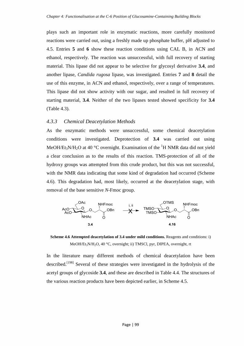
Chapter 4: Functionalisation at the C-6 Position of Glucosamine-Containing Building Blocks
Page | 99
plays such an important role in enzymatic reactions, more carefully monitored
reactions were carried out, using a freshly made up phosphate buffer, pH adjusted to
4.5. Entries 5 and 6 show these reaction conditions using CAL B, in ACN and
ethanol, respectively. The reaction was unsuccessful, with full recovery of starting
material. This lipase did not appear to be selective for glycosyl derivative 3.4, and
another lipase, Candida rugosa lipase, was investigated. Entries 7 and 8 detail the
use of this enzyme, in ACN and ethanol, respectively, over a range of temperatures.
This lipase did not show activity with our sugar, and resulted in full recovery of
starting material, 3.4. Neither of the two lipases tested showed specificity for 3.4
(Table 4.3).
4.3.3 Chemical Deacetylation Methods
As the enzymatic methods were unsuccessful, some chemical deacetylation
conditions were investigated. Deprotection of 3.4 was carried out using
MeOH/Et3N/H2O at 40 °C overnight. Examination of the 1H NMR data did not yield
a clear conclusion as to the results of this reaction. TMS-protection of all of the
hydroxy groups was attempted from this crude product, but this was not successful,
with the NMR data indicating that some kind of degradation had occurred (Scheme
4.6). This degradation had, most likely, occurred at the deacetylation stage, with
removal of the base sensitive N-Fmoc group.
Scheme 4.6 Attempted deacetylation of 3.4 under mild conditions. Reagents and conditions: i)
MeOH/Et3N/H2O, 40 °C, overnight; ii) TMSCl, pyr, DIPEA, overnight, rt
In the literature many different methods of chemical deacetylation have been
described.[198]
Several of these strategies were investigated in the hydrolysis of the
acetyl groups of glycoside 3.4, and these are described in Table 4.4. The structures of
the various reaction products have been depicted earlier, in Scheme 4.5.

Chapter 4: Functionalisation at the C-6 Position of Glucosamine-Containing Building Blocks
Page | 100
Entry Reagents Conditions Outcome
1 DIPEA/H2O/MeOH Overnight, rt 4.14
2 KCN, MeOH KCN 5 eq, rt,
overnight
4.15
3 KCN, MeOH KCN 3 eq, 0 °C, 2
h
Not conclusive from
NMR, acetyls still
present
4 DBTO, MeOH DBTO 1 eq, 50 °C,
3 h → rt, overnight
→ 50 °C, 3 h
Starting material, 3.4,
still present. Presence of
4.13 observed
5 i) Zeolite clay,
MeOH, DCM
iii) AcCl
i) rt, overnight
ii) 50 °C overnight
iii) AcCl 0.2 eq
Full recovery of starting
material, 3.4
6 pTsOH, DCM,
MeOH
i) pTsOH 1 eq, 3 h,
rt
ii) pTsOH extra 1
eq, overnight, rt
i) Starting material, 3.4
ii) Starting material, 3.4.
Minor presence of
various deacetylation
products
7 AcCl, MeOH i) AcCl 0.1 eq, 3 h,
rt
ii) AcCl extra 0.1
eq, overnight
iii) AcCl extra 0.2
eq, overnight
i) Starting material, 3.4
ii) Starting material, 3.4
iii) Starting material, 3.4
8 AcCl, MeOH i) AcCl 0.5 eq,
overnight, rt
ii) AcCl extra 0.5
eq, overnight, rt
Minor presence of 4.13.
Starting material, 3.4
detected.
Various deprotection
products observed.
9 AcCl, MeOH AcCl 5 eq,
overnight, rt
4.13, 4.12, and various
deprotection products
observed.
Table 4.4 Various methods investigated for the chemical O-deacetylation of 3.4

Chapter 4: Functionalisation at the C-6 Position of Glucosamine-Containing Building Blocks
Page | 101
The basic conditions which have been described previously resulted in a loss of the
N-Fmoc protecting group, and so alternative strategies were investigated. The N-
Fmoc group has been demonstrated earlier to be stable to DIPEA, in an overnight
reaction during solid phase peptide synthesis. Reaction conditions of
DIPEA/MeOH/H2O were investigated for the O-deacetylation of glycoside 3.4
(Entry 1). N-Fmoc deprotection was indicated by a red stain on the TLC in the
presence of ninhydrin. The 1H NMR spectrum indicated deprotection of the O-acetyl
groups, with the benzyl ester protection remaining intact (4.14) (Table 4.4).
The use of KCN as a milder method of acetyl hydrolysis has been described.[199]
Entries 2 and 3 show the reaction being carried out using KCN and MeOH. The first
attempt at this involved 5 eq of KCN, and this appeared to cause hydrolysis of the
acetyl groups, and also the benzyl ester and N-Fmoc groups. The reaction was
repeated at 0 °C, with 3 eq of KCN. The 1H NMR spectrum of this reaction was
unclear, but deprotection of the benzyl ester looks to have occurred, indicated by the
disappearance of the characteristic aromatic protons at approx. 7.3 ppm, with the
Fmoc and acetyl peaks still present (Table 4.4).
Entry 4 shows the conditions for deacetylation using dibutyltin oxide (DBTO). This
procedure has been demonstrated in the literature, for deprotection of glucose
derivatives.[200]
The result of this reaction was partial O-deacetylation, and removal
of the benzyl ester group. The N-Fmoc group remained intact, and some starting
material was observed (Table 4.4).
Entry 5 details the use of a zeolite clay for deacetylation. Zeolite is a catalyst which
has been used in mild hydrolysis reactions.[201]
The type of zeolite which was used in
this reaction is called montmorillonite K-10. The reaction was unsuccessful, with full
recovery of starting material, glycoside 3.4 (Table 4.4).
Entry 6 details the reaction carried out using para-toulenesulfonic acid (pTsOH).
The hydrolysis of O-acetyl groups by this method has been described in the literature
for glucose derivatives.[202]
For our sugar, there appeared to be some deprotection,
but mainly starting material, glycoside 3.4, was recovered (Table 4.4).
The cleavage of O-acetyl groups of glucosamine derivatives with acetyl chloride
(AcCl) and MeOH has been described by Jung et al.[203]
Entry 7 shows the details of

Chapter 4: Functionalisation at the C-6 Position of Glucosamine-Containing Building Blocks
Page | 102
how this was attempted with our sugar, 3.4, in the presence of 0.1 eq of AcCl in
MeOH. This low equivalence of AcCl was used to investigate if selective
deprotection of the primary alcohol could be carried out in the presence of the
protected secondary alcohols. The reaction did not proceed after 3 h, and so a further
0.1 eq of AcCl was added, and allowed to stir overnight. No change was observed by
TLC, and a further 0.2 eq of AcCl was added (0.4 eq in total). This did not have any
effect on the sugar, with the starting material, 3.4, being recovered in full. On further
examination of the literature, it was observed that Yeom et al carry out the
hydrolysis of O-acetyl groups in the presence of benzyl esters, at 0.15 eq of AcCl. In
the same work, deacetylation at different positions of glucose and galactose
derivatives was carried out, but required a slightly higher equivalence of AcCl, 0.3
eq.[204]
Entry 8 details the attempted hydrolysis of the acetyl groups using 0.5 eq of
AcCl in MeOH. The reaction was allowed to stir overnight, and TLC analysis
indicated partial deprotection had occurred, but still showed the presence of some
starting material. An extra 0.5 eq of AcCl was added. This reaction resulted in partial
deprotection of the alcohols and removal of the benzyl ester group. In order to
investigate if a large excess of AcCl would facilitate deacetylation, 5 eq of AcCl in
MeOH was added to the sugar. This is described in entry 9, which shows that
deprotection of the alcohols was successful, leaving the NHAc intact, but also caused
the hydrolysis of the benzyl ester protecting group, resulting in what was thought to
be glycoside 4.13 (Table 4.4).
4.3.4 Introduction of Azide Functionality at C-6 of Glycosylated Serine
Building Block
On review of the above deacetylation reactions, we observed that the selective
removal of the acetyl esters, in the presence of the benzyl ester, was difficult to
achieve. Therefore, a new scheme was devised. From compound 3.4, hydrogenolysis
of the benzyl ester protecting group was carried out, using Pd/C and H2, to give the
free acid 3.5, which was earlier used for glycopeptide synthesis. This was carried out
to encourage the formation of a single product upon reaction with AcCl, which was
the method of deacetylation that had shown the most promising results. The free acid
3.5 was then treated with an excess of AcCl in MeOH. Although, using these
conditions, full removal of the O-acetyl groups was achieved, the product of this
reaction was found to be methyl ester, 4.17. This product was identified by a distinct

Chapter 4: Functionalisation at the C-6 Position of Glucosamine-Containing Building Blocks
Page | 103
signal in the 1H NMR spectrum which was attributed to the methyl ester CH3, and
confirmed by MS. On review of 1H NMR data from the previous deacetylation
reactions involving AcCl, as described in Table 4.4, the mild conditions in entry 8
did not result in formation of the methyl ester. The conditions described in entry 9,
however, did produce the methyl ester 4.17, among a mixture of products, including
the benzyl ester, 4.12, and the free acid, 4.13.
Compound 4.17 was then selectively tosylated at the C-6 position of the sugar
moiety, by reaction with tosyl chloride at 0 °C, to give glycoside 4.18. The next
stage in the synthesis was the conversion of the tosyl group to an azide functionality,
using NaN3 in DMF (Scheme 4.7). These conditions have been shown in the
literature to be compatible with functionalised GlcNAc derivatives.[205, 206]
For our
sugar, this reaction was not successful, and several degradation products were
observed. The main product of this reaction had undergone Fmoc cleavage under
these standard reaction conditions, but otherwise still contained the sugar and serine
moieties, with the NHAc and OMe groups still intact. Further analysis of this
compound by IR revealed an absorbance band at 2101 cm-1
which indicated that the
azide had indeed replaced the tosyl group.[207]
The conditions required to introduce
an azide into the C-6 position proved to be too harsh in the presence of the Fmoc
protecting group. The Fmoc protection is needed if this building block is to be
suitable for introduction into a glycopeptide, using SPPS methodologies.
Scheme 4.7 Attempted introduction of an azide functionality at the C-6 position. Reagents and
conditions: i) AcCl, MeOH, overnight, rt, 89 %; ii) TsCl, pyr, 0 °C → rt, overnight, 24 %; iii) NaN3,
DMF, 75 °C, 2.5 h

Chapter 4: Functionalisation at the C-6 Position of Glucosamine-Containing Building Blocks
Page | 104
There is precedence of a linker attached azide being incorporated into a peptide
molecule for use in biological systems, and so this approach was considered.[208]
The
linker attached azide used in this synthesis was 3-azido-propionic acid, 4.21. This
was synthesised, following literature procedures, via a substitution reaction from 3-
bromopropionic acid, 4.20, using an excess of NaN3 (Scheme 4.8).[209]
Scheme 4.8 Synthesis of linker attached azide 4.21. Reagents and conditions: i) NaN3, MeCN,
reflux, 3 h, 36 %
In order to introduce this azide linker into the sugar moiety, the free hydroxy groups
at the C-3 and C-4 positions needed to be protected to prevent unwanted coupling
reactions. Fernández et al have shown that trimethyl silyl (TMS) ether protection at
the C-6 position can be selectively removed in the presence of other TMS groups in
glucose. This strategy would be compatible for the introduction of the azide
functionality.[210]
The hydroxy groups of 4.17 were TMS protected, using TMSCl in the presence of
HMDS, resulting in 4.22. The primary TMS group at the C-6 position of the sugar
was selectively deprotected, using AcOH, to give 4.23. Using standard coupling
conditions of TBTU/HOBt in the presence of DIPEA, introduction of the linker
attached azide, 4.21, into the C-6 position of 4.23 was attempted. However, this
experiment was not successful, with recovery of 4.17 as the main product (Scheme
4.9), with no evidence of compounds 4.22 or 4.23.

Chapter 4: Functionalisation at the C-6 Position of Glucosamine-Containing Building Blocks
Page | 105
Scheme 4.9 Attempted synthesis of C-6 functionalised 4.24. Reagents and conditions: i)
TMSCl, HMDS, pyr, 0 °C → rt, overnight, 90 %; ii) AcOH, acetone:MeOH, 0 °C → rt, 7 h, 67
%; iii) TBTU, HOBt, DIPEA, 4.21, overnight, 0 °C → rt
Introduction of the azide functionality was also investigated with alternative
conditions, using DCC as the coupling reagent, in the presence of DMAP (Scheme
4.10). The product of this reaction was a complicated mixture of products, due to the
presence of the labile TMS ethers. Some of the desired product, 4.25, was isolated
from the reaction mixture, with the major product found to be 4.23. The yield of this
reaction could be greatly improved by increasing the reaction time, for this
investigation a reaction time of 5 h was allowed, due to the base sensitivity of the
Fmoc group. No product was found in which the Fmoc group had been deprotected
during this reaction, and so the DCC mediated coupling seems to be more suitable
for the formation of 4.24.

Chapter 4: Functionalisation at the C-6 Position of Glucosamine-Containing Building Blocks
Page | 106
Scheme 4.10 Introduction of the linker attached azide under DCC coupling conditions.
Reagents and conditions: i) DCC, DMAP, DCM, 4.21, 0 °C → rt, 5 h, 9 %; ii) 2 % TFA in DCM;
iii) LiI
From 4.25, the remaining TMS ethers can be removed with a 2 % solution of TFA in
DCM. Following this, the acid could be deprotected, using LiI.[211]
LiI is anticipated
to be selective for the methyl ester, and should not hydrolyse the ester bond linking
the azide to the sugar. These mild deprotection conditions are also suited to the
presence of the sensitive glycosidic bond. This acid should be ready for introduction
into a glycopeptide, using SPPS techniques. This functionalised carbohydrate
derivative would present the opportunity to introduce many different biologically
relevant moieties into the C-6 position, such as phosphates or aromatic group. This
may modulate the activity of compounds containing this building block. This type of
functionalisation may be beneficial in the case of GSK-3. It is known that GSK-3 has
a binding site for ATP, and many aromatic compounds take advantage of this to
increase their affinity for the kinase. GSK-3 also has a binding pocket for the
phosphate portion of ATP, and introduction of a phosphate, or phosphonate, would
also have the potential to increase the binding affinity of the glycopeptide for GSK-
3. As ATP-competitive inhibitors are often not as kinase selective as those which are
non-competitive, a combination of a peptide, substrate based sequence, which also
contains the added binding affinity of the ATP site, may be a successful candidate.

Chapter 4: Functionalisation at the C-6 Position of Glucosamine-Containing Building Blocks
Page | 107
4.4 Chapter Conclusion
Important progress towards the synthesis of building block 4.27 was achieved.
Selectively functionalised glycoside, 4.27, could be incorporated into a glycopeptide
using standard SPPS conditions. Many problems were encountered throughout this
synthesis. The first difficulty was met when attempting to introduce a selective
orthogonal protecting group into the C-6 position of compound 4.1. This was mostly
unsuccessful, and, when the desired product was produced, the results were not
reproducible.
These problems were not overcome when using the benzyl glycoside, 4.7. Side
products, such as acetyl pyridinium derivative 4.8, were obtained and identified from
these attempts. When working with the serine-glycosylated derivative, 3.4,
incomplete hydrolysis of the O-acetyl groups presented some difficulty. Several
different deacetylation reactions were investigated, with the use of AcCl in the
presence of MeOH found to be the most successful method. From the deprotected
glycoside, 4.16, it was attempted to introduce an azide functionality into the C-6
position. This did not prove successful, with removal of the Fmoc group occurring.
Finally, a linker attached azide was investigated. Standard coupling conditions of
TBTU/HOBt were not found to be suitable for this reaction, with none of the desired
product being isolated. Alternative coupling conditions of DCC did produce the
TMS protected building block, 4.25. The yield of this reaction could be improved
with longer reaction times.
For future work, this azide containing glycoside can be incorporated into a
glycopeptide sequence, and the affinity of a kinase, like GSK-3, for this glycopeptide
can be compared to that of the non-functionalised derivative.
From work carried out in chapter 5, on a glycosyl-dopamine prodrug, it is thought
that perhaps a similar approach would be best for introducing an azide into the C-6
position. Building from a TMS protected glycoside, which has not been
functionalised with Fmoc protected serine, may allow for introduction of the azide
under harsher conditions.

Chapter 5: The Synthesis of a Glycosylated-Dopamine Prodrug
Page | 108
Chapter 5: The Synthesis of a Glycosylated-Dopamine
Prodrug
5.1 Introduction: Prodrugs
5.1.1 What is a Prodrug
A prodrug is a drug which does not have any biological effect, but, once
administered, will be transformed into a biologically active molecule by metabolic
reactions in the body. It has been defined as a bioreversible derivative of a drug
molecule, which will undergo enzymatic or chemical transformation in-vivo to give
the active drug.[212]
Several therapeutically important substances are degraded too
quickly by the body, or are not delivered to the area where they are needed.
Disguising these substances in the form of a prodrug can reverse these drawbacks.
The use of prodrugs has become widespread in the pharmaceutical community,
including many drugs which are currently on the market. A prodrug must possess
specific properties: it should not display activity against any pharmaceutical target,
have high aqueous solubility, be resistant to hydrolysis during the absorption phase,
be converted into the active drug once it has reached the target location, and any
groups which are cleaved must be non-toxic.[213]
For these reasons, glycosylation has
become the modification of choice for many products. Examples of glycosylated
prodrugs are Fabrazyme®, which is a treatment for Fabry disease, and also Rebif
®
which is used in the treatment of multiple sclerosis.[214]
5.1.2 Improving the Pharmacokinetic Properties of a Drug
Several advancements have been made in the discovery of new drug treatments. The
disadvantage is that some of these drugs, especially those which are peptides or
proteins, display a poor pharmacokinetic profile. Potential drug candidates will
display good pharmacodynamic properties, but this is not enough to ensure the
desired response. The pharmacokinetic properties of a compound are an important
consideration in drug design. This refers to processes like absorption, distribution,
metabolism, and excretion. Many therapeutics can undergo elimination from the
body, even before they have had any benefit. This elimination is facilitated by

Chapter 5: The Synthesis of a Glycosylated-Dopamine Prodrug
Page | 109
processes such as enzymatic degradation, renal filtration, and liver clearance.
Accumulation within tissues can also remove the drug from the bloodstream.[215]
One example of a modification carried out on peptide drugs is PEGylation (Fig. 5.1).
Poly-ethylene-glycol (PEG) has been shown to alter the pharmacokinetic and
biodistribution properties of protein conjugates. It appears to prolong the life of the
drug in the body, and to enhance localisation of the drug to specific disease sites. It
does this in a number of ways, such as size enlargement of the molecule, disguising
the peptide from glycosylation or other surface modifications which would ordinarily
occur, and by charge modification. Size enlargement has been demonstrated to slow
down kidney filtration, allowing the drug more time to reach the target area. The
prevention of chemical transformations, such as glycosylation, can alter targeting of
the drug by the immune system, also allowing the drug to have a longer half-life.[216]
Figure 5.1 Poly-ethylene-glycol
Khondee et al have shown that complexation of Keratinocyte Growth Factor-2
(KGF-2) with PEGylated polyanions has a stabilising effect. KGF-2 is a heparin-
binding protein which has potent regenerative properties. The disadvantage of using
KGF-2 as a therapeutic is that it unfolds and aggregates at approximately 37 °C. It
was observed that once KGF-2 had been complexed with the PEGylated polyanions,
dextrin sulphate or pentosan polysulphate, the melting temperature of the protein was
increased by 9-17 °C.[217]
5.2 Glycosylation can Enhance Pharmacological Profile of Drugs
5.2.1 Glycosyl Prodrugs
Glycosylation has been shown to alter the pharmacokinetic and pharmacodynamic
profiles of drugs. Attachment of a sugar moiety can increase the amount of drug that
reaches the target area, for example, via transporters such as the glucose transporter
(GLUT 1-4). It can also reduce the toxicity of some drugs, allowing a higher dose to
be administered.[218]
Glycosylation of drugs has allowed for targeted delivery to the
desired site, for example in the treatment of lysosomal diseases, like Gaucher and

Chapter 5: The Synthesis of a Glycosylated-Dopamine Prodrug
Page | 110
Fabry disease. The active drug is transported, via receptor-mediated endocytosis, to
the lysosome, after targeting specific mannose-6-phosphate receptors. This targeting
is achieved by glyco-engineering of the active proteins to express either mannose or
mannose-6-phosphate at their termini. While not strictly a prodrug, this illustrates
how glycosylation could be used to enhance a drugs pharmacological profile.[214]
Larrosa et al have synthesised several glycosyl prodrugs based on the naturally
occurring polyphenol, reservatol (Fig. 5.2). Reservatol has shown anti-inflammatory
activity, and has become of interest in the treatment of inflammatory bowel diseases
(IBD). While reservatol shows promise as a therapeutic due to its pharmacodynamic
properties, the pharmacokinetic profile is not as attractive. Reservatol is rapidly
metabolised in the body, lessening its efficacy in the colon. It was found that, by
conjugating a sugar moiety, this rapid metabolism was prevented, allowing the
reservatol prodrug to reach its target site, the colon. An added effect of
glycosylation was a reduction in mucosal barrier imbalance, preventing diarrhea in
the subjects, again increasing the amount of active drug which could act on the colon
tissue.[219]
Figure 5.2 Glycosyl reservatol prodrug (Reservatol portion shown in red)
Glycosyl prodrugs have also been developed for the selective treatment of cancer.
Duocarmycin is an antibiotic chemotherapeutic, but cannot be administered in high
enough doses to be effective, without adverse side-effects. Tietz et al have developed
glucosylated duocarmycin prodrugs which are effectively delivered to the cancer
tissue through an antibody-directed enzyme prodrug therapy (ADEPT) approach.
The conjugation of the sugar moiety allowed for localisation of the prodrug in the
tumor tissues, where the active drug was released through enzymatic hydrolysis of
the glycosidic bond. The duocarmycin exhibited cytotoxic effects on the cancer
cells.[220]

Chapter 5: The Synthesis of a Glycosylated-Dopamine Prodrug
Page | 111
5.2.2 Barriers to the Treatment of CNS Disorders
The uptake of solutes into the brain is controlled by the BBB. It separates the central
nervous system (CNS) from the rest of the systemic circulation, and is the “gate-
keeper” of the brain. The BBB plays a vital role in maintaining homeostasis,
allowing nutrients into the brain, and protecting the brain from harmful
substances.[221]
The blood capillaries which feed the brain are lined with tight-fitting
cells, which, unlike other capillaries in the body, do not contain any pores. In
addition, these cells are covered with a fatty layer, formed by nearby cells. Together,
this results in the BBB being an effective barrier to substances which may be
harmful to the brain. In order to enter the brain drugs must be able to pass through
the fatty cells coating the capillaries, and also though their poreless cell
membrane.[222]
The treatment of many brain related diseases has been hampered by the inability of
substances, known to alleviate symptoms, to cross the BBB. It has been estimated
that 98 % of small-molecular weight drugs, and 100 % of large-molecular weight
drugs, which have been developed for the treatment of CNS diseases do not readily
cross the BBB.[223]
Many drugs which have shown CNS activity have been discarded
in the early trial stages, due to their inability to reach their target.[224]
The BBB allows nutrients to enter the brain through transport channels, for example
the large neutral amino acid transporter (LAT-1) and glucose transporters (GLUT 1-
4) carriers, or by receptor-mediated transport, like insulin. By administering the
active substance as a prodrug, these transport mechanisms can be exploited to deliver
the drug into the brain, where the prodrug is broken down into the active substance,
and a harmless by-product, for example a sugar (Fig. 5.3).[212]

Chapter 5: The Synthesis of a Glycosylated-Dopamine Prodrug
Page | 112
Figure 5.3 Illustration of prodrug transport across the BBB
5.2.3 GLUT Transporter
One mechanism of glucose delivery across membranes is via the GLUT carrier, and
there are several different sub-types of this transporter (GLUT 1-4). The transporter
which has been associated with glycosyl dopamine prodrugs is GLUT-1, which
carries glucose and other hexoses through the BBB into the brain. The large energy
demand of the brain is primarily satisfied by glucose, but, in the mammalian brain,
the cells do not keep a large storage of glycogen. In order to maintain healthy brain
function there must be a mechanism to take in glucose across the BBB. Cellular
diffusion of glucose does not occur at any of the membranes in the body, and, as the
BBB is no exception, a carrier is required to deliver it to the brain. This process is
carried out by the GLUT-1 transport protein, in a regulated manner. The uptake of
glucose is very close to the amount of glucose utilised in maintaining brain function.
GLUT-1 has the highest transport capacity of all the carrier-mediated transporters at
the BBB.[225]
The GLUT transporter generally associated with glucosamine is the
GLUT-2 carrier.[226]
There has also been evidence to demonstrate that a decrease in
GLUT-1 and GLUT-3 in the BBB is associated with AD and diabetes. This has been
shown to correlate to a decrease in the level of O-GlcNAcylation of proteins in the
brain.[227]
This well characterised transporter has been investigated for the delivery of CNS
active compounds across the BBB.

Chapter 5: The Synthesis of a Glycosylated-Dopamine Prodrug
Page | 113
5.2.4 Glycosylated Prodrugs for CNS Targeting
Work carried out by Gynther et al investigated the ability of glyco-conjugates to
cross the BBB via GLUT-1. The drugs which they studied were Ketoprofen and
Indomethacin, which were modified with D-glucose using a bioreversible ester
linkage (Fig. 5.4). They found that the glyco-conjugates did bind to GLUT-1, and
also that the prodrug was detected in brain tissue. To further prove that the method of
delivery to the brain was through GLUT-1 transport, glucose was added to the
perfusion medium. It was found that this decreased the amount of prodrug which was
detected in the brain tissue, showing that both D-glucose and the glyco-conjugate
shared a transport mechanism. These prodrugs were shown to be susceptible to
hydrolysis by rat brain homogenate, to give glucose and the active substance.[228]
Figure 5.4 Ketoprofen glyco-conjugate (Ketoprofen portion shown in red)
Some studies have shown that GLUT-1 is a bidirectional transporter, mediating both
blood to brain, and brain to blood transportation of hexoses. This may limit the
effectiveness of some prodrugs, with elimination from the brain before hydrolysis
and release of the active drug can occur.[229]
In order to investigate if a complex kind
of linkage would prevent this bidirectional transport from occurring, Fan et al
synthesised an array of glucosyl thiamine disulfide prodrugs of naproxen (Fig. 5.5).
The thiamine disulfide moiety is quickly reduced by the abundant disulfide
reductase, and ring-closed to a thiazolium once it has entered the brain. This
thiazolium locks the prodrug into the brain, and renders the GLUT-1 transporter
unidirectional for this substance. The ester linkage then becomes hydrolysed at a
slower pace, resulting in a sustained release of the active naproxen. The prodrugs
synthesised and tested in this study were transported across the BBB, and high levels
of naproxen were measured in the brain.[230]

Chapter 5: The Synthesis of a Glycosylated-Dopamine Prodrug
Page | 114
Figure 5.5 Thiamine disulfide derivatives of naproxen (Naproxen portion shown in red)
5.3 Dopamine
5.3.1 The Neurotransmitter Dopamine
Dopamine is a catecholamine neurotransmitter, which plays many roles in the CNS.
It is derived from the amino acid tyrosine (Scheme 5.1). Dopamine is involved in
many processes, including movement, learning, and motivation. Dopamine is a
precursor in the biosynthesis of norepinephrine and epinephrine (Scheme 5.1). It
plays a part in the regulation of complex processes such as mood, attentiveness and
emotion.[231]
Scheme 5.1 Catecholamine biosynthetic pathway. i) Tyrosine hydroxylase; ii) Aromatic L-amino
acid decarboxylase; iii) Dopamine β-hydroxylase; iv) Phenylethanolamine N-methyltransferase
There are two main projections in the brain associated with dopamine; the
mesolimbic pathway, which can also be activated by most psychoactive substances,
and the nigrostriatal pathway. Degradation of dopaminergic neurons in the
nigrostriatal pathway is related to Parkinson’s disease.[232]
Five different dopamine

Chapter 5: The Synthesis of a Glycosylated-Dopamine Prodrug
Page | 115
receptors are known in the body; D1 – D5. D2 appears to be the main receptor
involved with Parkinson’s disease. Parkinson’s disease is characterised by a large
reduction in dopamine levels in the basal ganglia, which occurs as a result of this
degradation. Dysregulation of dopaminergic activity has also been implicated in
schizophrenia, with an excessive release of dopamine in the striatum.[233]
Parkinson’s disease is a progressive neurodegenerative disorder, which is
characterised by a reduction in the levels of dopamine in the brain, as discussed
above. Symptoms of this disease include tremors, rigidity, slow movement and poor
balance. As dopamine stimulates the motor neurons involved in muscle movement,
sufferers of Parkinson’s disease become unable to control movement and
coordination.[234]
Much research has gone into the use of dopamine to treat diseases
such as Parkinson’s. Dopamine cannot cross the BBB, because of its hydrophilicity
and the lack of a specific transport system, and so treatment directly with this
neurotransmitter is not possible, without using intrusive methods.[235]
L-Dopa or Levodopa (L-3,4-dihydroxyphenyl alanine) is the natural precursor of
dopamine, as illustrated earlier (Scheme 5.1). This has become the prodrug of choice
in the treatment of the motor impairments of Parkinson’s disease. L-Dopa has the
ability to cross the BBB though the LAT-1 carrier, and is converted to dopamine in
the brain. Prolonged treatment with L-Dopa has unwanted side-effects, such as the
appearance of abnormal involuntary movements (dyskinesia). This has been
attributed to overstimulation of dopaminergic receptors in the substantia nigra.[236]
Also, L-Dopa can be converted to dopamine in the peripheral tissues, causing
unwanted side-effects, and reducing the amount of dopamine which reaches the
brain.[237]
New treatments are, therefore, vital to the quality of life of Parkinson’s
disease sufferers.
5.3.2 Glycosylation as a Transport Mechanism for Dopamine Delivery
Fernández et al have synthesised several glycosyl dopamine derivatives as
antiparkinsonian agents (Fig. 5.6). These prodrugs consist of the active
neurotransmitter, dopamine, linked to D-glucose through a succinyl linker. The
compounds contain ester, amide and glycosidic linkages, which can be cleaved by
enzymes in the brain tissue. The synthesised prodrugs were tested for activity on the
dopamine D2 receptor, to check if they would be pharmacologically active before

Chapter 5: The Synthesis of a Glycosylated-Dopamine Prodrug
Page | 116
enzymatic release of the dopamine molecule. They did not show any binding
affinity, as expected. This demonstrates that both the amino and phenol groups are
required for dopamine binding to the D2 receptor. Stability studies were carried out
to investigate the enzymatic degradation of the ester, amide and glycosidic bonds in
brain tissue. It was found that the ester bond is cleaved via esterase-catalysed
hydrolysis, and that the amide bond is then cleaved by amidase or protease activation
to release dopamine. Glycosidic bonds were broken by glycosidase activity. The
prodrugs synthesised did not have the desired effect when tested on mouse models of
Parkinson’s disease. It was thought that either the compounds were not able to cross
the BBB, or that the bioconversion into dopamine, by enzymatic degradation, was
occurring too slowly.[210]
Figure 5.6 Glucosyl-dopamine prodrugs synthesised by Fernández et al[210]
The same group later synthesised more glycosyl-dopamine prodrugs (Fig. 5.7),
which also showed affinity for the GLUT-1 carrier. They explored the effects of
different linkers, or attachment at different positions of the sugar moiety. The
compound 5.4, containing a carbamate bond, was found not to release dopamine
when exposed to rat brain tissue. It was thought that perhaps oxidation of the
catechol ring was happening faster than hydrolysis of the carbamate bond, and so the
free dopamine molecule was not formed. Glycoside 5.5 exhibited the properties of a
desired prodrug, in that the dopamine was released upon hydrolysis of the glycosidic
bond, but did not show affinity for the GLUT-1 carrier. Carbamate 5.6 was the best
candidate from this study, with a high affinity for GLUT-1, and efficient hydrolysis
to release dopamine.[238]

Chapter 5: The Synthesis of a Glycosylated-Dopamine Prodrug
Page | 117
Figure 5.7 Second generation of glycosyl-dopamine prodrugs synthesised by Fernández et al[238]
Dalpiaz et al have tested the affinity of glucosyl-dopamine, linked at the C-3
position, for the GLUT-1 transporter (Fig. 5.8). They found that this analogue had a
greater level of binding to GLUT-1 than some other analogues.[234]
Figure 5.8 Glucose-dopamine linked at C-3
Bonnina et al synthesised some of the same derivatives as Fernández et al, but via a
different synthetic scheme. They also synthesised the L-Dopa derivatives. It was
observed that the galactose-dopamine prodrug, 5.7, and the glucose-L-Dopa
analogue, 5.8, were the best candidates (Fig. 5.9). They were shown to be
transported across the BBB, utilizing the GLUT-1 carrier, and were hydrolysed in
brain tissue to release dopamine or L-Dopa. They were active in reducing morphine
induced locomotion in the mouse model of Parkinson’s.[239]
Figure 5.9 Glycosyl-dopamine derivatives synthesised by Bonina et al[239]

Chapter 5: The Synthesis of a Glycosylated-Dopamine Prodrug
Page | 118
Yoon et al have synthesised α-glycosyl-dopamine and α-glycosyl-L-Dopa derivatives
via enzymatic transglycosylation reactions (Fig. 5.10). The stability profile of these
compounds under physiological conditions was assessed. It was found that the α-
derivatives were less susceptible to non-enzymatic oxidation than L-Dopa. It was
postulated that, while the β-anomeric prodrugs synthesised by Fernández et al did
not induce a strong enough response to be used as therapeutics in Parkinson’s
disease, the α-anomer might be broken down quicker once it reached the brain. The
reasoning behind this is that there are more α-glucosidases in the body than β-
glucosidases, and so more dopamine would be released in the brain, over a shorter
period, than with a β-glycoside. These compounds were not tested for their affinity
for GLUT-1 in this this study, or their ability to cross the BBB.[240]
Figure 5.10 α-Glucosyl-dopamine derivative
5.3.3 Non-Glycosidic Dopamine Prodrug Approaches
Alternative approaches utilising other transport mechanisms of the brain have also
been investigated.
Trapani et al have used chitosan nanoparticles to encapsulate dopamine. These
biodegradable carriers have been tested and used to transport anticancer drugs to the
brain, and so have been shown to be safe. The surface of the chitosan nanoparticle is
positively charged, and it was hypothesised that this would result in transport across
the BBB via adsorptive-mediated transcytosis (AMT). This transport mechanism is
used to allow cationic substances through the BBB. In-vivo experiments on rats
showed an increase in the levels of dopamine detected in the brain after
administration of the chitosan/dopamine nanoparticle.[241]
Work carried out by Carafa et al investigated the use of prodrug-loaded liposomes as
a delivery method. It has been shown that condensation of dopamine with neutral
amino acids could utilise the LAT-1 transporter to carry the prodrug across the BBB.
The dopamine prodrug 2-amino-N-[2-(3,4-dihydroxyphenyl)-ethyl]-3-phenyl-

Chapter 5: The Synthesis of a Glycosylated-Dopamine Prodrug
Page | 119
propionamide (DOPH) employs such a strategy. The downside of DOPH is its low
bioavailability; it is sensitive to photodegradation, and also to enzymatic
degradation. DOPH was encapsulated into a liposome made of
dimiristoylphosphatidylcholine (CMPC). It was found that this process did infer
protection against photodegradation, and also increased the half-life of DOPH in the
blood, in-vitro.[242]
It is uncertain whether an amino acid linked molecule would be
able to cross the BBB in patients suffering from Parkinson’s. Ohtsuki et al have
carried out research into the effect that Parkinson’s has on the BBB transport
mechanisms. In their mouse model it was observed that the level of substances
transported via the LAT-1 carrier was severely decreased, while no significant
decrease was observed in other carriers such as GLUT-1. It was postulated that this
could be the reason for the low activity of the antiparkinsonian drug levodopa.[243]
5.4 Aim
The aim of this section was the synthesis and biological evaluation of a novel
glucosamine-based prodrug, which has potential to improve the pharmacokinetic
properties of dopamine. This compound includes a succinyl linker bridging the
dopamine and glucosamine molecules. The target compound 5.9 is shown below
(Fig 5.11).
Figure 5.11 Glucosamine-based dopamine prodrug
There were several reasons for selecting GlcNAc as the sugar moiety. Similar
compounds have been synthesised utilising glucose and galactose to transport the
dopamine molecule across the BBB, these have been detailed earlier in this chapter.
We wanted to investigate if a GlcNAc derivative would display an increased affinity
for the GLUT-1 carrier. Some synthetic carbohydrate receptors have been designed
by Davis and co-workers, which show an increased affinity for GlcNAc over some
other sugars, such as glucose.[244]
It was postulated that natural receptors may follow
this trend, perhaps increasing the amount of prodrug, and therefore dopamine, which

Chapter 5: The Synthesis of a Glycosylated-Dopamine Prodrug
Page | 120
is transported across the BBB. GlcNAc may also have an alternative mechanism of
crossing the BBB, which would be more efficient. As mentioned previously, other
GLUT transporters, including GLUT-2 and -3, have been associated with GlcNAc
and GlcN. Syntheses of a prodrug involving GlcNAc may therefore be rewarding,
albeit more challenging.
5.5 The Synthesis of Glucosamine-Based Dopamine Prodrug 5.9
5.5.1 The Synthesis of Linker-Attached Dopamine
Dopamine was attached to a succinic acid linker, following literature procedures.[210]
A succinic linker was chosen as the ester bond between the linker and sugar will be
easily hydrolysed by enzymes in the brain. The amide bond between the linker and
dopamine molecules should also be labile, and cleaved in the brain by a protease or
amidase, as discussed earlier.[210]
The first step in this synthesis was the protection of
the hydroxy groups on the dopamine aromatic ring. Dopamine hydrochloride, 5.10,
was N-Boc protected using Boc anhydride, in the presence of Et3N. This was carried
out to prevent the reactive amine from becoming alkylated in the next step. The
hydroxy groups of the dopamine molecule were then benzylated, by treatment with
benzyl bromide, in the presence of K2CO3 to give protected dopamine 5.11. The acid
labile N-Boc group was removed with a 5 % solution of TFA in DCM, resulting in
the free amine 5.12. This benzyl protected dopamine 5.12 could then carry out
nucleophilic attack on the carbonyl carbon of succinic anhydride, to give free acid
5.13 (Scheme 5.2).

Chapter 5: The Synthesis of a Glycosylated-Dopamine Prodrug
Page | 121
Scheme 5.2 The synthesis of linker attached dopamine 5.13. Reagents and conditions: i)
MeOH:Et3N (9:1), Boc anhydride, 1 h, 50 °C; ii) K2CO3, DMF, BnBr, overnight, rt, 43 % over two
steps; iii) 5 % TFA, DCM, 6 h, rt, 87 %; iv) Succinic anhydride, pyr, 3 h, rt, 82 %
5.5.2 The Synthesis of Sugar Moiety 5.18
As mentioned in chapter 4, Fernández et al have used TMS ether protection to
selectively introduce the succinic acid dopamine derivative, 5.13, into the C-6
position of glucose and galactose derivatives.[210]
According to work by Gervay-
Hague and co-workers, this transformation worked well with most sugars, but, when
this selective deprotection was carried out on glucosamine, the TMS group at C-1
was also removed (Scheme 5.3).[245]
Scheme 5.3 Selective TMS protection demonstrated by Gervais-Hague and co-workers.
Reagents and conditions: i) AcOH, Ac2O, pyr, rt, 48 h
To avoid unwanted coupling of the dopamine-linker to a free hydroxyl at C-1, it was
decided to differentially protect the C-1 hydroxy as a benzyl ether. D-Glucosamine
hydrochloride was N-Troc protected at the amine functionality, by reacting with
TrocCl, in the presence of NaHCO3, as discussed earlier in chapter 2. This was
followed by acetylation of the hydroxy groups with acetic anhydride, in the presence
of pyridine, to give the anomeric mixture of 2.26. The protected glucosamine 2.26
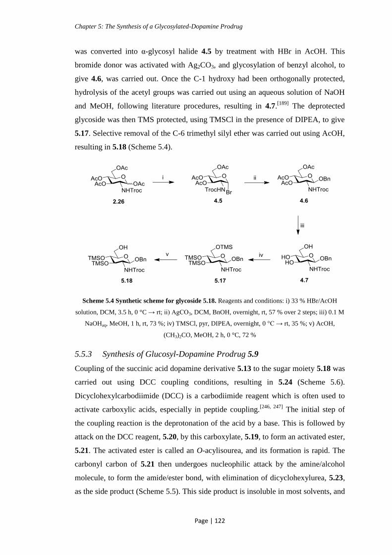
Chapter 5: The Synthesis of a Glycosylated-Dopamine Prodrug
Page | 122
was converted into α-glycosyl halide 4.5 by treatment with HBr in AcOH. This
bromide donor was activated with Ag2CO3, and glycosylation of benzyl alcohol, to
give 4.6, was carried out. Once the C-1 hydroxy had been orthogonally protected,
hydrolysis of the acetyl groups was carried out using an aqueous solution of NaOH
and MeOH, following literature procedures, resulting in 4.7.[189]
The deprotected
glycoside was then TMS protected, using TMSCl in the presence of DIPEA, to give
5.17. Selective removal of the C-6 trimethyl silyl ether was carried out using AcOH,
resulting in 5.18 (Scheme 5.4).
Scheme 5.4 Synthetic scheme for glycoside 5.18. Reagents and conditions: i) 33 % HBr/AcOH
solution, DCM, 3.5 h, 0 °C → rt; ii) AgCO3, DCM, BnOH, overnight, rt, 57 % over 2 steps; iii) 0.1 M
NaOHaq, MeOH, 1 h, rt, 73 %; iv) TMSCl, pyr, DIPEA, overnight, 0 °C → rt, 35 %; v) AcOH,
(CH3)2CO, MeOH, 2 h, 0 °C, 72 %
5.5.3 Synthesis of Glucosyl-Dopamine Prodrug 5.9
Coupling of the succinic acid dopamine derivative 5.13 to the sugar moiety 5.18 was
carried out using DCC coupling conditions, resulting in 5.24 (Scheme 5.6).
Dicyclohexylcarbodiimide (DCC) is a carbodiimide reagent which is often used to
activate carboxylic acids, especially in peptide coupling.[246, 247]
The initial step of
the coupling reaction is the deprotonation of the acid by a base. This is followed by
attack on the DCC reagent, 5.20, by this carboxylate, 5.19, to form an activated ester,
5.21. The activated ester is called an O-acylisourea, and its formation is rapid. The
carbonyl carbon of 5.21 then undergoes nucleophilic attack by the amine/alcohol
molecule, to form the amide/ester bond, with elimination of dicyclohexylurea, 5.23,
as the side product (Scheme 5.5). This side product is insoluble in most solvents, and

Chapter 5: The Synthesis of a Glycosylated-Dopamine Prodrug
Page | 123
so purification of the desired product is simple. When used in peptide chemistry, for
the formation of an amide bond, an additive such as HOBt is generally used to help
prevent racemisation during the reaction.[248]
In our case we are not concerned about
racemisation, and so HOBt was not required. When this coupling reaction is carried
out in the presence of DMAP it is known as a Steglich esterification.
Scheme 5.5 Mechanism of ester formation using DCC as the coupling reagent

Chapter 5: The Synthesis of a Glycosylated-Dopamine Prodrug
Page | 124
Scheme 5.6 DCC mediated coupling reaction. Reagents and conditions: i) DCC, DMAP, DCM, 3
h, rt, 60 %
Following formation of the ester bond to give 5.24, deprotection of the remaining
TMS groups was carried out by treating with a 2 % solution of TFA in DCM to give
5.25. The removal of the N-Troc protecting group was then accomplished using the
standard conditions of Zn in acetic anhydride. The N-Troc group was successfully
reduced, but, as well as the amine functionality becoming acetylated, as was desired,
partial acetylation of the hydroxyl groups was observed (Scheme 5.7).

Chapter 5: The Synthesis of a Glycosylated-Dopamine Prodrug
Page | 125
Scheme 5.7 TMS removal, followed by N-Troc reduction. Reagents and conditions: i) 2 % TFA in
DCM solution, 0 °C, 1 h, 98 %; ii) Zn, Ac2O, overnight, rt; iii) MeOH/H2O/Et3N, 45 °C, overnight
Hydrolysis of the O-acetyls from 5.26 was attempted using mild conditions of
MeOH/H2O/Et3N at 45 °C overnight, but no reaction occurred, with full recovery of
starting material. Mild conditions were required due to the presence of the ester bond
between the sugar and the succinic linker, which prevented the use of most standard
deacetylation procedures.
As this deprotection was posing a problem, removal of the N-Troc group was carried
out prior to removal of the TMS hydroxy protecting groups (Scheme 5.8). The N-
Troc deprotection was carried out in the presence of Et3N, in an attempt to neutralise
the Lewis acidic ZnCl2 produced during this reaction, which may have caused
removal of the TMS groups. Under these conditions, elimination of the TMS groups
and full acetylation of the free hydroxyl groups was observed, resulting in 5.28.

Chapter 5: The Synthesis of a Glycosylated-Dopamine Prodrug
Page | 126
Scheme 5.8 N-Troc deprotection. Reagents and conditions: i) Zn, Et3N, Ac2O, rt, 3 h, 40 %; ii)
Conditions in Table 5.1; iii) H2, EtOAc/MeOH, overnight, rt, 82 % of 5.29; iv) MeOH/H2O/Et3N,
overnight, 45 °C
Selective O-deacetylation was attempted under various different conditions,
summarised in Table 5.1, but was unsuccessful. Deacetylation carried out using mild
basic methods of MeOH/H2O/Et3N, shown in entry 1, resulted in recovery of starting
material. Entries 2 and 3 show the conditions for deacetylation using NaOMe over a
range of temperatures. Work by Xia et al shows acetyl removal in the presence of a
pivaloyl ester, at low temperatures using NaOMe,[249]
but this did not work for our
compound. When carried out at very low temperatures of -20 → -15 °C no reaction
occurred, being confirmed by NMR analysis. When the experiment was carried out
at higher temperatures of 0 °C, in addition to O-deacetylation of the sugar moiety,
hydrolysis of the labile ester linkage was observed. Compound 5.34 was isolated
from the reaction mixture. Entry 4 details this reaction carried out in the presence of
aqueous NaOH and MeOH, again resulting in hydrolysis of the ester linkage.
Enzymatic methods of deprotection have been discussed earlier, in chapter 3. They
are highly dependent on the structure of the substrate, and usually several different
enzymes are tested for each compound.[195]
Two enzymes were investigated for this
deacetylation, shown in entries 5 and 6, using Candida rugosa lipase (CRL) and
Candida antarctica lipase (CAL B) respectively. Neither enzyme had any effect,
with recovery of starting material.

Chapter 5: The Synthesis of a Glycosylated-Dopamine Prodrug
Page | 127
Mild acidic conditions were also investigated, as discussed previously in chapter 3.
Acetyl chloride has been used for the removal of acetyl groups.[204]
When the
reaction was carried out in the presence of AcCl (entry 7), this resulted in hydrolysis
of the labile ester linkage, with recovery of deprotected glycoside 5.34.
Entry Conditions Result
1 MeOH/H2O/Et3N, 45 °C, overnight Recovery of starting material, 5.28
2 NaOMe, -20 → -15 °C Recovery of starting material, 5.28
3 NaOMe, 0 °C Hydrolysis of the ester linkage
4 NaOHaq, MeOH, 0 °C Hydrolysis of the ester linkage
5 CRL, phosphate buffer, pH 7.4,
ACN, 25 → 45 °C
Recovery of starting material, 5.28
6 CAL B, ethanol, 45 °C Recovery of starting material, 5.28
7 AcCl, MeOH, rt, 2 days Hydrolysis of the ester linkage
Table 5.1 Conditions investigated for deacetylation
As an alternative strategy, removal of the benzyl ester protecting groups, under
standard conditions of Pd/C in EtOAc/MeOH was carried out on 5.28. The benzyl
protection was easily removed from the hydroxyl groups of the dopamine moiety,
but the C-1 benzyl protection was not removed under these conditions, resulting in
5.29. Acetyl deprotection was attempted on this compound using standard conditions
of MeOH/Et3N/H2O at 45 °C, which had no effect on 5.28. The result was
degradation of 5.29, with recovery of deprotected glycoside 5.34 (Scheme 5.8).
As it did not appear to be possible to remove the acetyl groups selectively in the
presence of the succinate ester linkage, another strategy was considered. From
compound 5.25, protection of the hydroxy groups with a less labile group than the
TMS ethers was attempted. The first protecting group investigated was triethyl silyl
(TES) ether, which is 10-100 times more stable to acid hydrolysis or nucleophilic
attack than TMS,[250]
but this reaction did not proceed, with recovery of starting
material. Protection as chloro-acetyls was also attempted, but again no reaction
occurred with recovery of starting material (Scheme 5.9).

Chapter 5: The Synthesis of a Glycosylated-Dopamine Prodrug
Page | 128
Scheme 5.9 Protection of hydroxy groups. Reagents and conditions: i) TESCl, imidazole, DMF, rt,
overnight; ii) AcCl2, pyr, 0 °C → rt, overnight
N-Troc deprotection at a later stage in the synthesis had proven problematic, and so
an alternative strategy was devised. Removal of the N-Troc group was carried out at
a much earlier stage, before introducing the succinic acid dopamine derivative 5.13.
N-Troc deprotection from 4.6 was carried out, as before, using Zn and acetic
anhydride, to give 5.33. Deacetylation was carried out in the presence of aqueous
NaOH and MeOH, to give the deprotected sugar 5.34, which was used as a crude.
TMS protection of the free hydroxyl groups was attempted, as described earlier, for
the N-Troc protected sugar (Scheme 5.10). This reaction did not proceed, with
recovery of starting material in the aqueous layer of the wash. The experiment was
repeated with more equivalents of TMSCl, but was still unsuccessful. Imidazole is a
common base used in silyl ether protections.[250]
The experiment was then carried out
in the presence of imidazole, but again, the starting glycoside 5.34 was recovered.
Scheme 5.10 Synthesis of TMS protected glycoside 5.35. Reagents and conditions: i) Ac2O, Zn,
Et3N, 5 h, rt, 71 %; ii) 0.1 M NaOH, MeOH, 1 h, rt, 80 %; iii) TMSCl, DIPEA, pyr, overnight, 0 °C
→ rt
The lack of reactivity of 5.34 was surprising as this compound appears in the
literature, and is involved in reactions at the hydroxyl groups.[251]
The difficulty

Chapter 5: The Synthesis of a Glycosylated-Dopamine Prodrug
Page | 129
experienced may have been due to hydroxide salts still present from the
deacetylation step, which were interfering with the reactions.
An alternative preparation of 5.34 was followed, according to literature procedures,
starting with N-acetyl glucosamine 5.36.[46]
From this compound, 5.36, the anomeric
position was selectively protected as the benzyl ether, using 1.2 equivalence of BnBr
in the presence of NaH, to give 5.34. TMS protection of the free hydroxyl groups
was then achieved using TMSCl, in the presence of DIPEA in pyridine, to give 5.35.
This reaction was also carried out using hexamethyldisilazane (HMDS) as the base,
which resulted in improved yields (44 → 74 %). Selective removal of the TMS ether
at C-6 was carried out in the presence of AcOH, resulting in an anomeric mixture of
5.37 (Scheme 5.12).
Scheme 5.12 Synthesis of glycoside 5.37. Reagents and conditions: i) BnBr, NaH, 4 h, 0 °C, 78 %;
ii) TMSCl, HMDS, pyr, overnight, 0 °C → rt, 74 %; iii) AcOH, MeOH, (CH3)2CO, 0 °C, 2 h, 86 %
The glycoside 5.37 was then coupled to the dopamine succinyl linker, 5.13, using
DCC in the presence of DMAP, resulting in 5.38. This was then treated with a 2 %
solution of TFA in DCM to remove the remaining TMS ether groups, to give 5.39.
Hydrogenolysis was carried out on 5.39, using Pd/C in a H2 atmosphere, to give the
deprotected glycoside 5.9, which will serve as the potential glycosyl dopamine
prodrug (Scheme 5.13). Benzyl deprotection at the dopamine hydroxy groups was
easily achieved using this method, but the anomeric benzyl ether required a greater
amount of Pd/C, with H2 bubbled through the reaction mixture, to go to completion.
Care had to be taken during this step, as, in an earlier trial reaction, using a large

Chapter 5: The Synthesis of a Glycosylated-Dopamine Prodrug
Page | 130
excess of Pd/C and longer reaction times, hydrolysis of the amide bond linking the
dopamine also occurred.
Scheme 5.13 Synthesis of glycosyl dopamine prodrug 5.9. Reagents and conditions: i) DCC,
DMAP, DCM, rt, 4 h, 52 %; ii) 2 % solution of TFA in DCM, 0 °C, 1 h, 50 %; iii) Pd/C, H2, MeOH,
rt, 4 h, 56 %
The 1H NMR spectrum of the deprotected glycosyl prodrug 5.9 is shown in Fig.
5.12. It is noteworthy that the configuration of the deprotected final product is
predominantly α. While the anomeric position was protected as a benzyl glycoside,
in compounds 5.38 and 5.39, the β-anomer was the only one observed.
Compound 5.9 will be tested for its ability to cross the BBB in mice, and, if
successful, further studies can be carried out into the mechanism of transport into the
brain capillaries, the affinity of the molecule for glucose transporters, and the
subsequent release of the dopamine molecule.

Chapter 5: The Synthesis of a Glycosylated-Dopamine Prodrug
Page | 131
Figure 5.12 1H NMR spectrum of 5.9 carried out in CD3OD
5.6 Chapter Conclusions
The glycosylated dopamine prodrug, 5.9, was successfully synthesised. Some
problems encountered during the synthesis include the difficulty presented by the
undesired acetylation of the hydroxy groups during the N-Troc deprotection step.
The removal of these O-acetyl groups was complicated by the presence of the ester
bond linking the dopamine to the C-6 of the sugar. No method was found which was
suitable to selectively deprotect the O-acetyl groups, while leaving the ester linker
intact. An alternative route was devised, which involved coupling of the dopamine
succinate to a sugar moiety in which the amine was protected as an NHAc instead of
NHTroc. This synthesis presented its own problems, with the sugar produced from a
deprotection step proving unreactive. It was found that some side-product of the
deprotection reaction was preventing the TMS protection of hydroxy groups. Again,
another route was conceived, synthesising the O-TMS protected sugar moiety, 5.35,

Chapter 5: The Synthesis of a Glycosylated-Dopamine Prodrug
Page | 132
from a different starting material, 5.36. This method proved successful, resulting in
the synthesis of the desired glycosylated dopamine prodrug.
Future work could involve investigation of a different linker to the succinamide one
used in this synthesis. Also, testing of 5.9 for its ability to cross the BBB, and an
investigation into the mechanism of transport into the brain capilliaries, should be
carried out.

Chapter 6: Experimental Details
Page | 133
Chapter 6: Experimental Details
6.1 General Procedures
6.1.1 Reagents
Reagents used in the experiments were purchased from Sigma Aldrich, Fluka, or
Novabiochem and used without further purification. All solvents for synthesis,
extraction and column chromatography were distilled and dried before use, if
necessary, or bought as anhydrous solvents or reagents, e.g.: DMF, pyridine, and
Et3N from Sigma Aldrich. Solvents were dried as follows: DCM was distilled over
calcium hydride, and THF was distilled from sodium wire and benzophenone.
6.1.2 Equipment
1H NMR and
13C NMR spectra were recorded on a Bruker Avance 300 MHz
spectrometer operated at 300 MHz for 1H and 75 MHz for
13C at 298 K, or on Bruker
Avance II 600 NMR operated at 600 MHz for 1H NMR spectroscopy in Trinity
College Dublin. NMR spectra were obtained by using CDCl3, CD3OD, C6D5N, or
(CD3)2SO as the deuterated solvent; chemical shifts are expressed as δ units (ppm)
relative to respective residual solvent peaks[252]
. The abbreviations s, d, dd, t, q, m
and bs refer to singlet, doublet, doublet of doublets, triplet, quartet, multiplet and
broad singlet signal, respectively, in 1H NMR spectroscopic data. In
13C NMR data s,
d, t, and q refer to quaternary C, CH, CH2, and CH3, respectively. COSY (Correlation
Spectroscopy), HSQC (Heteronuclear Single Quantum Correlation), NOESY
(Nuclear Overhauser Effect Spectroscopy), and Dept 135 (Distortionless
Enhancement by Polarization Transfer) were used to aid assignation of 1H and
13C
NMR spectra. Mass spectrometry was carried out on samples using a Liquid
chromatography/Time of flight-mass spectrometer. The LC was a model 1200 Series
(Agilent Corp, Santa Clara, CA). Standard injection volumes were 1 µL, with a
mobile phase of A = ACN with 0.1 % formic acid and B = 0.1 % formic acid in
water (A:B, 0:100 to 50:50 to 100:0 to 50:50 to 0:100 over 20 mins). The
spectrometer was a model 6210 Time-Of-Flight LC/MS (Agilent Corp, Santa Clara,
CA) with an electrospray source both positive and negative (ESI+/-), capillary 3,500
V, nebuliser spray 30 psig, drying gas 5 L/min, source temperature 325 °C. The

Chapter 6: Experimental Details
Page | 134
fragmentor voltage was used at 175 V. Reference masses (Agilent Solution) were
121.050873, 149.02332, 322.048121, 922.009798, 1221.990633, 1521.971475 and
2421.91399 m/z. Optical rotations were measured on an ADP410 polarimeter
(sensitivity one millidegree) at room temperature. The light source was a light
emitting diode with interface filter (589 nm). The concentrations of solutions
prepared for [α]D measurements are given in g/ 100 mL of corresponding solvent.
Column chromatography was performed using Merck silica gel 60 (particle size
0.040- 0.063 mm). Reactions were monitored by thin layer chromatography (TLC)
using Merck silica gel 60, F254. Sample spots were visualized by staining with a
suitable dye followed by charring (either 5 % sulphuric acid in ethanol or 0.5 %
ninhydrin in ethanol). Infrared spectra were obtained, either as a film on a NaCl plate
or as a solid in a KBr disc, in the region 4000–400 cm−1
on a Nicolet Impact 400 D
spectrophotometer. Melting points of recrystallised compounds were obtained using
a Stuart Scientific Melting Point SMP1, and are uncorrected. Temperatures given for
experiments requiring heating of the reaction mixture are given in ˚C of the silicon
oil bath. All air and moisture sensitive reactions were carried out under a nitrogen
atmosphere.

Chapter 6: Experimental Details
Page | 135
6.2 Experimental Details
Nα-[(1,1-Dimethylethoxy)carbonyl]-O-[(2-deoxy-2-acetamino-3,4,6-tri-O-acetyl)-
β-D-glucopyranosyl]-L-serine-phenylmethyl ester (2.21)[87]
Protected glycoside 2.22 (92 mg, 0.12 mmol) was dissolved in acetic anhydride (3
mL, 31.8 mmol). Freshly activated zinc powder (171 mg, 2.63 mmol) (activated by
rinsing twice with 2 M HCl, followed by H2O, acetone, and diethyl ether) and Et3N
(0.04 mL, 0.26 mmol) were added to the reaction mixture, and this was allowed to
stir at rt for 4 h. The reaction mixture was filtered through celite to remove the zinc
salts. The solvent was co-evaporated with toluene under reduced pressure, then the
remaining residue was dissolved in DCM and washed twice with sat. NaHCO3
solution, followed by brine, then dried (MgSO4), and filtered. The organic layer was
concentrated under reduced pressure, and the crude product was purified by column
chromatography (Hex:EtOAc, 2:1 to 1:1 to MeOH) to give the N-acetyl protected
glycoside 2.21 as a white solid (70 mg, 86 %). Spectroscopic data was in agreement
with reported values.
Rf: 0.2 (Hex:EtOAc, 1:1); 1H NMR: (CDCl3, 300 MHz) δ = 7.40-7.33 (m, 5 H, Ph-
H), 5.69 (d, 1 H, NHC(O)CH3, J = 12 Hz), 5.44 (d, 1 H, NHC(O)OC(CH3)3, J = 9
Hz), 5.23-5.20 (m, 1 H, H-3), 5.17-5.08 (m, 2 H, Ph-CH2), 5.03 (t, 1 H, H-4, J = 9
Hz), 4.71 (d, 1 H, H-1, J = 9 Hz), 4.46-4.45 (m, 1 H, α-H), 4.28-4.21 (m, 2 H,
overlapping β-Ha and H-6a), 4.11-4.06 (m, 1 H, H-6b), 3.86-3.81 (dd, 1 H, β-Hb, J = 3
Hz, 12 Hz), 3.79-3.75 (m, 1 H, H-2), 3.67-3.63 (m, 1 H, H-5), 2.06, 2.02, 2.01 (each
s, 9 H, OC(O)CH3), 1.93 (s, 3 H, NHC(O)CH3), 1.43 (s, 9 H, C(CH3)3); (Lit[87]
: 1H
NMR: (CDCl3, 300 MHz) δ = 7.36-7.28, 5.53, 5.43, 5.24, 5.18, 5.12, 5.00, 4.68, 4.45,
4.25-4.18, 4.06, 3.79, 3.72, 3.60, 2.03, 2.00, 1.99, 1.91, 1.41);13
C NMR: (CDCl3, 75
MHz) δ = 170.8, 170.7, 170.6 (each s, OC(O)CH3), 169.4 (s, NHC(O)CH3), 166.4
(d, C(O)OBn) 155.4 (NHC(O)OC(CH3)3), 135.4 (s, Ph-C), 128.5, 128.4, 128.2 (d,
Ph-CH), 100.8 (d, C-1), 80.1 (s, C(CH3)3), 72.0 (d, C-3), 71.9 (d, C-5), 69.4 (t, β-C),
68.5 (d, C-4), 67.3 (t, Ph-CH2), 62.0 (t, C-6), 54.6 (d, C-2), 54.0 (d, α-C), 28.3 (q,
C(CH3)3), 22.2 (q, NHC(O)CH3), 20.7, 20.7, 20.6 ( each q, OC(O)CH3); IR: (DCM

Chapter 6: Experimental Details
Page | 136
film on NaCl plate) 3288, 3066, 2978, 1748, 1500, 1231 cm-1
; MS-ESI/TOF:
Calculated for C29H41N2O13 (M + H)+ : 635.2603, found 625.2600.
Nα-[(1,1-Dimethylethoxy)carbonyl]-O-[(2-deoxy-2-[(2,2,2-trichloroethoxy
carbonyl)amino]-3,4,6-tri-O-acetyl)-β-D-glucopyranosyl]-L-serine-phenylmethyl
ester (2.22)[87]
Imidate donor 2.34 (1.28 g, 2.06 mmol) and protected serine acceptor 2.24 (490 mg,
1.72 mmol) were placed under N2, in the presence of 4Å molecular sieves, and
dissolved in dry DCM (2 mL). A freshly made solution of TMSOTf in dry DCM
(0.04 M, 4.3 mL, 0.17 mmol) was added, and the reaction was left to stir at rt
overnight. The reaction was quenched by adding solid NaHCO3 (100 mg) to the
reaction mixture and allowed to stir for 5 mins. H2O (50 mL) was added to the flask.
The product was extracted twice with DCM. The organic layers were combined and
washed with H2O, then dried (MgSO4), and filtered. The organic layer was
concentrated under reduced pressure, and the remaining residue was purified by
column chromatography (Hex:EtOAc, 1:1) to give glycoside 2.22 as a yellow oil
(640 mg, 49 %). Spectroscopic data was in agreement with reported values.
Rf: 0.8 (Hex:EtOAc, 1:1); 1H NMR: (CDCl3, 300 MHz) δ = 7.37-7.34 (m, 5 H, Ph-
H), 5.28-5.13 (m, 5 H, overlapping H-3, Ph-CH2, NHC(O)OCH2, and
NHC(O)OC(CH3)3) , 5.00 (t, 1 H, H-4, J = 9 Hz), 4.82-4.77 (m, 1 H,
NHC(O)OCH2a), 4.67-4.61 (m, 2 H, overlapping NHC(O)OCH2b and H-1), 4.32-
4.31 (m, 1 H, α-H), 4.28-4.13 (m, 2 H, overlapping β-Ha and H-6a), 4.10-4.06 (m, 1
H, H-6b), 3.84-3.80 (dd, 1 H, β-Hb, J = 3 Hz, 9 Hz), 3.65-3.61 (m, 1 H, H-5), 3.58-
3.51 (m, 1 H, H-2), 2.08, 2.04, 2.02 (each s, 9 H, OC(O)CH3), 1.45 (s, 9 H,
C(CH3)3); (Lit[87]
: 1H NMR: (CDCl3, 250 MHz) δ = 7.35-7.30, 5.40, 5.22, 5.18, 5.12,
5.00, 4.77, 4.64, 4.61, 4.45, 4.28, 4.22, 4.06, 3.79, 3.60, 3.50, 2.00, 1.97, 1.42); 13
C
NMR: (CDCl3, 75 MHz) δ = 170.6 (s, CHC(O)OBn), 169.9, 169.8, 169.5 (each s,
OC(O)CH3), 155.5 (s, NHC(O)OC(CH3)3), 154.0 (s, NHC(O)OCH2), 135.4 (s, Ph-
C), 128.6, 128.4, 128.3 (each d, Ph-CH), 100.8 (d, C-1), 95.3 (s, CCl3), 80.3 (s,
C(CH3)3), 74.5 (t, NHC(O)OCH2), 72.1 (d, C-3), 69.7 (t, β-C), 68.5 (d, C-5), 68.0 (d,

Chapter 6: Experimental Details
Page | 137
C-4), 67.1 (t, Ph-CH2), 62.0 (t, C-6), 56.2 (d, C-2), 54.7 (d, α-C), 28.3 (q, C(CH3)3),
21.1, 20.7, 20.6 (each q, OC(O)CH3); IR: (DCM film on NaCl plate) 3350, 2978,
1751, 1234 cm-1
; MS-ESI/TOF: Calculated for C30H40Cl3N2O14 (M + H)+: 757.1540,
found 757.1567.
Nα-[(1,1-Dimethylethoxy)carbonyl]-L-serine-phenylmethyl ester (2.24)
[88, 253]
L-Serine-Nα-[(1,1dimethylethoxy)carbonyl] (1.50 g, 7.3 mmol) and K2CO3 (1.49 g,
10 mmol) were placed under N2. Anhydrous DMF (30 mL) was added and the
resulting suspension was stirred for 5 mins. Benzyl bromide (1.1 mL, 9.3 mmol) was
added slowly. The reaction mixture was stirred at rt overnight. The solvent was
evaporated under reduced pressure. The organic layer was concentrated under
reduced pressure, and the remaining residue was dissolved in DCM, then washed
with H2O, and brine. The organic phase was then dried (MgSO4), and filtered. The
solvent was evaporated under reduced pressure to give protected serine derivative
2.24 as a white solid (1.46 g, 68 %). Spectroscopic data was in agreement with
reported values.
Rf: 0.5 (Hex:EtOAc, 2:1); 1H-NMR: (CDCl3, 300 MHz): δ = 7.34 (s, 5 H, Ph-H),
5.57 (d, 1 H, NH, J = 6 Hz), 5.21 (d, 2 H, Ph-CH2, J = 3 Hz), 4.41-4.39 (m, 1 H, α-
H), 3.99-3.88 (m, 2 H, β-H), 2.90 (t, 1 H, OH, J = 6 Hz), 1.43 (s, 9 H, C(CH3)3);
(Lit[253]
: 1H-NMR: (CDCl3, 300 MHz): δ = 7.35, 5.47, 5.21, 4.42, 3.98-3.88, 2.34,
2.04); 13
C-NMR: (CDCl3, 75 MHz): = 170.8 (s, OC(O)NH), 155.7 (s, OC(O)CH),
135.3 (s, C(CH3)3), 128.5, 128.3, 128.0 (each d, Ph-C), 80.0 (s, Ph-C), 67.2 (t, Ph-
CH2), 63.2 (t, β-C), 55.9 (d, α-C), 34.6, 28.2, 20.9 (each q, C(CH3)3); IR: (KBr disc)
3421, 3366, 2980, 1761, 1670, 1527 cm -1
; MS-ESI/TOF: Calculated for C15H22NO5
(M + H)+: 296.1492, found 296.1480.

Chapter 6: Experimental Details
Page | 138
(2-Deoxy-2-[(2,2,2-trichloroethoxycarbonyl)amino]-1,3,4,6-tetra-O-acetyl)-α/β-
D-glucopyranoside (2.26)[89, 254]
D (+) -Glucosamine hydrochloride (2.00 g, 9.0 mmol) and NaHCO3 (2.27 g, 27
mmol) were suspended/ dissolved in H2O (20 mL). The resulting suspension was
stirred for 5 mins at rt. Trichloroethoxy carbonyl chloride (TrocCl) (1.45 mL, 10.8
mmol) was added dropwise. The reaction mixture was stirred at rt for 2 h, then
neutralized (1 M HCl). The solvent was evaporated under reduced pressure, and the
remaining residue was dissolved in pyridine (8.9 mL). Acetic anhydride (4.5 mL, 47
mmol) was added dropwise at 0 ˚C. The reaction mixture was allowed to reach rt,
and stir overnight. The solvent was evaporated using a rotary evaporator, and the
remaining residue was dissolved in DCM, and washed with sat. NaHCO3 solution,
and brine, then dried (MgSO4), and filtered. The organic layer was concentrated
under reduced pressure, and the remaining residue was purified by column
chromatography (Hex:EtOAc, 4:1 to 3:1 to 2:1 to 1:1) to leave the protected sugar
2.26 as an off-white solid (4.08 g, 87 %). Spectroscopic data was in agreement with
reported values. 2.26 was an anomeric mixture α:β in the ratio of 1:0.3.
Rf: 0.7 (Hex:EtOAc, 1:1); 1H NMR: (CDCl3, 300 MHz) δ = 6.25 (d, 1 H, H-1 (α-
anomer), J = 3 Hz), 5.81-5.76 (m, 2 H, overlapping NHC(O)OCH2 and H-1 (β-
anomer)), 5.35-5.26 (m, 1 H, H-3), 5.22-5.11 (m, 1 H, H-4), 4.86-4.60 (m, 2 H,
NHC(O)CH2), 4.30-4.19 (m, 2 H, overlapping H-2 and H-6a), 4.09-4.01 (m, 2 H,
overlapping H-6b and H-5), 2.15, 2.10, 2.07, 2.04 (each s, 12 H, OC(O)CH3);
(Lit[254]
: 1H NMR for β-anomer: (CDCl3, 300 MHz) δ = 5.73, 5.22, 5.11, 5.14, 4.72,
4.29, 4.10, 3.93, 3.81, 2.10, 2.08, 2.03; Lit[255]
: 1H NMR for α-anomer: (CDCl3) δ =
6.25, 5.28, 4.64, 4.30, 4.22, 4.07, 4.03, 2.22, 2.12, 2.06); 13
C NMR: (CDCl3, 75
MHz) δ = 170.6, 170.4, 169.1, 168.6 (each s, OC(O)CH3), 154.2 (s, NHC(O)OCH2),
95.3 (s, CCl3), 92.2 (d, C-1 (β-anomer)), 90.4 (d, C-1 (α-anomer)), 74.5 (t,
NHC(O)OCH2), 70.4 (d, C-3), 69.6 (d, C-5), 67.7 (d, C-4), 61.5 (t, C-6), 53.1 (d, C-
2), 20.9, 20.7, 20.5, 20.4 (each q, OC(O)CH3); IR: (film on NaCl) 3327, 2959, 1755,

Chapter 6: Experimental Details
Page | 139
1226 cm-1
; MS-ESI/TOF: Calculated for C17H26Cl3N2O11 (M + NH4)+: 539.0597,
found 539.0621.
(2-Deoxy-2-acetamido-1,3,4,6-tetra-O-acetyl)-α/β-D-glucopyranoside (2.30)[256]
D (+) -Glucosamine hydrochloride (3.00 g, 1.4 mmol) was suspended in pyridine (30
mL). To this suspension, acetic anhydride (4.2 mL, 44.7 mmol) was added slowly at
0 ˚C. The reaction mixture was allowed to reach rt and stirred for 48 h. The solvent
was evaporated under reduced pressure. The resulting residue was dissolved in
DCM, and washed with 0.5 M HCl, and twice with sat. NaHCO3 solution, followed
by H2O, dried (MgSO4), and filtered. The solvent was evaporated using a rotary
evaporator, and the crude product was crystallized (Hex and EtOAc) to give
peracetylated derivative 2.30 as a white solid (1.82 g, 34 %). Spectroscopic data was
in agreement with reported values.
Rf: 0.2 (Hex:EtOAc, 1:2); 1H-NMR: (CDCl3, 300 MHz): δ = 6.18 (d, 1 H, H-1, J = 3
Hz), 5.53 (d, 1 H, N-H, J = 9 Hz), 5.24-5.20 (m, 2 H, overlapping H-3 and H-4),
4.52-4.46 (m, 1 H, H-2), 4.28-4.22 (dd, 1 H, H-6a, J = 6 Hz, 12 Hz), 4.09-4.04 (dd, 1
H, H-6b, J = 3 Hz, 12 Hz), 4.01-3.98 (m, 1 H, H-5), 2.19, 2.09, 2.05, 2.04, 1.94 (each
s, 15 H, NHC(O)CH3 and OC(O)CH3); (Lit[256]
: 1H-NMR: (CDCl3, 400 MHz): δ =
6.31, 5.70, 5.45, 5.24, 5.15-5.06, 4.27, 4.11-4.06, 3.83, 2.16, 2.10, 2.07, 2.02, 2.01,
2.00); 13
C-NMR: (CDCl3, 75 MHz): δ = 171.7, 170.7, 169.9, 169.1, 168.6 (each s,
NHC(O)CH3 and OC(O)CH3), 90.7 (d, C-1), 70.7 (d, C-4), 69.7 (d, C-5), 67.5 (d, C-
3), 61.5 (t, C-6), 51.1 (d, C-2), 23.1, 20.9, 20.7, 20.7, 20.6 (each q, C(O)CH3); m.p.:
134-136 ˚C; IR: (KBr disc) 3439, 2953, 1741, 1674, 1519, 1241 cm-1
; MS-ESI/TOF:
Calculated for C16H27N2O10 (M + NH4)+: 407.1660, found 407.1676.

Chapter 6: Experimental Details
Page | 140
(2-Deoxy-1-(O:2-N,N-ethylidyne)-3,4,6-tri-O-acetyl)-α-D-glucopyranoside
(2.32)[94]
Peracetylated glucosamine 2.30 (800 mg, 2.1 mmol) was dissolved in dry DCM (4
mL). HBr (33 % solution in AcOH) (4.0 mL, 70 mmol) was added to the reaction
mixture at 0 ˚C. The reaction mixture was allowed to reach rt, and stirred overnight.
The solvent was co-evaporated with toluene under reduced pressure, and the
remaining residue was dissolved in DCM, and washed with sat. NaHCO3 solution,
followed by brine, then dried (MgSO4), and filtered. The organic layer was
concentrated under reduced pressure, and the crude product was purified by column
chromatography (Hex:EtOAc, 1:2) to give compound 2.32 as a yellow oil (300 mg,
44 %). Spectroscopic data was in agreement with reported values.
Rf: 0.1 (Hex:EtOAc, 1:2); 1H NMR: (CDCl3, 300 MHz) δ = 5.98 (d, 1 H, H-1, J = 9
Hz), 5.26 (t, 1 H, H-3, J = 6 Hz), 4.95-4.91 (m, 1 H, H-4), 4.18-4.08 (m, 3 H,
overlapping H-2 and H-6), 3.64-3.58 (m, 1 H, H-5), 2.11, 2.10, 2.09, 2.08 (each s, 12
H, N(C)CH3 and OC(O)CH3); (Lit[94]
: 1H NMR: (CDCl3, 60 MHz) δ = 5.9, 5.2, 4.9,
4.2, 4.1, 3.6, 2.1); IR: (DCM film on NaCl plate) 3340, 2959, 1747, 1660, 1543,
1230 cm-1
; MS-ESI/TOF: Calculated for C14H20NO8 (M + H)+: 330.1183, found
330.1192.
(2-Deoxy-2-[(2,2,2-trichloroethoxycarbonyl)amino]-3,4,6-tri-O-acetyl)-α/β-D-
glucopyranose (2.33)[89]
Method A
To a solution of protected glycoside 2.26 (2.46 g, 4.7 mmol) in THF (15 mL)
benzylamine (0.62 mL, 5.6 mmol) was added. The reaction mixture was allowed to

Chapter 6: Experimental Details
Page | 141
stir at 50 ˚C overnight. The solvent was evaporated under reduced pressure to leave a
brown oil. This residue was dissolved in DCM, then washed with 0.5 M HCl and
brine, then dried (MgSO4), and filtered. The organic layer was concentrated under
reduced pressure, and the remaining residue was then purified by column
chromatography (Hex:EtOAc, 1:1) to give hemiacetal 2.33 as a white solid (1.98 g,
88 %). Spectroscopic data was in agreement with reported values.
Method B
To a solution of protected glycoside 2.26 (5.00 g, 9.5 mmol) in THF (60 mL)
dimethylamine (2 M solution in THF, 9.5 mL, 19.1 mmol) was added. The reaction
mixture was allowed to stir at 80 ˚C overnight. The solvent was evaporated under
reduced pressure to leave a brown oil. This residue was dissolved in DCM, and
washed with 1.0 M HCl, followed by brine, then dried (MgSO4), and filtered. The
organic layer was concentrated under reduced pressure, and the remaining residue
was purified by column chromatography (Hex:EtOAc, 1:1) to give hemiacetal 2.33
as a white solid (3.69 g, 81 %). Spectroscopic data was in agreement with reported
values.
Method C
Protected glycoside 2.26 (2.66 g, 5.1 mmol) and hydrazine acetate (550 mg, 6.0
mmol) were dissolved in anhydrous DMF, under N2. The reaction mixture was
stirred at rt for 2 h, then further hydrazine acetate (230 mg, 2.6 mmol) was added.
This was allowed to stir at rt for 2.5 h. The reaction mixture was diluted with EtOAc
(20 mL), and this was washed twice with H2O. The organic layer was dried (MgSO-
4), and filtered, then concentrated under reduced pressure to give hemiacetal 2.33 as
a white solid (2.40 g crude, 98 %), which was used without further purification.
Spectroscopic data was in agreement with reported values.
Rf: 0.4 and 0.5 (α and β anomers respectively, Hex:EtOAc, 1:1); 1H NMR: (CDCl3,
300 MHz) δ = 5.66 (d, 1 H, NHC(O)OCH2, J = 9 Hz), 5.37-5.30 (m, 2 H,
overlapping H-1 and H-3), 5.12 (t, 1 H, H-4, J = 9 Hz), 4.83 (d, 1 H, NHC(O)OCH2a,
J = 12 Hz), 4.63 (d, 1 H, NHC(O)OCH2b, J = 12 Hz), 4.26-4.01 (m, 4 H, overlapping
H-2, H-5, and H-6), 2.10, 2.04, 2.01 (each s, 9 H, OC(O)CH3); (Lit[89]
: 1H NMR:
(CDCl3, 250 MHz) δ = 5.48-5.09, 4.83-4.61, 4.29-4.01, 3.65, 2.21-2.01); 13
C NMR:

Chapter 6: Experimental Details
Page | 142
(CDCl3, 75 MHz) δ = 171.6, 171.1, 169.6 (each s, OC(O)CH3), 154.3 (s,
NHC(O)OCH2), 95.4 (s, CCl3), 91.6 (d, C-1), 74.5 (t, NHC(O)OCH2), 70.8 (d, C-5),
68.3 (d, C-3), 67.6 (d, C-4), 62.0 (t, C-6), 54.2 (d, C-2), 21.0, 20.7, 20.6 (each t,
OC(O)CH3); IR: (KBr disc) 3583, 3338, 2958, 1747, 1235 cm-1
; MS-ESI/TOF:
Calculated for C15H24Cl3N2O10 (M + NH4)+: 497.0491, found 497.0509.
(2-Deoxy-2-[(2,2,2-trichloroethoxycarbonyl)amino]-3,4,6-tri-O-acetyl-1-
trichloroacetimido)-α-D-glucopyranoside (2.34)[257]
Method A
Hemiacetal 2.33 (1.60 g, 3.3 mmol) was placed under N2, in the presence of 4 Å
molecular sieves, and dissolved in dry DCM (15 mL). Cl3CCN (2.34 mL, 2.33
mmol) was added dropwise. NaH (60 % in mineral oil) (66 mg, 1.6 mmol) was
carefully added to the reaction mixture, which was allowed to stir under N2, at rt for
1 h. The reaction mixture was filtered through celite (prewashed with wet DCM).
The solvent was evaporated under reduced pressure to leave a brown oil, which was
purified by column chromatography (Hex:EtOAc, 2:1) to give imidate derivative
2.34 as a yellow oil (1.77 g, 86 %). Spectroscopic data was in agreement with
reported values.
Method B
Hemiacetal 2.33 (3.37 g, 7.03 mmol) was placed under N2, in the presence of 4Å
molecular sieves, and dissolved in dry DCM (40 mL). Cl3CCN (5.03 mL, 49 mmol)
was added dropwise, followed by addition of DBU (0.26 mL, 1.75 mmol). The
reaction mixture was allowed to stir at rt for 4 h. The reaction was filtered to remove
the molecular sieves, and the solvent was evaporated under reduced pressure. The
remaining residue was purified by column chromatography (Hex:EtOAc, 2:1 to 1:1)
to give imidate derivative 2.34 as a yellow oil (2.73 g, 72 %). Spectroscopic data was
in agreement with reported values.

Chapter 6: Experimental Details
Page | 143
Rf: 0.6 (Hex:EtOAc, 2:1); 1H NMR: (CDCl3, 300 MHz) δ = 8.81 (s, 1 H, C(NH)),
6.43 (d, 1 H, H-1, J = 3 Hz), 5.39-5.21 (m, 3 H, overlapping H-3, H-4, and NH
C(O)OCH2), 4.72 (d, 2 H, NHC(O)OCH2, J = 3 Hz), 4.31-4.25 (m, 2 H, overlapping
H-2 and H-6a), 4.10-4.16 (m, 2 H, overlapping H-5 and H-6b), 2.08, 2.06, 2.04 (each
s, 9 H, OC(O)CH3); (Lit[257]
: 1H NMR: (CDCl3, 400 MHz) δ = 8.80, 6.42, 5.35, 5.25,
5.19, 4.73, 4.70, 4.29, 4.28, 4.12, 2.08, 2.06); 13
C NMR: (CDCl3, 75 MHz) δ = 170.6
(s, C(NH)), 169.3 (s, NHC(O)OCH2), 160.4, 160.4, 154.1 (each s, OC(O)CH3), 95.2
(s, CCl3), 94.5 (d, C-1), 90.6 (s, CCl3), 74.6 (t, NHC(O)OCH2), 70.3, 70.3 (each s, C-
5 and C-3), 67.4 (s, C-4), 61.4 (t, C-6), 53.9 (s, C-2), 21.0, 20.7, 20.6 (each q,
OC(O)CH3); IR: (DCM film on a NaCl plate) 3336, 3000, 1728, 1235 cm-1
; MS-
ESI/TOF: Calculated for C17H20Cl6N2NaO10 (M + Na)+: 644.9141, found 644.9163.
Nα-[(1,1-Dimethylethoxy)carbonyl]-O-[(2-deoxy-2-[(2,2,2-trichloroethoxy
carbonyl)amino]-3,4,6-tri-O-acetyl)-β-D-glucopyranosyl]-L-serine (2.38)
Glycoside 2.22 (100 mg, 0.13 mmol) was dissolved in distilled EtOAc (5 mL) and
placed under N2. 10 % Pd/C (w/w) (10 mg) was added to the reaction mixture, and
the flask was placed under H2. The reaction mixture was allowed to stir at rt for 1.5
h. The Pd/C was filtered off over celite. The solvent was evaporated under reduced
pressure, and the remaining residue was dissolved in DCM and washed twice with
H2O, then dried (MgSO4), and filtered to give the free acid 2.38 as a white foam (70
mg, 75 %).
Rf: 0.3 (DCM:MeOH, 9:1); 1
H NMR: (CDCl3, 300 MHz) δ = 5.72 (s, 1 H,
NHC(O)OCH2), 5.52 (s, 1 H, NHC(O)OC(CH3)3), 5.32-5.25 (m, 1 H, H-3), 5.06 (t, 1
H, H-4, J = 9 Hz), 4.81 (d, 1 H, NHC(O)OCH2a, J = 12 Hz), 4.73-4.66 (m, 2 H,
overlapping NHC(O)OCH2b and H-1), 4.46 (s, 1 H, α-H), 4.26-4.08 (m, 3 H,
overlapping β-Ha, and H-6), 3.92 (d, 1 H, β-Hb, J = 9 Hz), 3.73-3.65 (m, 2 H,
overlapping H-5 and H-2), 2.10, 2.04, 2.03 (each s, 9 H, OC(O)CH3), 1.46 (s, 9 H,
C(CH3)3); 13
C NMR: (CDCl3, 75 MHz) δ = 173.2 (s, C(O)OH), 171.1, 170.7, 169.6
(each s, OC(O)CH3), 155.9 (s, NHC(O)OCH2), 154.4 (s, NHC(O)OC(CH3)3), 101.1
(d, C-1), 95.5 (s, CCl3), 80.6 (s, C(CH3)3), 74.5 (t, NHC(O)OCH2), 71.9 (d,

Chapter 6: Experimental Details
Page | 144
overlapping C-5 and C-3), 69.6 (t, β-C), 68.6 (d, C-4), 62.0 (t, C-6), 55.9 (d, C-2),
53.7 (d, α-C), 28.3 (q, C(CH3)3), 20.8, 20.6, 19.2 (each q, OC(O)CH3); IR: (DCM
film on NaCl plate) 3410, 2980, 2127, 1652, 1250 cm-1
; [α]D: + 9.3 (c 1.5, CH2Cl2);
MS-ESI/TOF: Calculated for C46H67Cl6N4O28 (2M + H)+: 1333.2068, found
1333.2131.
Nα-[(1,1-Dimethylethoxy)carbonyl]-O-[(2-deoxy-2-[(2,2,2-trichloroethoxy
carbonyl)amino]-3,4,6-tri-O-acetyl)-β-D-glucopyranosyl]-L-serine-
tetradecylamide (2.39)
Free acid 2.38 (73 mg, 0.11 mmol) was placed under N2, and dissolved in anhydrous
DMF (2 mL). To this solution, HOBt (16 mg, 0.12 mmol) and TBTU (38 mg, 0.12
mmol) were added and allowed to stir for 5 mins. Tetradecylamine (25 mg, 0.12
mmol) and Et3N (0.02 mL, 0.13 mmol) were added to the reaction mixture. This was
allowed to stir at rt overnight. The solvent was evaporated under reduced pressure,
and the remaining residue was dissolved in DCM. The organic layer was washed
with 0.5 M HCl, followed by brine, then dried (MgSO4), and filtered. The organic
layer was concentrated under reduced pressure, and the crude product was purified
by column chromatography (Hex:EtOAc, 1:2) to give the tetradecyl derivative 2.39
as a clear oil (72 mg, 77 %).
Rf: 0.3 (Pet. Ether: EtOAc, 1:1); 1H NMR: (CDCl3, 300 MHz) δ = 6.52 (s, 1 H,
C(O)NHCH2), 5.67 (d, 1 H, NHC(O)OCH2, J = 9 Hz), 5.45 (s, 1 H,
NHC(O)OC(CH3)3), 5.25 (t, 1 H, H-3, J = 9 Hz), 5.04 (t, 1 H, H-4, J = 9 Hz), 4.82 (d,
1 H, NHC(O)OCH2a, J = 12 Hz), 4.70 (d, 1 H, H-1, J = 9 Hz), 4.63 (d, 1 H,
NHC(O)OCH2b, J = 12 Hz), 4.28-4.16 (m, 3 H, overlapping α-H and H-6) 4.05-4.02
(m, 1 H, β-Ha), 3.80-3.69 (m, 3 H, overlapping β-Hb, H-5 and H-2), 3.24 (d, 2 H,
C(O)NHCH2, J = 9 Hz), 2.09, 2.04, 2.02 (each s, 9 H, OC(O)CH3), 1.44 (s, 9 H,
C(CH3)3), 1.25 (m, 24 H, (CH2)12), 0.87 (t, 3 H, CH2CH3, J = 6 Hz); 13
C NMR:
(CDCl3, 75 MHz) δ = 170.7, 169.6, 169.5 (each s, OC(O)CH3 and C(O)NHCH2),
155.4 (s, NHC(O)OCH2), 154.4 (s, NHC(O)OC(CH3)3), 102.1 (d, C-1), 95.5 (s,
CCl3), 80.3 (s, OC(CH3)3), 74.5 (t, NHC(O)OCH2), 72.0 (d, overlapping C-3 and C-

Chapter 6: Experimental Details
Page | 145
5), 70.4 (t, β-C), 68.4 (d, C-4), 61.8 (t, C-6), 56.1 (d, C-2), 53.6 (d, α-C), 38.7, 31.9,
29.7, 29.7, 29.6, 29.4, 29.3, 29.3, 26.9, 23.7, 23.0, 22.7 (each t, CH2), 28.3 (q,
C(CH3)3), 20.7, 20.6 (each q, OC(O)CH3), 14.1 (q, CH2CH3); IR: (DCM film on
NaCl plate) 3329, 2925, 1749, 1654, 1552, 1235 cm-1
; [α]D: + 10 (c 0.6, CH2Cl2);
MS-ESI/TOF: Calculated for C37H63Cl3N3O13 (M + H)+: 862.3421, found 862.3434.
Nα-[(1,1-Dimethylethoxy)carbonyl]-O-[(2-deoxy-2-[(2,2,2-trichloroethoxy
carbonyl)amino]-3,4,6-tri-O-acetyl)-β-D-glucopyranosyl]-L-serine-propylamide
(2.50)
Free acid 2.38 (124 mg, 0.19 mmol) was placed under N2, and dissolved in
anhydrous DCM (10 mL). To this solution, HOBt (27 mg, 0.20 mmol) and TBTU
(65 mg, 0.20 mmol) were added and allowed to stir for 5 mins. Propylamine (0.02
mL, 0.20 mmol) and Et3N (0.03 mL, 0.22 mmol) were added to the reaction mixture,
and this was allowed to stir at rt overnight. The solvent was evaporated, and the
remaining residue was dissolved in DCM. The organic layer was washed with 0.5 M
HCl, followed by brine, then dried (MgSO4), and filtered. The organic layer was
concentrated under reduced pressure, and the remaining residue was purified by
column chromatography (Hex:EtOAc, 1:2) to give the propyl derivative 2.50 as a
white paste (70 mg, 87 %).
Rf: 0.5 (Hex:EtOAc, 1:2); 1H NMR: (CDCl3, 300 MHz) δ = 6.52 (bs, 1 H,
C(O)NHCH2), 5.58-5.41 (m, 2 H, overlapping peaks NHC(O)OCH2 and
NHC(O)OC(CH3)3), 5.30-5.20 (m, 1 H, H-3), 5.10-5.01 (m, 1 H, H-4), 4.83- 4.58
(m, 3 H, overlapping NHC(O)OCH2 and H-1), 4.30-4.08 (m, 3 H, overlapping α-H
and H-6), 4.01-3.90 (m, 1 H, β-Ha), 3.76-3.64 (m, 3 H, overlapping β-Hb, H-5, and
H-2), 3.28-3.20 (m, 2 H, NHCH2), 2.09, 2.04, 2.02 (each s, 9 H, OC(O)CH3), 1.58-
1.49 (m, 2 H, NHCH2CH2), 1.44 (s, 9 H, C(CH3)3), 0.94 (t, 3 H, CH2CH3, J = 6 Hz);
13C NMR: (CDCl3, 75 MHz) δ = 170.8, 170.7, 169.7, 169.5 (each s, OC(O)CH3 and
C(O)NHCH2), 154.4 (s, NHC(O)OCH2), 154.4 (s, NHC(O)OC(CH3)3), 102.1 (d, C-
1), 95.4 (s, CCl3), 80.4 (s, OC(CH3)3), 74.5 (t, NHC(O)OCH2), 72.1 (t, β-C), 72.0,
71.9 (each d, C-3 and C-5), 68.4 (d, C-4), 61.9 (t, C-6), 56.2 (d, C-2), 52.9 (d, α-C),

Chapter 6: Experimental Details
Page | 146
41.5 (t, C(O)NHCH2), 28.5 (q, C(CH3)3), 22.6 (t, C(O)NHCH2CH2), 20.8, 20.7, 20.7
(each q, OC(O)CH3), 11.3 (q, CH2CH3); IR: (DCM film on NaCl plate) 3326, 2967,
1749, 1654, 1538, 1235 cm-1
; [α]D: - 2.7 (c 0.75, CH2Cl2); MS-ESI/TOF: Calculated
for C26H41Cl3N3O13 (M + H)+: 712.1657, found 712.1689.
O-[(2-Deoxy-2-[(2,2,2-trichloroethoxycarbonyl)amino]-3,4,6-tri-O-acetyl)-β-D-
glucopyranosyl]-L-serine-propylamide (2.51)
Propyl derivative 2.50 (13 mg, 0.02 mmol) was dissolved in DCM (2 mL) and
cooled to 0 °C. A 50 % (v/v) solution of TFA in DCM (0.03 mL, 1.42 mmol) was
added to the reaction mixture, and this was allowed to return to rt, then stirred at rt
overnight. The reaction mixture was washed with a sat. solution of NaHCO3,
followed by brine. The organic layer was dried (MgSO4), filtered, and concentrated
under reduced pressure. The remaining residue was purified by column
chromatography (Hex:EtOAc, 1:1 to 1:3) to give the free amine 2.51 as a clear oil (9
mg, 97 %).
Rf: 0.53 (DCM:MeOH, 9:1); 1H NMR: (CDCl3, 300 MHz) δ = 7.51-7.42 (m, 1 H,
C(O)NHCH2), 5.48 (d, 1H, NHC(O)CH2, J = 9 Hz), 5.24 (t, 1 H, H-3, J = 6 Hz),
5.07 (t, 1 H, H-4, J = 9 Hz), 4.84-4.59 (m, 3 H, overlapping NHC(O)CH2 and H-1),
4.30-4.23 (dd, 1 H, H-6a, J = 6 Hz, 15 Hz), 4.16-4.11 (m, 1 H, H-6b), 4.06-3.92 (m, 2
H, β-H), 3.70-3.62 (m, 2 H, overlapping H-2 and H-5), 3.52-3.49 (m, 1 H, α-H),
3.22-3.10 (m, 2 H, C(O)NHCH2), 2.09, 2.03 (each s, 9 H, OC(O)CH3), 1.91 (bs, 2 H,
NH2), 1.57-1.50 (m, 2 H, NHCH2CH2), 0.93 (t, 3 H, CH2CH3, J = 9 Hz); 13
C NMR:
(CDCl3, 75 MHz) δ = 169.7, 168.4, 100.1, 94.4, 73.5, 70.9, 67.6, 60.9, 58.0, 55.1,
53.9, 40.1, 29.9, 21.7, 19.7, 19.6, 10.4; [α]D: - 4.4 (c 0.45, CH2Cl2); MS-ESI/TOF:
Calculated for C42H64Cl6N6NaO22 (2M + Na)+: 1237.2097, found 1237.2160.

Chapter 6: Experimental Details
Page | 147
O-[(2-Deoxy-2-[(2,2,2-trichloroethoxycarbonyl)amino]-3,4,6-tri-O-acetyl)-β-D-
glucopyranosyl]-L-serine-phenylmethyl ester (2.52)
Protected glycoside 2.22 (200 mg, 0.26 mmol) was placed under N2, and dissolved in
dry DCM (5 mL). A 50 % (v/v) solution of TFA in DCM (0.4 mL, 2.63 mmol) was
added to the reaction at 0 ˚C, and allowed to stir for 1 h. The solvent was evaporated
under reduced pressure to leave a yellow oil. The reaction mixture was purified by
column chromatography (Hex:EtOAc, 2:1 to 1:1 to MeOH) to give free amine 2.52
as a yellow oil (160 mg, 90 %).
Rf: 0.53 (DCM:MeOH, 9:1); 1H NMR: (CD3OD, 300 MHz) δ = 7.43-7.39 (m, 5 H,
Ph-H), 5.32 (s, 2 H, Ph-CH2), 5.24 (t, 1 H, H-3, J = 9 Hz), 4.98 (t, 1 H, H-4, J = 9
Hz), 4.92-4.82 (Overlapped by H2O peak: NH2, NHC(O)OCH2 and NHC(O)OCH2a),
4.75 (d, 1 H, H-1, J = 9 Hz), 4.66 (d, 1 H, NHC(O)OCH2b, J = 12 Hz), 4.36-4.35 (m,
1 H, α-H), 4.30-4.28 (m, 1 H, β-Ha), 4.25-4.24 (m, 1 H, H-6a), 4.16-4.15 (m, 1 H, H-
6b), 4.12-4.11 (m, 1 H, β-Hb), 3.76-3.73 (m, 1 H, H-5), 3.68-3.62 (m, 1 H, H-2), 2.02
(s, 6 H, OC(O)CH3), 1.99 (s, 3 H, OC(O)CH3); 13
C NMR: (CD3OD, 75 MHz) δ =
170.9, 170.2, 169.6 (each s, OC(O)CH3), 166.6 (s, C(O)OBn), 155.5 (s,
NHC(O)OCH2), 134.9 (s, Ph-C), 128.4, 128.3, 128.2 (each d, Ph-CH), 100.1 (d, C-
1), 95.6 (s, CCl3), 74.1 (t, NHC(O)OCH2), 72.3 (d, C-3), 71.7 (d, C-5), 68.5 (d, C-4),
68.0 (t, Ph-CH2), 66.0 (t, β-C), 61.6 (t, C-6), 55.4 (d, C-2), 52.9 (d, α-C), 19.3, 19.2,
19.2 (each q, OC(O)CH3); IR (DCM film on NaCl plate): 2917, 1750, 1677, 1206
cm-1
; [α]D: - 2.4 (c 0.83, CH2Cl2); MS-ESI/TOF: Calculated for C25H32Cl3N2O12 (M
+ H)+: 657.1015, found 657.1026.

Chapter 6: Experimental Details
Page | 148
Nα-(1-Oxodecyl)-O-[(2-deoxy-2-[(2,2,2-trichloroethoxycarbonyl)amino]-3,4,6-
tri-O-acetyl)-β-D-glucopyranosyl]-L-serine-phenylmethyl ester (2.53)
Decanoic acid (20 mg, 0.13 mmol), TBTU (40 mg, 0.13 mmol), and HOBt (20 mg,
0.13 mmol) were dissolved in dry DCM (7 mL) under N2, in the presence of 4Å
molecular sieves. Et3N (0.05 mL, 0.36 mmol) was added to the reaction mixture.
This was allowed to stir at rt for 15 mins. A solution of free amine 2.52 (80 mg, 0.19
mmol) in dry DCM (10 mL), and THF (1 mL for solubility) was then added to the
reaction mixture. This was allowed to stir at rt overnight. The solution was filtered to
remove the molecular sieves, then washed with a sat. NaHCO3 solution, and 1.0 M
HCl. The organic layer was dried (MgSO4), and filtered. The organic layer was
concentrated under reduced pressure, and the remaining residue was purified by
column chromatography (Hex:EtOAc, 1:1) to give the oxodecyl derivative 2.53 as a
clear oil (80 mg, 88 %).
Rf: 0.4 (Hex:EtOAc, 1:1); 1
H NMR: (CDCl3, 300 MHz) δ = 7.40-7.31 (m, 5 H, Ph-
H), 6.49 (d, 1 H, NHC(O)CH2, J = 6 Hz), 5.43 (d, 1 H, NHC(O)OCH2, J = 9 Hz),
5.24-5.13 (m, 3 H, overlapping H-3 and Ph-CH2), 5.02 (t, 1 H, H-4, J = 9 Hz), 4.84-
4.64 (m, 4 H, overlapping NHC(O)OCH2, H-1, and α-H), 4.26-4.20 (m, 2 H,
overlapping H-6a and β-Ha), 4.13-4.07 (m, 1 H, H-6b), 3.92-3.87 (dd, 1 H, β-Hb, J =
3 Hz, 9 Hz), 3.62-3.52 (m, 2 H, overlapping H-5 and H-2), 2.25 (t, 2 H,
NHC(O)CH2, J = 6 Hz), 2.05-2.01 (m, 9 H, OC(O)CH3), 1.61 (t, 2 H, C(O)CH2CH2,
J = 9 Hz), 1.26-1.37 (m, 12 H, (CH2)6), 0.87 (t, 3 H, CH2CH3, J = 9 Hz); 13
C NMR:
(CDCl3, 75 MHz) δ = 173.5 (s, NHC(O)CH2), 171.2 (s, C(O)OBn), 170.6, 169.7,
169.4 (each s, OC(O)CH3), 154.3 (s, NHC(O)OCH2), 135.2 (s, Ph-C), 128.6, 128.5,
128.3 (each d, Ph-C), 100.3 (d, C-1), 95.4 (s, CCl3), 74.5 (t, NHC(O)OCH2), 71.9 (d,
C-3), 71.8 (d, C-5), 69.1 (t, β-C), 68.4 (d, C-4), 67.5 (t, CH2-Ph), 61.9 (t, C-6), 56.2
(d, C-2), 52.3 (d, α-C), 36.4 (t, NHC(O)CH2), 33.8, 31.9, 29.5, 29.4, 29.3, 29.3, 22.6
(each t, CH2), 25.5 (t, NHC(O)CH2CH2), 20.7, 20.6 (each q, OC(O)CH3), 14.1 (q,
CH2CH3); IR: (DCM film on NaCl plate) 3308, 2927, 2855, 1752, 1655, 1536, 1234

Chapter 6: Experimental Details
Page | 149
cm-1
; [α]D: -1.1 (c 1.85, CH2Cl2); MS-ESI/TOF: Calculated for C35H49Cl3KN2O13 (M
+ K)+: 849.1932, found 849.1934.
Nα-(1-Oxodecyl)-O-[(2-deoxy-2-[(2,2,2-trichloroethoxycarbonyl)amino]-3,4,6-
tri-O-acetyl)-β-D-glucopyranosyl]-L-serine (2.54)
The oxodecyl derivative 2.53 (514 mg, 0.63 mmol) was dissolved in distilled EtOAc
(15 mL) and placed under N2. 10 % Pd/C (w/w) (100 mg) was added to the reaction
mixture, and the flask was placed under H2. The reaction mixture was allowed to stir
at rt for 3 h. The reaction mixture was filtered through celite, and the solvent was
evaporated under reduced pressure to give the free acid 2.54 as a clear oil (449 mg,
98 %).
Rf: 0.26 (DCM:MeOH, 9:1); 1
H NMR: (CDCl3, 300 MHz) δ = 6.80 (s, 1 H,
NHC(O)CH2), 6.04 (s, 1 H, NHC(O)OCH2), 5.25 (t, 1 H, H-3, J = 9 Hz), 5.07 (t, 1
H, H-4, J = 9 Hz), 4.85-4.65 (m, 4 H, overlapping NHC(O)OCH2, H-1 and α-H),
4.25-4.22 (m, 3 H, overlapping H-6 and β-Ha), 3.99-3.97 (m, 1 H, β-Hb), 3.75-3.69
(m, 2 H, overlapping H-5 and H-2), 2.27-2.25 (m, 2 H, NHC(O)CH2), 2.05-2.01 (m,
9 H, OC(O)CH3), 1.61 (bs, 2 H, NHC(O)CH2CH2), 1.26 (m, 12 H, (CH2)6), 0.87 (t, 3
H, CH2CH3, J = 6 Hz); 13
C NMR: (CDCl3, 75 MHz) δ = 183.6 (s, C(O)OH), 170.7,
169.5 (each s, OC(O)CH3 and NHC(O)CH2), 152.6 (s, NHC(O)OCH2), 100.7 (d, C-
1), 95.4 (s, CCl3), 74.6 (t, NHC(O)OCH2), 71.9 (d, C-3 and C-5), 68.5 (d, C-4), 68.3
(t, β-C), 62.0 (t, C-6), 56.4 (d, C-2), 53.3 (d, α-C), 36.3 (t, NHC(O)CH2), 31.9, 29.5,
29.4, 29.3, 22.6 (each t, CH2), 25.4 (t, NHC(O)CH2CH2), 20.8, 20.6 ( each q,
OC(O)CH3), 14.3 (q, CH2CH3); IR: (DCM film on NaCl plate) 3408, 2927, 2856,
1750, 1649, 1547, 1236 cm-1
; [α]D: - 4 (c 1.0, CH2Cl2); MS-ESI/TOF: Calculated for
C28H44Cl3N2O13 (M + H)+: 721.1903, found 721.1933.

Chapter 6: Experimental Details
Page | 150
Nα-(1-Oxodecyl)-O-[(2-deoxy-2-[(2,2,2-trichloroethoxycarbonyl)amino]-3,4,6-
tri-O-acetyl)-β-D-glucopyranosyl]-L-serine-tetradecylamide (2.55)
Free acid 2.54 (40 mg, 0.06 mmol), TBTU (20 mg, 0.06 mmol), and HOBt (8 mg,
0.06 mmol) were dissolved in anhydrous DCM (5 mL) under N2, in the presence of
3Å molecular sieves. The reaction mixture was allowed to stir at rt for 15 mins. A
solution of tetradecylamine (13 mg, 0.06 mmol) in anhydrous DCM (3 mL) was then
added, and the reaction mixture was allowed to stir at rt overnight. The solvent was
evaporated under reduced pressure, and the remaining residue was washed with 1.0
M HCl, dried (MgSO4), and filtered. The organic layer was concentrated using a
rotary evaporator, and the crude product was purified by column chromatography
(Hex:EtOAc, 1:1) to give glycolipid derivative 2.55 as a cream solid (16 mg, 13 %).
Nα-(1-Oxodecyl)-β-dehydroxy-(β,β-dibromo)-L-dehydroserine (2.58)
The free acid 2.54 (40 mg, 0.05 mmol) was dissolved in anhydrous DCM (3 mL). To
this was added HOBt (7.4 mg, 0.06 mmol), TBTU (17 mg, 0.06 mmol), Et3N (0.01
mL, 0.06 mmol) and NBS (19 mg, 0.11 mmol). The reaction mixture was allowed to
stir at rt for 2 h, then washed with H2O. The organic layer was reduced using a rotary
evaporator, to leave the crude dibrominated product 2.58 and hemiacetal 2.33.

Chapter 6: Experimental Details
Page | 151
Nα-(N
α-[(1,1-Dimethylethoxy)carbonyl]-L-leucine-)-O-[(2-deoxy-2-[(2,2,2-
trichloroethoxycarbonyl)amino]-3,4,6-tri-O-acetyl)-β-D-glucopyranosyl]-L-
serine (2.59)
Nα-[(1,1-Dimethylethoxy)carbonyl]-L-leucine (38 mg, 0.17 mmol), TBTU (53 mg,
0.17 mmol), and HOBt (22 mg, 0.17 mmol) were dissolved in dry DCM (10 mL)
under N2, in the presence of 4 Å molecular sieves. Et3N (0.06 mL, 0.45 mmol) was
added to the reaction mixture. This was allowed to stir at rt for 15 mins. A solution
of free amine 2.52 (89 mg, 0.13 mmol) in dry DCM (5 mL), and THF (1 mL to aid
solubility) was then added to the reaction mixture. This was allowed to stir at rt
overnight. The solution was filtered to remove the molecular sieves, then washed
with a sat. NaHCO3 solution, followed by 1.0 M HCl. The organic layer was dried
(MgSO4), and filtered. The organic layer was concentrated under reduced pressure,
and the remaining residue was purified by column chromatography (Hex:EtOAc,
1:1) to give the benzylated glycoside as a clear oil (64 mg, 56 %).
Rf: 0.8 (Pet. Ether:EtOAc, 1:1); 1
H NMR: (CDCl3, 300 MHz) δ = 7.36-7.34 (m, 5 H,
Ar-H), 7.13, 6.78 (each d, 1 H, N-H, J = 6 Hz), 7.01, 6.70 (each d, 1 H, N-H, J = 9
Hz), 5.03-4.96 (m, 7 H, overlapping including ph-CH2, H-3, H-4, N-H, and β-Ha),
4.62-4.58 (m, 2 H, overlapping α-H and β-Hb), 4.50-4.47 (m, 1 H, H-6a), 4.41-4.21
(m, 5 H, overlapping H-1, NHC(O)CH2a, β-Ha (Leu), α-H (Leu), and H-6b), 4.08-
4.00 (m, 3 H, overlapping including β-Hb (Leu) and NHC(O)CH2b), 3.87-3.77 (m, 1
H, H-5), 3.57-3.49 (m, 1 H, H-2), 2.04, 2.02, 1.99 (each s, 9 H, OC(O)CH3), 1.71-
1.67 (m, 1 H, CH(CH3)2), 1.48 (s, 9 H, C(CH3)3), 0.96-0.83 (m, 6 H, CH(CH3)2); 13
C
NMR: (CDCl3, 75 MHz) δ = 170.7, 169.1, 134.9, 128.7, 128.4, 128.0, 103.3, 95.5,
80.7, 74.7, 72.2, 71.9, 68.7, 67.7, 62.0, 53.7, 83.4, 41.2, 31.5, 29.0, 28.4, 26.9, 24.8,
22.6, 20.8, 20.1, 18.8, 14.1; IR: (DCM film on NaCl plate) 3367, 2926, 1751, 1677,
1527, 1232 cm-1
; [α]D: + 6.7 (c 0.3, CH2Cl2); MS-ESI/TOF: Calculated for
C36H50Cl3N3NaO15 (M + Na)+: 892.2200, found 892.2241.

Chapter 6: Experimental Details
Page | 152
The benzylated glycoside (58 mg, 0.07 mmol) was dissolved in EtOAc (8 mL) and
10 % Pd/C (w/w) (20 mg) was added. The reaction vessel was placed under a H2
atmosphere, and the reaction mixture was stirred at rt for 3 h, then filtered over
celite. The solvent was evaporated under reduced pressure, and the remaining
residue was purified by column chromatography (EtOAc:Hex, 1:1 to MeOH). 2.59
was obtained as a white oily solid (30 mg, 60 %).
Nα-[(1,1-Dimethylethoxy)carbonyl]-L-serine-tetradecylamide (2.61)
[258]
Nα-[(1,1-Dimethylethoxy)carbonyl]-L-serine (300 mg, 1.46 mmol) and TBTU (510
mg, 1.60 mmol) were dissolved in dry DCM (10 mL) under N2, in the presence of
4Å molecular sieves. Et3N (0.41 mL, 2.9 mmol) was added to the reaction mixture.
This was allowed to stir at rt for 15 mins. A solution of tetradecylamine (342 mg, 1.6
mmol) in dry DCM (5 mL) was then added to the reaction mixture. This was allowed
to stir at rt overnight. The solution was filtered to remove the molecular sieves, and
washed with 1.0 M HCl, then dried (MgSO4), and filtered. The organic layer was
concentrated under reduced pressure, and the remaining residue was purified by
column chromatography (Hex: EtOAc, 1:1) to give tetradecyl derivative 2.61 as a
white solid (436 mg, 75 %). Spectroscopic data was in agreement with reported
values.
Rf: 0.48 (Hex:EtOAc, 1:1); 1
H NMR: (CDCl3, 300 MHz) δ = 6.82 (s, 1 H,
C(O)NHCH2), 5.68 (s, 1 H, NHC(O)OC(CH3)3), 4.13-4.06 (m, 2 H, overlapping α-H
and β-Ha), 3.66-3.61 (m, 1 H, β-Hb), 3.33-3.12 (m, 3 H, overlapping NHCH2 and
CH2OH), 1.49-1.46 (m, 11 H, overlapping NHCH2CH2 and C(CH3)3), 1.25 (m, 22 H,
(CH2)11), 0.88 (t, 3 H, CH2CH3, J = 6 Hz); (Lit[258]
: 1
H NMR: (DMSO-d6, 60 MHz) δ
= 7.70, 6.50, 4.76, 3.88, 3.52, 3.07, 1.40, 1.58-0.74); 13
C NMR: (CDCl3, 75 MHz) δ
= 169.6 (s, C(O)NHCH2), 154.9 (s, NHC(O)OCH2), 66.7 (t, β-C), 58.4 (d, α-C), 43.3
(t, C(O)NHCH2), 35.7, 33.4, 33.4, 33.3, 33.2, 33.1, 33.0, 30.6, 26.4 (each t, CH2),
32.1 (q, CH2CH3); IR: (DCM film on NaCl plate) 3403, 2921, 2851, 2112, 1653,
1529, cm-1
.

Chapter 6: Experimental Details
Page | 153
L-Serine-tetradecylamide (2.62)[258]
The Boc protected serine derivative 2.61 (436 mg, 1.09 mmol) was placed under N2,
and dissolved in dry DCM (10 mL). A 50 % solution (v/v) of TFA in DCM (1.68
mL, 10 mmol) was added to the reaction at 0 ˚C, and allowed to stir at rt for 5 h. The
solvent was evaporated under reduced pressure, and the remaining residue was
purified through a silica plug (Hex:EtOAc, 1:2 to MeOH) to give the free amine 2.62
as a white solid (300 mg, 91 %).
Rf: 0.1 (DCM:MeOH, 9:1); 1
H NMR: ((CD3)2SO, 300 MHz) δ = 8.32 (t, 1 H,
C(O)NHCH2, J = 6 Hz), 8.07 (s, 2 H, NH2), 5.48 (s, 1 H, OH), 3.74-3.63 (m, 3 H,
overlapping β-H and α-H), 3.13-3.07 (m, 2 H, C(O)NHCH2), 1.41-1.39 (m, 2 H,
C(O)NHCH2CH2), 1.24 (m, 22 H, (CH2)11), 0.86 (t, 3 H, CH2CH3, J = 6 Hz);
(Lit[258]
: 1H NMR: ((CD3)2SO, 60 MHz) δ = 8.35, 8.11, 4.58, 3.71, 3.09, 1.53-0.67);
13C NMR: ((CD3)2SO, 75 MHz) δ = 166.3 (s, C(O)NHCH2), 60.2 (t, β-C), 54.2 (d, α-
C), 31.1, 30.6, 28.8, 28.7, 28.5, 26.1, 21.9 (each t, CH2), 13.8 (q, CH2CH3); MS-
ESI/TOF: Calculated for C16H36N2O2 (M + H)+: 301.285, found 301.286.
Nα-(1-Oxodecyl)-L-serine-tetradecylamide (2.63)
[105]
Decanoic acid (139 mg, 1.1 mmol) and TBTU (353 mg, 1.1 mmol) were dissolved in
anhydrous DMF (7 mL), under N2, in the presence of 4Å molecular sieves. Et3N
(0.27 mL, 2.0 mmol) was added to the reaction mixture. This was allowed to stir at rt
for 15 mins. A solution of the free acid 2.62 (300 mg, 1.0 mmol) in anhydrous DMF
(10 mL) was then added to the reaction mixture. This was allowed to stir at rt
overnight. The molecular sieves were removed by filtration. The organic layer was
concentrated under reduced pressure. The remaining residue was dissolved in DCM,

Chapter 6: Experimental Details
Page | 154
and washed with sat. NaHCO3 solution, followed by brine, then dried (MgSO4), and
filtered. The organic layer was concentrated under reduced pressure, and the crude
product was purified by column chromatography (Hex:EtOAc, 1:1) to give the lipid
derivative 2.63 as a white solid (144 mg, 32 %). Spectroscopic data was in
agreement with reported values.
Rf: 0.2 (Hex:EtOAc, 1:1); 1
H NMR: (CDCl3, 300 MHz) δ = 6.87 (bs, 1 H,
C(O)NHCH2), 6.66 (d, 1 H, NHC(O)CH2, J = 6 Hz), 4.42-4.36 (m, 2 H, overlapping
CH2OH and α-H), 4.14 (d, 1 H, β-Ha, J = 12 Hz), 3.61-3.56 (dd, 1 H, β-Hb, J = 3 Hz,
12 Hz), 3.30-3.19 (m, 2 H, C(O)NHCH2), 2.33-2.25 (m, 2 H, NHC(O)CH2), 1.76-
1.61 (m, 2 H, NHC(O)CH2CH2), 1.48-1.44 (m, 2 H, NHCH2CH2), 1.25 (m, 34 H,
CH2 x 17), 0.88 (t, 6 H, CH2CH3); (Lit[105]
: 1H NMR: (CDCl3, 250 MHz) δ = 6.83,
6.64, 4.45-4.30, 4.20-4.10, 3.70-3.50, 3.30-3.15, 2.30-2.15, 1.75-1.40, 1.40-1.15,
0.88); 13
C NMR: (CDCl3, 75 MHz) δ = 174.5, 173.0 (each s, NHC(O)CH2 and
C(O)NHCH2), 63.3 (t, β-C), 53.3 (d, α-C), 39.5 (t, C(O)NHCH2), 36.4, 31.9, 31.9,
29.7, 29.6, 29.5, 29.4, 29.4, 29.3, 29.2, 26.9, 25.6, 22.7 (each t, CH2), 14.1 (q,
CH2CH3); MS-ESI/TOF: Calculated for C27H54N2O3K2 (M + 2K)+2
266.1699, found
266.1683.
Nα-(Benzyloxycarbonyl)-L-serine-tetradecylamide (2.65)
[43]
Nα-(Benzyloxycarbonyl)-L-serine (500 mg, 2.09 mmol), TBTU (738 mg, 2.29 mmol)
and HOBt (309 mg, 2.29 mmol) were dissolved in dry DCM (25 mL), under N2, in
the presence of 4Å molecular sieves. Et3N (0.58 mL, 4.18 mmol) was added to the
reaction mixture. This was allowed to stir at room temp for 15 mins. A solution of
tetradecylamine (488 mg, 2.29 mmol) in dry DCM (5 mL) was then added to the
reaction mixture, and allowed to stir at rt overnight. The solution was filtered to
remove the molecular sieves, then dissolved in DCM. The organic layer was washed
with 1.0 M HCl, followed by brine, then dried (MgSO4), and filtered. The organic
layer was concentrated under reduced pressure, and the crude product was purified
by column chromatography (Hex:EtOAc, 2:1 to 1:1) to give the tetradecyl derivative

Chapter 6: Experimental Details
Page | 155
2.65 as an off-white solid (500 g, 55 %). Spectroscopic data was in agreement with
reported values.
Rf: 0.4 (Hex:EtOAc, 1:1); 1H NMR: (CDCl3, 300 MHz) δ = 7.37-7.31 (m, 5 H, Ph-
H), 6.70 (s, 1 H, C(O)NHCH2), 5.99 (d, 1 H, NHC(O)OBn, J = 6 Hz), 5.10 (s, 2 H,
Ph-CH2), 4.22-4.16 (m, 1 H, α-H), 4.07-4.03 (dd, 1 H, β-Ha, J = 3 Hz, 9 Hz), 3.77-
3.63 (m, 2 H, overlapping β-Hb and CH2OH), 3.24-3.17 (m, 2 H, NHCH2), 1.44 (s, 2
H, NHCH2CH2), 1.25 (m, 22 H, (CH2)11), 0.87 (t, 3 H, CH2CH3, J = 12 Hz); (Lit[43]
:
1H NMR: (CDCl3, 270 MHz) δ = 7.36, 5.14, 4.15, 3.65, 3.23, 1.47, 1.25, 0.88);
13C
NMR: (CDCl3, 75 MHz) δ = 170.8 (s, C(O)NHCH2), 156.8 (s, NHC(O)OBn), 135.9
(s, Ph-C), 128.6, 128.3, 128.0 (each d, Ph-CH), 67.3 (t, Ph-CH2), 62.8 (t, β-C), 55.5
(d, α-C), 39.7 (t, C(O)NHCH2), 31.9 (t, NHCH2CH2), 29.7, 29.6, 29.6, 29.5, 29.4,
29.3, 29.3, 26.8, 22.7 (each t, CH2), 14.1 (q, CH2CH3); IR: (KBr disc) 3294, 2919,
2850, 1694, 1650, 1546, 1240 cm-1
; MS-ESI/TOF: Calculated for C25H43N2O4 (M +
H)+: 437.3278, found 437.3265.
Nα-(Benzyloxycarbonyl)-O-[(2-deoxy-2-[(2,2,2-trichloroethoxycarbonyl)amino]-
3,4,6-tri-O-acetyl)-β-D-glucopyranosyl]-L-serine-tetradecylamide (2.67)
Imidate donor 2.34 (425 mg, 0.68 mmol) and protected serine derivative 2.65 (248
mg, 0.57 mmol) were placed under N2, and dissolved in dry DCM (30 mL), in the
presence of 3Å molecular sieves. A freshly made solution of 0.04 M TMSOTf (1.42
mL, 0.06 mmol) was added. The reaction was left to stir at rt overnight. The reaction
was quenched by adding solid NaHCO3 (100 mg) to the reaction mixture and
allowed to stir for 5 mins. H2O (50 mL) was added to the flask. The product was
extracted twice with DCM. The organic layers were combined and washed with
H2O, dried (MgSO4), and filtered to leave a brown, foamy oil. The crude product
was purified by column chromatography (Hex:EtOAc, 2:1 to 1:1) to give glycoside
2.67 as an off-white solid (450 mg, 88 %).
Rf: 0.6 (Hex:EtOAc, 1:1); 1
H NMR: (CDCl3, 300 MHz) δ = 7.39-7.31 (m, 5 H, Ph-
H), 6.44 (s, 1 H, C(O)NHCH2), 5.76 (s, 1 H, NHC(O)OBn), 5.46 (s, 1 H,

Chapter 6: Experimental Details
Page | 156
NHC(O)OCH2), 5.25-5.00 (m, 4 H, overlapping H-3, H-4, and Ph-CH2), 4.84-4.47
(m, 3 H, overlapping NHC(O)OCH2 and H-1), 4.37-4.36 (m, 1 H, α-H), 4.25-4.01
(m, 3 H, overlapping H-6 and β-Ha), 3.84-3.64 (m, 3 H, overlapping β-Hb, H-5, and
H-2), 3.28-3.19 (m, 2 H, C(O)NHCH2), 2.05, 2.03, 2.02 (each s, 9 H, OC(O)CH3),
1.49 (bs, 2 H, NHCH2CH2), 1.25 (m, 22 H, (CH2)11), 0.88 (t, 3 H, CH2CH3, J = 6
Hz); 13
C NMR: (CDCl3, 75 MHz) δ = 170.7 (s, C(O)NHCH2), 169.4, 169.1 (each s,
OC(O)CH3), 154.4 (s, NHC(O)OCH2), 154.4 (s, NHC(O)OBn), 136.1 (s, Ph-C),
101.9 (d, C-1), 95.4 (s, CCl3), 74.5 (t, NHC(O)OCH2), 72.1 (d, C-5), 71.8 (d, C-3),
70.2 (t, β-C), 68.3 (d, C-4), 67.2 (t, Ph-CH2), 61.8 (t, C-6), 56.1 (d, C-2), 53.9 (d, α-
C), 39.8 (t, C(O)NHCH2), 31.9, 29.7, 29.7, 29.6, 29.4, 29.3, 29.3, 26.9, 22.7 (each t,
CH2), 20.7, 20.6 (each q, OC(O)CH3), 14.1 (q, CH2CH3); m.p.: 116-120 ˚C; IR:
(DCM film on NaCl plate) 3375, 2928, 1748, 1652, 1535, 1233 cm-1
; [α]D: + 8 (c
1.0, CH2Cl2); MS-ESI/TOF: Calculated for C40H60Cl3KN3O13 (M + K)+: 935.2856,
found 935.2859.
Nα-(Benzyloxycarbonyl)-O-[(2-deoxy-2-acetamido-3,4,6-tri-O-acetyl)-β-D-
glucopyranosyl]-L-serine-tetradecylamide (2.68)
Glycoside 2.67 (225 mg, 0.25 mmol) and freshly activated zinc powder (activated by
rinsing three times with 2 M HCl, followed by H2O, then acetone, and ether) (330
mg, 5.0 mmol) were dissolved/ suspended in acetic anhydride (8 mL, 84 mmol) and
THF (2 mL) was added to aid solubility. Et3N (0.1 mL, 0.70 mmol) was added to the
reaction mixture. The reaction mixture was allowed to stir at rt for 2 h, and then
filtered through celite to remove zinc salts. The solvent was evaporated under
reduced pressure, and the remaining residue was dissolved in DCM. This was
washed with brine, then dried (MgSO4), and filtered. The organic layer was
concentrated under reduced pressure, and the crude product was purified by column
chromatography (Hex:EtOAc, 1:1 to MeOH) to give N-acetyl protected glycoside
2.68 as an off-white solid (132 mg, 69 %).
Rf: 0.6 (DCM:MeOH, 9:1); 1H NMR: (CDCl3, 300 MHz) δ = 7.34 (s, 5 H, Ph-H),
6.56 (s, 1 H, C(O)NHCH2), 6.03 (d, 1 H, NHC(O)CH3, J = 9 Hz), 5.87 (s, 1 H,

Chapter 6: Experimental Details
Page | 157
NHC(O)OBn), 5.19-5.01 (m, 4 H, overlapping H-3, Ph-CH2, and H-4), 4.62 (d, 1 H,
H-1, J = 6 Hz), 4.36 (s, 1 H, α-H), 4.24-4.11 (m, 2 H, H-6), 4.07-3.96 (m, 2 H,
overlapping β-Ha and H-2), 3.79-3.73 (m, 2 H, overlapping β-Hb and H-5), 3.21 (t, 2
H, C(O)NHCH2, J = 6 Hz), 2.05, 2.02, 1.89 (each s, 12 H, OC(O)CH3 and
NHC(O)CH3), 1.48 (s, 2 H, NHCH2CH2), 1.30 (m, 22 H, (CH2)11), 0.87 (t, 3 H,
CH2CH3, J = 6 Hz); 13
C NMR: (CDCl3, 75 MHz) δ = 171.1 (s, NHC(O)CH3), 170.7
(s, C(O)NHCH2), 169.3, 169.2 (each s, OC(O)CH3), 156.3 (s, NHC(O)OBn), 136.1
(s, Ph-C), 128.5, 128.3, 128.1 (each d, Ph-CH), 101.8 (d, C-1), 72.4 (d, C-5), 72.0 (d,
C-3), 69.6 (t, β-C), 68.3 (d, C-4), 67.1 (t, Ph-CH2), 61.9 (t, C-6), 54.1 (d, overlapping
α-C and C-2), 39.8 (t, C(O)NHCH2), 31.9, 29.7, 29.6, 29.6, 29.4, 29.3, 26.9, 22.7
(each t, CH2), 23.2 (q, NHC(O)CH3), 20.7, 20.6 (each q, OC(O)CH3), 14.1 (q,
CH2CH3); m.p.: 156-160 ˚C; IR: (DCM film on NaCl plate) 3431, 2923, 2107, 1742,
1650, 1239 cm-1
; [α]D: - 10 (c 0.20 , CH2Cl2); MS-ESI/TOF: Calculated for
C39H65N4O12 (M + NH4)+: 781.4593, found 781.4556.
O-[(2-Deoxy-2-acetamido-3,4,6-tri-O-acetyl)-β-D-glucopyranosyl]-L-serine-
tetradecylamide (2.69)[83]
Protected glycoside 2.68 (100 mg, 0.13 mmol) was dissolved in EtOAc (25 mL) and
placed under N2. 10 % Pd/C (w/w) (50 mg) was added to the reaction mixture, and
the flask was placed under H2. The reaction mixture was allowed to stir at rt for 5 h.
The reaction mixture was filtered through celite, and the solvent was evaporated
under reduced pressure to leave free amine 2.69 as a white solid (80 mg, 98 %).
Spectroscopic data was in agreement with reported values.
1H NMR: (CD3OD, 300 MHz) δ = 5.23 (t, 1 H, H-3, J = 9 Hz), 4.99 (t, 1 H, H-4, J =
9 Hz), 4.73 (d, 1 H, H-1, J = 9 Hz), 4.34-4.28 (dd, 1 H, H-6a, J = 6 Hz, 12 Hz), 4.18-
4.14 (m, 1 H, H-6b), 3.94-3.84 (m, 4 H, overlapping H-2, H-5, and β-H), 3.23-3.15
(m, 2 H, C(O)NHCH2), 2.07 (s, 3 H, NHC(O)CH3), 2.02, 1.99, 1.94 (each s, 9 H,
OC(O)CH3), 1.55-1.51 (m, 2 H, NHCH2CH2), 1.17 (m, 22 H, (CH2)11), 0.91 (t, 3 H,
CH2CH3, J = 6 Hz); (Lit[83]
: 1H NMR: (CDCl3, 270 MHz) δ = 5.71, 5.17, 5.07, 4.59,
4.25, 4.13, 3.98, 3.90, 3.82, 3.70-3.64, 3.54, 3.26-3.18, 2.09, 2.03, 1.95, 1.50, 1.26,

Chapter 6: Experimental Details
Page | 158
0.88); 13
C NMR: (CD3OD, 75 MHz) δ = 170.9 (s, NHC(O)CH3), 170.4 (s,
C(O)NHCH2), 169.0 (s, OC(O)CH3), 100.7 (d, C-1), 72.7 (d, C-5), 71.6 (d, C-3),
69.7 (t, β-C), 68.7 (d, C-4), 61.8 (t, C-6), 53.9 (d, overlapping α-C and C-2), 39.1 (t,
C(O)NHCH2), 31.7, 29.4, 29.1, 26.6, 22.3 (each t, CH2), 21.5 (q, NHC(O)CH3),
19.3, 19.2 (each q, OC(O)CH3), 13.1 (q, CH2CH3); MS-ESI/TOF: Calculated for
C62H114N7O20 (2M + NH4)+: 1276.8113, found 1276.8155.
Nα-(1-Oxodecyl)-O-[(2-deoxy-2-acetamido-3,4,6-tri-O-acetyl)-β-D-
glucopyranosyl]-L-serine-tetradecylamide (2.70)
Decanoic acid (30 mg, 0.19 mmol), TBTU (60 mg, 0.19 mmol), and HOBt (30 mg,
0.19 mmol) were dissolved in anhydrous DMF (20 mL) under N2, in the presence of
3Å molecular sieves. Anhydrous Et3N (0.05 mL, 0.34 mmol) was added to the
reaction mixture. This was allowed to stir at rt for 15 mins. A solution of free amine
2.69 (106 mg, 0.16 mmol) in anhydrous DMF (5 mL) was then added to the reaction
mixture, and allowed to stir at rt overnight. The solvent was evaporated under
reduced pressure, and the remaining residue was washed with 1.0 M HCl, dried
(MgSO4), and filtered. The organic layer was concentrated using a rotary evaporator,
and the crude product was purified by column chromatography (Hex:EtOAc, 1:2 to
DCM:MeOH, 9:1) to give glycolipid derivative 2.70 as a cream solid (96 mg, 72 %).
Rf: 0.4 (DCM:MeOH, 9:1); 1
H NMR: (CDCl3, 300 MHz) δ = 6.64-6.60 (m, 2 H,
overlapping C(O)NHCH2 and NHC(O)CH2), 5.99 (d, 1 H, NHC(O)CH3, J = 9 Hz),
5.17 (t, 1 H, H-3, J = 9 Hz), 5.06 (t, 1 H, H-4, J = 9 Hz), 4.69 (d, 1 H, H-1, J = 9 Hz),
4.63-4.57 (m, 1 H, α-H), 4.29-4.23 (dd, 1 H, H-6a, J = 6 Hz, 12 Hz), 4.18-4.14 (m, 1
H, H-6b), 4.03-3.98 (m, 2 H, overlapping β-Ha and H-2), 3.77-3.71 (m, 2 H,
overlapping β-Hb and H-5), 3.28-3.15 (m, 2 H, C(O)NHCH2), 2.22 (t, 2 H,
NHC(O)CH2, J = 6 Hz), 2.08, 2.03, 1.94 (s, 12 H, OC(O)CH3 and NHC(O)CH3),
1.61 (s, 2 H, NHC(O)CH2CH2), 1.49 (s, 2 H, NHCH2CH2), 1.30 (m, 34 H, CH2 x
17), 0.87 (t, 6 H, CH2CH3, J = 6 Hz); 13
C NMR: (CDCl3, 75 MHz) δ = 172.7 (s,

Chapter 6: Experimental Details
Page | 159
overlapping C(O)NHCH2 and NHC(O)CH2), 170.0 (s, NHC(O)CH3), 169.6, 169.5,
168.3 (each s, OC(O)CH3), 101.1 (d, C-1), 71.5 (d, C-5), 71.1 (d, C-3), 68.5 (t, β-C),
67.2 (d, C-4), 60.9 (t, C-6), 53.1 (d, C-2), 51.1(d, α-C), 38.8 (t, C(O)NHCH2), 35.4
(t, NHC(O)CH2), 30.9 (t, NHCH2CH2), 30.8, 28.7, 28.6, 28.6, 28.4, 28.4, 28.3, 25.9,
24.6, 21.7 (each t, CH2), 22.3 (q, NHC(O)CH3), 19.7, 19.6 (each q, OC(O)CH3), 13.0
(q, CH2CH3); m.p.: 162-168 ˚C; IR: (DCM film on NaCl plate) 3419, 2091, 1641
cm-1
; [α]D: - 20 (c 0.1, CH2Cl2); MS-ESI/TOF: Calculated for C41H74N3O11 (M +
H)+: 784.5318, found 784.5316.
Nα-(1-Oxodecyl)-O-[(2-deoxy-2-acetamido)-β-D-glucopyranosyl]-L-serine-
tetradecylamide (2.71)
Protected glycolipid 2.70 (48 mg, 0.06 mmol) was dissolved in MeOH (5 mL). H2O
(2.5 mL) was added, followed by Et3N (0.10 mL). THF (2 mL) was added to aid
solubility. The reaction mixture was stirred at 40 ˚C overnight. A white precipitate
crashed out of solution. The solvent was evaporated under reduced pressure to leave
a yellow oil and a white solid, which were triturated with MeOH (3 x 5 mL) to give
the deprotected glycolipid 2.71 as a white solid (insoluble in MeOH) (16 mg, 41 %).
Rf: 0.23 (DCM:MeOH, 9:1); 1
H NMR: (C5D5N, 600 MHz) δ = 9.00 (d, 1 H,
NHC(O)CH3, J = 6 Hz), 8.68-8.67 (m, 2 H, overlapping C(O)NHCH2 and
NHC(O)CH2), 5.36 (d, 1 H, α-H, J = 6 Hz), 5.09 (d, 1 H, H-1, J = 12 Hz), 4.57-4.53
(m, 1 H, H-2), 4.51-4.48 (m, 2 H, overlapping H-6a and β-Ha), 4.34-4.31 (m, 1 H, β-
Hb), 4.29-4.25 (m, 2 H, overlapping H-3 and H-6b), 4.13 (t, 1 H, H-4, J = 6 Hz),
3.87-3.84 (m, 1 H, H-5), 3.61-3.41 (m, 2 H, C(O)NHCH2), 2.47 (t, 2 H,
NHC(O)CH2, J = 6 Hz), 2.16 (s, 3 H, NHC(O)CH3), 1.80-1.77 (m, 2 H,
NHC(O)CH2CH2), 1.64-1.61 (m, 2 H, NHCH2CH2), 1.33-1.17 (m, 34 H, CH2 x 17),
0.88-0.83 (m, 6 H, CH2CH3); 13
C NMR: (C5D5N, 150 MHz) δ = 174.4, 172.2, 171.6
(each s, C(O)NHCH2, NHC(O)CH2, and NHC(O)CH3), 103.3(d, C-1), 80.1 (d, C-5),
77.2 (d, C-3), 73.0 (d, C-4), 70.2 (t, β-C), 63.3 (t, C-6), 57.9 (d, C-2), 55.8 (d, α-C),

Chapter 6: Experimental Details
Page | 160
40.7 (t, C(O)NHCH2), 37.2 (t, NHC(O)CH2), 32.8, 32.7, 30.7, 30.6, 30.4, 30.3, 30.2,
28.0, 26.8, 23.7, 23.6 (each t, CH2), 24.3 (q, NHC(O)CH3), 15.0 (q, CH2CH3); MS-
ESI/TOF: Calculated for C35H68N3O8 (M + H)+: 658.5001, found 658.4972.
Nα-(1-Oxotetracosanoyl)-O-[(2-deoxy-2-acetamido-3,4,6-tri-O-acetyl)-β-D-
glucopyranosyl]-L-serine-tetradecylamide (2.73)
Tetracosanoic acid (182 mg, 0.45 mmol), TBTU (159 mg, 0.49 mmol), and HOBt
(67 mg, 0.49 mmol) were dissolved in anhydrous DMF (15 mL) under N2, in the
presence of 4Å molecular sieves. Anhydrous Et3N (0.13 mL, 0.9 mmol) was added
to the reaction mixture. This was allowed to stir at rt for 15 mins. A solution of free
amine 2.69 (283 mg, 0.45 mmol) in anhydrous DMF (5 mL) was added to the
reaction mixture, and allowed to stir at rt for 5 h. The solvent was evaporated under
reduced pressure. The remaining residue was dissolved in DCM and washed with 1.0
M HCl, then dried (MgSO4), and filtered. The organic layer was concentrated under
reduced pressure, and the crude product was purified by column chromatography
(DCM:MeOH, 12:1) to give glycolipid derivative 2.73 as a white solid (63 mg, 17
%).
Rf: 0.47 (DCM:MeOH, 12:1); 1
H NMR: (CDCl3, 300 MHz) δ = 6.58-6.56 (m, 2 H,
overlapping C(O)NHCH2 and NHC(O)CH2), 5.80 (d, 1 H, NHC(O)CH3, J = 9 Hz),
5.14-5.07 (m, 2 H, overlapping H-3 and H-4), 4.68 (d, 1 H, H-1, J = 9 Hz), 4.64-4.60
(m, 1 H, α-H), 4.27-4.14 (m, 2 H, H-6), 4.05-3.98 (m, 2 H, overlapping β-Ha and H-
2), 3.77-3.71 (m, 2 H, overlapping β-Hb and H-5), 3.24-3.20 (m, 2 H, C(O)NHCH2),
2.24-2.18 (m, 2 H, NHC(O)CH2), 2.08, 2.04, 1.95 (each s, 12 H, OC(O)CH3 and
NHC(O)CH3), 1.61 (bs, 2 H, NHC(O)CH2CH2), 1.49 (bs, 2 H, NHCH2CH2), 1.25
(m, 62 H, CH2 x 31), 0.87 (t, 6 H, CH2CH3, J = 6 Hz); 13
C NMR: (CDCl3, 75 MHz) δ
= 173.6, 171.2, 170.7, 170.5, 169.4, 169.3 (each s, C(O)NHCH2, NHC(O)CH2,
NHC(O)CH3, and OC(O)CH3), 102.1 (d, C-1), 72.4 (d, C-3), 72.2 (d, C-5), 69.6 (t,
β-C), 68.0 (d, C-4), 61.8 (t, C-6), 54.2 (d, C-2), 52.0 (d, α-C), 39.8 (t, C(O)NHCH2),

Chapter 6: Experimental Details
Page | 161
36.5 (t, NHC(O)CH2CH2), 32.0, 29.7, 29.6, 29.4, 29.3, 26.9, 25.6, 22.7 (each t, CH2),
23.4 (q, NHC(O)CH3), 20.8, 20.7, 20.7 (each q, OC(O)CH3), 14.0 (q, CH2CH3);
m.p.: 164-168 ˚C; [α]D: - 23 (c 0.26, CH2Cl2); IR: (DCM film on NaCl plate) 3293,
2919, 2850, 1745, 1667, 1643, 1554, 1260 cm-1
; MS-ESI/TOF: Calculated for
C55H102N3O11 (M + H)+: 980.7509, found 980.7513.
Nα-(1-Oxotetracosanoyl)-O-[(2-deoxy-2-acetamido)-β-D-glucopyranosyl]-L-
serine-tetradecylamide (2.74)
Protected glycolipid 2.73 (22 mg, 0.02 mmol) was dissolved in warm DCM. MeOH
(3 mL) and H2O (1.5 mL) were added to the reaction mixture, followed by Et3N
(0.10 mL). The reaction mixture was stirred at 45 ˚C overnight. The solvent was
evaporated under reduced pressure to leave a yellow oil and a white solid, which was
triturated with MeOH (3 x 5 mL) to give deprotected glycolipid 2.74 as a white solid
(insoluble in MeOH) (11 mg, 57 %).
Rf: 0.18 (DCM: MeOH, 12:1); 1H NMR: (C5D5N, 600 MHz) δ = 9.03 (d, 1 H,
NHC(O)CH3, J = 6 Hz), 8.67 (m, 2 H, overlapping NHCH2 and NHC(O)CH2,
partially overlapped by solvent peak), 5.39-5.35 (m, 1 H, α-H), 5.12-5.11 (m, 1 H,
H-1, overlapped by H2O peak), 4.56-4.35 (m, 3 H, overlapping H-2, H-6a, and β-Ha),
4.33-4.27 (m, 3 H, overlapping β-Hb, H-3, and H-6b), 4.15 (t, 1 H, H-4, J = 6 Hz),
3.87-3.84 (m, 1 H, H-5), 3.47-3.43 (m, 2 H, C(O)NHCH2), 2.50 (t, 2 H,
NHC(O)CH2, J = 6 Hz), 2.18 (s, 3 H, NHC(O)CH3), 1.82-1.77 (m, 2 H,
NHC(O)CH2CH2), 1.65-1.61 (m, 2 H, NHCH2CH2), 1.41-1.21 (m, 62 H, CH2 x 31),
0.98-0.86 (m, 6 H, CH2CH3); 13
C NMR: (C5D5N, 150 MHz) δ = 174.4, 172.2, 171.6
(each s, C(O)NHCH2, NHC(O)CH2, and NHC(O)CH3), 103.4 (d, C-1), 79.4 (d, C-5),
77.3 (d, C-3), 73.1 (d, C-4), 70.2 (t, β-C), 63.3 (t, C-6), 58.1 (d, C-2), 54.8 (d, α-C),
40.7 (t, C(O)NHCH2), 37.2 (t, NHC(O)CH2), 32.9, 30.7, 28.1, 26.9, 23.7 (each t,
CH2), 24.2 (q, NHC(O)CH3), 15.1 (q, CH2CH3); MS-MALDI-TOF: Calculated for
C49H95N3O8Na (M + Na)+: 876.7017, found 876.7047.

Chapter 6: Experimental Details
Page | 162
Nα-(9-Fluorenylmethoxycarbonyl)-L-serine-phenylmethyl ester (3.2)
[259]
Method A
Nα-(9-Fluorenylmethoxycarbonyl)-L-serine (800 mg, 2.4 mmol) and K2CO3 (500 mg,
3.6 mmol) were dissolved/suspended in anhydrous DMF (5 mL). Benzyl bromide
(0.38 mL, 3.17 mmol) was then added to the reaction, and it was allowed to stir at rt
overnight. The solvent was concentrated under reduced pressure, and the remaining
residue washed with 1.0 M HCl and brine, then dried (MgSO4), and filtered. The
organic layer was concentrated under reduced pressure, and the remaining residue
was purified by column chromatography (Hex:EtOAc, 2:1 to 1:1) to give benzyl
protected 3.2 as a white syrup (822 mg, 81 %). Spectroscopic data is in agreement
with literature values.
Method B
Nα-(9-Fluorenylmethoxycarbonyl)-L-serine (800 mg, 2.4 mmol) and K2CO3 (500 mg,
3.6 mmol) were dissolved/suspended in anhydrous DMF (5 mL), under N2.
Tetrabutylammonium iodide (90 mg, 0.24 mmol) was added to the reaction mixture,
and this was allowed to stir for 10 mins at rt. Benzyl bromide (0.84 mL, 7.32 mmol)
was then added to the reaction, and it was allowed to stir at rt for 7 h. The solvent
was concentrated under reduced pressure. The remaining residue was dissolved in
EtOAc, and washed twice with sat. NaHCO3 solution, then dried (MgSO4), and
filtered. The organic layer was concentrated under reduced pressure, and the
remaining residue was purified by column chromatography (Hex:EtOAc, 2:1 to 1:1)
to give benzyl protected 3.2 as a white syrup (842 mg, 82 %). Spectroscopic data is
in agreement with literature values.
Method C
Nα-(9-Fluorenylmethoxycarbonyl)-L-serine (680 mg, 2.0 mmol) and DIPEA (0.25
mL, 3.0 mmol) were dissolved in anhydrous DMF (6 mL). Benzyl bromide (0.32
mL, 2.7 mmol) was then added to the reaction, and it was allowed to stir at rt

Chapter 6: Experimental Details
Page | 163
overnight. The solvent was concentrated under reduced pressure, and the remaining
residue was dissolved in DCM, and washed with 1.0 M HCl and brine, then dried
(MgSO4), and filtered. The organic layer was concentrated under reduced pressure,
and the remaining residue was purified by column chromatography (Hex:EtOAc,
1:1) to give benzyl protected 3.2 as a white syrup (722 mg, 85 %). Spectroscopic
data is in agreement with literature values.
Rf: 0.6 (Hex:EtOAc, 1:1); 1H NMR: (CDCl3, 300 MHz) δ = 7.79 (d, 2 H, Fmoc-H, J
= 9 Hz), 7.63 (d, 2 H, Fmoc-H, J = 6 Hz), 7.42-7.28 (m, 9 H, overlapping Fmoc-H
and Ph-H), 6.01 (d, 1 H, NH-Fmoc, J = 6 Hz), 5.23 (s, 2 H, Ph-CH2), 4.52-4.37 (m, 3
H, overlapping α-H and Fmoc-CH2), 4.25-4.13 (m, 1 H, Fmoc-CH), 4.02 (bs, 1 H, β-
Ha), 3.94 (bs, 1 H, β-Hb), 2.84 (bs, 1 H, CH2OH); (Lit[259]
: 1
H NMR: (CDCl3, 300
MHz) δ = 7.77, 7.60, 7.43-7.26, 5.75, 5.23, 4.50-4.20, 4.22, 4.07, 2.15); 13
C NMR:
(CDCl3, 75 MHz) δ = 170.6 (s, C(O)OBn), 156.4 (s, Fmoc-C(O)), 143.8, 143.7,
141.4, 141.3 (each s, Fmoc-C), 135.2 (s, Ph-C), 128.6, 128.5, 128.3, 128.2, 127.7,
127.2 (each d, Fmoc-CH and Ph-CH), 125.1, 120.4 (each d, Fmoc-CH), 67.5 (t, Ph-
CH2), 67.3 (t, Fmoc-CH2), 63.2 (t, β-C), 56.3 (d, α-C), 47.1 (d, Fmoc-CH); IR:
(DCM film on NaCl plate) 3375, 3065, 2950, 1719, 1520, 1450, 1195 cm-1
; MS-
ESI/TOF: Calculated for C25H24NO5 (M + H)+
: 418.1649, found 418.1654.
Nα-(9-Fluorenylmethoxycarbonyl)-O-[(2-deoxy-2-[(2,2,2-
trichloroethoxycarbonyl)amino]-3,4,6-tri-O-acetyl)-β-D-glucopyranosyl]-L-
serine-phenylmethyl ester (3.3)[178]
Fmoc protected serine derivative 3.2 (411 mg, 0.97 mmol) and imidate 2.34 (666
mg, 1.07 mmol) were dissolved in dry DCM, under N2. A freshly made solution of
0.04 N TMSOTf in dry DCM (2.42 mL, 0.10 mmol) was then added to the reaction
mixture. This was stirred at rt overnight. The reaction mixture was washed with sat.
NaHCO3 solution, followed by brine, then the organic layer was dried (MgSO4), and
filtered. The organic layer was concentrated under reduced pressure, and the crude
product was purified by column chromatography (Hex:EtOAc, 2:1 to 1:1) to yield

Chapter 6: Experimental Details
Page | 164
glycoside 3.3 as a yellow oil (540 mg, 62 %). Spectroscopic data was in agreement
with reported values.
Rf: 0.67 (Hex:EtOAc, 1:1); 1H NMR: (CDCl3, 300 MHz) δ = 7.77 (d, 2 H, Fmoc-H, J
= 9 Hz), 7.61 (d, 2 H, Fmoc-H, J = 6 Hz), 7.42-7.32 (m, 9 H, overlapping Fmoc-H
and Ph-H), 5.80 (d, 1 H, NH-Fmoc, J = 6 Hz), 5.29-5.18 (m, 4 H, overlapping H-3,
NHC(O)OCH2 and Ph-CH2), 5.03 (t, 1 H, H-4, J = 9 Hz), 4.79-4.72 (m, 1 H,
NHC(O)OCH2a), 4.62-4.50 (m, 4 H, overlapping α-H, H-1, Fmoc-CH2a and
NHC(O)OCH2b), 4.26-4.20 (m, 4 H, overlapping Fmoc-CH, β-Ha, H-6a and Fmoc-
CH2b ), 4.09 (d, 1 H, H-6b, J = 9 Hz), 3.92-3.89 (m, 1 H, β-Hb), 3.62-3.51 (m, 2 H,
overlapping H-2 and H-5), 2.03 (s, 9 H, OC(O)CH3); (Lit[178]
: 1H NMR: (CDCl3, 400
MHz) δ = 7.76, 7.61, 7.42-7.27, 5.81, 5.31-5.17, 5.02, 4.81-4.58, 4.53-4.39, 4.23,
4.10, 3.92, 3.59, 2.07, 2.03, 2.02); 13
C NMR: (CDCl3, 75 MHz) δ = 170.6, 170.5,
169.4 (each s, OC(O)CH3 and C(O)OBn), 156.1 (s, NHC(O)OCH2), 154.1 (s, Fmoc-
C(O)), 143.9, 143.6, 141.3 (each s, Fmoc-C), 135.2 (s, Ph-C), 128.6, 128.5, 128.3,
127.8, 127.2 (each d, Fmoc-CH and Ph-CH), 125.2, 125.1, 120.0 (each d, Fmoc-
CH), 95.3 (d, C-1), 83.2 (s, CCl3), 74.5 (t, NHC(O)OCH2), 71.9 (d, C-5), 71.7 (d, C-
3), 69.7 (t, β-C), 68.5 (d, C-4), 67.6 (t, Fmoc-CH2), 67.4 (t, Ph-CH2), 61.9 (t, C-6),
56.4 (d, C-2), 56.1 (d, α-C), 47.1 (d, Fmoc-CH), 20.7, 20.6 (each q, OC(O)CH3);
m.p.: 46-50 ˚C; IR: (DCM film on NaCl plate) 3342, 3066, 2957, 1750, 1531, 1233
cm-1
; MS-ESI/TOF: Calculated for C40H42Cl3N2O14 (M + H)+ : 879.1696, found
879.1678.
Nα-(9-Fluorenylmethoxycarbonyl)-O-[(2-deoxy-2-acetamido-3,4,6-tri-O-acetyl)-
β-D-glucopyranosyl]-L-serine-phenylmethyl ester (3.4)[178]
Protected glycoside 3.3 (135 mg, 0.15 mmol) was dissolved in Ac2O (6 mL) and
freshly activated zinc dust (Activated by rinsing twice with 2 M HCl, followed by
H2O, then acetone, and ether) (197 mg, 3.0 mmol) was added. The reaction mixture
was stirred at rt overnight, then filtered over celite to remove the zinc salts. The
solvent was concentrated under reduced pressure, and the remaining residue was
purified by column chromatography (Hex:EtOAc, 2:1 to 1:2) to give the N-acetyl
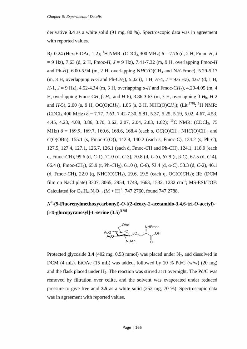
Chapter 6: Experimental Details
Page | 165
derivative 3.4 as a white solid (91 mg, 80 %). Spectroscopic data was in agreement
with reported values.
Rf: 0.24 (Hex:EtOAc, 1:2); 1H NMR: (CDCl3, 300 MHz) δ = 7.76 (d, 2 H, Fmoc-H, J
= 9 Hz), 7.63 (d, 2 H, Fmoc-H, J = 9 Hz), 7.41-7.32 (m, 9 H, overlapping Fmoc-H
and Ph-H), 6.00-5.94 (m, 2 H, overlapping NHC(O)CH3 and NH-Fmoc), 5.29-5.17
(m, 3 H, overlapping H-3 and Ph-CH2), 5.02 (t, 1 H, H-4, J = 9.6 Hz), 4.67 (d, 1 H,
H-1, J = 9 Hz), 4.52-4.34 (m, 3 H, overlapping α-H and Fmoc-CH2), 4.20-4.05 (m, 4
H, overlapping Fmoc-CH, β-Ha, and H-6), 3.86-3.63 (m, 3 H, overlapping β-Hb, H-2
and H-5), 2.00 (s, 9 H, OC(O)CH3), 1.85 (s, 3 H, NHC(O)CH3); (Lit[178]
: 1H NMR:
(CDCl3, 400 MHz) δ = 7.77, 7.63, 7.42-7.30, 5.81, 5.37, 5.25, 5.19, 5.02, 4.67, 4.53,
4.45, 4.23, 4.08, 3.86, 3.70, 3.62, 2.07, 2.04, 2.03, 1.82); 13
C NMR: (CDCl3, 75
MHz) δ = 169.9, 169.7, 169.6, 168.6, 168.4 (each s, OC(O)CH3, NHC(O)CH3, and
C(O)OBn), 155.1 (s, Fmoc-C(O)), 142.8, 140.2 (each s, Fmoc-C), 134.2 (s, Ph-C),
127.5, 127.4, 127.1, 126.7, 126.1 (each d, Fmoc-CH and Ph-CH), 124.1, 118.9 (each
d, Fmoc-CH), 99.6 (d, C-1), 71.0 (d, C-3), 70.8 (d, C-5), 67.9 (t, β-C), 67.5 (d, C-4),
66.4 (t, Fmoc-CH2), 65.9 (t, Ph-CH2), 61.0 (t, C-6), 53.4 (d, α-C), 53.3 (d, C-2), 46.1
(d, Fmoc-CH), 22.0 (q, NHC(O)CH3), 19.6, 19.5 (each q, OC(O)CH3); IR: (DCM
film on NaCl plate) 3307, 3065, 2954, 1748, 1663, 1532, 1232 cm-1
; MS-ESI/TOF:
Calculated for C39H43N2O13 (M + H)+: 747.2760, found 747.2788.
Nα-(9-Fluorenylmethoxycarbonyl)-O-[(2-deoxy-2-acetamido-3,4,6-tri-O-acetyl)-
β-D-glucopyranosyl]-L-serine (3.5)[178]
Protected glycoside 3.4 (402 mg, 0.53 mmol) was placed under N2, and dissolved in
DCM (4 mL). EtOAc (15 mL) was added, followed by 10 % Pd/C (w/w) (20 mg)
and the flask placed under H2. The reaction was stirred at rt overnight. The Pd/C was
removed by filtration over celite, and the solvent was evaporated under reduced
pressure to give free acid 3.5 as a white solid (252 mg, 70 %). Spectroscopic data
was in agreement with reported values.

Chapter 6: Experimental Details
Page | 166
Rf: 0.2 (DCM:MeOH, 10:1); 1H NMR: (CD3OD, 300 MHz) δ = 7.82 (d, 2 H, Fmoc-
H, J = 9 Hz), 7.70 (d, 2 H, Fmoc-H, J = 9 Hz), 7.43-7.31 (m, 4 H, Fmoc-H), 5.27-
5.20 (m, 1 H, H-3), 4.99 (t, 1 H, H-4, J = 9 Hz), 4.74 (d, 1 H, H-1, J = 9 Hz), 4.47-
4.35 (m, 3 H, overlapping α-H and Fmoc-CH2), 4.29-4.23 (m, 2 H, overlapping
Fmoc-CH and H-6a), 4.16-4.10 (m, 2 H, overlapping β-Ha and H-6b), 3.96-3.91 (dd,
1 H, β-Hb, J = 6 Hz, 12 Hz), 3.85-3.76 (m, 2 H, overlapping H-2 and H-5), 2.04,
2.01, 1.99 (each s, 9 H, OC(O)CH3), 1.87 (s, 3 H, NHC(O)CH3); (Lit[178]
: 1H NMR:
(CD3OD, 400 MHz) δ = 7.94, 7.82, 7.54, 7.47, 5.36, 5.11, 4.83, 4.55, 4.49, 4.43-4.36,
4.33-4.21, 4.18, 4.04-3.91, 2.17, 2.15, 2.11, 2.00); IR: (DCM film on NaCl plate)
3427, 2096, 1649 cm-1
; MS-ESI/TOF: Calculated for C25H24NO5 (M + H)+
657.2290, found 657.2299.
(2-Deoxy-2-[(2,2,2-trichloroethoxycarbonyl)amino]-1,3,4-tri-O-acetyl-6-O-
(triphenylmethyl))-α/β-D-glucopyranoside (4.2)
D (+) -Glucosamine hydrochloride (2.00 g, 9.0 mmol) and solid NaHCO3 (2.27 g, 27
mmol) were dissolved in H2O (20 mL). The reaction mixture was stirred for 5 mins
at rt. TrocCl (1.45 mL, 10.8 mmol) was added dropwise. The reaction mixture was
stirred at rt for 2 h, then neutralized (1 M HCl). The solvent was evaporated under
reduced pressure. The NH-Troc protected glucosamine was obtained as a cream solid
which was used without further purification. A portion of this intermediate (150 mg,
0.46 mmol) was dissolved in dry pyridine (10 mL). Et3N (0.02 mL, 0.12 mmol) and
TrCl (640 mg, 2.3 mmol) was added to the reaction mixture. This was stirred at 95
°C for 3 h. The reaction mixture was cooled to 0 °C, then acetyl chloride (0.19 mL,
2.76 mmol) was added slowly. The reaction mixture was allowed to return to rt, then
stirred at rt overnight. The solvent was evaporated under reduced pressure, and the
remaining residue was dissolved in DCM, and washed with brine. The organic layer
was dried (MgSO4), and filtered, then concentrated under reduced pressure. The
crude product was purified by column chromatography (Hex:EtOAc, 2:1) to give
protected glucosamine derivative 4.2 as a clear oil (132 mg, 39 %) (Anomeric
mixture of α:β in the ratio 1:0.4).

Chapter 6: Experimental Details
Page | 167
Rf: 0.53 (Pet. Ether: EtOAc, 2:1); 1H NMR: (CDCl3, 300 MHz) δ = 7.44-7.19 (m, 15
H, Ph-H), 6.35 (d, 1 H, H-1 (α-anomer), J = 3 Hz), 5.72 (d, 1 H, H-1 (β-anomer), J =
9 Hz), 5.35 (t, 1 H, H-4, J = 9 Hz), 5.24-5.15 (m, 1 H, H-3), 5.11 (d, 1 H,
NHC(O)OCH2, J = 9 Hz), 4.83 (d, 1 H, NHC(O)OCH2a (α-anomer), J = 12 Hz), 4.73
(s, 2 H, NHC(O)OCH2 (β-anomer)), 4.63 (d, 1 H, NHC(O)OCH2b (α-anomer), J = 12
Hz), 4.27-4.13 (m, 1 H, H-2 (α-anomer)), 4.03-3.97 (m, 1 H, H-2 (β-anomer)), 3.93-
3.86 (m, 1 H, H-5 (α-anomer)), 3.68-3.63 (m, 1 H, H-5 (β-anomer)), 3.35-3.27 (m, 2
H, H-6a (both anomers)), 3.10-3.01 (m, 2 H, H-6b (both anomers)), 2.17-2.01 (m, 18
H, OC(O)CH3 (both anomers)); 13
C NMR: (CDCl3, 75 MHz) δ = 171.5, 169.3, 168.7
(each s, OC(O)CH3), 154.1 (s, NHC(O)OCH2), 143.5 (s, Ph-C), 128.7, 127.9, 127.8,
127.3, 127.0 (each d, Ph-H), 95.3 (s, CCl3), 92.6 (s, C(ph)3), 90.7 (d, C-1), 74.7 (t,
NHC(O)OCH2), 71.2 (d, C-5), 71.0 (d, C-3), 67.8 (d, C-4), 61.4 (t, C-6), 53.4 (d, C-
2), 21.0, 20.7, 20.4 (each q, OC(O)CH3); [α]D: + 41.6 (c 1.2, CH2Cl2); IR: (DCM
film on NaCl plate) 3421, 3059, 1753, 1647, 1219 cm-1
; MS-ESI/TOF: Calculated
for C15H20Cl3NNaO10 (M (-Tr) + Na)+
(Tr removed by formic acid in the mobile
phase): 502.0045, found 502.0061.
(2-Deoxy-2-[(2,2,2-trichloroethoxycarbonyl)amino]-1,3,4-tri-O-acetyl-6-O-(tert-
butyldimethylsilyl))-α/β-D-glucopyranoside (4.3)
D (+) -Glucosamine hydrochloride (2.00 g, 9.0 mmol) and solid NaHCO3 (2.27 g, 27
mmol) were dissolved in H2O (20 mL). The reaction mixture was stirred for 5 mins
at rt. TrocCl (1.45 mL, 10.8 mmol) was added dropwise. The reaction mixture was
stirred at rt for 2 h, then neutralized (1 M HCl). The solvent was evaporated under
reduced pressure. The NH-Troc protected glucosamine was obtained as a cream solid
which was used without further purification. This intermediate (307 mg, 0.85 mmol)
was dissolved in dry pyridine (3 mL) under N2, in the presence of 3Å molecular
sieves. Imidazole (63 mg, 0.94 mmol) and TBDMSCl (154 mg, 1.02 mmol) were
added to the reaction mixture. This was stirred at rt overnight. Acetic anhydride
(0.48 mL, 5.1 mmol) was added, and the reaction was left to stir at rt overnight. The
solvent was evaporated under reduced pressure, and the remaining residue was

Chapter 6: Experimental Details
Page | 168
dissolved in DCM, and washed twice with water. The organic layer was dried
(MgSO4), and filtered, then concentrated under reduced pressure. The crude product
was purified by column chromatography (Hex:EtOAc, 4:3) to give the protected
glucosamine derivative 4.3 as a yellow oil (131 mg, 26 %) (Anomeric mixture of α:β
in the ratio 3:1).
Rf: 0.9 (Hex:EtOAc, 4:1); 1H NMR: (CDCl3, 300 MHz) δ = 6.20 (d, 1 H, H-1 (α-
anomer), J = 3 Hz), 5.68 (d, 1 H, H-1 (β-anomer), J = 9 Hz), 5.44 (d, 1 H,
NHC(O)OCH2 (β-anomer), J = 9 Hz), 5.27-5.12 (m, 4 H, overlapping H-3,
NHC(O)OCH2, and H-4 (α-anomer), and H-3 (β-anomer)), 5.12-5.06 (m, 1 H, H-4
(β-anomer)), 4.78 (d, 1 H, NHC(O)OCH2a (α-anomer), J = 12 Hz), 4.72-4.69 (m, 2
H, NHC(O)OCH2 (β-anomer)), 4.59 (d, 1 H, NHC(O)OCH2b (α-anomer), J = 12 Hz),
4.15-4.07 (m, 2 H, overlapping H-2 (α-anomer) and H-6a (β-anomer)) 3.83-3.78 (m,
2 H, overlapping H-5 (α-anomer) and H-2 (β-anomer)), 3.64-3.60 (m, 4 H,
overlapping H-5 and H-6b (β-anomer), and H-6 (α-anomer)), 2.14-2.00 (m, 18 H,
OC(O)CH3 (both anomers)), 0.84 (s, 18 H, SiC(CH3)3 (both anomers)), 0.03- -0.01
(m, 12 H, SiCH3 (both anomers)); 13
C NMR: (CDCl3, 75 MHz) δ = 170.4, 169.9,
168.3, 168.0, 167.7 (each s, OC(O)CH3), 153.1 (s, NHC(O)OCH2), 94.5, 94.3 (each
s, CCl3 (both anomers)), 91.3 (d, C-1 (β-anomer)), 89.5 (d, C-1 (α-anomer)), 73.9 (t,
NHC(O)OCH2 (β-anomer)), 73.4 (t, NHC(O)OCH2 (α-anomer)), 71.5 (d, C-5), 69.9
(d, C-3), 67.5 (d, C-4 (β-anomer)), 67.0 (d, C-4 (α-anomer)), 61.07, 60.8 (each t, C-6
(both anomers)), 52.3 (d, C-2), 24.8, 24.5 (each q, SiC(CH3)3), 19.9, 19.8, 19.7, 19.6
(each q, OC(O)CH3), -6.41 (q, SiCH3); IR: (as a DCM film on NaCl plate) 3327,
2957, 1755, 1538, 1368, 1219 cm-1
.
(2-Deoxy-2-[(2,2,2-trichloroethoxycarbonyl)amino]-3,4,6-tri-O-acetyl-1-O-
phenylmethyl)-β-D-glucopyranoside (4.6)[189, 260]
Acetylated derivative 2.26 (1.32 g, 2.5 mmol) was dissolved in DCM (10 mL), and
cooled to 0 ˚C. To this a 33 % solution (v/v) of HBr in AcOH (2.4 mL) was added,
and the reaction mixture was stirred for 45 mins. The reaction mixture was allowed
to reach rt, and stirred for 45 mins. A further portion of HBr solution (1.2 mL) was
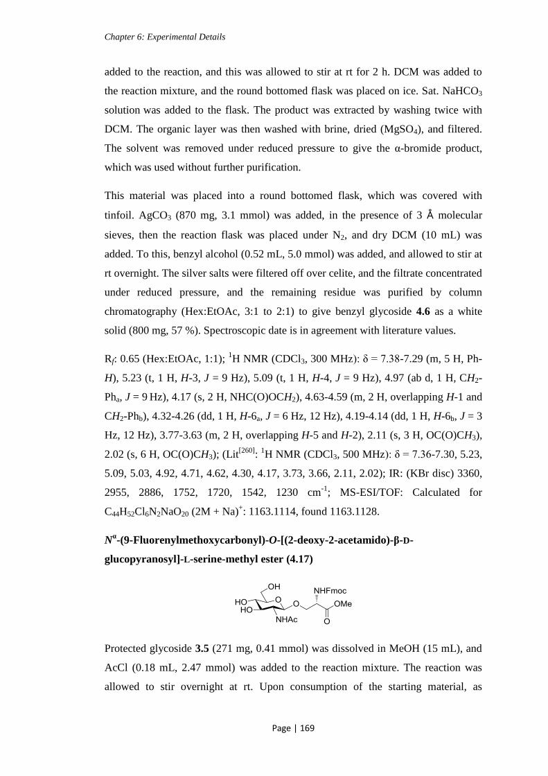
Chapter 6: Experimental Details
Page | 169
added to the reaction, and this was allowed to stir at rt for 2 h. DCM was added to
the reaction mixture, and the round bottomed flask was placed on ice. Sat. NaHCO3
solution was added to the flask. The product was extracted by washing twice with
DCM. The organic layer was then washed with brine, dried (MgSO4), and filtered.
The solvent was removed under reduced pressure to give the α-bromide product,
which was used without further purification.
This material was placed into a round bottomed flask, which was covered with
tinfoil. AgCO3 (870 mg, 3.1 mmol) was added, in the presence of 3 Å molecular
sieves, then the reaction flask was placed under N2, and dry DCM (10 mL) was
added. To this, benzyl alcohol (0.52 mL, 5.0 mmol) was added, and allowed to stir at
rt overnight. The silver salts were filtered off over celite, and the filtrate concentrated
under reduced pressure, and the remaining residue was purified by column
chromatography (Hex:EtOAc, 3:1 to 2:1) to give benzyl glycoside 4.6 as a white
solid (800 mg, 57 %). Spectroscopic date is in agreement with literature values.
Rf: 0.65 (Hex:EtOAc, 1:1); 1H NMR (CDCl3, 300 MHz): δ = 7.38-7.29 (m, 5 H, Ph-
H), 5.23 (t, 1 H, H-3, J = 9 Hz), 5.09 (t, 1 H, H-4, J = 9 Hz), 4.97 (ab d, 1 H, CH2-
Pha, J = 9 Hz), 4.17 (s, 2 H, NHC(O)OCH2), 4.63-4.59 (m, 2 H, overlapping H-1 and
CH2-Phb), 4.32-4.26 (dd, 1 H, H-6a, J = 6 Hz, 12 Hz), 4.19-4.14 (dd, 1 H, H-6b, J = 3
Hz, 12 Hz), 3.77-3.63 (m, 2 H, overlapping H-5 and H-2), 2.11 (s, 3 H, OC(O)CH3),
2.02 (s, 6 H, OC(O)CH3); (Lit[260]
: 1H NMR (CDCl3, 500 MHz): δ = 7.36-7.30, 5.23,
5.09, 5.03, 4.92, 4.71, 4.62, 4.30, 4.17, 3.73, 3.66, 2.11, 2.02); IR: (KBr disc) 3360,
2955, 2886, 1752, 1720, 1542, 1230 cm-1
; MS-ESI/TOF: Calculated for
C44H52Cl6N2NaO20 (2M + Na)+: 1163.1114, found 1163.1128.
Nα-(9-Fluorenylmethoxycarbonyl)-O-[(2-deoxy-2-acetamido)-β-D-
glucopyranosyl]-L-serine-methyl ester (4.17)
Protected glycoside 3.5 (271 mg, 0.41 mmol) was dissolved in MeOH (15 mL), and
AcCl (0.18 mL, 2.47 mmol) was added to the reaction mixture. The reaction was
allowed to stir overnight at rt. Upon consumption of the starting material, as

Chapter 6: Experimental Details
Page | 170
indicated by TLC, the solvent was evaporated under reduced pressure, and the
remaining residue was purified by column chromatography (DCM:MeOH, 9:1 to
7:1) to give glycoside 4.17 as a white solid (198 mg, 89 %).
Rf: 0.2 (DCM:MeOH, 7:1); 1H MNR (CD3OD (a drop of D2O to improve solubility)
300 MHz): δ = 7.83 (d, 2 H, Ph-H, J = 6 Hz), 7.69 (d, 2 H, Ph-H, J = 9 Hz), 7.46-
7.34 (m, 4 H, Ph-H), 4.49-4.38 (m, 4 H, overlapping H-1, Fmoc-CH2, and α-H), 4.25
(t, 1 H, Fmoc-CH, J = 6 Hz), 4.18-4.13 (dd, 1 H, β-Ha, J = 6 Hz, 9 Hz), 3.91-3.83
(m, 2 H, overlapping β-Hb and H-6a), 3.75 (s, 3 H, C(O)OCH3), 3.73-3.61 (m, 2 H,
overlapping H-2 and H-6b), 3.51-3.45 (m, 1 H, H-3), 3.35-3.30 (m, 2 H, (overlapped
by MeOH peak) overlapping H-5 and H-4), 1.94 (s, 3 H, NHC(O)CH3); 13
C NMR:
(CD3OD (a drop of D2O to improve solubility), 75 MHz) δ = 173.3, 167.5 (each s,
C(O)OCH3 and NHC(O)CH3), 143.6, 141.1 (each s, Fmoc-C(O) and Ph-C), 127.7,
127.1, 124.8, 119.7 (each d, Fmoc-CH and Ph-CH), 101.2 (d, C-1), 76.4, 74.1, 70.2,
66.8, 55.8, 54.3, 52.3, 29.7 (not assigned due to poor solubility of sample), 21.9 (q,
NHC(O)CH3); IR: (KBr disc) 3319, 2952, 1748, 1690, 1654, 1535 cm-1
; [α]D: - 1.7 (c
1.3, CH3OH); MS-ESI/TOF: Calculated for C27H33N2O10 (M + H)+: 546.2162, found
546.2184.
Nα-(9-Fluorenylmethoxycarbonyl)-O-[(2-deoxy-2-acetamido-6-O-p-tosyl)-β-D-
glucopyranosyl]-L-serine-methyl ester (4.18)
Glycoside 4.17 (60 mg, 0.11 mmol) was placed under N2 and dissolved in anhydrous
pyridine (2 mL). The reaction mixture was cooled to 0 °C, and a solution of TsCl
(100 mg, 0.52 mmol) in anhydrous pyridine (2 mL) was added. The reaction was
stirred at 0 °C for 1 h, allowed to return to rt, and stirred at rt overnight. The solvent
was co-evaporated with toluene under reduced pressure, and the remaining residue
was purified by column chromatography (DCM:MeOH, 15:1 to 10:1) to give the
tosylated glycoside 4.18 as a clear oil (19 mg, 24 %).
Rf: 0.5 (DCM:MeOH, 7:1); 1H MNR (CDCl3, 300 MHz): δ = 7.74 (t, 4 H, Ph-H, J =
6 Hz), 7.57 (d, 2 H, Ph-H, J = 6 Hz), 7.37 (t, 2 H, Ph-H, J = 6 Hz), 7.29-7.23 (m, 4

Chapter 6: Experimental Details
Page | 171
H, Ph-H), 6.91 (bs, 1 H, NHC(O)CH3), 5.99 (d, 1 H, NHC(O)OCH2, J = 9 Hz), 4.50-
4.36 (m, 2 H, overlapping H-1 and α-H), 4.27-4.14 (m, 5 H, overlapping H-6, Fmoc-
CH, and Fmoc-CH2), 4.05-4.00 (dd, 1 H, β-Ha, J = 6 Hz, 9 Hz), 3.86-3.83 (m, 1 H,
β-Hb), 3.72 (s, 3 H, C(O)OCH3), 3.65-3.39 (m, 4 H, overlapping H-2, H-3, H-4, and
H-5), 2.36 (s, 3 H, Ph-CH3), 1.95 (s, 3 H, NHC(O)CH3); 13
C NMR: (CDCl3, 75
MHz) δ = 173.2, 170.4 (each s, C(O)OCH3 and NHC(O)CH3), 156.4 (s, Fmoc-C(O)),
145.1, 143.7, 143.6, 141.23, 132.48 (each s, Ph-C), 129.9, 127.9, 127.8, 127.1,
125.1, 120.0 (each d, Fmoc-CH and Ph-CH), 100.3 (d, C-1), 74.9, 73.5, 70.3 (each d,
C-2, C-3, C-4, and C-5), 69.1, 37.2 (each t, C-6, Fmoc-CH2, and β-C), 53.9 (d, α-C),
52.89 (q, C(O)OCH3), 23.2 (q, NHC(O)CH3), 21.61 (q, Ph-CH3); IR: (DCM film on
NaCl plate) 3349, 2984, 1723, 1655, 1548, 1176 cm-1
; [α]D: - 2.8 (c 0.85, CH2Cl2);
MS-ESI/TOF: Calculated for C34H39N2O12S (M + H)+: 700.2250, found 700.2268.
3-Azido-propionic acid (4.21)[209]
3-Bromopropionic acid (1.50 g, 9.8 mmol) was dissolved in MeCN (4 mL). NaN3
(956 mg, 14.7 mmol) was added and the reaction mixture, and heated at reflux (85
°C) for 3 h. The reaction was allowed to return to rt, then carefully washed with 0.1
M HCl, dried (MgSO4) and filtered. The solvent was evaporated to almost dryness
under reduced pressure, with care taken that the water bath was not heated. The
remaining orange oil was used without further purification (400 mg, 36 % crude
yield).
1H NMR: (CDCl3, 300 MHz) δ = 9.87 (bs, 1 H, O-H), 3.58 (t, 2 H, CH2N3, J = 6
Hz), 2.63 (t, 2 H, OC(O)CH2, J = 6 Hz); (Lit[209]
: 1H NMR: (CDCl3, 400 MHz) δ =
11.00, 3.60, 2.65); IR: (Liquid IR in CDCl3) 3165, 2941, 2104, 1718 cm-1
.
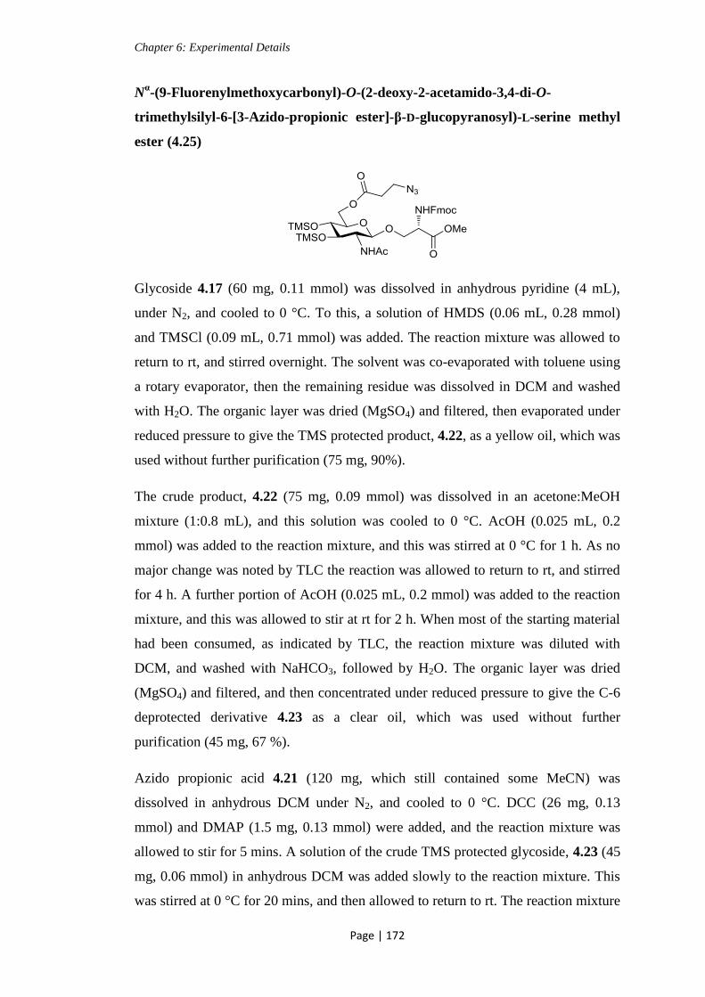
Chapter 6: Experimental Details
Page | 172
Nα-(9-Fluorenylmethoxycarbonyl)-O-(2-deoxy-2-acetamido-3,4-di-O-
trimethylsilyl-6-[3-Azido-propionic ester]-β-D-glucopyranosyl)-L-serine methyl
ester (4.25)
Glycoside 4.17 (60 mg, 0.11 mmol) was dissolved in anhydrous pyridine (4 mL),
under N2, and cooled to 0 °C. To this, a solution of HMDS (0.06 mL, 0.28 mmol)
and TMSCl (0.09 mL, 0.71 mmol) was added. The reaction mixture was allowed to
return to rt, and stirred overnight. The solvent was co-evaporated with toluene using
a rotary evaporator, then the remaining residue was dissolved in DCM and washed
with H2O. The organic layer was dried (MgSO4) and filtered, then evaporated under
reduced pressure to give the TMS protected product, 4.22, as a yellow oil, which was
used without further purification (75 mg, 90%).
The crude product, 4.22 (75 mg, 0.09 mmol) was dissolved in an acetone:MeOH
mixture (1:0.8 mL), and this solution was cooled to 0 °C. AcOH (0.025 mL, 0.2
mmol) was added to the reaction mixture, and this was stirred at 0 °C for 1 h. As no
major change was noted by TLC the reaction was allowed to return to rt, and stirred
for 4 h. A further portion of AcOH (0.025 mL, 0.2 mmol) was added to the reaction
mixture, and this was allowed to stir at rt for 2 h. When most of the starting material
had been consumed, as indicated by TLC, the reaction mixture was diluted with
DCM, and washed with NaHCO3, followed by H2O. The organic layer was dried
(MgSO4) and filtered, and then concentrated under reduced pressure to give the C-6
deprotected derivative 4.23 as a clear oil, which was used without further
purification (45 mg, 67 %).
Azido propionic acid 4.21 (120 mg, which still contained some MeCN) was
dissolved in anhydrous DCM under N2, and cooled to 0 °C. DCC (26 mg, 0.13
mmol) and DMAP (1.5 mg, 0.13 mmol) were added, and the reaction mixture was
allowed to stir for 5 mins. A solution of the crude TMS protected glycoside, 4.23 (45
mg, 0.06 mmol) in anhydrous DCM was added slowly to the reaction mixture. This
was stirred at 0 °C for 20 mins, and then allowed to return to rt. The reaction mixture

Chapter 6: Experimental Details
Page | 173
was stirred at rt for 5 h, and then washed with H2O. The organic layer was dried
(MgSO4) and filtered, and then concentrated under reduced pressure to almost
dryness. The remaining oil was purified by column chromatography (Pet. Ether:
EtOAc, 1:1 →1:2) to give the functionalised glycoside 4.25 as a clear oil (5 mg, 9
%).
Rf: 0.7 (Pet. Ether:EtOAc, 1:3); 1H NMR (CDCl3, 300 MHz): δ = 7.77 (d, 2 H, Ph-H,
J = 6 Hz), 7.69-7.65 (m, 2 H, Ph-H), 7.43-7.30 (m, 4 H, Ph-H), 5.91 (d, 1 H,
NHC(O)OCH2, J = 6 Hz), 5.86 (d, 1 H, NHAc, J = 9 Hz), 4.88 (d, 1 H, H-1, J = 6
Hz), 4.52-4.43 (m, 3 H, overlapping α-H, β-Ha, and H-6a), 4.38-4.32 (m 1 H, β-Hb),
4.28-4.13 (m, 3 H, overlapping H-6b and Fmoc-CH2), 3.89 (t, 1 H, H-3, J = 6 Hz),
3.82-3.77 (m, 1 H, Fmoc-CH), 3.75 (s, 3 H, C(O)OCH3), 3.60-3.49 (m, 4 H,
overlapping H-4, H-5, and CH2N3), 3.45-3.37 (m, 1 H, H-2), 2.64-2.58 (m, 2 H,
OC(O)CH2), 1.97 (s, 3 H, NHC(O)CH3), 0.17, 0.16 (each s, 18 H, Si(CH3)3); 13
C
NMR (CDCl3, 75 MHz): δ = 155.2, 151.3, 126.7, 126.1, 124.2, 118.9, 99.1, 71.1,
66.1, 54.9, 53.2, 51.5, 30.1, 28.7, 22.7, -0.3, -0.4; IR: (Liquid IR in CDCl3) 3155,
2962, 2104, 1816, 1712, 1563, 1249 cm-1
; [α]D: + 8 (c 0.25, CH2Cl2); MS-ESI/TOF:
Calculated for C33H44N5O11Si (M (-TMS) + H)+: 714.2801, found 714.2818 (1 X
TMS removed by formic acid in the mobile phase).
(2-Deoxy-2-acetamido-6-O-[N-(3,4-hydoxyphenylethyl)-succinamide])-α/β-D-
glucopyranose (5.9)
Glycosyl derivative 5.39 (40 mg, 0.06 mmol) was dissolved in MeOH (5 mL), and
10 % Pd/C (w/w) (40 mg) was added. The reaction was carried out by bubbling H2
through the reaction mixture, at rt for 4 h. The reaction mixture was filtered through
celite, and the solvent was removed under reduced pressure. The remaining residue
was purified by column chromatography (EtOAc:MeOH, 4:1) to give the
deprotected glycosyl dopamine derivative 5.9 as a white oily solid (14 mg, 56 %).
(Anomeric ratio 1:0.6, α:β)
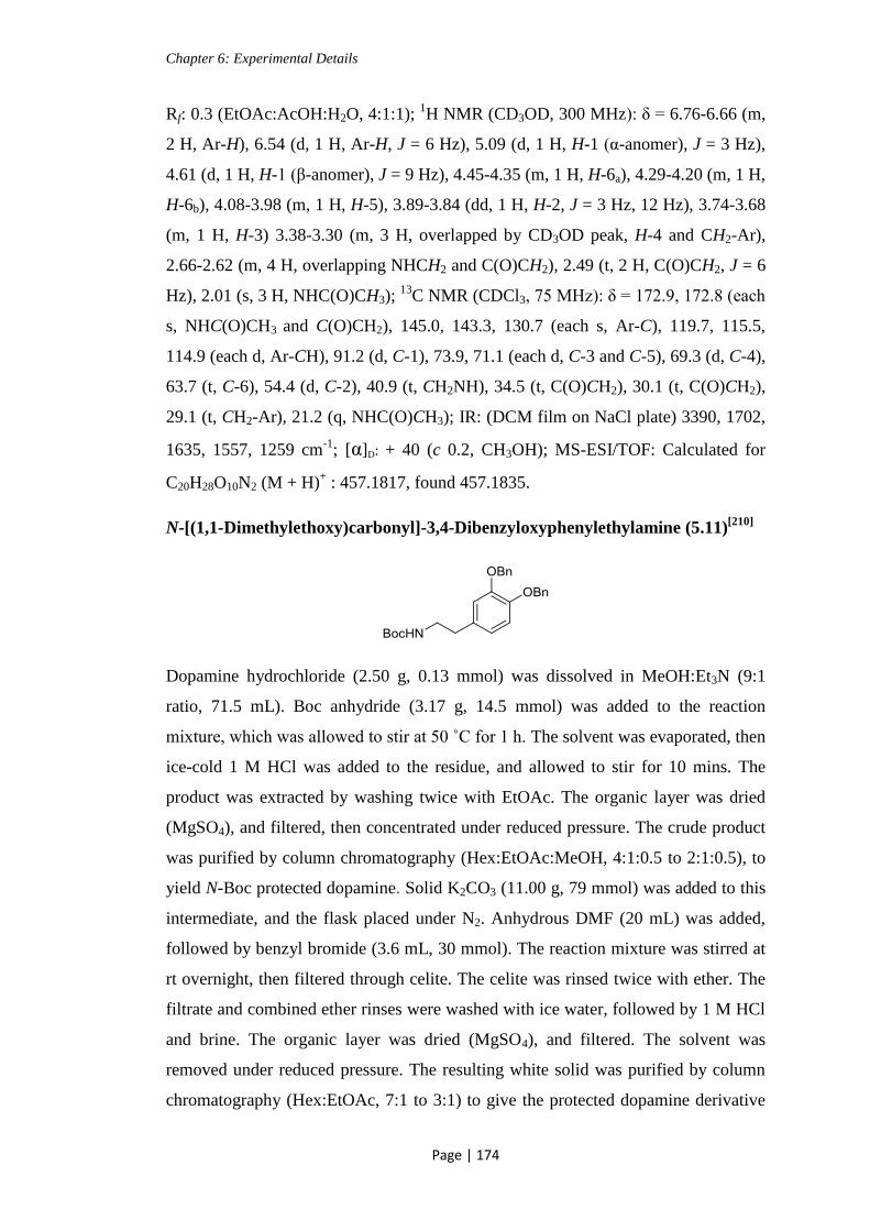
Chapter 6: Experimental Details
Page | 174
Rf: 0.3 (EtOAc:AcOH:H2O, 4:1:1); 1H NMR (CD3OD, 300 MHz): δ = 6.76-6.66 (m,
2 H, Ar-H), 6.54 (d, 1 H, Ar-H, J = 6 Hz), 5.09 (d, 1 H, H-1 (α-anomer), J = 3 Hz),
4.61 (d, 1 H, H-1 (β-anomer), J = 9 Hz), 4.45-4.35 (m, 1 H, H-6a), 4.29-4.20 (m, 1 H,
H-6b), 4.08-3.98 (m, 1 H, H-5), 3.89-3.84 (dd, 1 H, H-2, J = 3 Hz, 12 Hz), 3.74-3.68
(m, 1 H, H-3) 3.38-3.30 (m, 3 H, overlapped by CD3OD peak, H-4 and CH2-Ar),
2.66-2.62 (m, 4 H, overlapping NHCH2 and C(O)CH2), 2.49 (t, 2 H, C(O)CH2, J = 6
Hz), 2.01 (s, 3 H, NHC(O)CH3); 13
C NMR (CDCl3, 75 MHz): δ = 172.9, 172.8 (each
s, NHC(O)CH3 and C(O)CH2), 145.0, 143.3, 130.7 (each s, Ar-C), 119.7, 115.5,
114.9 (each d, Ar-CH), 91.2 (d, C-1), 73.9, 71.1 (each d, C-3 and C-5), 69.3 (d, C-4),
63.7 (t, C-6), 54.4 (d, C-2), 40.9 (t, CH2NH), 34.5 (t, C(O)CH2), 30.1 (t, C(O)CH2),
29.1 (t, CH2-Ar), 21.2 (q, NHC(O)CH3); IR: (DCM film on NaCl plate) 3390, 1702,
1635, 1557, 1259 cm-1
; [α]D: + 40 (c 0.2, CH3OH); MS-ESI/TOF: Calculated for
C20H28O10N2 (M + H)+ : 457.1817, found 457.1835.
N-[(1,1-Dimethylethoxy)carbonyl]-3,4-Dibenzyloxyphenylethylamine (5.11)[210]
Dopamine hydrochloride (2.50 g, 0.13 mmol) was dissolved in MeOH:Et3N (9:1
ratio, 71.5 mL). Boc anhydride (3.17 g, 14.5 mmol) was added to the reaction
mixture, which was allowed to stir at 50 ˚C for 1 h. The solvent was evaporated, then
ice-cold 1 M HCl was added to the residue, and allowed to stir for 10 mins. The
product was extracted by washing twice with EtOAc. The organic layer was dried
(MgSO4), and filtered, then concentrated under reduced pressure. The crude product
was purified by column chromatography (Hex:EtOAc:MeOH, 4:1:0.5 to 2:1:0.5), to
yield N-Boc protected dopamine. Solid K2CO3 (11.00 g, 79 mmol) was added to this
intermediate, and the flask placed under N2. Anhydrous DMF (20 mL) was added,
followed by benzyl bromide (3.6 mL, 30 mmol). The reaction mixture was stirred at
rt overnight, then filtered through celite. The celite was rinsed twice with ether. The
filtrate and combined ether rinses were washed with ice water, followed by 1 M HCl
and brine. The organic layer was dried (MgSO4), and filtered. The solvent was
removed under reduced pressure. The resulting white solid was purified by column
chromatography (Hex:EtOAc, 7:1 to 3:1) to give the protected dopamine derivative
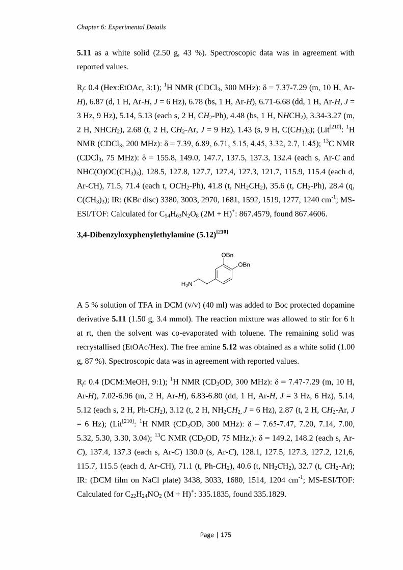
Chapter 6: Experimental Details
Page | 175
5.11 as a white solid (2.50 g, 43 %). Spectroscopic data was in agreement with
reported values.
Rf: 0.4 (Hex:EtOAc, 3:1); 1H NMR (CDCl3, 300 MHz): δ = 7.37-7.29 (m, 10 H, Ar-
H), 6.87 (d, 1 H, Ar-H, J = 6 Hz), 6.78 (bs, 1 H, Ar-H), 6.71-6.68 (dd, 1 H, Ar-H, J =
3 Hz, 9 Hz), 5.14, 5.13 (each s, 2 H, CH2-Ph), 4.48 (bs, 1 H, NHCH2), 3.34-3.27 (m,
2 H, NHCH2), 2.68 (t, 2 H, CH2-Ar, J = 9 Hz), 1.43 (s, 9 H, C(CH3)3); (Lit[210]
: 1H
NMR (CDCl3, 200 MHz): δ = 7.39, 6.89, 6.71, 5.15, 4.45, 3.32, 2.7, 1.45); 13
C NMR
(CDCl3, 75 MHz): δ = 155.8, 149.0, 147.7, 137.5, 137.3, 132.4 (each s, Ar-C and
NHC(O)OC(CH3)3), 128.5, 127.8, 127.7, 127.4, 127.3, 121.7, 115.9, 115.4 (each d,
Ar-CH), 71.5, 71.4 (each t, OCH2-Ph), 41.8 (t, NH2CH2), 35.6 (t, CH2-Ph), 28.4 (q,
C(CH3)3); IR: (KBr disc) 3380, 3003, 2970, 1681, 1592, 1519, 1277, 1240 cm-1
; MS-
ESI/TOF: Calculated for C54H63N2O8 (2M + H)+: 867.4579, found 867.4606.
3,4-Dibenzyloxyphenylethylamine (5.12)[210]
A 5 % solution of TFA in DCM (v/v) (40 ml) was added to Boc protected dopamine
derivative 5.11 (1.50 g, 3.4 mmol). The reaction mixture was allowed to stir for 6 h
at rt, then the solvent was co-evaporated with toluene. The remaining solid was
recrystallised (EtOAc/Hex). The free amine 5.12 was obtained as a white solid (1.00
g, 87 %). Spectroscopic data was in agreement with reported values.
Rf: 0.4 (DCM:MeOH, 9:1); 1H NMR (CD3OD, 300 MHz): δ = 7.47-7.29 (m, 10 H,
Ar-H), 7.02-6.96 (m, 2 H, Ar-H), 6.83-6.80 (dd, 1 H, Ar-H, J = 3 Hz, 6 Hz), 5.14,
5.12 (each s, 2 H, Ph-CH2), 3.12 (t, 2 H, NH2CH2, J = 6 Hz), 2.87 (t, 2 H, CH2-Ar, J
= 6 Hz); (Lit[210]
: 1H NMR (CD3OD, 300 MHz): δ = 7.65-7.47, 7.20, 7.14, 7.00,
5.32, 5.30, 3.30, 3.04); 13
C NMR (CD3OD, 75 MHz,): δ = 149.2, 148.2 (each s, Ar-
C), 137.4, 137.3 (each s, Ar-C) 130.0 (s, Ar-C), 128.1, 127.5, 127.3, 127.2, 121,6,
115.7, 115.5 (each d, Ar-CH), 71.1 (t, Ph-CH2), 40.6 (t, NH2CH2), 32.7 (t, CH2-Ar);
IR: (DCM film on NaCl plate) 3438, 3033, 1680, 1514, 1204 cm-1
; MS-ESI/TOF:
Calculated for C22H24NO2 (M + H)+: 335.1835, found 335.1829.

Chapter 6: Experimental Details
Page | 176
N-(3,4-Dibenzyloxyphenylethyl)-succinamic acid (5.13)[210]
The free amine 5.12 (1.00 g, 2.99 mmol) was dissolved in pyridine (2 mL), and
succinic anhydride (0.54 g, 5.41 mmol) was added to the reaction mixture. This was
allowed to stir at rt for 3 h. The solvent was co-evaporated with toluene under
reduced pressure, and the remaining residue was purified by column chromatography
(DCM:MeOH, 15:1 to 10:1) to give succinyl-dopamine derivative 5.13 as a white
solid (1.06 g, 82 %). Spectroscopic data was in agreement with reported values.
Rf: 0.5 (DCM:MeOH, 9:1); 1H NMR (CDCl3, 300 MHz): δ = 7.43-7.28 (m, 10 H,
Ar-H), 6.86 (d, 1 H, Ar-H, J = 9 Hz), 6.76 (d, 1 H, Ar-H, J = 3 Hz), 6.68-6.65 (m, 1
H, Ar-H), 5.71 (bs, 1 H, NHCH2), 5.12, 5.11 (each s, 2 H, Ph-CH2), 3.45-3.39 (m, 2
H, NHCH2), 2.68 (t, 2 H, CH2-Ar, J = 6 Hz), 2.61 (t, 2 H, C(O)CH2, J = 6 Hz), 2.37
(t, 2 H, C(O)CH2, J = 6 Hz); (Lit[210]
: 1H NMR (CDCl3, 200 MHz): δ = 7.39, 6.89,
6.80, 6.71, 5.55, 5.17, 5.15, 3.45, 2.70, 2.65, 2.38); 13
C NMR (CDCl3, 75 MHz): δ =
149.0, 147.8 (each s, CH2C(O)), 137.3, 131.9 (each s, Ar-C), 128.5, 127.8, 127.4,
127.3, 121.7, 115.9, 115.2 (each d, Ar-CH), 40.85 (t, NHCH2), 34.7, 31.7 (each t,
CH2C(O)), 30.2 (t, CH2-Ph); IR: (KBr disc) 3324, 3035, 2927, 1694, 1640, 1523,
1266 cm-1
.
(2-Deoxy-2-[(2,2,2-trichloroethoxycarbonyl)amino]-3,4,6-tri-O-trimethylsilyl-1-
O-phenylmethyl ether)-β-D-glucopyranoside (5.17)
Benzyl glycoside 4.6 (1.04 g, 2.2 mmol) was dissolved in distilled MeOH (18 mL),
and 0.1 M NaOH(aq) (18 mL) was added. The reaction mixture was stirred at rt for 1
h, then neutralized with AcOH (10 mL). The organic solvent was evaporated using a
rotary evaporator, and the remaining aqueous solution was extracted with EtOAc.
The organic layer was washed twice with sat. NaHCO3 solution, then dried (MgSO4),
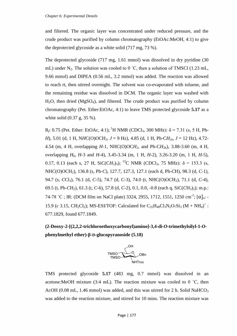
Chapter 6: Experimental Details
Page | 177
and filtered. The organic layer was concentrated under reduced pressure, and the
crude product was purified by column chromatography (EtOAc:MeOH, 4:1) to give
the deprotected glycoside as a white solid (717 mg, 73 %).
The deprotected glycoside (717 mg, 1.61 mmol) was dissolved in dry pyridine (30
mL) under N2. The solution was cooled to 0 ˚C, then a solution of TMSCl (1.23 mL,
9.66 mmol) and DIPEA (0.56 mL, 3.2 mmol) was added. The reaction was allowed
to reach rt, then stirred overnight. The solvent was co-evaporated with toluene, and
the remaining residue was dissolved in DCM. The organic layer was washed with
H2O, then dried (MgSO4), and filtered. The crude product was purified by column
chromatography (Pet. Ether:EtOAc, 4:1) to leave TMS protected glycoside 5.17 as a
white solid (0.37 g, 35 %).
Rf: 0.75 (Pet. Ether: EtOAc, 4:1); 1
H NMR (CDCl3, 300 MHz): δ = 7.31 (s, 5 H, Ph-
H), 5.01 (d, 1 H, NHC(O)OCH2, J = 9 Hz), 4.85 (d, 1 H, Ph-CH2a, J = 12 Hz), 4.72-
4.54 (m, 4 H, overlapping H-1, NHC(O)OCH2, and Ph-CH2b), 3.88-3.60 (m, 4 H,
overlapping H6, H-3 and H-4), 3.45-3.34 (m, 1 H, H-2), 3.26-3.20 (m, 1 H, H-5),
0.17, 0.13 (each s, 27 H, SiC(CH3)3); 13
C NMR (CDCl3, 75 MHz): δ = 153.3 (s,
NHC(O)OCH2), 136.8 (s, Ph-C), 127.7, 127.3, 127.1 (each d, Ph-CH), 98.3 (d, C-1),
94.7 (s, CCl3), 76.1 (d, C-5), 74.7 (d, C-3), 74.0 (t, NHC(O)OCH2), 71.1 (d, C-4),
69.5 (t, Ph-CH2), 61.3 (t, C-6), 57.8 (d, C-2), 0.1, 0.0, -0.8 (each q, SiC(CH3)3); m.p.:
74-78 ˚C ; IR: (DCM film on NaCl plate) 3324, 2955, 1712, 1551, 1250 cm-1
; [α]D: -
15.9 (c 3.15, CH2Cl2); MS-ESI/TOF: Calculated for C25H48Cl3N2O7Si3 (M + NH4)+ :
677.1829, found 677.1849.
(2-Deoxy-2-[(2,2,2-trichloroethoxycarbonyl)amino]-3,4-di-O-trimethylsilyl-1-O-
phenylmethyl ether)-β-D-glucopyranoside (5.18)
TMS protected glycoside 5.17 (483 mg, 0.7 mmol) was dissolved in an
acetone:MeOH mixture (3:4 mL). The reaction mixture was cooled to 0 ˚C, then
AcOH (0.08 mL, 1.46 mmol) was added, and this was stirred for 2 h. Solid NaHCO3
was added to the reaction mixture, and stirred for 10 mins. The reaction mixture was

Chapter 6: Experimental Details
Page | 178
diluted with DCM (20 mL), and the solid NaHCO3 was removed by filtration. The
filtrate was concentrated under reduced pressure, and the remaining residue was
purified by column chromatography (Hex:EtOAc, 9:1 to 3:1) to give the selectively
deprotected glycoside 5.18 as a white solid (298 mg, 72 %).
Rf: 0.32 (Pet. Ether: EtOAc, 4:1); 1H NMR (CDCl3, 300 MHz): δ = 7.31-7.26 (m, 5
H, Ph-H), 5.14 (bs, 1 H, NHC(O)OCH2), 4.85 (d, 1 H, Ph-CH2a, J = 12 Hz), 4.69-
4.66 (m, 3 H, overlapping H-1 and NHC(O)OCH2), 4.59 (d, 1 H, Ph-CH2b, J = 12
Hz), 3.86-3.65 (m, 3 H, overlapping H6a, H-3 and H-4), 3.56 (t, 1 H, H6b, J = 9 Hz),
3.41-3.29 (m, 2 H, overlapping H-2 and H-5), 0.17, 0.13 (each s, 18 H, SiC(CH3)3);
13C NMR (CDCl3, 75 MHz): δ = 153.4 (s, NHC(O)OCH2), 136.4 (s, Ph-C), 127.7,
127.2, 127.1 (each d, Ph-CH), 98.8 (d, C-1), 94.5 (s, CCl3), 75.4 (d, C-5), 74.5 (d, C-
3), 74.0 (t, NHC(O)OCH2), 71.5 (d, C-4), 70.3 (t, Ph-CH2), 61.2 (t, C-6), 58.1 (d, C-
2), 0.12, 0.00 (each q, SiC(CH3)3); m.p.: 70- 74 ˚C; [α]D: - 7.3 (c 0.55, CH2Cl2); IR:
(DCM film on NaCl plate) 3351, 2098, 1642, 1546, 1251 cm-1
; MS-ESI/TOF:
Calculated for C22H40Cl3N2O7Si2 (M + NH4)+: 607.1409, found 607.1417.
(2-Deoxy-2-[(2,2,2-trichloroethoxycarbonyl)amino]-3,4-di-O-trimethylsilyl-6-O-
[N-(3,4-dibenzyloxyphenylethyl)-succinamyl]-1-O-phenylmethyl)-β-D-
glucopyranoside (5.24)
Benzyl glycoside 5.18 (298 mg, 0.51 mmol) was dissolved in dry DCM (10 mL),
under N2, and DCC (120 mg, 0.58 mmol) was added. This was then added to a
solution of the succinyl-dopamine derivative 5.13 (242 mg, 0.56 mmol) in dry DCM
(25 mL) under N2. DMAP (4.3 mg, 0.03 mmol) was added to the reaction mixture,
and this was allowed to stir at rt for 3 h. The reaction mixture was filtered through
celite. The filtrate was diluted with DCM (20 mL) and washed with an aqueous
solution of 5 % AcOH, followed by H2O. The organic layer was dried (MgSO4), and
filtered. The crude product was purified by column chromatography (Pet.
Ether:EtOAc, 4:1 to 2:1) to give the protected glycoside 5.24 as a clear syrup (306
mg, 60 %).

Chapter 6: Experimental Details
Page | 179
Rf: 0.6 (Pet. Ether: EtOAc, 1:1); 1H NMR (CDCl3, 300 MHz): δ = 7.45-7.25 (m, 15
H, Ar-H), 6.87 (d, 1 H, Ar-H, J = 9 Hz), 6.79-6.78 (m, 1 H, Ar-H), 6.70-6.66 (dd, 1
H, Ar-H, J = 3 Hz, 9 Hz), 5.60 (bs, 1 H, NHC(O)CH2), 5.14-5.12 (m, 5 H,
overlapping Ph-CH2 and NHC(O)OCH2), 4.85 (d, 1 H, Ph-CH2a, J = 12 Hz), 4.67-
4.48 (m, 5 H, overlapping H-1, Ph-CH2b, NHC(O)OCH2 and H-6a), 4.15-4.09 (dd, 1
H, H-6b, J = 6 Hz, 12 Hz), 3.73 (t, 1 H, H-3, J = 9 Hz), 3.57 (t, 1 H, H-4, J = 9 Hz),
3.49-3.34 (m, 4 H, overlapping H-5, H-2 and CH2-Ar), 2.73-2.66 (m, 4 H,
overlapping NHCH2 and C(O)CH2), 2.39-2.35 (m, 2 H, C(O)CH2), 0.15, 0.13 (each
s, 18 H, SiC(CH3)3) ; 13
C NMR (CDCl3, 75 MHz): δ = 171.9, 170.3 (each s,
C(O)CH2), 153.2 (s, NHC(O)OCH2), 148.3, 146.9, 136.7, 136.5, 136.4, 131.6 (each
s, Ar-C), 127.7, 127.6, 127.5, 127.2, 127.1, 127.0, 126.6, 126.5, 120.9, 115.0, 114.7
(each d, Ar-CH), 98.4 (d, C-1), 94.5 (s, CCl3), 74.2 (d, C-3), 73.9 (t, NHC(O)OCH2),
73.2, 57.5 (each d, C-2 and C-5), 71.7 (d, C-4), 70.7, 70.5 (each t, Ph-CH2), 69.8 (t,
Ph-CH2), 63.0 (t, C-6), 40.0 (t, CH2NH), 34.4, 30.2 (each t, C(O)CH2), 28.7 (t, CH2-
Ph), 0.1, 0.0 (each q, SiC(CH3)3; IR: (DCM film on NaCl plate) 3368, 3065, 2956,
1737, 1650, 1512, 1252 cm-1
; [α]D: - 7.3 (c 1.9, CH2Cl2); MS-ESI/TOF: Calculated
for C42H45Cl3KN2O11 (M + K[– 2 x TMS])+ : 897.1721, found 897.1766.
(2-Deoxy-2-[(2,2,2-trichloroethoxycarbonyl)amino]-6-O-[N-(3,4-dibenzyloxy
phenylethyl)-succinamyl]-1-O-phenylmethyl)-β-D-glucopyranoside (5.25)
A 2 % solution of TFA in DCM (v/v) (12.25 mL, 3.18 mmol) was added to the
protected glycoside 5.24 (200 mg, 0.19 mmol) at 0 ˚C, and this was allowed to stir
for 1 h. To the reaction mixture solid NaHCO3 (400 mg) was added, and stirred for 2
mins. The solid was removed by gravity filtration, and the filtrate was concentrated
under reduced pressure. The remaining residue was purified by column
chromatography (DCM:MeOH, 50:1 to 30:1) to give glycoside 5.25 as a white solid
(160 mg, 98 %)
Rf: 0.55 (DCM:MeOH, 9:1); 1H NMR (CDCl3, 300 MHz): δ = 7.44-7.17 (m, 15 H,
Ar-H), 6.86 (d, 1 H, Ar-H, J = 9 Hz), 6.78 (d, 1 H, Ar-H, J = 1.8 Hz), 6.68-6.65 (dd,
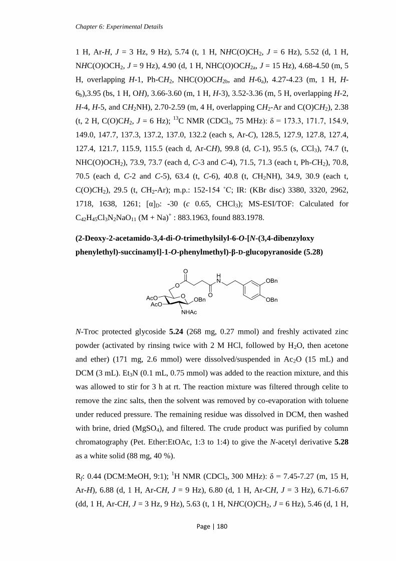
Chapter 6: Experimental Details
Page | 180
1 H, Ar-H, J = 3 Hz, 9 Hz), 5.74 (t, 1 H, NHC(O)CH2, J = 6 Hz), 5.52 (d, 1 H,
NHC(O)OCH2, J = 9 Hz), 4.90 (d, 1 H, NHC(O)OCH2a, J = 15 Hz), 4.68-4.50 (m, 5
H, overlapping H-1, Ph-CH2, NHC(O)OCH2b, and H-6a), 4.27-4.23 (m, 1 H, H-
6b),3.95 (bs, 1 H, OH), 3.66-3.60 (m, 1 H, H-3), 3.52-3.36 (m, 5 H, overlapping H-2,
H-4, H-5, and CH2NH), 2.70-2.59 (m, 4 H, overlapping CH2-Ar and C(O)CH2), 2.38
(t, 2 H, C(O)CH2, J = 6 Hz); 13
C NMR (CDCl3, 75 MHz): δ = 173.3, 171.7, 154.9,
149.0, 147.7, 137.3, 137.2, 137.0, 132.2 (each s, Ar-C), 128.5, 127.9, 127.8, 127.4,
127.4, 121.7, 115.9, 115.5 (each d, Ar-CH), 99.8 (d, C-1), 95.5 (s, CCl3), 74.7 (t,
NHC(O)OCH2), 73.9, 73.7 (each d, C-3 and C-4), 71.5, 71.3 (each t, Ph-CH2), 70.8,
70.5 (each d, C-2 and C-5), 63.4 (t, C-6), 40.8 (t, CH2NH), 34.9, 30.9 (each t,
C(O)CH2), 29.5 (t, CH2-Ar); m.p.: 152-154 ˚C; IR: (KBr disc) 3380, 3320, 2962,
1718, 1638, 1261; [α]D: -30 (c 0.65, CHCl3); MS-ESI/TOF: Calculated for
C42H45Cl3N2NaO11 (M + Na)+ : 883.1963, found 883.1978.
(2-Deoxy-2-acetamido-3,4-di-O-trimethylsilyl-6-O-[N-(3,4-dibenzyloxy
phenylethyl)-succinamyl]-1-O-phenylmethyl)-β-D-glucopyranoside (5.28)
N-Troc protected glycoside 5.24 (268 mg, 0.27 mmol) and freshly activated zinc
powder (activated by rinsing twice with 2 M HCl, followed by H2O, then acetone
and ether) (171 mg, 2.6 mmol) were dissolved/suspended in Ac2O (15 mL) and
DCM (3 mL). Et3N (0.1 mL, 0.75 mmol) was added to the reaction mixture, and this
was allowed to stir for 3 h at rt. The reaction mixture was filtered through celite to
remove the zinc salts, then the solvent was removed by co-evaporation with toluene
under reduced pressure. The remaining residue was dissolved in DCM, then washed
with brine, dried (MgSO4), and filtered. The crude product was purified by column
chromatography (Pet. Ether:EtOAc, 1:3 to 1:4) to give the N-acetyl derivative 5.28
as a white solid (88 mg, 40 %).
Rf: 0.44 (DCM:MeOH, 9:1); 1H NMR (CDCl3, 300 MHz): δ = 7.45-7.27 (m, 15 H,
Ar-H), 6.88 (d, 1 H, Ar-CH, J = 9 Hz), 6.80 (d, 1 H, Ar-CH, J = 3 Hz), 6.71-6.67
(dd, 1 H, Ar-CH, J = 3 Hz, 9 Hz), 5.63 (t, 1 H, NHC(O)CH2, J = 6 Hz), 5.46 (d, 1 H,

Chapter 6: Experimental Details
Page | 181
NHC(O)CH3, J = 9 Hz), 5.21-5.12 (m, 5 H, overlapping H-3 and Ph-CH2), 5.04 (t, 1
H, H-4, J = 9 Hz), 4.87 (d, 1 H, Ph-CH2a, J = 12 Hz), 4.62-4.55 (m, 2 H, overlapping
H-1 and PhCH2b), 4.20-4.12 (m, 2 H, H-6), 4.01-3.91 (m, 1 H, H-2), 3.68-3.62 (m, 1
H, H-5), 3.46-3.39 (m, 2 H, NHCH2), 2.71-2.67 (m, 4 H, overlapping C(O)CH2 and
CH2-Ar), 2.41-2.33 (m, 2 H, C(O)CH2), 1.99, 1.98 (each s, 6 H, OC(O)CH3), 1.88 (s,
3 H, NHC(O)CH3); 13
C NMR (CDCl3, 75 MHz): δ = 172.5, 171.1, 170.9, 170.1,
169.48 (each s, C(O)CH2, NHC(O)CH3, and OC(O)CH3), 149.0, 147.7, 137.4, 137.3,
136.9, 132.4 (each s, Ar-C), 128.5, 128.0, 127.8, 127.7, 127.4, 127.3, 121.6, 116.6,
115.8, 115.5 (each d, Ar-CH), 99.5 (d, C-1), 72.5 (d, C-3), 71.8, 71.5 (each t, Ph-
CH2), 70.7 (d, C-5), 68.7 (d, C-4), 62.4 (t, Ph-CH2), 54.5 (d, C-2), 40.8 (t, NHCH2),
35.1 (t, C(O)CH2), 30.9 (t, C(O)CH2), 29.4 (t, CH2-Ph), 23.3 (q, NHC(O)CH3), 20.7,
20.6 (each q, OC(O)CH3); m.p.: 120-124 ˚C; IR: (DCM film on NaCl plate) 3431,
2100, 1650, 1237 cm-1
; [α]D: -16.5 (c 1.7, CH2Cl2); MS-ESI/TOF: Calculated for
C45H52N2O12 (M + 2H)+2
: 406.1755, found 406.1739 .
(2-Deoxy-2-acetamido-3,4,6-tri-O-acetyl-1-O-phenylmethyl)-β-D-
glucopyranoside (5.33)[251, 261]
N-Troc protected glycoside 3.18 (736 mg, 1.29 mmol) and freshly activated zinc dust
(activated by rinsing twice with 2 M HCl, then H2O, followed by acetone and ether)
(1.26 g, 19 mmol) were dissolved/suspended in Ac2O (20 mL). Et3N (0.35 mL, 2.58
mmol) was added to the reaction mixture, and this was allowed to stir at rt for 5 h.
The solvent was evaporated, and the remaining residue was purified by column
chromatography (Hex: EtOAc, 1:1 to EtOAc flush) to give the N-acetyl derivative
5.33 as a white solid (400 mg, 71 %). Spectroscopic data was in agreement with
reported values.
Rf: 0.67 (DCM:MeOH, 9:1); 1H NMR (CDCl3, 300 MHz): δ = 7.38-7.29 (m, 5 H,
Ar-H), 5.39 (d, 1 H, NHC(O)CH3, J = 9 Hz), 5.20 (t, 1 H, H-3, J = 9 Hz), 5.09 (t, 1
H, H-4, J = 9 Hz), 4.89 (d, 1 H, Ph-CH2a, J = 12 Hz), 4.64-4.58 (m, 2 H, overlapping
Ph-CH2b and H-1), 4.30-25 (dd, 1 H, H-6a, J = 4.8 Hz, 12 Hz), 4.19-4.14 (dd, 1 H, H-
6b, J = 3 Hz, 12 Hz), 4.02-3.92 (m, 1 H, H-2), 3.70-3.64 (m, 1 H, H-5), 2.10, 2.01

Chapter 6: Experimental Details
Page | 182
(each s, 9 H, OC(O)CH3), 1.90 (s, 3 H, NHC(O)CH3); (Lit[251]
: 1H NMR (CDCl3, 200
MHz): δ = 7.28-7.26, 5.78, 5.21, 5.04, 4.85, 4.64, 4.56, 4.24, 4.12, 3.92, 3.66, 2.05,
1.97, 1.85); 13
C NMR (CDCl3, 75 MHz): δ = 170.9, 170.8, 170.3, 169.4 (each s,
OC(O)CH3 and NHC(O)CH3), 136.9 (s, Ar-C), 128.6, 128.5, 128.3, 128.0 (each d,
Ar-CH), 99.5 (d, C-1), 72.5 (d, C-3), 71.9 (d, C-5), 70.7 (t, Ph-CH2), 68.7 (d, C-4),
62.2 (t, C-6), 54.5 (d, C-2), 23.3 (q, NHC(O)CH3), 20.8, 20.7, 20.6 (each q,
OC(O)CH3); MS-ESI/TOF: Calculated for C21H28NO9 (M + H)+: 439.1792, found
439.1807.
(2-Deoxy-2-acetamido-1-O-phenylmethyl)-α/β-D-glucopyranoside (5.34)[261, 262]
Method A
Acetylated derivative 5.33 (140 mg, 0.32 mmol) was dissolved in distilled MeOH
(10 mL), and H2O (5 mL) was added. The reaction mixture was stirred at 45 °C
overnight. The organic solvent was evaporated using a rotary evaporator, and the
remaining aqueous solution was extracted with EtOAc. The organic layer was then
washed twice with sat. NaHCO3 solution, then dried (MgSO4), and filtered. The
organic layer was concentrated under reduced pressure, to give the crude deprotected
product, 5.34, as a white oily solid (80 mg, 80 %). Following this method only the β-
anomer is obtained.
Method B
(2-Deoxy-2-acetamido)-β-D-glucopyranose (500 mg, 2.26 mmol) was dissolved in
anhydrous DMF (5 mL) and placed under N2 at 0 °C. BnBr (0.57 mL, 4.79 mmol)
and NaH (120 mg, 5.0 mmol) was added to the reaction in four portions, over 2 h,
then the reaction was allowed to stir at 0 °C for a further 2 h. The reaction mixture
was filtered through celite. The celite was rinsed with DMF, and the filtrate was
reduced using a rotary evaporator. The remaining residue was dissolved in H2O and
washed three times with DCM. The aqueous layer was co-evaporated with toluene
under reduced pressure to give an anomeric mixture of the benzyl glycoside 5.34 as a

Chapter 6: Experimental Details
Page | 183
white oily solid (547 mg, 78 %). Spectroscopic data is in agreement with literature
values. (Anomeric ratio 0.5:1, α:β)
1H NMR (D2O, 300 MHz): δ = 7.58-7.38 (m, 5 H, Ph-CH), 4.96 (d, 1 H, H-1 (α-
anomer), J = 3 Hz), 4.91 (d, 1 H, Ph-CH2a, J = 12 Hz), 4.70 (d, 1 H, Ph-CH2b, J = 12
Hz), 4.59-4.54 (m, 2 H, overlapping H-1 (β-anomer) and Ph-CH2b (α-anomer)), 3.99-
3.47 (m, 12 H, overlapping H-3, H-4, H-5, H-6, and H-2 (both anomers)), 1.99, 1.95
(each s, 3 H, NHC(O)CH3 (both anomers)); (Lit[262]
: 1H NMR (D2O, 300 MHz): δ =
7.46-7.37, 4.89, 4.68, 4.54, 3.95, 3.76, 3.70-3.46, 1.94); 13
C NMR (D2O, 75 MHz): δ
= 174.5, 174.3 (each s, NHC(O)CH3 (both anomers)), 137.0, 136.8 (each s, Ph-C
(both anomers)), 129.2, 128.7, 128.6, 128.5, 128.4, 128.4 (each d, Ph-CH), 99.9 (d,
C-1 (β-anomer)), 95.8 (d, C-1 (α-anomer)), (75.9, 73.7, 72.1, 71.6, 71.4, 70.8, 70.1,
69.9, 60.8, 60.6, 60.5, 55.53, 53.7), 22.1, 21.8 (each q, NHC(O)CH3 (both anomers));
IR: (KBr disc) 3291, 2870, 2419, 1651, 1553, 1080 cm-1
; MS-ESI/TOF: Calculated
for C15H22NO6 (M + H)+: 312.1442, found 312.1449.
(2-Deoxy-2-acetamido-3,4,6-tri-O-trimethylsilyl-1-O-phenylmethyl)-α/β-D-
glucopyranoside (5.35)
Method A
Glycoside 5.34 (268 mg, 0.64 mmol) was dissolved in dry pyridine (6 mL). The
solution was cooled to 0 ˚C, then a solution of TMSCl (0.49 mL, 3.84 mmol) and
DIPEA (0.30 mL, 1.28 mmol) was added. The reaction was allowed to reach rt, then
stirred overnight at rt. The solvent was co-evaporated with toluene, and the
remaining residue was dissolved in DCM. The organic layer was washed with H2O,
then dried (MgSO4), and filtered. The crude product was purified by column
chromatography (Pet. Ether:EtOAc, 1:1) to give an anomeric mixture of TMS
protected glycoside 5.35 as a white solid (150 mg, 44 %).

Chapter 6: Experimental Details
Page | 184
Method B
Glycoside 5.34 (268 mg, 0.64 mmol) was dissolved in dry pyridine (6 mL). The
solution was cooled to 0 ˚C, then a solution of TMSCl (0.49 mL, 3.84 mmol) and
hexamethyldisilazane (0.27 mL, 1.28 mmol) was added. The reaction was allowed to
reach rt, then stirred overnight at rt. The solvent was co-evaporated with toluene, and
the remaining residue was dissolved in DCM. The organic layer was washed with
H2O, then dried (MgSO4), and filtered. The crude product was purified by column
chromatography (Pet. Ether:EtOAc, 1:1) to give an anomeric mixture of the TMS
protected glycoside 5.35 as a white solid (250 mg, 74 %). (Anomeric ratio 0.5:1, α:β)
Rf: 0.8 (Pet. Ether: EtOAc, 1:2); 1
H NMR (CDCl3, 300 MHz): δ = 7.35-7.26 (m, 10
H, Ph-H (both anomers)), 5.61 (d, 1 H, NHC(O)CH3 (β-anomer), J = 9 Hz), 5.44 (d,
1 H, NHC(O)CH3 (α-anomer), J = 9 Hz), 5.05 (d, 1 H, H-1 (α-anomer), J = 3 Hz),
4.82 (d, 1 H, Ph-CH2a (β-anomer), J = 12 Hz), 4.71-4.65 (m, 2 H, overlapping H-1
(β-anomer) and Ph-CH2a (α-anomer)), 4.54 (d, 1 H, Ph-CH2b (β-anomer), J = 12 Hz),
4.43 (d, 1 H, Ph-CH2b (α-anomer), J = 12 Hz), 4.14-3.99 (m, 2 H, overlapping H-2
(both anomers)), 3.98-3.54 (m, 8 H, overlapping H-6, H-3 and H-4 (both anomers)),
3.34-3.23 (m, 2 H, H-5 (both anomers)), 1.93, 1.92 (each s, 6 H, NHC(O)CH3 (both
anomers)) 0.16, 0.14, 0.13, 0.12, 0.01, 0.09 (each s, 54 H, SiC(CH3)3 (both
anomers)); 13
C NMR (CDCl3, 75 MHz): δ = 170.0, 169.9 (each s, NHC(O)CH3 (both
anomers)), 138.1, 137.6 (each s, Ph-C (both anomers)), 128.7, 128.6, 128.5, 128.4,
128.2, 128.0, 127.7 (each d, Ph-CH (both anomers)), 99.4 (d, C-1 (β-anomer)), 92.8
(d, C-1 (α-anomer)), 76.9 (d, C-5), 75.1, 74.5, 73.1, 71.9 (each d, C-3 and C-4 (both
anomers)), 70.2, 69.5 (each t, Ph-CH2 (both anomers)), 62.3, 61.9 (each t, C-6 (both
anomers)), 54.8 (d, C-2 (α-anomer)), 53.9 (d, C-2 (β-anomer)), 1.3, 1.2, 1.1, 1.0, 0.9,
0.8 (each q, SiC(CH3)3 (both anomers)); IR: (DCM film on NaCl plate) 3288, 2956,
1655, 1562, 1250 cm-1
; [α]D: + 10 (c 1.0, CH2Cl2); MS-ESI/TOF: Calculated for
C21H36NO6Si2 (M (-TMS) – H)- (1 x TMS removed by formic acid in mobile phase):
454.2087, found 454.2089.

Chapter 6: Experimental Details
Page | 185
(2-Deoxy-2-acetamido-3,4-di-O-trimethylsilyl-1-O-phenylmethyl)-α/β-D-
glucopyranoside (5.37)
TMS protected glycoside 5.35 (51 mg, 0.10 mmol) was dissolved in an
acetone:MeOH mixture (0.8:0.6 mL). The reaction mixture was cooled to 0 ˚C, then
AcOH (0.01 mL, 0.2 mmol) was added, and this was stirred for 2 h at 0 ˚C. Solid
NaHCO3 (10 mg) was added and stirred for 10 mins. The reaction mixture was
diluted with DCM (20 mL), and the solid NaHCO3 was removed by filtration. The
filtrate was washed with a sat. solution of NaHCO3, then dried (MgSO4) and filtered.
The organic layer was concentrated under reduced pressure, and the remaining
residue was purified by column chromatography (Pet. Ether:EtOAc, 1:1) to give the
selectively deprotected derivative 5.37 as a clear oil (38 mg, 86 %). (Anomeric ratio
0.6:1, α:β)
Rf: 0.6 (Pet. Ether: EtOAc, 1:2); 1H NMR (CDCl3, 300 MHz): δ = 7.35-7.30 (m, 10
H, Ph-H (both anomers)), 5.50 (d, 1 H, NHC(O)CH3 (β-anomer), J = 9 Hz), 5.31 (d,
1 H, NHC(O)CH3 (α-anomer), J = 6 Hz), 5.09 (d, 1 H, H-1 (α-anomer), J = 3 Hz),
4.89-4.82 (m, 2 H, overlapping H-1 (β-anomer) and Ph-CH2a), 4.59 (d, 1 H, Ph-
CH2b, J = 12 Hz), 4.06-3.99 (td, 1 H, H-2 (α-anomer), J = 9 Hz, 15 Hz), 3.96-3.89
(dd, 1 H, H-3 (β-anomer), J = 9 Hz, 12 Hz), 3.85-3.80 (dd, 1 H, H-6a (β-anomer), J =
3 Hz, 9 Hz),), 3.73-3.59 (m, 5 H, overlapping H-6b (β-anomer), H-3, H-4 and H-6 (α-
anomer)), 3.53 (t, 1 H, H-4 (β-anomer), J = 9 Hz), 3.44-3.32 (m, 2 H, overlapping H-
2 and H-5 (β-anomer)), 1.99, 1.94 (each s, 6 H, NHC(O)CH3 (both anomers)), 0.18,
0.16, 0.13, 0.10 (each s, 36 H, SiC(CH3)3 (both anomers)); 13
C NMR (CDCl3, 75
MHz): δ = 169.5, 168.9 (each s, NHC(O)CH3 (both anomers)), 136.8 (s, Ph-C),
127.7, 127.2, 127.1 (each d, Ph-CH), 98.7 (d, C-1 (β-anomer)), 91.8 (d, C-1 (α-
anomer)), 75.4 (d, C-5), 74.0 (d, C-3), 71.6 (d, C-4), 70.4 (t, Ph-CH2), 61.4, 60.9
(each t, C-6 (both anomers)), 57.3 (d, C-2 (β-anomer)), 53.8 (d, C-2 (α-anomer)),
23.2, 22.9 (each q, NHC(O)CH3 (both anomers)), 0.3, 0.2, 0.1, -0.9 (each q, SiC(CH-
3)3 (both anomers)); IR: (DCM film on NaCl plate) 3431, 2925, 1625, 1243 cm-1
;
[α]D: - 9.4 (c 0.85, CH3OH); MS-ESI/TOF: Calculated for C15H20NO6 (M (-2 x

Chapter 6: Experimental Details
Page | 186
TMS) - H)- (TMS removed by formic acid mobile phase): 310.1296, found
310.1310.
(2-Deoxy-2-acetamido-6-O-[N-(3,4-dibenzyloxyphenylethyl)-succinamyl]-1-O-
phenylmethyl)-β-D-glucopyranoside (5.39)
Glycoside 5.37 (100 mg, 0.21 mmol) was dissolved in dry DCM (4 mL), under N2,
and DCC (52 mg, 0.25 mmol) was added. This was then added to a solution of 5.13
(104 mg, 0.24 mmol) in dry DCM (2 mL) under N2. DMAP (1.8 mg, 0.001 mmol)
was then added to the reaction mixture, and this was allowed to stir at rt for 4 h. The
reaction mixture was filtered through celite. The filtrate was diluted with DCM and
washed with an aqueous solution of 5 % AcOH (v/v), followed by H2O. The organic
layer was dried (MgSO4), and filtered. The crude product was purified by column
chromatography (Pet. Ether:EtOAc, 1:1) to give glycosyl-dopamine derivative 5.38
as a clear syrup (100 mg, 52 %).
This glycosyl-dopamine derivative 5.38 (100 mg, 0.11 mmol) was dissolved in a 2 %
solution of TFA (v/v) in DCM (7.5 mL) at 0 °C. The reaction mixture was allowed
to stir at 0 °C for 1 h, then solid NaHCO3 (50 mg) was added, and stirred for 5 mins.
The reaction mixture was diluted with DCM, then filtered by gravity to remove the
NaHCO3. The filtrate was concentrated under reduced pressure, and purified by
column chromatography (DCM:MeOH, 30:1 to 20:1 to 9:1) to give glycoside 5.39 as
a clear oil (44 mg, 50 %).
Rf: 0.5 (DCM:MeOH, 9:1); 1H NMR (CDCl3 with a drop of CD3OD, 300 MHz): δ =
7.46-7.25 (m, 15 H, Ar-H), 6.87 (d, 1 H, Ar-H, J = 6 Hz), 6.81 (d, 1 H, Ar-H, J =
2.1 Hz), 6.71-6.68 (dd, 1 H, Ar-H, J = 3 Hz, 6 Hz), 5.14, 5.11 (each s, 4 H, Ph-CH2),
4.86 (d, 1 H, Ph-CH2a, J = 15 Hz), 4.56 (d, 1 H, Ph-CH2b, J = 15 Hz), 4.48 (d, 1 H,
H-1, J = 9 Hz), 4.44 (bs, 2 H, H-6), 3.66 (t, 1 H, H-2, J = 9 Hz), 3.52-3.35 (m, 5 H,
overlapping H-4, H-5, H-3 and CH2-Ar), 2.70-2.65 (m, 4 H, overlapping NHCH2 and
C(O)CH2), 2.41 (t, 2 H, C(O)CH2, J = 6 Hz), 1.95 (s, 3 H, NHC(O)CH3); 13
C NMR

Chapter 6: Experimental Details
Page | 187
(CDCl3, 75 MHz): δ = 173.2 (s, NHC(O)CH3) 172.4, 172.1 (each s, C(O)CH2),
148.9, 147.6, 137.3, 137.2, 132.5 (each s, Ar-C), 128.5, 128.4, 128.0, 127.9, 127.9,
127.5, 127.4, 121.7, 115.8, 115.5 (each d, Ar-CH), 99.8 (d, C-1), 74.4 (d, C-3), 73.8
(d, C-5), 71.5, 71.4 (each t, Ph-CH2), 70.7 (t, Ph-CH2), 70.4 (d, C-4), 63.5 (t, C-6),
56.3 (d, C-2), 40.9 (t, CH2NH), 35.0 (t, C(O)CH2), 30.6 (t, C(O)CH2), 29.4 (t, CH2-
Ar), 23.0 (q, NHC(O)CH3); IR: (DCM film on NaCl plate) 3299, 2984, 1736, 1641,
1554, 1260 cm-1
; [α]D: - 21.5 (c 0.65, CH3OH).
6.3 General Procedures for Solid Phase Peptide Synthesis
(SPPS)[172]
6.3.1 Swelling of the Resin
2-Chlorotrityl chloride (CTC) resin (300 mg) was placed into SPPS apparatus. Dry
DCM (3 mL) was added, and the vessel was gently shaken for 15 mins. The solvent
was removed by vacuum filtration. This was repeated twice.
6.3.2 Introduction of the First Amino Acid (AA)
The recommended loading of CTC resin is 1.0-1.6 mmol AA/g of resin. The first
NHFmoc-AA (0.42 mmol) was dissolved in dry DCM (3 mL), and DIPEA (0.28 mL,
4 eq to carboxylic acid, 1.68 mmol) was added. This solution was gently syringed
onto the swollen resin, and stirred for 1.75 h. The liquid was then removed by
vacuum filtration.
6.3.3 Capping of the First AA
The resin was washed with 3 x DCM/ MeOH/ DIPEA (17:2:1 mL), followed by 3 x
DCM (2 mL), 2 x DMF (2 mL), 2 x DCM (2 mL), then dried by vacuum filtration.
6.3.4 Estimation of Loading of the First AA[180]
The resin (50 mg) was weighed into a 100 mL beaker, DMF (20 mL) was added.
This suspension was stirred for 30 min, then DBU (0.4 mL) was added. (This gave a
2 % DBU/DMF soln.) The mixture was stirred for 30 mins then diluted to 100 mL
with acetonitrile. 2 mL of this solution was transferred to a 25 mL beaker and diluted
with acetonitrile to 25 mL. A reference solution was prepared in the same way,
without addition of the resin. The sample and reference solutions were measured by
UV in quartz cuvettes at 294 and 304 nm, and the absorbance resulting from the

Chapter 6: Experimental Details
Page | 188
dibenzofulvene released upon Fmoc cleavage was used to calculate the level of
loading of the first AA (Fig. 6.1).
Figure 6.1 Equation for the calculation of the level of attachment of the first AA
The literature value for CTC resin loading is 0.86 mmol/g.
6.3.5 Removal of Fmoc Protecting Group
Swelling of the resin was carried out, as described above. A 20 % piperidine solution
in DMF (v/v) (1.5 mL) was added to the resin, and gently shaken for 1 min. The
liquid was removed by vacuum filtration, and a further 1.5 mL of this solution was
added. The resin was gently shaken for 10 mins, then the liquid removed. The resin
was washed with DMF x 7 (2 mL).
6.3.6 Monitoring Fmoc Removal/ Coupling of AA – The Kaiser Test:
Figure 6.2 Reagents required for the Kaiser test
A few beads were transferred to a glass vial and washed three times with EtOH
(EtOH added, swirled, then decanted). Each of the above solutions (0.10 mL of A
and 0.10 mL of B) was added to the beads, and the vial was heated with a heat gun
for 2 mins (Fig. 6.2). The colour change of the beads was noted (Fig. 6.3). It is
important to take note of the colour of the resin, and not the liquid. This test can be
carried out with pyridine and reagent A, but the colour change may be different.

Chapter 6: Experimental Details
Page | 189
Figure 6.3 Colour changes in the Kaiser test
The Kaiser test was also used to monitor the success of coupling reactions, as
denoted by a colour change to clear or yellow. The Kaiser test became less useful for
glycopeptides with colour changes of red being observed at certain stages.
6.3.7 Coupling of the Second, and Each Consecutive AA
NHFmoc-AA (0.42 mmol), TBTU (148 mg, 1.1 eq, 0.46 mmol), and HOBt (62 mg,
1.1 eq) were dissolved in DMF (3 mL). DIPEA (0.15 mL, 2 eq) was added to the
mixture. This was allowed to react for 10 mins, then added to the pre-swelled resin.
The resin was gently shaken for 1.5 h, then filtered. The liquid was removed from
the resin by vacuum filtration, and the resin was washed with DMF (2 mL x 7
times), followed by DCM (3 mL x 3 times). (Wash with DCM ensures removal of
DMF which may cause unwanted Fmoc removal over time).
6.3.8 Capping of the Second, and Consecutive AA After Coupling
Capping of any unreacted free amines was carried out after each coupling. The resin
was swelled, then DMF (1.4 mL), DIPEA (0.2 mL), and Ac2O (0.2 mL) were added.
The resin was gently shaken for 15 mins, then filtered. The resin was washed with
DMF (2 mL x 7 times).
6.3.9 Cleavage from the Resin for Mass Spectrometry and for Final Peptide
Monitoring of the intermediates of each stage in the synthesis was carried out by
removing a few beads from the reaction vessel. These were washed with EtOH x 3
times. A 1 % TFA solution (v/v) in DCM (3 mL) was added to the resin and left to
react at rt for 10 mins. The liquid was removed (filtered through cotton wool to
remove the beads) and concentrated under reduced pressure. The resulting oil was
dissolved in DMF or DCM and sent for analysis.
The cleavage of the final glycopeptide was carried out with 50 % TFA (v/v) in
DCM, for 1 h. Purification of the final glycopeptides was carried out by HPLC.

Chapter 6: Experimental Details
Page | 190
Peptide 3.6:
The bulk of the peptide was cleaved from the resin as described above, to give a
mixture of crude products. The solvent was evaporated under reduced pressure, and
the remaining residue was triturated with a 10 % solution of TFA in DCM (4 x 5
mL). The combined DCM/TFA extracts were concentrated under reduced pressure to
give a clear oil (12 mg, 16 % crude yield). This oil was redissolved in a 10 %
solution of TFA in DCM, and purified by RP-HPLC. Preparative HPLC was carried
out using an Agilent Zorbex Eclipse XDB-C18 column, using a mobile phase of A =
H2O with 0.1 % formic acid, and B = MeCN with 0.1 % formic acid, gradient elution
was used (A:B, 100:0 to 50:50 to 0:100 to 50:50 to 100:0 over 20 mins). The
presence of the peptide, 3.6, was confirmed by MS analysis in fractions collected at
6.5-8 and 8-9.5 mins run time.
Glycopeptide 3.8:
The bulk of the peptide was cleaved from the resin as described above, to give a
crude mixture of products. The solvent was evaporated under reduced pressure, and
the remaining residue was triturated with a 10 % solution of TFA in DCM (4 x 5
mL). The combined DCM/TFA extracts were concentrated under reduced pressure to
give a yellow oil (12 mg, 30 % crude yield). This oil was redissolved in a 10 %
solution of TFA in DCM, and purified by RP-HPLC. Preparative HPLC was carried
out using an Agilent Zorbex Eclipse XDB-C18 column, using a mobile phase of A =
H2O with 0.1 % formic acid, and B = MeCN with 0.1 % formic acid, gradient elution
was used (A:B, 100:0 to 50:50 to 0:100 to 50:50 to 100:0 over 20 mins). The
presence of the peptide, 3.8, was confirmed by MS analysis in fractions collected at
6-7 and 7-8 mins run time.
Glycopeptide 3.10:
The bulk of the peptide was cleaved from the resin as described above, to give a
crude mixture of products. The solvent was evaporated under reduced pressure, and
the remaining residue was triturated with a 10 % solution of TFA in DCM (4 x 5
mL). The combined DCM/TFA extracts were concentrated under reduced pressure to
give a yellow oil (13 mg, 26 % crude yield). This oil was redissolved in a 10 %
solution of TFA in DCM, and purified by RP-HPLC. Preparative HPLC was carried

Chapter 6: Experimental Details
Page | 191
out using an Agilent Zorbex Eclipse XDB-C18 column, using a mobile phase of A =
H2O with 0.1 % formic acid, and B = MeCN with 0.1 % formic acid, gradient elution
was used (A:B, 100:0 to 50:50 to 0:100 to 50:50 to 100:0 over 20 mins). The
presence of the peptide, 3.10, was confirmed by MS analysis in fractions collected at
7-8 and 8-9 mins run time.
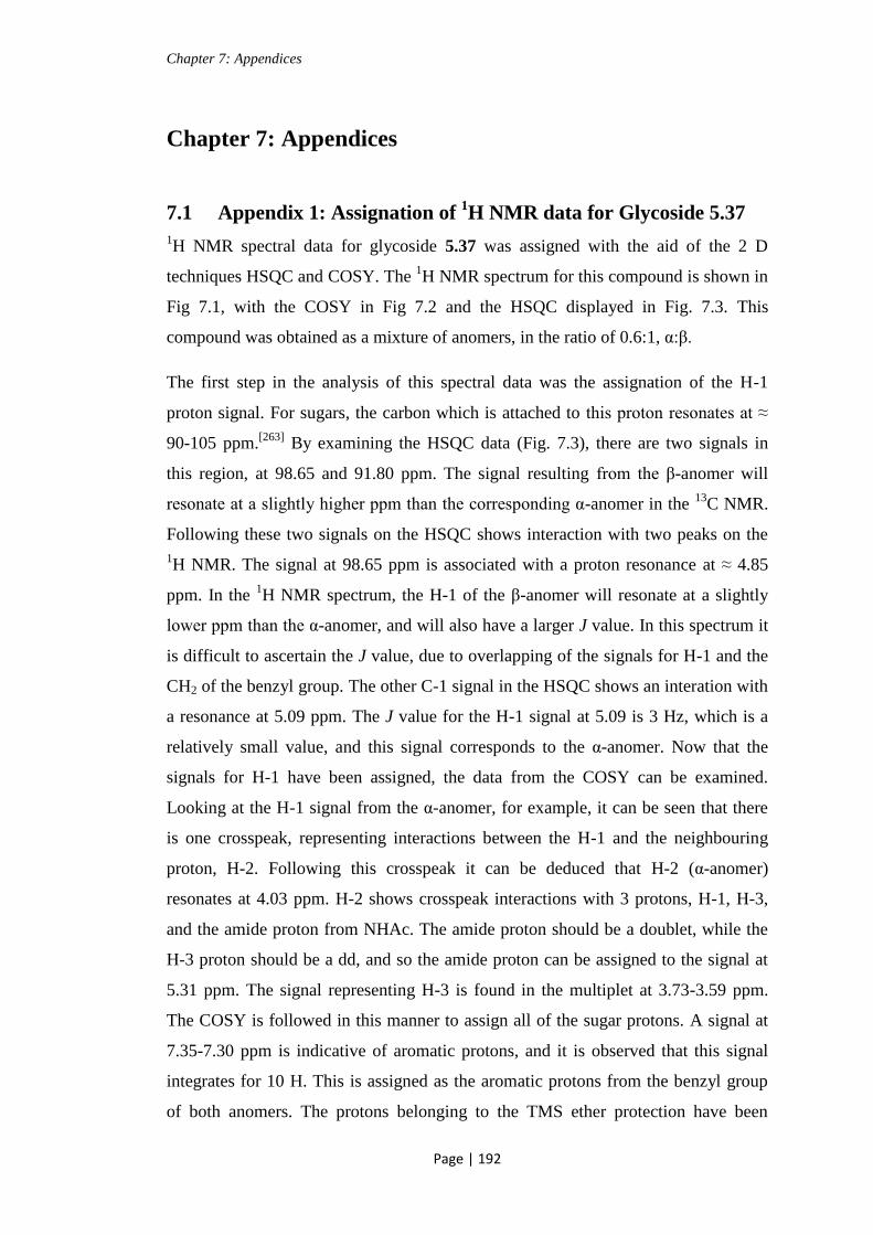
Chapter 7: Appendices
Page | 192
Chapter 7: Appendices
7.1 Appendix 1: Assignation of 1H NMR data for Glycoside 5.37
1H NMR spectral data for glycoside 5.37 was assigned with the aid of the 2 D
techniques HSQC and COSY. The 1H NMR spectrum for this compound is shown in
Fig 7.1, with the COSY in Fig 7.2 and the HSQC displayed in Fig. 7.3. This
compound was obtained as a mixture of anomers, in the ratio of 0.6:1, α:β.
The first step in the analysis of this spectral data was the assignation of the H-1
proton signal. For sugars, the carbon which is attached to this proton resonates at ≈
90-105 ppm.[263]
By examining the HSQC data (Fig. 7.3), there are two signals in
this region, at 98.65 and 91.80 ppm. The signal resulting from the β-anomer will
resonate at a slightly higher ppm than the corresponding α-anomer in the 13
C NMR.
Following these two signals on the HSQC shows interaction with two peaks on the
1H NMR. The signal at 98.65 ppm is associated with a proton resonance at ≈ 4.85
ppm. In the 1H NMR spectrum, the H-1 of the β-anomer will resonate at a slightly
lower ppm than the α-anomer, and will also have a larger J value. In this spectrum it
is difficult to ascertain the J value, due to overlapping of the signals for H-1 and the
CH2 of the benzyl group. The other C-1 signal in the HSQC shows an interation with
a resonance at 5.09 ppm. The J value for the H-1 signal at 5.09 is 3 Hz, which is a
relatively small value, and this signal corresponds to the α-anomer. Now that the
signals for H-1 have been assigned, the data from the COSY can be examined.
Looking at the H-1 signal from the α-anomer, for example, it can be seen that there
is one crosspeak, representing interactions between the H-1 and the neighbouring
proton, H-2. Following this crosspeak it can be deduced that H-2 (α-anomer)
resonates at 4.03 ppm. H-2 shows crosspeak interactions with 3 protons, H-1, H-3,
and the amide proton from NHAc. The amide proton should be a doublet, while the
H-3 proton should be a dd, and so the amide proton can be assigned to the signal at
5.31 ppm. The signal representing H-3 is found in the multiplet at 3.73-3.59 ppm.
The COSY is followed in this manner to assign all of the sugar protons. A signal at
7.35-7.30 ppm is indicative of aromatic protons, and it is observed that this signal
integrates for 10 H. This is assigned as the aromatic protons from the benzyl group
of both anomers. The protons belonging to the TMS ether protection have been

Chapter 7: Appendices
Page | 193
assigned to the singlets at 0.18-0.10 ppm. Due to the presence of the electropositive
Si atom these protons are very shielded, and so resonate at almost 0 ppm. The acetyl
CH3 are assigned to the singlets at 1.99 and 1.94 ppm (both anomers). CH3 signals
typically appear at a slightly lower ppm, but due to the presence of the
electronegative oxygen of the acetyl group these protons resonate further downfield.
Figure 7.11 H NMR spectrum for glycoside 5.37

Chapter 7: Appendices
Page | 194
Figure 7.2 COSY of glycoside 5.37
Figure 7.3 HSQC of glycoside 5.37

Chapter 7: Appendices
Page | 195
7.2 Appendix 2: Experimental Data for Attempted Reactions
Table 2.1 Attempted synthesis of 2.55 using acetyl chloride coupling methology
The free acid 2.54 (100 mg, 0.14 mmol) was dissolved in anhydrous DCM (5 mL)
and placed under a N2 atmosphere at 0 °C, in the presence of 3.4 Å molecular sieves.
Oxalyl chloride (0.01 mL, 0.15 mmol) and DMF (0.005 mL) were added to the
reaction mixture. This was allowed to stir at 0 °C for 1 h. A solution of
tetradecylamine (29 mg, 0.14 mmol) and pyridine (0.012 mL, 0.15 mmol) in DCM
(3 mL) was added dropwise to the reaction mixture, which was allowed to return to
rt. The reaction mixture was stirred at rt for 4.5 h, then the solvent was evaporated
under reduced pressure. The remaining residue was dissolved in DCM, and washed
with 1.0 M HCl. The organic layer was evaportated using a rotary evaporator. This
reaction failed to yield the desired product.
Scheme 2.17 Attempted direct glycosylation of acceptor 2.63
Imidate donor 2.34 (18 mg, 0.03 mmol) and lipid acceptor 2.63 (11 mg, 0.02 mmol)
were dissolved in anhydrous DCM (5 mL) under a N2 atmosphere, in the presence of
3.4 Å molecular sieves. To this BF3OEt2 (0.002 mL, 0.01 mmol) was added, and the
reaction mixture was allowed to stir at rt for 3 h. The solvent was evaporated under
reduced pressure to leave a white oily solid. The desired product was not
synthesised, and hemiacetal 2.33 and lipid starting material 2.63 were recovered.
Scheme 2.21 N-Cbz removal from Troc protected derivative 2.67
Troc protected derivative 2.67 (230 mg, 0.3 mmol) was dissolved in EtOAc (13 mL).
Pd/C 10 % (w/w) (100 mg) was added and the reaction mixture was placed under a
H2 atmosphere, then stirred at rt overnight. The reaction did not proceed with
recovery of starting material. The reaction mixture was filtered to remove the Pd/C,
then the experiment was repeated for a further 24 h. Full recovery of starting
material 2.67 was achieved, with non of the desired product synthesised.
Table 4.1 Attempted tritylation of 4.1
The Troc protected glucosamine 4.1 (400 mg, 1.1 mmol) was dissolved in dry
pyridine (15 mL). Et3N (0.02 mL, 0.12 mmol) and TrCl (379 mg, 1.4 mmol) was
added to the reaction mixture. This was stirred at 70 °C for 4 h. The reaction mixture

Chapter 7: Appendices
Page | 196
was cooled to 0 °C, then Ac2O (0.64 mL, 6.8 mmol) was added slowly. The reaction
mixture was allowed to return to rt, then stirred at rt overnight. The solvent was
evaporated under reduced pressure, and the remaining residue was dissolved in
DCM, and washed with brine. The organic layer was dried (MgSO4), and filtered,
then concentrated under reduced pressure. The crude product was purified by column
chromatography (Hex:EtOAc, 2:1) to give protected glucosamine derivative 4.2 as a
clear oil (83 mg, 5 %), and mainly the acetylated derivative 2.26.
Table 4.2 i) Attempted O-Fmoc protection of the C-6 position
Glycoside 4.7 (55 mg, 0.12 mmol) was dissolved in anhydrous pyridine (10 mL)
under a N2 atmosphere, and cooled to 0 °C. Fmoc-Cl (47 mg, 0.19 mmol) was added
to the reaction mixture, and this was allowed to stir at 0 °C for 2 h. The reaction
mixture was allowed to return to rt, then stirred for a further 4 h. Another equivalent
of Fmoc-Cl was added (32 mg), and the reaction mixture was allowed to stir at rt
overnight. Ac2O (8 mL) was added, and the reaction mixture was allowed to stir for
4 h. The solvent was evaporated under reduced pressure, and the remaining residue
was dissolved in DCM then washed with 1.0 M HCl, followed by a sat. solution of
NaHCO3. The organic layer was concentrated under reduced pressure. The desired
product was not obtained from this experiment.
Table 4.2 ii) Attempted O-MOM protection of the C-6 position
Glycoside 4.7 (55 mg, 0.12 mmol) was dissolved in acetone (10 mL) and K2CO3 (62
mg, 0.45 mmol) was added. MOM-Br (0.14 mL mg, 0.17 mmol) was added
dropwise to the reaction mixture, and this was heated at reflux (56 °C) overnight.
The reaction mixture was allowed to return to rt, then MeOH (6 mL) was added to
quench the remaining MOM-Br. The solvent was evaporated under reduced pressure,
and the remaining reside dissolved in pyridine (6 Ml). Ac2O (6 mL) was added, and
the reaction mixture was allowed to stir at rt overnight. The solvent was evaporated
under reduced pressure, and the remaining residue was dissolved in DCM then
washed with 1.0 M HCl, followed by a sat. solution of NaHCO3. The organic layer
was concentrated under reduced pressure. The desired product was not obtained from
this experiment.

Chapter 7: Appendices
Page | 197
Table 4.3 Attempted enzymatic deacetylation (general procedure)
Glycoside 3.4 (83 mg, 0.11 mmol) was dissolved in ethanol or ACN (10 mL).
Phosphate buffer was added to adjust the pH of the reaction mixture to the desired
value. Enzyme (CRL or CAL B) (42 mg) was added to the reaction mixture, and this
was gently shaken in a temperature controlled water bath (temperature range from 20
→ 50 °C) for 2 days. None of these enzymatic experiments yielded the desired
product, with full recovery of starting material 3.4 in all cases.
Table 4.4 i) Attempted chemical deacetylation using DBTO
Glycoside 3.4 (22 mg, 0.03 mmol) was dissolved in MeOH (5 mL) and DBTO (7.3
mg, 0.03 mmol) was added. The reaction mixture was heated to 50 °C and allowed
to stir for 3 h. The reaction mixture was allowed to return to rt, then stirred
overnight. The reaction had not gone to completion, and was again heated to 50 °C,
and stirred for 3 h. Partial deacetylation was observed, with mainly the starting
material recovered.
Table 4.4 ii) Attempted chemical deacetylation using KCN
Glycoside 3.4 (22 mg, 0.03 mmol) was dissolved in MeOH (5 mL) and cooled to 0
°C. KCN (5.7 mg, 0.09 mmol) was added and the reaction mixture was stirred at 0
°C for 0.5 h. The reaction mixture was allowed to return to rt, then stirred for 1.5 h.
The solvent was evaporated under reduced pressure. The desired product was not
obtained under these reaction conditions.
Scheme 4.7 Attempted introduction of azide functionality into 4.18
Tosyl protected glycoside 4.18 (20 mg, 0.03 mmol) was dissolved in DMF (5 mL)
and NaN3 (18 mg, 0.3 mmol) was added. The reaction mixture was heated to 70 °C
and allowed to stir for 2.5 h. The solvent was evaporated under reduced pressure,
and the remaining residue was purified by column chromatography (DCM:MeOH,
15:1 to 10:1 to 8:2). The desired product was not synthesised, and removal of the
Fmoc group was noted by NMR.

Chapter 7: Appendices
Page | 198
Scheme 4.9 Attempted introduction of linker attached azide into 4.23 using
TBTU/HOBt methodology
TMS protected glycoside 4.23
Azido propionic acid 4.21 (110 mg, which still contained some MeCN) was
dissolved in anhydrous DCM (3 mL) under N2, and cooled to 0 °C. HOBt (17 mg,
0.13 mmol), TBTU (41 mg, 0.13 mmol) and DIPEA (0.02 mL, 0.13 mmol) were
added to the reaction mixture, and this was allowed to stir for 5 mins. A solution of
the crude TMS protected glycoside, 4.23 (45 mg, 0.06 mmol) in anhydrous DCM
was added slowly to the reaction mixture. This was stirred at 0 °C for 1.5 h, and then
allowed to return to rt. The reaction mixture was stirred at rt overnight, and then
washed with H2O. The organic layer was concentrated under reduced pressure to
almost dryness. The remaining oil was purified by column chromatography
(DCM:MeOH, 15:1 to 9:1). The desired product was not synthesised under these
reaction conditions.

Chapter 8: Bibliography
Page | 199
Chapter 8: Bibliography
1. Varki, A., Biological roles of oligosaccharides: All of the theories are
correct. Glycobiology 1993, 3(2): p. 97-130.
2. M. E. Taylor and Drickamer, K., Introduction to glycobiology. 3rd ed. 2011,
New York: Oxford University Press Inc.
3. Lindhorst, T.K., Essentials of carbohydrate chemistry and biochemistry. 3rd
ed. 2007, Weinheim: John Wiley and Sons.
4. van Kasteren, S.I., Kramer, H.B., Gamblin, D.P., and Davis, B.G., Site-
selective glycosylation of proteins: creating synthetic glycoproteins. Nat.
Protoc., 2007, 2(12): p. 3185-3194.
5. Bongat, A.F.G. and Demchenko, A.V., Recent trends in the synthesis of O-
glycosides of 2-amino-2-deoxysugars. Carbohydr. Res., 2007, 342(3-4): p.
374-406.
6. Galan, M.C., Benito-Alifonso, D., and Watt, G.M., Carbohydrate chemistry
in drug discovery. Org. Biomol. Chem., 2011, 9(10): p. 3598-3610.
7. Zhu, X. and Schmidt, R.R., New principles for glycoside-bond formation.
Angew Chem Int Ed Engl, 2009, 48(11): p. 1900-34.
8. Dube, D.H. and Bertozzi, C.R., Glycans in cancer and inflammation -
potential for therapeutics and diagnostics. Nat. Rev. Drug Discovery 2005,
4(6): p. 477-488.
9. Huang, C.-Y., Thayer, D.A., Chang, A.Y., Best, M.D., Hoffmann, J., Head,
S., and Wong, C.-H., Carbohydrate microarray for profiling the antibodies
interacting with Globo H tumor antigen. Proc. Natl. Acad. Sci. U. S. A.,
2006, 103(1): p. 15-20.
10. Suzuki-Anekoji, M., Suzuki, M., Kobayashi, T., Sato, Y., Nakayama, J.,
Suzuki, A., Bao, X., Angata, K., and Fukuda, M., HNK-1 glycan functions as
a tumor suppressor for astrocytic tumor. J. Biol. Chem., 2011, 286(37): p.
32824-32833, S32824/1-S32824/9.
11. El Ashry, E.S.H. and Aly, M.R.E., Synthesis and biological relevance of N-
acetylglucosamine-containing oligosaccharides. Pure Appl. Chem., 2007,
79(12): p. 2229-2242.
12. Yan, Y., Wanshun, L., Baoqin, H., Changhong, W., Chenwei, F., Bing, L.,
and Liehuan, C., The antioxidative and immunostimulating properties of D-
glucosamine. Int Immunopharmacol 2007, 7(1): p. 29-35.
13. Nakamura, H., Application of glucosamine on human disease-Osteoarthritis.
Carbohydr. Polym., 2011, 84(2): p. 835-839.
14. Torres, C.-R. and Hart, G.W., Topography and polypeptide distribution of
terminal N-acetylglucosamine residues on the surfaces of intact lymphocytes.
Evidence for O-linked GlcNAc. J. Biol. Chem., 1984, 259(5): p. 3308-17.
15. Hart, G.W., Housley, M.P., and Slawson, C., Cycling of O-linked -N-
acetylglucosamine on nucleocytoplasmic proteins. Nature (London, U. K.),
2007, 446(7139): p. 1017-1022.
16. Xu, W., Yang, S., Bhadury, P., He, J., He, M., Gao, L., Hu, D., and Song, B.,
Synthesis and bioactivity of novel sulfone derivatives containing 2,4-
dichlorophenyl substituted 1,3,4-oxadiazole/thiadiazole moiety as chitinase
inhibitors. Pestic. Biochem. Physiol., 2011, 101(1): p. 6-15.

Chapter 8: Bibliography
Page | 200
17. Lane, J.A., Mehra, R.K., Carrington, S.D., and Hickey, R.M., Development
of biosensor-based assays to identify anti-infective oligosaccharides. Anal.
Biochem., 2011, 410(2): p. 200-205.
18. Butkinaree, C., Park, K., and Hart, G.W., O-linked -N-acetylglucosamine
(O-GlcNAc): Extensive crosstalk with phosphorylation to regulate signaling
and transcription in response to nutrients and stress. Biochim. Biophys.
Acta, Gen. Subj., 2010, 1800(2): p. 96-106.
19. Fang, B. and Miller, M.W., Use of Galactosyltransferase to Assess the
Biological Function of O-linked N-Acetyl-D-Glucosamine: A Potential Role
for O-GlcNAc during Cell Division. Exp. Cell Res., 2001, 263(2): p. 243-
253.
20. O'Donnell, N., Zachara, N.E., Hart, G.W., and Marth, J.D., Ogt-dependent X-
chromosome-linked protein glycosylation is a requisite modification in
somatic cell function and embryo viability. Mol. Cell. Biol., 2004, 24(4): p.
1680-1690.
21. Zachara, N.E. and Hart, G.W., Cell signaling, the essential role of O-
GlcNAc! Biochim. Biophys. Acta, Mol. Cell Biol. Lipids 2006, 1761(5-6): p.
599-617.
22. Griffith, L.S. and Schmitz, B., O-linked N-acetylglucosamine levels in
cerebellar neurons respond reciprocally to perturbations of phosphorylation.
Eur. J. Biochem., 1999, 262(3): p. 824-831.
23. Hu, P., Shimoji, S., and Hart, G.W., Site-specific interplay between O-
GlcNAcylation and phosphorylation in cellular regulation. FEBS Lett., 2010,
584(12): p. 2526-2538.
24. Rechsteiner, M. and Rogers, S.W., PEST sequences and regulation by
proteolysis. Trends Biochem. Sci., 1996, 21(7): p. 267-271.
25. Garner, K., Li, M., Ugwuanya, N., and Cockcroft, S., The
phosphatidylinositol transfer protein RdgB- binds 14-3-3 via its
unstructured C-terminus, whereas its lipid-binding domain interacts with the
integral membrane protein ATRAP (angiotensin II type I receptor-associated
protein). Biochem. J., 2011, 439(1): p. 97-111.
26. Cheng, X. and Hart, G.W., Alternative O-glycosylation/O-phosphorylation of
serine-16 in murine estrogen receptor-. Post-translational regulation of
turnover and transactivation activity. J. Biol. Chem., 2001, 276(13): p.
10570-10575.
27. Sayat, R., Leber, B., Grubac, V., Wiltshire, L., and Persad, S., O-GlcNAc-
glycosylation of -catenin regulates its nuclear localization and
transcriptional activity. Exp. Cell Res., 2008, 314(15): p. 2774-2787.
28. Rajamani, U., Joseph, D., Roux, S., and Essop, M.F., The hexosamine
biosynthetic pathway can mediate myocardial apoptosis in a rat model of
diet-induced insulin resistance. Acta Physiol., 2011, 202(2): p. 151-157.
29. Fernandez-Gonzalez, M., Boutureira, O., Bernardes, G.J.L., Chalker, J.M.,
Young, M.A., Errey, J.C., and Davis, B.G., Site-selective chemoenzymatic
construction of synthetic glycoproteins using endoglycosidases. Chem. Sci.,
2010, 1(6): p. 709-715.
30. Hudak, J.E., Yu, H.H., and Bertozzi, C.R., Protein glycoengineering enabled
by the versatile synthesis of aminooxy glycans and the genetically encoded
aldehyde tag. J. Am. Chem. Soc., 2011, 133(40): p. 16127-16135.

Chapter 8: Bibliography
Page | 201
31. Morales-Serna, J.A., Boutureira, O., Diaz, Y., Matheu, M.I., and Castillon,
S., Recent advances in the glycosylation of sphingosines and ceramides.
Carbohydr. Res., 2007, 342(12-13): p. 1595-1612.
32. Kolter, T., A view on sphingolipids and disease. Chem. Phys. Lipids 2011,
164(6): p. 590-606.
33. Zhu, Y., Gumlaw, N., Karman, J., Zhao, H., Zhang, J., Jiang, J.-L., Maniatis,
P., Edling, A., Chuang, W.-L., Siegel, C., Shayman, J.A., Kaplan, J., Jiang,
C., and Cheng, S.H., Lowering glycosphingolipid levels in CD4+ T cells
attenuates T cell receptor signaling, cytokine production, and differentiation
to the Th17 lineage. J. Biol. Chem., 2011, 286(17): p. 14787-14794.
34. Ciarlo, L., Manganelli, V., Garofalo, T., Matarrese, P., Tinari, A., Misasi, R.,
Malorni, W., and Sorice, M., Association of fission proteins with
mitochondrial raft-like domains. Cell Death Differ., 2010, 17(6): p. 1047-
1058.
35. Posse de Chaves, E. and Sipione, S., Sphingolipids and gangliosides of the
nervous system in membrane function and dysfunction. FEBS Lett., 2010,
584(9): p. 1748-1759.
36. Sonnino, S., Mauri, L., Chigorno, V., and Prinetti, A., Gangliosides as
components of lipid membrane domains. Glycobiology 2007, 17(1): p. 1R-
13R.
37. Lopez, P.H.H. and Schnaar, R.L., Gangliosides in cell recognition and
membrane protein regulation. Curr. Opin. Struct. Biol., 2009, 19(5): p. 549-
557.
38. Schnaar, R.L., Brain gangliosides in axon-myelin stability and axon
regeneration. FEBS Lett., 2010, 584(9): p. 1741-1747.
39. Walkley, S.U. and Vanier, M.T., Secondary lipid accumulation in lysosomal
disease. Biochim. Biophys. Acta, Mol. Cell Res., 2009, 1793(4): p. 726-736.
40. Pannuzzo, G., Cardile, V., Costantino-Ceccarini, E., Alvares, E., Mazzone,
D., and Perciavalle, V., A galactose-free diet enriched in soy isoflavones and
antioxidants results in delayed onset of symptoms of Krabbe disease in
twitcher mice. Mol. Genet. Metab., 2010, 100(3): p. 234-240.
41. Roeske-Nielsen, A., Buschard, K., Maanson, J.E., Rastam, L., and Lindblad,
U., A variation in the cerebroside sulfotransferase gene is linked to exercise-
modified insulin resistance and to type 2 diabetes. Exp. Diabetes Res., 2009:
p. 5.
42. Esteves, A.I.S., Nicolai, M., Humanes, M., and Goncalves, J., Sulfated
polysaccharides in marine sponges: extraction methods and anti-HIV
activity. Mar. Drugs, 2011, 9: p. 139-153.
43. Yoshida, H., Ikeda, K., Achiwa, K., and Hoshino, H., Synthesis of sulfated
cerebroside analogs having mimics of ceramide and their anti-human
immunodeficiency virus type 1 activities. Chem. Pharm. Bull., 1995, 43(4): p.
594-602.
44. Meyers, R.A., Immunology; From cell biology to disease. 1st ed. 2007,
Weinheim: Wiley-VCH.
45. Terabe, M. and Berzofsky, J.A., The role of NKT cells in tumor immunity.
Adv. Cancer Res., 2008, 101: p. 277-348.
46. Wu, L. and Van Kaer, L., Natural killer T cells and autoimmune disease.
Curr. Mol. Med., 2009, 9(1): p. 4-14.
47. Bendelac, A., Savage, P.B., and Teyton, L., The biology of NKT cells. Annu.
Rev. Immunol., 2007, 25: p. 297-336.

Chapter 8: Bibliography
Page | 202
48. Silk, J.D., Salio, M., Brown, J., Jones, E.Y., and Cerundolo, V., Structural
and functional aspects of lipid binding by CD1 molecules. Annu Rev Cell
Dev Biol, 2008, 24: p. 369-95.
49. Florence William, C., Bhat Rakesh, K., and Joyce, S., CD1d-restricted
glycolipid antigens: presentation principles, recognition logic and functional
consequences. Expert Rev Mol Med 2008, 10: p. e20.
50. Zhou, L.-d., Zhang, Q.-h., Zhang, Y., Liu, J., and Cao, Y.-m., The shiitake
mushroom-derived immuno-stimulant lentinan protects against murine
malaria blood-stage infection by evoking adaptive immune-responses. Int.
Immunopharmacol., 2009, 9(4): p. 455-462.
51. Sakuishi, K., Oki, S., Araki, M., Porcelli, S.A., Miyake, S., and Yamamura,
T., Invariant NKT cells biased for IL-5 production act as crucial regulators
of inflammation. J. Immunol., 2007, 179(6): p. 3452-3462.
52. Blomqvist, M., Rhost, S., Teneberg, S., Loefbom, L., Oesterbye, T., Brigl,
M., Mansson, J.-E., and Cardell, S.L., Multiple tissue-specific isoforms of
sulfatide activate CD1d-restricted type II NKT cells. Eur. J. Immunol., 2009,
39(7): p. 1726-1735.
53. Venkataswamy, M.M. and Porcelli, S.A., Lipid and glycolipid antigens of
CD1d-restricted natural killer T cells. Semin. Immunol., 2010, 22(2): p. 68-
78.
54. Yin, N., Long, X., Goff, R.D., Zhou, D., Cantu, C., III, Mattner, J., Saint
Mezard, P., Teyton, L., Bendelac, A., and Savage, P.B., -Anomers of iGb3
and Gb3 stimulate cytokine production by Natural Killer T Cells. ACS
Chem. Biol., 2009, 4(3): p. 191-197.
55. Gapin, L., iNKT cell autoreactivity: what is 'self' and how is it recognized?
Nat. Rev. Immunol., 2010, 10(4): p. 272-277.
56. Brennan, P.J., Tatituri, R.V.V., Brigl, M., Kim, E.Y., Tuli, A., Sanderson,
J.P., Gadola, S.D., Hsu, F.-F., Besra, G.S., and Brenner, M.B., Invariant
natural killer T cells recognize lipid self antigen induced by microbial danger
signals. Nat. Immunol., 2011, 12(12): p. 1202-1211.
57. Stanic, A.K., De Silva, A.D., Park, J.-J., Sriram, V., Ichikawa, S., Hirabyashi,
Y., Hayakawa, K., Van Kaer, L., Brutkiewicz, R.R., and Joyce, S., Defective
presentation of the CD1d1-restricted natural Va14Ja18 NKT lymphocyte
antigen caused by -D-glucosylceramide synthase deficiency. Proc. Natl.
Acad. Sci. U. S. A., 2003, 100(4): p. 1849-1854.
58. Adar, T. and Ilan, Y., -Glycosphingolipids as immune modulators. J.
Immunotoxicol., 2008, 5(2): p. 209-220.
59. van den Heuvel, M.J., Garg, N., Van Kaer, L., and Haeryfar, S.M.M., NKT
cell costimulation: Experimental progress and therapeutic promise. Trends
Mol. Med., 2011, 17(2): p. 65-77.
60. Van Kaer, L., -Galactosylceramide therapy for autoimmune diseases:
prospects and obstacles. Nat. Rev. Immunol., 2005, 5(1): p. 31-42.
61. Tupin, E., Nicoletti, A., Elhage, R., Rudling, M., Ljunggren, H.-G., Hansson,
G.K., and Berne, G.P., CD1d-dependent activation of NKT cells aggravates
atherosclerosis. J. Exp. Med., 2004, 199(3): p. 417-422.
62. Miyamoto, K., Miyake, S., and Yamamura, T., A synthetic glycolipid
prevents autoimmune encephalomyelitis by inducing Th2 bias of natural
killer T cells. Nature (London, U. K.) 2001, 413(6855): p. 531-534.
63. Oki, S., Chiba, A., Yamamura, T., and Miyake, S., The clinical implication
and molecular mechanism of preferential IL-4 production by modified

Chapter 8: Bibliography
Page | 203
glycolipid-stimulated NKT cells. J. Clin. Invest., 2004, 113(11): p. 1631-
1640.
64. Henon, E., Dauchez, M., Haudrechy, A., and Banchet, A., Molecular
dynamics simulation study on the interaction of KRN 7000 and three
analogues with human CD1d. Tetrahedron 2008, 64(40): p. 9480-9489.
65. Goff, R.D., Gao, Y., Mattner, J., Zhou, D., Yin, N., Cantu, C., III, Teyton, L.,
Bendelac, A., and Savage, P.B., Effects of lipid chain lengths in -
galactosylceramides on cytokine release by Natural Killer T cells. J. Am.
Chem. Soc., 2004, 126(42): p. 13602-13603.
66. Veerapen, N., Reddington, F., Salio, M., Cerundolo, V., and Besra, G.S.,
Synthesis of truncated analogues of the iNKT cell agonist, -galactosyl
ceramide (KRN7000), and their biological evaluation. Bioorg. Med. Chem.,
2011, 19(1): p. 221-228.
67. Kenna, T., Mason, L.G., Porcelli, S.A., Koezuka, Y., Hegarty, J.E.,
O'Farrelly, C., and Doherty, D.G., NKT Cells from normal and tumor-
bearing human livers are phenotypically and functionally distinct from
murine NKT cells. J. Immunol., 2003, 171(4): p. 1775-1779.
68. Fujio, M., Wu, D., Garcia-Navarro, R., Ho, D.D., Tsuji, M., and Wong, C.-
H., Structure-based discovery of glycolipids for CD1d-mediated NKT cell
activation: Tuning the adjuvant versus immunosuppression activity. J. Am.
Chem. Soc., 2006, 128(28): p. 9022-9023.
69. Fan, G.-T., Pan, Y.-s., Lu, K.-C., Cheng, Y.-P., Lin, W.-C., Lin, S., Lin, C.-
H., Wong, C.-H., Fang, J.-M., and Lin, C.-C., Synthesis of -galactosyl
ceramide and the related glycolipids for evaluation of their activities on
mouse splenocytes. Tetrahedron 2005, 61(7): p. 1855-1862.
70. Schmieg, J., Yang, G., Franck, R.W., and Tsuji, M., Superior protection
against malaria and melanoma metastases by a C-glycoside analogue of the
natural killer T cell ligand -galactosylceramide. J. Exp. Med., 2003,
198(11): p. 1631-1641.
71. Li, X., Chen, G., Garcia-Navarro, R., Franck, R.W., and Tsuji, M.,
Identification of C-glycoside analogues that display a potent biological
activity against murine and human invariant natural killer T cells.
Immunology 2009, 127(2): p. 216-225.
72. Hogan, A.E., O'Reilly, V., Dunne, M.R., Dere, R.T., Zeng, S.G., O'Brien, C.,
Amu, S., Fallon, P.G., Exley, M.A., O'Farrelly, C., Zhu, X., and Doherty,
D.G., Activation of human invariant natural killer T cells with a
thioglycoside analogue of -galactosylceramide. Clin. Immunol.
(Amsterdam, Neth.) 2011, 140(2): p. 196-207.
73. Ben Ya'acov, A., Lalazar, G., Livovsky, D.M., Kanovich, D., Axelrod, E.,
Preston, S., Schwarzmann, G., and Ilan, Y., Decreased STAT-1
phosphorylation by a thio analogue of -glucosylceramide is associated with
altered NKT lymphocyte polarization. Mol. Immunol., 2009, 47(2-3): p. 526-
533.
74. Pellicci, D.G., Clarke, A.J., Patel, O., Mallevaey, T., Beddoe, T., Le Nours,
J., Uldrich, A.P., McCluskey, J., Besra, G.S., Porcelli, S.A., Gapin, L.,
Godfrey, D.I., and Rossjohn, J., Recognition of -linked self glycolipids
mediated by natural killer T cell antigen receptors. Nat. Immunol., 2011,
12(9): p. 827-833.

Chapter 8: Bibliography
Page | 204
75. Yu, E.D., Girardi, E., Wang, J., and Zajonc, D.M., Cutting edge: Structural
basis for the recognition of -linked glycolipid antigens by Invariant NKT
cells. J. Immunol., 2011, 187(5): p. 2079-2083.
76. Kobayashi, E., Motoki, K., Yamaguchi, Y., Uchida, T., Fukushima, H., and
Koezuka, Y., Enhancing effects of -, -monoglycosylceramides on natural
killer cell activity. Bioorg. Med. Chem., 1996, 4(4): p. 615-619.
77. Oku, H., Li, C., Shimatani, M., Iwasaki, H., Toda, T., Okabe, T., and
Watanabe, H., Tumor specific cytotoxicity of -glucosylceramide: structure-
cytotoxicity relationship and anti-tumor activity in vivo. Cancer Chemother.
Pharmacol., 2009, 64(3): p. 485-496.
78. Trappeniers, M., Van Beneden, K., Decruy, T., Hillaert, U., Linclau, B.,
Elewaut, D., and Van Calenbergh, S., 6'-Derivatised -GalCer Analogues
Capable of Inducing Strong CD1d-Mediated Th1-Biased NKT Cell
Responses in Mice. J. Am. Chem. Soc., 2008, 130(49): p. 16468-16469.
79. Aspeslagh, S., Li, Y.-L., Yu, E.D., Pauwels, N., Trappeniers, M., Girardi, E.,
Decruy, T., Van Beneden, K., Venken, K., Drennan, M., Leybaert, L., Wang,
J., Franck, R.W., Van Calenbergh, S., Zajonc, D.M., and Elewaut, D.,
Galactose-modified iNKT cell agonists stabilized by an induced fit of CD1d
prevent tumour metastasis. EMBO J., 2011, 30(11): p. 2294-2305.
80. O'Konek, J.J., Illarionov, P., Khursigara, D.S., Ambrosino, E., Izhak, L.,
Castillo, B.F., II, Raju, R., Khalili, M., Kim, H.-Y., Howell, A.R., Besra,
G.S., Porcelli, S.A., Berzofsky, J.A., and Terabe, M., Mouse and human
iNKT cell agonist -mannosylceramide reveals a distinct mechanism of
tumor immunity. J. Clin. Invest., 2011, 121(2): p. 683-694.
81. Silk, J.D., Salio, M., Reddy, B.G., Shepherd, D., Gileadi, U., Brown, J.,
Masri, S.H., Polzella, P., Ritter, G., Besra, G.S., Jones, E.Y., Schmidt, R.R.,
and Cerundolo, V., Cutting edge: nonglycosidic CD1d lipid ligands activate
human and murine invariant NKT cells. J Immunol, 2008, 180(10): p. 6452-
6.
82. Banchet-Cadeddu, A., Henon, E., Dauchez, M., Renault, J.-H., Monneaux,
F., and Haudrechy, A., The stimulating adventure of KRN 7000. Org. Biomol.
Chem., 2011, 9(9): p. 3080-3104.
83. Ikeda, K., Asahara, T., Achiwa, K., and Hoshino, H., Synthesis of N-
acetylglucosaminyl- and N-acetylgalactosaminylceramides as cerebroside
analogs and their anti-human immunodeficiency virus type 1 activities.
Chem. Pharm. Bull., 1997, 45(2): p. 402-405.
84. Huang, L.-D., Lin, H.-J., Huang, P.-H., Hsiao, W.-C., Raghava Reddy, L.V.,
Fu, S.-L., and Lin, C.-C., Synthesis of serine-based glycolipids as potential
TLR4 activators. Org. Biomol. Chem., 2011, 9(7): p. 2492-2504.
85. Faroux-Corlay, B., Greiner, J., Terreux, R., Cabrol-Bass, D., Aubertin, A.-
M., Vierling, P., and Fantini, J., Amphiphilic anionic analogues of
galactosylceramide: Synthesis, anti-HIV-1 activity, and gp120 binding. J.
Med. Chem., 2001, 44(13): p. 2188-2203.
86. Fukunaga, K., Yoshida, M., Nakajima, F., Uematsu, R., Hara, M., Inoue, S.,
Kondo, H., and Nishimura, S.-I., Design, synthesis, and evaluation of -
galactosylceramide mimics promoting -glucocerebrosidase activity in
keratinocytes. Bioorg. Med. Chem. Lett., 2003, 13(5): p. 813-815.
87. Sahha, U.K. and Schmidt, R.R., Glycosylimidates. Part 77. Efficient
synthesis of O-(2-acetamido-2-deoxy--D-glucopyranosyl)-serine and -
Chapter 8: Bibliography
Page | 205
threonine building blocks for glycopeptide formation. J. Chem. Soc., Perkin
Trans. 1 : Organic and Bio-Organic Chemistry, 1997(12): p. 1855-1860.
88. Clemens, J.J., Davis, M.D., Lynch, K.R., and MacDonald, T.L., Synthesis of
para-alkyl aryl amide analogues of sphingosine-1-phosphate: discovery of
potent S1P receptor agonists. Bioorg. Med. Chem. Lett., 2003, 13(20): p.
3401-3404.
89. Dullenkopf, W., Castro-Palomino, J.C., Manzoni, L., and Schmidt, R.R.,
Glycosyl imidates. Part 77. N-trichloroethoxycarbonyl-glucosamine
derivatives as glycosyl donors. Carbohydr. Res., 1996, 296: p. 135-147.
90. Salvador, L.A., Elofsson, M., and Kihlberg, J., Preparation of building
blocks for glycopeptide synthesis by glycosylation of Fmoc amino acids
having unprotected carboxyl groups. Tetrahedron 1995, 51(19): p. 5643-56.
91. Pazynina, G., Nasonov, V., Belyanchikov, I., Brossmer, R., Maisel, M.,
Tuzikov, A., and Bovin, N., Koenigs-Knorr glycosylation with neuraminic
acid derivatives. Int. J. Carbohydr. Chem., 2010: p. 594247, 8 pp.
92. Davis, B.G., Recent developments in oligosaccharide synthesis. Perkin 1
2000(14): p. 2137-2160.
93. Davis, B.G. and Fairbanks, A.J., Carbohydrate chemistry. 1st ed. Oxford
Chemistry Primers. 2002, New York: Oxford University Press.
94. Imoto, M., Yoshimura, H., Yamamoto, M., Shimamoto, T., Kusumoto, S.,
and Shiba, T., Chemical synthesis of a biosynthetic precursor of lipid A with
a phosphorylated tetraacyl disaccharide structure. Bull. Chem. Soc. Jpn.,
1987, 60(6): p. 2197-204.
95. Kamst, E., Zegelaar-Jaarsveld, K., van der Marel, G.A., van Boom, J.H.,
Lugtenberg, B.J.J., and Spaink, H.P., Chemical synthesis of N-
acetylglucosamine derivatives and their use as glycosyl acceptors by the
Mesorhizobium loti chitin oligosaccharide synthase NodC. Carbohydr. Res.,
1999, 321(3-4): p. 176-189.
96. Joullie, M.M. and Lassen, K.M., Evolution of amide bond formation.
ARKIVOC (Gainesville, FL, U. S.) 2010(8): p. 189-250.
97. Reid, G.E. and Simpson, R.J., Automated solid-phase peptide synthesis: use
of 2-(1H-benzotriazol-1-yl)-1,1,3,3-tetramethyluronium tetrafluoroborate for
coupling of tert-butyloxycarbonyl amino acids. Anal. Biochem., 1992,
200(2): p. 301-9.
98. Abdelmoty, I., Albericio, F., Carpino, L.A., Foxman, B.M., and Kates, S.A.,
Structural studies of reagents for peptide bond formation: Crystal and
molecular structures of HBTU and HATU. Lett. Pept. Sci., 1994, 1(2): p. 57-
67.
99. Jones, J., Amino acid and peptide synthesis. 2nd ed. Oxford Chemistry
Primers. 2002, New York: Oxford University Press Inc.
100. Baumann, K., Kowalczyk, D., and Kunz, H., Total synthesis of the
glycopeptide recognition domain of the P-selectin glycoprotein ligand 1.
Angew Chem Int Ed Engl, 2008, 47(18): p. 3445-9.
101. Seitz, O., Glycopeptide synthesis and the effects of glycosylation on protein
structure and activity. ChemBioChem 2000, 1(4): p. 214-246.
102. Ramesh, R., De, K., and Chandrasekaran, S., An efficient synthesis of
dehydroamino acids and dehydropeptides from O-Cbz and O-Eoc derivatives
of serine and threonine. Tetrahedron 2007, 63(42): p. 10534-10542.
103. Ferreira, P.M.T., Monteiro, L.S., Pereira, G., Ribeiro, L., Sacramento, J., and
Silva, L., Reactivity of dehydroamino acids and dehydrodipeptides towards

Chapter 8: Bibliography
Page | 206
N-bromosuccinimide: synthesis of -bromo- and , -dibromodehydroamino
acid derivatives and of substituted 4-imidazolidinones. Eur. J. Org. Chem.,
2007(35): p. 5934-5949.
104. Zhu, X. and Schmidt, R.R., Selective conversion of N-
trichloroethoxycarbonyl (Troc) groups into N-acetyl groups in the presence
of N-tert-butoxycarbonyl (Boc) protecting groups. Synthesis 2003(8): p.
1262-1266.
105. Yoshiizumi, K., Nakajima, F., Dobashi, R., Nishimura, N., and Ikeda, S.,
Studies on scavenger receptor inhibitors. Part 1: synthesis and structure-
activity relationships of novel derivatives of sulfatides. Bioorg. Med. Chem.,
2002, 10(8): p. 2445-2460.
106. Petrasca, A., Characterisation of the stimulatory properties of synthetic
glycolipids for human invariant natural killer T cells, 2011, Trinity College
Dublin, M.Sc. Thesis.
107. Aktas, E., Kucuksezer, U.C., Bilgic, S., Erten, G., and Deniz, G.,
Relationship between CD107a expression and cytotoxic activity. Cell.
Immunol., 2009, 254(2): p. 149-154.
108. Grewal, I.S. and Flavell, R.A., CD40 and CD154 in cell-mediated immunity.
Annu. Rev. Immunol., 1998, 16: p. 111-135.
109. O'Reilly, V., Zeng Shijuan, G., Bricard, G., Atzberger, A., Hogan Andrew,
E., Jackson, J., Feighery, C., Porcelli Steven, A., and Doherty Derek, G.,
Distinct and overlapping effector functions of expanded human CD4, CD8α
and CD4CD8α Invariant Natural Killer T Cells. PLoS One, 2011, 6(12): p.
e28648.
110. Kornbluth, R.S. and Stone, G.W., Immunostimulatory combinations:
designing the next generation of vaccine adjuvants. J. Leukocyte Biol., 2006,
80(5): p. 1084-1102.
111. Slobedman, B., Barry, P.A., Spencer, J.V., Avdic, S., and Abendroth, A.,
Virus-encoded homologs of cellular interleukin-10 and their control of host
immune function. J. Virol., 2009, 83(19): p. 9618-9629.
112. http://www.alzheimer.ie/eng/Alzheimer-Dementia/Dementia-and-
Alzheimer's-disease/Alzheimer. 04/01/2012].
113. Alzheimer, A., Stelzmann, R.A., Schnitzlein, H.N., and Murtagh, F.R., An
English translation of Alzheimer's 1907 paper, "Uber eine eigenartige
Erkankung der Hirnrinde". Clin Anat, 1995, 8(6): p. 429-31.
114. Shankar, G.M., Li, S., Mehta, T.H., Garcia-Munoz, A., Shepardson, N.E.,
Smith, I., Brett, F.M., Farrell, M.A., Rowan, M.J., Lemere, C.A., Regan,
C.M., Walsh, D.M., Sabatini, B.L., and Selkoe, D.J., Amyloid-β protein
dimers isolated directly from Alzheimer's brains impair synaptic plasticity
and memory. Nat. Med. (N. Y., NY, U. S.), 2008, 14(8): p. 837-842.
115. Brunden, K.R., Trojanowski, J.Q., and Lee, V.M., Advances in tau-focused
drug discovery for Alzheimer's disease and related tauopathies. Nat Rev
Drug Discov, 2009, 8(10): p. 783-93.
116. Nyakas, C., Granic, I., Halmy, L.G., Banerjee, P., and Luiten, P.G.M., The
basal forebrain cholinergic system in aging and dementia. Rescuing
cholinergic neurons from neurotoxic amyloid-β42 with memantine. Behav.
Brain Res., 2011, 221(2): p. 594-603.
117. Teipel, S.J., Buchert, R., Thome, J., Hampel, H., and Pahnke, J.,
Development of Alzheimer-disease neuroimaging-biomarkers using mouse

Chapter 8: Bibliography
Page | 207
models with amyloid-precursor protein-transgene expression. Prog.
Neurobiol. (Oxford, U. K.), 2011, 95(4): p. 547-556.
118. Drechsel, D.N., Hyman, A.A., Cobb, M.H., and Kirschner, M.W.,
Modulation of the dynamic instability of tubulin assembly by the
microtubule-associated protein tau. Mol Biol Cell, 1992, 3(10): p. 1141-54.
119. Mazanetz, M.P. and Fischer, P.M., Untangling tau hyperphosphorylation in
drug design for neurodegenerative diseases. Nat Rev Drug Discov, 2007,
6(6): p. 464-79.
120. Arnold, C.S., Johnson, G.V.W., Cole, R.N., Dong, D.L.Y., Lee, M., and Hart,
G.W., The microtubule-associated protein tau is extensively modified with O-
linked N-acetylglucosamine. J. Biol. Chem., 1996, 271(46): p. 28741-28744.
121. Dias, W.B. and Hart, G.W., O-GlcNAc modification in diabetes and
Alzheimer's disease. Mol. BioSyst., 2007, 3(11): p. 766-772.
122. Goedert, M., Wischik, C.M., Crowther, R.A., Walker, J.E., and Klug, A.,
Cloning and sequencing of the cDNA encoding a core protein of the paired
helical filament of Alzheimer disease: identification as the microtubule-
associated protein tau. Proc. Natl. Acad. Sci. U. S. A., 1988, 85(11): p. 4051-
5.
123. Goedert, M., Spillantini, M.G., Jakes, R., Rutherford, D., and Crowther,
R.A., Multiple isoforms of human microtubule-associated protein tau:
sequences and localization in neurofibrillary tangles of Alzheimer's disease.
Neuron, 1989, 3(4): p. 519-26.
124. Lee, V.M.Y., Goedert, M., and Trojanowski, J.Q., Neurodegenerative
tauopathies. Annu. Rev. Neurosci., 2001, 24: p. 1121-1159.
125. Anderton, B.H., Betts, J., Blackstock, W.P., Brion, J.P., Chapman, S.,
Connell, J., Dayanandan, R., Gallo, J.M., Gibb, G., Hanger, D.P., Hutton, M.,
Kardalinou, E., Leroy, K., Lovestone, S., Mack, T., Reynolds, C.H., and Van
Slegtenhorst, M., Sites of phosphorylation in tau and factors affecting their
regulation. Biochem Soc Symp, 2001(67): p. 73-80.
126. Matsuo, E.S., Shin, R.-W., Billingsley, M.L., Van deVoorde, A., O'Connor,
M., Trojanowski, J.Q., and Lee, V.M.Y., Biopsy-derived adult human brain
tau is phosphorylated at many of the same sites as Alzheimer's disease paired
helical filament tau. Neuron, 1994, 13(4): p. 989-1002.
127. Roberson, E.D., Scearce-Levie, K., Palop, J.J., Yan, F., Cheng, I.H., Wu, T.,
Gerstein, H., Yu, G.-Q., and Mucke, L., Reducing endogenous Tau
ameliorates amyloid β-induced deficits in an Alzheimer's disease mouse
model. Science (Washington, DC, U. S.), 2007, 316(5825): p. 750-754.
128. Small, S.A. and Duff, K., Linking Aβ and tau in late-onset Alzheimer's
disease: a dual pathway hypothesis. Neuron, 2008, 60(4): p. 534-542.
129. Vossel, K.A., Zhang, K., Brodbeck, J., Daub, A.C., Sharma, P., Finkbeiner,
S., Cui, B., and Mucke, L., Tau reduction prevents Aβ-induced defects in
axonal transport. Science (Washington, DC, U. S.), 2010, 330(6001): p. 198.
130. Han, D., Qureshi, H.Y., Lu, Y., and Paudel, H.K., Familial FTDP-17
Missense Mutations Inhibit Microtubule Assembly-promoting Activity of Tau
by Increasing Phosphorylation at Ser202 in Vitro. J Biol Chem, 2009,
284(20): p. 13422-33.
131. Bramblett, G.T., Goedert, M., Jakes, R., Merrick, S.E., Trojanowski, J.Q.,
and Lee, V.M.Y., Abnormal tau phosphorylation at Ser396 in Alzheimer's
disease recapitulates development and contributes to reduced microtubule
binding. Neuron, 1993, 10(6): p. 1089-99.
Chapter 8: Bibliography
Page | 208
132. Pritchard, S.M., Dolan, P.J., Vitkus, A., and Johnson, G.V.W., The toxicity of
tau in Alzheimer disease: turnover, targets and potential therapeutics. J.
Cell. Mol. Med., 2011, 15(8): p. 1621-1635.
133. Steinhilb, M.L., Dias-Santagata, D., Mulkearns, E.E., Shulman, J.M.,
Biernat, J., Mandelkow, E.-M., and Feany, M.B., S/P and T/P
phosphorylation is critical for tau neurotoxicity in Drosophila. J. Neurosci.
Res., 2007, 85(6): p. 1271-1278.
134. Ishihara, T., Hong, M., Zhang, B., Nakagawa, Y., Lee, M.K., Trojanowski,
J.Q., and Lee, V.M., Age-dependent emergence and progression of a
tauopathy in transgenic mice overexpressing the shortest human tau isoform.
Neuron, 1999, 24(3): p. 751-62.
135. Santacruz, K., Lewis, J., Spires, T., Paulson, J., Kotilinek, L., Ingelsson, M.,
Guimaraes, A., DeTure, M., Ramsden, M., McGowan, E., Forster, C., Yue,
M., Orne, J., Janus, C., Mariash, A., Kuskowski, M., Hyman, B., Hutton, M.,
and Ashe, K.H., Tau suppression in a neurodegenerative mouse model
improves memory function. Science, 2005, 309(5733): p. 476-81.
136. Arrasate, M., Mitra, S., Schweitzer, E.S., Segal, M.R., and Finkbeiner, S.,
Inclusion body formation reduces levels of mutant huntingtin and the risk of
neuronal death. Nature (London, U. K.), 2004, 431(7010): p. 805-810.
137. Zhang, B., Maiti, A., Shively, S., Lakhani, F., McDonald-Jones, G., Bruce, J.,
Lee, E.B., Xie, S.X., Joyce, S., Li, C., Toleikis, P.M., Lee, V.M.Y., and
Trojanowski, J.Q., Microtubule-binding drugs offset tau sequestration by
stabilizing microtubules and reversing fast axonal transport deficits in a
tauopathy model. Proc. Natl. Acad. Sci. U. S. A., 2005, 102(1): p. 227-231.
138. Matsuoka, Y., Gray, A.J., Hirata-Fukae, C., Minami, S.S., Waterhouse, E.G.,
Mattson, M.P., LaFerla, F.M., Gozes, I., and Aisen, P.S., Intranasal NAP
administration reduces accumulation of amyloid peptide and tau
hyperphosphorylation in a transgenic mouse model of Alzheimer's disease at
early pathological stage. J. Mol. Neurosci., 2007, 31(2): p. 165-170.
139. Renna, M., Jimenez-Sanchez, M., Sarkar, S., and Rubinsztein, D.C.,
Chemical Inducers of autophagy that enhance the clearance of mutant
proteins in neurodegenerative diseases. J. Biol. Chem., 2010, 285(15): p.
11061-11067.
140. Rodriguez-Navarro, J.A., Rodriguez, L., Casarejos, M.J., Solano, R.M.,
Gomez, A., Perucho, J., Cuervo, A.M., Garcia de Yebenes, J., and Mena,
M.A., Trehalose ameliorates dopaminergic and tau pathology in parkin
deleted/tau overexpressing mice through autophagy activation. Neurobiol.
Dis., 2010, 39(3): p. 423-438.
141. Boimel, M., Grigoriadis, N., Lourbopoulos, A., Haber, E., Abramsky, O., and
Rosenmann, H., Efficacy and safety of immunization with phosphorylated tau
against neurofibrillary tangles in mice. Exp. Neurol., 2010, 224(2): p. 472-
485.
142. Schirmer, R.H., Adler, H., Pickhardt, M., and Mandelkow, E., "Lest we
forget you - methylene blue ...". Neurobiol. Aging, 2011, 32(12): p. 2325 e7-
2325 e16.
143. Cuny, G.D., Kinase inhibitors as potential therapeutics for acute and chronic
neurodegenerative conditions. Curr. Pharm. Des., 2009, 15(34): p. 3919-
3939.
Chapter 8: Bibliography
Page | 209
144. Wang, Z., Pandey, A., and Hart, G.W., Dynamic interplay between O-linked
N-acetylglucosaminylation and glycogen synthase kinase-3-dependent
phosphorylation. Mol. Cell. Proteomics, 2007, 6(8): p. 1365-1379.
145. Steinacker, P., Klafki, H., Lehnert, S., Jesse, S., v. Arnim, C.A.F., Tumani,
H., Pabst, A., Kretzschmar, H.A., Wiltfang, J., and Otto, M., ERK2 is
Increased in cerebrospinal fluid of Creutzfeldt-Jakob Disease patients. J.
Alzheimer's Dis., 2010, 22(1): p. 119-128.
146. Zeitlin, R., Patel, S., Burgess, S., Arendash, G.W., and Echeverria, V.,
Caffeine induces beneficial changes in PKA signaling and JNK and ERK
activities in the striatum and cortex of Alzheimer's transgenic mice. Brain
Res., 2011, 1417: p. 127-136.
147. Su, S.C. and Tsai, L.-H., Cyclin-dependent kinases in brain development and
disease. Annu. Rev. Cell Dev. Biol., 2011, 27: p. 465-491.
148. Sato, K., Minegishi, S., Takano, J., Plattner, F., Saito, T., Asada, A.,
Kawahara, H., Iwata, N., Saido, T.C., and Hisanaga, S.-i., Calpastatin, an
endogenous calpain-inhibitor protein, regulates the cleavage of the Cdk5
activator p35 to p25. J. Neurochem., 2011, 117(3): p. 504-515.
149. Jain, P., Flaherty, P.T., Yi, S., Chopra, I., Bleasdell, G., Lipay, J., Ferandin,
Y., Meijer, L., and Madura, J.D., Design, synthesis, and testing of an 6-O-
linked series of benzimidazole based inhibitors of CDK5/p25. Bioorg. Med.
Chem., 2011, 19(1): p. 359-373.
150. Mapelli, M., Massimiliano, L., Crovace, C., Seeliger, M.A., Tsai, L.-H.,
Meijer, L., and Musacchio, A., Mechanism of CDK5/p25 binding by CDK
inhibitors. J. Med. Chem., 2005, 48(3): p. 671-679.
151. Wen, Y., Planel, E., Herman, M., Figueroa, H.Y., Wang, L., Liu, L., Lau, L.-
F., Yu, W.H., and Duff, K.E., Interplay between cyclin-dependent kinase 5
and glycogen synthase kinase 3 mediated by neuregulin signaling leads to
differential effects on Tau phosphorylation and amyloid precursor protein
processing. J. Neurosci., 2008, 28(10): p. 2624-2632.
152. Zhang, J. and Herrup, K., Nucleocytoplasmic Cdk5 is involved in neuronal
cell cycle and death in post-mitotic neurons. Cell Cycle, 2011, 10(8): p.
1208-1214.
153. Zhang, J., Yang, P.L., and Gray, N.S., Targeting cancer with small molecule
kinase inhibitors. Nat. Rev. Cancer, 2009, 9(1): p. 28-39.
154. Fischer, P.M., The design of drug candidate molecules as selective inhibitors
of therapeutically relevant protein kinases. Curr. Med. Chem., 2004, 11(12):
p. 1563-1583.
155. Garuti, L., Roberti, M., and Bottegoni, G., Non-ATP competitive protein
kinase inhibitors. Curr. Med. Chem., 2010, 17(25): p. 2804-2821.
156. Chico, L.K., Van Eldik, L.J., and Watterson, D.M., Targeting protein kinases
in central nervous system disorders. Nat Rev Drug Discov, 2009, 8(11): p.
892-909.
157. Plotkin, B., Kaidanovich, O., Talior, I., and Eldar-Finkelman, H., Insulin
mimetic action of synthetic phosphorylated peptide inhibitors of glycogen
synthase kinase-3. J. Pharmacol. Exp. Ther., 2003, 305(3): p. 974-980.
158. Spittaels, K., Van den Haute, C., Van Dorpe, J., Geerts, H., Mercken, M.,
Bruynseels, K., Lasrado, R., Vandezande, K., Laenen, I., Boon, T., Van Lint,
J., Vandenheede, J., Moechars, D., Loos, R., and Van Leuven, F., Glycogen
synthase kinase-3β phosphorylates protein tau and rescues the axonopathy in

Chapter 8: Bibliography
Page | 210
the central nervous system of human four-repeat tau transgenic mice. J. Biol.
Chem., 2000, 275(52): p. 41340-41349.
159. Gomez-Sintes, R., Hernandez, F., Lucas, J.J., and Avila, J., GSK-3 mouse
models to study neuronal apoptosis and neurodegeneration. Front. Mol.
Neurosci., 2011, 4(Nov.): p. 45.
160. Gong, C.X. and Iqbal, K., Hyperphosphorylation of microtubule-associated
protein tau: a promising therapeutic target for Alzheimer disease. Curr. Med.
Chem., 2008, 15(23): p. 2321-2328.
161. Dajani, R., Fraser, E., Roe, S.M., Young, N., Good, V., Dale, T.C., and Pearl,
L.H., Crystal structure of glycogen synthase kinase 3β: structural basis for
phosphate-primed substrate specificity and autoinhibition. Cell (Cambridge,
MA, U. S.), 2001, 105(6): p. 721-732.
162. Martinez, A., Alonso, M., Castro, A., Perez, C., and Moreno, F.J., First non-
ATP competitive Glycogen Synthase Kinase 3β (GSK-3β) inhibitors:
Thiadiazolidinones (TDZD) as potential drugs for the treatment of
Alzheimer's disease. J. Med. Chem., 2002, 45(6): p. 1292-1299.
163. Eldar-Finkelman, H. and Martinez, A., GSK-3 inhibitors: preclinical and
clinical focus on CNS. Front. Mol. Neurosci., 2011, 4(Oct.): p. 32.
164. Selenica, M.L., Jensen, H.S., Larsen, A.K., Pedersen, M.L., Helboe, L., Leist,
M., and Lotharius, J., Efficacy of small-molecule glycogen synthase kinase-3
inhibitors in the postnatal rat model of tau hyperphosphorylation. Br. J.
Pharmacol., 2007, 152(6): p. 959-979.
165. Stukenbrock, H., Mussmann, R., Geese, M., Ferandin, Y., Lozach, O.,
Lemcke, T., Kegel, S., Lomow, A., Burk, U., Dohrmann, C., Meijer, L.,
Austen, M., and Kunick, C., 9-Cyano-1-azapaullone (Cazpaullone), a
Glycogen Synthase Kinase-3 (GSK-3) inhibitor activating pancreatic β cell
protection and replication. J. Med. Chem., 2008, 51(7): p. 2196-2207.
166. Chen, Q., Cui, W., Cheng, Y., Zhang, F., and Ji, M., Studying the mechanism
that enables paullones to selectively inhibit glycogen synthase kinase 3
rather than cyclin-dependent kinase 5 by molecular dynamics simulations
and free-energy calculations. J. Mol. Model., 2011, 17(4): p. 795-803.
167. Martinez, A. and Perez, D.I., GSK-3 inhibitors: a ray of hope for the
treatment of Alzheimer's disease? J Alzheimers Dis, 2008, 15(2): p. 181-91.
168. Hampel, H., Ewers, M., Buerger, K., Annas, P., Moertberg, A., Bogstedt, A.,
Froelich, L., Schroeder, J., Schoenknecht, P., Riepe, M.W., Kraft, I., Gasser,
T., Leyhe, T., Moeller, H.-J., Kurz, A., and Basun, H., Lithium trial in
Alzheimer's disease: a randomized, single-blind, placebo-controlled,
multicenter 10-week study. J. Clin. Psychiatry (Memphis, TN, U. S.), 2009,
70(6): p. 922-931.
169. Eldar-Finkelman, H. and Eisenstein, M., Peptide inhibitors targeting protein
kinases. Curr. Pharm. Des., 2009, 15(21): p. 2463-2470.
170. Chen, G., Bower, K.A., Ma, C., Fang, S., Thiele, C.J., and Luo, J., Glycogen
synthase kinase 3β (GSK3β) mediates 6-hydroxydopamine-induced neuronal
death. Faseb J., 2004, 18(10): p. 1162-1164, 10 1096/fj 04-1551fje.
171. Licht-Murava, A., Plotkin, B., Eisenstein, M., and Eldar-Finkelman, H.,
Elucidating substrate and inhibitor binding sites on the surface of GSK-3β
and the refinement of a competitive inhibitor. J. Mol. Biol., 2011, 408(2): p.
366-378.

Chapter 8: Bibliography
Page | 211
172. Coin, I., Beyermann, M., and Bienert, M., Solid-phase peptide synthesis:
from standard procedures to the synthesis of difficult sequences. Nat. Protoc.,
2007, 2(12): p. 3247-3256.
173. Amino acids, peptides and proteins in organic chemistry, ed. A.B. Hughes.
Vol. 3. 2011, Weinheim: Wiley-VCH.
174. Mende, F. and Seitz, O., 9-Fluorenylmethoxycarbonyl-based solid-phase
synthesis of peptide α-thioesters. Angew. Chem., Int. Ed., 2011, 50(6): p.
1232-1240.
175. Herzner, H., Reipen, T., Schultz, M., and Kunz, H., Synthesis of
glycopeptides containing carbohydrate and peptide recognition motifs.
Chem. Rev. (Washington, D. C.), 2000, 100(12): p. 4495-4537.
176. Werz, D.B., Castagner, B., and Seeberger, P.H., Automated synthesis of the
tumor-associated carbohydrate antigens Gb-3 and Globo-H: Incorporation
of -galactosidic linkages. J. Am. Chem. Soc., 2007, 129(10): p. 2770-2771.
177. Polt, R., Szabo, L., Treiberg, J., Li, Y., and Hruby, V.J., General methods for
- or -O-Ser/Thr glycosides and glycopeptides. Solid-phase synthesis of O-
glycosyl cyclic enkephalin analogs. J. Am. Chem. Soc., 1992, 114(26): p.
10249-58.
178. Schlummer, S., Vetter, R., Kuder, N., Henkel, A., Chen, Y.-X., Li, Y.-M.,
Kuhlmann, J., and Waldmann, H., Influence of serine O-glycosylation or O-
phosphorylation close to the vJun nuclear localisation sequence on nuclear
import. ChemBioChem 2006, 7(1): p. 88-97.
179. Albericio, F., Developments in peptide and amide synthesis. Curr. Opin.
Chem. Biol., 2004, 8(3): p. 211-221.
180. Gude, M., Ryf, J., and White, P.D., An accurate method for the quantitation
of Fmoc-derivatized solid phase supports. Lett. Pept. Sci., 2003, 9(4-5): p.
203-206.
181. Aitken, A. and Learmonth, M., Protein determination by UV absorption.
Protein Protoc. Handb., 1996: p. 3-6.
182. Kuipers, B.J.H. and Gruppen, H., Prediction of molar extinction coefficients
of proteins and peptides using UV absorption of the constituent amino acids
at 214 nm to enable quantitative reverse phase high-performance-liquid
chromatography-mass spectrometry analysis. J. Agric. Food Chem., 2007,
55(14): p. 5445-5451.
183. Kunz, H., Glycopeptides of biological interest: a challenge for chemical
synthesis. Pure Appl. Chem., 1993, 65(6): p. 1223-32.
184. Buskas, T., Ingale, S., and Boons, G.-J., Glycopeptides as versatile tools for
glycobiology. Glycobiology, 2006, 16(8): p. 113R-136R.
185. Rawe, S.L., Zaric, V., O'Boyle, K.M., and Murphy, P.V., N-Glycosyl-
thiophene-2-carboxamides: Effects on endothelial cell growth in the presence
and absence of bFGF-A significant increase in potency using per-O-
acetylated sugar analogues. Bioorg. Med. Chem. Lett., 2006, 16(5): p. 1316-
1319.
186. Meutermans, W., Le, G.T., and Becker, B., Carbohydrates as scaffolds in
drug discovery. ChemMedChem, 2006, 1(11): p. 1164-1194.
187. Sletten, E.M. and Bertozzi, C.R., Bioorthogonal chemistry: Fishing for
selectivity in a sea of functionality. Angew. Chem., Int. Ed., 2009, 48(38): p.
6974-6998.
188. Boyce, M., Carrico, I.S., Ganguli, A.S., Yu, S.-H., Hangauer, M.J., Hubbard,
S.C., Kohler, J.J., and Bertozzi, C.R., Metabolic cross-talk allows labeling of

Chapter 8: Bibliography
Page | 212
O-linked β-N-acetylglucosamine-modified proteins via the N-
acetylgalactosamine salvage pathway. Proc. Natl. Acad. Sci. U. S. A., 2011,
108(8): p. 3141-3146, S3141/1-S3141/10.
189. Bennett, C.S., Dean, S.M., Payne, R.J., Ficht, S., Brik, A., and Wong, C.-H.,
Sugar-assisted glycopeptide ligation with complex oligosaccharides: Scope
and limitations. J. Am. Chem. Soc., 2008, 130(36): p. 11945-11952.
190. Sarkar, S. and Sucheck, S.J., Comparing the use of 2-methylenenaphthyl, 4-
methoxybenzyl, 3,4-dimethoxybenzyl and 2,4,6-trimethoxybenzyl as N-H
protecting groups for p-tolyl 2-acetamido-3,4,6-tri-O-acetyl-2-deoxy-1-thio-
β-D-glucosides. Carbohydr. Res., 2011, 346(3): p. 393-400.
191. Fleming, I. and Mason, J.B., Product from the reaction of pyridine with
acetic anhydride. J. Chem. Soc. C, 1969(18): p. 2509-10.
192. Abramov, M., Schepers, G., Van Aerschot, A., and Herdewijn, P., Fmoc-
protected altritol phosphoramidite building blocks and their application in
the synthesis of altritol nucleic acids (ANAs). Eur. J. Org. Chem., 2007(9): p.
1446-1456.
193. Vogel, S. and Heilmann, J., Synthesis, Cytotoxicity, and Antioxidative
Activity of Minor Prenylated Chalcones from Humulus lupulus. J. Nat. Prod.,
2008, 71(7): p. 1237-1241.
194. Gudino, E.D., Iribarren, A.M., and Iglesias, L.E., Candida antarctica B
lipase-catalysed alcoholysis of peracetylated alkyl D-ribofuranosides.
Biocatal. Biotransform., 2010, 28(4): p. 267-271.
195. Filice, M., Bavaro, T., Fernandez-Lafuente, R., Pregnolato, M., Guisan, J.M.,
Palomo, J.M., and Terreni, M., Chemo-biocatalytic regioselective one-pot
synthesis of different deprotected monosaccharides. Catal. Today 2009,
140(1-2): p. 11-18.
196. Perez, J.A., Trujillo, J.M., Lopez, H., Aragon, Z., and Boluda, C.,
Regioselective enzymatic acylation and deacetylation of secoiridoid
glucosides. Chem. Pharm. Bull., 2009, 57(8): p. 882-884.
197. Singh, S.K., Sharma, V.K., Olsen, C.E., Wengel, J., Parmar, V.S., and
Prasad, A.K., Biocatalytic Separation of N-7/N-9 Guanine Nucleosides. J.
Org. Chem., 2010, 75(22): p. 7932-7935.
198. Troiani, V., Cluzeau, J., and Casar, Z., Application of chemoselective
pancreatin powder-catalyzed deacetylation reaction in the synthesis of key
statin side chain intermediate (4R,6S)-4-(tert-butyldimethylsilyloxy)-6-
(hydroxymethyl)tetrahydropyran-2-one. Org. Process Res. Dev., 2011, 15(3):
p. 622-630.
199. Velasco-Torrijos, T., Towards a second generation of macrotricyclic
receptors for carbohydrate recognition, PhD thesis, 2002, University of
Bristol.
200. Wang, S.-M., Zhu, W.-G., Kang, J.-X., Liu, H.-M., Chen, J.-M., Li, C.-P.,
and Zhang, K., ESIMS and NMR studies on the selective deprotection of
acetylated glucosides by dibutyltin oxide. Carbohydr. Res., 2011, 346(2): p.
203-209.
201. Gurjar, M.K., Mondal, D., Ravindranadh, S.V., and Chorghade, M.S., Clay-
mediated selective hydrolysis of 5'-O-acetyl-2',3'-
isopropylidene/cyclohexylidene nucleosides. Synth. Commun., 2006, 36(16):
p. 2321-2327.
202. Gonzalez, A.G., Brouard, I., Leon, F., Padron, J.I., and Bermejo, J., A facile
chemoselective deacetylation in the presence of benzoyl and p-bromobenzoyl

Chapter 8: Bibliography
Page | 213
groups using p-toluenesulfonic acid. Tetrahedron Lett., 2001, 42(18): p.
3187-3188.
203. Jung, M.E. and Koch, P., An efficient synthesis of the protected carbohydrate
moiety of Brasilicardin A. Org. Lett., 2011, 13(14): p. 3710-3713.
204. Yeom, C.-E., Lee, S.Y., Kim, Y.J., and Kim, B.M., Mild and chemoselective
deacetylation method using a catalytic amount of acetyl chloride in
methanol. Synlett, 2005(10): p. 1527-1530.
205. Wong, C.-H., Hendrix, M., Manning, D.D., Rosenbohm, C., and Greenberg,
W.A., A library approach to the discovery of small molecules that recognize
RNA: Use of a 1,3-hydroxyamine motif as core. J. Am. Chem. Soc., 1998,
120(33): p. 8319-8327.
206. Mayer, A., Gloster, T.M., Chou, W.K., Vocadlo, D.J., and Tanner, M.E., 6''-
Azido-6''-deoxy-UDP-N-acetylglucosamine as a glycosyltransferase
substrate. Bioorg. Med. Chem. Lett., 2011, 21(4): p. 1199-1201.
207. Lieber, E., Rao, C.N.R., Chao, T.S., and Hoffman, C.W.W., Infrared spectra
of organic azides. Anal. Chem., 1957, 29: p. 916-18.
208. Brabez, N., Lynch, R.M., Xu, L., Gillies, R.J., Chassaing, G., Lavielle, S.,
and Hruby, V.J., Design, synthesis, and biological studies of efficient
multivalent melanotropin ligands: Tools toward melanoma diagnosis and
treatment. J. Med. Chem., 2011, 54(20): p. 7375-7384.
209. Grandjean, C., Boutonnier, A., Guerreiro, C., Fournier, J.-M., and Mulard,
L.A., On the preparation of carbohydrate-protein conjugates using the
traceless staudinger ligation. J. Org. Chem., 2005, 70(18): p. 7123-7132.
210. Fernandez, C., Nieto, O., Rivas, E., Montenegro, G., Fontenla, J.A., and
Fernandez-Mayoralas, A., Synthesis and biological studies of glycosyl
dopamine derivatives as potential antiparkinsonian agents. Carbohydr. Res.,
2000, 327(4): p. 353-365.
211. Mayato, C., Dorta, R.L., and Vazquez, J.T., Methyl esters: an alternative
protecting group for the synthesis of O-glycosyl amino acid building blocks.
Tetrahedron Lett., 2008, 49(8): p. 1396-1398.
212. Rautio, J., Laine, K., Gynther, M., and Savolainen, J., Prodrug approaches
for CNS delivery. AAPS J., 2008, 10(1): p. 92-102.
213. Beaumont, K., Webster, R., Gardner, I., and Dack, K., Design of ester
prodrugs to enhance oral absorption of poorly permeable compounds:
Challenges to the discovery scientist. Curr. Drug Metab., 2003, 4(6): p. 461-
485.
214. Sola, R.J. and Griebenow, K., Glycosylation of therapeutic proteins an
effective strategy to optimize efficacy. BioDrugs 2010, 24(1): p. 9-21.
215. Graham, P.L., An introduction to Medicinal Chemistry. 3rd ed. 2005, New
York: Oxford University Press Inc.
216. Caliceti, P. and Veronese, F.M., Pharmacokinetic and biodistribution
properties of poly(ethylene glycol)-protein conjugates. Adv. Drug Delivery
Rev., 2003, 55(10): p. 1261-1277.
217. Khondee, S., Olsen, C., Zeng, Y., Middaugh, C.R., and Berkland, C.,
Noncovalent PEGylation by Polyanion Complexation as a Means To
Stabilize Keratinocyte Growth Factor-2 (KGF-2). Biomacromolecules 2011,
12(11): p. 3880-3894.
218. Gantt, R.W., Peltier-Pain, P., and Thorson, J.S., Enzymatic methods for
glyco(diversification/randomization) of drugs and small molecules. Nat.
Prod. Rep., 2011, 28(11): p. 1811-1853.

Chapter 8: Bibliography
Page | 214
219. Larrosa, M., Tome-Carneiro, J., Yanez-Gascon, M.J., Alcantara, D., Selma,
M.V., Beltran, D., Garcia-Conesa, M.T., Urban, C., Lucas, R., Tomas-
Barberan, F., Morales, J.C., and Espin, J.C., Preventive oral treatment with
resveratrol pro-prodrugs drastically reduce colon inflammation in rodents. J.
Med. Chem., 2010, 53(20): p. 7365-7376.
220. Tietze, L.F., von Hof, J.M., Mueller, M., Krewer, B., and Schuberth, I.,
Glycosidic prodrugs of highly potent bifunctional duocarmycin derivatives
for selective treatment of cancer. Angew. Chem., Int. Ed., 2010, 49(40): p.
7336-7339, S7336/1-S7336/33.
221. de Lange, E.C.M., Potential role of ABC transporters as a detoxification
system at the blood-CSF barrier. Adv. Drug Delivery Rev., 2004, 56(12): p.
1793-1809.
222. Patrick, G.L., An introduction to medicinal chemistry. 3rd ed. 2005, New
York: Oxford University Press.
223. Pardridge William, M., The blood-brain barrier: bottleneck in brain drug
development. NeuroRx, 2005, 2(1): p. 3-14.
224. Begley, D.J., Delivery of therapeutic agents to the central nervous system:
the problems and the possibilities. Pharmacol. Ther., 2004, 104(1): p. 29-45.
225. Cura, A.J. and Carruthers, A., Acute Modulation of Sugar Transport in Brain
Capillary Endothelial Cell Cultures during Activation of the Metabolic Stress
Pathway. J. Biol. Chem., 2010, 285(20): p. 15430-15439.
226. Cunningham, P., Afzal-Ahmed, I., and Naftalin, R.J., Docking studies show
that D-glucose and quercetin slide through the transporter GLUT1. J. Biol.
Chem., 2006, 281(9): p. 5797-5803.
227. Liu, Y., Liu, F., Grundke-Iqbal, I., Iqbal, K., and Gong, C.-X., Brain glucose
transporters, O-GlcNAcylation and phosphorylation of tau in diabetes and
Alzheimer's disease. J. Neurochem., 2009, 111(1): p. 242-249.
228. Gynther, M., Ropponen, J., Laine, K., Leppanen, J., Haapakoski, P., Peura,
L., Jarvinen, T., and Rautio, J., Glucose Promoiety Enables Glucose
Transporter Mediated Brain Uptake of Ketoprofen and Indomethacin
Prodrugs in Rats. J. Med. Chem., 2009, 52(10): p. 3348-3353.
229. Farrell, C.L. and Pardridge, W.M., Blood-brain barrier glucose transporter
is asymmetrically distributed on brain capillary endothelial lumenal and
ablumenal membranes: an electron microscopic immunogold study. Proc.
Natl. Acad. Sci. U. S. A., 1991, 88(13): p. 5779-83.
230. Fan, W., Wu, Y., Li, X.-K., Yao, N., Li, X., Yu, Y.-G., and Hai, L., Design,
synthesis and biological evaluation of brain-specific glucosyl thiamine
disulfide prodrugs of naproxen. Eur. J. Med. Chem., 2011, 46(9): p. 3651-
3661.
231. Golan, D.E., Principles of pharmacology: the pathophysiologic basis of drug
therapy. 2nd ed. 2008, Philadelphia: Lippincott, Williams and Wilkins.
232. WHO, Neuroscience of psychoactive substance use and dependence. 1st ed.
2004, Geneva: WHO.
233. Miyake, N., Thompson, J., Skinbjerg, M., and Abi-Dargham, A., Presynaptic
dopamine in schizophrenia. CNS Neurosci. Ther., 2010, 17(2): p. 104-109.
234. Dalpiaz, A., Filosa, R., de Caprariis, P., Conte, G., Bortolotti, F., Biondi, C.,
Scatturin, A., Prasad, P.D., and Pavan, B., Molecular mechanism involved in
the transport of a prodrug dopamine glycosyl conjugate. Int. J. Pharm., 2007,
336(1): p. 133-139.

Chapter 8: Bibliography
Page | 215
235. Mosley, A.D., Romaine, D.S., and Samii, A., The encyclopedia of
Parkinson's Disease. 2nd ed. 2010, New York: Facts on File, inc.
236. Santini, E., Valjent, E., and Fisone, G., Parkinson's disease: levodopa-
induced dyskinesia and signal transduction. FEBS J., 2008, 275(7): p. 1392-
1399.
237. Malvindi, M.A., Di Corato, R., Curcio, A., Melisi, D., Rimoli, M.G.,
Tortiglione, C., Tino, A., George, C., Brunetti, V., Cingolani, R., Pellegrino,
T., and Ragusa, A., Multiple functionalization of fluorescent nanoparticles
for specific biolabeling and drug delivery of dopamine. Nanoscale, 2011,
3(12): p. 5110-5119.
238. Fernandez, C., Nieto, O., Fontenla, J.A., Rivas, E., de Ceballos, M.L., and
Fernandez-Mayoralas, A., Synthesis of glycosyl derivatives as dopamine
prodrugs: Interaction with glucose carrier GLUT-1. Org. Biomol. Chem.,
2003, 1(5): p. 767-771.
239. Bonina, F., Puglia, C., Rimoli, M.G., Melisi, D., Boatto, G., Nieddu, M.,
Calignano, A., La Rana, G., and De Caprariis, P., Glycosyl derivatives of
dopamine and L-dopa as Anti-Parkinson prodrugs: Synthesis,
pharmacological activity and in vitro stability studies. J. Drug Targeting
2003, 11(1): p. 25-36.
240. Yoon, S.-H., Bruce Fulton, D., and Robyt, J.F., Synthesis of dopamine and L-
DOPA--glycosides by reaction with cyclomaltohexaose catalyzed by
cyclomaltodextrin glucanyltransferase. Carbohydr. Res., 2009, 344(17): p.
2349-2356.
241. Trapani, A., De Giglio, E., Cafagna, D., Denora, N., Agrimi, G., Cassano, T.,
Gaetani, S., Cuomo, V., and Trapani, G., Characterization and evaluation of
chitosan nanoparticles for dopamine brain delivery. Int. J. Pharm., 2011,
419(1-2): p. 296-307.
242. Carafa, M., Marianecci, C., Di Marzio, L., De Caro, V., Giandalia, G.,
Giannola, L.I., and Santucci, E., Potential dopamine prodrug-loaded
liposomes: preparation, characterization, and in vitro stability studies. J.
Liposome Res., 2010, 20(3): p. 250-257.
243. Ohtsuki, S., Yamaguchi, H., Kang, Y.-S., Hori, S., and Terasaki, T.,
Reduction of L-type amino acid transporter 1 mRNA expression in brain
capillaries in a mouse model of Parkinson's disease. Biol. Pharm. Bull.,
2010, 33(7): p. 1250-1252.
244. Barwell, N.P. and Davis, A.P., Substituent Effects in Synthetic Lectins -
Exploring the Role of CH-π Interactions in Carbohydrate Recognition. J.
Org. Chem., 2011, 76(16): p. 6548-6557.
245. Witschi, M.A. and Gervay-Hague, J., Selective Acetylation of per-O-TMS-
Protected Monosaccharides. Org. Lett., 2010, 12(19): p. 4312-4315.
246. Slebioda, M., Wodecki, Z., and Kolodziejczyk, A.M., Formation of optically
pure N-acyl-N,N'-dicyclohexylurea in N,N'-dicyclohexylcarbodiimide-
mediated peptide synthesis. Int. J. Pept. Protein Res., 1990, 35(6): p. 539-41.
247. Katoh, A., Ohkanda, J., Itoh, Y., and Mitsuhashi, K., N-hydroxyamide-
containing heterocycles. New effective additives for peptide synthesis by the
dicyclohexylcarbodiimide method. Chem. Lett., 1992(10): p. 2009-12.
248. Ulrich, H., Chemistry and Technology of Carbodiimides. 1st ed. 2007,
London: John Wiley and Sons.

Chapter 8: Bibliography
Page | 216
249. Xia, J., Alderfer, J.L., Srikrishnan, T., Chandrasekaran, E.V., and Matta,
K.L., A convergent synthesis of core 2 branched sialylated and sulfated
oligosaccharides. Bioorg. Med. Chem., 2002, 10(11): p. 3673-3684.
250. Kocienski, P.J., Protecting Groups. 3rd ed. 2005, Stuttgart: Thieme.
251. Aguilera, B., Fernandez-Mayoralas, A., and Jaramillo, C., Use of cyclic
sulfamidates derived from D-allosamine in nucleophilic displacements: scope
and limitations. Tetrahedron 1997, 53(16): p. 5863-5876.
252. Gottlieb, H.E., Kotlyar, V., and Nudelman, A., NMR chemical shifts of
common laboratory solvents as trace impurities. J. Org. Chem., 1997,
62(21): p. 7512-7515.
253. Tummatorn, J., Albiniak, P.A., and Dudley, G.B., Synthesis of benzyl esters
using 2-benzyloxy-1-methylpyridinium triflate. J. Org. Chem., 2007, 72(23):
p. 8962-8964.
254. Ellervik, U. and Magnusson, G., Glycosylation with N-Troc-protected
glycosyl donors. Carbohydr Res, 1996, 280(2): p. 251-60.
255. Johnson, D.A. and Sowell, C.G., Aminoalkyl glucosaminide phosphate
compounds and their use as adjuvants and immunoeffectors, 2001, (Corixa
Corporation, USA). Application: US. p. 44 pp , Cont -in-part of U S
6,113,918.
256. Sudibya, H.G., Ma, J., Dong, X., Ng, S., Li, L.-J., Liu, X.-W., and Chen, P.,
Interfacing glycosylated carbon-nanotube-network devices with living cells
to detect dynamic secretion of biomolecules. Angew. Chem., Int. Ed., 2009,
48(15): p. 2723-2726.
257. Paulsen, H. and Helpap, B., Building blocks of oligosaccharides. Part XCVI.
Synthesis and partial structures of the N-glycoproteins of the complex type.
Carbohydr. Res., 1991, 216: p. 289-313.
258. Antoon, J.W., Liu, J., Gestaut, M.M., Burow, M.E., Beckman, B.S., and
Foroozesh, M., Design, Synthesis, and Biological Activity of a Family of
Novel Ceramide Analogues in Chemoresistant Breast Cancer Cells. J. Med.
Chem., 2009, 52(18): p. 5748-5752.
259. Huang, Y., Dey, S., Zhang, X., Soennichsen, F., and Garner, P., The -
helical peptide nucleic acid concept: Merger of peptide secondary structure
and codified nucleic acid recognition. J. Am. Chem. Soc., 2004, 126(14): p.
4626-4640.
260. Higashi, K., Nakayama, K., Shioya, E., and Kusama, T., Direct
transformation of O-glycoside into glycosyl bromide with the combination of
trimethylsilyl bromide and zinc bromide. Chem. Pharm. Bull., 1991, 39(10):
p. 2502-4.
261. Bera, S. and Linhardt, R.J., Design and synthesis of unnatural heparosan and
chondroitin building blocks. J. Org. Chem., 2011, 76(9): p. 3181-3193.
262. Meng, Y., Guo, Y., Ling, Y., Zhao, Y., Zhang, Q., Zhou, X., Ding, F., and
Yang, Y., Synthesis and protective effects of aralkyl alcoholic 2-acetamido-2-
deoxy-β-D-pyranosides on hypoglycemia and serum limitation induced
apoptosis in PC12 cell. Bioorg. Med. Chem., 2011, 19(18): p. 5577-5584.
263. Robyt, J.F., Essentials of carbohydrate chemistry. 1st ed. 1998, New York:
Springer-Verlag New York, Inc.