the structure and ultra structure of anther epidermis and...
TRANSCRIPT

Introduction
Lagerstroemia indica L. (Lythraceae) is a widespreadgarden plant appreciated for its long lasting summerflowers. The family is the most palynologically diverse ofthe order Myrtales. The most simple pollen types and theones common to the largest number of genera areprolate-spheroidal to prolate, tricolporate withoutpseudocolpi, psilate, scabrate, or finely verrucated.Specialisations include oblate grains, development ofpseudocolpi (3 or 6 in number), diversification of exinesculpturing, broadening of the colpal and pseudocolpalareas, and reduction in the conspicuousness of the colpi(Graham et al., 1990). The outermost cell layer of theanther is epidermis, which in most plants is slightlymodified during development. This layer is amultifunctional tissue, playing important roles in waterrelations, defence, and pollinator attraction. In manyplants, anther epidermis comprises simple and small cells,but studies on the anther of Aristolochia L. (Johri &Bhotnagar, 1955), Calycanthus L. (Mature, 1968), andChelone glabra L. (Arekal, 1963) showed that epidermalcells are as epidermis hairs. Despite the global problem ofair pollution in urban areas, little attention has been paidto the effect of air pollution on the development and
structure of the anther and pollen. Studies by Pfahler(1981), Majd et al. (1996), and Emberlin (1998) showedthat air pollutants affect pollen structure, viability,germination, and tube growth. These effects cause pollensterility and reduced fertilisation. The purpose of thisstudy was to investigate the structure of the antherepidermis, its contents, and pollen structure, and theeffects of air pollution on these characters inLagerstroemia indica L.
Materials and Methods
Preparation of Microscopic Slides
Samples were collected from Lagerstroemia indicaplants grown in a control area (National Botanical Garden,Paykanshahr, 30 km from Tehran) and from plantsgrown in a polluted area surrounded by heavy traffic (thecity centre). According to reports by the Air QualityCentre at the Environment Protection Agency, the meanair pollutant concentrations in the control and pollutedstudy areas are shown in the Table. The flower buds andanthers were fixed with FAA (formalin:acetic acid:alcoholethyl 96%, 2:1:17) at different stages of development.Then, they were dehydrated in a graded alcohol series
Turk J Bot32 (2008) 35-42© TÜB‹TAK
35
The Structure and Ultra Structure of Anther Epidermis and Pollenin Lagerstroemia indica L. (Lythraceae) in Response to Air Pollution
Farkhondeh REZANEJAD
Department of Biology, Shahid Bahounar University, Kerman - IRAN
Received: 17.11.2006Accepted: 04.10.2007
Abstract: The structure of the anthers and pollen of Lagerstroemia indica L. (crepe myrtles) (Lythraceae) in samples collected fromclean and polluted areas was studied by OM, SEM, and TEM. The epidermal cells of the anthers enlarged during anther development.Their cuticle content increased and became thick and folded. The cytoplasm of epidermal cells was peripheral and degenerated inmature anthers. At this time, their major content was phenolic compounds. The epidermal cells in the anthers collected from pollutedareas were shrunken, fragile, and burned at the tip, compared to those collected from non-polluted areas. Flavonoid stainability wasgreater in the anthers collected from polluted areas than in control samples. In addition, the cuticles were thinner, unfolded, andirregular. Pollen grains in anthers collected from polluted areas were irregular, shrunken, and smaller in comparison to the controls.Pollen cytoplasm in polluted samples was less dense and without cellular differentiation.
Key Words: Air pollution, anther epidermis, flavonoids, Lagerstroemia indica, pollen
Research Article
E-mail: [email protected]

and embedded in paraffin. Serial sections of 8-12 µmwere prepared and examined by light microscopy (LM).
Some samples were fixed in 3% glutaraldehyde and1% paraformaldehyde in 0.1 M phosphate buffer (pH7.2) for 5 h at room temperature. After washing withrinsing buffer (0.2 M phosphate buffer, pH 7.2), thesamples were postfixed in 1% osmium tetra oxide in 0.1M phosphate buffer for 2 h at 4 °C. They weredehydrated in a graded alcohol series and embedded inSpurr’s resin. For LM, sections 2 µm thick were preparedusing glass knives on an Ultracut microtome (MT 5000,Sorvall Ultra Microtome), and then stained with toluidineblue O. Ultrathin sections were prepared with a diamondknife on the same microtome. The sections wereobserved by TEM (JEOL JEM. 1010) and the imageswere photographed.
Examination of Pollen Structure
Structure, agglomeration of airborne particulatematerial (APM), and cellular material release in pollengrains were examined by LM and SEM. Pollen regularitywas assessed based on the percentage of well-regulatedand normal pollen grains. At least 100 pollen grains werestudied. The size of pollen grains from the control andpolluted study areas was compared by measuring thediameter of 20 pollen grains on each slide. Allexperiments were repeated 4 times. The results wereanalysed using one-way ANOVA in SPSS to test thesignificance (P < 0.05) of the treatments (Wang et al.,2006).
Study of the Epidermal Compounds
Hand sections were prepared and stained with specificdyes for protein, starch, lipid, lignin, and phenolicmaterial. The presence of phenolic compounds wasdetermined using Fast Blue BB salt, which has the abilityto react specifically with these compounds (O’Brian &McCully, 1981) and gives a characteristic reddish-brownreaction product.
Results
LM showed that epidermal cells extended anddifferentiated into the upper and lower epidermis inimmature anthers (Figure 1a). During development, the
length of epidermal cells increased. Consequently, theywere observed as completely enlarged cells during theanther dehiscence stage (Figures 1b-f). At this stage ofanther development, the upper and lower epidermis hada fully enlarged shape. The epidermal cells located at themargins (right and left) of the anthers were small andidentical to the epidermis in most plants (Figures 1a-f).The outer surfaces of the upper and lower epidermiswere covered by an intact cuticle-like film (Figure 1e).This layer was stained by Sudan Black B (Figure 1e). Atthe early stages of anther development it was smooth(Figure 2a) and with further development became thickerand folded (Figures 2b-c).
The various staining reactions of epidermal cellssuggested that flavonoids were the main compounds(Figure 3). These compounds were visualised by reactionwith Fast Blue BB salt, resulting in a reddish-brownreaction product (Figures 1e-f). The effects of airpollution on epidermal cells were observed as plasmolysis,shrinkage, fragility, and burning at the tip (Figure 4). Anenhanced accumulation of flavonoids was observed insome samples collected from the polluted area (Figure4a). In some other samples, degeneration and collapse ofepidermal cells, as well as their compounds wereobserved (Figures 4b-c). The cuticle showed fragility anddecreased folding (Figures 4b,d-f).
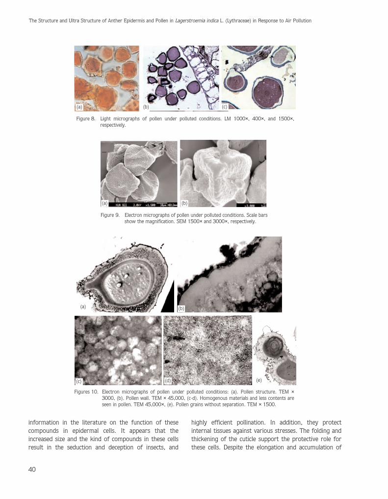
Pollen grains collected from the control area wereregular (Figures 5-6), 4-colporate (Figure 5b, asterisksshow 4-colporate pollen grains), usually 3-colporate(Figures 5a and 6a), sometimes had pseudopores (Figure6b), and contained differentiated cellular organelles(Figure 7). Ornamentation was verrucated (Figure 6b).Pollen grains from the polluted area showed structuralirregularities, such as shrinkage (Figure 8a) andprecocious pollen germination from one or moreapertures (Figures 8-10). The pollen walls were irregularas well (Figure 10b). In some pollen from the pollutedarea, cytoplasm had less content and differentiatedcellular organelles were not seen (Figures 10c-d). Therewere some pollen grains that adhered to each other(Figure 10e). The mean pollen regularity and pollen sizeare shown in Figures 36 and 37, respectively. Statisticalanalysis of the regularity and diameter of pollen showedsignificant differences among samples collected frompolluted and control sites (Figures 11 and 12).
36
The Structure and Ultra Structure of Anther Epidermis and Pollen in Lagerstroemia indica L. (Lythraceae) in Response to Air Pollution

Discussion
Lagerstroemia indica L. pollen grains have differentforms. They are triporate, tetraporate, andpseudoporate. Studies by Graham et al. (1987) andTutuncu et al. (2007) on the pollen morphology of thisspecies report results similar to ours. Both studiesreported that pollen grains are spheroidal andtricolporate, and that ornamentation is verrucated. Pacini& Bellani (1986) and Nepi et al. (2003) reported thepresence of 3- and 4-porate pollen grains. They did notindicate the presence of colpi in the pollen of this species.The outermost cellular layer of the anther is epidermis.This layer protects inner tissues against various stresses.The anther epidermal cells in most plants are the same
size and relatively small. They do not undergo obviouschanges in comparison to other cellular layers duringanther development; therefore they are slightly modifiedduring development. Previous studies on the anther ofAristolochia L. (Johri & Bhotnagar, 1955), Calycanthus L.(Mature, 1968), and Chelone glabra L. (Arekal, 1963)showed that anther epidermis in these plants differentiateas epidermal hairs. These researchers did not indicate thecompounds of this layer. In Lagerstroemia indica L., theelongations of anther epidermal cells was worthy of note.They differentiated at a very early stage of antherdevelopment, prior to the differentiation of the walllayers, as upper and lower epidermis. The compounds inepidermal cells are often flavonoids. There was no
F. REZANEJAD
37
(c)
(a)
(b)
(d)
(e) (f)
Figure 1. Light micrographs of the cross section of an anther under normal conditions (lesspolluted): (a). Epidermal cells in young anther. LM 400×, (b-c). Epidermal cells inmature anther before dehiscence. LM 100× and 150×. respectively, (d).Epidermal cells in mature anther after dehiscence. LM 100×, (e). Epidermal cells inyoung anther stained with fast blue BB, phenolic compounds stained with fast blueBB. LM 400, (f). Epidermis stained with fast blue BB and cuticle stained with Sudanblack B. LM 150. Ue: upper epidermis ; Le: lower epidermis; F: flavonoid; C:cuticle.

38
The Structure and Ultra Structure of Anther Epidermis and Pollen in Lagerstroemia indica L. (Lythraceae) in Response to Air Pollution
Figure 3. Electron micrographs of the anther epidermis and its cuticle.TEM 2000× and 14,000×, respectively (F: flavonoid; C: cuticle).
Figure 4. Light micrographs of the cross section of an anther and its epidermis under airpolluted conditions: (a). Staining by Haematoxylin. LM 400×, (b-c). Staining byAniline Blue. LM 1000× and 3000×, respectively, (d). Electron micrograph of thecross section of epidermis under air polluted conditions. TEM 4000×, (e-f).Electron micrographs of the cross section of a cuticle under air polluted conditions.TEM 14,000× and 8000×, respectively.
(a) (b) (c)
Figure 2. Electron micrographs of a cuticle: (a). Smooth cuticle, (b-c). Folded cuticle. Scale barsshow the magnification.
(a) (b)
(a)
(b)
(c)
(d) (e) (f)

F. REZANEJAD
39
Figure 6. Electron micrographs of pollen under normal conditions (Figure 6aimmature pollen before anther dehiscence and Figure 6b mature pollen),SEM 3000×.
Figure 7. Electron micrographs of pollen under normal conditions. TEM 3000×, TEM 45,000×,and TEM 45,000×, respectively.
Figure 5. Light micrographs of pollen under normal conditions (asterisks in Figure 5b shows 4-colporatepollen).as LM 1000×, 400×, and 1500×, respectively.
(a) (b)
(c)
(a) (b)
(a) (b) (c)
information in the literature on the function of thesecompounds in epidermal cells. It appears that theincreased size and the kind of compounds in these cellsresult in the seduction and deception of insects, and
highly efficient pollination. In addition, they protectinternal tissues against various stresses. The folding andthickening of the cuticle support the protective role forthese cells. Despite the elongation and accumulation of
40
The Structure and Ultra Structure of Anther Epidermis and Pollen in Lagerstroemia indica L. (Lythraceae) in Response to Air Pollution
Figure 9. Electron micrographs of pollen under polluted conditions. Scale barsshow the magnification. SEM 1500× and 3000×, respectively.
Figures 10. Electron micrographs of pollen under polluted conditions: (a). Pollen structure. TEM ×3000, (b). Pollen wall. TEM × 45,000, (c-d). Homogenous materials and less contents areseen in pollen. TEM 45,000×, (e). Pollen grains without separation. TEM × 1500.
Figure 8. Light micrographs of pollen under polluted conditions. LM 1000×, 400×, and 1500×,respectively.
(a)
(a)
(b)
(b)
(a) (b)
(c) (d) (e)
(c)

phenolic compounds in epidermal cells, due to a highconcentration of air pollutants, especially particulatematter that in most days is 3-4 times greater than thestandard dose, the epidermal cells and pollen sufferserious damage. The epidermal cells showed plasmolysis,shrinkage, fragility, and burning at the tip. Although weconsidered the cause as enhanced accumulation ofphenolic compounds in some samples collected from thepolluted region, quantitative study is necessary. A studyon the needles of Picea abies (L.) H.Karst. by Soukupova(2001) considers the enhanced accumulation of phenolicmaterials to be one of the most common reactions ofplants to stresses. Studies show contradictory results, i.e.enhanced accumulation and the opposite results understress have also been reported in the literature.
Therefore, it appears that the type of plant species andthe type of stress are important. Pollen grains grown inthe air polluted environment showed structuralabnormality, decreased size, and fragility. Some studieshave reported that airborne particle materials adhere tothe pollen surface and cause the collapse and degradationof the exine surface, and shrinkage and abnormality ofpollen (Majd & Mohamadi, 1992; Emberlin, 1998, 2000;Parui et al., 1998; Pelter, 1998). Pfahler (1981) showedthat a high level of pollution could induce mutagenicityand physiologic changes; therefore, a high concentrationof air pollutants, especially APM, causes abnormality inanther layers, microspore mother cells, and developingpollen. Because of the nutritional role of the tapetal layer,abnormality of the tapetum induces pollen sterility.
F. REZANEJAD
41
0
20
40
60
80
100
Non-polluted Polluted
Polle
n re
gula
rity
(%
)
05
101520253035
Polluted Non-polluted
Polle
n di
amet
er (
µm)
Figure 11. The mean pollen regularity (%) in the control (non-polluted)and polluted areas (mean ± SE).
Figure 12. The mean pollen diameter (µm) in the control (non-polluted)and polluted areas (mean ± SE).
Table. Mean air pollutant concentrations in the control and polluted study areas.
SO2, ppm NO2, ppm CO, ppm HC, ppm APM, µg m-3
Polluted area 0.07 0.1 8.4 2.7 154
Control area 0.003 0.01 0.65 0.12 60
References
Arekal GD (1963). Contribution to embryology of Chelone glabra.Phytomorphology 13: 376-388.
Emberlin J (1998). The effects of air pollution on allergenic pollen. EurRespir Rev 8: 164-1672.
Emberlin J (2000). The problem of pollen. Allergy 8: 25-28.
Graham A, Graham SA, Nowicke JW, Patel V & Lee S (1990).Palynology and Systematics of the Lythraceae III. GeneraPhysocalymma through Woodfordia, Addenda, and Conclusions.Am J Bot 77: 159-177.
Johri BM & Bhotnagar (1955). A contribution to the morphology andlife history of Aristolochia. Phytomorphology 5: 123- 137.
Majd A & Mohamadi S (1992). Effect of certain toxins and air pollutionon pollen development of Glycine max. J Islamic Azad University649-651.
Majd A, Sharife MR & Zare H (1996). The effect of air pollutants ofArak Aluminum factory on growth and development of certainspecies of Leguminosae. J. of Science, University for TeacherEducation 7: 27-31.
Mature SL (1968). Development of the male and female gametophytesof Calycanthus fertilis. Proc, Natl Inst Sci India part B Biol Sci 34:323-329.
Nepi M, Guarnieri M & Pacini E (2003). Real and feed pollen ofLagerstroemia indica: Ecophysiological differences. Plant Biol 5:311-314.

42
The Structure and Ultra Structure of Anther Epidermis and Pollen in Lagerstroemia indica L. (Lythraceae) in Response to Air Pollution
O’Brian TP & McCully ME (1981). The Study of Plant Structure:Principles and Selected Methods. Melbourne, Australia:Termarcarphi Pty., Ltd.
Pacini E & Bellani LM (1986). Lagerstroemia indica L. pollen: Form andfunction. In: Blackmore S, Ferguson IK (eds.) Pollen and sporesform and function Pp. 347-357. London: Academic Press.
Parui S,, Mondal AK & Mandal S (1998). Protein content and proteinskin test sensitivity of the pollen of Argemona on exposure to SO2.Grana 37: 121-124.
Pelter G (1998). Interaction between pollens and air pollution. AllergieImmunol 30: 324-326.
Pfahler PL (1981). In vitro germination characteristics of maize pollento detect biological activity of environmental pollutants. EnvironHealth Persp 37: 125-132.
Soukupova J (2001). Comparative study of two spruce species in apolluted mountains region. New Phytologist 150: 133-145.
Tutuncu S, Dane F & Tutuncu S (2007). Examination of pollenmorphology of some exotic trees and shrubs found in the parksand the gardens of Edirne (European Turkey) I. J Appl Bio Sci 1:45-55.
Wang Y, Zhang N, Qiang W, Xiong Z & Du G (2006). Effects of reduced,ambient, and enhanced UV-B radiation on pollen germination andpollen tube growth of six alpine meadow annual species. EnvironExp Bot 57: 296-302.