the spleen of the one humped camel (camelus dromedarius) has a unique histological structure
TRANSCRIPT
J. Anat. (2000) 196, pp. 425–432, with 9 figures Printed in the United Kingdom 425
The spleen of the one humped camel (Camelus dromedarius)
has a unique histological structure
MOHAMED ZIDAN1,3, ANWAR KASSEM1, ALI DOUGBAG1, EBTEHAG EL GHAZZAWI2,
MOHAMED ABD EL AZIZ1 AND REINHARD PABST3
"Department of Histology, Faculty of Veterinary Medicine, and #Department of Histology, Faculty of Medicine,
Alexandria University, Egypt, and $Department of Functional and Applied Anatomy, Medical School of Hannover,
Germany
(Accepted 20 December 1999)
The histology and structure of 38 spleens of the dromedary (aged 0±5–15 y) were studied in relation to age.
The spleen was found to have a thick capsule (292³106 mm) divided into an outer layer (113³39 mm)
composed mainly of connective tissue and an inner layer (180³81 mm) consisting mainly of smooth muscle
cells. Vascular and avascular trabeculae extend from the capsule, the former containing arteries and nerves
but no trabecular veins, the latter being divided structurally into primary and secondary trabeculae.
Subcapsular and peritrabecular blood sinuses around primary and vascular trabeculae are unique to the
camel spleen. The central artery emerges from the periarterial lymphatic sheath and branches into up to 4
penicilli which extend as sheathed arterioles (42³8 µm). These are found near or surrounded by blood
sinusoids of the red pulp. A wide marginal zone surrounds the white pulp and contains sheathed arteries but
no marginal sinuses. The red pulp is characteristically divided into cords by secondary trabeculae and
contains venous sinusoids of different sizes. The camel spleen is of a sinusal type that can store blood. The
thick muscular capsule and trabeculae pump the stored blood according to the body’s need. Both closed and
open circulations are found. The venous return is unique as the blood flow is from the venous sinusoids of
the red pulp to the peritrabecular sinuses to the subcapsular sinuses to the splenic vein. No significant
structural differences related to age were found.
Key words : White pulp; red pulp; marginal zone; periarterial macrophage sheath.
The camel is an important source of meat, milk and
hide in several countries and there is growing interest
in its meat and milk products (Hamers & Muylder-
mans, 1998). The most important disease to affect the
camel is the blood parasite, Trypanosoma evansi
(Ngeranwa et al. 1993). The blood parasites are
removed and phagocytosed in the spleen (Schnizer et
al. 1972; Chen & Weiss, 1973). In general, it is the
largest lymphoid organ and the most important organ
of immunological defence for the blood (Pabst, 1993).
The white pulp of the mammalian spleen is composed
of the periarterial lymphatic sheath (PALS) and
lymph follicles (Raviola, 1994). Lymph follicles
predominate in the human spleen whereas the PALS
Correspondence to: Prof. Reinhard Pabst, Department of Functional and Applied Anatomy, Medical School of Hannover, 30625 Hannover,
Germany. Tel. : 49 511 5326740; fax: 49 511 5322948; e-mail : Pabst.Reinhard!MH-Hannover.de
predominates in the rat (Steiniger et al. 1997). The
reticular framework of the white pulp is divided into
2 parts in mice (Tanaka et al. 1996) and man (Satoh,
1997), one part surrounding the PALS and the other
surrounding the lymph follicles. The marginal zone is
the site of heavy blood traffic and filtration. Venous
sinusoids are found near or within it (More et al.
1964; Weiss, 1964; Burk & Simon, 1970). It envelops
the white pulp in rodents and rabbits (Snook, 1950)
but surrounds only the lymph follicles in humans and
contains no marginal or perimarginal sinuses. It is
separated from the red pulp by a perifollicular zone
(Steiniger et al. 1997). The red pulp of several species
is composed of splenic cords separated by blood
sinusoids (Dellmann & Brown, 1976). The periarterial
macrophage sheath (PAMS) is composed of macro-
Figs 1–4. For legends see p. 429.
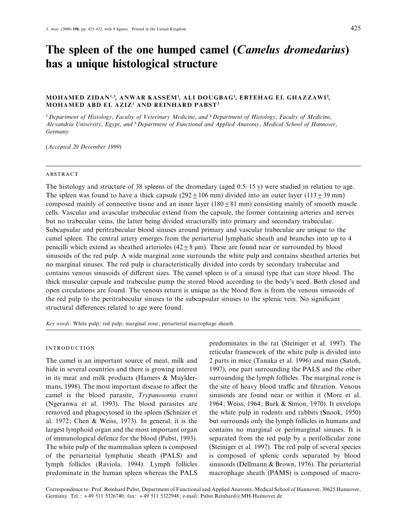
Fig. 1. (a) Overview of the camel spleen showing the capsule (C), trabecula (T), subcapsular sinus (arrow), white pulp (WP) and red pulp
(RP). H&E. (b) The splenic capsule is divided into an outer layer (O) composed mainly of connective tissue and an inner layer (I)
composed mainly of smooth muscle cells. S, subcapsular blood sinus; RP, red pulp. Trichrome.
426 M. Zidan and others
Figs 5–8. For legends see p. 429.
Histology of the camel spleen 427
phages supported by a reticular network (Raviola,
1994). Mice, rats and rabbits lack sheathed arteries
(Snook, 1950). The diameter of the PAMS has been
determined in several animals (Seki & Abe, 1985). The
splenic circulation may be open (Thomas, 1967;
Fukuta et al. 1976; Irino et al. 1977; Blue & Weiss,
1981a), closed (Simon, 1970a ; Murkami et al. 1973)
or a combination of both (Barnhart et al. 1976; Chen,
1978). There are sinusal type spleens, e.g. in horses,
dogs and pigs (Brown & Dellmann, 1976), man, rats
and rabbits (Blue & Weiss, 1981b) and nonsinusal
types, e.g. in cats, ruminants (Brown & Dellmann,
1976) and mice (Blue & Weiss, 1981a). The age-
structure relationship of the human spleen has been
studied (Timens et al. 1989) and the relationship
between the regeneration of the splenic transplant and
the age of the host and the donor has been investigated
in rats (Westermann et al. 1988). In spite of the
immunological and haematological importance of the
spleen and the advantages of knowledge of its cell
physiology, very little is known about the spleen of the
camel (Hegazi, 1953; Bareedy et al. 1982; Abd El Aal,
1994; Radmehr, 1997). It has been shown that the
white pulp is composed of lymph follicles around
blood vessels. The marginal zone is poorly delimited
from the white and the red pulp and composed of
diffuse lymphatic tissue (Bareedy et al. 1982; Abd El
Aal, 1994). There are numerous ellipsoids in the red
pulp (Hegazi, 1953) and the marginal zone (Abd El
Aal, 1994). Radmehr (1997) described the branching
of the splenic artery. The following structural charac-
teristics found in other species have not been described
in the camel : the PALS, corona and the reticular
framework of the white pulp, splenic circulation,
marginal sinuses and morphometry. The aim of this
study was therefore to examine the different splenic
compartments and age-structure relationships using
morphometry and to describe the splenic circulation
in the camel.
The study was carried out on 38 spleens obtained
from clinically healthy camels of both sexes with a
mean age 9±5 y (range 0±5–15 y). Fresh specimens were
obtained from 35 spleens directly after slaughter and
were fixed in 10% buffered formalin. As these spleens
probably contracted during bleeding to an unphysi-
ologically small size, to show the splenic com-
partments of a normal spleen 3 spleens were perfused
with 0±9% saline through the splenic artery to remove
the blood from the blood vessels and avoid blood
clots. The splenic vein was then ligated and each
spleen infused with 10 l of 10% buffered formalin
through the splenic artery, after which the splenic
artery was tied off. The infused spleens were immersed
in the same fixative for 24 h before specimen col-
lection. All specimens were prepared for paraffin
sections and stained with haematoxylin and eosin,
(H&E) Crossman trichrome, Gomori’s reticulin,
Orcein, PAS (Bo$ ck, 1989) and Giemsa (Burck, 1988).
The thickness of the capsule and the diameter of the
PAMS of all samples were measured using a mi-
crometer eye piece (Carl Zeiss, Germany) and com-
pared at different ages. The results are given as
mean³standard deviation.
The camel spleen was composed of a thick capsule
surrounding the splenic parenchyma (Fig. 1a). The
capsule was 292³106 µm thick and was divided into
clearly demarcated outer and inner layers (Fig. 1b).
The outer layer (113³39 µm) consisted mainly of
connective tissue including collagen, elastic and
reticular fibres with few smooth muscle cells. The
inner layer (180³81 µm) was composed pre-
dominantly of smooth muscle cells supported by
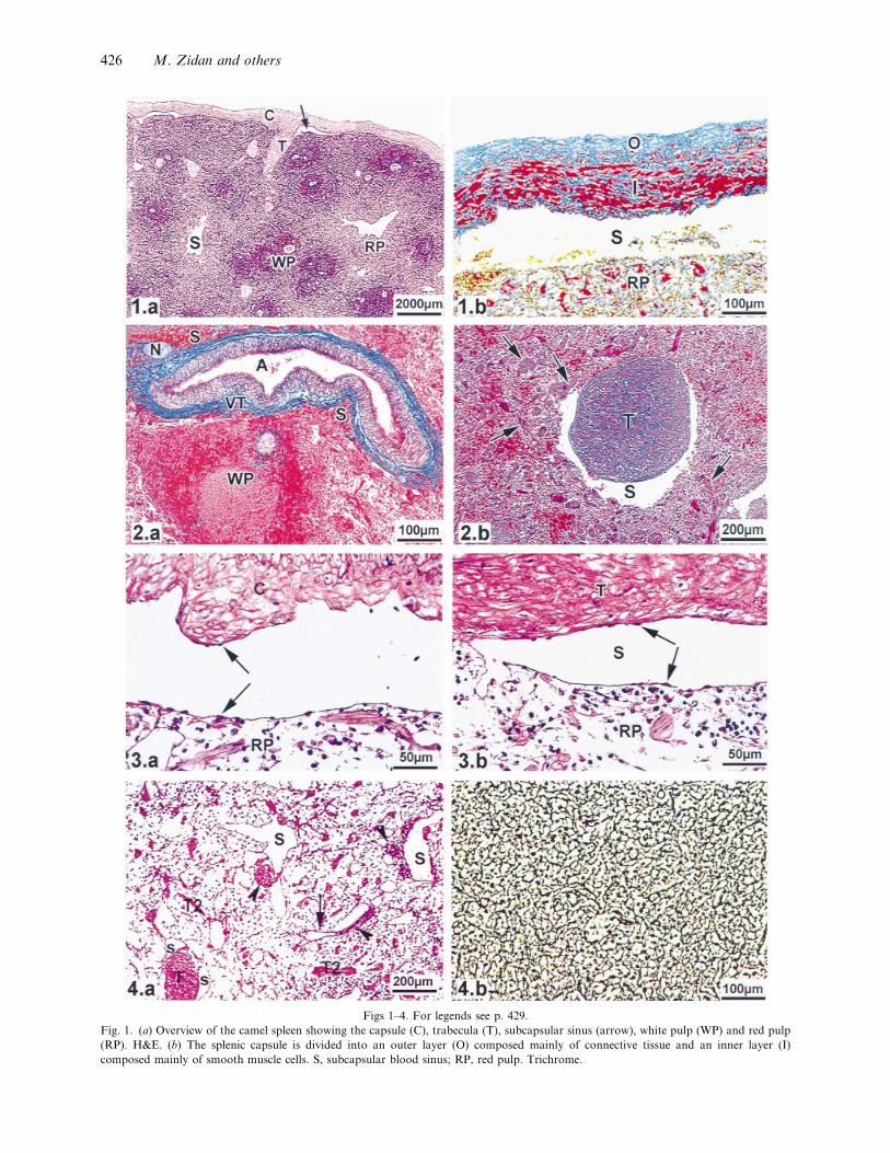
reticular, collagen and elastic fibres. Vascular and
avascular trabeculae extended from the capsule into
the splenic parenchyma. The former contained tra-
becular arteries and nerves but no veins and were
composed mainly of connective tissue made up of
collagen, reticular and elastic fibres and a few laterally
located smooth muscle cells parallel to the longi-
tudinal axis of the trabeculae (Fig. 2a). The latter
were divided into primary and secondary trabeculae.
The primary trabeculae originated from the capsule
and had a similar structure to that of the inner layer
of the capsule, being composed mainly of smooth
muscle cells lying parallel to the longitudinal axis of
the trabeculae and supported by reticular, collagen
and elastic fibres (Fig. 2b). The secondary trabeculae
were composed mainly of parallel smooth muscle cells
with reticular fibres among them. Subcapsular blood
sinuses extended under the capsule and connected to
peritrabecular sinuses which surrounded both the
primary and the vascular trabeculae (Fig. 3a, b). The
red pulp was divided into small compartments (cords)
by the secondary trabeculae (Fig. 4a), each cord being
composed of a reticular network containing the
different blood cells (Fig. 4b). Blood sinusoids with a
continuous basement membrane were irregularly
distributed in the red pulp (Figs 4a, 5a) and connected
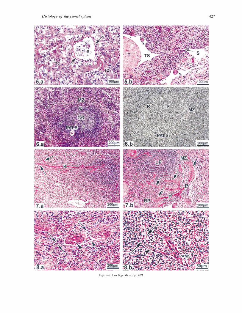
to the peritrabecular sinuses (Fig. 5b). The white pulp
was demarcated by circumferential reticular fibres
428 M. Zidan and others

that clearly divided the compartments of the white
pulp into PALS and lymphoid follicle (Fig. 6a, b).
The lymphoid follicles were spherical in shape and
sometimes indented at the site of the PALS. Secondary
follicles had germinal centres surrounded by a corona
(Fig. 6a). After emerging from the PALS, the central
artery branched into a maximum of 4 straight
branches (penicillary arteries), which were able to
branch further (Fig. 7a, b). The extensions of the
penicillary arteries were ensheathed by cylindrical
PAMS (42³8 µm) forming the sheathed arteries
which were possibly surrounded by or associated with
blood sinusoids (Fig. 8a). The arterial capillaries
extended beyond the edge of the PAMS into the red
pulp to open directly into the sinusoids (Fig. 8b) or
into splenic cords. The white pulp was separated from
the red pulp by a marginal zone that was clearly
separated from the white pulp by reticular fibres, but
there was no clear line of demarcation from the red
pulp (Fig. 6a, b). It contained PAMS but no marginal
sinuses or secondary trabeculae (Fig. 7b). There was
no significant difference in the morphometric measure-
ments of the capsule or the PAMS at different ages. A
schematic overview of the different compartments and
the circulation of the camel spleen is depicted in
Figure 9.
The present study has shown that the capsule of the
camel spleen is characteristically thick and divided
into outer connective tissue and inner smooth muscle
layers constituting about 1}3 and 2}3 of the capsule
thickness, respectively. Abd El Aal (1994) described
Fig. 2. (a) A vascular trabecula (VT) composed mainly of connective tissue with a few laterally located smooth muscle cells and containing
a trabecular artery (A) and nerve (N) but no vein. S, peritrabecular blood sinus; WP, white pulp. Trichrome. (b) An avascular trabecula
divided into a primary trabecula (T) which is composed of parallel smooth muscle cells and connective tissue, and secondary trabeculae
(arrows) composed of parallel smooth muscles and reticular fibres. S, peritrabecular blood sinus. Trichrome.
Fig. 3. Photomicrograph of an infused fixed spleen showing (a) a subcapsular sinus lined with flat endothelial cells (arrows) lying on a PAS
positive basement membrane. C, capsule; RP, red pulp. PAS (b) A peritrabecular sinus (S) lined with flat endothelial cells (arrows) resting
on a PAS-positive basement membrane. T, trabecula; RP, red pulp. PAS.
Fig. 4. (a) Photomicrograph of an infused fixed spleen showing red pulp composed of splenic cords separated by secondary trabeculae (T2)
and containing blood sinusoids (S) and PAMS (arrowheads) which may open into blood sinusoids (arrow). T, primary trabecula; S,
peritrabecular sinus. PAS. (b) Red pulp supported by a reticular connective tissue network. Gomori’s reticulin.
Fig. 5. (a) Red pulp composed of splenic cords separated by secondary trabeculae (T2) and containing blood sinusoids (S) with a continuous
basement membrane (arrow). PAS. (b) A red pulp sinusoid (S) connected to a peritrabecular sinus (TS). The arrow shows the site of
connection. The PAMS (arrowheads) is located inside the blood sinusoid. T, primary trabecula. PAS.
Fig. 6. (a) White pulp composed of PALS and a lymph follicle which possesses a germinal centre (GC) and corona (C). MZ, marginal zone.
Giemsa. (b) White pulp divided by reticular fibres into clearly demarcated PALS and a lymph follicle (LF), separated from the marginal zone
(MZ) by circumferential reticular fibres (R). Gomori’s reticulin.
Fig. 7. (a) Central artery continuing as a penicillary artery (P) which divides into 2 sheathed arteries (arrows). PAS. (b) Central artery
dividing into 4 penicillary arteries (P) which extend as sheathed arteries surrounded by cylindrical PAMS (arrows) located in the marginal
zone (MZ) or in the red pulp (RP). LF, lymph follicle. PAS.
Fig. 8. (a) PAMS (arrowhead) surrounded by a blood sinusoid (arrows) in the red pulp. (b) A sheathed artery extends beyond the edge of
the PAMS as an arterial capillary (arrowhead) and opens into a blood sinusoid which is surrounded by basement membrane (arrows). PAS.
these 2 layers only in passing without quantitative
measurements and reported that the outer layer was
thicker than the inner. Ruminants have only 2 thin
layers of smooth muscle cells. In the horse, the capsule
consists of an outer thick connective tissue and an
inner thinner smooth muscle layer, in the pig the
capsule is formed mainly from smooth muscle, while
in the dog and cat smooth muscle makes up about 2}3
of the capsule thickness (Brown & Dellmann, 1976).
The capsule of the human spleen is composed of
connective tissue with little smooth muscle (Weiss,
1983). As in other species (Brown & Dellmann, 1976),
the splenic trabeculae extend from the capsule into the
parenchyma, but in the camel they were uniquely
divided into vascular and avascular trabeculae. The
vascular trabeculae contained arteries and nerves but
no veins. The avascular outnumbered the vascular
trabeculae and were divided into primary and sec-
ondary trabeculae. The primary trabeculae have a
similar structure to the inner layer of the capsule,
while the secondary trabeculae are composed mainly
of smooth muscle and extend through the red pulp as
in the pig (Ueda et al. 1991).
The current work revealed a unique structure which
has not previously been observed in other species in
that the trabecular and capsular veins were replaced
by peritrabecular and subcapsular sinuses respect-
ively. The peritrabecular sinuses were noted by
Bareedy et al. (1982) and collecting veins similar to
large sinuses extending under the capsule were
recorded by Abd El Aal (1994). The peritrabecular
and the subcapsular sinuses play the role of the
trabecular and capsular veins in collecting the venous
blood from the spleen to the splenic vein. Bareedy et
Histology of the camel spleen 429

Fig. 9. Schematic diagram showing the different compartments and the circulation of the camel spleen.
al. (1982) and Abd El Aal (1994) found that the red
pulp was divided into cords by blood sinusoids. The
present study has revealed that the cords are separated
by secondary trabeculae and the sinusoids are distrib-
uted irregularly in the red pulp. The massive dis-
tribution of the secondary trabeculae among the
splenic cords and sinusoids facilitates quick outflow of
blood during splenic contraction.
Due to the presence of blood sinusoids in the red
pulp of the camel spleen it can be classified, in
agreement with Bareedy et al. (1982), as sinusal in
type. The increase in the diameter of the blood
sinusoids in the infused spleen further supports its
blood storage function. The volume of the stored
blood may exceed 10 l as although we infused 10 l of
fixative the spleen appeared to be able to accom-
modate more. The type of spleen found in the camel
differs from that in other ruminants which are of
nonsinusal type (Brown & Dellmann, 1976). The
camel spleen can store blood like other sinusoidal type
spleens of the pig, horse and dog (Brown & Dellmann,
1976) and human, rat and rabbit (Blue & Weiss,
1981b). The present finding indicates that the spleen
of the one humped camel with its muscular capsule
and trabeculae represents a highly contractile bag that
can force the stored blood out to the circulation
according to the needs of the body, e.g. on extreme
physical activity or in haemorrhagic conditions. The
white pulp was demarcated by circumferential re-
ticular fibres that divided it into 2 compartments, one
surrounding the lymph follicles and the other forming
a network at the PALS. This is in agreement with the
finding of Tanaka et al. (1996) in mice and Sataho et
al. (1997) in man. They concluded that this reticular
framework plays an essential role in lymphocyte
homing and compartmentalisation, which may also be
true for the camel white pulp. The marginal zone
surrounds all the white pulp as in other species
(Brown & Dellmann, 1976), except in man where it
surrounds only the lymphoid follicles (Steiniger et al.
1997).
The PAMS was cylindrical in shape as in man
(Raviola, 1994). This finding is in contrast to the
finding of Bareedy et al. (1982) and Abd El Aal (1994)
who described an ellipsoid, rounded or oval shaped
PAMS in the camel. It is ellipsoid shape in the dog
(Jacobsen, 1971). The PAMS of the camel (42³8 µm)
was smaller than that of the pig (60–90 µm), similar to
that of the horse (40–60 µm) and larger than that of
the dog, cat (30–40 µm) and cow (! 30 µm) (Seki &
Abe, 1985) and also man (20–30 µm) (Raviola, 1994).
There is no PAMS in rats, rabbits or mice (Snook,
1950). The sheathed arteries were clearly demarcated
from the red pulp. Blood sinusoids extended adjacent
430 M. Zidan and others

to the sheathed arteries as in dogs (Jacobsen, 1971),
or—uniquely in the camel—surrounded them. The
current study showed that the arterial capillary may
extend over the end of the PAMS in the red pulp.
Similar results were recorded by Seki & Abe (1985) in
the cow, horse, pig, dog and rat, but not in the cat.
This arterial capillary may open directly into sinusoids
or into splenic cords. Therefore both closed and open
circulations were observed in the camel spleen. This
agrees with the finding of Barnhard et al. (1976) in
man and of Seki & Abe (1985) in the dog and rat.
The camel spleen is of sinusal type that can store
blood. The thick muscular capsule and trabeculae
pump the stored blood according to the body’s need.
Both closed and open circulations are found. The
venous return is unique as the blood flows from the
venous sinusoids of the red pulp to the peritrabecular
sinsuses to the subcapsular sinuses to the splenic vein.
The absence of a marginal sinus and the presence of a
closed circulation in the camel spleen may reduce the
role of the marginal zone in blood filtration. This may
explain why blood parasites are the main health
problem in the camel.
The study was supported by the Ministry of Higher
Education, Egypt through a scholarship to Mohamed
Zidan. The facilities provided by Dr M. A. Abo Maze
(director of the Koom Hamada Abattoir, Egypt)
which enabled us to obtain the specimens, as well as
linguistic corrections by Ms S. Fryk and preparation
of the diagram by Ms M. Peter are gratefully
acknowledged.
ABD EL AAL E (1994) Some histological and histochemical
studies on the spleen of the dromedary camel (Camelus
dromedarius). M.Sc. thesis, Faculty of Veterinary Medicine
Moshtohor, Zagazig University, Egypt.
BAREEDY M, ANIS H, EWAIS M, AMMER S (1982) Some
anatomical and histological studies on the spleen of one humped
camel (Camelus dromedarius). Egyptian Journal of Histology 5,
75–83.
BARNHART M, BAECHLER C, USHER J (1976) Arteriovenous
shunts in the human spleen. American Journal of Hematology 1,
105–114.
BLUE J, WEISS L (1981a) Vascular pathways in non sinusal red
pulp. An electron microscopic study of cat spleen. American
Journal of Anatomy 161, 135–186.
BLUE J, WEISS L (1981b) Electron microscopy of the red pulp of
the dog spleen including vascular arrangements, periarterial
macrophage sheaths (Ellipsoids) and the contractile, innervated
reticular meshwork. American Journal of Anatomy 161, 189–218.
BO$ CK P (1989) Romeis Mikroskopische Technik, 17th edn, p. 697.
Mu$ nchen, Wien, Baltimore: Urban & Schwarzenberg.
BROWN E, DELLMANN H-D (1976) Lymphatic system. In
Textbook of Veterinary Histology (ed. Dellmann H-D, Brown E),
pp. 161–184. Philadelphia: Lea & Febiger.
BURCK H-C (1998) Histologische Technik, 6th edn, pp. 129–130.
Stuttgart: Thieme.
BURKE J, SIMON G (1970) Electron microscopy of the spleen: 1
Anatomy and microcirculation. American Journal of Pathology
58, 127–155.
CHEN L (1978) Microcirculation of the spleen: an open or closed
circulation? Science 201, 157–159.
CHEN L, WEISS L (1972) Electron microscopy of the red pulp of
human spleen. American Journal of Anatomy 134, 425–485.
CHEN L, WEISS L (1973) The role of the sinus wall in the passage
of the erythrocytes through the spleen. Blood 41, 529–537.
FUJITA T (1974) A scanning electron microscope study of the
human spleen. Archivum Histolologicum Japonicum 37, 187–216.
FUKUTA K, NISHIDA T, MOCHIZUKI K (1976) Electron
microscopy of the splenic circulation in chicken. Japanese Journal
of Veterinary Science 38, 241–254.
HAMERS R, MUYLDERMANS S (1998) Immunology of the
camel and llamas. In Handbook of Vertebrate Immunology (ed.
Pastoret P, Griebel P, Bazin H, Govaerts, A), pp. 421–438.
California : Academic Press.
HEGAZI A (1953) The spleen of the camel compared with other
domesticated animals and its microscopic examination. Journal
of the American Veterinary Medical Association 122, 182–184.
IRINO S, MURAKAMI T, FUJITA T (1977) Open circulation in
the human spleen. Dissection scanning electron microscopy of
conductive stained tissue and observation of resin vascular cast.
Archivum Histolologicum Japonicum 40, 297–304.
JACOBSEN G (1971) Morphological-histochemical comparison of
dog and cat splenic ellipsoid sheaths. Anatomical Record 169,
105–114.
MOORE R, MUMAW V, SCHOENBERG M (1964) The structure
of the spleen and its functional implications. Experimental and
Molecular Pathology 3, 31–50.
MURAKAMI T, FUJITA T, MAYOSHI M (1973) Closed
circulation in the rat spleen as evidenced by scanning electron
microscopy of vascular casts. Experientia 29, 1374–1375.
NGERANWA J, GATHUMBI P, MUTIGA E, AGUMBAH G
(1993) Pathogenesis of Trypanosoma (brucei) evensi in small east
African goats. Research in Veterinary Science 54, 283–289.
PABST R (1993) Anatomische und physiologische Vorausset-
zungen zur Erhaltung der postoperativen Milzfunktion nach
milzerhaltenden Eingriffen. Chirurgische Gastroenterologie 9
(suppl. 2), 19–22.
RADMEHR R (1997) Vascular segments in the spleen of one
humped camel (Camelus dromedarius). Journal of Camel Practice
and Research 4, 45–46.
RAVIOLA E (1994) The spleen. In Bloom and Fawcett. A Text
Book of Histology, 12th edn (ed. Fawcett D), pp. 460–472. New
York: Chapman & Hall.
SATOH T, TAKEDA R, OIKAWA H, SATODATE R (1997)
Immunohistochemical and structural characteristics of the re-
ticular framework of the white pulp and marginal zone in the
human spleen. Anatomical Record 249, 486–494.
SCHNIZER B, SODEMAN T, MEAD M, CONTACOS P (1972)
Pitting function of the spleen in malaria : ultrastructural
observations. Science 177, 175–184.
SEKI A, ABE M (1985) Scanning electron microscopic studies on
the microvascular system of the spleen in rat, dog, pig, horse and
cow. Japanese Journal of Veterinary Science 47, 237–249.
Histology of the camel spleen 431
SNOOK T (1950) A comparative study of the vascular arrange-
ments in mammalian spleen. American Journal of Anatomy 87,
31–78.
STEINIGER B, BARTH P, HERBEST B, HARTNELL A,
CROCKER P (1997) The species-specific structure of micro-
anatomical compartments in the human spleen: strongly sialo-
adhesin-positive macrophages occur in the perifollicular zone,
but not in the marginal zone. Immunology 92, 307–316.
TANAKA H, HATABA Y, SAITO S, FUKUSHIMA O,
MIYASAKA M (1996) Phenotypic characteristics and sig-
nificance of reticular meshwork surrounding splenic white pulp
of mice. Journal of Electron Microscopy 45, 407–416.
THOMAS C (1967) An electron and light microscope study of sinus
structure in perfused rabbit and dog spleen. American Journal of
Anatomy 120, 527–533.
TIMENS W, BOES A, ROZEBOOM-UITERWIJK T, POPPEMA
S (1989) Immaturity of the human splenic marginal zone in
infancy: possible contribution to the deficient infant immune
response. Journal of Immunology 143, 3200–3206.
UEDA H, ABE M, TAKEHANA K, TAWASA K, HIRAGATA
T (1991) Ultrastructure of the red pulp and spleen enervation in
horse and pig. Acta Anatomica 141, 151–158.
WEISS L (1964) The white pulp of the spleen. The relationships of
arterial vessels, reticulum and free cells in the peripheral
lymphatic sheath. Bulletin of Johns Hopkins Hospital 115,
99–173.
WEISS L (1983) The spleen. In Histology, Cell and Tissue Biology,
5th edn (ed. Weiss L), pp. 544–568. London and Basingstoke:
Macmillan.
WESTERMANN J, WILLFU$ HR K-H, PABST R (1988) Influence
of donor and host age on the regeneration and blood flow of
splenic transplants. Journal of Pediatric Surgery 23, 835–838.
432 M. Zidan and others