the spinal cord || development of the spinal cord
TRANSCRIPT
2 Development of the Spinal Cord
Ken WS Ashwell
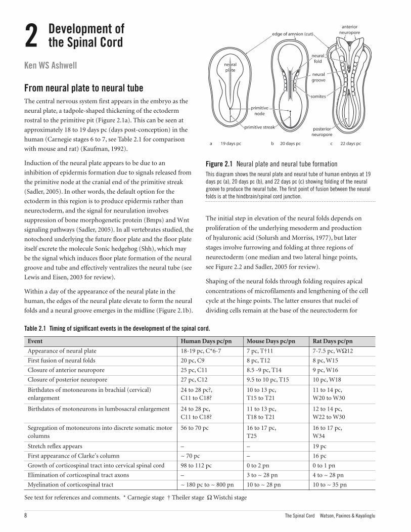
From neural plate to neural tubeThe central nervous system first appears in the embryo as the
neural plate, a tadpole-shaped thickening of the ectoderm
rostral to the primitive pit (Figure 2.1a). This can be seen at
approximately 18 to 19 days pc (days post-conception) in the
human (Carnegie stages 6 to 7, see Table 2.1 for comparison
with mouse and rat) (Kaufman, 1992).
Induction of the neural plate appears to be due to an
inhibition of epidermis formation due to signals released from
the primitive node at the cranial end of the primitive streak
(Sadler, 2005). In other words, the default option for the
ectoderm in this region is to produce epidermis rather than
neurectoderm, and the signal for neurulation involves
suppression of bone morphogenetic protein (Bmps) and Wnt
signaling pathways (Sadler, 2005). In all vertebrates studied, the
notochord underlying the future floor plate and the floor plate
itself excrete the molecule Sonic hedgehog (Shh), which may
be the signal which induces floor plate formation of the neural
groove and tube and effectively ventralizes the neural tube (see
Lewis and Eisen, 2003 for review).
Within a day of the appearance of the neural plate in the
human, the edges of the neural plate elevate to form the neural
folds and a neural groove emerges in the midline (Figure 2.1b).
Table 2.1 Timing of significant events in the development of the spinal cord.
Event Human Days pc/pn Mouse Days pc/pn Rat Days pc/pn
Appearance of neural plate 18-19 pc, C*6-7 7 pc, T†11 7-7.5 pc, WΩ12
First fusion of neural folds 20 pc, C9 8 pc, T12 8 pc, W15
Closure of anterior neuropore 25 pc, C11 8.5 -9 pc, T14 9 pc, W16
Closure of posterior neuropore 27 pc, C12 9.5 to 10 pc, T15 10 pc, W18
Birthdates of motoneurons in brachial (cervical)enlargement
24 to 28 pc?,C11 to C18?
10 to 13 pc,T15 to T21
11 to 14 pc,W20 to W30
Birthdates of motoneurons in lumbosacral enlargement 24 to 28 pc, C11 to C18?
11 to 13 pc,T18 to T21
12 to 14 pc,W22 to W30
Segregation of motoneurons into discrete somatic motorcolumns
56 to 70 pc 16 to 17 pc,T25
16 to 17 pc,W34
Stretch reflex appears – – 19 pc
First appearance of Clarke’s column ~ 70 pc – 16 pc
Growth of corticospinal tract into cervical spinal cord 98 to 112 pc 0 to 2 pn 0 to 1 pn
Elimination of corticospinal tract axons – 3 to ~ 28 pn 4 to ~ 28 pn
Myelination of corticospinal tract ~ 180 pc to ~ 800 pn 10 to ~ 28 pn 10 to ~ 35 pn
See text for references and comments. * Carnegie stage † Theiler stage Ω Wistchi stage
Figure 2.1 Neural plate and neural tube formationThis diagram shows the neural plate and neural tube of human embryos at 19days pc (a), 20 days pc (b), and 22 days pc (c) showing folding of the neuralgroove to produce the neural tube. The first point of fusion between the neuralfolds is at the hindbrain/spinal cord junction.
8 The Spinal Cord Watson, Paxinos & Kayalioglu
The initial step in elevation of the neural folds depends on
proliferation of the underlying mesoderm and production
of hyaluronic acid (Solursh and Morriss, 1977), but later
stages involve furrowing and folding at three regions of
neurectoderm (one median and two lateral hinge points,
see Figure 2.2 and Sadler, 2005 for review).
Shaping of the neural folds through folding requires apical
concentrations of microfilaments and lengthening of the cell
cycle at the hinge points. The latter ensures that nuclei of
dividing cells remain at the base of the neurectoderm for
Spinal Cord Atlas Text+Index.qxp 21/08/08 4:39 PM Page 8
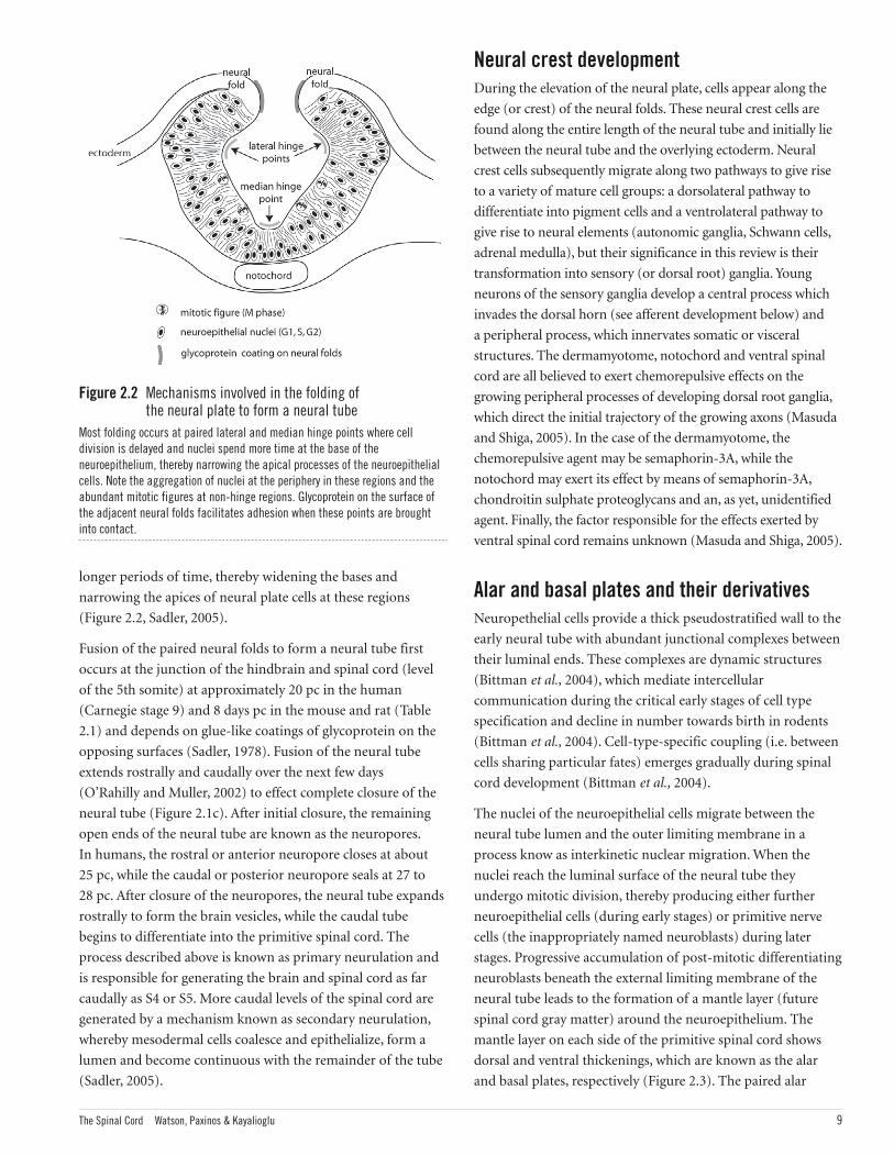
Figure 2.2 Mechanisms involved in the folding of the neural plate to form a neural tube
Most folding occurs at paired lateral and median hinge points where celldivision is delayed and nuclei spend more time at the base of theneuroepithelium, thereby narrowing the apical processes of the neuroepithelialcells. Note the aggregation of nuclei at the periphery in these regions and theabundant mitotic figures at non-hinge regions. Glycoprotein on the surface ofthe adjacent neural folds facilitates adhesion when these points are broughtinto contact.
The Spinal Cord Watson, Paxinos & Kayalioglu 9
Neural crest developmentDuring the elevation of the neural plate, cells appear along the
edge (or crest) of the neural folds. These neural crest cells are
found along the entire length of the neural tube and initially lie
between the neural tube and the overlying ectoderm. Neural
crest cells subsequently migrate along two pathways to give rise
to a variety of mature cell groups: a dorsolateral pathway to
differentiate into pigment cells and a ventrolateral pathway to
give rise to neural elements (autonomic ganglia, Schwann cells,
adrenal medulla), but their significance in this review is their
transformation into sensory (or dorsal root) ganglia. Young
neurons of the sensory ganglia develop a central process which
invades the dorsal horn (see afferent development below) and
a peripheral process, which innervates somatic or visceral
structures. The dermamyotome, notochord and ventral spinal
cord are all believed to exert chemorepulsive effects on the
growing peripheral processes of developing dorsal root ganglia,
which direct the initial trajectory of the growing axons (Masuda
and Shiga, 2005). In the case of the dermamyotome, the
chemorepulsive agent may be semaphorin-3A, while the
notochord may exert its effect by means of semaphorin-3A,
chondroitin sulphate proteoglycans and an, as yet, unidentified
agent. Finally, the factor responsible for the effects exerted by
ventral spinal cord remains unknown (Masuda and Shiga, 2005).
Alar and basal plates and their derivativesNeuropethelial cells provide a thick pseudostratified wall to the
early neural tube with abundant junctional complexes between
their luminal ends. These complexes are dynamic structures
(Bittman et al., 2004), which mediate intercellular
communication during the critical early stages of cell type
specification and decline in number towards birth in rodents
(Bittman et al., 2004). Cell-type-specific coupling (i.e. between
cells sharing particular fates) emerges gradually during spinal
cord development (Bittman et al., 2004).
The nuclei of the neuroepithelial cells migrate between the
neural tube lumen and the outer limiting membrane in a
process know as interkinetic nuclear migration. When the
nuclei reach the luminal surface of the neural tube they
undergo mitotic division, thereby producing either further
neuroepithelial cells (during early stages) or primitive nerve
cells (the inappropriately named neuroblasts) during later
stages. Progressive accumulation of post-mitotic differentiating
neuroblasts beneath the external limiting membrane of the
neural tube leads to the formation of a mantle layer (future
spinal cord gray matter) around the neuroepithelium. The
mantle layer on each side of the primitive spinal cord shows
dorsal and ventral thickenings, which are known as the alar
and basal plates, respectively (Figure 2.3). The paired alar
longer periods of time, thereby widening the bases and
narrowing the apices of neural plate cells at these regions
(Figure 2.2, Sadler, 2005).
Fusion of the paired neural folds to form a neural tube first
occurs at the junction of the hindbrain and spinal cord (level
of the 5th somite) at approximately 20 pc in the human
(Carnegie stage 9) and 8 days pc in the mouse and rat (Table
2.1) and depends on glue-like coatings of glycoprotein on the
opposing surfaces (Sadler, 1978). Fusion of the neural tube
extends rostrally and caudally over the next few days
(O’Rahilly and Muller, 2002) to effect complete closure of the
neural tube (Figure 2.1c). After initial closure, the remaining
open ends of the neural tube are known as the neuropores.
In humans, the rostral or anterior neuropore closes at about
25 pc, while the caudal or posterior neuropore seals at 27 to
28 pc. After closure of the neuropores, the neural tube expands
rostrally to form the brain vesicles, while the caudal tube
begins to differentiate into the primitive spinal cord. The
process described above is known as primary neurulation and
is responsible for generating the brain and spinal cord as far
caudally as S4 or S5. More caudal levels of the spinal cord are
generated by a mechanism known as secondary neurulation,
whereby mesodermal cells coalesce and epithelialize, form a
lumen and become continuous with the remainder of the tube
(Sadler, 2005).
Spinal Cord Atlas Text+Index.qxp 21/08/08 4:39 PM Page 9
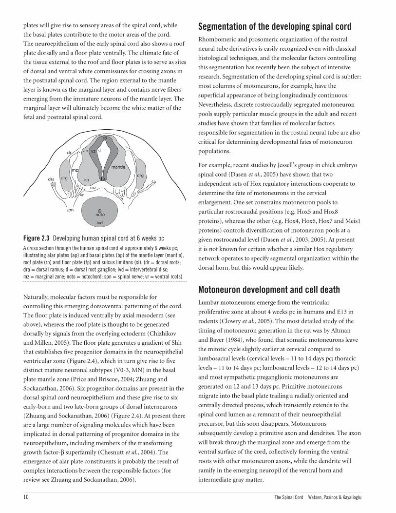
plates will give rise to sensory areas of the spinal cord, while
the basal plates contribute to the motor areas of the cord.
The neuroepithelium of the early spinal cord also shows a roof
plate dorsally and a floor plate ventrally. The ultimate fate of
the tissue external to the roof and floor plates is to serve as sites
of dorsal and ventral white commissures for crossing axons in
the postnatal spinal cord. The region external to the mantle
layer is known as the marginal layer and contains nerve fibers
emerging from the immature neurons of the mantle layer. The
marginal layer will ultimately become the white matter of the
fetal and postnatal spinal cord.
Naturally, molecular factors must be responsible for
controlling this emerging dorsoventral patterning of the cord.
The floor plate is induced ventrally by axial mesoderm (see
above), whereas the roof plate is thought to be generated
dorsally by signals from the overlying ectoderm (Chizhikov
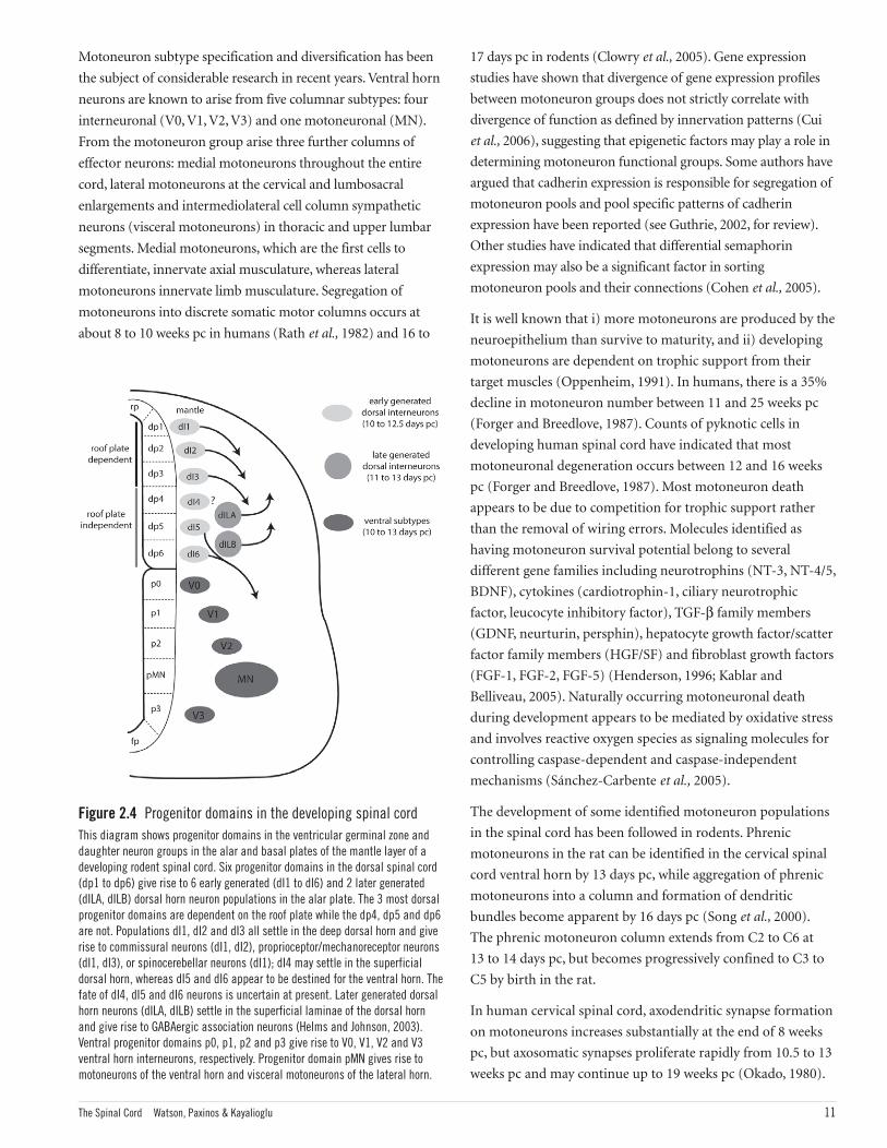
and Millen, 2005). The floor plate generates a gradient of Shh
that establishes five progenitor domains in the neuroepithelial
ventricular zone (Figure 2.4), which in turn give rise to five
distinct mature neuronal subtypes (V0-3, MN) in the basal
plate mantle zone (Price and Briscoe, 2004; Zhuang and
Sockanathan, 2006). Six progenitor domains are present in the
dorsal spinal cord neuroepithelium and these give rise to six
early-born and two late-born groups of dorsal interneurons
(Zhuang and Sockanathan, 2006) (Figure 2.4). At present there
are a large number of signaling molecules which have been
implicated in dorsal patterning of progenitor domains in the
neuroepithelium, including members of the transforming
growth factor-β superfamily (Chesnutt et al., 2004). The
emergence of alar plate constituents is probably the result of
complex interactions between the responsible factors (for
review see Zhuang and Sockanathan, 2006).
Segmentation of the developing spinal cordRhombomeric and prosomeric organization of the rostral
neural tube derivatives is easily recognized even with classical
histological techniques, and the molecular factors controlling
this segmentation has recently been the subject of intensive
research. Segmentation of the developing spinal cord is subtler:
most columns of motoneurons, for example, have the
superficial appearance of being longitudinally continuous.
Nevertheless, discrete rostrocaudally segregated motoneuron
pools supply particular muscle groups in the adult and recent
studies have shown that families of molecular factors
responsible for segmentation in the rostral neural tube are also
critical for determining developmental fates of motoneuron
populations.
For example, recent studies by Jessell’s group in chick embryo
spinal cord (Dasen et al., 2005) have shown that two
independent sets of Hox regulatory interactions cooperate to
determine the fate of motoneurons in the cervical
enlargement. One set constrains motoneuron pools to
particular rostrocaudal positions (e.g. Hox5 and Hox8
proteins), whereas the other (e.g. Hox4, Hox6, Hox7 and Meis1
proteins) controls diversification of motoneuron pools at a
given rostrocaudal level (Dasen et al., 2003, 2005). At present
it is not known for certain whether a similar Hox regulatory
network operates to specify segmental organization within the
dorsal horn, but this would appear likely.
Motoneuron development and cell deathLumbar motoneurons emerge from the ventricular
proliferative zone at about 4 weeks pc in humans and E13 in
rodents (Clowry et al., 2005). The most detailed study of the
timing of motoneuron generation in the rat was by Altman
and Bayer (1984), who found that somatic motoneurons leave
the mitotic cycle slightly earlier at cervical compared to
lumbosacral levels (cervical levels – 11 to 14 days pc; thoracic
levels – 11 to 14 days pc; lumbosacral levels – 12 to 14 days pc)
and most sympathetic preganglionic motoneurons are
generated on 12 and 13 days pc. Primitive motoneurons
migrate into the basal plate trailing a radially oriented and
centrally directed process, which transiently extends to the
spinal cord lumen as a remnant of their neuroepithelial
precursor, but this soon disappears. Motoneurons
subsequently develop a primitive axon and dendrites. The axon
will break through the marginal zone and emerge from the
ventral surface of the cord, collectively forming the ventral
roots with other motoneuron axons, while the dendrite will
ramify in the emerging neuropil of the ventral horn and
intermediate gray matter.
10 The Spinal Cord Watson, Paxinos & Kayalioglu
Figure 2.3 Developing human spinal cord at 6 weeks pcA cross section through the human spinal cord at approximately 6 weeks pc,illustrating alar plates (ap) and basal plates (bp) of the mantle layer (mantle),roof plate (rp) and floor plate (fp) and sulcus limitans (sl). (dr = dorsal roots;dra = dorsal ramus; d = dorsal root ganglion; ivd = intervertebral disc; mz = marginal zone; noto = notochord; spn = spinal nerve; vr = ventral roots).
Spinal Cord Atlas Text+Index.qxp 21/08/08 4:39 PM Page 10
Motoneuron subtype specification and diversification has been
the subject of considerable research in recent years. Ventral horn
neurons are known to arise from five columnar subtypes: four
interneuronal (V0, V1, V2, V3) and one motoneuronal (MN).
From the motoneuron group arise three further columns of
effector neurons: medial motoneurons throughout the entire
cord, lateral motoneurons at the cervical and lumbosacral
enlargements and intermediolateral cell column sympathetic
neurons (visceral motoneurons) in thoracic and upper lumbar
segments. Medial motoneurons, which are the first cells to
differentiate, innervate axial musculature, whereas lateral
motoneurons innervate limb musculature. Segregation of
motoneurons into discrete somatic motor columns occurs at
about 8 to 10 weeks pc in humans (Rath et al., 1982) and 16 to
17 days pc in rodents (Clowry et al., 2005). Gene expression
studies have shown that divergence of gene expression profiles
between motoneuron groups does not strictly correlate with
divergence of function as defined by innervation patterns (Cui
et al., 2006), suggesting that epigenetic factors may play a role in
determining motoneuron functional groups. Some authors have
argued that cadherin expression is responsible for segregation of
motoneuron pools and pool specific patterns of cadherin
expression have been reported (see Guthrie, 2002, for review).
Other studies have indicated that differential semaphorin
expression may also be a significant factor in sorting
motoneuron pools and their connections (Cohen et al., 2005).
It is well known that i) more motoneurons are produced by the
neuroepithelium than survive to maturity, and ii) developing
motoneurons are dependent on trophic support from their
target muscles (Oppenheim, 1991). In humans, there is a 35%
decline in motoneuron number between 11 and 25 weeks pc
(Forger and Breedlove, 1987). Counts of pyknotic cells in
developing human spinal cord have indicated that most
motoneuronal degeneration occurs between 12 and 16 weeks
pc (Forger and Breedlove, 1987). Most motoneuron death
appears to be due to competition for trophic support rather
than the removal of wiring errors. Molecules identified as
having motoneuron survival potential belong to several
different gene families including neurotrophins (NT-3, NT-4/5,
BDNF), cytokines (cardiotrophin-1, ciliary neurotrophic
factor, leucocyte inhibitory factor), TGF-β family members
(GDNF, neurturin, persphin), hepatocyte growth factor/scatter
factor family members (HGF/SF) and fibroblast growth factors
(FGF-1, FGF-2, FGF-5) (Henderson, 1996; Kablar and
Belliveau, 2005). Naturally occurring motoneuronal death
during development appears to be mediated by oxidative stress
and involves reactive oxygen species as signaling molecules for
controlling caspase-dependent and caspase-independent
mechanisms (Sánchez-Carbente et al., 2005).
The development of some identified motoneuron populations
in the spinal cord has been followed in rodents. Phrenic
motoneurons in the rat can be identified in the cervical spinal
cord ventral horn by 13 days pc, while aggregation of phrenic
motoneurons into a column and formation of dendritic
bundles become apparent by 16 days pc (Song et al., 2000).
The phrenic motoneuron column extends from C2 to C6 at
13 to 14 days pc, but becomes progressively confined to C3 to
C5 by birth in the rat.
In human cervical spinal cord, axodendritic synapse formation
on motoneurons increases substantially at the end of 8 weeks
pc, but axosomatic synapses proliferate rapidly from 10.5 to 13
weeks pc and may continue up to 19 weeks pc (Okado, 1980).
The Spinal Cord Watson, Paxinos & Kayalioglu 11
Figure 2.4 Progenitor domains in the developing spinal cordThis diagram shows progenitor domains in the ventricular germinal zone anddaughter neuron groups in the alar and basal plates of the mantle layer of adeveloping rodent spinal cord. Six progenitor domains in the dorsal spinal cord(dp1 to dp6) give rise to 6 early generated (dI1 to dI6) and 2 later generated(dILA, dILB) dorsal horn neuron populations in the alar plate. The 3 most dorsalprogenitor domains are dependent on the roof plate while the dp4, dp5 and dp6are not. Populations dI1, dI2 and dI3 all settle in the deep dorsal horn and giverise to commissural neurons (dI1, dI2), proprioceptor/mechanoreceptor neurons(dI1, dI3), or spinocerebellar neurons (dI1); dI4 may settle in the superficialdorsal horn, whereas dI5 and dI6 appear to be destined for the ventral horn. Thefate of dI4, dI5 and dI6 neurons is uncertain at present. Later generated dorsalhorn neurons (dILA, dILB) settle in the superficial laminae of the dorsal hornand give rise to GABAergic association neurons (Helms and Johnson, 2003).Ventral progenitor domains p0, p1, p2 and p3 give rise to V0, V1, V2 and V3ventral horn interneurons, respectively. Progenitor domain pMN gives rise tomotoneurons of the ventral horn and visceral motoneurons of the lateral horn.
Spinal Cord Atlas Text+Index.qxp 21/08/08 4:39 PM Page 11
12 The Spinal Cord Watson, Paxinos & Kayalioglu
within the central nervous system. In the spinal cord,
oligodendrocyte precursors arise from a restricted region in
the ventral ventricular zone of both rodents and humans near
the floor plate (Noll and Miller, 1993; Hajihosseini et al., 1996),
an area which also includes a motoneuron progenitor domain
(Richardson et al., 1997). Originally this region was believed
not to give rise to astrocytes, but more recent lineage studies
have demonstrated that astrocyte and ependymal cells may
also be derived from this part of the ventricular zone
(Masahira et al., 2006). The emergence of oligodendrocyte
precursors is under the influence of inductive signaling by Shh
derived from the floor plate (Oh et al., 2005), whereas Wnt
proteins have been identified as dorsal factors that directly
inhibit oligodendrocyte development (Shimizu et al., 2005).
The subsequent dispersal and development of
oligodendrocytes appears to be dependent on the guidance
molecule netrin-1 (Tsai et al., 2006), which is also secreted
from the floor plate region. In the human, oligodendrocyte
precursors may be detected in the dorsal spinal cord at 74 days
pc and in the ventral roots at 83 pc (Hajihosseini et al., 1996).
Colonization of the developing human spinal cord by
microglia appears to coincide with vascularization and
neuronal migration, with the invasion of these cells from the
meninges following a progression along the vasculature from
white to gray matter (Rezaie and Male, 1999). The earliest
arrival of microglia is around 9 weeks pc in the human,
although the major influx and distribution of microglia occurs
from 16 weeks (Rezaie and Male, 1999).
Development of major ascending and descending tractsIn the developing rat spinal cord, the initial step in the
development of the dorsal column pathways, i.e. the
bifurcation of the central processes of dorsal root ganglion
cells, occurs at 14 days pc (Altman and Bayer, 1984). The dorsal
columns as a group appear at 17 days and a distinction
between the fasciculus cuneatus and gracilis first becomes
apparent a day later (Altman and Bayer, 1984).
Immunohistochemical studies in the human spinal cord have
shown that non-phosphorylated neurofilament protein appears
in the spinocerebellar neurons of Clarke’s column as early as 10
weeks pc (Clowry et al., 2005). By 14 weeks pc, dorsal
spinocerebellar tract axons can be seen emerging from the
nucleus and coursing through the gray matter and by 16 weeks
pc these axons can be seen entering the lateral funiculus
(Clowry et al., 2005). Spinocerebellar neurons of Clarke’s
column in the rat leave the mitotic cycle between 13 and 15
days pc, slightly behind the time of generation of spinothalamic
neurons in the same segmental level (Beal and Bice, 1994).
Development of spinal cord afferents and dorsal horn interneuronsThe development of dorsal root ganglion cells has been most
closely studied in rodents (Altman and Bayer, 1984). The
majority of dorsal root ganglion cells in the rat are produced
between 12 and 15 days pc with a rostrocaudal gradient of
production and larger ganglion cells appear to be produced
before smaller ones. By 13 days pc, many dorsal root ganglion
cells of the rat adopt a bipolar shape, coinciding with the
outgrowth of central processes into the dorsal horn and
peripheral processes to somatic targets. Transformation of
dorsal root ganglion cells into a pseudo-unipolar morphology
occurs on 15 and 16 days pc. Dorsal horn interneurons are
generated on 15 and 16 days pc, after the initial ingrowth of
dorsal root ganglion cell central processes (Altman and Bayer,
1984), and there appears to be a ventral-to-dorsal gradient of
neurogenesis within the dorsal horn interneuron population.
The invasion of the dorsal horn by afferents has also been
studied in rodents. The central processes of phrenic nerve
afferent fibers invade the dorsal horn at 14 days pc and spindle
afferents distribute to the ventral horn and appear to make
contact with motoneurons as early as 16 days pc (Song et al.,
1999). Some pruning of phrenic nerve afferents may occur
during development, in that afferents were seen to cross the
midline at birth but these were lost by P4 (Song et al., 1999).
In the developing human cervical spinal cord, central processes
of muscle spindle afferents cross the dorsal horn by 7.5 weeks
pc and form contacts with motoneurons by 9 weeks pc
(Clowry et al., 2005). This coincides with an abrupt increase
in the density of axo-dendritic synapses in the ventral horn
(Okado, 1980).
Development of glia in the spinal cordRecent studies in laboratory animals have shown that Olig
genes are important in regulating glial differentiation. During
late embryonic and early fetal life in rodents, Olig2 expression
identifies a domain in developing spinal cord which appears to
give rise to a broad range of neural stem and glial progenitor
cells (Liu and Rao, 2004). Proliferating stem cells within the
neural tube do not express any glial markers until 10.5 days pc.
By 11 days pc, glial precursors have begun to differentiate and
at least two regions containing glial precursors can be
identified in the ventral neural tube. Protoplasmic and fibrous
astrocytes develop from radial glia (McDermott et al., 2005)
and (as identified by glial fibrillary acidic protein) can first be
detected at 16 days pc in rodents (Liu et al., 2002).
Oligodendrocytes are the glial cells responsible for myelination
Spinal Cord Atlas Text+Index.qxp 21/08/08 4:39 PM Page 12
The Spinal Cord Watson, Paxinos & Kayalioglu 13
Myelination of spinal cord pathwaysMyelinated fibers can be found in the early fetal human spinal
cord (e.g. 10 weeks pc – Okado, 1982; less than 16 weeks pc –
Niebroj-Dobosz et al., 1980), but most significant myelination
does not occur until the second trimester. In the developing
human spinal cord, mRNA for key markers of myelination (i.e.
myelin basic protein, proteolipid protein and myelin associated
glycoprotein) all undergo rapid rises between 15 and 22 weeks
pc (Grever et al., 1997). This corresponds with a transition in
the human spinal cord from only sparse myelination to well
myelinated tracts, but not all tracts appear to myelinate at the
same rate. The descending medial longitudinal fasciculus
(medial vestibulospinal tract), for example, myelinates earliest
at about 20 weeks pc, whereas the corticospinal tract seems to
lag behind other pathways in the extent of myelination and is
incompletely myelinated at birth (Tanaka et al., 1995;
Weidenheim et al., 1996). There also appear to be anterior-to-
posterior and rostral-to-caudal gradients in spinal cord
myelination (Weidenheim et al., 1996).
Myelination of the corticospinal tract has been followed in
BALB/cByJ mice (Hsu et al., 2006). Pro-myelinated axons (axons
surrounded by only one layer of oligodendrocyte process) were
first seen at 2 pn and 4 pn at segmental levels C7 and L4,
respectively, but a dramatic increase in myelinated axons does
not occur until 14 pn at both levels. In the rat, myelination of the
corticospinal tract starts around 10 pn and continues into the
second postnatal month (Gorgels et al., 1989).
Relative growth of the spinal cord and vertebral columnUp until 14 weeks pc, the human spinal cord extends the entire
length of the embryo and spinal nerves exit the vertebral
column through intervertebral foramina situated alongside
their point of emergence from the spinal cord. With progressive
growth during the fetal period, the vertebral column, dura and
arachnoid elongate more rapidly than the developing spinal
cord so that the caudal end of the spinal cord comes to lie
progressively higher up the vertebral column. By the end of the
fifth month, the caudal end of the spinal cord is alongside the
caudal edge of the S1 vertebra and by birth it lies beside the L3
vertebra. The adult position (alongside the L2 vertebra) is
attained by the second year of life. Naturally, this position
change necessitates profound lengthening of the dorsal and
ventral roots, particularly at the sacral segmental levels.
Spinothalamic pathways probably develop during the period
from 13 to 15 days pc in the rat. The initial outgrowth of these
axons is towards the floor plate region to effect decussation
and the factors responsible for controlling this initial trajectory
have been the subject of considerable recent interest. Netrin-1,
a long-range guidance cue expressed by floor plate cells, acts in
concert with Shh to attract commissural axons like the
spinothalamic tract fibers to the ventral midline (Salinas,
2003). Once these axons have crossed the midline, the pattern
of expression of molecules on the growing axons is altered so
that the floor plate subsequently exerts a repulsive force for the
growth cones (Garbe and Bashaw, 2004).
In the human, the corticospinal tract has been reported to
reach the caudal medulla at about 13 weeks pc, with
completion of the pyramidal decussation by 15 weeks (for
review see ten Donkelaar et al., 2004). Invasion of cervical
levels of the cord occurs between 14 and 16 weeks, but caudal
spinal cord is not reached until much later (lower thoracic cord
– 17 weeks pc; lumbosacral cord – 27 weeks pc). This early
contact between the corticospinal tract axons and at least
upper spinal cord probably allows activity dependent
maturation of spinal motor centers (Eyre et al., 2000), but
myelination in the corticospinal tract occurs over a protracted
period and is not complete until the age of two to three years.
There is also evidence for activity dependent withdrawal of
corticospinal projections during human development, much
as has been seen in rodents (Eyre et al., 2001).
In contrast to humans, the growth of the corticospinal tract
into the rodent spinal cord occurs entirely postnatally. The
leading axons of the decussating component of the rat
corticospinal tract reach the cervical spinal segments at the
time of birth, midthoracic levels at postnatal day 2 (2 pn) and
the lumbar enlargement at 5 pn (Gribnau et al., 1986; Joosten
et al., 1987; Gorgels, 1990). On the other hand, the murine
crossed corticospinal tract does not reach mid-thoracic levels
until 4 pn and lumbar levels until the second postnatal week
(Gianino et al., 1999; Hsu et al., 2006). The number of viable
axons on one side of the murine corticospinal tract peaks at
6 pn and 14 pn at the level of C7 and at 14 pn at the L4 level
(Hsu et al., 2006). Axonal degeneration immediately follows
the zenith in axon numbers: estimates of degenerating axons
show peaks at 6 pn and 14 pn at the C7 level and at about
14 pn at the L4 level. As in other major pathways of the
developing central nervous system, exuberant axonal growth
followed by substantial axonal loss is evident in the developing
corticospinal tract in both rodents and humans.
Spinal Cord Atlas Text+Index.qxp 21/08/08 4:40 PM Page 13
14 The Spinal Cord Watson, Paxinos & Kayalioglu
Forger NG, Breedlove SM (1987) Motoneuronal death during
human fetal development. J Comp Neurol 264, 118-122.
Garbe DS, Bashaw GJ (2004) Axon guidance at the midline:
From mutants to mechanisms. Crit Rev Biochem Mol Biol 39,
319-341.
Gianino S, Stein SA, Li H, Lu X, Biesiada E, Ulas J, Xu XM
(1999) Postnatal growth of corticospinal axons in the spinal
cord of developing mice. Dev Brain Res 112, 189-204.
Gorgels TG (1990) A quantitative analysis of axon outgrowth,
axon loss, and myelination in the rat pyramidal tract. Dev
Brain Res 54 51-61.
Gorgels TG, de Kort EJ, van Aanholt HT, Nieuwenhuys R
(1989) A quantitative analysis of the development of the
pyramidal tract in the cervical spinal cord in the rat. Anat
Embryol 179, 377-385.
Grever WE, Weidenheim KM, Tricoche M, Rashbaum WK,
Lyman WD (1997) Oligodendrocyte gene expression in the
human fetal spinal cord during the second trimester of
gestation. J Neurosci Res 47, 332-340.
Gribnau AAM, de Kort EJM, van Aaholt HTH Nieuwenhuys R
(1986) On the development of the pyramidal tract in the rat:
II. An anterograde tracer study of the outgrowth of the
corticospinal fibers. Anat Embryol 175, 101-110.
Guthrie S (2002) Neuronal development: sorting out motor
neurons. Curr Biol 12, R488-R490.
Hajihosseini M, Tham TN, Dubois-Dalcq M (1996) Origin of
oligodendrocytes within the human spinal cord. J Neurosci 16,
7981-7994.
Helms AW, Johnson JE (2003) Specification of dorsal spinal
cord interneurons. Curr Opin Neurobiol 13, 42-49.
Henderson CE (1996) Role of neurotrophic factors in neuronal
development. Curr Opin Neurobiol 6, 64-70
Hsu J-YC, Stein SA, Xu X-M (2006) Development of the
corticospinal tract in the mouse spinal cord: A quantitative
ultrastructural analysis. Brain Res 1084, 16-27.
Joosten EAJ, Gribnau AAM, Dederen PJWC (1987) An
anterograde tracer study of the developing corticospinal tract
in the rat: three components. Dev Brain Res 36, 121-130.
Kablar B, Belliveau AC (2005) Presence of neurotrophic factors
in skeletal muscle correlates with survival of spinal cord motor
neurons. Dev Dyn 234, 659-669.
Kaufman MH (1992) The Atlas of Mouse Development.
Academic Press: London
ReferencesAltman J, Bayer SA (1984) The development of the rat spinal
cord. Adv Anat Embryol Cell Biol 85, 1-164.
Beal JA, Bice TN (1994) Neurogenesis of spinothalamic and
spinocerebellar tract neurons in the lumbar spinal cord of the
rat. Brain Res Dev Brain Res 78, 49-56.
Bittman KS, Panzer JA, Balice-Gordon RJ (2004) Patterns of
cell-cell coupling in embryonic spinal cord studied via ballistic
delivery of gap-junction-permeable dyes. J Comp Neurol 480,
273-285.
Chesnutt C, Burrus LW, Brown AMC, Niswander L (2004)
Coordinate regulation of neural tube patterning and
proliferation by TGFb and WNT activity. Dev Biol 274,
334-347.
Chizhikov VV, Millen KJ (2005) Roof-plate dependent
patterning of the vertebrate dorsal central nervous system.
Dev Biol 277, 287-295.
Clowry GJ, Moss JA, Clough RL (2005) An
immunohistochemical study of the development of
sensorimotor components of the early fetal human spinal cord.
J Anat 207, 313-324.
Cohen S, Funkelstein L, Livet J, Rougon G, Henderson CE,
Castellani V, Mann F (2005) A semaphorin code defines
subpopulations of spinal motor neurons during mouse
development. Eur J Neurosci 21, 1767-1776.
Cui D, Dougherty KJ, Machacek DW, Sawchuk M, Hochman S,
Baro DJ (2006) Divergence between motoneurons: gene
expression profiling provides a molecular characterization of
functionally discrete somatic and autonomic motoneurons.
Physiol Genomics 24, 276-289.
Dasen JS, Liu JP, Jessell TM (2003) MN columnar fate imposed
by sequential phases of Hox-c activity. Nature 425, 926-933.
Dasen JS, Tice BC, Brenner-Morton S, Jessell TM (2005) A Hox
regulatory network establishes motor neuron pool identity and
target-muscle connectivity. Cell 123, 477-491.
Eyre JA, Miller S, Clowry GJ, Conway EA, Watts C (2000)
Functional corticospinal projections are established prenatally
in the human foetus permitting involvement in the
development of spinal motor centers. Brain 123, 51-64.
Eyre JA, Taylor JP, Villagra F, Smith M, Miller S (2001)
Evidence of activity-dependent withdrawal of corticospinal
projections during human development. Neurology 57,
1543-1554.
Spinal Cord Atlas Text+Index.qxp 21/08/08 4:40 PM Page 14
The Spinal Cord Watson, Paxinos & Kayalioglu 15
Price SR, Briscoe J (2004) The generation and diversification of
spinal motor neurons: signals and responses. Mech Dev 121,
1103-1115.
Rath G, Gopinath G, Bijlani V (1982) Prenatal development of
the human spinal cord. I. Ventral motor neurons. J Neurosci
Res 7, 437-441.
Rezaie P, Male D (1999) Colonisation of the developing human
brain and spinal cord by microglia: A review. Microsc Res Tech
45,359-382.
Richardson WD, Pringle NP, Yu WP, Hall AC (1997) Origins of
spinal cord oligodendrocytes: possible developmental and
evolutionary relationships with motor neurons. Dev Neurosci
19, 58-68.
Sadler TW (1978) Distribution of surface coat material on
fusing neural folds of mouse embryos during neurulation.
Anat Rec 191, 345-350.
Sadler TW (2005) Embryology of neural tube development.
Am J Med Gen C (Semin Med Genet) 135C, 2-8.
Salinas PC (2003) The morphogen Sonic Hedgehog
collaborates with netrin-1 to guide axons in the spinal cord.
Trends Neurosci 26, 641-643.
Sánchez-Carbente MR, Castro-Obregón S, Covarrubias L,
Narváez V (2005) Motoneuronal death during spinal cord
development is mediated by oxidative stress. Cell Death Diff
12, 279-291.
Shimizu T, Kagawa T, Wada T, Muroyama Y, Takada S, Ikenaka
K (2005) Wnt signalling controls the timing of
oligodendrocyte development in the spinal cord. Dev Biol 282,
397-410.
Solursh M, Morriss GM (1977) Glycosaminoglycan synthesis
in rat embryos during formation of the primary mesenchyme
and neural folds. Dev Biol 57, 75-86.
Song A, Tracey DJ, Ashwell KWS (1999) Development of the
rat phrenic nerve and the terminal distribution of phrenic
afferents in the cervical cord. Anat Embryol 200, 625-643.
Song A, Ashwell KWS, Tracey DJ (2000) Development of the
rat phrenic nucleus and its connections with brainstem
respiratory nuclei. Anat Embryol 202, 159-77
Tanaka S, Mito T, Takashima S (1995) Progress of myelination
in the human fetal spinal nerve roots, spinal cord and
brainstem with myelin basic protein immunohistochemistry.
Early Human Dev 41, 49-59.
Lewis KE, Eisen JS (2003) From cells to circuits: development
of the zebrafish spinal cord. Prog Neurobiol 69, 419-449.
Liu Y, Wu Y, Lee JC, Xue H, Pevny LH, Kaprielian Z, Rao MS
(2002) Oligodendrocyte and astrocyte development in rodents:
an in situ and immunohistological analysis during embryonic
development. Glia 40, 25-43.
Liu Y, Rao MS (2004) Olig genes are expressed in a
heterogeneous population of precursor cells in the developing
spinal cord. Glia 45, 67-74.
Masahira N, Takebayashi H, Ono K, Watanabe K, Ding L,
Furusho M, Ogawa Y, Nabeshima Y, Alvarez-Buylla A, Shimizu
K, Ikenaka K (2006) Olig2-positive progenitors in the
embryonic spinal cord give rise not only to motoneurons and
oligodendrocytes, but also to a subset of astrocytes and
ependymal cells. Dev Biol 293, 358-369.
Masuda T, Shiga T (2005) Chemorepulsion and cell adhesion
molecules in patterning trajectories of sensory axons. Neurosci
Res 51, 337-347.
McDermott KW, Barry DS, McMahon SS (2005) Role of radial
glia in cytogenesis, patterning and boundary formation in the
developing spinal cord. J Anat 207, 241-250.
Niebroj-Dobosz I, Fidzianska A, Rafalowska J, Sawicka E
(1980) Correlative biochemical and morphological studies of
myelination in human ontogenesis. I. Myelination of the spinal
cord. Acta Neuropathologica 49, 145-152.
Noll E, Miller RH (1993) Oligodendrocyte precursors originate
at the ventral ventricular zone dorsal to the ventral midline
region in the embryonic rat spinal cord. Development 118,
563-573.
Oh S, Huang X, Chiang C (2005) Specific requirements of
sonic hedgehog signaling during oligodendrocyte
development. Dev Dyn 234, 489-496.
Okado N (1980) Development of the human spinal cord with
reference to synapse formation in the motor nucleus. J Comp
Neurol 191, 495-513.
Okado N (1982) Early myelin formation and glial cell
development in the human spinal cord. Anat Rec 202, 483-490.
Oppenheim RW (1991) Cell death during development of the
nervous system. Annu Rev Neurosci 14, 453-501.
O’Rahilly R, Muller F (2002) The two sites of fusion of the
neural folds and the two neuropores in the human embryo.
Teratology 65, 162-170.
Spinal Cord Atlas Text+Index.qxp 21/08/08 4:40 PM Page 15
16 The Spinal Cord Watson, Paxinos & Kayalioglu
ten Donkelaar, HJ, Lammens M, Wesseling P, Hori A, Keyser A,
Rotteveel J (2004) Development and malformations of the
human pyramidal tract. J Neurol 251, 1429-1442.
Tsai H-H, Macklin WB, Miller RH (2006) Netrin-1 is required
for the normal development of spinal cord oligodendrocytes.
J Neurosci 26, 1913-1922.
Weidenheim KM, Bodhireddy SR, Rashbaum WK, Lyman WD
(1996) Temporal and spatial expression of major myelin
proteins in the human fetal spinal cord during the second
trimester. J Neuropathol Exp Neurol 55, 734-745.
Zhuang BQ, Sockanathan S (2006) Dorsal-ventral patterning:
a view from the top. Curr Opin Neurobiol 16, 20-24.
Spinal Cord Atlas Text+Index.qxp 21/08/08 4:40 PM Page 16