the skin is a significant but overlooked anatomical ...eprints.gla.ac.uk/130638/1/130638.pdf · the...
TRANSCRIPT
For correspondence annette
macleodglasgowacuk
daggerThese authors contributed
equally to this workDaggerThese authors also contributed
equally to this work
Competing interests The
authors declare that no
competing interests exist
Funding See page 13
Received 12 May 2016
Accepted 12 September 2016
Published 22 September 2016
Reviewing editor Photini
Sinnis Johns Hopkins Bloomberg
School of Public Health United
States
Copyright Capewell et al This
article is distributed under the
terms of the Creative Commons
Attribution License which
permits unrestricted use and
redistribution provided that the
original author and source are
credited
The skin is a significant but overlookedanatomical reservoir for vector-borneAfrican trypanosomesPaul Capewell123dagger Christelle Cren-Travaille45dagger Francesco Marchesi6Pamela Johnston6 Caroline Clucas123 Robert A Benson278Taylor-Anne Gorman12378 Estefania Calvo-Alvarez45 Aline Crouzols45Gregory Jouvion9 Vincent Jamonneau10 William Weir123 M Lynn Stevenson6Kerry OrsquoNeill123 Anneli Cooper123 Nono-raymond Kuispond Swar11Bruno Bucheton10 Dieudonne Mumba Ngoyi12 Paul Garside278Brice Rotureau45Dagger Annette MacLeod123Dagger
1Wellcome Trust Centre for Molecular Parasitology University of GlasgowGlasgow United Kingdom 2College of Medical Veterinary and Life SciencesUniversity of Glasgow Glasgow United Kingdom 3Henry Wellcome Building forComparative Medical Sciences University of Glasgow Glasgow United Kingdom4Trypanosome Transmission Group Trypanosome Cell Biology Unit INSERMU1201 Paris France 5Department of Parasites and Insect Vectors Institut PasteurParis France 6Veterinary Diagnostic Services Veterinary School University ofGlasgow Glasgow United Kingdom 7Institute of Infection Immunology andInflammation University of Glasgow Glasgow United Kingdom 8GlasgowBiomedical Research Centre University of Glasgow Glasgow United Kingdom9Human Histopathology and Animal Models Unit Institut Pasteur Paris France10Institut de Recherche pour le Developpement Unite Mixte de Recherche IRD-CIRAD 177 Campus International de Baillarguet Montpellier France 11Universityof Kinshasa Kinshasa Democratic Republic of the Congo 12Department ofParasitology National Institute of Biomedical Research Kinshasa DemocraticRepublic of the Congo
Abstract The role of mammalian skin in harbouring and transmitting arthropod-borne protozoan
parasites has been overlooked for decades as these pathogens have been regarded primarily as
blood-dwelling organisms Intriguingly infections with low or undetected blood parasites are
common particularly in the case of Human African Trypanosomiasis caused by Trypanosoma brucei
gambiense We hypothesise therefore the skin represents an anatomic reservoir of infection Here
we definitively show that substantial quantities of trypanosomes exist within the skin following
experimental infection which can be transmitted to the tsetse vector even in the absence of
detectable parasitaemia Importantly we demonstrate the presence of extravascular parasites in
human skin biopsies from undiagnosed individuals The identification of this novel reservoir
requires a re-evaluation of current diagnostic methods and control policies More broadly our
results indicate that transmission is a key evolutionary force driving parasite extravasation that
could further result in tissue invasion-dependent pathology
DOI 107554eLife17716001
Capewell et al eLife 20165e17716 DOI 107554eLife17716 1 of 17
SHORT REPORT
IntroductionUnderstanding the process of parasite transmission is essential for the design of rational control
measures to break the disease cycle and requires the identification of all reservoirs of infection In a
number of vector-borne diseases it is becoming evident that asymptomatic individuals be they
humans or animals can represent a significant proportion of the infected population and therefore
an important reservoir of disease that requires targeting by control measures (Lindblade et al
2014 Fakhar 2013 Koffi et al 2006 Berthier et al 2016) The recent identification in West
Africa of asymptomatic individuals with human trypanosomiasis (long-term seropositives) but unde-
tected parasitaemia raises the question of what role these individuals play in disease transmission
(Koffi et al 2006 Jamonneau et al 2012 Bucheton et al 2011 Kanmogne et al 1996) Ther-
apy is currently only directed towards microscopy-positive individuals and thus a proportion of the
infected population remain untreated
There is convincing evidence that seropositive individuals with low or undetected parasitaemia
contain transmissible trypanosomes Xenodiagnosis experiments in which tsetse flies are fed on
microscopy-negative infected humans (Frezil 1971) or more recently experimentally-infected pigs
(Wombou Toukam et al 2011) have shown that these apparently aparasitaemic hosts contain the
parasite since the tsetse flies became infected It is uncertain where the trypanosomes reside in the
host but given the telmophagus (slash and suck) feeding habit of the tsetse fly they could be skin-
dwelling parasites ingested with the blood meal Our findings suggest that parasites may be suffi-
ciently abundant in the skin to allow transmission and therefore the skin may represent an anatomical
reservoir of infection
Detection of trypanosomes in the skin is not well documented although there are descriptions of
cutaneous symptoms associated with African trypanosomiasis and distinct lsquotrypanidrsquo skin lesions
(McGovern et al 1995) Imaging data from mouse models of infection suggest that trypanosomes
sequester to major organs such as the spleen liver and brain (Blum et al 2008 Kennedy 2004)
and recent evidence has demonstrated trypanosomes in extravascular adipose tissue
(Trindade et al 2016) These adipose-associated trypanosomes appear to be a new life-cycle stage
with a distinct transcriptional profile and while tsetse bite-site associated transmission has been sug-
gested (Caljon et al 2016) and a historical study made a passing observation of localised deposi-
tion of trypanosomes in the skin matrix (Goodwin 1971) the broader role of skin-dwelling
trypanosomes in transmission remains unclear In this paper we report the investigation of a possible
anatomical reservoir in the skin of the mammalian host We provide conclusive evidence of Tb bru-
cei (a causative agent of animal trypanosomiasis) and the human-infective trypanosome Tb gam-
biense invading the extravascular tissue of the skin (including but not restricted to the adipose
tissue) and undergoing onward transmission despite undetected vascular parasitaemia We also pro-
vide evidence of localisation of trypanosomes within the skin of undiagnosed humans The presence
of a significant transmissible population of T brucei in this anatomical compartment is likely to
impact future control and elimination strategies for both animal and human trypanosomiases
ResultsIn order to investigate the potential for extravascular skin invasion by T brucei BALBc mice were
inoculated via IP injection with the low virulence STIB247 strain of T brucei and skin sections were
assessed over a 36-day time-course The presence and relative quantities of extravascular parasites
were evaluated by semi-quantitative scoring of the histological samples (Figure 1mdashsource data 1)
and compared to blood parasitaemia (Figure 1mdashsource data 2) Extravascular parasites were first
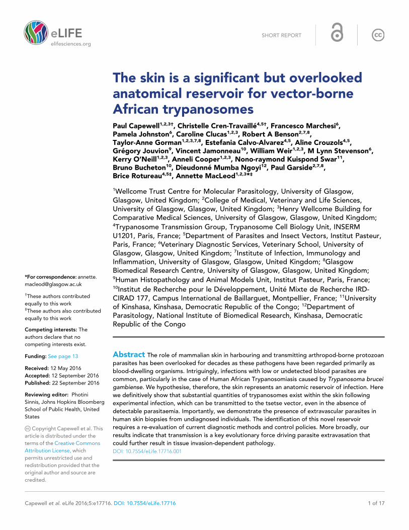
observed in the skin 12 days post-infection and remained throughout the experiment Skin parasite
numbers fluctuated to a lesser extent than blood parasitaemia and the apparent periodicity in the
skin may be due to one particularly high data point on day 24 (Figure 1) Parasites were found in the
dermis subcutaneous adipose tissue (Figure 2) and in fascia beneath the panniculus carnosus mus-
cle We did not detect any particular clustering around dermal adipocytes The presence of parasites
in the skin was not associated with major inflammation (Supplementary file 1) To confirm that skin
invasion by this parasite was not strain or sub-species specific the more virulent TREU927 strain of
T brucei and the human-infective Tb gambiense strain PA were used to infect mice Extravascular
skin invasion of the dermis subcutaneous adipose tissue and fascial planes (Figure 2mdashfigure
Capewell et al eLife 20165e17716 DOI 107554eLife17716 2 of 17
Short report Epidemiology and Global Health Microbiology and Infectious Disease
supplement 1) was evident with associated mild to moderate inflammation (Supplementary file 2)
in some instances the degree of skin invasion in the Tb gambiense infected mice was far greater
than Tb brucei perhaps suggesting a greater propensity for sequestration in this sub-species
To confirm that the extravascular distribution of parasites was not an artefact of the route of inoc-
ulation infections by natural vector transmission were carried out using a bioluminescent Tb brucei
strain AnTat11E AMLuctdTomato Mice were infected by a single infective bite of an individual G
m morsitans After 4 to 11 days and up to the end of the experiment parasites were observed in
the skin with a dynamic distribution (Figure 3A) and a variable density (Figure 3B) Parasites were
first detected in the blood between 5 and 19 days after natural transmission and parasitaemia
remained lower than 10 (Kanmogne et al 1996) parasitesml Observed bioluminescence directly
reflects the total number of living parasites in the entire organism including blood and viscera but
the intensity of the signal decreases with tissue depth Therefore at the end of each experiment
mice were sacrificed and their organs were checked for bioluminescence The presence of extravas-
cular parasites in cutaneous and subcutaneous tissues was first demonstrated by bioluminescence
imaging in entire dissected skins (Figure 3C) and was not necessarily localised to the bite site In
addition to the skin only the spleen some lymph nodes and adipose tissue were observed to be
positive for bioluminescence in several individuals (Figure 3mdashfigure supplement 1) This suggests
that the observed bioluminescence is likely to originate predominantly but not solely from parasites
in the skin In addition only mild inflammation was observed after 29 days (Figure 3mdashfigure supple-
ment 2)
To confirm that trypanosomes in the skin are a viable population fluorescent parasites were mon-
itored by two intravital imaging methods following IP injection and natural transmission (Figure 4)
First IP-injected fluorescent T brucei STIB247 were imaged in vivo using 2-photon microscopy
(Figure 4A) Extravascular trypanosomes observed in the dermal layer of dorsal skin were highly
motile consistent with viability (Video 1) Second naturally-transmitted fluorescent T brucei
AnTat11E AMLuctdTomato were imaged in vivo using spinning-disk confocal microscopy in the
C57BL6J-Flk1-EGFP mouse line that has green fluorescent endothelial cells in the lymphatic and
Figure 1 Parasite densities in the blood and in the extravascular tissue of the skin over a time-course The blood
parasitaemia of Tb brucei strain STIB247 (red) and the semi-quantitative score of extravascular parasites in the
skin (blue) are shown over a 36-day time-course following infection in BalbC mice Blood parasitaemia was
measured using phase microscopy using methodology outlined in (Lumsden 1963) Skin parasite burden is an
average of five high-power fields scored by histological analysis (0 = no parasites detectable 1 = low numbers of
parasites 2 = moderate numbers of parasites 3 = large numbers of parasites) Standard error shown (n = 3)
DOI 107554eLife17716002
The following source data is available for figure 1
Source data 1 Semi-quantitative evaluation of the parasite burden in skin sections (STIB247) Every three days for
36 days of a STIB247 Tb
DOI 107554eLife17716003
Source data 2 Daily parasitaemia during STIB247 infection in BalbC mice The daily parasitaemia during a 36-day
STIB247 Tb
DOI 107554eLife17716004
Capewell et al eLife 20165e17716 DOI 107554eLife17716 3 of 17
Short report Epidemiology and Global Health Microbiology and Infectious Disease
blood vessels (Ema et al 2006) (Figure 4B) Extravascular trypanosomes were observed in the der-
mal layer of the ear and were highly motile (Videos 2 3 and 4)
Differentiation of dividing trypanosomes to non-dividing stumpy forms is essential for transmis-
sion to the tsetse Using the relative transcript abundance of the stumpy marker Protein Associated
with Differentiation 1 (PAD1) (Dean et al 2009) to an endogenous control Zinc Finger Protein 3
(ZFP3) (Walrad et al 2009) we estimated that approximately 20 of skin-dwelling parasites were
stumpy (Supplementary file 3) To directly determine the proportion of stumpy forms in skin sec-
tions histological staining for PAD1 was also performed (Table 1mdashsource data 4) PAD1-positive
cells were observed in variable proportions (from 8 to 80) in all bioluminescent skin samples exam-
ined after IP injection (Supplementary file 4) Following natural transmission up to 38 of parasites
detected using VSG surface markers (Figure 3mdashfigure supplement 3AndashB) also expressed PAD1
(Figure 3mdashfigure supplement 3CndashD) (n = 441 cells from 8 skin sections) In all skin sections stumpy
parasites were homogenously distributed in the dermis and subcutaneous adipose tissues
We next assessed the ability of skin-dwelling parasites to infect tsetse flies Teneral flies (imma-
ture flies that have not yet taken a blood meal) were fed on different regions of skin from mice
infected with AnTat11E AMLucTY1tdTomato with differing levels of bioluminescence across the
skin (Table 1mdashsource data 1 and Table 1mdashsource data 2) This was repeated in 20 mice with differ-
ing levels of parasitaemia Flies were dissected and checked for the presence of fluorescent trypano-
somes after two days (Table 1) In mice with undetectable or low parasitaemia (lt5 104 parasites
Figure 2 Extravascular localisation of trypanosomes during an infection Histological sections of dorsal skin from
uninfected and infected BalbC mice stained with trypanosome-specific anti-ISG65 antibody (brown)
counterstained with Gillrsquos Haematoxylin stain (blue) at 12 days and 24 days post-inoculation with Tb brucei strain
STIB247 Parasites are visible in extravascular locations of the skin including the deep dermis and subcutaneous
adipose tissue from day 12 The scale bar represents 20 mm
DOI 107554eLife17716005
The following figure supplement is available for figure 2
Figure supplement 1 Skin invasion by Tb brucei strain TREU927 and Tb gambiense strain PA
DOI 107554eLife17716006
Capewell et al eLife 20165e17716 DOI 107554eLife17716 4 of 17
Short report Epidemiology and Global Health Microbiology and Infectious Disease
Figure 3 Dynamics of parasite distribution in the extravascular tissue of the skin and in the blood during a representative course of infection following
natural transmission A total of seven mice were infected by the single infective bite of an individual Gm morsitans on the belly with the Tb brucei
AnTat11E AMLuctdTomato strain Panels A and C depict representative patterns (A) Examples of bioluminescence profiles of 3 mice (+ bitten by an
infected fly - bitten by an uninfected fly and 0 not bitten) 6 12 20 and 26 days after the bite are shown (B) Ventral (blue) and dorsal (green)
bioluminescence (BL) intensities (in pscm2sr on the left Y-axis) and parasitaemia (in parasitesml in red on the right Y-axis) were measured daily for 29
days and plotted as mean plusmn SD (n = 7 mice) (C) The entire skins of mice (+) and (-) were dissected for bioluminescence imaging 29 days after the bite
For the mouse (+) Figure 3mdashfigure supplement 1 shows the bioluminescence profile of dissected organs Figure 3mdashfigure supplement 2 presents
the skin inflammation and Figure 3mdashfigure supplement 3 shows labelled parasites in skin sections
DOI 107554eLife17716007
The following figure supplements are available for figure 3
Figure supplement 1 Bioluminescence mostly originates from parasites in the skin
DOI 107554eLife17716008
Figure supplement 2 Mild inflammation of skin tissues one month after an infection by natural transmission
DOI 107554eLife17716009
Figure supplement 3 Extravascular parasites in the skin express both VSGs and PAD1 surface markers
DOI 107554eLife17716010
Capewell et al eLife 20165e17716 DOI 107554eLife17716 5 of 17
Short report Epidemiology and Global Health Microbiology and Infectious Disease
ml) no parasites were found in flies fed on non-
bioluminescent regions Conversely a median of
36 (plusmn 23 n = 70) of flies that fed on bioluminescent regions of low parasitaemic mice became
infected (Table 1 and Table 1mdashsource data 3) This demonstrates that skin parasites from mice
without visible parasitaemia can contribute to tsetse infection possibly reflecting human asymptom-
atic infections When parasites were detected in the blood and skin tsetse infection rates increased
up to 100 (median of 61 plusmn 22 n = 120) (Table 1 and Table 1mdashsource data 3) Parasites taken-
up from the skin were able to further develop through the life-cycle to early procyclic forms in the fly
as evidenced by GPEET-procyclin marker on the parasites surface (n = 721 cells from 16 flies)
(Table 1mdashsource data 4 and Supplementary file 4)
To demonstrate that skin invasion by trypanosomes can occur in humans as well as the murine
model slides of historical skin biopsies taken as part of a diagnostic screening programme for
Onchocerca microfilaria in the trypanosomiasis-endemic Democratic Republic of the Congo were
examined for trypanosomes At the time of sampling the incidence of trypanosomiasis was 15ndash2
and we hypothesised that some individuals may have harboured undiagnosed infections Re-exami-
nation of 1121 skin biopsies by microscopy revealed six individuals with trypanosomes in their skin
(Figure 5) These individuals had not previously been diagnosed with human trypanosomiasis by clin-
ical signs or the presence of blood parasites
DiscussionWe have shown that there exists a significant yet overlooked population of live motile extravascular
T brucei in the dermis and subcutis of animal models infected by artificial routes or by vector trans-
mission It is likely that once injected the parasites disseminate via the lymph and blood to the skin
where they are ingested during tsetse fly pool-feeding and readily initiate cyclical development in
the vector Given the relative volume of the skin organ compared to the vasculature it is possible
depending on the density of skin invasion that more parasites exist in the skin than in the blood
Figure 4 Extravascular localisation of trypanosomes
during an infection visualised using multi-photon
microscopy (A) and spinning-disk confocal microscopy
(B) (A) Still-image extracted from video (Video 1) of
multi-photon live imaging of dorsal skin during a
trypanosome infection Intravenous non-targeted
quantum dots (white) highlight blood vessels Tb
brucei STIB 247 parasites transfected with mCherry to
aid visualisation (red) are clearly visible and motile
outside the vasculature and within the extravascular
skin matrix (green) (B) Still-image extracted from
(Video 3) of spinning-disk confocal live imaging of the
ear of an Kdr (Flk1) C57BL6J Rj mouse during a
trypanosome infection Tb brucei AnTat11E AMLuc
tdTomato parasites expressing tdTomato (red) are
moving in the extravascular region surrounding a vessel
of the dermis (green)
DOI 107554eLife17716011
Video 1 Extravascular trypanosomes visualised in the
skin using 2-photon microscopy Intravital multi-photon
imaging of the flank skin during trypanosome infection
10 days after IP inoculation An intravenous injection of
non-targeted quantum dots prior to imaging allowed
visualisation of blood vessels mCherry STIB247 Tb
brucei parasites (red) are observed moving in the
extravascular region surrounding blood vessels of the
dermis (white) Collagen auto-fluorescence is visible as
green
DOI 107554eLife17716012
Capewell et al eLife 20165e17716 DOI 107554eLife17716 6 of 17
Short report Epidemiology and Global Health Microbiology and Infectious Disease
The skin therefore represents an unappreciated
reservoir of infection The extravasation of trypa-
nosomes was described previously in major
organs such as liver spleen (Blum et al 2008
Kennedy 2004) and visceral adipose tissue
(Trindade et al 2016) but the importance of
these parasites in transmission was not investi-
gated Here we show that these skin-dwelling try-
panosomes contribute to transmission and could
explain the maintenance of disease foci despite
active screening and treatment Skin invasion for
enhanced transmission is likely a powerful evolu-
tionary force driving extravasation suggesting
that the generalised tissue penetration underlying
pathogenesis (ie splenomegaly hepatomegaly
CNS invasion) is a secondary epiphenomenon A
skin reservoir also presents a novel target for
diagnostics (eg skin biopsies) allowing the prev-
alence of infection to be accurately determined
and the identification of any previously unde-
tected animal reservoirs of human disease
The skin as an anatomical reservoir of parasites
is a recurring theme in arthropod-borne human
diseases such as Leishmania (Sacks 2008
Schlein 1993) and Onchocerca (Symptomatol-
ogy 1974 Dalmat 1955) Here we present evi-
dence of trypanosomes in the skin of hitherto
undiagnosed individuals This anatomical reservoir
may serve to explain how HAT foci re-emerge and
persist despite low numbers of reported cases even in the absence of an animal reservoir
(Kagbadouno et al 2012 Cordon-Obras et al 2009 Balyeidhusa et al 2012)
Video 2 Extravascular trypanosomes visualised in the
skin using spinning-disk confocal microscopy Spinning-
disk confocal live imaging of the ear of anKdr (Flk1)
C57BL6J Rj mouse during a trypanosome infection
after natural transmission Tb brucei AnTat11E
AMLuctdTomato parasites expressing tdTomato (red)
are observed moving in the extravascular region
surrounding blood and or lymphatic vessels of the
dermis (green)
DOI 107554eLife17716013
Video 3 Extravascular trypanosomes visualised in the
skin using spinning-disk confocal microscopy
DOI 107554eLife17716014
Video 4 Extravascular trypanosomes visualised in the
skin using spinning-disk confocal microscopy
DOI 107554eLife17716015
Capewell et al eLife 20165e17716 DOI 107554eLife17716 7 of 17
Short report Epidemiology and Global Health Microbiology and Infectious Disease
Table 1 Skin parasites are ingested during tsetse pool-feeding Mice were IP infected with Tb brucei AnTat11E AMLucTY1
tdTomato and the parasitaemia and bioluminescence were monitored daily until the day of xenodiagnosis The number of parasites in
the blood was determined using a haemocytometer or a flux cytometer The number of parasites in the skin was estimated from the
measured bioluminescence intensity by using a standard curve (Table 1mdashsource data 1 and Table 1mdashsource data 2) Batches of
teneral flies were fed on different skin regions of mice infected with differing levels of bioluminescence across the skin and with
differing levels of parasitaemia (Table 1mdashsource data 2 and Table 1mdashsource data 3) Fly batches A4A A2A B1A 3B and 3A were
used to assess tsetse transmission in hosts with low numbers of blood parasites but high numbers skin parasites while fly batches 1B
1A 4B 4A 2B 2A B2B and B4A were used to investigate the impact of high numbers of parasites in both the skin and blood Flies
were dissected and their midguts checked for the presence of fluorescent trypanosomes after two days to determine the proportion
of infected flies (Table 1mdashsource data 4AndashB) For some of these experiments results of an in-depth quantification of parasite stages
by IFA is provided in Supplementary file 4 Stumpy forms were observed only in the blood of mice with parasitaemia values
highlighted in purple Bioluminescence was detected in the skin of mice with values highlighted in grey
Fly batches Parasites in blood (per ml) Parasites in skin (per cm2) Dissected flies Fly infection rates ()
C1 0 0 32 0
C 0 0 8 0
A4B lt 104 lt 103 16 0
B1B 22 104 lt 103 13 0
A4A lt 104 66 105 17 35
A2A 11 104 38 106 7 86
B1A 22 104 46 107 16 31
3B 44 104 26 104 14 36
3A 44 104 26 104 16 38
1B 18 105 80 103 12 67
1A 18 105 80 103 14 79
4B 22 105 12 104 17 53
4A 22 105 12 104 18 56
2B 16 106 80 103 14 36
2A 16 106 32 104 18 39
B4B 43 106 67 107 10 80
B4A 43 106 67 107 17 100
DOI 107554eLife17716016
Source data 1 Characterisation of the AnTat 11E AMLucTY1tdTomato sub-clone (A) The in vitro growth of the selected AnTat11E AMLucTY1tdTo-
mato sub-clone (red) was similar to that of the parental wild-type strain (blue) Bloodstream forms were cultured in HMI11 counted daily in a Muse
cytometer (Merck-Millipore) and diluted after 4 days (B) A parasite density bioluminescence intensity analysis was performed by measuring the biolu-
minescence in successive 2-fold dilutions in 96-micro-well plates with an IVIS Spectrum imager (Perkin Elmer) When plotted as mean plusmn SD (n = 3) para-
site densities and bioluminescence intensities were correlated when the bioluminescence levels were higher than 10 (Berthier et al 2016) pscm2sr
corresponding to about 10 (Koffi et al 2006) parasites allowing estimation of the parasite density from in vivo imaging over this threshold This stan-
dard curve was used to estimate the number of parasites in the skin from measured values of bioluminescence (C) This correlation was verified by quan-
tification in a microplate reader Infinite 200 (Tecan) at the very beginning of the first in vivo experiment as well as the end of the last one (mean plusmn SD
n = 3)
DOI 107554eLife17716017
Source data 2 Parasite densities in extravascular tissue of the skin and in the blood of mice used for differential xenodiagnosis Mice were injected IP
with AnTat11E AMLucTY1tdTomato and monitored daily for bioluminescence and parasitaemia (A) Bioluminescence profile of four mice (- uninfected
control and (1ndash3) three infected mice) four days after infection (B) The entire skins of the uninfected control mouse (-) and mouse 3 were dissected for
bioluminescence imaging four days after infection (C) Parasite densities in the blood and in the skin (calculated from the mean dorsal bioluminescence
intensity measurement and from the standard curve in Source data 1 in parasitescm (Fakhar 2013) in blue) were calculated daily over one week and
plotted as mean plusmn SD (n = 13 mice)
DOI 107554eLife17716018
Source data 3 Skin parasites are sufficient to initiate a tsetse infection Schematics summarising the principal results from the xenodiagnosis experi-
ment In a mouse with no detected transmissible parasites in the blood (absence of stumpy forms by IFA and absence of infection of flies fed on a non-
bioluminescent region of the skin) flies can ingest transmissible parasites from the bioluminescent region of the skin (left panel) When a mouse
Capewell et al eLife 20165e17716 DOI 107554eLife17716 8 of 17
Short report Epidemiology and Global Health Microbiology and Infectious Disease
HAT was once widespread across much of sub-Saharan Africa but concerted control efforts
brought it close to elimination during the 1960s (World Health Organization 2000 2013) How-
ever disease foci persisted with a resurgence in the number of reported cases to over 300000 in
the 1990s (World Health Organization 2000 Steverding 2008) Currently HAT is approaching
elimination in many areas (World Health Organization 2013 Simarro et al 2012) Understanding
how HAT evaded elimination in the latter half of the 20th century and how it continues to persist is
vital to efforts to eliminate the disease For example our results indicate that it may be necessary to
develop novel therapeutics capable of accessing extravascular compartments The current policy in
most endemic countries is not to treat serologically positive individuals unless they demonstrate
active infection due to the long duration and high toxicity of treatment and the low predictive value
of the serological tests We suggest that this policy should be reconsidered in light of our compel-
ling evidence that they represent a carrier population which may maintain HAT foci and explain pre-
viously thwarted efforts to eliminate this major pathogen
Materials and methods
Trypanosome strains and culturesAll strains used in this study are pleomorphic STIB247 is a low virulence Tb brucei strain that indu-
ces a chronic infection and was isolated from a hartebeest in the Serengeti in 1971 (Geigy et al
1973) TREU927 is the genome reference strain for T brucei and is more virulent than STIB247 This
strain was isolated from a tsetse fly in Kenya in 19691970 (Goedbloed et al 1973) PA is a human-
infective group 1 Tb gambiense strain isolated from a patient in the Democratic Republic of the
Congo in 1975 (Tait et al 1984) mCherry STIB247 was created by transfection of STIB247 with
pHD1034-mCherry (Myburgh et al 2013) This strain constitutively expresses fluorescent mCherry
from the ribosomal RNA promoter and its expression is stable over repeated passage
The AnTat 11E clone of Tb brucei was derived from a strain originally isolated from a bushbuck
in Uganda in 1966 (Le Ray et al 1977) Bloodstream forms were cultivated in HMI11 medium sup-
plemented with 10 foetal calf serum at 37˚C in 5 CO2 This strain was genetically engineered to
produce two strains continuously expressing the red-shifted luciferase (PpyRE9H) and the tdTomato
red fluorescent protein either individually or in combination
For the first AnTat 11E strain the pTbAMLuc plasmid (M Taylor London School of Hygiene and
Tropical Medicine UK) was used for continuous cytosolic expression of the red-shifted luciferase
PpyRE9H The tdTomato coding sequence was cloned with HindIII and BamHI into the pTSARib
Figure 5 Extravascular localisation of trypanosomes in previously unidentified human cases of trypanosomiasis Histological sections of skin collected
from previously unidentified cases of human trypanosomiasis from the Democratic Republic of Congo showing the presence of extravascular parasites
in biopsies from three individuals (A B and C) Skin biopsies were collected as part of a national onchocerciasis screening programme that took place
in the same geographic region as an active trypanosomiasis focus Slides were stained with Giemsa and examined under oil immersion at 100x
magnification In addition to visible slender forms (black arrows) in the extravascular tissue of the skin a clearly identifiable stumpy transmission form
with typical morphology and an unattached undulating membrane is also present in the skin of one individual (red arrow in A) The scale bar represents
5 mm
DOI 107554eLife17716021
Capewell et al eLife 20165e17716 DOI 107554eLife17716 9 of 17
Short report Epidemiology and Global Health Microbiology and Infectious Disease
vector (Xong et al 1998) generating the final pTSARib-tdTomato construct The two plasmids
were linearised with KpnI and SphI restriction enzymes respectively and used to transform procyclic
parasites with an Amaxa Nucleofector (Lonza) (Burkard et al 2007) After 24 hr transfected cells
were selected by the addition of blasticidin or puromycin (10 mgml) After one week the population
was examined (i) for red fluorescence by fluorescence microscopy and (ii) for both red fluorescence
and bioluminescence by using a fluorimeter Infinite 200 (Tecan Switzerland) Cells were sub-cloned
by limiting dilution and clone selection was performed after 15 days by measuring both biolumines-
cence in a microplate reader Infinite 200 (Tecan) and fluorescence with a Muse cell Analyzer (Merck-
Millipore) This strain named AnTat11E AMLuctdTomato was used for natural transmission
experiments
The second AnTat 11E strain expressing the 31 Kb chimeric multiplex reporter protein
PpyRE9HTY1tdTomato was named AnTat 11E AMLucTY1tdTomato This cytoplasmic reporter
is composed of three distinct markers the red-shifted luciferase (PpyRE9H) is fused to the tdTomato
red fluorescent protein by a TY1 tag Briefly the 168 Kb optimised version of the North American
firefly Photinus pyralis luciferase (Branchini et al 2005) was fused with a 10-bp sequence known as
TY1-tag (Bastin et al 1996) and cloned into the pTSARib plasmid (Xong et al 1998) by using
XhoI and HindIII restriction enzymes to obtain the pTSARib-PpyRE9H-TY1 plasmid Finally the 14
Kb coding region of the tdTomato fluorescent protein was inserted downstream using HindIII and
BamHI The resulting 89 Kb vector containing a blasticidin S resistance cassette was linearised with
SphI to integrate the rDNA promoter locus Bloodstream parasites were transformed with an Amaxa
Nucleofector (Lonza) (Burkard et al 2007) sub-cloned by limiting dilution and clone selection was
performed by measuring both bioluminescence in a microplate reader Infinite 200 (Tecan) and fluo-
rescence with a Muse cell Analyzer (Merck-Millipore) The selected AnTat 11E AMLucTY1tdTo-
mato sub-clone was comparable to the parental wild-type strain in terms of growth rate (Table 1mdash
source data 1A) pleomorphism (Table 1mdashsource data 4C) tsetse infectivity and virulence in mice
(Table 1mdashsource data 2) In order to verify the reliability of the bioluminescent marker as well as to
define the bioluminescence detection threshold of the AnTat 11E AMLucTY1tdTomato selected
sub-clone a parasite density bioluminescence intensity analysis was performed in 96-micro-well
plates with an IVIS Spectrum imager (Perkin Elmer) Parasite density and bioluminescence intensity
were correlated when bioluminescence levels were higher than 104 pscm2sr corresponding to
about 103 parasites allowing estimation of the parasite density from in vivo imaging over this thresh-
old (Table 1mdashsource data 1B) This correlation was verified by quantification in a microplate reader
Infinite 200 (Tecan) at the very beginning of the first in vivo experiment as well as the end of the last
one demonstrating the stability of the triple reporter expression in the AnTat 11E AMLucTY1
tdTomato selected sub-clone over time especially after at least one full in vivo parasite cycle in the
tsetse fly and the mammalian host (Table 1mdashsource data 1C) This strain was used for xenodiagno-
sis experiments and quantification of parasite densities
Mouse strainsBALBc and C57BL6J mice were used as models for chronic disease (Magez and Caljon 2011) In
addition to allow for further 3D intravital imaging of the lymphatic and blood systems C57BL6J-
Flk1-EGFP mice expressing a GFP tagged Kdr (Flk1) gene encoding the vascular endothelial growth
factor receptor 2 (VEGFR-2) were used (Ema et al 2006)
Ethical statementsThis study was conducted under Home Office and SAPO regulations in the UK and in strict accor-
dance with the recommendations from the Guide for the Care and Use of Laboratory Animals of the
European Union (European Directive 201063UE) and the French Government The protocol was
approved by the rsquoComite drsquoethique en experimentation animale de lrsquoInstitut Pasteurrsquo CETEA 89 (Per-
mit number 2012ndash0043 and 2016ndash0017) and undertaken in compliance with Institut Pasteur Bio-
safety Committee (protocol CHSCT 12131)
Skin invasion time-courseA total of 36 eight-week old BALBc mice (Harlan UK) were inoculated by intra-peritoneal (IP) injec-
tion with 104 parasites of strain STIB 247 Parasitaemia was assayed on each subsequent day using
Capewell et al eLife 20165e17716 DOI 107554eLife17716 10 of 17
Short report Epidemiology and Global Health Microbiology and Infectious Disease
phase microscopy (Lumsden 1963) Twenty-four uninfected animals served as controls Every three
days for 36 days three infected animals and two uninfected animals were culled and 2 cm2 skin sam-
ples removed from the dorsum Skin samples were fixed in 10 neutral buffered formalin prior to
histological analysis
Natural infections using tsetse fliesTsetse flies (Glossina morsitans morsitans) were maintained infected and dissected at the Institut
Pasteur as described previously (Rotureau et al 2012) Flies were infected with AnTat11E AMLuc
tdTomato parasites Positive flies were selected first by screening the abdominal fluorescence (mid-
gut infection) 15 days after the infective meal and then by a salivation test (mature salivary gland
infection) after one month Single flies with salivary gland infections were used to infect the abdo-
men of mice anaesthetised by IP injection of ketamine (Imalgene 1000 at 125 mgkg) and xylazine
(Rompun 2 at 125 mgkg) and feeding was confirmed by visual observation of the fly abdomen
full of blood Control mice were either not bitten or bitten by uninfected flies The presence and
density of parasites in the blood was determined daily by automated fluorescent cell counting with a
Muse cytometer (Merck-Milllipore detection limit 5102 parasitesml) or by direct examination under
a phase microscope with standardised one-use haemocytometers (Hycor Kova detection limit 104
parasitesml) according to the manufacturerrsquos recommendations
Infection for xenodiagnosisA total of 20 seven-week-old male C57BL6J Rj mice (Janvier France) were IP injected with 10
(Jamonneau et al 2012) AnTat 11E AMLucTY1tdTomato bloodstream forms Parasitaemia was
assayed daily by automated fluorescent cell counting with a Muse cytometer (Merck-Millipore
detection limit 5102 parasitesml) according to the manufacturerrsquos recommendations
PAD1ZFP3 relative expressionThree BALBc were infected with Tb brucei strain TREU927 and culled at day 11 The mice were
perfused and a 2 cm2 region of skin removed from the flank Skin sections were lysed using a Qiagen
Tissuelyzer LT and RNA extracted using a Qiagen RNAeasy kit following the manufacturerrsquos instruc-
tions 100 ng of RNA from each sample was reverse-transcribed using an Invitrogen Superscript III
RT kit qPCR was performed on each sample using 5 ml of cDNA using a protocol and primers vali-
dated previously (MacGregor et al 2011) on an Agilent Technologies Stratagene Mx3005P qPCR
machine The ratio of PAD1 to ZFP3 and hence the proportion of cells transcribing the PAD1 gene
was estimated using the Agilent Technologies MXPro software
XenodiagnosisMice were first anaesthetised by IP injection of ketamine (Imalgene 1000 at 125 mgkg) and xylazine
(Rompun 2 at 125 mgkg) Batches of 10 teneral male tsetse flies (from 8 to 24 hr post-eclosion)
were then placed in 50 ml Falcon tubes closed with a piece of net through which they were allowed
to feed directly on mouse skin regions of interest for 10 min The selection of the skin regions for fly
feeding was based on mice bioluminescence profiles and parasitaemia Unfed flies were discarded
and fed flies were maintained as previously described Anaesthetised mice were finally sacrificed by
cervical dislocation and their skin was dissected for controlling bioluminescence with an IVIS Spec-
trum imager (Perkin Elmer) All the flies were dissected and checked for the presence of trypano-
somes either 2 or 14 days after their meal on mouse skin by two entomologists blinded to group
assignment and experimental procedures Dissections were performed as previously described
(Rotureau et al 2012) entire midguts were scrutinised by fluorescence microscopy to detect and
count living red fluorescent parasites and positive midguts were further treated for IFA A total of
420 flies were used in 3 independent xenodiagnosis experiments
In vitro bioluminescence imagingTo perform the parasite density bioluminescence intensity assay with AnTat 11E AMLucTY1tdTo-
mato bloodstream forms parasites were counted centrifuged and resuspended in fresh HMI11
medium at 10106 cellsml Then 100 ml (or 106 parasites) of this suspension were transferred into
black clear-bottom 96-well plates and serial 2-fold dilutions were performed in triplicate adjusting
Capewell et al eLife 20165e17716 DOI 107554eLife17716 11 of 17
Short report Epidemiology and Global Health Microbiology and Infectious Disease
the final volume to 200 ml of HMI11 with 300 mgml of beetle luciferin (Promega France) Luciferase
activity was quantified after 10 min of incubation with a microplate reader Infinite 200 (Tecan) fol-
lowing the instructions of the Promega Luciferase Assay System After background removal results
were analysed as mean plusmn SD of three independent experiments
In vivo bioluminescence imagingInfection with bioluminescent parasites was monitored daily by detecting the bioluminescence in
whole animals with the IVIS Spectrum imager (Perkin Elmer) The equipment consists of a cooled
charge-coupled camera mounted on a light-tight chamber with a nose cone delivery device to keep
the mice anaesthetised during image acquisition with 15 isofluorane D-luciferin potassium salt
(Promega) stock solution was prepared in phosphate buffered saline (PBS) at 3333 mgml filter-
sterilised and stored in a 20˚C freezer To produce bioluminescence mice were inoculated IP with
150 ml of D-luciferin stock solution (250 mgkg) After 10 min of incubation to allow substrate dis-
semination all mice were anaesthetised in an oxygen-rich induction chamber with 2 isofluorane
and images were acquired by using automatic exposure (30 s to 5 min) depending on signal inten-
sity Images were analysed with Living Image software version 431 (Perkin Elmer) Data were
expressed in average radiance (pscm2sr) corresponding to the total flux of bioluminescent signal
according to the selected area (total body of the mouse here) The background noise was removed
by subtracting the bioluminescent signal of the control mouse from the infected ones for each
acquisition
2-photon microscopyIntravital multi-photon microscopy studies were carried out using a Zeiss LSM7 MP system equipped
with a tuneable titaniumsapphire solid-state two-photon excitation source (4W Chameleon Ultra II
Coherent Laser Group) coupled to an Optical Parametric Oscillator (Chameleon Compact OPO
Coherent) Movies were acquired for 10 to 15 min with an XY pixel resolution of 512 512 in 2 mm
Z increments producing up to 40 mm stacks 3D tracking was performed using Volocity 611 (Perkin
Elmer Cambridge UK) Values representing the mean velocity displacement and meandering index
were calculated for each object Mice were anaesthetised IP using medetomidine (Domitor 05 mg
kg) and ketamine (50 mgkg) and placed on a heated stage Following removal of hair with a depila-
tory cream dorsal skin was imaged An intravenous injection of non-targeted quantum dots
(Qdot705) (Life Technologies UK) prior to imaging allowed visualisation of blood vessels
Spinning-disk confocal microscopyAnTat11E AMLuctdTomato parasites were monitored in the ear of Kdr (Flk1) C57BL6J Rj mice by
spinning-disk confocal microscopy as described previously (Rotureau et al 2012) Briefly mice
were first anaesthetised by IP injection of ketamine (Imalgene 1000 at 125 mgkg) and xylazine
(Rompun 2 at 125 mgkg) Mice were wrapped in a heating blanket and placed on an aluminium
platform with a central round opening of 21 mm in diameter A coverslip was taped on the central
hole and the mouse was positioned so that the ear was lying on this oiled coverslip Imaging was
performed using an UltraView ERS spinning-disk confocal system (Perkin Elmer) with a x40 oil objec-
tive (13 numerical aperture) Movies were acquired by an EM-CCD camera (Hamamatsu) controlled
by the Volocity software (Perkin Elmer) with an exposure time of 500 ms for a total of 30 to 120 s
Images were analysed using ImageJ 148v and its plugin Bio-formats importer (NIH)
Histological and immunohistochemical evaluation of the skinParaformaldehyde-fixed skin samples were trimmed and processed into paraffin blocks Sections
were stained with Haematoxylin and Eosin (HE) Additional serial sections were processed for immu-
nohistochemical staining using a polyclonal rabbit antibody raised against the invariant surface gly-
coprotein 65 (IGS65) (M Carrington Cambridge UK) using a Dako Autostainer Link 48 (Dako
Denmark) and were subsequently counterstained with Gillrsquos Haematoxylin
Histopathological assessment of inflammation in the skinThe extent of cutaneous inflammatory cell infiltration was assessed in haematoxylin and eosin stained
sections with a semi-quantitative scoring system applied by two pathologists blinded to group
Capewell et al eLife 20165e17716 DOI 107554eLife17716 12 of 17
Short report Epidemiology and Global Health Microbiology and Infectious Disease
assignment and experimental procedures The extent of mixed inflammatory cell infiltration in the
dermis andor subcutis was assessed on a 0 to 3 grading scale (0 = no inflammation or only few scat-
tered leukocytes 1 = low numbers of inflammatory cells 2 = moderate numbers of inflammatory
cells 3 = large numbers of inflammatory cells) Ten high-power fields were scored for each skin sam-
ple An inflammation score calculated as the average of the scores in the 10 high-power fields was
determined for each sample
Semi-quantitative evaluation of the parasite burden in skin sectionsParasite burden was assessed in skin sections stained with anti-IGS65 antibody by two pathologists
blinded to group assignment and experimental procedures Presence of parasites defined as intra-
vascular (parasites within the lumen of dermal or subcutaneous small to medium-sized vessels) and
extravascular (parasites located outside blood vessels scattered in the connective tissue of the der-
mis or in the subcutis) was evaluated in 5 randomly selected high-power fields at x40 magnification
with a 0 to 3 semi-quantitative grading scale (0 = no parasites detectable 1 = low numbers of para-
sites (lt20) 2 = moderate numbers of parasites (20 lt 50) 3 = large numbers of parasites (gt50) An
average parasite burden score was calculated for each sample
Immunofluorescence analysisCells were treated for immunofluorescence after paraformaldehyde or methanol fixation as
described previously (Dean et al 2009) Parasites were stained with one or two of the following
antibodies (i) the anti-CRD polyclonal rabbit antibody (1300) to label the cross-reactive determinant
of the glycosylphosphatidylinositol anchors of proteins predominantly the variant surface glycopro-
teins (Zamze et al 1988) (ii) the anti-PAD1 polyclonal rabbit antibody (1100) targeting the carbox-
ylate-transporter Proteins Associated with Differentiation 1 (PAD1) (Keith Matthews Edinburgh UK)
(Dean et al 2009) (iii) the anti-GPEET mouse IgG3 monoclonal antibody (1500) targeting the T
brucei GPEET-rich procyclin (Acris Antibodies GmbH San Diego USA) (iv) the L8C4 mouse IgG1
monoclonal antibody labelling an epitope of the PFR2 protein (Kohl et al 1999) Specific antibod-
ies with minimal cross-reactions with mice and coupled to AlexaFluor 488 Cy3 or Cy5 (Jackson
ImmunoResearch USA) were used as secondary antibodies DNA was stained with 46-diamidino-2-
phenylindole (DAPI) IFA image acquisition was carried out on a Leica 4000B microscope with a x100
objective lens using a Hamamatsu ORCA-03G camera controlled by Micro-manager and images
were normalised and analysed with ImageJ 149v (NIH)
Histopathology of historical human skin samplesHistorical human skin samples were collected from 1991 to 1995 as part of The National Onchocerci-
asis Task Force (NOTF) (Makenga Bof et al 2015) Of this collection 1121 paraffin embedded skin
samples were cut into 25 micron sections and stained with Giemsa (Sigma-Aldrich) Slides were
screened for the presence of parasites by two pathologists independently and representative images
taken at x100 magnification
AcknowledgementsWe acknowledge P Solano D Engman K Matthews M Taylor M Carrington R Amino E Myburgh
and K Gull for providing various material cell lines antibodies and plasmids and to A Tait for criti-
cally reviewing this manuscript
Additional information
Funding
Funder Grant reference number Author
Wellcome Trust Senior Fellowship (AnnetteMacleod) - 095201Z10Z
Paul CapewellCaroline ClucasWilliam WeirAnneli CooperAnnette MacLeod
Capewell et al eLife 20165e17716 DOI 107554eLife17716 13 of 17
Short report Epidemiology and Global Health Microbiology and Infectious Disease
Wellcome Trust Wellcome Trust Centre forMolecular Parasitology CoreFunding - 085349
Paul CapewellCaroline ClucasWilliam WeirAnneli CooperAnnette MacLeod
Agence Nationale de la Re-cherche
Young Researcher Grant(ANR-14-CE14-0019-01)
Brice Rotureau
Agence Nationale de la Re-cherche
Post-doctoral fellowship(ANR-14-CE14-0019-01)
Estefania Calvo-Alvarez
Agence Nationale de la Re-cherche
Investissement drsquoAvenirprogramme LaboratoiredrsquoExcellence (ANR-10-LABX-62-IBEID)
Paul Capewell
Institut Pasteur Estefania Calvo-AlvarezAline CrouzolsGregory JouvionBrice Rotureau
Institut National de la Sante etde la Recherche Medicale
Estefania Calvo-AlvarezAline CrouzolsGregory JouvionBrice Rotureau
The funders had no role in study design data collection and interpretation or the decision tosubmit the work for publication
Author contributions
PC CC-T FM PJ CC RAB ACo N-rKS DMN Acquisition of data Analysis and interpretation of
data Drafting or revising the article T-AG ACr GJ MLS KO Acquisition of data Analysis and
interpretation of data EC-A Acquisition of data Analysis and interpretation of data VJ BB Acquisi-
tion of data WW Analysis and interpretation of data Drafting or revising the article PG Concep-
tion and design BR Conception and design Acquisition of data Analysis and interpretation of
data Drafting or revising the article AM Conception and design Analysis and interpretation of
data Drafting or revising the article
Author ORCIDs
Annette MacLeod httporcidorg0000-0002-0150-5049
Ethics
Animal experimentation This study was conducted and licenced under Home Office and SAPO reg-
ulations in the UK and in strict accordance with the recommendations from the Guide for the Care
and Use of Laboratory Animals of the European Union (European Directive 201063UE) and the
French Government The protocol was approved by the Comite drsquoethique en experimentation ani-
male de lrsquoInstitut Pasteur CETEA 89 (Permit number 2012-0043 and 2016-0017) and carried out in
compliance with Institut Pasteur Biosafety Committee (protocol CHSCT 12131)
Additional filesSupplementary files Supplementary file 1 Histopathological assessment of inflammation in the skin during STIB247
infection The extent of cutaneous inflammatory cell infiltration during the 36-day STIB247 experi-
ment was assessed on haematoxylin and eosin stained sections with a semi-quantitative scoring sys-
tem applied by two pathologists blinded to group assignment and experimental proceduresThe
extent of mixed inflammatory cell infiltration in the dermis andor subcutis was assessed on a 0 to 3
grading scale (0 = no inflammation or only few scattered leukocytes 1 = low numbers of inflamma-
tory cells 2 = moderate numbers of inflammatory cells 3 = large numbers of inflammatory cells)
Ten high-power fields (HPFs) were scored for each skin sample
DOI 107554eLife17716022
Capewell et al eLife 20165e17716 DOI 107554eLife17716 14 of 17
Short report Epidemiology and Global Health Microbiology and Infectious Disease
Supplementary file 2 Histopathological assessment of inflammation in the skin during TREU927
infection The extent of cutaneous inflammatory cell infiltration at day 10 of infection by strain
TREU927 experiment was assessed on haematoxylin and eosin stained sections with a semi-quantita-
tive scoring system applied by two pathologists blinded to group assignment and experimental pro-
cedures The extent of mixed inflammatory cell infiltration in the dermis andor subcutis was
assessed on a 0 to 3 grading scale (0 = no inflammation or only few scattered leukocytes 1 = low
numbers of inflammatory cells 2 = moderate numbers of inflammatory cells 3 = large numbers of
inflammatory cells) Ten high-power fields (HPFs) were scored for each skin sample
DOI 107554eLife17716023
Supplementary file 3 Expression of PAD1 relative to ZFP3 The relative abundance stumpy cells in
the skin of three BALBc was estimated using qPCR at day 11 post-inoculation with Tb brucei strain
TREU927 Mice were culled and perfused to remove blood parasites and a 2 cm (Fakhar 2013)
region of skin removed from the flank The tissue was homogenised and RNA extracted 100 ng of
RNA from each sample was reverse-transcribed and qPCR performed to estimate the cycle thresh-
olds (CT) of the stumpy marker PAD1 and the endogenous control ZFP3 As CT is inversely propor-
tional to amount of target cDNA in the sample and PAD1 and ZFP3 have similar qPCR efficiencies a
comparison of the delta (D) of CT between PAD1 and ZFP3 transcripts reveals the relative ratio of
PAD1 to ZFP3 transcripts and hence the proportion of differentiated parasites transcribing the
PAD1 gene
DOI 107554eLife17716024
Supplementary file 4 Both the respective densities and the proportions of transmissible forms of
parasites in the skin and in the blood govern the tsetse infection rates during pool feeding For
some of the xenodiagnosis experiments shown in Table 1 identification and quantification of para-
site stages was performed by IFA on blood smears and skin sectionsStumpy forms were observed
only in the blood of mice with parasitaemia values highlighted in light grey Bioluminescence was
detected in the skin of mice with values highlighted in dark grey The number of parasites in the skin
was calculated according to the values obtained in the standard in vitro assay (Table 1mdashsource data
1) and is therefore probably an underestimate Tsetse flies were dissected 2 days after xenodiagno-
sis Populations of intermediate and stumpy form cells were assessed in blood smears and in succes-
sive skin sections stained either with the anti-CRD antibody or the anti-PAD1 antibody (see
Materials and method section) Populations of early procyclic cells were assessed in dissected fly
midguts stained with the anti-GPEET antibody (see Materials and method section) ND not
determined
DOI 107554eLife17716025
ReferencesBalyeidhusa AS Kironde FA Enyaru JC 2012 Apparent lack of a domestic animal reservoir in Gambiensesleeping sickness in northwest Uganda Veterinary Parasitology 187157ndash167 doi 101016jvetpar201112005
Bastin P Bagherzadeh Z Matthews KR Gull K 1996 A novel epitope tag system to study protein targeting andorganelle biogenesis in Trypanosoma brucei Molecular and Biochemical Parasitology 77235ndash239 doi 1010160166-6851(96)02598-4
Berthier D Breniere SF Bras-Goncalves R Lemesre JL Jamonneau V Solano P Lejon V Thevenon S BuchetonB 2016 Tolerance to Trypanosomatids A threat or a key for disease elimination Trends in Parasitology 32157ndash168 doi 101016jpt201511001
Blum JA Zellweger MJ Burri C Hatz C 2008 Cardiac involvement in African and American trypanosomiasisThe Lancet Infectious Diseases 8631ndash641 doi 101016S1473-3099(08)70230-5
Branchini BR Southworth TL Khattak NF Michelini E Roda A 2005 Red- and green-emitting firefly luciferasemutants for bioluminescent reporter applications Analytical Biochemistry 345140ndash148 doi 101016jab200507015
Bucheton B MacLeod A Jamonneau V 2011 Human host determinants influencing the outcome ofTrypanosoma brucei gambiense infections Parasite Immunology 33438ndash447 doi 101111j1365-3024201101287x
Buck AA (Ed) 1974 Onchocerciasis Symptomatology Pathology Diagnosis Geneva WHOBurkard G Fragoso CM Roditi I 2007 Highly efficient stable transformation of bloodstream forms ofTrypanosoma brucei Molecular and Biochemical Parasitology 153220ndash223 doi 101016jmolbiopara200702008
Capewell et al eLife 20165e17716 DOI 107554eLife17716 15 of 17
Short report Epidemiology and Global Health Microbiology and Infectious Disease
Caljon G Van Reet N De Trez C Vermeersch M Perez-Morga D Van Den Abbeele J 2016 The Dermis as aDelivery Site of Trypanosoma brucei for Tsetse Flies PLoS Pathogens 12e1005744 doi 101371journalppat1005744
Cordon-Obras C Berzosa P Ndong-Mabale N Bobuakasi L Buatiche JN Ndongo-Asumu P Benito A Cano J2009 Trypanosoma brucei gambiense in domestic livestock of Kogo and Mbini foci (Equatorial Guinea)Tropical Medicine amp International Health 14535ndash541 doi 101111j1365-3156200902271x
Dalmat HT 1955 The black flies (Diptera Simuliidae) of Guatemala and their role as vectors of onchocerciasisSmithsonian Miscellaneous Collections 1251ndash425 doi 101093aibsbulletin5510-c
Dean S Marchetti R Kirk K Matthews KR 2009 A surface transporter family conveys the trypanosomedifferentiation signal Nature 459213ndash217 doi 101038nature07997
Ema M Takahashi S Rossant J 2006 Deletion of the selection cassette but not cis-acting elements in targetedFlk1-lacZ allele reveals Flk1 expression in multipotent mesodermal progenitors Blood 107111ndash117 doi 101182blood-2005-05-1970
Fakhar M Motazedian MH Hatam GR Asgari Q Kalantari M Mohebali M 2013 Asymptomatic human carriersof Leishmania infantum possible reservoirs for Mediterranean visceral leishmaniasis in southern Iran Annals ofTropical Medicine amp Parasitology 102577ndash583 doi 101179136485908X337526
Frezil JL 1971 [Application of xenodiagnosis in the detection of T gambiense trypanosomiasis inimmunologically suspect patients] Bulletin De La SocieTeDe Pathologie Exotique Et De Ses Filiales 64871ndash878
Geigy R Kauffmann M Jenni L 1973 Wild mammals as reservoirs for Rhodesian sleeping sickness in theSerengeti 1970-71 Transactions of the Royal Society of Tropical Medicine and Hygiene 67284ndash286 doi 1010160035-9203(73)90202-2
Goedbloed E Ligthart GS Minter DM Wilson AJ Dar FK Paris J 1973 Serological studies of trypanosomiasis inEast Africa II Comparisons of antigenic types of Trypanosoma brucei subgroup organisms isolated from wildtsetse flies Annals of Tropical Medicine and Parasitology 6731ndash43 doi 10108000034983197311686861
Goodwin LG 1971 Pathological effects of Trypanosoma brucei on small blood vessels in rabbit ear-chambersTransactions of the Royal Society of Tropical Medicine and Hygiene 6582ndash88 doi 1010160035-9203(71)90189-1
Jamonneau V Ilboudo H Kabore J Kaba D Koffi M Solano P Garcia A Courtin D Laveissiere C Lingue KBuscher P Bucheton B 2012 Untreated human infections by Trypanosoma brucei gambiense are not 100fatal PLoS Neglected Tropical Diseases 6e1691 doi 101371journalpntd0001691
Kagbadouno MS Camara M Rouamba J Rayaisse JB Traore IS Camara O Onikoyamou MF Courtin F RavelS de Meeus T Bucheton B Jamonneau V Solano P 2012 Epidemiology of sleeping sickness in Boffa (Guinea)where are the trypanosomes PLoS Neglected Tropical Diseases 6e1949 doi 101371journalpntd0001949
Kanmogne GD Asonganyi T Gibson WC 1996 Detection of Trypanosoma brucei gambiense in serologicallypositive but aparasitaemic sleeping-sickness suspects in Cameroon by PCR Annals of Tropical Medicine andParasitology 90475ndash483
Kennedy PG 2004 Human African trypanosomiasis of the CNS current issues and challenges Journal of ClinicalInvestigation 113496ndash504 doi 101172JCI21052
Koffi M Solano P Denizot M Courtin D Garcia A Lejon V Buscher P Cuny G Jamonneau V 2006Aparasitemic serological suspects in Trypanosoma brucei gambiense human African trypanosomiasis apotential human reservoir of parasites Acta Tropica 98183ndash188 doi 101016jactatropica200604001
Kohl L Sherwin T Gull K 1999 Assembly of the paraflagellar rod and the flagellum attachment zone complexduring the Trypanosoma brucei cell cycle Journal of Eukaryotic Microbiology 46105ndash109 doi 101111j1550-74081999tb04592x
Le Ray D Barry JD Easton C Vickerman K 1977 First tsetse fly transmission of the AnTat serodeme ofTrypanosoma brucei Annales De La SocieTeBelge De MeDecine Tropicale 57
Lindblade KA Steinhardt L Samuels A Kachur SP Slutsker L 2013 The silent threat asymptomatic parasitemiaand malaria transmission Expert Review of Anti-Infective Therapy 11623ndash639 doi 101586eri1345
Lumsden WH 1963 Quantitative methods in the study of trypanosomes and their applications With specialreference to diagnosis Bulletin of the World Health Organization 28745ndash752
MacGregor P Savill NJ Hall D Matthews KR 2011 Transmission stages dominate trypanosome within-hostdynamics during chronic infections Cell Host amp Microbe 9310ndash318 doi 101016jchom201103013
Magez S Caljon G 2011 Mouse models for pathogenic African trypanosomes unravelling the immunology ofhost-parasite-vector interactions Parasite Immunology 33423ndash429 doi 101111j1365-3024201101293x
Makenga Bof JC Maketa V Bakajika DK Ntumba F Mpunga D Murdoch ME Hopkins A Noma MM Zoure HTekle AH Katabarwa MN Lutumba P 2015 Onchocerciasis control in the Democratic Republic of Congo(DRC) challenges in a post-war environment Tropical Medicine amp International Health 2048ndash62 doi 101111tmi12397
McGovern TW Williams W Fitzpatrick JE Cetron MS Hepburn BC Gentry RH 1995 Cutaneous manifestationsof African trypanosomiasis Archives of Dermatology 1311178ndash1182 doi 101001archderm199501690220084016
Myburgh E Coles JA Ritchie R Kennedy PG McLatchie AP Rodgers J Taylor MC Barrett MP Brewer JMMottram JC 2013 In vivo imaging of trypanosome-brain interactions and development of a rapid screeningtest for drugs against CNS stage trypanosomiasis PLoS Neglected Tropical Diseases 7e2384 doi 101371journalpntd0002384
Capewell et al eLife 20165e17716 DOI 107554eLife17716 16 of 17
Short report Epidemiology and Global Health Microbiology and Infectious Disease
Rotureau B Subota I Buisson J Bastin P 2012 A new asymmetric division contributes to the continuousproduction of infective trypanosomes in the tsetse fly Development 1391842ndash1850 doi 101242dev072611
Sacks D Lawyer P Kamhawi S 2008 The biology of Leishmaniandashsand fly interactions In Myler P (Ed)Leishmania After the Genome p 205ndash238
Schlein Y 1993 Leishmania and Sandflies interactions in the life cycle and transmission Parasitology Today 9doi 1010160169-4758(93)90070-V
Simarro PP Cecchi G Franco JR Paone M Diarra A Ruiz-Postigo JA Fevre EM Mattioli RC Jannin JG 2012Estimating and mapping the population at risk of sleeping sickness PLoS Neglected Tropical Diseases 6e1859doi 101371journalpntd0001859
Steverding D 2008 The history of African trypanosomiasis Parasites amp Vectors 13 doi 1011861756-3305-1-3Tait A Babiker EA Le Ray D 1984 Enzyme variation in Trypanosoma brucei spp I Evidence for the sub-speciation of Trypanosoma brucei gambiense Parasitology 89311ndash326 doi 101017S0031182000001335
Trindade S Rijo-Ferreira F Carvalho T Pinto-Neves D Guegan F Aresta-Branco F Bento F Young SA Pinto AVan Den Abbeele J Ribeiro RM Dias S Smith TK Figueiredo LM 2016 Trypanosoma brucei parasites occupyand functionally adapt to the adipose tissue in mice Cell Host amp Microbe 19 doi 101016jchom201605002
Walrad P Paterou A Acosta-Serrano A Matthews KR 2009 Differential trypanosome surface coat regulation bya CCCH protein that co-associates with procyclin mRNA cis-elements PLoS Pathogens 5e1000317 doi 101371journalppat1000317
Wombou Toukam CM Solano P Bengaly Z Jamonneau V Bucheton B 2011 Experimental evaluation ofxenodiagnosis to detect trypanosomes at low parasitaemia levels in infected hosts Parasite 18295ndash302 doi101051parasite2011184295
World Health Organization Department of Epidemic and Pandemic Alert and Response 2000 WHO Reporton Global Surveillance of Epidemic-Prone Infectious Diseases Geneva World Health Organization
World Health Organization 2013 Control and surveillance of human African trypanosomiasis World HealthOrganization Technical Report Series1ndash237
Xong HV Vanhamme L Chamekh M Chimfwembe CE Van Den Abbeele J Pays A Van Meirvenne N HamersR De Baetselier P Pays E 1998 A VSG expression site-associated gene confers resistance to human serum inTrypanosoma rhodesiense Cell 95839ndash846 doi 101016S0092-8674(00)81706-7
Zamze SE Ferguson MA Collins R Dwek RA Rademacher TW 1988 Characterization of the cross-reactingdeterminant (CRD) of the glycosyl-phosphatidylinositol membrane anchor of Trypanosoma brucei variantsurface glycoprotein European Journal of Biochemistry 176527ndash534 doi 101111j1432-10331988tb14310x
Capewell et al eLife 20165e17716 DOI 107554eLife17716 17 of 17
Short report Epidemiology and Global Health Microbiology and Infectious Disease
IntroductionUnderstanding the process of parasite transmission is essential for the design of rational control
measures to break the disease cycle and requires the identification of all reservoirs of infection In a
number of vector-borne diseases it is becoming evident that asymptomatic individuals be they
humans or animals can represent a significant proportion of the infected population and therefore
an important reservoir of disease that requires targeting by control measures (Lindblade et al
2014 Fakhar 2013 Koffi et al 2006 Berthier et al 2016) The recent identification in West
Africa of asymptomatic individuals with human trypanosomiasis (long-term seropositives) but unde-
tected parasitaemia raises the question of what role these individuals play in disease transmission
(Koffi et al 2006 Jamonneau et al 2012 Bucheton et al 2011 Kanmogne et al 1996) Ther-
apy is currently only directed towards microscopy-positive individuals and thus a proportion of the
infected population remain untreated
There is convincing evidence that seropositive individuals with low or undetected parasitaemia
contain transmissible trypanosomes Xenodiagnosis experiments in which tsetse flies are fed on
microscopy-negative infected humans (Frezil 1971) or more recently experimentally-infected pigs
(Wombou Toukam et al 2011) have shown that these apparently aparasitaemic hosts contain the
parasite since the tsetse flies became infected It is uncertain where the trypanosomes reside in the
host but given the telmophagus (slash and suck) feeding habit of the tsetse fly they could be skin-
dwelling parasites ingested with the blood meal Our findings suggest that parasites may be suffi-
ciently abundant in the skin to allow transmission and therefore the skin may represent an anatomical
reservoir of infection
Detection of trypanosomes in the skin is not well documented although there are descriptions of
cutaneous symptoms associated with African trypanosomiasis and distinct lsquotrypanidrsquo skin lesions
(McGovern et al 1995) Imaging data from mouse models of infection suggest that trypanosomes
sequester to major organs such as the spleen liver and brain (Blum et al 2008 Kennedy 2004)
and recent evidence has demonstrated trypanosomes in extravascular adipose tissue
(Trindade et al 2016) These adipose-associated trypanosomes appear to be a new life-cycle stage
with a distinct transcriptional profile and while tsetse bite-site associated transmission has been sug-
gested (Caljon et al 2016) and a historical study made a passing observation of localised deposi-
tion of trypanosomes in the skin matrix (Goodwin 1971) the broader role of skin-dwelling
trypanosomes in transmission remains unclear In this paper we report the investigation of a possible
anatomical reservoir in the skin of the mammalian host We provide conclusive evidence of Tb bru-
cei (a causative agent of animal trypanosomiasis) and the human-infective trypanosome Tb gam-
biense invading the extravascular tissue of the skin (including but not restricted to the adipose
tissue) and undergoing onward transmission despite undetected vascular parasitaemia We also pro-
vide evidence of localisation of trypanosomes within the skin of undiagnosed humans The presence
of a significant transmissible population of T brucei in this anatomical compartment is likely to
impact future control and elimination strategies for both animal and human trypanosomiases
ResultsIn order to investigate the potential for extravascular skin invasion by T brucei BALBc mice were
inoculated via IP injection with the low virulence STIB247 strain of T brucei and skin sections were
assessed over a 36-day time-course The presence and relative quantities of extravascular parasites
were evaluated by semi-quantitative scoring of the histological samples (Figure 1mdashsource data 1)
and compared to blood parasitaemia (Figure 1mdashsource data 2) Extravascular parasites were first
observed in the skin 12 days post-infection and remained throughout the experiment Skin parasite
numbers fluctuated to a lesser extent than blood parasitaemia and the apparent periodicity in the
skin may be due to one particularly high data point on day 24 (Figure 1) Parasites were found in the
dermis subcutaneous adipose tissue (Figure 2) and in fascia beneath the panniculus carnosus mus-
cle We did not detect any particular clustering around dermal adipocytes The presence of parasites
in the skin was not associated with major inflammation (Supplementary file 1) To confirm that skin
invasion by this parasite was not strain or sub-species specific the more virulent TREU927 strain of
T brucei and the human-infective Tb gambiense strain PA were used to infect mice Extravascular
skin invasion of the dermis subcutaneous adipose tissue and fascial planes (Figure 2mdashfigure
Capewell et al eLife 20165e17716 DOI 107554eLife17716 2 of 17
Short report Epidemiology and Global Health Microbiology and Infectious Disease
supplement 1) was evident with associated mild to moderate inflammation (Supplementary file 2)
in some instances the degree of skin invasion in the Tb gambiense infected mice was far greater
than Tb brucei perhaps suggesting a greater propensity for sequestration in this sub-species
To confirm that the extravascular distribution of parasites was not an artefact of the route of inoc-
ulation infections by natural vector transmission were carried out using a bioluminescent Tb brucei
strain AnTat11E AMLuctdTomato Mice were infected by a single infective bite of an individual G
m morsitans After 4 to 11 days and up to the end of the experiment parasites were observed in
the skin with a dynamic distribution (Figure 3A) and a variable density (Figure 3B) Parasites were
first detected in the blood between 5 and 19 days after natural transmission and parasitaemia
remained lower than 10 (Kanmogne et al 1996) parasitesml Observed bioluminescence directly
reflects the total number of living parasites in the entire organism including blood and viscera but
the intensity of the signal decreases with tissue depth Therefore at the end of each experiment
mice were sacrificed and their organs were checked for bioluminescence The presence of extravas-
cular parasites in cutaneous and subcutaneous tissues was first demonstrated by bioluminescence
imaging in entire dissected skins (Figure 3C) and was not necessarily localised to the bite site In
addition to the skin only the spleen some lymph nodes and adipose tissue were observed to be
positive for bioluminescence in several individuals (Figure 3mdashfigure supplement 1) This suggests
that the observed bioluminescence is likely to originate predominantly but not solely from parasites
in the skin In addition only mild inflammation was observed after 29 days (Figure 3mdashfigure supple-
ment 2)
To confirm that trypanosomes in the skin are a viable population fluorescent parasites were mon-
itored by two intravital imaging methods following IP injection and natural transmission (Figure 4)
First IP-injected fluorescent T brucei STIB247 were imaged in vivo using 2-photon microscopy
(Figure 4A) Extravascular trypanosomes observed in the dermal layer of dorsal skin were highly
motile consistent with viability (Video 1) Second naturally-transmitted fluorescent T brucei
AnTat11E AMLuctdTomato were imaged in vivo using spinning-disk confocal microscopy in the
C57BL6J-Flk1-EGFP mouse line that has green fluorescent endothelial cells in the lymphatic and
Figure 1 Parasite densities in the blood and in the extravascular tissue of the skin over a time-course The blood
parasitaemia of Tb brucei strain STIB247 (red) and the semi-quantitative score of extravascular parasites in the
skin (blue) are shown over a 36-day time-course following infection in BalbC mice Blood parasitaemia was
measured using phase microscopy using methodology outlined in (Lumsden 1963) Skin parasite burden is an
average of five high-power fields scored by histological analysis (0 = no parasites detectable 1 = low numbers of
parasites 2 = moderate numbers of parasites 3 = large numbers of parasites) Standard error shown (n = 3)
DOI 107554eLife17716002
The following source data is available for figure 1
Source data 1 Semi-quantitative evaluation of the parasite burden in skin sections (STIB247) Every three days for
36 days of a STIB247 Tb
DOI 107554eLife17716003
Source data 2 Daily parasitaemia during STIB247 infection in BalbC mice The daily parasitaemia during a 36-day
STIB247 Tb
DOI 107554eLife17716004
Capewell et al eLife 20165e17716 DOI 107554eLife17716 3 of 17
Short report Epidemiology and Global Health Microbiology and Infectious Disease
blood vessels (Ema et al 2006) (Figure 4B) Extravascular trypanosomes were observed in the der-
mal layer of the ear and were highly motile (Videos 2 3 and 4)
Differentiation of dividing trypanosomes to non-dividing stumpy forms is essential for transmis-
sion to the tsetse Using the relative transcript abundance of the stumpy marker Protein Associated
with Differentiation 1 (PAD1) (Dean et al 2009) to an endogenous control Zinc Finger Protein 3
(ZFP3) (Walrad et al 2009) we estimated that approximately 20 of skin-dwelling parasites were
stumpy (Supplementary file 3) To directly determine the proportion of stumpy forms in skin sec-
tions histological staining for PAD1 was also performed (Table 1mdashsource data 4) PAD1-positive
cells were observed in variable proportions (from 8 to 80) in all bioluminescent skin samples exam-
ined after IP injection (Supplementary file 4) Following natural transmission up to 38 of parasites
detected using VSG surface markers (Figure 3mdashfigure supplement 3AndashB) also expressed PAD1
(Figure 3mdashfigure supplement 3CndashD) (n = 441 cells from 8 skin sections) In all skin sections stumpy
parasites were homogenously distributed in the dermis and subcutaneous adipose tissues
We next assessed the ability of skin-dwelling parasites to infect tsetse flies Teneral flies (imma-
ture flies that have not yet taken a blood meal) were fed on different regions of skin from mice
infected with AnTat11E AMLucTY1tdTomato with differing levels of bioluminescence across the
skin (Table 1mdashsource data 1 and Table 1mdashsource data 2) This was repeated in 20 mice with differ-
ing levels of parasitaemia Flies were dissected and checked for the presence of fluorescent trypano-
somes after two days (Table 1) In mice with undetectable or low parasitaemia (lt5 104 parasites
Figure 2 Extravascular localisation of trypanosomes during an infection Histological sections of dorsal skin from
uninfected and infected BalbC mice stained with trypanosome-specific anti-ISG65 antibody (brown)
counterstained with Gillrsquos Haematoxylin stain (blue) at 12 days and 24 days post-inoculation with Tb brucei strain
STIB247 Parasites are visible in extravascular locations of the skin including the deep dermis and subcutaneous
adipose tissue from day 12 The scale bar represents 20 mm
DOI 107554eLife17716005
The following figure supplement is available for figure 2
Figure supplement 1 Skin invasion by Tb brucei strain TREU927 and Tb gambiense strain PA
DOI 107554eLife17716006
Capewell et al eLife 20165e17716 DOI 107554eLife17716 4 of 17
Short report Epidemiology and Global Health Microbiology and Infectious Disease
Figure 3 Dynamics of parasite distribution in the extravascular tissue of the skin and in the blood during a representative course of infection following
natural transmission A total of seven mice were infected by the single infective bite of an individual Gm morsitans on the belly with the Tb brucei
AnTat11E AMLuctdTomato strain Panels A and C depict representative patterns (A) Examples of bioluminescence profiles of 3 mice (+ bitten by an
infected fly - bitten by an uninfected fly and 0 not bitten) 6 12 20 and 26 days after the bite are shown (B) Ventral (blue) and dorsal (green)
bioluminescence (BL) intensities (in pscm2sr on the left Y-axis) and parasitaemia (in parasitesml in red on the right Y-axis) were measured daily for 29
days and plotted as mean plusmn SD (n = 7 mice) (C) The entire skins of mice (+) and (-) were dissected for bioluminescence imaging 29 days after the bite
For the mouse (+) Figure 3mdashfigure supplement 1 shows the bioluminescence profile of dissected organs Figure 3mdashfigure supplement 2 presents
the skin inflammation and Figure 3mdashfigure supplement 3 shows labelled parasites in skin sections
DOI 107554eLife17716007
The following figure supplements are available for figure 3
Figure supplement 1 Bioluminescence mostly originates from parasites in the skin
DOI 107554eLife17716008
Figure supplement 2 Mild inflammation of skin tissues one month after an infection by natural transmission
DOI 107554eLife17716009
Figure supplement 3 Extravascular parasites in the skin express both VSGs and PAD1 surface markers
DOI 107554eLife17716010
Capewell et al eLife 20165e17716 DOI 107554eLife17716 5 of 17
Short report Epidemiology and Global Health Microbiology and Infectious Disease
ml) no parasites were found in flies fed on non-
bioluminescent regions Conversely a median of
36 (plusmn 23 n = 70) of flies that fed on bioluminescent regions of low parasitaemic mice became
infected (Table 1 and Table 1mdashsource data 3) This demonstrates that skin parasites from mice
without visible parasitaemia can contribute to tsetse infection possibly reflecting human asymptom-
atic infections When parasites were detected in the blood and skin tsetse infection rates increased
up to 100 (median of 61 plusmn 22 n = 120) (Table 1 and Table 1mdashsource data 3) Parasites taken-
up from the skin were able to further develop through the life-cycle to early procyclic forms in the fly
as evidenced by GPEET-procyclin marker on the parasites surface (n = 721 cells from 16 flies)
(Table 1mdashsource data 4 and Supplementary file 4)
To demonstrate that skin invasion by trypanosomes can occur in humans as well as the murine
model slides of historical skin biopsies taken as part of a diagnostic screening programme for
Onchocerca microfilaria in the trypanosomiasis-endemic Democratic Republic of the Congo were
examined for trypanosomes At the time of sampling the incidence of trypanosomiasis was 15ndash2
and we hypothesised that some individuals may have harboured undiagnosed infections Re-exami-
nation of 1121 skin biopsies by microscopy revealed six individuals with trypanosomes in their skin
(Figure 5) These individuals had not previously been diagnosed with human trypanosomiasis by clin-
ical signs or the presence of blood parasites
DiscussionWe have shown that there exists a significant yet overlooked population of live motile extravascular
T brucei in the dermis and subcutis of animal models infected by artificial routes or by vector trans-
mission It is likely that once injected the parasites disseminate via the lymph and blood to the skin
where they are ingested during tsetse fly pool-feeding and readily initiate cyclical development in
the vector Given the relative volume of the skin organ compared to the vasculature it is possible
depending on the density of skin invasion that more parasites exist in the skin than in the blood
Figure 4 Extravascular localisation of trypanosomes
during an infection visualised using multi-photon
microscopy (A) and spinning-disk confocal microscopy
(B) (A) Still-image extracted from video (Video 1) of
multi-photon live imaging of dorsal skin during a
trypanosome infection Intravenous non-targeted
quantum dots (white) highlight blood vessels Tb
brucei STIB 247 parasites transfected with mCherry to
aid visualisation (red) are clearly visible and motile
outside the vasculature and within the extravascular
skin matrix (green) (B) Still-image extracted from
(Video 3) of spinning-disk confocal live imaging of the
ear of an Kdr (Flk1) C57BL6J Rj mouse during a
trypanosome infection Tb brucei AnTat11E AMLuc
tdTomato parasites expressing tdTomato (red) are
moving in the extravascular region surrounding a vessel
of the dermis (green)
DOI 107554eLife17716011
Video 1 Extravascular trypanosomes visualised in the
skin using 2-photon microscopy Intravital multi-photon
imaging of the flank skin during trypanosome infection
10 days after IP inoculation An intravenous injection of
non-targeted quantum dots prior to imaging allowed
visualisation of blood vessels mCherry STIB247 Tb
brucei parasites (red) are observed moving in the
extravascular region surrounding blood vessels of the
dermis (white) Collagen auto-fluorescence is visible as
green
DOI 107554eLife17716012
Capewell et al eLife 20165e17716 DOI 107554eLife17716 6 of 17
Short report Epidemiology and Global Health Microbiology and Infectious Disease
The skin therefore represents an unappreciated
reservoir of infection The extravasation of trypa-
nosomes was described previously in major
organs such as liver spleen (Blum et al 2008
Kennedy 2004) and visceral adipose tissue
(Trindade et al 2016) but the importance of
these parasites in transmission was not investi-
gated Here we show that these skin-dwelling try-
panosomes contribute to transmission and could
explain the maintenance of disease foci despite
active screening and treatment Skin invasion for
enhanced transmission is likely a powerful evolu-
tionary force driving extravasation suggesting
that the generalised tissue penetration underlying
pathogenesis (ie splenomegaly hepatomegaly
CNS invasion) is a secondary epiphenomenon A
skin reservoir also presents a novel target for
diagnostics (eg skin biopsies) allowing the prev-
alence of infection to be accurately determined
and the identification of any previously unde-
tected animal reservoirs of human disease
The skin as an anatomical reservoir of parasites
is a recurring theme in arthropod-borne human
diseases such as Leishmania (Sacks 2008
Schlein 1993) and Onchocerca (Symptomatol-
ogy 1974 Dalmat 1955) Here we present evi-
dence of trypanosomes in the skin of hitherto
undiagnosed individuals This anatomical reservoir
may serve to explain how HAT foci re-emerge and
persist despite low numbers of reported cases even in the absence of an animal reservoir
(Kagbadouno et al 2012 Cordon-Obras et al 2009 Balyeidhusa et al 2012)
Video 2 Extravascular trypanosomes visualised in the
skin using spinning-disk confocal microscopy Spinning-
disk confocal live imaging of the ear of anKdr (Flk1)
C57BL6J Rj mouse during a trypanosome infection
after natural transmission Tb brucei AnTat11E
AMLuctdTomato parasites expressing tdTomato (red)
are observed moving in the extravascular region
surrounding blood and or lymphatic vessels of the
dermis (green)
DOI 107554eLife17716013
Video 3 Extravascular trypanosomes visualised in the
skin using spinning-disk confocal microscopy
DOI 107554eLife17716014
Video 4 Extravascular trypanosomes visualised in the
skin using spinning-disk confocal microscopy
DOI 107554eLife17716015
Capewell et al eLife 20165e17716 DOI 107554eLife17716 7 of 17
Short report Epidemiology and Global Health Microbiology and Infectious Disease
Table 1 Skin parasites are ingested during tsetse pool-feeding Mice were IP infected with Tb brucei AnTat11E AMLucTY1
tdTomato and the parasitaemia and bioluminescence were monitored daily until the day of xenodiagnosis The number of parasites in
the blood was determined using a haemocytometer or a flux cytometer The number of parasites in the skin was estimated from the
measured bioluminescence intensity by using a standard curve (Table 1mdashsource data 1 and Table 1mdashsource data 2) Batches of
teneral flies were fed on different skin regions of mice infected with differing levels of bioluminescence across the skin and with
differing levels of parasitaemia (Table 1mdashsource data 2 and Table 1mdashsource data 3) Fly batches A4A A2A B1A 3B and 3A were
used to assess tsetse transmission in hosts with low numbers of blood parasites but high numbers skin parasites while fly batches 1B
1A 4B 4A 2B 2A B2B and B4A were used to investigate the impact of high numbers of parasites in both the skin and blood Flies
were dissected and their midguts checked for the presence of fluorescent trypanosomes after two days to determine the proportion
of infected flies (Table 1mdashsource data 4AndashB) For some of these experiments results of an in-depth quantification of parasite stages
by IFA is provided in Supplementary file 4 Stumpy forms were observed only in the blood of mice with parasitaemia values
highlighted in purple Bioluminescence was detected in the skin of mice with values highlighted in grey
Fly batches Parasites in blood (per ml) Parasites in skin (per cm2) Dissected flies Fly infection rates ()
C1 0 0 32 0
C 0 0 8 0
A4B lt 104 lt 103 16 0
B1B 22 104 lt 103 13 0
A4A lt 104 66 105 17 35
A2A 11 104 38 106 7 86
B1A 22 104 46 107 16 31
3B 44 104 26 104 14 36
3A 44 104 26 104 16 38
1B 18 105 80 103 12 67
1A 18 105 80 103 14 79
4B 22 105 12 104 17 53
4A 22 105 12 104 18 56
2B 16 106 80 103 14 36
2A 16 106 32 104 18 39
B4B 43 106 67 107 10 80
B4A 43 106 67 107 17 100
DOI 107554eLife17716016
Source data 1 Characterisation of the AnTat 11E AMLucTY1tdTomato sub-clone (A) The in vitro growth of the selected AnTat11E AMLucTY1tdTo-
mato sub-clone (red) was similar to that of the parental wild-type strain (blue) Bloodstream forms were cultured in HMI11 counted daily in a Muse
cytometer (Merck-Millipore) and diluted after 4 days (B) A parasite density bioluminescence intensity analysis was performed by measuring the biolu-
minescence in successive 2-fold dilutions in 96-micro-well plates with an IVIS Spectrum imager (Perkin Elmer) When plotted as mean plusmn SD (n = 3) para-
site densities and bioluminescence intensities were correlated when the bioluminescence levels were higher than 10 (Berthier et al 2016) pscm2sr
corresponding to about 10 (Koffi et al 2006) parasites allowing estimation of the parasite density from in vivo imaging over this threshold This stan-
dard curve was used to estimate the number of parasites in the skin from measured values of bioluminescence (C) This correlation was verified by quan-
tification in a microplate reader Infinite 200 (Tecan) at the very beginning of the first in vivo experiment as well as the end of the last one (mean plusmn SD
n = 3)
DOI 107554eLife17716017
Source data 2 Parasite densities in extravascular tissue of the skin and in the blood of mice used for differential xenodiagnosis Mice were injected IP
with AnTat11E AMLucTY1tdTomato and monitored daily for bioluminescence and parasitaemia (A) Bioluminescence profile of four mice (- uninfected
control and (1ndash3) three infected mice) four days after infection (B) The entire skins of the uninfected control mouse (-) and mouse 3 were dissected for
bioluminescence imaging four days after infection (C) Parasite densities in the blood and in the skin (calculated from the mean dorsal bioluminescence
intensity measurement and from the standard curve in Source data 1 in parasitescm (Fakhar 2013) in blue) were calculated daily over one week and
plotted as mean plusmn SD (n = 13 mice)
DOI 107554eLife17716018
Source data 3 Skin parasites are sufficient to initiate a tsetse infection Schematics summarising the principal results from the xenodiagnosis experi-
ment In a mouse with no detected transmissible parasites in the blood (absence of stumpy forms by IFA and absence of infection of flies fed on a non-
bioluminescent region of the skin) flies can ingest transmissible parasites from the bioluminescent region of the skin (left panel) When a mouse
Capewell et al eLife 20165e17716 DOI 107554eLife17716 8 of 17
Short report Epidemiology and Global Health Microbiology and Infectious Disease
HAT was once widespread across much of sub-Saharan Africa but concerted control efforts
brought it close to elimination during the 1960s (World Health Organization 2000 2013) How-
ever disease foci persisted with a resurgence in the number of reported cases to over 300000 in
the 1990s (World Health Organization 2000 Steverding 2008) Currently HAT is approaching
elimination in many areas (World Health Organization 2013 Simarro et al 2012) Understanding
how HAT evaded elimination in the latter half of the 20th century and how it continues to persist is
vital to efforts to eliminate the disease For example our results indicate that it may be necessary to
develop novel therapeutics capable of accessing extravascular compartments The current policy in
most endemic countries is not to treat serologically positive individuals unless they demonstrate
active infection due to the long duration and high toxicity of treatment and the low predictive value
of the serological tests We suggest that this policy should be reconsidered in light of our compel-
ling evidence that they represent a carrier population which may maintain HAT foci and explain pre-
viously thwarted efforts to eliminate this major pathogen
Materials and methods
Trypanosome strains and culturesAll strains used in this study are pleomorphic STIB247 is a low virulence Tb brucei strain that indu-
ces a chronic infection and was isolated from a hartebeest in the Serengeti in 1971 (Geigy et al
1973) TREU927 is the genome reference strain for T brucei and is more virulent than STIB247 This
strain was isolated from a tsetse fly in Kenya in 19691970 (Goedbloed et al 1973) PA is a human-
infective group 1 Tb gambiense strain isolated from a patient in the Democratic Republic of the
Congo in 1975 (Tait et al 1984) mCherry STIB247 was created by transfection of STIB247 with
pHD1034-mCherry (Myburgh et al 2013) This strain constitutively expresses fluorescent mCherry
from the ribosomal RNA promoter and its expression is stable over repeated passage
The AnTat 11E clone of Tb brucei was derived from a strain originally isolated from a bushbuck
in Uganda in 1966 (Le Ray et al 1977) Bloodstream forms were cultivated in HMI11 medium sup-
plemented with 10 foetal calf serum at 37˚C in 5 CO2 This strain was genetically engineered to
produce two strains continuously expressing the red-shifted luciferase (PpyRE9H) and the tdTomato
red fluorescent protein either individually or in combination
For the first AnTat 11E strain the pTbAMLuc plasmid (M Taylor London School of Hygiene and
Tropical Medicine UK) was used for continuous cytosolic expression of the red-shifted luciferase
PpyRE9H The tdTomato coding sequence was cloned with HindIII and BamHI into the pTSARib
Figure 5 Extravascular localisation of trypanosomes in previously unidentified human cases of trypanosomiasis Histological sections of skin collected
from previously unidentified cases of human trypanosomiasis from the Democratic Republic of Congo showing the presence of extravascular parasites
in biopsies from three individuals (A B and C) Skin biopsies were collected as part of a national onchocerciasis screening programme that took place
in the same geographic region as an active trypanosomiasis focus Slides were stained with Giemsa and examined under oil immersion at 100x
magnification In addition to visible slender forms (black arrows) in the extravascular tissue of the skin a clearly identifiable stumpy transmission form
with typical morphology and an unattached undulating membrane is also present in the skin of one individual (red arrow in A) The scale bar represents
5 mm
DOI 107554eLife17716021
Capewell et al eLife 20165e17716 DOI 107554eLife17716 9 of 17
Short report Epidemiology and Global Health Microbiology and Infectious Disease
vector (Xong et al 1998) generating the final pTSARib-tdTomato construct The two plasmids
were linearised with KpnI and SphI restriction enzymes respectively and used to transform procyclic
parasites with an Amaxa Nucleofector (Lonza) (Burkard et al 2007) After 24 hr transfected cells
were selected by the addition of blasticidin or puromycin (10 mgml) After one week the population
was examined (i) for red fluorescence by fluorescence microscopy and (ii) for both red fluorescence
and bioluminescence by using a fluorimeter Infinite 200 (Tecan Switzerland) Cells were sub-cloned
by limiting dilution and clone selection was performed after 15 days by measuring both biolumines-
cence in a microplate reader Infinite 200 (Tecan) and fluorescence with a Muse cell Analyzer (Merck-
Millipore) This strain named AnTat11E AMLuctdTomato was used for natural transmission
experiments
The second AnTat 11E strain expressing the 31 Kb chimeric multiplex reporter protein
PpyRE9HTY1tdTomato was named AnTat 11E AMLucTY1tdTomato This cytoplasmic reporter
is composed of three distinct markers the red-shifted luciferase (PpyRE9H) is fused to the tdTomato
red fluorescent protein by a TY1 tag Briefly the 168 Kb optimised version of the North American
firefly Photinus pyralis luciferase (Branchini et al 2005) was fused with a 10-bp sequence known as
TY1-tag (Bastin et al 1996) and cloned into the pTSARib plasmid (Xong et al 1998) by using
XhoI and HindIII restriction enzymes to obtain the pTSARib-PpyRE9H-TY1 plasmid Finally the 14
Kb coding region of the tdTomato fluorescent protein was inserted downstream using HindIII and
BamHI The resulting 89 Kb vector containing a blasticidin S resistance cassette was linearised with
SphI to integrate the rDNA promoter locus Bloodstream parasites were transformed with an Amaxa
Nucleofector (Lonza) (Burkard et al 2007) sub-cloned by limiting dilution and clone selection was
performed by measuring both bioluminescence in a microplate reader Infinite 200 (Tecan) and fluo-
rescence with a Muse cell Analyzer (Merck-Millipore) The selected AnTat 11E AMLucTY1tdTo-
mato sub-clone was comparable to the parental wild-type strain in terms of growth rate (Table 1mdash
source data 1A) pleomorphism (Table 1mdashsource data 4C) tsetse infectivity and virulence in mice
(Table 1mdashsource data 2) In order to verify the reliability of the bioluminescent marker as well as to
define the bioluminescence detection threshold of the AnTat 11E AMLucTY1tdTomato selected
sub-clone a parasite density bioluminescence intensity analysis was performed in 96-micro-well
plates with an IVIS Spectrum imager (Perkin Elmer) Parasite density and bioluminescence intensity
were correlated when bioluminescence levels were higher than 104 pscm2sr corresponding to
about 103 parasites allowing estimation of the parasite density from in vivo imaging over this thresh-
old (Table 1mdashsource data 1B) This correlation was verified by quantification in a microplate reader
Infinite 200 (Tecan) at the very beginning of the first in vivo experiment as well as the end of the last
one demonstrating the stability of the triple reporter expression in the AnTat 11E AMLucTY1
tdTomato selected sub-clone over time especially after at least one full in vivo parasite cycle in the
tsetse fly and the mammalian host (Table 1mdashsource data 1C) This strain was used for xenodiagno-
sis experiments and quantification of parasite densities
Mouse strainsBALBc and C57BL6J mice were used as models for chronic disease (Magez and Caljon 2011) In
addition to allow for further 3D intravital imaging of the lymphatic and blood systems C57BL6J-
Flk1-EGFP mice expressing a GFP tagged Kdr (Flk1) gene encoding the vascular endothelial growth
factor receptor 2 (VEGFR-2) were used (Ema et al 2006)
Ethical statementsThis study was conducted under Home Office and SAPO regulations in the UK and in strict accor-
dance with the recommendations from the Guide for the Care and Use of Laboratory Animals of the
European Union (European Directive 201063UE) and the French Government The protocol was
approved by the rsquoComite drsquoethique en experimentation animale de lrsquoInstitut Pasteurrsquo CETEA 89 (Per-
mit number 2012ndash0043 and 2016ndash0017) and undertaken in compliance with Institut Pasteur Bio-
safety Committee (protocol CHSCT 12131)
Skin invasion time-courseA total of 36 eight-week old BALBc mice (Harlan UK) were inoculated by intra-peritoneal (IP) injec-
tion with 104 parasites of strain STIB 247 Parasitaemia was assayed on each subsequent day using
Capewell et al eLife 20165e17716 DOI 107554eLife17716 10 of 17
Short report Epidemiology and Global Health Microbiology and Infectious Disease
phase microscopy (Lumsden 1963) Twenty-four uninfected animals served as controls Every three
days for 36 days three infected animals and two uninfected animals were culled and 2 cm2 skin sam-
ples removed from the dorsum Skin samples were fixed in 10 neutral buffered formalin prior to
histological analysis
Natural infections using tsetse fliesTsetse flies (Glossina morsitans morsitans) were maintained infected and dissected at the Institut
Pasteur as described previously (Rotureau et al 2012) Flies were infected with AnTat11E AMLuc
tdTomato parasites Positive flies were selected first by screening the abdominal fluorescence (mid-
gut infection) 15 days after the infective meal and then by a salivation test (mature salivary gland
infection) after one month Single flies with salivary gland infections were used to infect the abdo-
men of mice anaesthetised by IP injection of ketamine (Imalgene 1000 at 125 mgkg) and xylazine
(Rompun 2 at 125 mgkg) and feeding was confirmed by visual observation of the fly abdomen
full of blood Control mice were either not bitten or bitten by uninfected flies The presence and
density of parasites in the blood was determined daily by automated fluorescent cell counting with a
Muse cytometer (Merck-Milllipore detection limit 5102 parasitesml) or by direct examination under
a phase microscope with standardised one-use haemocytometers (Hycor Kova detection limit 104
parasitesml) according to the manufacturerrsquos recommendations
Infection for xenodiagnosisA total of 20 seven-week-old male C57BL6J Rj mice (Janvier France) were IP injected with 10
(Jamonneau et al 2012) AnTat 11E AMLucTY1tdTomato bloodstream forms Parasitaemia was
assayed daily by automated fluorescent cell counting with a Muse cytometer (Merck-Millipore
detection limit 5102 parasitesml) according to the manufacturerrsquos recommendations
PAD1ZFP3 relative expressionThree BALBc were infected with Tb brucei strain TREU927 and culled at day 11 The mice were
perfused and a 2 cm2 region of skin removed from the flank Skin sections were lysed using a Qiagen
Tissuelyzer LT and RNA extracted using a Qiagen RNAeasy kit following the manufacturerrsquos instruc-
tions 100 ng of RNA from each sample was reverse-transcribed using an Invitrogen Superscript III
RT kit qPCR was performed on each sample using 5 ml of cDNA using a protocol and primers vali-
dated previously (MacGregor et al 2011) on an Agilent Technologies Stratagene Mx3005P qPCR
machine The ratio of PAD1 to ZFP3 and hence the proportion of cells transcribing the PAD1 gene
was estimated using the Agilent Technologies MXPro software
XenodiagnosisMice were first anaesthetised by IP injection of ketamine (Imalgene 1000 at 125 mgkg) and xylazine
(Rompun 2 at 125 mgkg) Batches of 10 teneral male tsetse flies (from 8 to 24 hr post-eclosion)
were then placed in 50 ml Falcon tubes closed with a piece of net through which they were allowed
to feed directly on mouse skin regions of interest for 10 min The selection of the skin regions for fly
feeding was based on mice bioluminescence profiles and parasitaemia Unfed flies were discarded
and fed flies were maintained as previously described Anaesthetised mice were finally sacrificed by
cervical dislocation and their skin was dissected for controlling bioluminescence with an IVIS Spec-
trum imager (Perkin Elmer) All the flies were dissected and checked for the presence of trypano-
somes either 2 or 14 days after their meal on mouse skin by two entomologists blinded to group
assignment and experimental procedures Dissections were performed as previously described
(Rotureau et al 2012) entire midguts were scrutinised by fluorescence microscopy to detect and
count living red fluorescent parasites and positive midguts were further treated for IFA A total of
420 flies were used in 3 independent xenodiagnosis experiments
In vitro bioluminescence imagingTo perform the parasite density bioluminescence intensity assay with AnTat 11E AMLucTY1tdTo-
mato bloodstream forms parasites were counted centrifuged and resuspended in fresh HMI11
medium at 10106 cellsml Then 100 ml (or 106 parasites) of this suspension were transferred into
black clear-bottom 96-well plates and serial 2-fold dilutions were performed in triplicate adjusting
Capewell et al eLife 20165e17716 DOI 107554eLife17716 11 of 17
Short report Epidemiology and Global Health Microbiology and Infectious Disease
the final volume to 200 ml of HMI11 with 300 mgml of beetle luciferin (Promega France) Luciferase
activity was quantified after 10 min of incubation with a microplate reader Infinite 200 (Tecan) fol-
lowing the instructions of the Promega Luciferase Assay System After background removal results
were analysed as mean plusmn SD of three independent experiments
In vivo bioluminescence imagingInfection with bioluminescent parasites was monitored daily by detecting the bioluminescence in
whole animals with the IVIS Spectrum imager (Perkin Elmer) The equipment consists of a cooled
charge-coupled camera mounted on a light-tight chamber with a nose cone delivery device to keep
the mice anaesthetised during image acquisition with 15 isofluorane D-luciferin potassium salt
(Promega) stock solution was prepared in phosphate buffered saline (PBS) at 3333 mgml filter-
sterilised and stored in a 20˚C freezer To produce bioluminescence mice were inoculated IP with
150 ml of D-luciferin stock solution (250 mgkg) After 10 min of incubation to allow substrate dis-
semination all mice were anaesthetised in an oxygen-rich induction chamber with 2 isofluorane
and images were acquired by using automatic exposure (30 s to 5 min) depending on signal inten-
sity Images were analysed with Living Image software version 431 (Perkin Elmer) Data were
expressed in average radiance (pscm2sr) corresponding to the total flux of bioluminescent signal
according to the selected area (total body of the mouse here) The background noise was removed
by subtracting the bioluminescent signal of the control mouse from the infected ones for each
acquisition
2-photon microscopyIntravital multi-photon microscopy studies were carried out using a Zeiss LSM7 MP system equipped
with a tuneable titaniumsapphire solid-state two-photon excitation source (4W Chameleon Ultra II
Coherent Laser Group) coupled to an Optical Parametric Oscillator (Chameleon Compact OPO
Coherent) Movies were acquired for 10 to 15 min with an XY pixel resolution of 512 512 in 2 mm
Z increments producing up to 40 mm stacks 3D tracking was performed using Volocity 611 (Perkin
Elmer Cambridge UK) Values representing the mean velocity displacement and meandering index
were calculated for each object Mice were anaesthetised IP using medetomidine (Domitor 05 mg
kg) and ketamine (50 mgkg) and placed on a heated stage Following removal of hair with a depila-
tory cream dorsal skin was imaged An intravenous injection of non-targeted quantum dots
(Qdot705) (Life Technologies UK) prior to imaging allowed visualisation of blood vessels
Spinning-disk confocal microscopyAnTat11E AMLuctdTomato parasites were monitored in the ear of Kdr (Flk1) C57BL6J Rj mice by
spinning-disk confocal microscopy as described previously (Rotureau et al 2012) Briefly mice
were first anaesthetised by IP injection of ketamine (Imalgene 1000 at 125 mgkg) and xylazine
(Rompun 2 at 125 mgkg) Mice were wrapped in a heating blanket and placed on an aluminium
platform with a central round opening of 21 mm in diameter A coverslip was taped on the central
hole and the mouse was positioned so that the ear was lying on this oiled coverslip Imaging was
performed using an UltraView ERS spinning-disk confocal system (Perkin Elmer) with a x40 oil objec-
tive (13 numerical aperture) Movies were acquired by an EM-CCD camera (Hamamatsu) controlled
by the Volocity software (Perkin Elmer) with an exposure time of 500 ms for a total of 30 to 120 s
Images were analysed using ImageJ 148v and its plugin Bio-formats importer (NIH)
Histological and immunohistochemical evaluation of the skinParaformaldehyde-fixed skin samples were trimmed and processed into paraffin blocks Sections
were stained with Haematoxylin and Eosin (HE) Additional serial sections were processed for immu-
nohistochemical staining using a polyclonal rabbit antibody raised against the invariant surface gly-
coprotein 65 (IGS65) (M Carrington Cambridge UK) using a Dako Autostainer Link 48 (Dako
Denmark) and were subsequently counterstained with Gillrsquos Haematoxylin
Histopathological assessment of inflammation in the skinThe extent of cutaneous inflammatory cell infiltration was assessed in haematoxylin and eosin stained
sections with a semi-quantitative scoring system applied by two pathologists blinded to group
Capewell et al eLife 20165e17716 DOI 107554eLife17716 12 of 17
Short report Epidemiology and Global Health Microbiology and Infectious Disease
assignment and experimental procedures The extent of mixed inflammatory cell infiltration in the
dermis andor subcutis was assessed on a 0 to 3 grading scale (0 = no inflammation or only few scat-
tered leukocytes 1 = low numbers of inflammatory cells 2 = moderate numbers of inflammatory
cells 3 = large numbers of inflammatory cells) Ten high-power fields were scored for each skin sam-
ple An inflammation score calculated as the average of the scores in the 10 high-power fields was
determined for each sample
Semi-quantitative evaluation of the parasite burden in skin sectionsParasite burden was assessed in skin sections stained with anti-IGS65 antibody by two pathologists
blinded to group assignment and experimental procedures Presence of parasites defined as intra-
vascular (parasites within the lumen of dermal or subcutaneous small to medium-sized vessels) and
extravascular (parasites located outside blood vessels scattered in the connective tissue of the der-
mis or in the subcutis) was evaluated in 5 randomly selected high-power fields at x40 magnification
with a 0 to 3 semi-quantitative grading scale (0 = no parasites detectable 1 = low numbers of para-
sites (lt20) 2 = moderate numbers of parasites (20 lt 50) 3 = large numbers of parasites (gt50) An
average parasite burden score was calculated for each sample
Immunofluorescence analysisCells were treated for immunofluorescence after paraformaldehyde or methanol fixation as
described previously (Dean et al 2009) Parasites were stained with one or two of the following
antibodies (i) the anti-CRD polyclonal rabbit antibody (1300) to label the cross-reactive determinant
of the glycosylphosphatidylinositol anchors of proteins predominantly the variant surface glycopro-
teins (Zamze et al 1988) (ii) the anti-PAD1 polyclonal rabbit antibody (1100) targeting the carbox-
ylate-transporter Proteins Associated with Differentiation 1 (PAD1) (Keith Matthews Edinburgh UK)
(Dean et al 2009) (iii) the anti-GPEET mouse IgG3 monoclonal antibody (1500) targeting the T
brucei GPEET-rich procyclin (Acris Antibodies GmbH San Diego USA) (iv) the L8C4 mouse IgG1
monoclonal antibody labelling an epitope of the PFR2 protein (Kohl et al 1999) Specific antibod-
ies with minimal cross-reactions with mice and coupled to AlexaFluor 488 Cy3 or Cy5 (Jackson
ImmunoResearch USA) were used as secondary antibodies DNA was stained with 46-diamidino-2-
phenylindole (DAPI) IFA image acquisition was carried out on a Leica 4000B microscope with a x100
objective lens using a Hamamatsu ORCA-03G camera controlled by Micro-manager and images
were normalised and analysed with ImageJ 149v (NIH)
Histopathology of historical human skin samplesHistorical human skin samples were collected from 1991 to 1995 as part of The National Onchocerci-
asis Task Force (NOTF) (Makenga Bof et al 2015) Of this collection 1121 paraffin embedded skin
samples were cut into 25 micron sections and stained with Giemsa (Sigma-Aldrich) Slides were
screened for the presence of parasites by two pathologists independently and representative images
taken at x100 magnification
AcknowledgementsWe acknowledge P Solano D Engman K Matthews M Taylor M Carrington R Amino E Myburgh
and K Gull for providing various material cell lines antibodies and plasmids and to A Tait for criti-
cally reviewing this manuscript
Additional information
Funding
Funder Grant reference number Author
Wellcome Trust Senior Fellowship (AnnetteMacleod) - 095201Z10Z
Paul CapewellCaroline ClucasWilliam WeirAnneli CooperAnnette MacLeod
Capewell et al eLife 20165e17716 DOI 107554eLife17716 13 of 17
Short report Epidemiology and Global Health Microbiology and Infectious Disease
Wellcome Trust Wellcome Trust Centre forMolecular Parasitology CoreFunding - 085349
Paul CapewellCaroline ClucasWilliam WeirAnneli CooperAnnette MacLeod
Agence Nationale de la Re-cherche
Young Researcher Grant(ANR-14-CE14-0019-01)
Brice Rotureau
Agence Nationale de la Re-cherche
Post-doctoral fellowship(ANR-14-CE14-0019-01)
Estefania Calvo-Alvarez
Agence Nationale de la Re-cherche
Investissement drsquoAvenirprogramme LaboratoiredrsquoExcellence (ANR-10-LABX-62-IBEID)
Paul Capewell
Institut Pasteur Estefania Calvo-AlvarezAline CrouzolsGregory JouvionBrice Rotureau
Institut National de la Sante etde la Recherche Medicale
Estefania Calvo-AlvarezAline CrouzolsGregory JouvionBrice Rotureau
The funders had no role in study design data collection and interpretation or the decision tosubmit the work for publication
Author contributions
PC CC-T FM PJ CC RAB ACo N-rKS DMN Acquisition of data Analysis and interpretation of
data Drafting or revising the article T-AG ACr GJ MLS KO Acquisition of data Analysis and
interpretation of data EC-A Acquisition of data Analysis and interpretation of data VJ BB Acquisi-
tion of data WW Analysis and interpretation of data Drafting or revising the article PG Concep-
tion and design BR Conception and design Acquisition of data Analysis and interpretation of
data Drafting or revising the article AM Conception and design Analysis and interpretation of
data Drafting or revising the article
Author ORCIDs
Annette MacLeod httporcidorg0000-0002-0150-5049
Ethics
Animal experimentation This study was conducted and licenced under Home Office and SAPO reg-
ulations in the UK and in strict accordance with the recommendations from the Guide for the Care
and Use of Laboratory Animals of the European Union (European Directive 201063UE) and the
French Government The protocol was approved by the Comite drsquoethique en experimentation ani-
male de lrsquoInstitut Pasteur CETEA 89 (Permit number 2012-0043 and 2016-0017) and carried out in
compliance with Institut Pasteur Biosafety Committee (protocol CHSCT 12131)
Additional filesSupplementary files Supplementary file 1 Histopathological assessment of inflammation in the skin during STIB247
infection The extent of cutaneous inflammatory cell infiltration during the 36-day STIB247 experi-
ment was assessed on haematoxylin and eosin stained sections with a semi-quantitative scoring sys-
tem applied by two pathologists blinded to group assignment and experimental proceduresThe
extent of mixed inflammatory cell infiltration in the dermis andor subcutis was assessed on a 0 to 3
grading scale (0 = no inflammation or only few scattered leukocytes 1 = low numbers of inflamma-
tory cells 2 = moderate numbers of inflammatory cells 3 = large numbers of inflammatory cells)
Ten high-power fields (HPFs) were scored for each skin sample
DOI 107554eLife17716022
Capewell et al eLife 20165e17716 DOI 107554eLife17716 14 of 17
Short report Epidemiology and Global Health Microbiology and Infectious Disease
Supplementary file 2 Histopathological assessment of inflammation in the skin during TREU927
infection The extent of cutaneous inflammatory cell infiltration at day 10 of infection by strain
TREU927 experiment was assessed on haematoxylin and eosin stained sections with a semi-quantita-
tive scoring system applied by two pathologists blinded to group assignment and experimental pro-
cedures The extent of mixed inflammatory cell infiltration in the dermis andor subcutis was
assessed on a 0 to 3 grading scale (0 = no inflammation or only few scattered leukocytes 1 = low
numbers of inflammatory cells 2 = moderate numbers of inflammatory cells 3 = large numbers of
inflammatory cells) Ten high-power fields (HPFs) were scored for each skin sample
DOI 107554eLife17716023
Supplementary file 3 Expression of PAD1 relative to ZFP3 The relative abundance stumpy cells in
the skin of three BALBc was estimated using qPCR at day 11 post-inoculation with Tb brucei strain
TREU927 Mice were culled and perfused to remove blood parasites and a 2 cm (Fakhar 2013)
region of skin removed from the flank The tissue was homogenised and RNA extracted 100 ng of
RNA from each sample was reverse-transcribed and qPCR performed to estimate the cycle thresh-
olds (CT) of the stumpy marker PAD1 and the endogenous control ZFP3 As CT is inversely propor-
tional to amount of target cDNA in the sample and PAD1 and ZFP3 have similar qPCR efficiencies a
comparison of the delta (D) of CT between PAD1 and ZFP3 transcripts reveals the relative ratio of
PAD1 to ZFP3 transcripts and hence the proportion of differentiated parasites transcribing the
PAD1 gene
DOI 107554eLife17716024
Supplementary file 4 Both the respective densities and the proportions of transmissible forms of
parasites in the skin and in the blood govern the tsetse infection rates during pool feeding For
some of the xenodiagnosis experiments shown in Table 1 identification and quantification of para-
site stages was performed by IFA on blood smears and skin sectionsStumpy forms were observed
only in the blood of mice with parasitaemia values highlighted in light grey Bioluminescence was
detected in the skin of mice with values highlighted in dark grey The number of parasites in the skin
was calculated according to the values obtained in the standard in vitro assay (Table 1mdashsource data
1) and is therefore probably an underestimate Tsetse flies were dissected 2 days after xenodiagno-
sis Populations of intermediate and stumpy form cells were assessed in blood smears and in succes-
sive skin sections stained either with the anti-CRD antibody or the anti-PAD1 antibody (see
Materials and method section) Populations of early procyclic cells were assessed in dissected fly
midguts stained with the anti-GPEET antibody (see Materials and method section) ND not
determined
DOI 107554eLife17716025
ReferencesBalyeidhusa AS Kironde FA Enyaru JC 2012 Apparent lack of a domestic animal reservoir in Gambiensesleeping sickness in northwest Uganda Veterinary Parasitology 187157ndash167 doi 101016jvetpar201112005
Bastin P Bagherzadeh Z Matthews KR Gull K 1996 A novel epitope tag system to study protein targeting andorganelle biogenesis in Trypanosoma brucei Molecular and Biochemical Parasitology 77235ndash239 doi 1010160166-6851(96)02598-4
Berthier D Breniere SF Bras-Goncalves R Lemesre JL Jamonneau V Solano P Lejon V Thevenon S BuchetonB 2016 Tolerance to Trypanosomatids A threat or a key for disease elimination Trends in Parasitology 32157ndash168 doi 101016jpt201511001
Blum JA Zellweger MJ Burri C Hatz C 2008 Cardiac involvement in African and American trypanosomiasisThe Lancet Infectious Diseases 8631ndash641 doi 101016S1473-3099(08)70230-5
Branchini BR Southworth TL Khattak NF Michelini E Roda A 2005 Red- and green-emitting firefly luciferasemutants for bioluminescent reporter applications Analytical Biochemistry 345140ndash148 doi 101016jab200507015
Bucheton B MacLeod A Jamonneau V 2011 Human host determinants influencing the outcome ofTrypanosoma brucei gambiense infections Parasite Immunology 33438ndash447 doi 101111j1365-3024201101287x
Buck AA (Ed) 1974 Onchocerciasis Symptomatology Pathology Diagnosis Geneva WHOBurkard G Fragoso CM Roditi I 2007 Highly efficient stable transformation of bloodstream forms ofTrypanosoma brucei Molecular and Biochemical Parasitology 153220ndash223 doi 101016jmolbiopara200702008
Capewell et al eLife 20165e17716 DOI 107554eLife17716 15 of 17
Short report Epidemiology and Global Health Microbiology and Infectious Disease
Caljon G Van Reet N De Trez C Vermeersch M Perez-Morga D Van Den Abbeele J 2016 The Dermis as aDelivery Site of Trypanosoma brucei for Tsetse Flies PLoS Pathogens 12e1005744 doi 101371journalppat1005744
Cordon-Obras C Berzosa P Ndong-Mabale N Bobuakasi L Buatiche JN Ndongo-Asumu P Benito A Cano J2009 Trypanosoma brucei gambiense in domestic livestock of Kogo and Mbini foci (Equatorial Guinea)Tropical Medicine amp International Health 14535ndash541 doi 101111j1365-3156200902271x
Dalmat HT 1955 The black flies (Diptera Simuliidae) of Guatemala and their role as vectors of onchocerciasisSmithsonian Miscellaneous Collections 1251ndash425 doi 101093aibsbulletin5510-c
Dean S Marchetti R Kirk K Matthews KR 2009 A surface transporter family conveys the trypanosomedifferentiation signal Nature 459213ndash217 doi 101038nature07997
Ema M Takahashi S Rossant J 2006 Deletion of the selection cassette but not cis-acting elements in targetedFlk1-lacZ allele reveals Flk1 expression in multipotent mesodermal progenitors Blood 107111ndash117 doi 101182blood-2005-05-1970
Fakhar M Motazedian MH Hatam GR Asgari Q Kalantari M Mohebali M 2013 Asymptomatic human carriersof Leishmania infantum possible reservoirs for Mediterranean visceral leishmaniasis in southern Iran Annals ofTropical Medicine amp Parasitology 102577ndash583 doi 101179136485908X337526
Frezil JL 1971 [Application of xenodiagnosis in the detection of T gambiense trypanosomiasis inimmunologically suspect patients] Bulletin De La SocieTeDe Pathologie Exotique Et De Ses Filiales 64871ndash878
Geigy R Kauffmann M Jenni L 1973 Wild mammals as reservoirs for Rhodesian sleeping sickness in theSerengeti 1970-71 Transactions of the Royal Society of Tropical Medicine and Hygiene 67284ndash286 doi 1010160035-9203(73)90202-2
Goedbloed E Ligthart GS Minter DM Wilson AJ Dar FK Paris J 1973 Serological studies of trypanosomiasis inEast Africa II Comparisons of antigenic types of Trypanosoma brucei subgroup organisms isolated from wildtsetse flies Annals of Tropical Medicine and Parasitology 6731ndash43 doi 10108000034983197311686861
Goodwin LG 1971 Pathological effects of Trypanosoma brucei on small blood vessels in rabbit ear-chambersTransactions of the Royal Society of Tropical Medicine and Hygiene 6582ndash88 doi 1010160035-9203(71)90189-1
Jamonneau V Ilboudo H Kabore J Kaba D Koffi M Solano P Garcia A Courtin D Laveissiere C Lingue KBuscher P Bucheton B 2012 Untreated human infections by Trypanosoma brucei gambiense are not 100fatal PLoS Neglected Tropical Diseases 6e1691 doi 101371journalpntd0001691
Kagbadouno MS Camara M Rouamba J Rayaisse JB Traore IS Camara O Onikoyamou MF Courtin F RavelS de Meeus T Bucheton B Jamonneau V Solano P 2012 Epidemiology of sleeping sickness in Boffa (Guinea)where are the trypanosomes PLoS Neglected Tropical Diseases 6e1949 doi 101371journalpntd0001949
Kanmogne GD Asonganyi T Gibson WC 1996 Detection of Trypanosoma brucei gambiense in serologicallypositive but aparasitaemic sleeping-sickness suspects in Cameroon by PCR Annals of Tropical Medicine andParasitology 90475ndash483
Kennedy PG 2004 Human African trypanosomiasis of the CNS current issues and challenges Journal of ClinicalInvestigation 113496ndash504 doi 101172JCI21052
Koffi M Solano P Denizot M Courtin D Garcia A Lejon V Buscher P Cuny G Jamonneau V 2006Aparasitemic serological suspects in Trypanosoma brucei gambiense human African trypanosomiasis apotential human reservoir of parasites Acta Tropica 98183ndash188 doi 101016jactatropica200604001
Kohl L Sherwin T Gull K 1999 Assembly of the paraflagellar rod and the flagellum attachment zone complexduring the Trypanosoma brucei cell cycle Journal of Eukaryotic Microbiology 46105ndash109 doi 101111j1550-74081999tb04592x
Le Ray D Barry JD Easton C Vickerman K 1977 First tsetse fly transmission of the AnTat serodeme ofTrypanosoma brucei Annales De La SocieTeBelge De MeDecine Tropicale 57
Lindblade KA Steinhardt L Samuels A Kachur SP Slutsker L 2013 The silent threat asymptomatic parasitemiaand malaria transmission Expert Review of Anti-Infective Therapy 11623ndash639 doi 101586eri1345
Lumsden WH 1963 Quantitative methods in the study of trypanosomes and their applications With specialreference to diagnosis Bulletin of the World Health Organization 28745ndash752
MacGregor P Savill NJ Hall D Matthews KR 2011 Transmission stages dominate trypanosome within-hostdynamics during chronic infections Cell Host amp Microbe 9310ndash318 doi 101016jchom201103013
Magez S Caljon G 2011 Mouse models for pathogenic African trypanosomes unravelling the immunology ofhost-parasite-vector interactions Parasite Immunology 33423ndash429 doi 101111j1365-3024201101293x
Makenga Bof JC Maketa V Bakajika DK Ntumba F Mpunga D Murdoch ME Hopkins A Noma MM Zoure HTekle AH Katabarwa MN Lutumba P 2015 Onchocerciasis control in the Democratic Republic of Congo(DRC) challenges in a post-war environment Tropical Medicine amp International Health 2048ndash62 doi 101111tmi12397
McGovern TW Williams W Fitzpatrick JE Cetron MS Hepburn BC Gentry RH 1995 Cutaneous manifestationsof African trypanosomiasis Archives of Dermatology 1311178ndash1182 doi 101001archderm199501690220084016
Myburgh E Coles JA Ritchie R Kennedy PG McLatchie AP Rodgers J Taylor MC Barrett MP Brewer JMMottram JC 2013 In vivo imaging of trypanosome-brain interactions and development of a rapid screeningtest for drugs against CNS stage trypanosomiasis PLoS Neglected Tropical Diseases 7e2384 doi 101371journalpntd0002384
Capewell et al eLife 20165e17716 DOI 107554eLife17716 16 of 17
Short report Epidemiology and Global Health Microbiology and Infectious Disease
Rotureau B Subota I Buisson J Bastin P 2012 A new asymmetric division contributes to the continuousproduction of infective trypanosomes in the tsetse fly Development 1391842ndash1850 doi 101242dev072611
Sacks D Lawyer P Kamhawi S 2008 The biology of Leishmaniandashsand fly interactions In Myler P (Ed)Leishmania After the Genome p 205ndash238
Schlein Y 1993 Leishmania and Sandflies interactions in the life cycle and transmission Parasitology Today 9doi 1010160169-4758(93)90070-V
Simarro PP Cecchi G Franco JR Paone M Diarra A Ruiz-Postigo JA Fevre EM Mattioli RC Jannin JG 2012Estimating and mapping the population at risk of sleeping sickness PLoS Neglected Tropical Diseases 6e1859doi 101371journalpntd0001859
Steverding D 2008 The history of African trypanosomiasis Parasites amp Vectors 13 doi 1011861756-3305-1-3Tait A Babiker EA Le Ray D 1984 Enzyme variation in Trypanosoma brucei spp I Evidence for the sub-speciation of Trypanosoma brucei gambiense Parasitology 89311ndash326 doi 101017S0031182000001335
Trindade S Rijo-Ferreira F Carvalho T Pinto-Neves D Guegan F Aresta-Branco F Bento F Young SA Pinto AVan Den Abbeele J Ribeiro RM Dias S Smith TK Figueiredo LM 2016 Trypanosoma brucei parasites occupyand functionally adapt to the adipose tissue in mice Cell Host amp Microbe 19 doi 101016jchom201605002
Walrad P Paterou A Acosta-Serrano A Matthews KR 2009 Differential trypanosome surface coat regulation bya CCCH protein that co-associates with procyclin mRNA cis-elements PLoS Pathogens 5e1000317 doi 101371journalppat1000317
Wombou Toukam CM Solano P Bengaly Z Jamonneau V Bucheton B 2011 Experimental evaluation ofxenodiagnosis to detect trypanosomes at low parasitaemia levels in infected hosts Parasite 18295ndash302 doi101051parasite2011184295
World Health Organization Department of Epidemic and Pandemic Alert and Response 2000 WHO Reporton Global Surveillance of Epidemic-Prone Infectious Diseases Geneva World Health Organization
World Health Organization 2013 Control and surveillance of human African trypanosomiasis World HealthOrganization Technical Report Series1ndash237
Xong HV Vanhamme L Chamekh M Chimfwembe CE Van Den Abbeele J Pays A Van Meirvenne N HamersR De Baetselier P Pays E 1998 A VSG expression site-associated gene confers resistance to human serum inTrypanosoma rhodesiense Cell 95839ndash846 doi 101016S0092-8674(00)81706-7
Zamze SE Ferguson MA Collins R Dwek RA Rademacher TW 1988 Characterization of the cross-reactingdeterminant (CRD) of the glycosyl-phosphatidylinositol membrane anchor of Trypanosoma brucei variantsurface glycoprotein European Journal of Biochemistry 176527ndash534 doi 101111j1432-10331988tb14310x
Capewell et al eLife 20165e17716 DOI 107554eLife17716 17 of 17
Short report Epidemiology and Global Health Microbiology and Infectious Disease
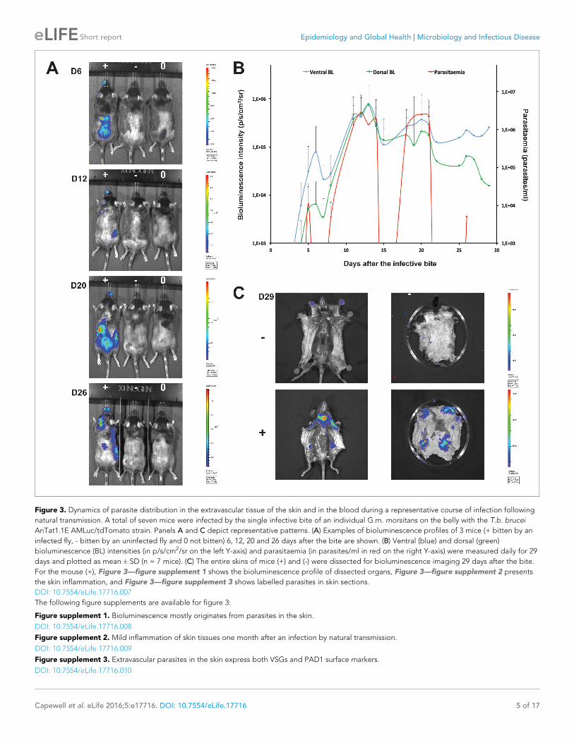
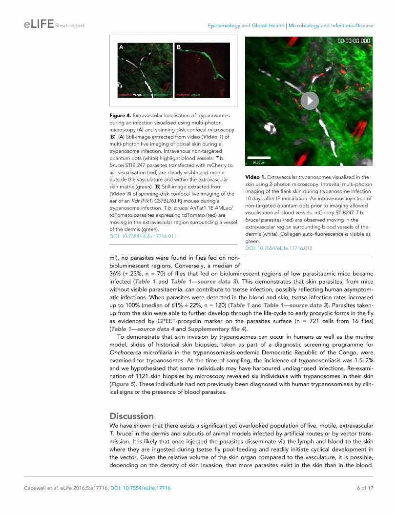
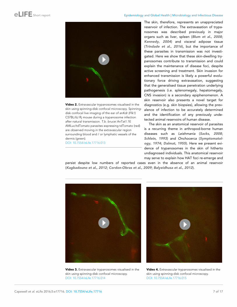
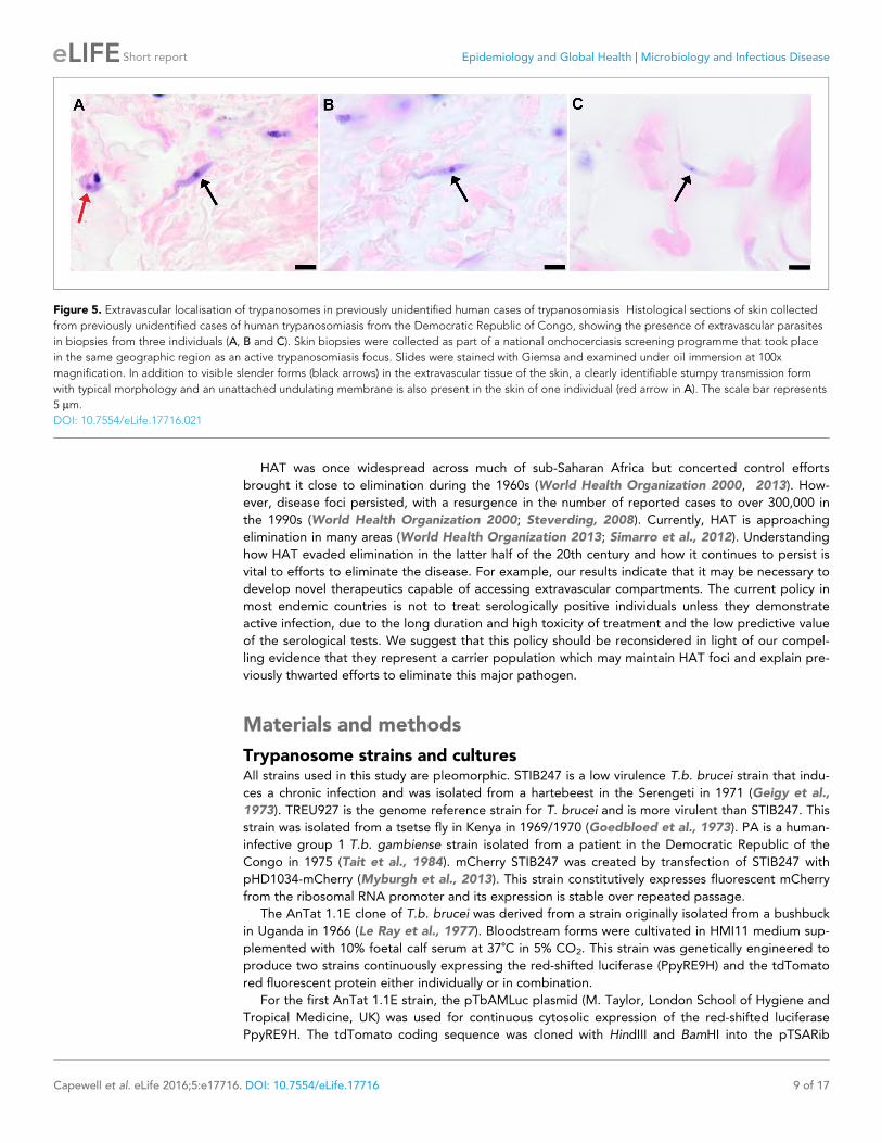