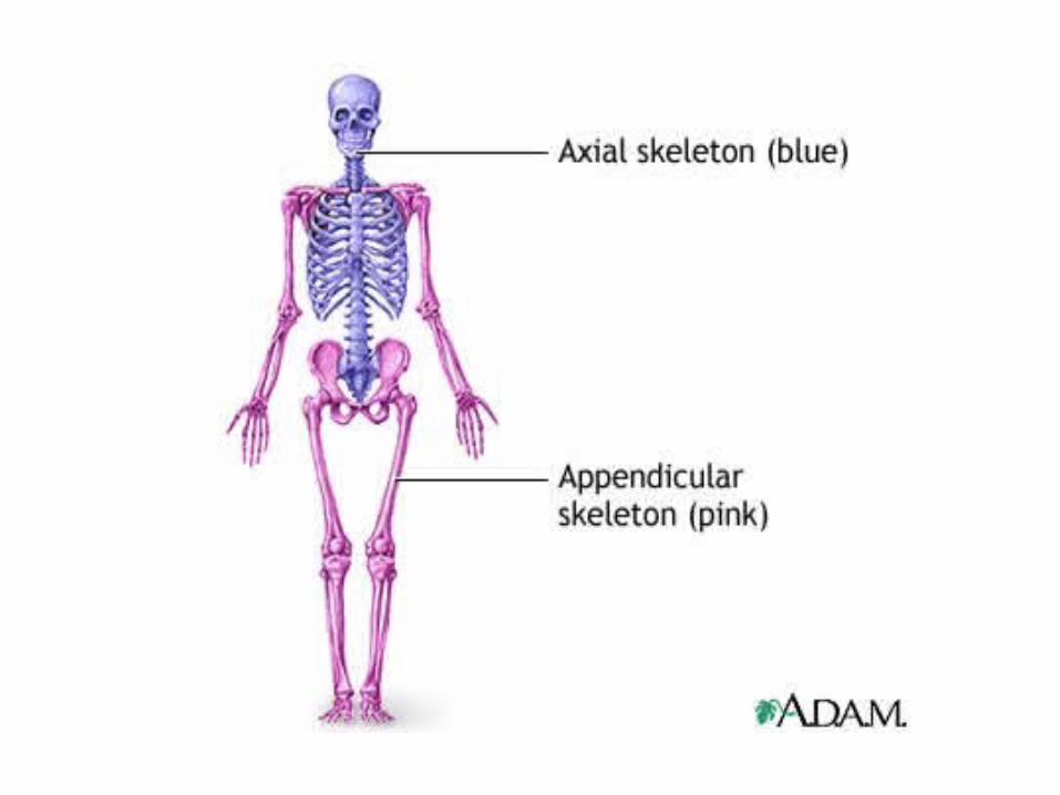
the skeletal system the skeletal system may be divided into two functional parts: the axial skeleton...
TRANSCRIPT

THE SKELETAL SYSTEM• The skeletal system may be divided into two
functional parts:
• The axial skeleton consists of the bones of the head (cranium or skull), neck (hyoid bone and cervical vertebrae),and trunk (ribs, sternum, vertebrae, and sacrum)
• • The appendicular skeleton consists of the bones of the limbs, including those forming the pectoral (shoulder) and pelvic girdles.
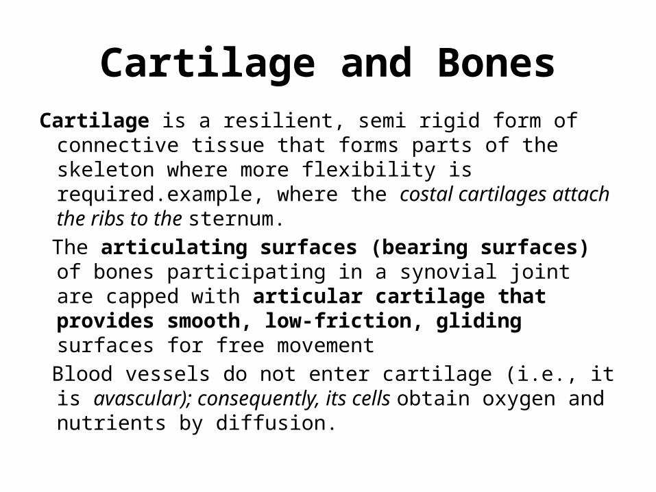
Cartilage and BonesCartilage is a resilient, semi rigid form of connective
tissue that forms parts of the skeleton where more flexibility is required.example, where the costal cartilages attach the ribs to the sternum.
The articulating surfaces (bearing surfaces) of bones participating in a synovial joint are capped with articular cartilage that provides smooth, low-friction, gliding surfaces for free movement
Blood vessels do not enter cartilage (i.e., it is avascular); consequently, its cells obtain oxygen and nutrients by diffusion.
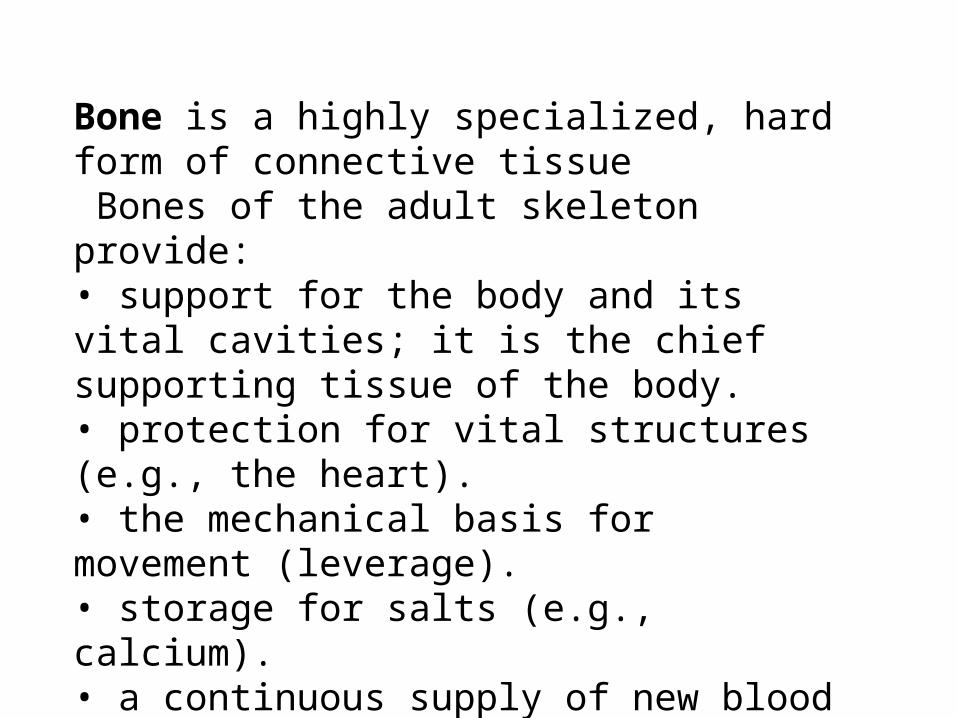
Bone is a highly specialized, hard form of connective tissue Bones of the adult skeleton provide:• support for the body and its vital cavities; it is the chief supporting tissue of the body.• protection for vital structures (e.g., the heart).• the mechanical basis for movement (leverage).• storage for salts (e.g., calcium).• a continuous supply of new blood cells (produced by the marrow in the medullary cavity of many bones).

• Periosteum A fibrous connective tissue covering surrounds
each skeletal element like a sleeve, except where articular cartilage occurs;
• And that of cartilages is called perichondrium. Both nourish the external aspects of the skeletal tissue.
They are capable of• laying down more cartilage or bone
(particularly during fracture healing) and • provide the interface for attachment of
tendons and ligaments.

• The two types of bone are compact bone and spongy (trabecular) bone.
• They are distinguished by the relative amount of solid matter and by the number and size of the spaces they contain
• Medullary cavity


• Some of the various markings and features of bones are :• Capitulum: small, round, articular head (e.g., capitulum of the
humerus).
• Condyle: rounded, knuckle-like articular area, often occurring in pairs (e.g., the lateral and medial femoral condyles).
• Crest: ridge of bone (e.g., the iliac crest).
• Epicondyle: eminence superior or adjacent to a condyle(e.g., lateral epicondyle of the humerus).
• Facet: smooth fl at area, usually covered with cartilage,where a bone articulates with another bone (e.g., superiorcostal facet on the body of a vertebra for articulation with a rib).

• Foramen: passage through a bone (e.g., obturator foramen).
• Fossa: hollow or depressed area (e.g., infraspinous fossa of the scapula).
• Groove: elongated depression or furrow (e.g., radial groove of the humerus).
• Head (L. caput): large, round articular end (e.g., head of the humerus).
• Line: linear elevation (e.g., soleal line of the tibia).
• Malleolus: rounded process (e.g., lateral malleolus of the fibula).
• Notch: indentation at the edge of a bone (e.g., greater sciatic notch).

• Spine: thorn-like process (e.g., the spine of the scapula).• Spinous process: projecting spine-like part (e.g., spinousprocess of a vertebra).• Trochanter: large blunt elevation (e.g., greater tro
chanter of the femur).• Trochlea: spool-like articular process or process that
acts as a pulley (e.g., trochlea of the humerus).• Tubercle: small raised eminence (e.g., greater tubercle
of the humerus• Tuberosity: large rounded elevation (e.g., ischial
tuberosity) Protuberance- projection of bone e.g. external occipital
protuberance

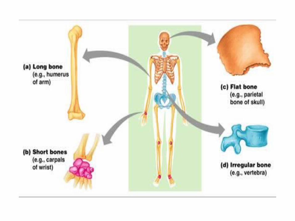
CLASSIFICATION OF BONES
Bones are classified according to their shape.• Long bones are tubular (e.g., the humerus in the arm).• Short bones are cuboidal and are found only in the tarsus
(ankle) and carpus (wrist).• Flat bones usually serve protective functions (e.g., the
flatbones of the cranium protect the brain).• Irregular bones have various shapes other than long, short,
or flat (e.g., bones of the face).• Sesamoid bones (e.g., the patella or knee cap) develop incertain tendons and are found where tendons cross the ends
of long bones in the limbs

BONE DEVELOPMENT
• Most bones take many years to grow and mature. The humerus (arm bone), for example, begins to ossify at the end of the embryonic period (8 weeks); however, ossification is not complete until age 20.
• All bones derive from mesenchyme (embryonic connective tissue) by two different processes: intramembranous ossifi cation (directly from mesenchyme) and endochondral ossifi cation (from cartilage derived from mesenchyme).

VASCULATURE AND INNERVATION OF BONES
• Most apparent are the nutrient arteries arise as branches of adjacent arteries outside the periosteum and pass obliquely through the compact bone of the shaft of a long bone via nutrient foramina.
• The nutrient artery divides in the medullary cavity into longitudinal branches that proceed toward each end, supplying the bone marrow, spongy bone, and deeper portions of the compact bone

osteoporosis
• During the aging process, the organic and inorganic components of bone both decrease, often resulting in osteoporosis, a reduction in the quantity of bone, or atrophy of skeletal tissue .Hence, the bones become brittle, lose the elasticity, and fracture easily.


NERVES
• Nerves accompany blood vessels supplying bones. The periosteum is richly supplied with sensory nerves (periosteal nerves.
• The periosteum is especially sensitive to tearing or tension, which explains the acute pain from bone fractures.
• Within bones, vasomotor nerves cause constriction or dilation of blood vessels, regulating blood flow through the bone marrow.

JOINTS
• CLASSIFICATION OF JOINTS• Three classes of joints are described, based on the manner or
type of material by which the articulating bones are united.• 1. SYNOVIAL JOINTS are united by a joint (articular) capsule
(composed of an outer fibrous layer lined by a serous synovial membrane) spanning and enclosing an articular cavity
• The joint cavity of a synovial joint contains a small amount of lubricating synovial fluid, secreted by the synovial membrane.
• internal surfaces are covered by synovial membrane.

• FIBROUS JOINTS: The sutures of the cranium are examples of fibrous joints
• These bones are close together, either interlocking along a wavy line or overlapping. A syndesmosis type of fibrous joint unites the bones with a sheet of fibrous tissue, either a ligament or a fibrous membrane.
• This type of joint is partially movableE.g. • 1. The interosseous membrane in the forearm is a sheet of
fibrous tissue that joins the radius and ulna in a syndesmosis
• 2. A dento-alveolar syndesmosis (gomphosis or socket) articulation between the root of the tooth and the alveolar process of the jaw.

• 3. Cartilaginous joints are united by hyaline cartilage or fibrocartilage.
• Primary cartilaginous joints permit growth in the length of a bone. When full growth is achieved, the epiphysial plate converts to bone and the epiphyses fuse with the diaphysis.(temporary unions)
• Secondary cartilaginous joints, or symphyses, are strong, slightly movable joints united by fibrocartilage. The fibrocartilaginous intervertebral disc
• SYNOVIAL JOINTS, the most common type of joint, provide free movement between the bones they join; they are joints of locomotion, typical of nearly all limb joints
The six major types of synovial joints are classified according to the shape of the articulating surfaces and/or the type of movement they permit

• 1. Plane joints permit gliding or sliding movements in the plane of the articular surfaces. The opposed surfaces of the bones are flat or almost flat, with movement limited by their tight joint capsules. Plane joints are numerous and are nearly always small. Eg acromioclavicular joint between the acromion of the scapula and the clavicle.
• 2. Hinge joints permit flexion and extension only, movements that occur in one plane (sagittal) uniaxial joints. The joint capsule of these joints is thin and lax anteriorly and posteriorly where movement occurs; however, the bones are joined by strong, laterally placed collateral ligaments. eg elbow joint

3. Saddle joints permit abduction and adduction as well as flexion and extension, biaxial joints that allow movement in two planes, sagittal and frontal. The performance of these movements in a circular sequence (circumduction) is also possible. The Carpometacarpal joint at the base of the 1st digit (thumb) is a saddle joint
4. Condyloid joints permit flexion and extension as well as abduction and adduction; thus condyloid joints are also biaxial. However, movement in one plane (sagittal) is usuallygreater (freer) than in the other. The metacarpophalangeal joints (knuckle joints) are condyloid joints

• 5. Ball and socket joints allow movement in multiple axesand planes: fl exion and extension, abduction and adduction,medial and lateral rotation, and circumduction; thus balland socket joints are multiaxial joints. The hip joint is a ball and
socket joint in which the spherical head of the femur rotates within the socket formed by the acetabulum of the hip bone.
s6. Pivot joints permit rotation around a central axis; thusthey are uniaxial. In these joints, a rounded process ofbone rotates within a sleeve or ring. The median atlantoaxialjoint is a pivot joint in which the atlas (C1 vertebra)rotates around a fi nger-like process, the dens of the axis(C2 vertebra), during rotation of the head.


JOINT VASCULATURE AND INNERVATION
• Joints receive blood from articular arteries that arise from the vessels around the joint.
• The arteries often anastomose (communicate) to form networks (peri-articular arterial anastomoses) to ensure a blood supply to and across the joint
• Articular veins are communicating veins that accompany arteries (L.venae comitantes) and, like the arteries, are located in the joint capsule, mostly in the synovial membrane.

anastomosis
• Joints have a rich nerve supply provided by articular nerves with sensory nerve endings in the joint capsule. In the distal parts of the limbs (hands and feet), the articular nerves are branches of the cutaneous nerves supplying the overlying skin.
The Hilton law states that the nerves supplying a joint also supply the muscles moving the joint and the skin covering their distal attachments.

Degenerative Joint Disease
• Synovial joints are well designed to withstand wear, but heavy use over several years can cause degenerative changes.
• Degenerative joint disease or osteoarthritis is often• accompanied by stiffness, discomfort, and pain.
Osteoarthritis• is common in older people and usually affects joints
that• support the weight of their bodies (e.g., the hips and
knees).
Arthroscopy
• The cavity of a synovial joint can be examined by inserting a cannula and an arthroscope (a small telescope) into it.
orthopedic surgeons use this to examine joints for abnormalities, such as torn menisci (partial articular discs of the knee joint).

Avascular necrosis
• Loss of arterial supply to an epiphysis or other parts of a bone results in the death of bone tissue— avascular necrosis (G. nekrosis, deadness). After every fracture, small areas of adjacent bone undergo necrosis.
• In some fractures, avascular necrosis of a large fragment of bone may occur
