the schistosoma japonicum genome reveals features of host–parasite interplay
TRANSCRIPT

ARTICLES
The Schistosoma japonicum genomereveals features of host–parasite interplayThe Schistosoma japonicum Genome Sequencing and Functional Analysis Consortium*
Schistosoma japonicum is a parasitic flatworm that causes human schistosomiasis, which is a significant cause of morbidity inChina and the Philippines. Here we present a draft genomic sequence for the worm. The genome provides a global insight intothe molecular architecture and host interaction of this complex metazoan pathogen, revealing that it can exploit hostnutrients, neuroendocrine hormones and signalling pathways for growth, development and maturation. Having a complexnervous system and a well-developed sensory system, S. japonicum can accept stimulation of the corresponding ligands as aphysiological response to different environments, such as fresh water or the tissues of its intermediate and mammalianhosts. Numerous proteases, including cercarial elastase, are implicated in mammalian skin penetration and haemoglobindegradation. The genomic information will serve as a valuable platform to facilitate development of new interventions forschistosomiasis control.
Schistosomiasis is an ancient scourge of mankind, depicted graphicallyin papyri from Pharaonic Egypt and known from human remains over2,000 years old from China1,2. Blood-dwelling trematodes (phylumPlatyhelminthes) of the genus Schistosoma cause this chronic anddebilitating disease, which afflicts more than 200 million people in76 tropical and subtropical countries. Morbidity is high and schistoso-miasis contributes to several hundreds of thousands of deathsannually3–5. Three principal species can infect humans: Schistosomajaponicum, Schistosoma mansoni and Schistosoma haematobium. Thefirst of these is prevalent in the Philippines and parts of Indonesia, andis a major disease risk for 66 million people living in southern China2.It remains a major public health concern in China despite over 50 yearsof concerted campaigns for its control2,6. Approximately one millionpeople in China, and more than 1.7 million bovines and othermammals, are currently infected2. Control measures includecommunity-based praziquantel chemotherapy, health education,improved sanitation, environmental modification and snail control.However, additional approaches, such as the development and deploy-ment of new drugs and anti-schistosome vaccines are urgentlyneeded to meet the prevailing challenges, which include the spectreof praziquantel-resistant parasites7,8.
During their complex developmental cycle, schistosomes alternatebetween a mammalian host and a snail host through the medium offresh water. After burrowing out of the snail host, free-swimmingcercariae penetrate the skin of the mammalian host, travel throughthe blood to the liver via the lungs, and transform into schistosomula.These mature in the hepatic portal vein, mate and, in the case ofS. japonicum, migrate to their final destination in the mesentericvenous plexus. Female worms release thousands of eggs daily, whichare discharged in the faeces after a damaging passage through theintestinal wall. If they reach fresh water, eggs hatch to release free-swimming ciliated miracidia, which, guided by light and chemicalstimuli, seek amphibious snails of the genus Oncomelania. Withinthe hemocoel of the snail, miracidia give rise asexually to numbers ofsporocysts, in which further asexual propagation produces numerouscercariae.
Eggs deposited by adult female schistosomes embolize in the liver,intestines and other tissue sites and are the key contributors to the
pathology and associated morbidity of schistosomiasis. Notably, thehighly adapted relationship between schistosomes and their snailintermediate and mammalian definitive hosts appears to involveexploitation by the parasite of host endocrine and immune signals9,10.The evasion strategies that underpin avoidance of the host immunesystem, allowing schistosomes to survive for years despite strong hostimmune responses, have long interested investigators intent ondevelopment of an efficacious vaccine.
Unlike most other platyhelminths, schistosomes are dioecious.The genome is arrayed on eight pairs of chromosomes, seven pairsof autosomes and one pair of sex chromosomes. Females are theheterogametic sex (ZW); males are homogametic (ZZ)11,12. No otherlophotrochozoan13 has yet been sequenced.
Genome features and evolutionGeneral information. The whole-genome shotgun (WGS) sequencingstrategy was used to decode the 397-megabase-pair (Mb) sequences,covering most (.90%) of the S. japonicum genome (SupplementaryTables 1 and 2 and Supplementary Fig. 1). A total of 13,469 protein-coding genes were identified, comprising about 4% of the draftS. japonicum genome (Supplementary Figs 2 and 3). Of the protein-coding genes, 6,972 (52%) were mapped to categories established by theGene Ontology project (Fig. 1a and Supplementary Fig. 4) and anorthologue relationship existed between 2,516 (19%) of them and1,546 Kyoto Encyclopedia of Genes and Genomes (KEGG) orthologygroups (Supplementary Fig. 5). Schistosoma japonicum has a relativelylarge genome and low gene density in comparison with other inverte-brates, including Brugia malayi (Table 1). On the basis of the outbredsource of the genomic libraries, high-quality discrepancies foundduring assembly were used to identify 557,739 single nucleotide poly-morphisms (SNPs) (Supplementary Table 3), with an average density of1.4 SNPs per kilobase pair, and the insertion and deletion (indel) rateswere much lower.Repeat sequences. A total of 657 different repeat families/elements,constituting 159 Mb (40.1%) of the S. japonicum genome wererevealed by comparing known repetitive sequences and using thesoftware REPEATSCOUT (version 1.0.3)14 (Fig. 2 and Supplemen-tary Table 4). Among them, 29 kinds of retrotransposon were found,
*Lists of participants and their affiliations appear at the end of the paper.
Vol 460 | 16 July 2009 | doi:10.1038/nature08140
345 Macmillan Publishers Limited. All rights reserved©2009

including known Gulliver, SjR1, SjR2 and Sj-pido elements as well as25 novel elements, together constituting 19.8% of the genome(Supplementary Table 5). Of the 25 novel retrotransposons, 18 arelong terminal repeat (LTR) forms, four are non-LTR forms and threeare Penelope-like elements—enigmatic retroelements that retainintrons15. Each type of retrotransposon was represented by one to793 intact copies or hundreds to thousands of partial copies. Thenon-LTR retrotransposons have significantly higher copy numbers,constituting 12.6% of the genome.Gene loss/duplication. It was intriguing to observe that schisto-somes share more orthologues with the vertebrates (SupplementaryTable 6), such as H. sapiens (4,324 pairs), than they do with theecdysozoans, for example C. elegans (3,292), despite the Ecdysozoaand Lophotrochozoa being phylogenetically adjacent13. Similarly, thecnidarians and vertebrates have been shown to share more ortholo-gous genes with each other than either does with the ecdysozoans16.One possible reason for this is that a higher evolutionary rate in theEcdysozoa causes an apparently larger orthologue divergence,although the scenario of functional selection of orthologue patternsin the context of parasite–host interplay is also worth consideration.
To test possible consequences of parasitism at the genome level, weinvestigated gene family and domain variations between schisto-somes and other metazoans. It is clear that there was minor variationin total numbers of protein families among S. japonicum (6,322) andthe other species, such as C. elegans (6,669), D. melanogaster (5,184)and H. sapiens (6,877) (Supplementary Table 7). However, a major
reduction in number, or even the elimination, of protein domainswas apparent in the S. japonicum genome, in that the great majority(3,654) of the 3,728 protein domains from the flatworm were sharedwith other species (Fig. 1b) and can thus be considered ubiquitousamong metazoans, whereas 3,834 domains found in at least one of theother species were not detected in schistosomes. Of these 3,834domains, 1,140 were shared by more than three taxa of vertebrates,insects, a nematode and sea anemones (Supplementary Fig. 6).Notably, domain-loss events seem to be more widespread inS. japonicum than in any other species studied so far, includingC. elegans, a model organism well known for rapid evolutionary ratesand a high degree of gene loss17. Roughly 1,000 protein domains havebeen abandoned by S. japonicum, including some involved in basicmetabolic pathways and defence, implying that loss of these domainscould be, at least partly, a consequence of the adoption of a parasiticway of life.
Against the background of extensive gene/domain loss, the findingof expanded gene families in schistosomes might provide clues to therequirements for a parasitic lifestyle. Among the most expanded genefamilies in schistosomes (Supplementary Tables 8 and 9), that encod-ing leishmanolysin (a major surface protease, also called gp63), amember of the metallopeptidase M8 family, has 12 putative familymembers in S. japonicum, but there is only one in human, fruit fly andnematode (C. elegans), and only three putative counterparts in thefree-living flatworm Schmidtea18 (Supplementary Information). Inaddition to elastase (see later), leishmanolysin-like proteases maycontribute to tissue invasion by schistosome cercariae19.
Development and metabolismCellular signalling pathways in development. To investigateregulatory networks involved in embryonic development and orga-nogenesis, we undertook comparative genomics analysis of well-characterized signalling pathways, including those for Wnt, notch,hedgehog and transforming growth factor b (TGF-b). Notably, theS. japonicum genome encodes these growth factors, receptors andessential components to regulate many cellular processes duringorganogenesis and tissue development (Fig. 3 and SupplementaryTables 10 and 11). Schistosoma japonicum also encodes endogenousepidermal growth factor (EGF)-like and fibroblast growth factor(FGF)-like peptides (Fig. 3). The intact downstream cascadecomposed of the RasRRafRmitogen-activated protein kinase(MAPK) and TGF-bRSMAD signalling pathways, including FGF-and EGF-receptors, has components sharing high identity withmammalian orthologues, which implies that schistosomes, in addi-tion to using their own pathways, can exploit host growth factors asdevelopmental signals. Indeed, we have identified an insulin receptorwith high sequence similarity with those of mammals20, whereas noinsulin growth factor or insulin molecules were found, further sup-porting the notion that schistosomes exploit key signalling pathwaysof their hosts for growth and metabolism.Metabolic pathways. Analysis of the KEGG pathways assigned tometabolic process (Supplementary Table 12 and SupplementaryFigs 7 and 8) indicates that S. japonicum can use carbohydrates asenergy/carbon sources. It is unable to de novo synthesize fatty acids,sterols, purines, nine human essential amino acids, arginine or tyro-sine (Supplementary Figs 9–11). Loss or degeneracy of fatty acid,sterol and purine synthesis pathways in schistosomes is probably aconsequence of the adoption of a parasitic lifestyle; notably, the genesencoding all the key enzymes for both the de novo fatty acid andpurine syntheses are complete in the free-living flatworm, Schmidteamediterranea18 (Supplementary Information). To obtain essentiallipid nutrients, the S. japonicum genome indeed encodes many trans-porters, including apolipoproteins, low-density lipoprotein receptor,scavenger receptor, fatty-acid-binding protein, ATP-binding-cassettetransporters and cholesterol esterase (Supplementary Table 13), toexploit fatty acids and cholesterol from host blood and plasma.
Locomotion (1%)
Immune system (1%)
Cellularhomeostasis (1%)
Biologicaladhesion (2%)
Reproduction (3%)
Response to stimulus (5%)
Metabolism (34%)Biologicalregulation
(16%)
Localization(14%)
Cellular componentorganization
and biogenesis (12%)
Development(11%)
S. japonicum
C. elegans
Insects
Vertebrates
N. vectensis
0 1,000 2,000 3,000 4,000 5,000 6,000
Number of domains
Domains in onespecific taxon(unique domains)
Domains sharedby two taxa
Domains sharedby three taxa
Domains sharedby four taxa
Domains sharedby five taxa
a
b
Figure 1 | Functional categorization of S. japonicum genes and protein-domain analysis. a, Proportion of the 6,972 S. japonicum proteins withfunctional information in different Gene Ontology categories. b, InS. japonicum, vertebrates (H. sapiens, G. gallus and D. rerio), insects(D. melanogaster and A. gambiae), C. elegans and Nematostella vectensis, atotal of 7,562 domains were detected. The majority of S. japonicum domainsare shared with other taxa, having the fewest unique domains, whereasvertebrates evolved significant numbers of unique protein domains.
ARTICLES NATURE | Vol 460 | 16 July 2009
346 Macmillan Publishers Limited. All rights reserved©2009

Nervous system and neuroendocrine system
Platyhelminths possess a central nervous system with a variety ofsensory structures that can transduce a wide range of stimuli, anduse a neuroendocrine system to regulate growth, metabolism andhomeostasis.Neurotransmitters and receptors. We characterized a number ofreceptors and transporters of neurotransmitters (SupplementaryTable 14) that may be required, for example, by miracidia andcercariae to navigate through water to locate new hosts and for theschistosomula and adult flukes to establish and reproduce within thehuman vasculature. In addition to known neurotransmitters andreceptors, we have identified a receptor for octopamine (Sup-plementary Table 14) and two key enzymes for synthesis of octopa-mine (Supplementary Fig. 12).
The nervous systems of flatworms can be considered to be predo-minantly peptidergic21. We found additional putative neuropeptidereceptors for opioids, galanin and melatonin. Thus, it appears thatschistosomes can accept stimulation of the corresponding ligands asa physiological response to different environments, such as freshwater or the tissues of their snail and mammalian hosts. There aregenes encoding receptors predicted to accept gastrointestinal neuro-peptide hormone signals including cholecystokinin, secretin, gastricinhibitory polypeptide and xenin, all of which are involved in func-tions promoting the release of alimentary tract fluids containingdigestive enzymes.
We also identified receptors for urotensin, angiotensin II and neu-romedin (types U and B), which have an important role in physio-logical regulation of the cardiovascular system, the hypothalamusand other vertebrate organs. Although schistosomes do not havethese organs, these components could have other effects on the cellsor tissues of the blood fluke, such as the regulation of cell growth or intissue remodelling. In addition, we found receptors for hypocretin(orexin), leptin and hypothalamic neuropeptides. Together, thesefeatures suggest that schistosomes have many advanced physiologicalfeatures regarded as more characteristic of higher metazoans.
Unexpectedly, a myokinin-like receptor was also observed(Supplementary Table 14). Myokinins are invertebrate neuropep-tides with myotropic and diuretic activities for which a receptor,called lymnokinin receptor, was first identified in the tickBoophilus microplus22. The discovery of such a receptor in schisto-somes supports the notion that they might synthesize myokininbecause their vertebrate hosts do not produce this neuropeptide.Additional examples of receptors found for other invertebrate neu-ropeptides included FMRFamide and myosuppressin23,24, bothbelonging to the FMRFamide-like peptide superfamily.Complex sensory system. Schistosomes have a variety of sensorystructures using which they, during their different life stages, pre-sumably respond to a myriad of environmental stimuli. Free-livingcercariae and miracidia can sense light, mechanical stimuli and tem-perature25, facilitating the finding of hosts, whereas the parasitic adult
Table 1 | Summary of S. japonicum genomic features in comparison with other organisms
Genome features Lophotrochozoa Ecdysozoa Deuterostomia
Schistosomajaponicum
Caenorhabditiselegans
Brugiamalayi
Drosophilamelanogaster
Anophelesgambiae
Gallusgallus
Daniorerio
Homosapiens
Total genome size (Mb) 398 100 88{
169 265 1,051 1,527 3,255
Total GC content (%) 34.1 35.4 30.5{40.2 42.2 41.2 34.4 36.2
GC content in coding regions (%) 36 42.7 39.6{53.3 44.3 42.9 34.4 37.5
GC content in intron regions (%) 33.8 32.1 27.6{39.5 43.9 41.3 34.1 36.6
GC content in intergenic regions (%) 34.7 35.4 30.9{40.2 42.2 41.2 34.4 36.2
Repeat rate (%) 40.1 18.3 ,15{
24.7 16.6 10.8 46.6 44
Total coding size (Mb)/ratio (%) 15.9/4 24.8/25 12.9/15 21.7/13 14.8/6 25.7/2 47.9/3 35.9/1
Number of coding genes 13,469 20,077 11,515 14,144 12,527 18,107 35,321 25,077
Gene density (genes per Mb) 34 200 130 84 47 17 23 8
Average CDS size (kb) 1.18 1.23 1.12 1.54 1.18 1.42 1.36 1.43
Average gene size 6 s.d. (kb) 10.5 6 16.0 2.8 6 3.2 2.8 6 2.9 4.3 6 10.6 4.6 6 10.0 21.6 6 46.8 19.5 6 35.1 41.3 6 98.0Number of transfer RNAs 153 608 (,590*) ,233
{314 450 189 2,010 129
Number of ribosomal RNAs 184 19 (,275*) ,400{
161 N/A 14 N/A 675
All coding sequence (CDS)-related features were calculated from the KEGG database, version 46 (ftp://ftp.genome.jp/pub/kegg/release/archive/kegg/46/). Caenorhabditis elegans and B. malayigenome feature files were downloaded from Wormbase (ftp://ftp.wormbase.org/pub/). Other genome-feature files and genome sequences were downloaded from Ensembl (ftp://ftp.ensembl.org/pub/release-49).*The features calculated from the feature file are different from those in the reference50.{Data obtained from previous publications51.
Non-repeat region (59.87%)
Undefined repeat(10.45%)
Transposon(0.22%)
3 Penelope-likeretrons (0.99%)
4 novel(6.97%) SjR2
(4.43%)SjR1
(0.84%)Sj-pido(0.39%)
18 novel(5.98%)
Gulliver(0.18%)
Sj-α2(0.76%)
Sj-α1(1.08%)
Satellite(4.23%)
Minisatellite(2.47%)
Microsatellite(0.63%)
Low complexity(0.51%)
Non-LTR retrotransposon(12.63%)
LTR retrotransposon(6.16%)
SINE(1.84%)
Figure 2 | The distribution of categories and composition of repeat elements in the S. japonicum genome. Retrons, retrotransposons; SINE, shortinterspersed nuclear element.
NATURE | Vol 460 | 16 July 2009 ARTICLES
347 Macmillan Publishers Limited. All rights reserved©2009

worms are able to respond to changes in levels of chemicals andnutrients. Using a top-down Gene-Ontology-based strategy tofacilitate the gene annotation (Supplementary Fig. 13), we identified71 genes encoding receptors, membrane channels, enzymes and othercomponents, such as rhodopsins/opsins26, phosrestins/arrestins27,transducins, cyclic nucleotide-gated channel, rhodopsin kinase andguanylate cyclase 2D (Supplementary Table 15). Both S. japonicumand S. mansoni have only two members of the rhodopsin family,unlike Drosophila, which possesses 13 members, and zebrafish, whichhas at least seven (Supplementary Fig. 14a). Phylogenetic analysisindicated that there are at least four schistosome transducins, eachof which could represent a divergent subtype of transducin super-family across chordates, echinoderms, molluscs and arthropods(Supplementary Fig. 14b), and could therefore mediate distinct res-ponses of sensors to signals.
The genome sequence analysis also revealed an array of genes encod-ing sensory proteins that could interact with chemical ligands andother stimuli. These included guanine-nucleotide-binding protein,potassium-voltage-gated-channel protein Shaker, the glutamate recep-tor for umami taste and protein Prospero28 (Supplementary Table 16and Supplementary Fig. 15a). Notably, the genome encodes most com-ponents of four of the five human gustatory sensation pathways: thesalty, sour, sweet and umami tastes. We also found several potentialsensors for sound perception, a common characteristic of vertebratesand arthropods29, in the genome (Supplementary Table 17).
We discovered an apparently intact olfaction pathway, includingcyclic nucleotide-gated olfactory channel, guanine-nucleotide-binding protein and adenylyl cyclase type 3 (SupplementaryTable 18 and Supplementary Fig. 15b). Moreover, mechanosensoryperception mediated by mechanically gated ion channels representsthe basis for the sensing of touch, balance, temperature and sound,and contributes essentially to the development and homeostasis of allEumetazoa30 (Supplementary Table 19). Putative sensory compo-nents for equilibrium/balance, mechanical stimulation, painand temperature (Supplementary Tables 20 and 21) were alsofound in the S. japonicum genome, including two proteins that have
similarities with the well-known mechanosensory protein, transientreceptor potential cation channel31, and several receptors such asmetabotropic glutamate receptor 3, which participate in the sensoryperception of pain, light and taste.Neuroendocrine system. Schistosomes have receptors that apparentlyevolved to accept endogenous hormones as well as those of the para-sitized mammalian host20,32. By surveying hormones and receptorsrelated to the classical neuroendocrine axis in the genomic sequenceof S. japonicum, we found (Fig. 4) putative receptors for hypothalamichormones such as thyrotropin-releasing hormone (TRH), prolactin-releasing hormone, somatostatin, melanin-concentrating hormone andleptin, as well as transmembrane proteins that have some similaritieswith receptors for gonadotropin-releasing hormone, corticotropin-releasing hormone and growth-hormone-releasing hormone. More-over, putative receptors are present that show weak similarity with thosein mammals for the pituitary hormones thyroid-stimulating hormone(TSH), luteinizing hormone, follicle-stimulating hormone, argininevasopressin and oxytocin.
Although a hypothalamus–pituitary-like organ has not beendescribed in schistosomes, it is possible that some neurons, similarto those in the hypothalamus and pituitary of vertebrates, could fulfilsimilar functions in terms of modulating the behaviour ofS. japonicum through peripheral endocrine tissues and cells. In thisregard, it is noteworthy that the genomic information suggests thepresence of an integral hypothalamic–pituitary–thyroid axis inS. japonicum. In addition to the superior TRH–TSH receptors, anintact system for synthesis of thyroxine and active triiodothyronine,as well as an inactivation mechanism of these hormones using deio-dination, was identified. Nuclear receptors for triiodothyronine andthyroxine were revealed with identity to mammalian orthologues.Hence, S. japonicum may use an endogenous thyroid hormone/receptor signalling pathway for growth and development (Fig. 4and Supplementary Table 22).
We confirmed that S. japonicum has receptors for steroid hor-mones such as progestin, progesterone and oestrogen32,33. In addi-tion, it possesses intricate pathways for processing steroid hormones
Frizzled Dishevelled
p
Geneexpression
Cell proliferation, differentiation,apoptosis
TCF
Notch
CytoplasmNucleus
Wnt
Delta
Activin
FRP
ProC
Notch
TACE
ProC
NICD
NICD
Activin R
? Fused
CI
ZIC2
IGF
IGFR
MAPKs
PI3K
p70S6K
Acetylcholine
AChR
Dopamine
DR
Histamine
HR
5-HT
HTR
GABA
GABAR
Glycine
Taurine
β-alanine
GlyR Glutamate
GluR
Neuropeptide Y
NPYR
mGlu
mGluR
Nodal
TGFRsDecorin
Ryanodine
RyR
Octopamine
OAR
BMPR
BMPNoggin
EGF-like
EGFR
MAPKs
GRB2EGFR
FGF
Ras
Suppressor of fused
Hedgehog
Patched
Patched
Smoothened
mRNA
TGF-β
Ras
GSK3β
β-catenin β-catenin
β-catenin
SMADs
SMADs
Neuroactivereceptors
Pathways for growthand development
Figure 3 | Putative signalling pathways for growth, development andneuroactive ligand-receptor interaction in S. japonicum. The pathways forgrowth and development (indicated with different colours), and theneuroactive ligand-receptor interactions in S. japonicum are shown on theleft and right, respectively. TACE, tumour-necrosis-factor-a-convertingenzyme; ProC, porcupine homologue (Drosophila); NICD, notchintracellular domain; FRP, frizzled-related protein 1; GSK3b, glycogensynthase kinase 3b; TCF, transcription factor 7; ‘p’ within cycle,
phosphorylation on the proteins indicated; BMP, bone morphogeneticprotein; IGF, insulin-like growth factor; mGlu, metabotropic glutamate;GlyR, glycine receptor; GluR, glutamate receptor; HR, histamine receptor;GABA, c-aminobutyric acid; DR, dopamine receptor; 5-HT,5-hydroxytryptamine; HTR, 5-hydroxytryptamine receptor; NPYR,neuropeptide Y receptor; AChR, acetylcholine receptor; RyR, ryanodinereceptor; OAR, octopamine receptor; ZIC2, Zic family member 2; CI,cubitus interruptus; suffix ‘R’ denotes receptor.
ARTICLES NATURE | Vol 460 | 16 July 2009
348 Macmillan Publishers Limited. All rights reserved©2009
to form other sex hormones. For example, there are putative enzymespresent that could convert the female hormones progesterone andpregnenolone to estriol, oestrone, androsterone and testosterone.Hence, schistosomes might use these pathways during their parasiticexistence. Schistosoma japonicum also encodes enzymes to catabolizeexcessive or used steroid hormones such as aldosterone.
With regard to the process of glycolysis for essential energy supply,receptors for adiponectin, an insulin-sensitizing hormone34, and lep-tin, a suppressor of the secretion of insulin35, are also encoded by thegenome of S. japonicum (Supplementary Table 22), providing furthersupport for the notion that the blood fluke modulates its energymetabolism in response to either its own insulin-like hormones orthose of its mammalian host.
The schistosomulum renews its tegument during maturation intoan adult schistosome under the effects of ecdysone36,37. In concordance,we identified an ecdysone-like receptor and its downstream effectorecdysone-induced protein 78C. In addition, allatostatin, a polypeptidehormone that suppresses the secretion of juvenile hormone, was previ-ously reported to be found throughout the schistosome nervoussystem38,39. An allatostatin-like receptor sequence that has highsimilarity with that of the cockroach was also identified (Supplemen-tary Table 22).
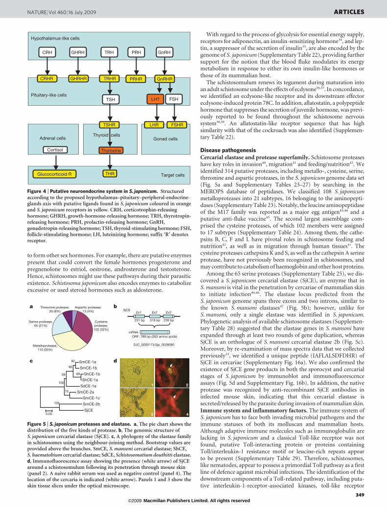
Disease pathogenesisCercarial elastase and protease superfamily. Schistosome proteaseshave key roles in invasion40, migration41 and feeding/nutrition42. Weidentified 314 putative proteases, including metallo-, cysteine, serine,threonine and aspartic proteases, in the S. japonicum genome data set(Fig. 5a and Supplementary Tables 23–27) by searching in theMEROPS database of peptidases. We classified 108 S. japonicummetalloproteases into 21 subtypes, 16 belonging to the aminopepti-dases (Supplementary Table 23). Notably, the leucine aminopeptidaseof the M17 family was reported as a major egg antigen43,44 and aputative anti-fluke vaccine45. The second largest assemblage com-prised the cysteine proteases, of which 102 members were assignedto 17 subtypes (Supplementary Table 24). Among them, the cathe-psins B, C, F and L have pivotal roles in schistosome feeding andnutrition42, as well as in migration through human tissues41. Thecysteine proteases cathepsins K and S, as well as the cathepsin A serineprotease, have not previously been recognized in schistosomes, andmay contribute to catabolism of haemoglobin and other host proteins.
Among the 65 serine proteases (Supplementary Table 25), we dis-covered a S. japonicum cercarial elastase (SjCE), an enzyme that inS. mansoni is vital in the penetration by cercariae of mammalian skinto initiate infection40,46. The elastase locus predicted from theS. japonicum genome spans three exons and two introns, similar tothe known S. mansoni elastases47 (Fig. 5b); however, unlike forS. mansoni, only a single elastase was identified in S. japonicum.Phylogenetic analysis of available schistosome elastases (Supplemen-tary Table 28) suggested that the elastase genes in S. mansoni haveexpanded through at least two rounds of gene duplication, whereasSjCE is an orthologue of S. mansoni cercarial elastase 2b (Fig. 5c).Moreover, by re-examination of mass spectra data that we collectedpreviously33, we identified a unique peptide (IAFLALSDFDHR) ofSjCE in cercariae (Supplementary Fig. 16a). We also confirmed theexistence of SjCE gene products in both the sporocyst and cercarialstages of S. japonicum by immunoblot and immunofluorescenceassays (Fig. 5d and Supplementary Fig. 16b). In addition, the nativeprotease was recognized by anti-recombinant SjCE antibodies ininfected mouse skin, indicating that this cercarial elastase issecreted/released by the parasite during invasion of mammalian skin.Immune system and inflammatory factors. The immune system ofS. japonicum has to face both invading microbial pathogens and theimmune statuses of both its molluscan and mammalian hosts.Although adaptive immune molecules such as immunoglobulin arelacking in S. japonicum and a classical Toll-like receptor was notfound, putative Toll-interacting protein or proteins containingToll/interleukin-1 resistance motif or leucine-rich repeats appearto be present (Supplementary Table 29). Therefore, schistosomes,like nematodes, appear to possess a primordial Toll pathway as a firstline of defence against microbial infections. The identification of thedownstream components of a Toll-related pathway, including puta-tive interleukin-1-receptor-associated kinases, toll-like receptor
Target cells
Adrenal cells Gonad cells
THR
Thyroxine
TRH
TRHR
TSH
TSHR
GHRH
GHRHR
Pituitary-like cells
Hypothalamus-like cells
Thyroid cells
PRH
PRHR
Cortisol
Glucocorticoid R
LH?
LHR
FSH
FSHR
GnRHR
GnRHCRH
CRHR
Figure 4 | Putative neuroendocrine system in S. japonicum. Structuredaccording to the proposed hypothalamus–pituitary–peripheral-endocrine-glands axis with putative ligands found in S. japonicum coloured in orangeand S. japonicum receptors in yellow. CRH, corticotrophin-releasinghormone; GHRH, growth-hormone-releasing hormone; TRH, thyrotropin-releasing hormone; PRH, prolactin-releasing hormone; GnRH,gonadotropin-releasing hormone; TSH, thyroid-stimulating hormone; FSH,follicle-stimulating hormone; LH, luteinizing hormone; suffix ‘R’ denotesreceptor.
c dSmCE-1a
SmCE-1b
ShCE-1b
ShCE-1a
SdCE-1a
SmCE-2a
SmCE-1c
SmCE-2b
SjCE
99
92
85
75
49
100
0.05
100
a b
SJC_S000173:Sjp_0028090
SjCE
mRNAORF: 789 bp (263 amino acids)
Ex1 Ex2 Ex3
1 2
3 4
Serine protease,65 (21%)
Threonine protease,26 (8%)
Aspartic protease,13 (4%)
Cysteineprotease,102 (32%)
Metalloprotease,110 (35%)
231 bp 319 bp 239 bp
Figure 5 | S. japonicum proteases and elastase. a, The pie chart shows thedistribution of the five kinds of protease. b, The genomic structure ofS. japonicum cercarial elastase (SjCE). c, A phylogeny of the elastase familyin schistosomes using the neighbour-joining method. Bootstrap values areprovided above the branches. SmCE, S. mansoni cercarial elastase; ShCE,S. haematobium cercarial elastase; SdCE, Schistosomatium douthitti elastase.d, Immunofluorescence assay showing the presence (white arrow) of SjCEaround a schistosomulum following its penetration through mouse skin(panel 2). A naive rabbit serum was used as negative control (panel 4). Thelocation of the cercaria is indicated (white arrow). Panels 1 and 3 show theskin tissue slices under the optical microscope.
NATURE | Vol 460 | 16 July 2009 ARTICLES
349 Macmillan Publishers Limited. All rights reserved©2009

adaptors, TNF-receptor-associated factor 6 (TRAF6), inhibitor ofnuclear factor kB kinase subunit epsilon (IKK-e) and p38 MAPK,further support the view that this primitive innate immune systemcould be crucial for the worm (Supplementary Table 30).
On the other hand, factors and metabolites in S. japonicum thatcould contribute to stimulation and regulation of mammalianimmunity were discovered. It is well accepted that glycans and lipidssynthesized by adult schistosomes or eggs may regulate secondarysignals through corresponding receptors on effector cells and access-ory cells of the mammalian host, thus compromising host immuno-logical defences targeting the parasite. We therefore searched forenzymes involved in the metabolism of various glycans or lipid anti-gens by interrogating this worm genome. It turned out that, with therare exception of enzymes such as a1,3-mannosyltransferase, a com-plete set of enzymatic machinery for biosynthesis and modification ofglycans and lipids exists (Supplementary Table 31).
In addition, prostaglandins, which are well-known mediators ofinflammation, can be synthesized by S. japonicum as a result of ara-chidonic-acid metabolism. It is feasible that S. japonicum synthesizesarachidonate by using lecithin, converting the arachidonate into leu-kotriene A4 using arachidonate 5-lipoxygenase, followed by the con-version of unstable leukotriene A4 into the active chemicalleukotriene B4 through leukotriene A4 hydrolase. The S. japonicumgenome also encodes putative receptors for leukotriene B4, cysteinylleukotriene and prostaglandins E2 and F2, suggesting that prosta-glandins could have an important role in the physiology of schisto-somes and also in the host–parasite interplay. Unexpectedly,S. japonicum possesses proteins paralogous to mammalian auto-immune-disease-related autoantigens; these include 69 kDa islet cellautoantigen (ICA1), islet antigen-2 (PTPRN) and glutamate decar-boxylase (GAD), known autoantigens related to type-I diabetes inb-cells, which raises the possibility that these autoantigen-mimickingmolecules could induce chemokine-receptor-mediated cell migra-tion and initiate leukocyte migration into inflamed tissue, whichultimately contribute to the granuloma formation that promotesparasite survival.
Concluding remarks
Lophotrochozoa, of which S. japonicum is a member, is a large taxonthat includes ,50% of all animal phyla. Our work provides a modelfor evaluating the genomic architecture, biology and evolution in thismajor taxon. Although the genome of S. japonicum has undergonesignificant protein-domain-loss events, a detailed molecular rep-ertoire exists to permit the pathogen to locate and penetrate hosts,nourish itself and interact with the environment and its host. Withthe release and analysis of the S. mansoni genome48, a comparative-genomics approach elucidating the similarities and differencesbetween these two closely related parasites will provide more cluesregarding these important pathways. Further functional analysis,using approaches such as RNA interference and translational studiesare essential to resolve uncertainties in the molecular physiology ofschistosomes and to illuminate mechanisms of pathogenesis in schis-tosomiasis, efforts that may lead to the development of new inter-ventions for its control and eventual elimination.
METHODS SUMMARY
We obtained adult worms and eggs of S. japonicum from infected rabbits. The
genomic DNA was extracted from ,1,000 mixed, outbred adult male and female
S. japonicum, perfused from rabbits infected with cercariae released by naturally
infected snails. Genomic libraries, including bacterial artificial chromosome
(BAC), fosmid and plasmid libraries, were constructed. We performed WGS
sequencing on capillary sequencers, and then used a modified PHUSION (ver-
sion 2.1c) package to assemble the reads. Protein-encoding genes were predicted
using EXONHUNTER (version 2.0)49. We used a stepwise method to predict the
gene functions. The metabolic and regulatory pathway of S. japonicum was
reconstructed with reference to the KEGG pathway database. Proteins were first
clustered using a Markov cluster algorithm and then merged according to
protein-domain information to establish protein-family clusters. We used
immunoblot and immunofluorescence assays to detect cercarial elastase.
Full Methods and any associated references are available in the online version ofthe paper at www.nature.com/nature.
Received 5 February; accepted 8 May 2009.
1. Adamson, P. B. Schistosomiasis in antiquity. Med. Hist. 20, 176–188 (1976).2. Zhou, X. N. et al. The public health significance and control of schistosomiasis in
China - then and now. Acta Trop. 96, 97–105 (2005).3. King, C. H., Dickman, K. & Tisch, D. J. Reassessment of the cost of chronic
helmintic infection: a meta-analysis of disability-related outcomes in endemicschistosomiasis. Lancet 365, 1561–1569 (2005).
4. Steinmann, P., Keiser, J., Bos, R., Tanner, M. & Utzinger, J. Schistosomiasis andwater resources development: systematic review, meta-analysis, and estimatesof people at risk. Lancet Infect. Dis. 6, 411–425 (2006).
5. Finkelstein, J. L., Schleinitz, M. D., Carabin, H. & McGarvey, S. T. Decision-modelestimation of the age-specific disability weight for Schistosomiasis japonica: asystematic review of the literature. PLoS Negl. Trop. Dis. 2, e158 (2008).
6. Utzinger, J., Zhou, X. N., Chen, M. G. & Bergquist, R. Conquering schistosomiasisin China: the long march. Acta Trop. 96, 69–96 (2005).
7. Li, Y. S. et al. Large water management projects and schistosomiasis control,Dongting Lake region, China. Emerg. Infect. Dis. 13, 973–979 (2007).
8. Bergquist, R., Utzinger, J. & McManus, D. P. Trick or treat: the role of vaccines inintegrated schistosomiasis control. PLoS Negl. Trop. Dis. 2, e244 (2008).
9. Amiri, P. et al. Tumour necrosis factor a restores granulomas and induces parasiteegg-laying in schistosome-infected SCID mice. Nature 356, 604–607 (1992).
10. Davies, S. J. et al. Modulation of blood fluke development in the liver by hepaticCD41 lymphocytes. Science 294, 1358–1361 (2001).
11. Hirai, H. et al. Chromosomal differentiation of the Schistosoma japonicum complex.Int. J. Parasitol. 30, 441–452 (2000).
12. Mone, H. & Boissier, J. Sexual biology of schistosomes. Adv. Parasitol. 57, 89–189(2004).
13. Halanych, K. M. The new view of animal phylogeny. Annu. Rev. Ecol. Evol. Syst. 35,229–256 (2004).
14. Price, A. L., Jones, N. C. & Pevzner, P. A. De novo identification of repeat families inlarge genomes. Bioinformatics 21, i351–i358 (2005).
15. Arkhipova, I. R., Pyatkov, K. I., Meselson, M. & Evgen’ev, M. B. Retroelementscontaining introns in diverse invertebrate taxa. Nature Genet. 33, 123–124 (2003).
16. Putnam, N. H. et al. Sea anemone genome reveals ancestral eumetazoan generepertoire and genomic organization. Science 317, 86–94 (2007).
17. Gamulin, V., Muller, I. M. & Muller, W. E. Sponge proteins are more similar tothose of Homo sapiens than to Caenorhabditis elegans. Biol. J. Linn. Soc. 71, 821–828(2000).
18. Robb, S. M., Ross, E. & Sanchez Alvarado, A. SmedGD: the Schmidtea mediterraneagenome database. Nucleic Acids Res. 36, D599–D606 (2008).
19. Curwen, R. S., Ashton, P. D., Sundaralingam, S. & Wilson, R. A. Identification ofnovel proteases and immunomodulators in the secretions of schistosomecercariae that facilitate host entry. Mol. Cell. Proteomics 5, 835–844 (2006).
20. Hu, W. et al. Evolutionary and biomedical implications of a Schistosoma japonicumcomplementary DNA resource. Nature Genet. 35, 139–147 (2003).
21. Mousley, A., Maule, A. G., Halton, D. W. & Marks, N. J. Inter-phyla studies onneuropeptides: the potential for broad-spectrum anthelmintic and/orendectocide discovery. Parasitology 131, S143–S167 (2005).
22. Holmes, S. P., Barhoumi, R., Nachman, R. J. & Pietrantonio, P. V. Functionalanalysis of a G protein-coupled receptor from the southern cattle tick Boophilusmicroplus (Acari: Ixodidae) identifies it as the first arthropod myokinin receptor.Insect Mol. Biol. 12, 27–38 (2003).
23. Egerod, K. et al. Molecular cloning and functional expression of the first twospecific insect myosuppressin receptors. Proc. Natl Acad. Sci. USA 100,9808–9813 (2003).
24. Scholler, S. et al. Molecular identification of a myosuppressin receptor from themalaria mosquito Anopheles gambiae. Biochem. Biophys. Res. Commun. 327, 29–34(2005).
25. Cohen, L. M., Neimark, H. & Eveland, L. K. Schistosoma mansoni: response ofcercariae to a thermal gradient. J. Parasitol. 66, 362–364 (1980).
26. Hoffmann, K. F., Davis, E. M., Fischer, E. R. & Wynn, T. A. The guanine proteincoupled receptor rhodopsin is developmentally regulated in the free-living stagesof Schistosoma mansoni. Mol. Biochem. Parasitol. 112, 113–123 (2001).
27. Matsumoto, H. & Yamada, T. Phosrestins I and II: arrestin homologs whichundergo differential light-induced phosphorylation in the Drosophilaphotoreceptor in vivo. Biochem. Biophys. Res. Commun. 177, 1306–1312 (1991).
28. Grosjean, Y., Lacaille, F., Acebes, A., Clemencet, J. & Ferveur, J. F. Taste,movement, and death: varying effects of new prospero mutants during Drosophiladevelopment. J. Neurobiol. 55, 1–13 (2003).
29. Robert, D. & Gopfert, M. C. Acoustic sensitivity of fly antennae. J. Insect Physiol.48, 189–196 (2002).
30. Walker, R. G., Willingham, A. T. & Zuker, C. S. A Drosophila mechanosensorytransduction channel. Science 287, 2229–2234 (2000).
31. Mutai, H. & Heller, S. Vertebrate and invertebrate TRPV-like mechanoreceptors.Cell Calcium 33, 471–478 (2003).
ARTICLES NATURE | Vol 460 | 16 July 2009
350 Macmillan Publishers Limited. All rights reserved©2009

32. Hu, W., Brindley, P. J., McManus, D. P., Feng, Z. & Han, Z. G. Schistosometranscriptomes: new insights into the parasite and schistosomiasis. Trends Mol.Med. 10, 217–225 (2004).
33. Liu, F. et al. New perspectives on host-parasite interplay by comparativetranscriptomic and proteomic analyses of Schistosoma japonicum. PLoS Pathog. 2,e29 (2006).
34. Heilbronn, L. K., Smith, S. R. & Ravussin, E. The insulin-sensitizing role of the fatderived hormone adiponectin. Curr. Pharm. Des. 9, 1411–1418 (2003).
35. Kieffer, T. J., Heller, R. S., Leech, C. A., Holz, G. G. & Habener, J. F. Leptinsuppression of insulin secretion by the activation of ATP-sensitive K1 channels inpancreatic beta-cells. Diabetes 46, 1087–1093 (1997).
36. Foster, J. M., Mercer, J. G. & Rees, H. H. Analysis of ecdysteroids in the trematodes,Schistosoma mansoni and Fasciola hepatica. Trop. Med. Parasitol. 43, 239–244 (1992).
37. Basch, P. F. Immunocytochemical localization of ecdysteroids in the life historystages of Schistosoma mansoni. Comp. Biochem. Physiol. Comp. Physiol. 83, 199–202(1986).
38. Smart, D. et al. Localization of Diploptera punctata allatostatin-likeimmunoreactivity in helminths: an immunocytochemical study. Parasitology 110,87–96 (1995).
39. Smart, D. et al. Peptides related to the Diploptera punctata allatostatins innonarthropod invertebrates: an immunocytochemical survey. J. Comp. Neurol.347, 426–432 (1994).
40. Dvorak, J. et al. Differential use of protease families for invasion by schistosomecercariae. Biochimie 90, 345–358 (2008).
41. Dvorak, J. et al. Multiple cathepsin B isoforms in schistosomula of Trichobilharziaregenti: identification, characterisation and putative role in migration andnutrition. Int. J. Parasitol. 35, 895–910 (2005).
42. Koehler, J. W., Morales, M. E., Shelby, B. D. & Brindley, P. J. Aspartic proteaseactivities of schistosomes cleave mammalian hemoglobins in a host-specificmanner. Mem. Inst. Oswaldo Cruz 102, 83–85 (2007).
43. Abouel-Nour, M. F., Lotfy, M., El-Kady, I., El-Shahat, M. & Doughty, B. L.Localization of leucine aminopeptidase in the Schistosoma mansoni eggs and inliver tissue from infected mice. J. Egypt. Soc. Parasitol. 35, 147–156 (2005).
44. Xu, Y. Z., Shawar, S. M. & Dresden, M. H. Schistosoma mansoni: purification andcharacterization of a membrane-associated leucine aminopeptidase. Exp.Parasitol. 70, 124–133 (1990).
45. Hillyer, G. V. Fasciola antigens as vaccines against fascioliasis andschistosomiasis. J. Helminthol. 79, 241–247 (2005).
46. Newport, G. R. et al. Cloning of the proteinase that facilitates infection byschistosome parasites. J. Biol. Chem. 263, 13179–13184 (1988).
47. Salter, J. P. et al. Cercarial elastase is encoded by a functionally conserved genefamily across multiple species of schistosomes. J. Biol. Chem. 277, 24618–24624(2002).
48. Berriman, M. et al. The genome of the blood fluke Schistosoma mansoni. Naturedoi:10.1038/nature08160 (this issue).
49. Brejova, B. et al. Finding genes in Schistosoma japonicum: annotating novelgenomes with help of extrinsic evidence. Nucleic Acids Res. 37, e52 (2009).
50. Stricklin, S. L., Griffiths-Jones, S. & Eddy, S. R. C. elegans noncoding RNA genes.WormBook 25, 1–7 (2005).
51. Ghedin, E. et al. Draft genome of the filarial nematode parasite Brugia malayi.Science 317, 1756–1760 (2007).
Supplementary Information is linked to the online version of the paper atwww.nature.com/nature.
Acknowledgements This investigation was mainly supported by the ChineseNational High-Tech Program (863 Program) (2004AA2Z1010, 2006AA02Z335,2006AA02Z318, 2007AA02Z153), the Chinese National Key Project for BasicResearch (973 Project) (2006CB708510, 2007CB513100), the Chinese Academyof Sciences, the Shanghai Municipal Commission for Science and Technology(04DZ14010, 055407031, 06JC14059, 07QA14043, 07DZ22915), and theNational Natural Science Foundation of China. Support from the US NationalInstitute of Allergy and Infectious Diseases (award number AI39461), the NationalScience and Engineering Research Council of Canada (OGP0046506), aInternational Collaborative Research Grants award from the National Health andMedical Research Council of Australia, and the Wellcome Trust, UK, is alsogratefully acknowledged. The Shanghai Supercomputer Center kindly providedcomputational facilities for some of the data analysis. The authors wish to thankP. De Jong and his colleagues for the BAC libraries construction of S. japonicum andN. M. El-Sayed for his contribution on the collaboration between the S. japonicumand S. mansoni sequencing consortia.
Author Contributions Y. Zhou, H.Z., F.L., W. Hu, Z.-Q.W., G.L. and S.R. contributedequally to this work.
Author Information The sequences of S. japonicum WGS assembly contigs andscaffolds, BACs, full-length complementary DNAs and retrotransposons have
been deposited in the European Molecular Biology Laboratory (EMBL) NucleotideSequence Database (http://www.ebi.ac.uk/embl/) and the Shanghai Center forLife Science & Biotechnology Information (LSBI; http://lifecenter.sgst.cn/schistosoma/en/schistosomaCnIndexPage.do), and can be freely downloaded.The EMBL accession numbers are CABF01000001–CABF01095265 (contigs)FN330975–FN356022 (scaffolds), FN293020–FN293041 (BACs),FN313573–FN330973 (full-length cDNAs), FN356203–FN356227(retrotransposons). The LSBI accession numbers areCNUS0000108051–CNUS0000203315 (contigs),CCON0000096785–CCON0000121832 (scaffolds),CNUS0000095394–CNUS0000108050 (predicted genes),CPRT0000000001–CPRT0000012657 (predicted proteins),CNUS0000203316–CNUS0000203337 (BACs),CNUS0000203338–CNUS0000220738 (full-length cDNAs),CNUS0000220739–CNUS0000220763 (retrotransposons). The sequences ofS. japonicum integrated protein-coding genes are available on the Chinese NationalHuman Genome Center at Shanghai website (http://www.chgc.sh.cn/japonicum). The BAC library (CHORI-108) is available from the laboratory of P. DeJong at the BACPAC Resources Center, Children’s Hospital Oakland ResearchInstitute, California (http://bacpac.chori.org/library.php?id5168). Reprints andpermissions information is available at www.nature.com/reprints. This paper isdistributed under the terms of the Creative CommonsAttribution-Non-Commercial-Share Alike licence, and is freely available to allreaders at www.nature.com/nature. Correspondence and requests for materialsshould be addressed to S.W. ([email protected]), Z.-G.H. ([email protected])or Z. Chen ([email protected]).
The Schistosoma japonicum Genome Sequencing and Functional AnalysisConsortium
Genome annotation and evolution analysis Yan Zhou1,2, Huajun Zheng1,2, YangyiChen1, Lei Zhang1, Kai Wang1, Jing Guo1, Zhen Huang1, Bo Zhang1, Wei Huang1, Ke Jin2,Tonghai Dou2, Masami Hasegawa2, Li Wang2,3, Yuan Zhang2, Jie Zhou2, Lin Tao3,Zhiwei Cao3, Yixue Li3, Tomas Vinar4, Brona Brejova4, Dan Brown4, Ming Li4, David J.Miller5, David Blair5, Yang Zhong (Principal Investigator)2,3, Zhu Chen (PrincipalInvestigator)1,6; Functional genomics analysis Feng Liu1,2, Wei Hu7, Zhi-Qin Wang1,Qin-Hua Zhang8, Huai-Dong Song6, Saijuan Chen6, Xuenian Xu7, Bin Xu7, Chuan Ju7,Yucheng Huang7, Paul J. Brindley9, Donald P. McManus10, Zheng Feng (PrincipalInvestigator)7, Ze-Guang Han (Principal Investigator)1; Sequencing and assemblyGang Lu1,6, Shuangxi Ren1, Yuezhu Wang1, Wenyi Gu1, Hui Kang1, Jie Chen1, XiaoyunChen1, Shuting Chen1, Lijun Wang1, Jie Yan 1, Biyun Wang1, Xinyan Lv1, Lei Jin1, BofeiWang1, Shiyin Pu1, Xianglin Zhang1, Wei Zhang1, Qiuping Hu1, Genfeng Zhu1, JunWang11, Jun Yu11, Jian Wang11, Huanming Yang11, Zemin Ning12, Matthew Beriman12,Chia-Lin Wei13, Yijun Ruan13, Guoping Zhao (Principal Investigator)1,2,14, ShengyueWang (Principal Investigator)1; Paper writing Feng Liu1,2, Yan Zhou1,2, Zhi-Qin Wang1,Gang Lu1,6, Huajun Zheng1,2, Paul J. Brindley9, Donald P. McManus10, David Blair5,Qin-hua Zhang8, Yang Zhong2,3, Shengyue Wang1, Ze-Guang Han1, Zhu Chen1,6;Project leaders Shengyue Wang1, Ze-Guang Han1, Zhu Chen1,6
1Shanghai-MOST Key Laboratory of Health and Disease Genomics, Chinese National
Human Genome Center at Shanghai, 250 Bi Bo Road, Shanghai 201203, China. 2School of
Life Science/Institutes of Biomedical Sciences, Fudan University, 220 Han Dan Road,
Shanghai 200433, China. 3Shanghai Center for Bioinformation Technology, 100 Qinzhou
Road, Shanghai 200235, China. 4Cheriton School of Computer Science, University of
Waterloo, 200 University Avenue West, Waterloo, Ontario N2L 3G1, Canada.5Comparative Genomics Centre/School of Tropical Biology, James Cook University,
Townsville, Queensland 4811, Australia. 6State Key Laboratory of Medical Genomics and
Shanghai Institute of Hematology, RuiJin Hospital, School of Medicine, Shanghai Jiao
Tong University, 197 Rui Jin Road II, Shanghai 200025, China. 7National Institute of
Parasitic Diseases, Chinese Center for Disease Control and Prevention, 207 Rui Jin Er
Road, Shanghai 200025, China. 8Shanghai Center for Biochip Engineering, 151 Li Bing
Road, Shanghai 201203, China. 9Department of Microbiology, Immunology & Tropical
Medicine, George Washington University Medical Center, Ross Hall, Room 448, 2300 I
Street, NW, Washington DC 20037, USA. 10Molecular Parasitology Laboratory,
Queensland Institute of Medical Research, 300 Herston Road, Brisbane, Queensland
4006, Australia. 11Beijing Institute of Genomics, Chinese Academy of Sciences/Beijing
Genomics Institute, B-6 Beijing Airport Industrial Zone, Beijing 101300, China.12Pathogen Sequencing Unit, Wellcome Trust Sanger Institute, Wellcome Trust Genome
Campus, Hinxton CB10 1SA, UK. 13Genome Institute of Singapore, 60 Biopolis Street,
Genome #02-01, 138672, Singapore. 14Shanghai Institutes for Biological Sciences,
Chinese Academy of Sciences, 320 Yue Yang Road, Shanghai 200031, China.
NATURE | Vol 460 | 16 July 2009 ARTICLES
351 Macmillan Publishers Limited. All rights reserved©2009

METHODSSchistosoma japonicum genomic and full-length cDNA library construction.Genomic DNA was extracted from ,1,000 mixed, outbred adult male and
female S. japonicum, perfused from rabbits infected with cercariae released by
naturally infected snails collected from an endemic focus in Anhui Province, as
described20. Four genomic libraries with different insert sizes were constructed,
one of bacterial artificial chromosomes (inserts, 80–120 kb), one of fosmids (36–
42 kb) and two of plasmids (6–10 kb and 1.6–4 kb) (Supplementary Table 1).
Total RNAs from S. japonicum adults and eggs were isolated using Trizol
(Invitrogen), after which mRNA was purified using the Poly(A) Purist mRNAPurification Kit (Ambion). Two full-length cDNA libraries, from adults and eggs
were constructed using a modified biotinylated CAP-trapper approach52,53.
WGS sequencing and assembly. After the clone ends of four discrete genomic
libraries were sequenced by capillary DNA sequencers ABI3700 (Applied
Biosystems) and MegaBACE 1000 or MegaBACE 4000 (General Electric),
PHRED (version 0.020425.c)54,55 was used for base calling. All reads were qualified
by removing clone vector and bacterial host sequences, as well as the host rabbit
(Oryctolagus cuniculus) DNA sequences (http://www.ensembl.org/Oryctolagus_
cuniculus/index.html). A modified PHUSION (version 2.1c) package56 was used
for assembly.
Repeat and retrotransposon identification. A repetitive sequence library of
S. japonicum was generated by the method of consensus seed extending using
REPEATSCOUT (version 1.0.3)14, with the k-mer size of 16. Tandem repeats in
the genome were identified using TANDEM REPEATS FINDER (version 4.00)57
and categorized using the tandem repeats analysis program TRAP (version 1.0)58.
Microsatellites, minisatellites and satellites are classically defined as repeat units of
1–6 bp, 11–100 bp and more than 100 bp, respectively. Polyprotein and reverse
transcriptase from GenBank were used as queries to search genome sequences ofS. japonicum using tBLASTN (e-value # 10210). The best hit sequences were then
used to query the genome, and those yielding multiple hits in the genome were
categorized as candidate retrotransposons. All candidate retrotransposons were
assembled to establish complete CDSs encoding polyprotein or reverse transcrip-
tase. Once the complete CDS was determined, sequences upstream and down-
stream of this CDS in the genome were analysed to identify LTRs which flank the
left and right termini of LTR retrotransposons and retroviruses.
Prediction and integration of protein-coding genes. Protein encoding genes
were predicted using EXONHUNTER (version 2.0)49. The prediction program
combined ab initio gene prediction with supporting evidence from S. japonicum
and S. mansoni expressed sequence tags, S. japonicum pair-end ditags, the Swiss-
Prot protein database59 and the Pfam protein-domain database (version 22.0)60.
Because there were few training sets available for S. japonicum or for any other
closely related species, we developed an iterative method that started from the
distantly related species C. elegans, and progressively improved parameters of the
gene finder on the basis of well-supported predicted gene fragments. The pre-
dicted genes were merged with putative expressed sequence tags and full-length
cDNA-derived CDSs (proteins), yielding an integrated protein-coding gene setfor further functional analysis. These genes were classified into categories estab-
lished by the Gene Ontology project through the encoding proteins or domains
matched to the Gene Ontology index provided by UniProt61 and InterPro62
(iprscan_DATA_17.0 and iprscan_PTHR_DATA_14.0).
Genome variation analysis. The PHUSION assembler56 does not provide align-
ment information of reads to its contig consensus, so BLASTN was used to
relocate reads to contig consensus, with overall identity of over 95%, and to
provide alignment information. We established a locally developed SNP pipeline
based on neighbourhood quality standard, with the following rules: for each
candidate SNP on shotgun reads, the 5-bp flanking sequences should be the same
as the contig consensus, the base quality on the SNP site should be no less than 23
and the base quality of the flanking 5 bp should be not less than 15 (refs 63, 64).
Pathway mapping. The metabolic and regulatory pathway of S. japonicum was
reconstructed on the basis of the KEGG pathway database65. The KEGG orthol-
ogy identifier was used as a linkage between genes and pathways. The assignment
of S. japonicum genes to KEGG orthologues was implemented with a modified
bidirectional-best-BLAST-hits method, which was adjusted using phylogenetic
information. The pathway mapping results for the S. japonicum genome areavailable at http://chgc.sh.cn/japonicum.
Gene-family analysis. Proteins of S. japonicum, C. elegans, D. melanogaster,
A. gambiae, D. rerio, G. gallus, H. sapiens and N. vectensis were first clustered
using a Markov cluster algorithm66 and then merged according to protein-
domain information to establish protein-family clusters. The S. japonicum
protein domains were scanned using INTERPROSCAN62. Protein-domain
information on other species was sourced from the KEGG database65.
Analysis of S. japonicum proteases. Putative proteases in the S. japonicum data
set were identified by comparing S. japonicum cDNA and predicted genes with
the MEROPS database67. The results were manually checked and compared with
annotations generated by BLAST searches against more comprehensive data-
bases as above. Results with inconsistent annotations from MEROPS and BLAST
were removed. For phylogenetic and evolutionary analyses of gene families,
deduced amino-acid sequences were aligned using CLUSTAL W (version
1.83)68. Phylogenetic trees were generated using MEGA (version 3.1)69 with
the neighbour-joining method and tested with 1,000 bootstrap replicates.
Immunofluorescence assay of S. japonicum cercarial elastase. A mouse anaes-
thetized with pentobarbital was infected with S. japonicum cercariae. After10 min, the skin was excised, finely diced, and embedded in OCT fixative. The
prepared 7-mm-thick frozen sections were incubated for 30 min in a solution of
20% goat serum in Tris-HCl-buffered saline. The sections were incubated with
the rabbit primary antiserum raised against purified recombinant SjCE or nor-
mal rabbit serum, followed by a FITC-conjugated second antibody. Fluorescence
was visualized using a Leica DM-2500 fluorescence microscope.
52. Seki, M., Carninci, P., Nishiyama, Y., Hayashizaki, Y. & Shinozaki, K. High-efficiency cloning of Arabidopsis full-length cDNA by biotinylated CAP trapper.Plant J. 15, 707–720 (1998).
53. Wei, C. L. et al. 59 long serial analysis of gene expression (LongSAGE) and 39
LongSAGE for transcriptome characterization and genome annotation. Proc. NatlAcad. Sci. USA 101, 11701–11706 (2004).
54. Ewing, B., Hillier, L., Wendl, M. C. & Green, P. Base-calling of automatedsequencer traces using phred. I. Accuracy assessment. Genome Res. 8, 175–185(1998).
55. Ewing, B. & Green, P. Base-calling of automated sequencer traces using phred. II.Error probabilities. Genome Res. 8, 186–194 (1998).
56. Mullikin, J. C. & Ning, Z. The phusion assembler. Genome Res. 13, 81–90 (2003).57. Benson, G. Tandem repeats finder: a program to analyze DNA sequences. Nucleic
Acids Res. 27, 573–580 (1999).58. Sobreira, T. J., Durham, A. M. & Gruber, A. TRAP: automated classification,
quantification and annotation of tandemly repeated sequences. Bioinformatics 22,361–362 (2006).
59. Gasteiger, E. et al. ExPASy: the proteomics server for in-depth protein knowledgeand analysis. Nucleic Acids Res. 31, 3784–3788 (2003).
60. Finn, R. D. et al. The Pfam protein families database. Nucleic Acids Res. 36,D281–D288 (2008).
61. UniProt Consortium. The universal protein resource (UniProt). Nucleic Acids Res.36, D190–D195 (2008).
62. Zdobnov, E. M. & Apweiler, R. InterProScan–an integration platform for thesignature-recognition methods in InterPro. Bioinformatics 17, 847–848 (2001).
63. Mullikin, J. C. et al. An SNP map of human chromosome 22. Nature 407, 516–520(2000).
64. Altshuler, D. et al. An SNP map of the human genome generated by reducedrepresentation shotgun sequencing. Nature 407, 513–516 (2000).
65. Kanehisa, M. et al. KEGG for linking genomes to life and the environment. NucleicAcids Res. 36, D480–D484 (2008).
66. Enright, A. J., Van Dongen, S. & Ouzounis, C. A. An efficient algorithm for large-scale detection of protein families. Nucleic Acids Res. 30, 1575–1584 (2002).
67. Rawlings, N. D., Morton, F. R. & Barrett, A. J. MEROPS: the peptidase database.Nucleic Acids Res. 34, D270–D272 (2006).
68. Thompson, J. D., Higgins, D. G. & Gibson, T. J. CLUSTAL W: improving thesensitivity of progressive multiple sequence alignment through sequenceweighting, position-specific gap penalties and weight matrix choice. Nucleic AcidsRes. 22, 4673–4680 (1994).
69. Kumar, S., Tamura, K. & Nei, M. MEGA3: integrated software for molecularevolutionary genetics analysis and sequence alignment. Brief. Bioinform. 5,150–163 (2004).
doi:10.1038/nature08140
Macmillan Publishers Limited. All rights reserved©2009