the role of transcription factors sp1 and yy1 in proximal promoter region in initiation of...
TRANSCRIPT

Journal of Cellular Biochemistry 104:237–250 (2008)
The Role of Transcription Factors Sp1 and YY1 inProximal Promoter Region in Initiation of Transcriptionof the Mu Opioid Receptor Gene in Human Lymphocytes
Hui Li,1 Han Liu,1 Zimei Wang,1 Xinhua Liu,1 Liyuan Guo,1 Lijun Huang,1 Libo Gao,1
Michael A. McNutt,2 and Gang Li1,3*1Department of Biochemistry and Molecular Biology, Peking University Health Science Center,Beijing 100083, China2Department of Pathology, Peking University Health Science Center, Beijing 100083, China3Infectious Diseases Center, Peking University, Beijing 100083, China
Abstract Although previous studies have shown that the mechanism of the lymphocyte mu opioid receptor (MOR)gene expression was distinctly different from that in the central nervous system, and is involved in several disparate aspectsof the immune response, its precise molecular mechanism is still undefined. In this study, we analyzed the proximalpromoter region of the MOR gene in lymphocytes to identify the influences of potential trans-acting factors in activatingthe initiation of the expression of the MOR gene in lymphocytes. The electrophoretic mobility shift assay showed that twotranscription factors, Sp1 and YY1, were able to bind the promoter region. Using sequence overlapping probes andmutation assays, we determined that the CCC sequence of Sp1 and the GGC sequence of YY1 binding elements were coresequences, and replacement of these sequences lead to substantial loss of promoter activity. Stimulation with morphinewas capable of up-regulating the intracellular level of Sp1 and YY1 proteins. Chromatin immunoprecipitation assaysshowed that the blockage of naloxone is achieved through down-regulation of transcription factor YY1. Furthermore,coimmunoprecipitation and transfection assays confirmed that the functional interaction of Sp1 and YY1 transcriptionfactors was a crucial step in the initiation of expression of the MOR in lymphocytes. Thus, we conclude that thecooperative interaction of Sp1 and YY1 transcription factors is the critical event triggering the initiation of transcription ofthe MOR gene in lymphocytes, and this finding will be helpful to understand the pharmacological effect of morphine onlymphocytes. J. Cell. Biochem. 104: 237–250, 2008. � 2007 Wiley-Liss, Inc.
Key words: mu opioid receptor; lymphocytes; expression; transcription factors; morphine; promoter
The human mu opioid receptor (MOR) is themajor molecular target of morphine and ispredominantly expressed in the central nervoussystem. It is involved in analgesia, as well as thedevelopment of opioid tolerance and physicaldependence [Waldhoer et al., 2004]. The novel
splice variant of the MOR supports the pre-sence of morphinergic signaling in animals[Cadet, 2004]. However, accumulated datain the past decade have also strongly suggest-ed the involvement of the MOR in immuneresponses, including innate and acquiredimmune responses [Pampusch et al., 1998;McCarthy et al., 2001; Suzuki et al., 2002,2003; Szabo et al., 2003; Royal et al., 2005]. Todate, all studies indicate that the effects ofmorphine on the immune system are achievedthrough opioid receptors. MOR mRNA tran-scripts like those found in the brain were in factreported in rat peritoneal macrophages and in avariety of human and monkey immune cells,human Raji B cells, human CD4þ and CD8þcells, human monocytes/macrophages, humanneutrophils, monkey peripheral blood mononu-clear cells and monkey neutrophils [Sedqi et al.,
� 2007 Wiley-Liss, Inc.
Grant sponsor: National Natural Science Foundation ofChina; Grant numbers: 30671856, 30772536; Grantsponsor: The Foundation of National Education Ministryfor Graduate Program; Grant number: 20030001028.
*Correspondence to: Gang Li, PhD, Professor, Departmentof Biochemistry and Molecular Biology, Peking UniversityHealth Science Center, Beijing 100083, China.E-mail: [email protected]
Received 12 June 2007; Accepted 24 September 2007
DOI 10.1002/jcb.21616
1995; Beagles et al., 2004; Wang et al., 2005].However, this conclusion is mainly derived frompharmacological and immunological experi-ments and the mechanism of expression of thelymphocytes MOR gene at the level of tran-scription is still unclear. It has been noted thatexpression of the MOR gene in lymphocytescan be up- and down-regulated in response tovarious stimuli though its low copy numbersof transcripts, indicating that the expression ofthe MOR gene in lymphocytes is subtly regu-lated [Suzuki et al., 2000; Borner et al., 2007].
Although there has been progress in thefunctional characterization of the MORgene 50-flanking region in neurons, there aresubstantial differences in lymphocytes. Forexample, nerve cells lines have multiple tran-scription initiation sites (TIS) as has beenshown by various studies [Wang et al., 1994;Liang et al., 1995; Choe et al., 1998; Ko et al.,1998; Wendel and Hoehe, 1998; Andria andSimon, 1999; Xu and Carr, 2000; Choi et al.,2005]. However, in lymphocytes, there is onlyone transcription initiation site located at110 bp upstream of the translation start codonas shown by data from our recent investigations[Wei et al., 2005]. The latter results may partlyaccount for the lower number of copies of themu transcripts that result in difficult detectionof mu mRNA in lymphocytes, and may alsoexplain why different cell types use the alter-native promoter to create more diversity inregulating developmental and tissue specificgene expression. This diversity and complexityof the MOR gene was reviewed recently [Pan,2005].
Our previous studies on the MOR gene inlymphocytes have also demonstrated that theregions from base pair �372 to �253 (trans-lational start site designed as þ1) located in the50 regulatory sequence of the MOR gene inlymphocytes contains one critical enhancer. Inaddition, deletion of 119 bp from the 50-terminalof the promoter has been shown to result ina remarkable decrease of firefly luciferaseactivity [Wei et al., 2005]. Further analysis ofthis sequence has shown that it contains twopotential binding sites for transcription factors,Sp1 and Ying Yang 1 (YY1). The binding of Sp1is necessary for a significant transcription rate[Nelson et al., 1995; Zaid et al., 1999]. AlthoughSp1 is known to mainly promote the constitutiveexpression of housekeeping genes, analysis ofseveral other types of genes also shows a strong
regulatory influence by Sp1 in various modelsof physiological adaptation, accompanied andprobably mediated by increased Sp1 phos-phorylation. In addition, Sp1 is known to playa role in the regulation of genes lacking afunctional TATA box. The sequence analysis ofthe lymphocyte MOR promoter region hasrevealed that the expected region 25–30 basesupstream of TIS lacks a classic TATA box. LikeSp1, the transcription factor YY1 is a generaltranscription regulator controlling a greatnumber of genes ranging from viral genes tostructural proteins such as a-actin [Goffart andWiesner, 2003]. Therefore, it is speculated thatthe binding sites for Sp1 and YY1 in this region,and the interaction between trans-factors mightplay a role in the expression of the MOR gene inlymphocytes.
The expressional and regulatory properties ofhuman MOR gene in lymphocytes have not yetbeen clearly reported. Two questions need to beclarified: (1) how the expression of MOR gene inlymphocytes is regulated since the evidenceson interaction of protein–DNA is lacked inlymphocytes and (2) how opioids, such asmorphine, impact the regulatory profile afterall the effect of opioids in immune system isdifferent from nerve system. The overall ob-jective of this study is to shed light on thepotential regulatory mechanism for the expres-sion of the MOR gene in lymphocytes. In thisstudy, we will further analyze the functionof the positive regulatory element located at�372 to �2 bp in the 50-flanking region ofthe MOR in lymphocytes. In addition, we alsoverify the impact of morphine stimulation onthe interrelationship of cis-acting elements andtranscription factors in the initiation of theMOR gene expression. It is hoped that clarifi-cation of the regulatory mechanism of the MORexpression in lymphocytes will provide furtherinsights for understanding the alterations ofthe immune system in morphine tolerance,dependence, and addiction, particularly incases of drug abuse associated with disease,such as AIDS [Hu et al., 2005; Mahajan et al.,2005].
MATERIALS AND METHODS
Cell Culture
1� 106 cells of the human lymphocyte cellline (CEM x174) were cultured in RPMI1640 medium (containing 10% fetal calf serum,
238 Li et al.
100 U/ml penicillin and 100 mg/L streptomycin)at 378C in a humidified atmosphere with 5%CO2 for 48 h. To observe the effects of morphineon the influence of transcriptional factorsSp1 and YY1 on the expression of the MORgene in lymphocytes, cells (5� 105 cells per ml)were treated with 10 mM morphine chloride,which has proved to be an optimal dose accord-ing to dose response curves as reported in ourprevious experiments [Li et al., 2003]. In thenaloxone blocking assay, the CEM x174 cellswere preincubated with 10 mM naloxone for30 min and subsequently treated with 10 mM ofmorphine for 12 h.
EMSA and Supershift EMSA
Nuclear extracts from CEM x174, SY5Y andHela cells were prepared with the methodas described by Koga et al. [2005]. Briefly,1� 105 cells per ml were cultured in RPMI1640 medium at 378C in a humidified atmos-phere with 5% CO2 for 48 h. After collection fromthe flask, cells were suspended in a solutioncontaining 20 mM Hepes (pH 7.9), 25% glycerin,0.02 mM KCl, 1.5 mM MgCl2, 0.05 mM dithio-threitol and 0.2 mM PMSF. The process wasrepeated as necessary to lyse the cells. Thepellets were then suspended in high saltbuffer containing 20 mM Hepes (pH 7.9), 25%glycerin, 1.2 mM KCl, 1.5 mM MgCl2, 0.5 mM
dithiothreitol and 0.2 mM PMSF to extractthe nuclear protein, which was collected as asupernatant after centrifugation at 25,000g for30 min and dialyzed for 6 h to remove the salt.After centrifugation at 25,000g for 20 min thesupernatant was stored in aliquots at �808C.The concentration of the solubulized nuclearprotein was conventionally measured by theCoomassie brilliant blue method [Bradford,1976]. Oligonucleotides of different sizes con-taining Sp1 and YY1 transcription factor bind-ing sites were used as probes for EMSA. Anoligonucleotide (120 bp) was derived from PstI/XhoI digestion on a fragment (419 bp, location atbases �372 to �2) of the MOR gene 50-flankingregion amplified from CEM x174 cells by PCRassay using primers P419U and P419D. PCRfor amplifying the 419 bp fragment was startedat 948C for 5 min, and subsequent conditionswere: 30 s at 948C, 30 s at 588C, and 45 s at 728Cfor 30 cycles, followed by a final extensionfor 10 min at 728C. The PCR product (120 bp)was analyzed by 2% agarose gel electrophoresis.A 120 bp probe was used to verify the potentialSp1 and YY1 binding sites in the regionof interest. The other synthetic oligo-nucleotides (AuGCT Biotech Co., China) usedin these experiments are listed in Table I(the nucleotides altered in mutant oligo-nucleotides are indicated in boldface). Partially
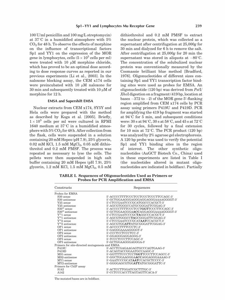
TABLE I. Sequences of Oligonucleotides Used as Primers orProbes for PCR Amplification and EMSA
Constructs Sequences
Probes for EMSAS30 sense 50-ACCCCTTTTCCCTCCTCCCTCCCTTCCAGC-30
S30 antisense 50-GCTGGAAGGGAGGGAGGAGGGAAAAGGGGT-30
Y25 sense 50-CTCCGAATCCCGCATGGCCCACGCT-30
Y25 antisense 50-AGCGTGGGCCATGCGGGATTCGGAG-30
S30m sense 50-ACCCCTTTTCCCTCCTGGTTCCCTTCCAGC-30
S30m antisense 50-GCTGGAAGGGAACCAGGAGGGAAAAGGGGT-30
Ym1 sense 50-CTCCGAATCCCGCTAGGCCCACGCT-30
Ym1 antisense 50-AGCGTGGGCCTAGCGGGATTCGGAG-30
Ym2 sense 50-CTCCGAATCCCGCATAATCCACGCT-30
Ym2 antisense 50-AGCGTGGATTATGCGGGATTCGGAG-30
OP1 sense 50-ACCCCTTTTCCCTC-30
OP1 antisense 50-GAGGGAAAAGGGGT-30
OP2 sense 50-CCCTCCTCCCTCC-30
OP2 antisense 50-GGAGGGAGGAGGG-30
OP3 sense 50-TCCCTCCCTTCCAGC-30
OP3 antisense 50-GCTGGAAGGGAGGGA-30
Primers for site-directed mutagenesis and EMSAP419U 50-ACCTCGAGAAGAGTGCCCAGTGAAG-30
P419D 50-ACAGTACCGGAATGCCAAGC-30
MS1-sense 50-GGTTTTCCCTCCTGGTTCCCTTCCAGCC-30
MS2-antisense 50-GGCTGGAAGGGAACCAGGAGGGAAAAG-30
MY1-sense 50-GAATCCCGCATAATCCACGCTCCCC-30
MY2-antisense 50-GGGGAGCGTGGATTATGCGGGATTC-30
Primers for ChIP assayS142 50-ACTCCTTGGATCGCTTTGC-30
A142 50-CCTCCCACCTTAGTAGTTCACA-30
The mutated bases are in boldface.
Sp1–YY1 and Lymphocytes Mu Receptor Gene 239
sequence-overlapping probes were used toidentify the precise Sp1 binding sites. Double-stranded oligonucleotides were generated byannealing the synthetic oligonucleotides withrespective complementary sequences. Comple-mentary oligonucleotides of equal quantity(2.5 mM each) were annealed in a thermocycler(Techgene, UK) at the following temperatures:888C, 2 min; 658C, 10 min; 378C, 10 min and258C, 5 min.
The 50-ends of the gel shift probes wereradiolabeled with g 32P-ATP using polynucleo-tide kinase (Promega). After labeling at 378C for10 min, the probes were purified with disposablecolumns containing Sephadex G-25 medium(Sigma Chemical Co.). Five micrograms ofthe nuclear extract was mixed with 2 mg ofsalmon sperm DNA, 2 ml of 5� binding buffer(50 mM Tris–HCl (pH 7.5), 250 mM NaCl,0.5 mM EDTA, 5 mM MgCl2, 20% glycerol,2.5 mM dithiothreitol) and H2O to a totalvolume of 9 ml for 10 min. Approximately0.2 mmol of the labeled DNA oligoprobe wasthen added to the nuclear mixture above to afinal volume of 10 ml, and incubated for 20 minat room temperature. Subsequently, the DNA–protein complexes were separated from theunbound DNA by electrophoresis via a 6%nondenaturing polyacrylamide gel in 0.5�TBE buffer at 350 V for 30 min. The gels weredried under vacuum and exposed for auto-radiography for 24 h at �808C. Competition ex-periments included the addition of a 125-foldexcess of unlabeled DNA oligonucleotides,while supershift analysis included the additionof 2 mg of the antibody against either Sp1 or YY1(Santa Cruz Biotechnology) to the reactionmixture for 30 min at room temperature beforethe addition of probes. A 125-fold excess ofunlabeled nonspecific probe was used as anonspecific reaction control. Mutated probeswere used to determine which base sequenceswere potential sites for protein binding incis-elements.
Verification of Sp1 and YY1 Capabilityfor DNA Binding by the ChromatinImmunoprecipitation (ChIP) Assay
CEM x174 cells were treated with 10 mMmorphine or/and 10 mM naloxone for 48 h. Thechromatin immunoprecipitation (ChIP) assaywas performed as described by Yin et al. [2004].CEM x174 cells (�2� 107 in a 75 ml cultureflask) were fixed with 1% formaldehyde for
12 min at 378C. The cell pellet was washedtwice with cold PBS and re-suspended incell lysis buffer (1% SDS, 5 mM EDTA, 50 mMTris–HCl at a pH of 8.1) containing 1% proteaseinhibitor cocktail (Sigma Chemical Co.). Thechromatin was sonicated (Ultrasonic Processorfrom Xinzhi Tech Ins, China) on ice with fivepulses of 10 s each with a 20 min interval toan average length of about 200–750 bp asdetermined by resolving the purified DNA on a1.5% agarose gel. The sample was then centri-fuged at 48C in an Eppendorf centrifuge (10 minat 15,000 rpm) to remove the cell debris from thecrude chromatin lysate. Twenty microliters ofthe lysate was diluted with 80 ml of dilutionbuffer (1% Triton X-100, 2 mM EDTA, 150 mMNaCl, 20 mM Tris–Cl at a pH of 8.1) and setaside as the input chromatin. The shearedchromatin from CEM x174 cells was diluted at1:10 and mixed with 50% protein A sepharose4B (Sigma Chemical Co.; about 50 ml in 1 mlchromatin), which was blocked with 2 mgof salmon sperm DNA at a concentration of2 mg/ml. After shaking 2 h, the sample wascentrifuged at 1,600 rpm for 5 min at 48C. Thesupernatant was collected and added eitherSp1 or YY1 polyclonal antibody to 2 mg/ml andshaken overnight at 48C. The sample was thenmixed again with 50% protein A sepharose4B (about 50 ml in 1 ml sample) and 2 mg ofsalmon sperm DNA to a final concentrationof 2 mg/ml. After shaking 1 h, the sample wascentrifuged at 1,600 rpm for 5 min to removethe supernatant. Pellets were consecutivelywashed with 1 ml of TSE I (0.1% SDS, 1%TritonX100, 2 mM EDTA, 20 mM Tris–HCl and150 mM NaCl at a pH of 8.1), TSE II (0.1% SDS,1% Triton X-100, 2 mM EDTA 20 mM Tris–HCland 500 mM NaCl at a pH of 8.1) and buffer III(1% NP40, 1% Deoxycholic acid, 1 mM EDTA,10 mM Tris–HCl and 0.25 M LiCl at a pH of 8.1)for 10 min each on a shaker at 48C. Antibody–protein–DNA complexes were eluted fromprotein A sepharose 4B with 100 ml of elutionbuffer (0.1 M NaHCO3, 1% SDS) by shaking on arotatory platform for 10 min. Eluted complexes,as well as the input chromatin, were incubatedin a water bath at 658C for 8 h to remove protein.DNA fragments were purified with a QiaquickPCR purification kit (QIAGEN) and stored at�208C until use. The PCR employed in the ChIPassay consisted of 25 ml of the PCR reaction mixcontaining 2 ml of the DNA template, 0.5 ml ofeach primer (S142 and A142; 0.5 mM), 2.5 ml of
240 Li et al.
PCR buffer (10�), 0.5 ml of 10 mM of dNTPs and1 ml of Taq polymerase (2.5 U/ml) which wassubjected to amplification in a thermocycler.The PCR parameters for the Sp1 and YY1binding regions were initially at 958C for2 min, followed by 30 cycles at 948C for 30 s fordenaturation, 528C for 30 s for annealing, and728C for 30 s to extend the DNA. The finalPCR amplified product was identified on a 2%agarose gel together with the 100 bp DNAladder (Promega). The predicted size was 142bp corresponding to bases �218 to �359 bp ofthe 50-flanking region of the MOR gene inlymphocytes. To further verify the effects ofmorphine on the influence of transcriptionalfactors Sp1 and YY1 on the expression of the mureceptor, cells were treated with 10 mM mor-phine chloride and/or 10 mM naloxone for 12 hbefore treatment with formaldehyde and ana-lyzed as described above.
Coimmunoprecipitation andSDS–PAGE/Western Blot Analysis
Coimmunoprecipitation (CoIP) experimentsto evaluated the interaction of Sp1 and YY1 wereperformed on cells solubilized in 1% Triton X-100, 0.5% deoxycholate, 0.1% SDS, 50 mM Tris,100 mM NaCl, 2 mM MgCl2, 10% glycerol, andproteinase inhibitor cocktail. For CoIPs of Sp1(or YY1), cells were solubilized in mediumcontaining 1% NP-40 in place of Triton andSDS. Lysates were clarified by centrifugation(50,000 rpm for 10 min with a TLA120.2 rotor)and then incubated overnight at 48C withantibody against YY1 (or Sp1; Santa CruzBiotechnology) bound to protein A-Sepharose.Non-immune rabbit IgG was also used as acontrol at the step. Immune complexes werewashed three times in solubilization mediumwithout DOC and SDS, washed once inphosphate-buffered saline, and eluted in samplebuffer containing 0.2 M DTT for SDS–PAGE.The samples were evaluatedby 12% SDS–PAGEelectrophoresis and Western blot analysis.Briefly, transfer of proteins from gels onto nitro-cellulose membrane (Amersham, UK) waselectrophoretically mediated in a transblottingcell at 48C for 2 h. Membranes were blocked byimmersion for 1 h in 5% non-fat milk (w/v)/PBS,and then incubated with horseradish peroxi-dase-conjugated anti-rabbit antibody (Promega)at room temperature for 1 h. Immunocomplexesresolved by electrophoresis were visualized byincubation of the membranes with Enhanced
Chemiluninescence (Zhongshang Boil Tech Co.,Beijing) and exposure on an X-ray film.
Plasmid Promoter Constructs andSite-Directed Mutagenesis
Creation of 50 deleted MOR-Luc fusion con-structs with the Luciferase Reporter Gene wasdescribed in our previous report as ‘‘Luc-5,’’ whichcontaining binding sites for Sp1 and YY1 [Weiet al., 2005]. Mutated MOR-Luc constructs of Sp1and YY1 were created by PCR using the mutationprimers MS1/MS2 and MY1/MY2 (Table I). Allconstructs were verified by DNA sequencing.
Transient Transfection andLuciferase Reporter Gene Assay
All the plasmids used in these transfectionexperiments were prepared by the Large-scalePurification Kit (Vigorous, China) following themanufacturer’s recommended protocol. Trans-fection efficiency was monitored by cotransfectionof the pRL-SV 40 promoter driven Renillaluciferase (Promega). CEM x174 cells were trans-fected by Lipofectamine 2000 Reagent Invitrogenas described in our previous report [Wei et al.,2005]. Briefly, the cells were pelleted and resus-pended in 50 ml of lysis buffer (Promega) at 36 hafter transfection. After one freeze/thaw cycle, thecell lysate (20 ml) was mixed with 100 ml of LAR IIreagent and the firefly luciferase activitywas measured as light output (for a 10 s interval)in a Centro LB 960 luminometer (Berthold,Germany). The Renilla luciferase activity wasestimated from the same lysates by the addition of100 ml of Stop and Glo reagent (Berthold), and thelight output (for a 10 s interval) was measuredseparately. The corrected pLuc promoter drivenluciferase activity was expressed as the ratio ofpLuc promoter driven luciferase activity to renillaluciferase activity. The promoter-less luciferasereporter vector (pLuc-basic) served as thenegative control. To observe the influence ofmorphine on expression of lymphocyte MOR,CEM x174 cells transfected with Luc 5 wasincubated with 10 mM morphine chloride and/or10 mM naloxone for 12 h before determination ofluciferase.
RESULTS
Identification of the Putative Sp1 and YY1 BindingRegion in the Lymphocyte MOR Promoter
To characterize the MOR promoter regionin lymphocytes, we amplified a 419 bp DNA
Sp1–YY1 and Lymphocytes Mu Receptor Gene 241

fragment containing the potential Sp1 and YY1binding region upstream of the human MORgene (GenBank accession number: AJ000341).The amplified fragment covers the region 377 bpupstream of the start codon which containsputative Sp1 and YY1 elements (Fig. 1A). Anoligonucleotide (120 bp) derived from PstI/XhoIdigestion of the 419 bp fragment was labeledwith [g-32P] and used as a probe to detect theputative binding sites for Sp1 and YY1 (Fig. 1B).A slow migrating band was observed in theEMSA experiment which could be specificallyabolished by competition with a 50-fold molarexcess of unlabeled oligonucleotide, but notby non-specific probe (Fig. 1C). The results
demonstrated the possibility of the existence ofbinding sites for Sp1 and YY1 proteins inMOR promoter region in lymphocytes. Also,complexes in various densities could be visual-ized with nuclear extracts from SY5Y neuronalcells (high expression of the MOR) and Helacells (no expression of the MOR; Fig. 1D).
Analysis of Binding of Sp1 and YY1 tothe MOR Promoter in Lymphocytes
To identify whether Sp1 element is able tobind to the proximal promoter of lymphocytes,complex formation with nuclear extracts fromCEM x174, SY5Y and Hela cells were compar-ed. The results showed that one complex was
Fig. 1. Verification of putative Sp1 and YY1 binding site inthe proximal promoter of the MOR gene in lymphocytes.A: Nucleotide sequence of the 50-region from the MOR gene.Nucleotide þ1 corresponds to the translation start codon, theboldfaced ATG. Underlined sequences represent the nucleotidesused as primers for PCR. Additional nucleotides with restrictionsites in the ends of primers are shown above or below thesequence. The putative Sp1 and YY1 cis-acting transcriptionelements are boxed and presented in italicized letters. Restrictionsites used for making the 120 bp probe are indicated by the closedtriangles. B: A fragment of 120 bp was visualized by amplificationfrom CEM x174 cells by PCR assay. M: DNA ladder; lane 1: PCR
product. C: EMSA experiment was performed with nuclearextracts from CEM x174 cells with a 120 bp probe. Lane 1, probealone; lane 2, probe plus 5 mg of total protein of nuclear extract;lane 3, 50-fold molar excess of unlabeled non-specific probe(N) plus 5 mg of total protein of nuclear extract; lane 4, 50-foldmolar excess of unlabeled double-stranded competitor (S) plus5 mg of total protein of nuclear extract. Weak bands are indicatedby horizontal arrows. D: Binding comparison of 120 bp probein different cell lines. ‘‘C’’ represents CEM x174 cells; ‘‘Y’’represents SY5Y cells; ‘‘H’’ represents Hela cells. All images wererepresentative of an experiment that was repeated three times.
242 Li et al.

visualized only in the lane of CEM x174 cells(Fig. 2A). Whereas, neither SY5Y neuronal cellsnor Hela cells showed any detectable bands.Non-specific probe was unable to abolishformation of the Sp1-complex (Fig. 2B). Asshown in Figure 2C, the Sp1-complex could beabolished by anti-Sp1 antibody, indicating thatSp1 proteins could bind to the region within�298 to �310 bp of the MOR promoter. To morespecifically identify the binding sites, threesequence overlap probes (OP1, OP2, and OP3)were designed as competitors. Since only crit-ical bases in probe could bind Sp1 protein innuclear extract, the precise Sp1 binding sitescould hereby be identified. The results showedthat the Sp1 complex could be abolished by OP2and OP3, but not by OP1, indicating that theCCC, the overlap region of OP2 and OP3, wascritical for the binding of Sp1 protein. Inter-estingly, if OP3 was used as the competitor,
another abolished region could be observed,indicating the existence of an additional, as yetunidentified, factor modulating the promoteractivity (Fig. 2C). We mutated the CCC of theOP2 probe to GGT and observed the effect ofthe mutated probe as a competitor (S30m).However, a 125-fold molar excess of unlabeledS30m was unable to compete with the bindingsite for the Sp1 complex, confirming the role ofCCC within the Sp1 binding site (Fig. 2D).
To analyze the role of the YY1 site inlymphocyte MOR promoter activity, we gene-rated a double-strand oligonucleotide (25 bp)as a probe, which contained the YY1 bindingsite and spanned the region between �268 and�286 bp. The comparison of the formation ofYY1 complex showed that a dense band wasobserved in the lane of CEM x174 cells com-pared with that of SY5Y and Hela cells (Fig. 3A).Combined with the results of Sp1, it was
Fig. 2. Binding of Sp1 transcription factor in the lymphocyteMOR promoter. Five micrograms of total proteins of nuclearextract from CEM x174 cells were used in the EMSA experiment.A: Comparison of probe S30 binding to Sp1 from different celllines. ‘‘C’’ represents CEM x174 cells; ‘‘Y’’ represents SY5Y cells;‘‘H’’ represents Hela cells. B: lane 1, probe S30 alone; lane 2,probe S30 plus nuclear extract; lane 3, probe S30 with nuclearextract plus 125-fold molar excess of unlabeled double-strandedcompetitor; lane 4, probe S30 with nuclear extract plus 125-foldmolar excess of unlabeled non-specific probe. ‘‘S’’ representsunlabeled competitor; ‘‘N’’ represents unlabeled non-specific
probe. C: lane 1, probe S30 alone; lane 2, S30 plus nuclearextract; lane 3, probe with nuclear extract plus 2 mg of anti-Sp1antibody; lanes 4–6, probe with nuclear extract plus varioussequence-overlapping probes as competitors. Upper arrowindicates the major complex. Lower arrow indicates theunidentified complex. D: The CCC of the OP2 was mutated toGGT as a competitor (S30m). Lane 1, OP2 probe only; lane 2,OP2 plus nuclear extract; lane 3, S30m with OP2; lane 4, S30m
with OP2 and nuclear extract. All images were representative ofan experiment that was repeated three times.
Sp1–YY1 and Lymphocytes Mu Receptor Gene 243

speculated that the mechanism in regulation ofthe expression of the MOR gene in lymphocyteswas different from other cell lines. Figure 3Bshows the specific complex which couldbe abolished by unlabeled competitor probe(125-fold excess), but not by non-specific probe(Fig. 3B). Since the sequence for the binding ofYY1 has been listed as having AGATGGC asthe consensus base sequence 9 [Emanuele et al.,1998], we generated two different mutations of
the probe (Ym1 and Ym2) to verify the functionalbases in the YY1 binding site. If Ym1 (mutatedfrom AT to TA) was used as probe, YY1 complexwas still detected. Accordingly, unlabeled Ym1could abolish the binding of wild-type probe,indicating the AT was not necessary for thebinding of YY1 (Fig. 3C). However, the mutationfrom GGC to AAT when tested as a competitorwas unable to efficiently compete with theformation of the complex of the wild-type
Fig. 3. Binding of transcription factor YY1 in the lymphocyteMOR promoter. Five micrograms of total protein of nuclearextract from CEM x174 cells were used in this EMSA experiment.A: Comparison of probe Y25 binding to YY1 from different celllines. ‘‘C’’ represents CEM x174 cells; ‘‘Y’’ represents SY5Y cells;‘‘H’’ represents Hela cells. B: lane 1, probe Y25 alone; lane 2,probe Y25 plus nuclear extract; lane 3, probe Y25 with nuclearextract plus 125-fold molar excess of unlabeled double-strandedcompetitor; lane 4, probe Y25 with nuclear extract plus 125-foldmolar excess of unlabeled non-specific probe. ‘‘S’’ representsunlabeled competitor; ‘‘N’’ represents unlabeled non-specificprobe. C: lane 1, probe Y25 alone; lane 2, probe Y25 plus nuclear
extract; lane 3, mutated YY1 probe (Ym1) alone; lane 4, probeYm1 plus nuclear extract; lane 5, probe Y25 with nuclear extractplus 125-fold molar excess of unlabeled probe Ym1. D: lane 1,probe Y25 alone; lane 2, Y25 plus nuclear extract; lane 3, probeY25 with nuclear extract plus 125-fold molar excess of unlabeledY25 competitor; lane 4, probe Y25 with nuclear extract plus125-fold molar excess of unlabeled Ym2 competitor. E: lane 1,probe Y25 alone; lane 2, Y25 plus nuclear extract; lane 3, probeY25 with nuclear extract plus 2 mg of anti-YY1 antibody. YY1complex was indicated by an arrow in each panel. All imageswere representative of an experiment that was repeatedthree times.
244 Li et al.

sequence, indicating the importance of GGC inthe binding of YY1 (Fig. 3D). Supershift EMSAwas performed to determine the specificity ofthe binding of YY1 protein to its putativebinding sites. The data showed that the retarda-tion caused by labeled probe could be signi-ficantly reduced by the addition of YY1 antibody(Fig. 3E). In concert, the data from the EMSAand supershift EMSA analysis confirmed thatSp1 and YY1 proteins bind to their putativebinding sites in the MOR gene promoter.
Effects of Morphine on the Formationof Sp1 and YY1 Complex
To investigate the regulatory mechanisms ofmorphine on the expression of the MOR inlymphocytes, 10 mM morphine chloride or/and 10 mM naloxone (30 min before the additionof morphine) was added into cultured CEMx174 cells for 12 h before nuclear extraction. Asshown in Figure 4A,B, morphine significantly
elevated the formation of Sp1 and YY1 com-plexes. However, compared with the anta-gonistic effect of naloxone on YY1 (Fig. 4B),no detectable change in Sp1 was observed(Fig. 4A). It seems like that naloxone does notalways antagonize the effect of morphine,particularly at the molecular level. An additiverather than antagonistic effect of naloxone withmorphine on the expression of some genes hasalso been previously reported [Xu et al., 2004].
Effects of Morphine on the Binding of Sp1and YY1 to the Lymphocyte MOR Promoter
Chromatin immunoprecipitation analysiswas performed in order to confirm that theeffect of morphine on the binding of Sp1 and YY1to the MOR promoter occurs in vivo. For theseexperiments, CEM x174 cells were treated withmorphine or/and naloxone as describe above for12 h, and then cross-linked with formaldehydeto fix the DNA–protein complexes. Next, cellswere sonicated to shear the DNA fragments,and then immunoprecipitation with antibodiesspecific to Sp1 and YY1 was performed. Finally,the cross-links were reversed, and the DNA waspurified and used as template for PCR ampli-fication. Primers S142 and A142 which con-tained the putative Sp1 and YY1 binding siteswere used and listed in Table I. The data fromthe ChIP assay showed that there was more Sp1protein binding its antibody after treatmentwith morphine. Consistent with the results ofthe EMSA, the antagonistic effect of naloxonewas not apparent (Fig. 5A). Similarly, theeffects of morphine on YY1 could be blocked bynaloxone (Fig. 5B). The fact that the Sp1 andYY1 antibodies could immunoprecipitate theSp1 and YY1 proteins binding to the MORpromoter region indicated that both Sp1 andYY1 transcription factors were involved in theexpressional regulation of the MOR gene inCEM x174 cells.
Evaluation of Interaction of TranscriptionFactor Sp1 and YY1
To reveal the occurrence of an interactionbetween Sp1 and YY1 at the protein level, CoIPanalysis was performed. Putative Sp1–YY1protein complexes were immunoprecipitatedwith anti-Sp1 or (anti-YY1) antibody and ana-lyzed by Western blotting. Immunoprecipita-tion with anti-YY1 led to the detection of a Sp1band and vice versa (Fig. 6, lanes 3 and 6).Whereas no Sp1 or YY1 was detected when we
Fig. 4. Effect of morphine on the formation of Sp1 and YY1complexes. Five micrograms of total protein of nuclear extractfrom CEM x174 cells was used in this EMSA experiment. A: lane1, probe S30 alone; lane 2, probe S30 plus nuclear extract; lane 3,treated with 10 mM morphine; lane 4, treated with 10 mMmorphine and 10 mM naloxone; lane 5, treated with 10 mMnaloxone alone. B: lane 1, probe Y25 alone; lane 2, probeY25 plus nuclear extract; lane 3, treated with 10 mM morphine;lane 4, treated with 10 mM morphine and 10 mM naloxone; lane5, treated with 10 mM naloxone alone. YY1 complex is indicatedby an arrow in each panel. M: morphine treated group;N: naloxone treated group; MN: morphine plus naloxone treatedgroup. All images were representative of an experiment that wasrepeated three times.
Sp1–YY1 and Lymphocytes Mu Receptor Gene 245

used a non-immune rabbit IgG as a control forthe immunoprecipitation (Fig. 6, lanes 1 and 5),indicating the specificity of the interactionbetween Sp1 and YY1 proteins. These resultssuggested the possibility of the interaction inthe regulation of expression of the MOR gene inlymphocytes.
Functional Analysis of the Mutation ofSp1 and YY1 Elements on the Activity
of the Lymphocyte MOR Promoter
To investigate the underlying role of Sp1 andYY1 elements in the lymphocyte MOR pro-moter, we introduced either a Sp1 mutation or aYY1 mutation, or both into a MOR-Luc con-struct containing the MOR promoter sequence
(Fig. 7A). Constructs with wild-type andmutated sites for Sp1 or/and YY1 were trans-fected into CEM x174 cells. Promoter activitywas determined by measuring luciferaselevels in transfected cells and normalizedwith cotransfected pRL-null Renilla luciferaseactivity. Data showed that Luc-5 with wild-typeSp1 and YY1 binding sites expressed a higherlevel of luciferase activity in CEM x174 cells(Fig. 7A). However, promoter activity from aconstruct containing a mutation either in theSp1 or YY1 element was significantly lessthan with the wild type construct. A constructmutated in both Sp1 and YY1 binding sitesresulted in the lowest level of luciferase activityand compared with Luc-5, the decrease inactivity was approximately 60% (P< 0.01).These results implied that the interaction oftranscription factors Sp1 and YY1 was anecessary event in the initiation of transcrip-tion of the MOR gene in lymphocytes. Morphinetreatment was able to stimulate the activityof luciferase in Luc-5 transfected cells, whichwas reversed by naloxone (Fig. 7B). The resultindicated that the region within �298 to�310 bp upstream of the start codon containingSp1 and YY1 elements might be one of sitesmorphine affected through influencing Sp1 andYY1. All finding taken together revealed thatthe bases CCT of Sp1 and the bases GGC of YY1elements in the promoter region were critical
Fig. 5. Confirmation of Sp1 and YY1 binding to the lymphocyteMOR promoter region with the Chromatin Immunoprecipitationassay (ChIP). Formaldehyde cross linked chromatin from CEMx174 cells treated with 10 mM morphine or/and 10 mM naloxonewas immunoprecipitated with anti-Sp1 or anti-YY1 antibody andsubjected to PCR as described under Materials and MethodsSection. PCR products were identified in a 2% agarose gel usingprimers containing the Sp1- or YY1-binding region of thelymphocyte MOR proximal promoter. Input represents totalchromatin applied for immunoprecipitation. A: lane 1, 100 bpDNA ladder; lanes 2–5, input of groups treated with 10 mMmorphine or/and 10 mM naloxone; lanes 6–9, PCR productdetected in the CEM x174 cells treated with 10 mM morphine or/and 10 mM naloxone, from which cellular chromatin complexeswere immunoprecipitated with anti-Sp1 antibody; lane 10,‘None’ represents a negative control of the CHIP assay withoutchromatin. Amplified product (142 bp) is indicated by an arrow.B: lane 1, 100 bp DNA ladder; lanes 2–5, input of groups treatedwith 10 mM morphine or/and 10 mM naloxone; lanes 6–9, PCRproduct detected in the CEM x174 cells treated with 10 mMmorphine or/and 10 mM naloxone, from which cellularchromatin complexes were immunoprecipitated with anti-YY1antibody; lane 10, ‘None’ represents a negative control of theChIP assay without chromatin. The 142 bp amplified product isindicated by an arrow. C: control; M: morphine treated group;N: naloxone treated group; MN: morphine plus naloxone treatedgroup. All images were representative of an experiment that wasrepeated three times.
Fig. 6. Coimmunoprecipitation analysis of the interaction oftranscription factors Sp1 and YY1. Lysates from CEM x174cells were immunoprecipitated (IP) with antibodies to Sp1 or YY1protein, or non-immune rabbit IgG (as indicated above eachlane) and separated on a SDS/PAGE gel. Coimmunoprecipitatedcomplexes were transferred to a nitrocellulose membrane,immunoblotted with anti-Sp1 (lanes 1–4) or anti-YY1 (lanes 5–8) antibody and exposed to X-ray film. Lanes 1 and 5,coimmunoprecipitated Sp1–YY1 complex with proteinA-Sepharose beads precoated with non-immune rabbit IgG andprobed with antibody to Sp1 (lane 1) or YY1 (lane 5); lanes 2 and6, coimmunoprecipitated Sp1–YY1 complex with proteinA-Sepharose beads precoatedwith anti-Sp1 antibody and probedwith antibody to Sp1 (lane 2) or YY1 (lane 6); lanes 3 and 7,coimmunoprecipitated Sp1–YY1 complex with proteinA-Sepharose beads precoated with anti-YY1 antibody andprobed with antibody to Sp1 (lane 3) or YY1(lane 7); lanes 4and 8, total protein immunoblotted with antibody to Sp1 (lane 4)or YY1(lane 8) as input. Image was representative of anexperiment that was repeated three times.
246 Li et al.

bases for the protein–DNA interaction(Fig. 7C). Morphine was capable to regulatethe expression of lymphocyte MOR throughaffecting the binding of Sp1 and YY1 to theirelements in promoter region.
DISCUSSION
Over the past decade there has been asubstantial increase in our understanding ofthe general genomic structure of the MOR geneand its 50 regulatory region [Liang et al., 1995;Wendel and Hoehe, 1998]. All of the advances inthis field have been helpful in explaining the
characteristic effects of the mu receptor inter-acting with its ligands in the nervous system,which include reward, tolerance, dependenceand analgesia. However, the evidence explain-ing immune regulation by opioid at the levelof transcription is still limited. It is likely thata dissimilar regulatory mechanism for theexpression of the MOR exists in lymphocytes.For example, as compared with the multipletranscription initiation sites found in rodentbrain cells and in human neuroblastoma SK-N-SH cells, only one initiation site has beendetected in lymphocytes, which might be thedirect reason accounting for the lower number
Fig. 7. Lymphocytes MOR promoter activity in transienttransfection assays. A series of MOR promoter/luciferase con-structs were prepared and introduced into CEM x174 cells. Afterculture for 24 h, cells were harvested for the luciferase activityassay. A: Various constructs containing MOR promoter withdeleted or mutated Sp1 or YY1 binding sites. The sequencesrepresent the potential Sp1 or YY1 cis-acting elements. Mutatedbases are italicized. M-Sp1: mutated Sp1 binding site; M-YY1:mutated YY1 binding site; M-SY: mutated in both Sp1 and YY1binding sites. The promoter activity of each construct wasstandardized by cotransfection of the internal control plasmid,renilla luciferase, and was expressed as the ratio of pRL-SV40 promoter driven renilla luciferase activity to the pLucpromoter driven firefly luciferase activity. B: The activity ofluciferase in Luc-5 transfected CEM x174 cells stimulated bymorphine. Luc5þM: morphine treated group; Luc5þMþN:morphine plus naloxone treated group; Luc5þN: naloxonetreated group. The bars in panel A and B showed the meanþ SD
of the results from four independent transfection experiments.*P<0.05 and **P< 0.01 was considered statistically significantbetween construct Luc-5 and all other constructs (A) or betweenmorphine treated group and all other groups (B). ##P<0.01between morphine treated group and morphine plus naloxone ornaloxone treated group (B). C: Schematic representing the role oftranscription factors Sp1 and YY1 in the initiation of the MORgene expression in lymphocytes. Both Sp1 and YY1 bind to theircis-elements in the proximal promoter of the lymphocyte MORgene and interact with each other. This interaction presumably isrelated to the activity of RNA polymerase II and the transcriptionof the MOR gene in lymphocytes is thereby triggered. The presentbases represent the core sequences for binding of transcriptionfactors. ATG represents the start codon. Upper arrow indicatesthe direction of transcription. Lower arrow indicates the possibleinitiation site of transcription about 110 bp upstream of thetranslation start codon [Wei et al., 2005]. The possible effects ofmorphine and naloxone are indicated.
Sp1–YY1 and Lymphocytes Mu Receptor Gene 247

of copies of the MOR transcripts in lymphocytesand the difficulty in detection of MOR mRNA inlymphocytes [Min et al., 1994; Liang et al., 1995;Andria and Simon, 1999; Madden et al., 2001;Wei et al., 2005]. In addition, two cis-actingelements, namely Sp1 and YY1 boxes, werepredicted to reside immediately upstream of thetranslation start site of the MOR gene in CEMx174 cells, and were identified as critical forcontrolling promoter activity of the MOR genein lymphocytes [Wei et al., 2005].
To clarify the mechanism underlying themorphine-stimulated transcription of the MORgene in lymphocytes, we focused on analysis ofthese two elements. The EMSA data showed thatthe putative Sp1- and YY1-binding sites in theregion of�372 to�2 bp identified inthesestudiesare required for Sp1 and YY1 binding. The factthat the binding of transcription factors to theproximal promoter is different in various cellscan partially explain the expressional differenceof the MOR gene. It has been reported that thepromoter of the MOR gene lacks a consensusTATA box and initiator [Gill and Tjian, 1992;Javahery et al., 1994]. Other analysis alsoshowed that this promoter does in fact possessa canonical Sp1 binding site and it has beenproven that the Sp1 protein can initiate trans-cription through binding the Sp1 element in aTATA-less promoter [Papadodima et al., 2005;Xia et al., 2005]. Using sequence-overlappingoligonucleotide as probes, we determined thatCCC was the critical base sequence for Sp1binding, and the mutation of this sequenceresulted in a loss of Sp1 binding. These resultsdemonstrated the precise binding site of trans-cription factor Sp1 in lymphocytes, which werenot described by other similar work in nerve celllines.
YY1 is a multifunctional transcription factorthat exerts positive and negative control on alarge number of cellular and viral genes bybinding to sites overlapping the transcriptionstart site [Goffart and Wiesner, 2003]. It isreported that YY1 regulates transcription inthree ways. It can activate, repress, or initiategene transcription depending on the promoterwhich is involved [Breslin and Vedeckis, 1998].Furthermore, the activity of YY1 in activation oftranscription may be related to the presence of aswitch region. Deletion of this region causesYY1 to act as a repressor of promoter activity[Bauknecht et al., 1995]. Several proposals formodels of YY1-mediated transcription of gene
have been put forward [Thomas and Seto, 1999;Sui et al., 2004]. Although the presence of theYY1 element has been identified within theproximal promoter of the MOR gene in lympho-cytes, its precise function in the initiationof transcription, particularly in regard to itsinteraction with other trans-factors, is as yet notunderstood in detail. Our data from mutationassay of EMSA and transfection experimentshas shown that transcription factor YY1 func-tions as an initiator of the MOR gene inlymphocytes with the core sequence GGCrather than AT as has been reported elsewhere[Gill and Tjian, 1992].
The latter part of our study was designed todefine the mechanism by which morphinetriggers the interaction of Sp1 and YY1, herebyinitiating the transcription of the MOR gene inlymphocytes. Despite accumulated knowledgeregarding the effect of morphine in lympho-cytes, neither the pharmacological and immu-nological findings, nor clinical studies in drugsabuse account fully for the mechanism of theMOR gene expression triggered by morphine inlymphocytes. Our previous work demonstratedthe relationship between morphine treatmentdose and the level of expression of the MOR genein lymphocytes, which was found to be relatedto the critical elements within the proximalpromoter [Wei et al., 2005]. In this study, thelevel of both Sp1 and YY1 proteins could be up-regulated by morphine. It is of interest that theblocking effect of naloxone on morphine could beobserved on the YY1 but not on the Sp1 protein.These results were consistent in data from bothEMSA and ChIP assays. Thus, these resultssuggest that the counteraction of the morphine-induced expression of the MOR gene by nalox-one is achieved through down-regulation ofthe YY1 protein, accounting partially for theblocking mechanism of naloxone. The precisemechanism by which this occurs will be a majorfocus of our future research.
Our previous work demonstrated the impor-tance of the proximal promoter containing Sp1and YY1 boxes on the transcriptional activityof the lymphocyte MOR promoter. There haspreviously been description of the physicalinteraction of these two transcription factorsin other work [Lee et al., 1993], and in fact, thisinteraction in the MOR proximal promoterin lymphocytes was existed as indicated in thepresent study by CoIP analysis. However, thefunctional interaction of these two molecules
248 Li et al.

remains as the focus of our ongoing study. Thefunctional significance of the critical bases onSp1 and YY1 binding sites was confirmed in thecurrent study by transfection assay of mutatedconstructs. As the double mutation of criticalbases on both Sp1 and YY1 binding sitesresulted in a decrease of luciferase activity, itis likely that the transcription of the MOR genein lymphocytes is regulated by a cooperativeinteraction of Sp1 and YY1 transcriptionfactors.
As reported previously, the MOR in lympho-cytes could be up-regulated by morphine[Suzuki et al., 2000]. However, the precisemolecular mechanism was not clarified. Ourdata that showed a significant elevation inthe formation of Sp1 and YY1 complexes bymorphine indicated one of possible explana-tions. Interestingly, the fact that the effect ofmorphine on the level of Sp1 was not abolishedby naloxone indicated that the expression ofthe MOR gene in lymphocytes is elaboratelyregulated.
In summary, the present study reveals themechanism affecting the activity of the proxi-mal promoter on transcription of the MOR genein lymphocytes. By identifying the functionalbases of Sp1 and YY1 binding sites, the relation-ship between protein–protein interaction andtranscriptional activity of the MOR promoter,and also the effect of morphine on Sp1 andYY1, we have been able to describe a part ofthe complicated mechanism for regulation ofmorphine on the expression of the MOR gene inlymphocytes.
ACKNOWLEDGMENTS
This work is supported by the NationalNatural Science Foundation of China (Nos.30671856 and 30772536) and The Foundationof National Education Ministry for graduateprogram (No. 20030001028).
REFERENCES
Andria ML, Simon EJ. 1999. Localization of promoterelements in the human mu-opioid receptor gene andregulation by DNA methylation. Brain Res Mol BrainRes 70:54–65.
Bauknecht T, Jundt F, Herr I, Oehler T, Delius H, Shi Y,Angel P, Zur Hausen H. 1995. A switch region deter-mines the cell type-specific positive or negative action ofYY1 on the activity of the human papillomavirus type 18promoter. J Virol 69:1–12.
Beagles K, Wellstein A, Bayer B. 2004. Systemic morphineadministration suppresses genes involved in antigenpresentation. Mol Pharmacol 65:437–442.
Borner C, Stumm R, Hollt V, Kraus J. 2007. Comparativeanalysis of mu-opioid receptor expression in immune andneuronal cells. J Neuroimmunol 188:56–63.
Bradford MM. 1976. A rapid and sensitive method for thequantitation of microgram quantities of protein utilizingthe principle of protein–dye binding. Anal Biochem 72:248–254.
Breslin MB, Vedeckis WV. 1998. The human glucocorticoidreceptor promoter upstream sequences contain bindingsites for the ubiquitous transcription factor, Yin Yang 1.J Steroid Biochem Mol Biol 67:369–381.
Cadet P. 2004. Mu opioid receptor subtypes. Med Sci Monit10:MS28–M32.
Choe CY, Im HG, Ko JL, Loh HH. 1998. Mouse m opioidreceptor gene expression. J Biol Chem 273:34926–34932.
Choi HS, Hwang CK, Kim CS, Song KY, Law PY, Wei LN,Loh HH. 2005. Transcriptional regulation of mouse muopioid receptor gene: Sp3 isoforms (M1, M2) function asrepressors in neuronal cells to regulate the mu opioidreceptor gene. Mol Pharmacol 67:1674–1683.
Emanuele DR, Giuseppe P, Olga CV, Elena B. 1998. Thebinding sites for Xenopus laevis FIII/YY1 in the first exonof L1 and L14 ribosomal protein genes are dispensable forpromoter expression. Eur J Biochem 255:563–569.
Gill G, Tjian R. 1992. Eukaryotic coactivators associatedwith the TATA box binding protein. Curr Opin Genet Dev2:236–242.
Goffart S, Wiesner RJ. 2003. Regulation and coordinationof nuclear gene expression during mitochondrial bio-genesis. Exp Physiol 88:33–40.
Hu S, Sheng WS, Lokensgard JR, Peterson PK. 2005.Morphine potentiates HIV-1 gp120-induced neuronalapoptosis. J Infect Dis 191:886–889.
Javahery R, Khachi A, Lo K, Zenzie Gregory B, Smale ST.1994. DNA sequence requirements for transcriptionalinitiator activity in mammalian cells. Mol Cell Biol14:116–127.
Ko JL, Liu HC, Minnerath SR, Loh HH. 1998. Transcrip-tional regulation of mouse m-opioid receptor gene. J BiolChem 273:27678–27685.
Koga T, Suicol AM, Nakamura H, Taura M, Lu Z, Shuto T,Okiyoneda T, Kai H. 2005. Sp1 dependent regulation ofMyeloid Elf-1 like factor in human epithelial cells. FEBSLett 579:2811–2816.
Lee JS, Galvint KM, Shit YS. 1993. Evidence for physicalinteraction between the zinc-finger transcription factorsYY1 and Spl. Proc Natl Acad Sci USA 90:6145–6149.
Li PF, Hao YS, Huang DA, Liu XH, Liu SL, Li G. 2003.Morphine-promoted survival of CEM x174 cells in earlystages of SIV infection in vitro: Involvement of themultiple molecular mechanisms. Toxicol Vitro 18:449–456.
Liang Y, Mestek A, Yu L, Carr LG. 1995. Cloning andcharacterization of the promoter region of the mouse muopioid receptor gene. Brain Res 679:82–88.
Madden JJ, Whaley WL, Ketelsen DK, Donahoe RM. 2001.The morphine-binding site on human activated T-cells isnot related to the mu opioid receptor. Drug AlcoholDependence 62:131–139.
Mahajan SD, Schwartz SA, Aalinkeel R, Chawda RP,Sykes DE, Nair MP. 2005. Morphine modulates
Sp1–YY1 and Lymphocytes Mu Receptor Gene 249

chemokine gene regulation in normal human astrocytes.Clin Immunol 115:323–332.
McCarthy L, Szabo I, Nitsche JF, Pintar JE, Rogers TJ.2001. Expression of functional mu-opioid receptorsduring T cell development. J Neuroimmunol 114:173–180.
Min BH, Augustin LB, Felsheim RF, Fuchs JA, Loh HH.1994. Genomicstructure analysis of promotersequence ofa mouse opioid receptor gene. Proc Natl Acad Sci USA91:9081–9085.
Nelson BD, Luciakova K, Li R, Betina S. 1995. The roleof thyroid hormone and promoter diversity in theregulation of nuclear encoded mitochondrial proteins.Biochim Biophys Acta 1271:85–91.
Pampusch MS, Osinski MA, Brown DR, Murtaugh MP.1998. The porcine mu opioid receptor: Molecular cloningand mRNA distribution in lymphoid tissues. J Neuro-immunol 90:192–198.
Pan YX. 2005. Diversity and complexity of the mu opioidreceptor gene: Alternative pre-mRNA splicing andpromoters. DNA Cell Biol 24:736–750.
Papadodima O, Sergaki M, Hurel C, Mamalaki A, MatsasR. 2005. Characterization of the BM88 promoter andidentification of an 88 bp fragment sufficient to driveneurone-specific expression. J Neurochem 95:146–159.
Royal W, Leander MV, Bissonnette R. 2005. Retinoid-induced mu opioid receptor expression by phytohemag-glutinin-stimulated U937 cells. J Neurovirol 11:157–165.
Sedqi M, Roy S, Ramakrishnan S, Elde R, Loh HH. 1995.Complementary DNA cloning of a mu-opioid receptorfrom rat peritoneal macrophages. Biochem Biophys ResCommun 209:563–574.
Sui GC, Affar EB, Shi YJ, Brignone C, Wal NR, Yin P,Donohoe M, Luke MP, Calvo D, Grossman SR, Shi Y.2004. Yin Yang 1 is a negative regulator of p53. Cell 117:859–872.
Suzuki S, Miyagi T, Chuang TK, Chuang LF, Doi RH,Chuang RY. 2000. Morphine upregulates mu opioidreceptors of human and monkey lymphocytes. BiochemBiophys Res Commun 279:621–628.
Suzuki S, Chuang LF, Yau P, Doi RH, Chuang RY. 2002.Interactions of opioid and chemokine receptors: Oligome-rization of mu, kappa, and delta with CCR5 on immunecells. Exp Cell Res 280:192–200.
Suzuki S, Chuang LF, Doi RH, Chuang RY. 2003. Morphinesuppresses lymphocyte apoptosis by blocking p53-
mediated death signaling. Biochem Biophys Res Com-mun 308:802–808.
Szabo I, Wetzel MA, Zhang N, Steele AD, Kaminsky DE,Chen C, Liu LY, Bednar F, Henderson EE, Howard OM,Oppenheim JJ, Rogers TJ. 2003. Selective inactivation ofCCR5 and decreased infectivity of R5 HIV-1 strainsmediated by opioid-induced heterologous desensitization.J Leukoc Biol 74:1074–1082.
Thomas MJ, Seto E. 1999. Unlocking the mechanisms oftranscription factor YY1 are chromatin modifyingenzymes the key. Gene 236:197–208.
Waldhoer M, Bartlett SE, Whistler JL. 2004. Opioidreceptor. Annu Rev Biochem 73:953–990.
Wang JB, Johnson PS, Persico AM. 1994. Hawkins AL,Griffin CA, Uhl GR. cDNA and genomic clones pharma-cologic characterization and chromosomal assignment.FEBS Lett 338:217–222.
Wang X, Tan N, Douglas SD, Zhang T, Wang YJ, Ho WZ.2005. Morphine inhibits CD8þ T cell-mediated, non-cytolytic, anti-HIV activity in latently infected immunecells. J Leukoc Biol 78:772–776.
Wei EM, Li PF, Liu XH, Qian CW, Li H, Xia WY, Li G. 2005.The potential promoter regions on the 50-flankingsequence of the mu opioid receptor gene in lymphocytes.J Cell Biochem 95:1204–1213.
Wendel B, Hoehe MR. 1998. The human mu opioid receptorgene: 50 regulatory and intronic sequences. J Mol Med 76:525–532.
Xia T, Zeng G, Gao L, Yu RK. 2005. Sp1 and AP2 enhancepromoter activity of the mouse GM3-synthase gene. Gene351:109–118.
Xu YH, Carr LG. 2000. Functional characterization of thepromoter region of the human m opioid receptor (hMOR)gene; identification of activating and inhibitory regions.Cell Mol Biol 47:OL29–OL38.
Xu J, Li PF, Liu XH, Li G. 2004. Morphine aggravates theapoptosis of simian immunodeficiency virus infectedCEM x174 cells in the prolonged culture in vitro. IntImmunopharmacol 4:1805–1816.
Yin N, Wang D, Zhang H, Yi X, Sun XJ, Shi B, Wu JH, WuG, Wang XJ, Shang YF. 2004. Molecular mechanismsinvolved in the growth stimulation of breast cancer cellsby leptin. Cancer Res 64:5870–5875.
Zaid A, Li R, Luciakova K, Barath P, Nery S, Nelson BD.1999. On the role of the general transcription factor Sp1in the activation and repression of diverse mammalianoxidative phosphorylation genes. J Bioenerg Biomembr31:129–135.
250 Li et al.