the role of the c8 proton of atp in the catalysis of
TRANSCRIPT

Kenyon and Roth BMC Biochemistry 2012, 13:15http://www.biomedcentral.com/1471-2091/13/15
RESEARCH ARTICLE Open Access
The role of the C8 proton of ATP in the catalysisof shikimate kinase and adenylate kinaseColin P Kenyon* and Robyn L Roth
Abstract
Background: It has been demonstrated that the adenyl moiety of ATP plays a direct role in the regulation of ATPbinding and/or phosphoryl transfer within a range of kinase and synthetase enzymes. The role of the C8-H of ATPin the binding and/or phosphoryl transfer on the enzyme activity of a number of kinase and synthetase enzymeshas been elucidated. The intrinsic catalysis rate mediated by each kinase enzyme is complex, yielding apparent KMvalues ranging from less than 0.4 μM to more than 1 mM for ATP in the various kinases. Using a combination ofATP deuterated at the C8 position (C8D-ATP) as a molecular probe with site directed mutagenesis (SDM) ofconserved amino acid residues in shikimate kinase and adenylate kinase active sites, we have elucidated amechanism by which the ATP C8-H is induced to be labile in the broader kinase family. We have demonstrated thedirect role of the C8-H in the rate of ATP consumption, and the direct role played by conserved Thr residuesinteracting with the C8-H. The mechanism by which the vast range in KM might be achieved is also suggested bythese findings.
Results: We have demonstrated the mechanism by which the enzyme activities of Group 2 kinases, shikimatekinase (SK) and adenylate kinase 1 (AK1), are controlled by the C8-H of ATP. Mutations of the conserved threonineresidues associated with the labile C8-H cause the enzymes to lose their saturation kinetics over the concentrationrange tested. The relationship between the role C8-H of ATP in the reaction mechanism and the ATP concentrationas they influence the saturation kinetics of the enzyme activity is also shown. The SDM clearly identified the aminoacid residues involved in both the catalysis and regulation of phosphoryl transfer in SK and AK1 as mediated byC8H-ATP.
Conclusions: The data outlined serves to demonstrate the “push” mechanism associated with the control of thesaturation kinetics of Group 2 kinases mediated by ATP C8-H. It is therefore conceivable that kinase enzymesachieve the observed 2,500-fold variation in KM through a combination of the various conserved “push” and “pull”mechanisms associated with the release of C8-H, the proton transfer cascades unique to the class of kinase inquestion and the resultant/concomitant creation of a pentavalent species from the γ-phosphate group of ATP. Alsodemonstrated is the interplay between the role of the C8-H of ATP and the ATP concentration in the observedenzyme activity. The lability of the C8-H mediated by active site residues co-ordinated to the purine ring of ATPtherefore plays a significant role in explaining the broad KM range associated with kinase steady state enzymeactivities.
* Correspondence: [email protected], Biosciences, Meiring Naude Road, Pretoria 0001Gauteng, South Africa
© 2012 Kenyon and Roth; licensee BioMed Central Ltd. This is an Open Access article distributed under the terms of theCreative Commons Attribution License (http://creativecommons.org/licenses/by/2.0), which permits unrestricted use,distribution, and reproduction in any medium, provided the original work is properly cited.
Kenyon and Roth BMC Biochemistry 2012, 13:15 Page 2 of 16http://www.biomedcentral.com/1471-2091/13/15
BackgroundIt has been demonstrated that the adenyl group of ATPplays a direct role in the control of ATP binding and/orphosphoryl transfer within a range of kinase and synthe-tase enzymes [1,2]. The role of the ATP C8-H in thebinding and/or phosphoryl transfer activity of a numberof kinase and synthetase enzymes was elucidated incomparative enzyme activity assays using ATP and ATPdeuterated at the C8 position. These comparisons dem-onstrated that a primary kinetic isotope effect (KIE) wasinvolved in all cases.Historically, the kinases have been classified into 25
families of homologous proteins, with the families as-sembled into 12 fold-groups based on the similarity oftheir structural folds [3,4]. This classification relays littleinformation on the catalytic mechanisms employed innucleotide binding and phosphoryl transfer. The func-tionality required for the catalysis and regulation ofphosphoryl transfer was found to be conserved withinthe families or fold-groups. As a result, a number of con-served mechanisms were proposed, wherein the C8-H ofthe adenyl moiety was found to play a direct role in thecontrol of phosphoryl transfer [2]. These mechanismswere developed using structurally conserved amino acidresidues within hydrogen-bonding distance of a nucleo-tide in the active sites of crystallised kinases of a particu-lar mechanistic class. On the basis of these conservedmechanisms, the role of the nucleotide C8-H in initiat-ing the formation of a pentavalent phosphorus inter-mediate from the γ-phosphate of ATP and the substratenucleophile was defined. All the kinase mechanisticclasses were clustered into two proposed mechanismsdepending on how the C8-H is induced to be labile,namely by either the co-ordination of a backbone car-bonyl to C6-NH2 of the adenyl moiety (a “push” mech-anism), or based on the protonation of N7 of the adenylmoiety (a “pull” mechanism). Associated with both the“push” and “pull” mechanisms is a proton transfer cas-cade via the tri-phosphate backbone, initiated fromC8-H, and mediated by specific conserved amino acidresidues unique to a particular mechanistic “class” of ki-nases, that culminates in a pentavalent species formedbetween the γ-phosphate of ATP and the substrate nu-cleophile. The “push” mechanism was defined within theGroup 2 kinases [2]. The Group 2 kinases are based onthe kinase organization outlined by Cheek et al. [3]).Two examples of enzymes falling within the Group 2
kinases are Mycobacterium tuberculosis shikimate kinase(SK) and human adenylate kinase 1 (AK1). SK belongsto the nucleoside monophosphate (NMP) kinase struc-tural family, a sub-family of the P-loop containing nu-cleoside tri-phosphate hydrolase superfamily (Pfam Clan:AAA:CL0023) [5]. The shikimate pathway is a seven-step biosynthetic route that links the metabolism of
carbohydrates to the synthesis of aromatic amino acidsby the conversion of erythrose-4-phosphate to chorismicacid [6]. SK (EC 2.7.1.71), the fifth enzyme in the shiki-mate biosynthetic pathway, catalyzes phosphate transferfrom ATP to the 3-hydroxy group of shikimate, formingshikimate-3-phosphate. Adenylate kinases (AKs) contrib-ute to the homeostasis of adenine nucleotides by main-taining intracellular nucleotide pools. Six isoenzymes ofadenylate kinase have been identified in mammaliancells with different subcellular localization and substratespecificity [7,8]. The AKs (ATP:AMP phosphotrans-ferases, EC 2.7.4.3) catalyse the reversible transfer of theγ-phosphate group from a phosphate donor (ATP, GTP,CTP, ITP) to ADP, with the phosphate donor usuallyATP. AK1 also belongs to the P-loop containing nucleo-side tri-phosphate hydrolase superfamily (Pfam Clan:AAA:CL0023). There is a size variation among the iso-enzymes: AK1, AK5 and AK6 are short type AKs, whileAK2, AK3 and AK4 are long type AKs that contain a 27amino acid insertion sequence in the central portion ofthe protein [8]. The mammalian AKs have a distinctintracellular compartmentalization, with AK1 in thecytosol, AK2 in the intermembrane space of mitochon-dria, AK3 in the mitochondrial matrix, AK4 beingmitochondrial in nature, AK5 (unknown) and AK6 inthe nucleus [9-15].Comparison of the active sites of SK and AK1 clearly
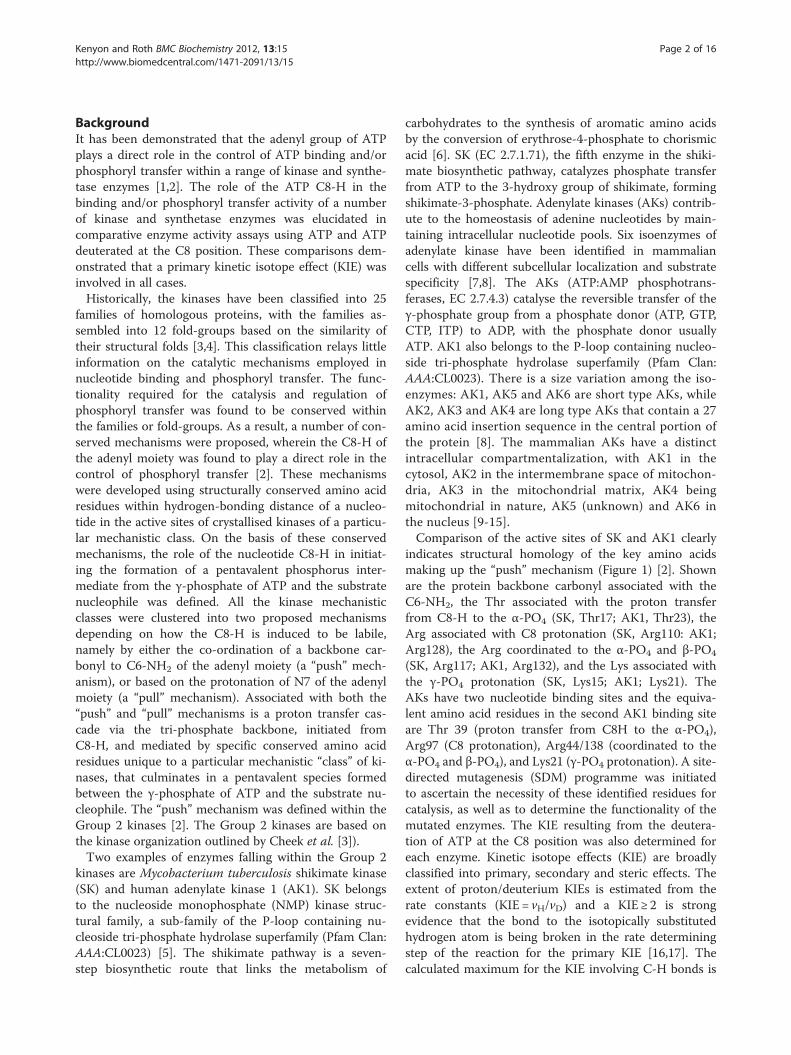
indicates structural homology of the key amino acidsmaking up the “push” mechanism (Figure 1) [2]. Shownare the protein backbone carbonyl associated with theC6-NH2, the Thr associated with the proton transferfrom C8-H to the α-PO4 (SK, Thr17; AK1, Thr23), theArg associated with C8 protonation (SK, Arg110: AK1;Arg128), the Arg coordinated to the α-PO4 and β-PO4
(SK, Arg117; AK1, Arg132), and the Lys associated withthe γ-PO4 protonation (SK, Lys15; AK1; Lys21). TheAKs have two nucleotide binding sites and the equiva-lent amino acid residues in the second AK1 binding siteare Thr 39 (proton transfer from C8H to the α-PO4),Arg97 (C8 protonation), Arg44/138 (coordinated to theα-PO4 and β-PO4), and Lys21 (γ-PO4 protonation). A site-directed mutagenesis (SDM) programme was initiatedto ascertain the necessity of these identified residues forcatalysis, as well as to determine the functionality of themutated enzymes. The KIE resulting from the deutera-tion of ATP at the C8 position was also determined foreach enzyme. Kinetic isotope effects (KIE) are broadlyclassified into primary, secondary and steric effects. Theextent of proton/deuterium KIEs is estimated from therate constants (KIE = vH/vD) and a KIE ≥ 2 is strongevidence that the bond to the isotopically substitutedhydrogen atom is being broken in the rate determiningstep of the reaction for the primary KIE [16,17]. Thecalculated maximum for the KIE involving C-H bonds is
A
B
Figure 1 Push mechanism” catalytic amino acid residues. Amino acid residues making up the “push” mechanism within the active sites of SK(A) and AK1 (B). Shown are the protein backbone carbonyl associated with the C6-NH2, the Thr associated with the proton transfer from C8H tothe α-PO4 (SK, Thr17; AK1, Thr 23), the Arg associated with C8 protonation (SK, Arg110: AK1; Arg128), the Arg coordinated to the α-PO4 and β-PO4
(SK, Arg117; AK1, Arg132), Lys associated with the γ-PO4 protonation (SK, Lys15; AK1; Lys21). The adenylate kinases have two nucleotide bindingsites and the equivalent amino acid residues in the second AK1 binding site are carbonyl associated with the C6-NH2, the Thr associated with theproton transfer from C8H to the α-PO4 (Thr 39), the Arg associated with C8 protonation (Arg97), the Arg coordinated to the α-PO4 and β-PO4
(Arg44/138), Lys associated with the γ-PO4 protonation (Lys21).
Kenyon and Roth BMC Biochemistry 2012, 13:15 Page 3 of 16http://www.biomedcentral.com/1471-2091/13/15
approximately 7 at room temperature as determined bythe difference in the zero-point energy-difference be-tween the bond to the deuterium and the bond to thehydrogen, at least in isolated chemical systems. The sec-ondary deuterium KIEs are defined as the isotope effectwhen the bond to the isotopically substituted atom is notcleaved but occur as a result of a hybridization change,with the KIE ranging between 0.7 and 1.4. Steric effectsinfluence KIEs to the same extent as the mechanisticevents affording secondary KIEs. The observed primaryKIEs resulting from ATP C8-H/D substitution in a var-iety of kinases, together with the proposed mechanismsassociated with each kinase family, prompted the under-taking of this SDM programme. The aim was to deter-mine the specific mechanistic role of the conservedamino acid residues in the active site, required both inbinding of ATP as well as the initiation phosphoryl trans-fer in SK and AK1 [1,2]. The SDM and the associatedsteady-state kinetics for each mutated enzyme in thepresence of ATP and C8-D ATP was used to delineatethe “push” mechanism, as defined by Kenyon et al.(2012). This was done by comparing the enzyme activityin the presence of ATP with that using C8D–ATP as ameasure of the intrinsic KIE for that enzyme, in conjunc-tion with SDM of the conserved amino acids implicatedin the “push” mechanism as a probe of the mechanisticrole of these residues.
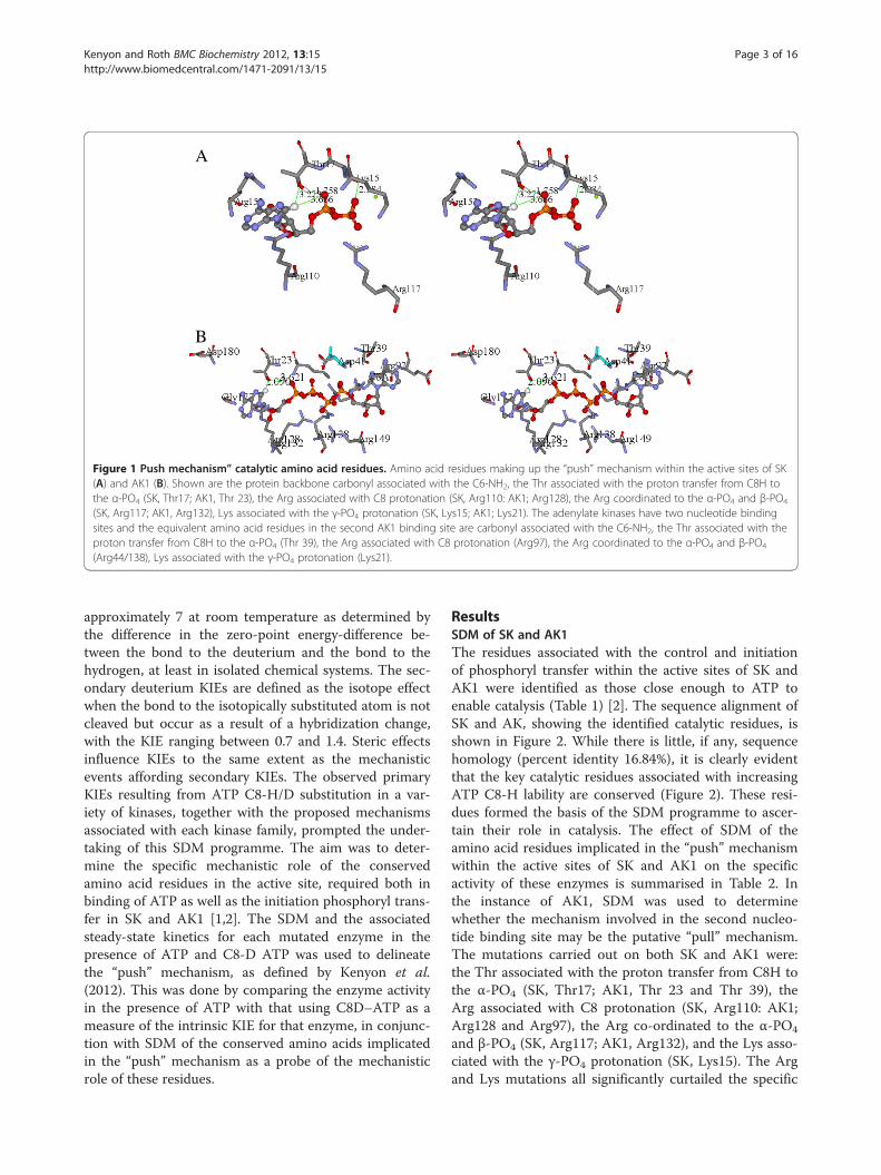
ResultsSDM of SK and AK1The residues associated with the control and initiationof phosphoryl transfer within the active sites of SK andAK1 were identified as those close enough to ATP toenable catalysis (Table 1) [2]. The sequence alignment ofSK and AK, showing the identified catalytic residues, isshown in Figure 2. While there is little, if any, sequencehomology (percent identity 16.84%), it is clearly evidentthat the key catalytic residues associated with increasingATP C8-H lability are conserved (Figure 2). These resi-dues formed the basis of the SDM programme to ascer-tain their role in catalysis. The effect of SDM of theamino acid residues implicated in the “push” mechanismwithin the active sites of SK and AK1 on the specificactivity of these enzymes is summarised in Table 2. Inthe instance of AK1, SDM was used to determinewhether the mechanism involved in the second nucleo-tide binding site may be the putative “pull” mechanism.The mutations carried out on both SK and AK1 were:the Thr associated with the proton transfer from C8H tothe α-PO4 (SK, Thr17; AK1, Thr 23 and Thr 39), theArg associated with C8 protonation (SK, Arg110: AK1;Arg128 and Arg97), the Arg co-ordinated to the α-PO4
and β-PO4 (SK, Arg117; AK1, Arg132), and the Lys asso-ciated with the γ-PO4 protonation (SK, Lys15). The Argand Lys mutations all significantly curtailed the specific
15
21
17
23 39 44
97
110
128 132
117
138
15
21
17
23 39 44
97
110
128 132
117
138
Figure 2 SK and AK1 sequence alignment. Sequence alignment of SK (pdb:1L4U) and AK1 (pdb:2C95) indicating the conserved amino acidresidues making up the catalytic residues responsible for inducing the C8-H of ATP to be labile. The red numbering of the residues are from thesecond AK1 active site. Also indicated are the secondary structure motifs as predicted by the Discovery StudioW (Accelrys Inc) assignment ofsecondary structure motifs according to Kabsch and Sander [25] as well as that obtained from the PDB. Back ground colours indicate;Red= identical residues, green= strong similarity, blue =weak similarity and no colour = no similarity.
Table 1 Catalytic residues associated with phosphoryl transfer
Enzyme SK AK1A AK1B
Residue Interatomic distance (Å) Residue Interatomic distance (Å) Residue Interatomic distance (Å)
1. C8-H to α-PO4 3.666 4.153 3.729
2. αC=O to C6-NH2 R153 1.893 G177 1.880 NR1
3. Thr-OH to C8H T17 3.273 T23 2.090 T392 1.786
4. Thr-OH to α-PO4 T17 1.758 T23 2.591 T39 5.648
5. Arg-NH1 to C8 R110 4.228 R128 4.781 R972 2.712
6. Arg-α-PO4 R117 2.757 R132 2.136 R44 1.904
7. Lys- γ-PO4 K15 1.882 K21 1.902 K21 1.865
NR=No coordinated residue.Coordinated to N7.The amino acid residues making up the “push” mechanism within the active sites of SK and AK1 identified by the inter-atomic distances between theco-crystallized nucleotide analogue and the amino acid residues within the active site. These residues included: the Thr associated with the proton transfer fromC8-H to the α-PO4 (SK, Thr17; AK1, Thr 23), the Arg associated with C8 protonation (SK, Arg110; AK1, Arg128), the Arg co-ordinated to the α-PO4 and β-PO4
(SK, Arg117; AK1, Arg132), and the Lys associated with the γ-PO4 protonation (SK, Lys15; AK1, Lys21). The residues identified in the AK1 second nucleotide bindingsite (AK1B) are: Thr39 (associated with the proton transfer from C8-H to the α-PO4), Arg97 (associated with C8 protonation), Arg44/138 (co-ordinated to the α-PO4
and β-PO4), and Lys21 (associated with the γ-PO4 protonation).
Kenyon and Roth BMC Biochemistry 2012, 13:15 Page 4 of 16http://www.biomedcentral.com/1471-2091/13/15

Table 2 Steady state specific activities of SK and AK1, WT and mutant enzymes
Enzyme SK AK1A AK1B
Residue Specific activity1 Residue Specific activity Residue Specific activity
1. Wild type WT 101,89 WT 110
2. Thr-C8H T17I 16.73 T23V 250 T39V 19.54
3. T17R 4.93
4. Arg-C8H R110A 0.92 R128A 0.128 R97A 2.61
5. R128Q <1 R97Q <1
6. R128K <1 R97K <1
7. Arg-α/β-PO4 R117 ND R132A 0.28 R44 ND
8. R132K 1.08
9. R132Q 0.00
10. Lys- γ-PO4 K15I 1.665 K21 ND
11. K15R 0.3861 umoles/mg protein/min.SDM of amino acid residues making up the “push” mechanism within the active sites of SK and AK1. These residues included: the Thr associated with the protontransfer from C8-H to the α-PO4 (SK, Thr17; AK1, Thr 23), the Arg associated with C8 protonation (SK, Arg110; AK1, Arg128), the Arg co-ordinated to the α-PO4 andβ-PO4 (SK, Arg117; AK1, Arg132), and the Lys associated with the γ-PO4 protonation (SK, Lys15; AK1; Lys21). The residues identified in the AK1 second nucleotidebinding site (AK1B) are: Thr 39 (associated with the proton transfer from C8H to the α-PO4), Arg 97 (associated with C8 protonation), Arg44/138 (co-ordinated tothe α-PO4 and β-PO4, and Lys21 (associated with the γ-PO4 protonation).
0 2 4 6 8 100
50
100
150
ATP (mM)
Spec
ific
Act
ivit
y (
mol
es.m
g
prot
ein-1 .m
in-1
)
Figure 3 Shikimate kinase specific activity of WT, K15I and T17Imutant enzymes. The effect of the concentration of ATP andC8D-ATP on the specific activity and KIE of M. tuberculosis shikimatekinase.○ = WT SK using ATP, □ = WT SK using C8D-ATP, ● = T17Imutant using ATP, ■ = T17I mutant using C8D-ATP, Δ = K15I mutantusing ATP, ▲= K15I mutant using C8D-ATP. Final enzymeconcentrations were: WT SK: 10 nM, T17I: 25 nM and K15I: 100 nM.The assays were run for 20 min (WT SK) or 80 min (T17I and K15I).
Kenyon and Roth BMC Biochemistry 2012, 13:15 Page 5 of 16http://www.biomedcentral.com/1471-2091/13/15
activity of both SK and AK1, reducing their specificactivity more than 100-fold. However, mutations of theinitial Thr residue showed a significantly weaker effectby comparison to the Lys and Arg mutations, with theSK-T17I and the AK1-T23I mutants giving approxi-mately 4.5 to 6 fold reduction in enzyme activity at lowATP concentrations. The inter-atomic distances betweenthese Thr residues and the α-phosphate of ATP are3.666 Å for SK and 4.153 Å for AK1, meaning they are inclose enough proximity for direct transfer of the C8-H tothe α-PO4 of ATP (Table 2).
Effect of C8D-ATP on specific activity of Thr mutantsThe effect of the ATP and C8D-ATP concentration onthe steady state specific activities of wild type (WT) SKand SK-T17I, as well as WT AK1, AK1-T23V and AK1-T39V was determined (Figures 3,4,5,6,7,8). The best-fitfor the data was obtained for the kinetic model using thenon-linear regression algorithms within the GraphPadPrismW 5 software (Table 3). As part of the software out-put, a data table containing 150 data points defining thebest computed fit for each enzyme’s kinetic response tothe presence of either ATP or C8D-ATP. These responsecurves were then used to define the KIE or inverse KIE(KIED) from KIE = vH/vD or KIED = vD/vH, respectively[1]. The enzyme activity data was used to determine thevalues of Kcat and Kcat/Km for each enzyme (Table 3).The SK-T17I mutant has significantly less activity than
WT SK (Figure 3). WT SK showed a classical KIE, withthe kinetic data reaching an asymptote at about 2 withdecreasing ATP concentration, and a similar asymptoteat 1 with increasing ATP concentration (Figure 4). It is
0 1 2 3 40
100
200
300
400
0.0
0.5
1.0
1.5
2.0
ATP (mM)
Spec
ific
Act
ivit
y (
mol
es.m
gpr
otei
n-1.m
in-1)
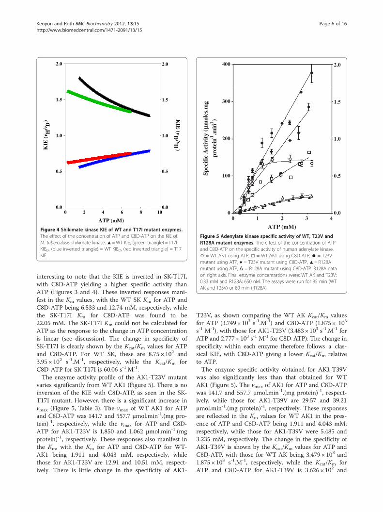
Figure 5 Adenylate kinase specific activity of WT, T23V andR128A mutant enzymes. The effect of the concentration of ATPand C8D-ATP on the specific activity of human adenylate kinase.○ = WT AK1 using ATP, □ = WT AK1 using C8D-ATP, ● = T23Vmutant using ATP, ♦ = T23V mutant using C8D-ATP, ▲= R128Amutant using ATP, Δ = R128A mutant using C8D-ATP. R128A dataon right axis. Final enzyme concentrations were: WT AK and T23V:0.33 mM and R128A: 650 nM. The assays were run for 95 min (WTAK and T23V) or 80 min (R128A).
0 2 4 6 8 100.0
0.5
1.0
1.5
2.0
0.0
0.5
1.0
1.5
2.0
ATP (mM)
KIE
(H
/D
) KIE
(D
/vH)
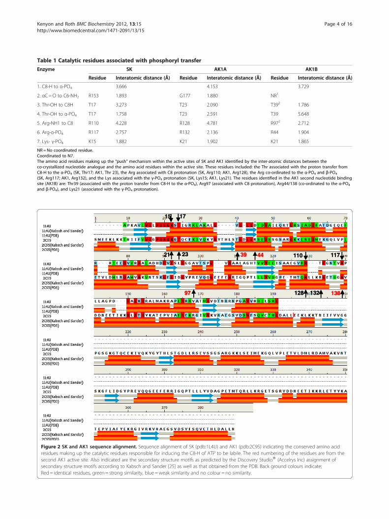
Figure 4 Shikimate kinase KIE of WT and T17I mutant enzymes.The effect of the concentration of ATP and C8D-ATP on the KIE ofM. tuberculosis shikimate kinase. ▲=WT KIE, (green triangle) = T17IKIED, (blue inverted triangle) = WT KIED, (red inverted triangle) = T17KIE.
Kenyon and Roth BMC Biochemistry 2012, 13:15 Page 6 of 16http://www.biomedcentral.com/1471-2091/13/15
interesting to note that the KIE is inverted in SK-T17I,with C8D-ATP yielding a higher specific activity thanATP (Figures 3 and 4). These inverted responses mani-fest in the Km values, with the WT SK Km for ATP andC8D-ATP being 6.533 and 12.74 mM, respectively, whilethe SK-T17I Km for C8D-ATP was found to be22.05 mM. The SK-T17I Km could not be calculated forATP as the response to the change in ATP concentrationis linear (see discussion). The change in specificity ofSK-T17I is clearly shown by the Kcat/Km values for ATPand C8D-ATP. For WT SK, these are 8.75 × 10² and3.95 × 102 s-1.M-1, respectively, while the Kcat/Km forC8D-ATP for SK-T17I is 60.06 s-1.M-1.The enzyme activity profile of the AK1-T23V mutant
varies significantly from WT AK1 (Figure 5). There is noinversion of the KIE with C8D-ATP, as seen in the SK-T17I mutant. However, there is a significant increase invmax (Figure 5, Table 3). The vmax of WT AK1 for ATPand C8D-ATP was 141.7 and 557.7 μmol.min-1.(mg pro-tein)-1, respectively, while the vmax for ATP and C8D-ATP for AK1-T23V is 1,850 and 1,062 μmol.min-1.(mgprotein)-1, respectively. These responses also manifest inthe Km, with the Km for ATP and C8D-ATP for WT-AK1 being 1.911 and 4.043 mM, respectively, whilethose for AK1-T23V are 12.91 and 10.51 mM, respect-ively. There is little change in the specificity of AK1-
T23V, as shown comparing the WT AK Kcat/Km valuesfor ATP (3.749× 10³ s-1.M-1) and C8D-ATP (1.875× 10³
s-1 M-1), with those for AK1-T23V (3.483× 10³ s-1.M-1 forATP and 2.777× 10³ s-1 M-1 for C8D-ATP). The change inspecificity within each enzyme therefore follows a clas-sical KIE, with C8D-ATP giving a lower Kcat/Km relativeto ATP.The enzyme specific activity obtained for AK1-T39V
was also significantly less than that obtained for WTAK1 (Figure 5). The vmax of AK1 for ATP and C8D-ATPwas 141.7 and 557.7 μmol.min-1.(mg protein)-1, respect-ively, while those for AK1-T39V are 29.57 and 39.21μmol.min-1.(mg protein)-1, respectively. These responsesare reflected in the Km values for WT AK1 in the pres-ence of ATP and C8D-ATP being 1.911 and 4.043 mM,respectively, while those for AK1-T39V were 5.485 and3.235 mM, respectively. The change in the specificity ofAK1-T39V is shown by the Kcat/Km values for ATP andC8D-ATP, with those for WT AK being 3.479 × 10³ and1.875 × 103 s-1.M-1, respectively, while the Kcat/Km forATP and C8D-ATP for AK1-T39V is 3.626 × 10² and
0 1 2 3 40
50
100
150
200
0
5
10
15
20
25
30
ATP (mM)
Spec
ific
Act
ivit
y (
mol
es.m
gpr
otei
n-1.m
in-1
)
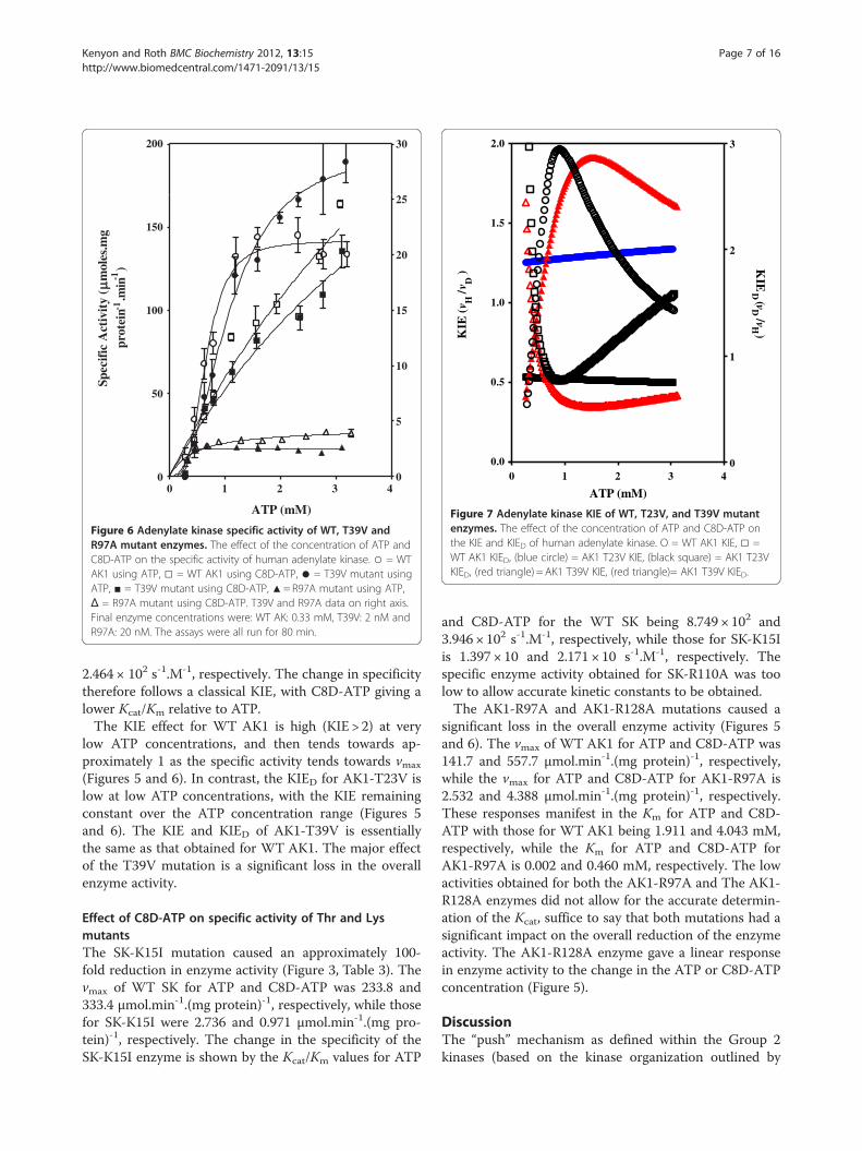
Figure 6 Adenylate kinase specific activity of WT, T39V andR97A mutant enzymes. The effect of the concentration of ATP andC8D-ATP on the specific activity of human adenylate kinase. ○ = WTAK1 using ATP, □ = WT AK1 using C8D-ATP, ● = T39V mutant usingATP, ■ = T39V mutant using C8D-ATP, ▲= R97A mutant using ATP,Δ = R97A mutant using C8D-ATP. T39V and R97A data on right axis.Final enzyme concentrations were: WT AK: 0.33 mM, T39V: 2 nM andR97A: 20 nM. The assays were all run for 80 min.
0 1 2 3 40.0
0.5
1.0
1.5
2.0
0
1
2
3
ATP (mM)
KIE
(H
/D
) KIE
D (vD/vH
)
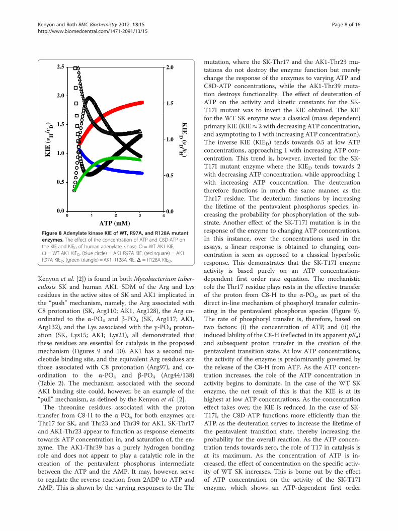
Figure 7 Adenylate kinase KIE of WT, T23V, and T39V mutantenzymes. The effect of the concentration of ATP and C8D-ATP onthe KIE and KIED of human adenylate kinase. O = WT AK1 KIE, □ =WT AK1 KIED, (blue circle) = AK1 T23V KIE, (black square) = AK1 T23VKIED, (red triangle) = AK1 T39V KIE, (red triangle)= AK1 T39V KIED.
Kenyon and Roth BMC Biochemistry 2012, 13:15 Page 7 of 16http://www.biomedcentral.com/1471-2091/13/15
2.464 × 10² s-1.M-1, respectively. The change in specificitytherefore follows a classical KIE, with C8D-ATP giving alower Kcat/Km relative to ATP.The KIE effect for WT AK1 is high (KIE > 2) at very
low ATP concentrations, and then tends towards ap-proximately 1 as the specific activity tends towards vmax
(Figures 5 and 6). In contrast, the KIED for AK1-T23V islow at low ATP concentrations, with the KIE remainingconstant over the ATP concentration range (Figures 5and 6). The KIE and KIED of AK1-T39V is essentiallythe same as that obtained for WT AK1. The major effectof the T39V mutation is a significant loss in the overallenzyme activity.
Effect of C8D-ATP on specific activity of Thr and LysmutantsThe SK-K15I mutation caused an approximately 100-fold reduction in enzyme activity (Figure 3, Table 3). Thevmax of WT SK for ATP and C8D-ATP was 233.8 and333.4 μmol.min-1.(mg protein)-1, respectively, while thosefor SK-K15I were 2.736 and 0.971 μmol.min-1.(mg pro-tein)-1, respectively. The change in the specificity of theSK-K15I enzyme is shown by the Kcat/Km values for ATP
and C8D-ATP for the WT SK being 8.749 × 10² and3.946 × 102 s-1.M-1, respectively, while those for SK-K15Iis 1.397 × 10 and 2.171 × 10 s-1.M-1, respectively. Thespecific enzyme activity obtained for SK-R110A was toolow to allow accurate kinetic constants to be obtained.The AK1-R97A and AK1-R128A mutations caused a
significant loss in the overall enzyme activity (Figures 5and 6). The vmax of WT AK1 for ATP and C8D-ATP was141.7 and 557.7 μmol.min-1.(mg protein)-1, respectively,while the vmax for ATP and C8D-ATP for AK1-R97A is2.532 and 4.388 μmol.min-1.(mg protein)-1, respectively.These responses manifest in the Km for ATP and C8D-ATP with those for WT AK1 being 1.911 and 4.043 mM,respectively, while the Km for ATP and C8D-ATP forAK1-R97A is 0.002 and 0.460 mM, respectively. The lowactivities obtained for both the AK1-R97A and The AK1-R128A enzymes did not allow for the accurate determin-ation of the Kcat, suffice to say that both mutations had asignificant impact on the overall reduction of the enzymeactivity. The AK1-R128A enzyme gave a linear responsein enzyme activity to the change in the ATP or C8D-ATPconcentration (Figure 5).
DiscussionThe “push” mechanism as defined within the Group 2kinases (based on the kinase organization outlined by
0 1 2 3 40.0
0.5
1.0
1.5
2.0
2.5
0.0
0.5
1.0
1.5
2.0
ATP (mM)
KIE
(H
/D
) KIE
D (D
/H )
Figure 8 Adenylate kinase KIE of WT, R97A, and R128A mutantenzymes. The effect of the concentration of ATP and C8D-ATP onthe KIE and KIED of human adenylate kinase. O = WT AK1 KIE,□ = WT AK1 KIED, (blue circle) = AK1 R97A KIE, (red square) = AK1R97A KIED, (green triangle) = AK1 R128A KIE, Δ = R128A KIED.
Kenyon and Roth BMC Biochemistry 2012, 13:15 Page 8 of 16http://www.biomedcentral.com/1471-2091/13/15
Kenyon et al. [2]) is found in both Mycobacterium tuber-culosis SK and human AK1. SDM of the Arg and Lysresidues in the active sites of SK and AK1 implicated inthe “push” mechanism, namely, the Arg associated withC8 protonation (SK, Arg110; AK1, Arg128), the Arg co-ordinated to the α-PO4 and β-PO4 (SK, Arg117; AK1,Arg132), and the Lys associated with the γ-PO4 proton-ation (SK, Lys15; AK1; Lys21), all demonstrated thatthese residues are essential for catalysis in the proposedmechanism (Figures 9 and 10). AK1 has a second nu-cleotide binding site, and the equivalent Arg residues arethose associated with C8 protonation (Arg97), and co-ordination to the α-PO4 and β-PO4 (Arg44/138)(Table 2). The mechanism associated with the secondAK1 binding site could, however, be an example of the“pull” mechanism, as defined by the Kenyon et al. [2].The threonine residues associated with the proton
transfer from C8-H to the α-PO4 for both enzymes areThr17 for SK, and Thr23 and Thr39 for AK1, SK-Thr17and AK1-Thr23 appear to function as response elementstowards ATP concentration in, and saturation of, the en-zyme. The AK1-Thr39 has a purely hydrogen bondingrole and does not appear to play a catalytic role in thecreation of the pentavalent phosphorus intermediatebetween the ATP and the AMP. It may, however, serveto regulate the reverse reaction from 2ADP to ATP andAMP. This is shown by the varying responses to the Thr
mutation, where the SK-Thr17 and the AK1-Thr23 mu-tations do not destroy the enzyme function but merelychange the response of the enzymes to varying ATP andC8D-ATP concentrations, while the AK1-Thr39 muta-tion destroys functionality. The effect of deuteration ofATP on the activity and kinetic constants for the SK-T17I mutant was to invert the KIE obtained. The KIEfor the WT SK enzyme was a classical (mass dependent)primary KIE (KIE� 2 with decreasing ATP concentration,and asymptoting to 1 with increasing ATP concentration).The inverse KIE (KIED) tends towards 0.5 at low ATPconcentrations, approaching 1 with increasing ATP con-centration. This trend is, however, inverted for the SK-T17I mutant enzyme where the KIED tends towards 2with decreasing ATP concentration, while approaching 1with increasing ATP concentration. The deuterationtherefore functions in much the same manner as theThr17 residue. The deuterium functions by increasingthe lifetime of the pentavalent phosphorus species, in-creasing the probability for phosphorylation of the sub-strate. Another effect of the SK-T17I mutation is in theresponse of the enzyme to changing ATP concentrations.In this instance, over the concentrations used in theassays, a linear response is obtained to changing con-centration is seen as opposed to a classical hyperbolicresponse. This demonstrates that the SK-T17I enzymeactivity is based purely on an ATP concentration-dependent first order rate equation. The mechanisticrole the Thr17 residue plays rests in the effective transferof the proton from C8-H to the α-PO4, as part of thedirect in-line mechanism of phosphoryl transfer culmin-ating in the pentavalent phosphorus species (Figure 9).The rate of phosphoryl transfer is, therefore, based ontwo factors: (i) the concentration of ATP, and (ii) theinduced lability of the C8-H (reflected in its apparent pKa)and subsequent proton transfer in the creation of thepentavalent transition state. At low ATP concentrations,the activity of the enzyme is predominantly governed bythe release of the C8-H from ATP. As the ATP concen-tration increases, the role of the ATP concentration inactivity begins to dominate. In the case of the WT SKenzyme, the net result of this is that the KIE is at itshighest at low ATP concentrations. As the concentrationeffect takes over, the KIE is reduced. In the case of SK-T17I, the C8D-ATP functions more efficiently than theATP, as the deuteration serves to increase the lifetime ofthe pentavalent transition state, thereby increasing theprobability for the overall reaction. As the ATP concen-tration tends towards zero, the role of T17 in catalysis isat its maximum. As the concentration of ATP is in-creased, the effect of concentration on the specific activ-ity of WT SK increases. This is borne out by the effectof ATP concentration on the activity of the SK-T17Ienzyme, which shows an ATP-dependent first order
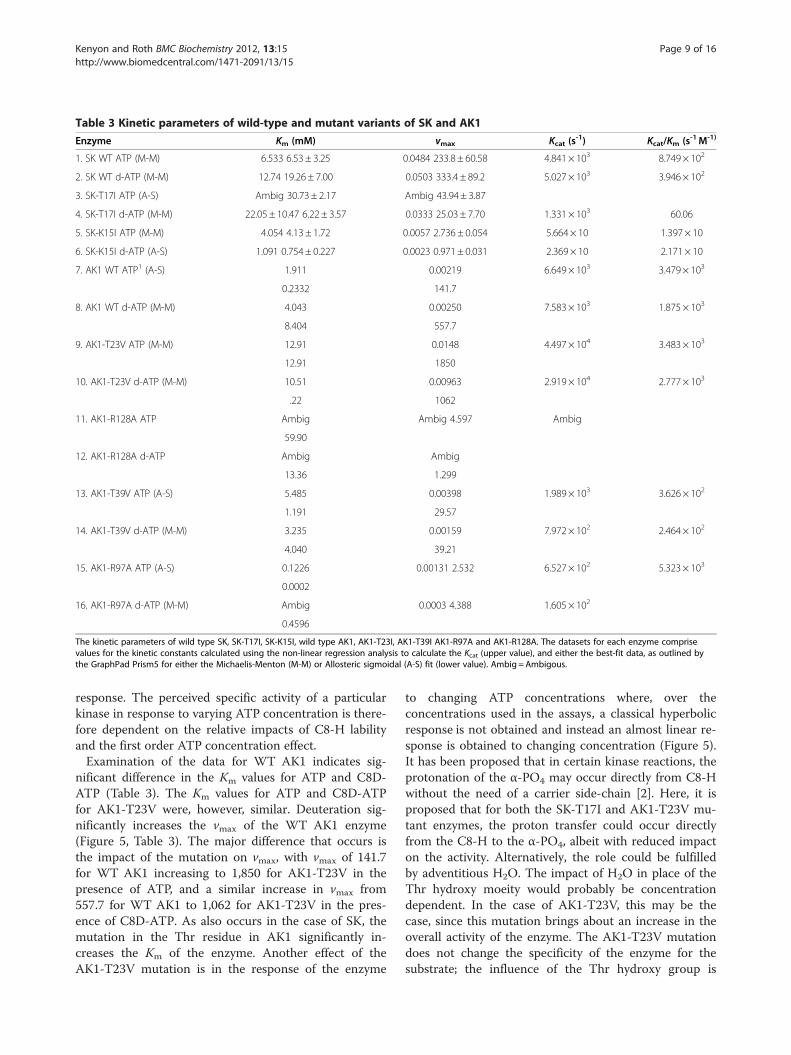
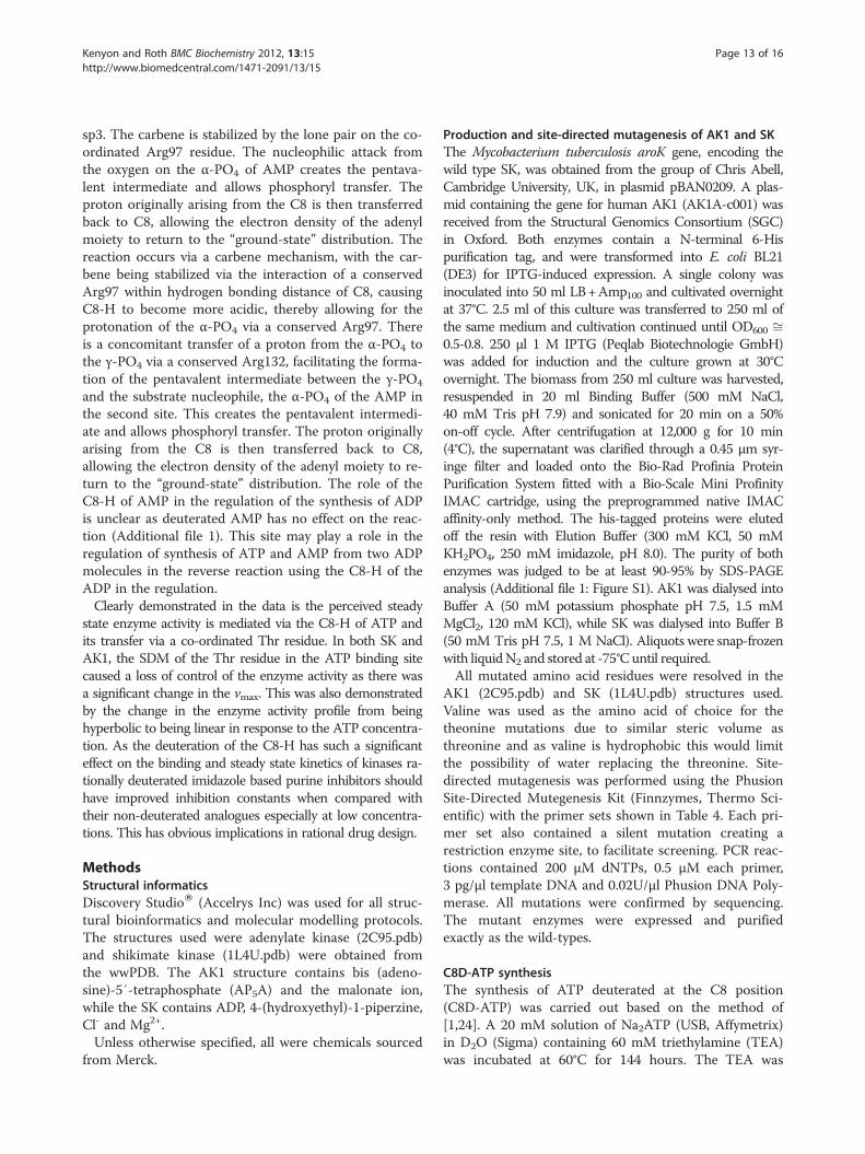
Table 3 Kinetic parameters of wild-type and mutant variants of SK and AK1
Enzyme Km (mM) vmax Kcat (s-1) Kcat/Km (s-1M-1)
1. SK WT ATP (M-M) 6.533 6.53 ± 3.25 0.0484 233.8 ± 60.58 4.841× 10³ 8.749 × 10²
2. SK WT d-ATP (M-M) 12.74 19.26 ± 7.00 0.0503 333.4 ± 89.2 5.027× 10³ 3.946 × 10²
3. SK-T17I ATP (A-S) Ambig 30.73 ± 2.17 Ambig 43.94 ± 3.87
4. SK-T17I d-ATP (M-M) 22.05 ± 10.47 6.22 ± 3.57 0.0333 25.03 ± 7.70 1.331× 10³ 60.06
5. SK-K15I ATP (M-M) 4.054 4.13 ± 1.72 0.0057 2.736 ± 0.054 5.664× 10 1.397× 10
6. SK-K15I d-ATP (A-S) 1.091 0.754± 0.227 0.0023 0.971 ± 0.031 2.369× 10 2.171× 10
7. AK1 WT ATP1 (A-S) 1.911 0.00219 6.649× 10³ 3.479 × 10³
0.2332 141.7
8. AK1 WT d-ATP (M-M) 4.043 0.00250 7.583× 10³ 1.875 × 10³
8.404 557.7
9. AK1-T23V ATP (M-M) 12.91 0.0148 4.497× 10⁴ 3.483 × 10³
12.91 1850
10. AK1-T23V d-ATP (M-M) 10.51 0.00963 2.919× 10⁴ 2.777 × 10³
.22 1062
11. AK1-R128A ATP Ambig Ambig 4.597 Ambig
59.90
12. AK1-R128A d-ATP Ambig Ambig
13.36 1.299
13. AK1-T39V ATP (A-S) 5.485 0.00398 1.989× 10³ 3.626 × 10²
1.191 29.57
14. AK1-T39V d-ATP (M-M) 3.235 0.00159 7.972× 10² 2.464 × 10²
4.040 39.21
15. AK1-R97A ATP (A-S) 0.1226 0.00131 2.532 6.527× 10² 5.323 × 10³
0.0002
16. AK1-R97A d-ATP (M-M) Ambig 0.0003 4.388 1.605× 10²
0.4596
The kinetic parameters of wild type SK, SK-T17I, SK-K15I, wild type AK1, AK1-T23I, AK1-T39I AK1-R97A and AK1-R128A. The datasets for each enzyme comprisevalues for the kinetic constants calculated using the non-linear regression analysis to calculate the Kcat (upper value), and either the best-fit data, as outlined bythe GraphPad Prism5 for either the Michaelis-Menton (M-M) or Allosteric sigmoidal (A-S) fit (lower value). Ambig =Ambigous.
Kenyon and Roth BMC Biochemistry 2012, 13:15 Page 9 of 16http://www.biomedcentral.com/1471-2091/13/15
response. The perceived specific activity of a particularkinase in response to varying ATP concentration is there-fore dependent on the relative impacts of C8-H labilityand the first order ATP concentration effect.Examination of the data for WT AK1 indicates sig-
nificant difference in the Km values for ATP and C8D-ATP (Table 3). The Km values for ATP and C8D-ATPfor AK1-T23V were, however, similar. Deuteration sig-nificantly increases the vmax of the WT AK1 enzyme(Figure 5, Table 3). The major difference that occurs isthe impact of the mutation on vmax, with vmax of 141.7for WT AK1 increasing to 1,850 for AK1-T23V in thepresence of ATP, and a similar increase in vmax from557.7 for WT AK1 to 1,062 for AK1-T23V in the pres-ence of C8D-ATP. As also occurs in the case of SK, themutation in the Thr residue in AK1 significantly in-creases the Km of the enzyme. Another effect of theAK1-T23V mutation is in the response of the enzyme
to changing ATP concentrations where, over theconcentrations used in the assays, a classical hyperbolicresponse is not obtained and instead an almost linear re-sponse is obtained to changing concentration (Figure 5).It has been proposed that in certain kinase reactions, theprotonation of the α-PO4 may occur directly from C8-Hwithout the need of a carrier side-chain [2]. Here, it isproposed that for both the SK-T17I and AK1-T23V mu-tant enzymes, the proton transfer could occur directlyfrom the C8-H to the α-PO4, albeit with reduced impacton the activity. Alternatively, the role could be fulfilledby adventitious H2O. The impact of H2O in place of theThr hydroxy moeity would probably be concentrationdependent. In the case of AK1-T23V, this may be thecase, since this mutation brings about an increase in theoverall activity of the enzyme. The AK1-T23V mutationdoes not change the specificity of the enzyme for thesubstrate; the influence of the Thr hydroxy group is

Arg110
Arg110
Arg110
Thr17
Arg110
Thr17
Glu106
Thr17
Glu/Asp
Lys15
Glu106
Lys15
Arg117Arg117
Thr17
Lys15
Glu/Asp
Lys15
Glu/Asp
Arg117
Glu106
Glu106
Arg117
Glu/Asp
Figure 9 Phosphoryl transfer mechanism found in the shikimate kinase. Phosphoryl transfer mechanism found in shikimate kinase. Theinitiation of phosphoryl transfer occurs via the coordination of the ATP C6-NH2 to a carbonyl arising from the protein backbone by the “push”mechanism resulting in the protonation of C8 via the coordination of a conserved Arg. This renders the C8-H more acidic, allowing for theprotonation of the α-PO4, via the conserved Thr17 carrier. There is a concomitant transfer of an H+ from the α-PO4 to β-PO4 via a conserved Arg,thereby facilitating the formation of the pentavalent intermediate between the γ-PO4 and the substrate nucleophile. There is a simultaneousATP-mediated deprotonation of the substrate -OH, allowing for the nucleophilic attack by the substrate to create the pentavalent intermediateand allow phosphoryl transfer. A protonated Lys then transfers the proton to the γ-PO4, changing the Mg2+ from being β-PO4 to γ-PO4
coordinated to being α-PO4 to β-PO4 coordinated. The H+ originally arising from the C8 is then transferred back to C8, allowing the electrondensity of the adenyl moiety to return to the “ground-state” distribution.
Kenyon and Roth BMC Biochemistry 2012, 13:15 Page 10 of 16http://www.biomedcentral.com/1471-2091/13/15
lacking at high concentration for this mutant, however.In the case of SK-T17I, the use of C8D-ATP effectivelymimics the presence of a functional T17 by stabilizingthe transition state intermediate and increasing theprobability of the reaction occurring, mimicking the roleof the Thr hydroxy group. The loss of the interplay be-tween the role of the Thr hydroxy group and the variationin ATP concentration due to the AK1-T23V mutationresulted in an increase in the vmax. In effect, the AK1-T23V mutation merely causes this enzyme to respond ina linear first order manner to the change in the ATP con-centration. The steady state enzyme activity is thereforeachieved by the interplay between the two effects, namely
the role of the pKa of the C8-H and the role of the ATPconcentration. In the case of the Thr mutations, H2Oprobably fulfills the role of the Thr hydroxy group andthe impact of the mutation is dependent on the H2Oconcentration. In the case of SK, the availability of waterto fulfill this role is lower than in the case of AK1 (asseen by the decreased overall the steady state activity ofthe mutant). In WTAK1, the role of Thr23 predominatesat low ATP concentrations, while increasing ATP con-centration steadily diminishes the importance of this rolein favour of mass action. Mutant AK1-T39V enzymeappears to have lost most of its functionality (Figure 5,Table 3). The second nucleotide binding site appears

:
Lys21
Arg132
Arg 128
Arg97
Thr39
Lys21
Arg132
Arg 128
Arg97
Asp141
Lys21
Arg132
Arg 128
Arg97Thr23
Arg149
Asp141
Glu176
Thr23
Glu176
Thr23
Thr39
Arg149
Glu176
Arg149
Asp141
Thr39
Figure 10 Phosphoryl transfer mechanism found in the adenylate kinase. Phosphoryl transfer mechanism found in adenylate kinase. Theinitiation of phosphoryl transfer occurs via the coordination of the AMP C6-NH2 to a carbonyl arising from the protein backbone by the “push”mechanism resulting in the protonation of C8 via the coordination of the conserved Arg128. This renders the C8-H more acidic, allowing for theprotonation of the α-PO4, via the conserved Thr23 carrier. There is a concomitant transfer of an H+ from the α-PO4 to β-PO4 via a conserved Arg,thereby facilitating the formation of the pentavalent intermediate between the γ-PO4 and the substrate nucleophile. There is a simultaneousATP-mediated deprotonation of the substrate -OH, allowing for the nucleophilic attack by the substrate to create the pentavalent intermediateand allow phosphoryl transfer. A protonated Lys then transfers the proton to the γ-PO 4, changing the Mg2+ from being β-PO4 to γ-PO4
coordinated to being α-PO4 to β-PO4 coordinated. There is a concomitant binding of AMP in the second nucleotide binding site which isactivated by the protonation of the N7 via the conserved Thr39 forming a carbene on the imidazole moiety causing a change in C8 hybridizationfrom sp2 to sp3 hybridized. The carbene being stabilized by the lone pair on the coordinated Arg97. The nucleophilic attack from the oxygen onthe α-PO4 of AMP creates the pentavalent intermediate and allows phosphoryl transfer. The H+ originally arising from the C8 is then transferredback to C8, allowing the electron density of the adenyl moiety to return to the “ground-state” distribution.
Kenyon and Roth BMC Biochemistry 2012, 13:15 Page 11 of 16http://www.biomedcentral.com/1471-2091/13/15
to function via the “pull” mechanism, with the Thr39serving to protonate AMP N7 - an intrinsic part of the“pull” mechanism [2]. Once this occurs, the lone pair onthe nitrogen of Arg97 serves stabilize the incipient car-bene which is forming at C8 of ATP (Figure 10). Removalof the Thr39 therefore destroys the functionality, even ifthe C8-H of the ATP is in close enough proximity of theα-PO4. This is because Thr39 assists this protonationevent by increasing the lability of ATP C8-H in the pro-cess of stabilising the nascent carbene that forms in the“pull” mechanism. Both the R128A and the R97A mutantenzymes have significantly reduced enzyme activities,with vmax values of 2.532 and 4.597 respectively (Figure 6,Table 3). The Arg128, which functions in conjunction
with Thr23, serves to protonate C8 and stabilize the car-bene. Furthermore, Arg97, which functions in conjunc-tion with Thr39, plays a key role in the formation of thecarbene in the “pull” mechanism. If the C8-H of AMPwas playing a direct role in the regulation of AK1 enzymeactivity, a KIE should be obtained on comparison of theactivity between enzyme activities obtained in the pres-ence of AMP and C8D-AMP. This was, however, notfound to be the case as AMP deuterated at the C8 pos-ition had no effect on the specific enzyme activity(Additional file 1: Figure S1).Taking these observations into account, it should be
borne in mind that the conformational changes arisingon nucleotide binding within the P-loop containing

Kenyon and Roth BMC Biochemistry 2012, 13:15 Page 12 of 16http://www.biomedcentral.com/1471-2091/13/15
nucleoside triphosphate hydrolase superfamily allow theresidues of the conserved Walker A motif to “lock” thephosphate backbone into position [18,19]. As a result,two residues that form part of the consensus sequence,GXXXXGKT/S, are brought directly into play, mechan-istically speaking. Significantly, these residues also playan integral part of the “push” mechanism in the guise ofthe conserved Thr residue coordinated to C8-H protonand the Lys residue responsible for the proton transfer be-tween the α- and β-phosphates. The catalytic role of theconserved Thr residue has previously been identified, in-cluding its hydrogen bonding interaction via the Thr-OH. . .O-Pα(ATP) [20]. Shi et al. (1993), in fact, ask thequestion, “How can Thr-23 participate in catalysis by inter-acting with the α-phosphate of ATP but not contribute tothe energetics of catalysis?” This research demonstratesthat the C8-H. . .HO-Thr and Thr-OH. . .O-Pα(ATP) inter-actions play a role in the initiation of phosphoryl transferand that the Thr residue, effectively, acts as a molecular“switch” on completion of nucleotide and substrate bind-ing. As a result, the phosphoryl transfer occurs, via theclassical in-line mechanism [21-23]. This in-line associativeSN2 type mechanism of phosphoryl transfer is pertinent tothis reaction mechanism as the phosphoryl transfer isdependent on the formation of a pentavalent phosphorusintermediate between the γ-phosphate and the substratenucleophile.
ConclusionsThe steady-state enzyme activity in the kinase enzymesis obviously more complex than merely the phosphoryl-ation of a substrate nucleophile. This complexity is ap-parent in the broad range of KM constants of the kinasesranging from less than 0.4 μM to in excess of 1,000 μMfor ATP (Carna Biosciences, Inc., Kinase Profiling Book:www.carnabio.com). The kinase enzymes have been clas-sified into 25 families of homologous proteins, with thefamilies assembled into 12 fold-groups based on thesimilarity of their structural folds, as well as conservedmechanisms associated with the regulation of the en-zyme activity via the C8-H of the adenyl moiety [2-4].Within a single group, both prokaryotic and eukaryoticorganisms are represented with kinase isoenzymes thatappear to be kinetically and functionally distinct basedon the rate of phosphoryl transfer and the regulationthereof. It is therefore conceivable that the various con-served “push” and “pull” mechanisms associated withthe release of C8-H, the proton transfer cascades andthe resultant/concomitant creation of the pentavalenttransition state are the mechanisms by which the kinaseenzymes achieve this 2,500-fold variation in the KM.Polyphosphate could serve as the energy “currency”within the cell however, ATP fulfils this role. It is neces-sary that the kinase enzymes carry out their specific
reactions at differential rates and to achieve these vary-ing rates the kinases have evolved a number of con-served mechanisms which broadly manifest in the“push” and the “pull mechanisms”. Data outlined aboveserves to demonstrate the “push” mechanism associatedwith the role of the C8-H in the regulation of the Group2 kinases. The phosphoryl transfer mechanism found inthe Group 2 kinases (Rossmann-like fold and phosphoe-nolpyruvte carboxykinase-like sequences) was assessedusing M. tuberculosis shikimate kinase and human ad-enylate kinase as the model systems. The initiation ofphosphoryl transfer in SK occurs via the co-ordinationof the ATP C6-NH2 to a carbonyl arising from the pro-tein backbone by the “push” mechanism, resulting in theprotonation of C8 via the co-ordination of a conservedArg residue (Arg110). This renders the C8-H moreacidic, allowing for the protonation of the α-PO4 via thehydroxyl of the conserved Thr carrier (Thr17). There isa concomitant transfer of an proton from the α-PO4 toβ-PO4 via Arg117, thereby facilitating the formation ofthe pentavalent intermediate between the γ-PO4 and thesubstrate nucleophile. Where required, there is a simul-taneous ATP-mediated deprotonation of the substrate-OH, allowing for the nucleophilic attack by the sub-strate to create the pentavalent intermediate and allow-ing phosphoryl transfer. In the case of adenylate kinase,the substrate is acidic, which does not require deproto-nation. The protonated Lys15 then transfers the protonto the γ-PO4, changing the Mg2+ from being β-PO4 toγ-PO4 co-ordinated to being α-PO4 to β-PO4 co-ordi-nated. The proton originally arising from the C8 is thentransferred back to C8, allowing the electron density ofthe adenyl moiety to return to the “ground-state” distri-bution. The human AK1 has two nucleotide bindingsites and the second site may utilise the “pull” mechan-ism as outlined by Kenyon et al. [2]. The “pull” mechan-ism is the phosphoryl transfer mechanism found in theGroup 4 kinases (hexokinase family with polyol sub-strate). The initiation of phosphoryl transfer occurs viathe coordination of the ATP C6-NH2 to a carbonyl aris-ing from the protein backbone by the “push” mechan-ism, resulting in the protonation of C8 via the co-ordination of a conserved Arg (Arg128). This rendersthe C8-H more acidic, allowing for the protonation ofthe α-PO4, via the hydroxyl of the conserved Thr carrier(Thr23). There is a concomitant transfer of a protonfrom the α-PO4 to the γ-PO4 via the conserved Arg132,thereby facilitating the formation of the pentavalentintermediate between the γ-PO4 and the substrate nu-cleophile, in this case the α-PO4 of the AMP in the sec-ond site. The AMP in the second nucleotide binding siteis activated by the protonation of AMP-N7 via the con-served Thr39, forming a carbene on the imidazole moi-ety that causes a change in C8 hybridization from sp2 to
Kenyon and Roth BMC Biochemistry 2012, 13:15 Page 13 of 16http://www.biomedcentral.com/1471-2091/13/15
sp3. The carbene is stabilized by the lone pair on the co-ordinated Arg97 residue. The nucleophilic attack fromthe oxygen on the α-PO4 of AMP creates the pentava-lent intermediate and allows phosphoryl transfer. Theproton originally arising from the C8 is then transferredback to C8, allowing the electron density of the adenylmoiety to return to the “ground-state” distribution. Thereaction occurs via a carbene mechanism, with the car-bene being stabilized via the interaction of a conservedArg97 within hydrogen bonding distance of C8, causingC8-H to become more acidic, thereby allowing for theprotonation of the α-PO4 via a conserved Arg97. Thereis a concomitant transfer of a proton from the α-PO4 tothe γ-PO4 via a conserved Arg132, facilitating the forma-tion of the pentavalent intermediate between the γ-PO4
and the substrate nucleophile, the α-PO4 of the AMP inthe second site. This creates the pentavalent intermedi-ate and allows phosphoryl transfer. The proton originallyarising from the C8 is then transferred back to C8,allowing the electron density of the adenyl moiety to re-turn to the “ground-state” distribution. The role of theC8-H of AMP in the regulation of the synthesis of ADPis unclear as deuterated AMP has no effect on the reac-tion (Additional file 1). This site may play a role in theregulation of synthesis of ATP and AMP from two ADPmolecules in the reverse reaction using the C8-H of theADP in the regulation.Clearly demonstrated in the data is the perceived steady
state enzyme activity is mediated via the C8-H of ATP andits transfer via a co-ordinated Thr residue. In both SK andAK1, the SDM of the Thr residue in the ATP binding sitecaused a loss of control of the enzyme activity as there wasa significant change in the vmax. This was also demonstratedby the change in the enzyme activity profile from beinghyperbolic to being linear in response to the ATP concentra-tion. As the deuteration of the C8-H has such a significanteffect on the binding and steady state kinetics of kinases ra-tionally deuterated imidazole based purine inhibitors shouldhave improved inhibition constants when compared withtheir non-deuterated analogues especially at low concentra-tions. This has obvious implications in rational drug design.
MethodsStructural informaticsDiscovery StudioW (Accelrys Inc) was used for all struc-tural bioinformatics and molecular modelling protocols.The structures used were adenylate kinase (2C95.pdb)and shikimate kinase (1L4U.pdb) were obtained fromthe wwPDB. The AK1 structure contains bis (adeno-sine)-5′-tetraphosphate (AP5A) and the malonate ion,while the SK contains ADP, 4-(hydroxyethyl)-1-piperzine,Cl- and Mg2+.Unless otherwise specified, all were chemicals sourced
from Merck.
Production and site-directed mutagenesis of AK1 and SKThe Mycobacterium tuberculosis aroK gene, encoding thewild type SK, was obtained from the group of Chris Abell,Cambridge University, UK, in plasmid pBAN0209. A plas-mid containing the gene for human AK1 (AK1A-c001) wasreceived from the Structural Genomics Consortium (SGC)in Oxford. Both enzymes contain a N-terminal 6-Hispurification tag, and were transformed into E. coli BL21(DE3) for IPTG-induced expression. A single colony wasinoculated into 50 ml LB+Amp100 and cultivated overnightat 37°C. 2.5 ml of this culture was transferred to 250 ml ofthe same medium and cultivation continued until OD600 ffi0.5-0.8. 250 μl 1 M IPTG (Peqlab Biotechnologie GmbH)was added for induction and the culture grown at 30°Covernight. The biomass from 250 ml culture was harvested,resuspended in 20 ml Binding Buffer (500 mM NaCl,40 mM Tris pH 7.9) and sonicated for 20 min on a 50%on-off cycle. After centrifugation at 12,000 g for 10 min(4°C), the supernatant was clarified through a 0.45 μm syr-inge filter and loaded onto the Bio-Rad Profinia ProteinPurification System fitted with a Bio-Scale Mini ProfinityIMAC cartridge, using the preprogrammed native IMACaffinity-only method. The his-tagged proteins were elutedoff the resin with Elution Buffer (300 mM KCl, 50 mMKH2PO4, 250 mM imidazole, pH 8.0). The purity of bothenzymes was judged to be at least 90-95% by SDS-PAGEanalysis (Additional file 1: Figure S1). AK1 was dialysed intoBuffer A (50 mM potassium phosphate pH 7.5, 1.5 mMMgCl2, 120 mM KCl), while SK was dialysed into Buffer B(50 mM Tris pH 7.5, 1 M NaCl). Aliquots were snap-frozenwith liquidN2 and stored at -75°C until required.All mutated amino acid residues were resolved in the
AK1 (2C95.pdb) and SK (1L4U.pdb) structures used.Valine was used as the amino acid of choice for thetheonine mutations due to similar steric volume asthreonine and as valine is hydrophobic this would limitthe possibility of water replacing the threonine. Site-directed mutagenesis was performed using the PhusionSite-Directed Mutegenesis Kit (Finnzymes, Thermo Sci-entific) with the primer sets shown in Table 4. Each pri-mer set also contained a silent mutation creating arestriction enzyme site, to facilitate screening. PCR reac-tions contained 200 μM dNTPs, 0.5 μM each primer,3 pg/μl template DNA and 0.02U/μl Phusion DNA Poly-merase. All mutations were confirmed by sequencing.The mutant enzymes were expressed and purifiedexactly as the wild-types.
C8D-ATP synthesisThe synthesis of ATP deuterated at the C8 position(C8D-ATP) was carried out based on the method of[1,24]. A 20 mM solution of Na2ATP (USB, Affymetrix)in D2O (Sigma) containing 60 mM triethylamine (TEA)was incubated at 60°C for 144 hours. The TEA was
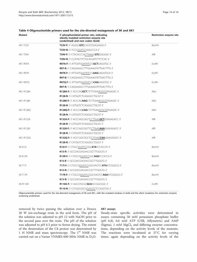
Table 4 Oligonucleotide primers used for the site-directed mutagenesis of SK and AK1
Mutant 5’-phosphorylated primer sets, indicatingsilently mutated restriction enzyme site(underlined) and new codon (bold)
Restriction enzyme site
AK1-T23V T23V-F: 5'-AGGGCGTCCAGTGTGAGAAGA-3' BamHI
T23V-R: 5'-TCCCGGATCCAGGCCCA-3'
AK1-T39V T39V-F: 5'-CTACACCCACTTAAGCGTCGGGGAC-3' AflII
T39V-R: 5'-CCATACTTCTGCACGATCTTCTCAC-3'
AK1-R97A R97A-F: 5'-ATTGATGGATATCCGGCTGAGGTGC-3' EcoRV
R97-R: 5'-CAGGAAGCCTTTGGAAGTATTGACTTTG-3'
AK1-R97K R97K-F: 5'-ATTGATGGATATCCGAAGGAGGTGCA-3' EcoRV
R97-R: 5'-CAGGAAGCCTTTGGAAGTATTGACTTTG-3'
AK1-R97Q R97Q-F: 5'-ATTGATGGATATCCGCAGGAGGTGC-3' EcoRV
R97-R: 5'-CAGGAAGCCTTTGGAAGTATTGACTTTG-3'
AK1-R128A R128A-F: 5'-ACCCAGGCTCTCTTGAAACGCGTAGAGAC-3' MluI
R128-R: 5'-CATGGTCTCAGGGCCTGCGT-3'
AK1-R128K R128K-F: 5'-ACCCAGAAGCTCTTGAAACGCGTAGAGAC-3' MluI
R128-R: 5'-CATGGTCTCAGGGCCTGCGT-3'
AK1-R128Q R128Q-F: 5'-ACCCAGCAGCTCTTGAAACGCGTAGAGAC-3' MluI
R128-R: 5'-CATGGTCTCAGGGCCTGCGT-3'
AK1-R132A R132A-F: 5'-ACCCAGCGGCTCCTTAAGGCTGGAGAGACC-3' AflII
R128-R: 5'-CATGGTCTCAGGGCCTGCGT-3'
AK1-R132K R132K-F: 5'-ACCCAGCGGCTCCTTAAGAAAGGAGAGACC-3' AflII
R128-R: 5'-CATGGTCTCAGGGCCTGCGT-3'
AK1-R132Q R132Q-F: 5'-ACCCAGCGGCTCCTTAAGCAAGGAGAGACC-3' AflII
R128-R: 5'-CATGGTCTCAGGGCCTGCGT-3'
SK-K15I K15I-F: 5'-CTGCCGGATCCGGCATATCCACCAT-3' BamHI
K15-R: 5'-GCCGACGAGAACCGCTTTGGGTG-3'
SK-K15R K15R-F: 5'-CTGCCGGGATCCGGCAGGTCCACCA-3' BamHI
K15-R: 5'-GCCGACGAGAACCGCTTTGGGTG-3'
SK-T17I T17I-F: 5'-CTGCCGGGATCCGGCAAGTCCATAATCGGGCG-3' BamHI
K15-R: 5'-GCCGACGAGAACCGCTTTGGGTG-3’
SK-T17R T17R-F: 5'-CTGCCGGGATCCGGCAAGTCCAGAATCGGGCG-3' BamHI
K15-R: 5'-GCCGACGAGAACCGCTTTGGGTG-3'
SK-R110A R110A-F: 5'-GGCGTGCGCGCAACCGGCGGC-3' EcoRV
R110-R: 5'-CTCGGCGGCGGATATCTCCAGGTAG-3'
Oligonucleotide primers used for the site-directed mutagenesis of SK and AK1, with the mutated residues in bold and the silent mutations for restriction enzymescreening underlined.
Kenyon and Roth BMC Biochemistry 2012, 13:15 Page 14 of 16http://www.biomedcentral.com/1471-2091/13/15
removed by twice passing the solution over a Dowex20 W ion-exchange resin in the acid form. The pH ofthe solution was adjusted to pH 12 with NaOH prior tothe second pass over the resin. The pH of the solutionwas adjusted to pH 6.3 prior to freeze drying. The extentof the deuteration of the C8 proton was determined by1 H NMR and mass spectroscopy. The 1H NMR wascarried out on a Varian VNMRS 600 MHz NMR in D2O.
AK1 assaysSteady-state specific activities were determined inassays containing 50 mM potassium phosphate buffer(pH 6.8), 0.6 mM ATP (USB, Affymetrix) and AMP(Sigma), 1 mM MgCl2 and differing enzyme concentra-tions, depending on the activity levels of the mutants.The reactions were incubated at 37°C for varyingtimes, again depending on the activity levels of the
Kenyon and Roth BMC Biochemistry 2012, 13:15 Page 15 of 16http://www.biomedcentral.com/1471-2091/13/15
mutants, before being terminated by the addition of10 mM EDTA.2Na.2H2O.ATP/C8D-ATP concentration gradient assays contained
50 mM potassium phosphate buffer (pH 6.8), 3.3 nM(WT AK1, AK1-T23V) or 2 nM (AK1-T39V) enzyme,and varying amounts of AMP, ATP or C8-D ATP, andMgCl2. These were kept at a constant ratio of 1:1:2.2 forAMP: ATP/C8-D ATP: MgCl2. The ATP concentrationsranged between 0.5 and 4 mM. The reactions were incu-bated at 37°C for varying times, before being terminatedby the addition of 10 mM EDTA.2Na.2H2O. Each datapoint is the average for triplicate reactions. The ADPconcentration was measured by HPLC.
SK assaysSteady-state specific activities were determined in assayscontaining: 100 mM potassium phosphate buffer (pH 6.8),500 mM KCl, 1 mM ATP and MgCl2, 2.4 mM shikimicacid (Sigma) and differing enzyme concentrations, depend-ing on the activity levels of the mutants. The reactionswere incubated at 37°C for varying times, again dependingon the activity levels of the mutants, before being termi-nated by the addition of 10 mM EDTA.2Na.2H2O. Eachdata point is the average for triplicate reactions.ATP/C8D-ATP concentration assays contained 100 mM
potassium phosphate buffer (pH 6.8), 500 mM KCl, 8 mMshikimic acid, 10 nM enzyme, and varying amounts ofATP or C8D-ATP, and MgCl2. These were kept at a con-stant ratio of 1:1 for ATP/C8D-ATP: MgCl2. The ATPconcentrations ranged between 0.5 and 10 mM. The re-actions were incubated at 30°C for varying times, beforebeing terminated by the addition of 10 mM EDTA.2-Na.2H2O. Each data point is the average for triplicate reac-tions. The ADP concentration was measured by HPLC.
HPLC analysisThe production of ADP was analysed by HPLC [1]. Theassay solutions were centrifuged prior to HPLC analysis.The assays for adenosine, AMP, ADP ATP were carriedout using Phenomenex 5 μ LUNA C18 column with themobile phase containing PIC AW (Waters Coorporation),250 ml acetonitrile, 7 g KH2PO4 per litre water. Theflow rate of the mobile phase was 1 ml/minute with UVdetection.
Additional file
Additional file 1: Figure S1. Effect of ATP and AMP concentrations onthe specific activity of AK1 showing no significant effect of deuteration ofAMP on the specific activity of AK1. Assays were run in 50 mM K2HPO4/KH2PO4 buffer (pH6.8), at MgCl2 concentrations equal to 1.1 times thesum of the ATP and AMP concentrations. ATP and AMP were added atequivalent concentrations to the assays. ● = AMP, ■ = deuterated AMP.
Authors' contributionsCPK defined the concept and experiments of this study. RLR expressed andpurified SK and AK1. RLR performed assays and collected data. CPK and RLRperformed calculations on data. CPK produced C8D-ATP. Quality control byMS and NMR on C8D-ATP was carried out by CPK. CPK drafted themanuscript and RLR assisted in editing the draft manuscript. Both authorshave read and approved the final manuscript.
AcknowledgementsWe thank the group of Chris Abell, Cambridge University, UK, for theMycobacterium tuberculosis aroK gene, encoding the wild type SK, and theStructural Genomics Consortium (SGC) in Oxford for the plasmid containingthe gene for human AK1 (AK1A-c001). We thank Dr Chris van derWesthuyzen for valuable comments review of the manuscript. This work wassupported by the Council for Scientific and Industrial Research (CSIR)parliamentary grant.
Received: 11 November 2011 Accepted: 26 July 2012Published: 10 August 2012
References1. Kenyon CP, Steyn A, Roth RL, Steenkamp PA, Nkosi TC, Oldfield LC: The role
of the C8 proton of ATP in the regulation of phosphoryl transfer withinkinases and synthetases. BMC Biochem 2011, 12:36–53.
2. Kenyon CP, Roth RL, Westhuyzen CW, Parkinson CJ: Conserved phosphoryltransfer mechanisms within kinase families and the role of the C8proton of ATP in the activation of phosphoryl transfer.BMC Research Notes 2012, 5:131.
3. Cheek S, Zhang H, Grishin NV: Sequence and structure classification ofkinases. J Mol Biol 2002, 320:855–881.
4. Cheek S, Ginalski K, Zhang H, Grishin NV: A comprehensive update of thesequence and structure classification of kinases. BMC Struc. Biol 2005,5(6):1–19.
5. Vonrhein C, Schlauderer GJ, Schulz GF: Movie of the structural changesduring a catalytic cycle of nucleoside monophosphate kinases.Structure 1995, 3:483–490.
6. Haslam E: Shikimic Acid: Metabolism and Metabolites. Chichester: Wiley; 1993.7. Van Rompay AR, Johansson M, Karlsson A: Phosphorylation of nucleosides
and nucleoside analogs by mammalian nucleoside monophosphatekinases. Pharmacol Ther 2000, 87:189–198.
8. Ren H, Wang L, Bennett M, Lian Y, Zheng X, Lu F, Li L, Nan J, Luo M,Eriksson S, Zhang C, Su X-D: The crystal structure of human adenylatekinase 6: an adenylate kinase localised to the cell nucleus. Proc Natl AcadSci 2005, 102:303–308.
9. Fukami-Kobayashi K, Nosaka M, Nakazawa A, Go M: Ancient divergence oflong and short isoforms of adenylate kinase: molecular evolution of thenucleoside monophosphate kinase family. FEBS Lett 1996, 385:214–220.
10. Atkinson DE: Cellular energy metabolism and its regulation.New York: Academic Press; 1979:85–107.
11. Nakazawa A, Yamada M, Tanaka H, Shahjahan M, Tanabe T: Gene structuresof three vertebrate adenylate kinase isoenzymes. Prog Clin Biol Res 1990,344:495–514.
12. Schulz GE: Cold Spring Harbor Symposium on Quantitative Biology. 1987,52:429–439.
13. Markland FS, Wadkins CL: Adenosine triphosphate-adenosine5'-monophosphate phosphotransferase of bovine liver mitochondria. I.Isolation and chemical properties. J Biol Chem 1966, 241:4124–4135.
14. Font B, Gautheron DC: General and kinetic properties of pig heartmitochondrial adenylate kinase. Biochim Biophys Acta 1980, 611:299–308.
15. Nakazawa A, Yamada M, Tanaka H, Shahjahan M, Tanake T: Isoenzymes. InStructure, Function and Use in Biology and Medicine. New York: Wiley-Liss;1990:495–514.
16. Wiberg KB: The deuterium isotope effect. Chem Rev 1955, 55:713–743.17. Carey FA, Sundberg RJ: Advanced organic chemistry. Part A. Structure and
Mechanisms. NewYork: Plenum Press; 2004.18. Müller CW, Schlauderer GJ, Reinstein J, Schulz GE: Adenylate kinase
motions during catalysis: and energetic counterweight balancingsubstrate binding. Structure 1996, 4:147–156.
19. Ramakrishnan C, Danl VS, Ramasarma T: A conformational anaylsis ofwalker motif A [GXXXXGKT (S)] in nucleotide binding and other proteins.Protein Eng 2002, 15:783–798.

Kenyon and Roth BMC Biochemistry 2012, 13:15 Page 16 of 16http://www.biomedcentral.com/1471-2091/13/15
20. Shi Z, Byeon I-JL, Jiang R-T, Tsai D-W: Mechanism of adenylate kinase.What can be learnt from a mutant enzyme with minor perturbation inkinetic parameters. Biochemistry 1993, 32:6450–6458.
21. Abele U, Schulz GE: High-resolution structures of adenylate kinase fromyeast ligated with inhibitor Ap5A, showing the pathway of phosphoryltransfer. Protein Sci 1995, 4:1262–1271.
22. Müller CW, Schulz GE: Structure of the complex between adenylatekinase from Escherichia coli and the inhibitor AP5A refined at 1.9Åresolution. J Mol Biol 1992, 224:159–177.
23. Fersht A: Structure and mechanism in protein science: guide to enzymecatalysis and protein folding. New York: W. H. Freeman & Co; 1999.
24. Heller S: 1H NMR studies on deuterium - hydrogen exchange at C-5 inuridines. Biochem Biophys Res Commun 1968, 32:998–1001.
25. Kabsch W, Sander C: Dictionary of protein secondary structure:pattern recognition of hydrogen-bonded and geometrical features.Biopolymers 1983, 22:2577–2637.
doi:10.1186/1471-2091-13-15Cite this article as: Kenyon and Roth: The role of the C8 proton of ATPin the catalysis of shikimate kinase and adenylate kinase. BMCBiochemistry 2012 13:15.
Submit your next manuscript to BioMed Centraland take full advantage of:
• Convenient online submission
• Thorough peer review
• No space constraints or color figure charges
• Immediate publication on acceptance
• Inclusion in PubMed, CAS, Scopus and Google Scholar
• Research which is freely available for redistribution
Submit your manuscript at www.biomedcentral.com/submit