the role of jak pathway dysregulation in the pathogenesis...
TRANSCRIPT

Review
The Role of JAK Pathway Dysregulation in the Pathogenesisand Treatment of Acute Myeloid Leukemia
Hun Ju Lee1, Naval Daver2, Hagop M. Kantarjian2, Srdan Verstovsek2, and Farhad Ravandi2
AbstractThe discovery of the Janus kinase 2 (JAK2) V617F mutation has improved our understanding of the
pathophysiology of myeloproliferative neoplasms such as polycythemia vera, essential thrombocythe-
mia, and primary myelofibrosis. Before discovery of the JAK2 V617F mutation, there were no specific
targeted therapies for patients with myeloproliferative neoplasms. More recently, several small-molecule
inhibitors have been developed that have shown therapeutic potential in the clinical setting. There is
evidence that the JAK2 pathway is dysregulated in some acute myeloid leukemias and may also represent
a novel therapeutic target in this disease. In this review, we describe the preclinical, clinical, and
pathophysiologic evidence for using JAK inhibitors in the treatment of acute myeloid leukemias. Clin
Cancer Res; 19(2); 1–9. �2012 AACR.
IntroductionAcute myeloid leukemia: current landscapeAcutemyeloid leukemia (AML) is a hematopoietic clonal
stemcell disorder characterizedby abnormaldifferentiationand proliferation of immature blast cells in the bone mar-row. AML is defined as 20% or more blasts in the bonemarrow or less than 20% blasts with recurrent cytogeneticabnormalities, according to the 2008 revision of the WorldHealth Organization classification of myeloid neoplasmsand acute leukemia (1, 2). AML accounts for approximately80% of all adult leukemia cases, and incidence increaseswith age, with a median age at diagnosis of 67 years (3).AML may arise de novo by transformation of the hemato-
poietic stem cell or progenitor cells of the myeloid lineage(4). Secondary AML can develop as a consequence ofchemoradiotherapy or evolution of preexisting myelodys-plastic syndrome (MDS) or myeloproliferative neoplasm(MPN). Both forms of secondary AML are associated withunfavorable chromosomal abnormalities and worse prog-nosis as compared with de novo AMLs (5). The primary goalof AML therapy is to achieve complete remission (CR) withinduction chemotherapy, defined as bone marrow blastsless than 5%, neutrophil count more than 1.0� 109/L, andplatelet count more than 100 � 109/L independent oftransfusion (6). Recent improvements in chemotherapeuticagents and supportive care have resulted in CR rates as high
as 85% in patients with AML. However, relapse occursfrequently as approximately 50% to 70% of patients withAML who achieve CR as a result of frontline therapy willexperience relapse within 3 years (7). As a result, consoli-dation chemotherapy or allogeneic stem cell transplanta-tion is often used to prevent relapse, with varying degrees ofsuccess.
In spite of advances in therapeutic options, overall pati-ent survival rate has not markedly improved, and AMLtherapy remains an area of unmet medical need. A largeproportion of patients with AML display mutations thatlead to signaling pathway dysregulation. The most well-known of these mutations is the FMS-like tyrosine kinase-3(FLT3) mutation, which occurs in approximately 30% ofpatientswithAML. The FLT3mutation is generated by eitherinternal tandemduplication of the juxtamembrane domainor pointmutations within the tyrosine kinase domain, bothresulting in constitutive activation of tyrosine kinase (8, 9).
The JAK/STAT PathwayThe JAK/STAT pathway has increasingly been implicated
in the pathogenesis of AML. The Janus kinases (JAK) anddownstream components of the pathway are involved incellular proliferation and differentiation and immunologicregulation. In humans, the JAK family of nonreceptortyrosine kinases consists of 4 knownmembers: JAK1, JAK2,JAK3, and TYK2 (10). JAK1, JAK2, and TYK2 are ubiqui-tously expressed, whereas JAK3 is expressed exclusivelyin hematopoietic, vascular smooth muscle, and endo-thelial cells (8). The JAK/STAT pathway is initiated via theextracellular binding of cytokines, including interleukins,IFNs, neurotrophic factors, and hormones, to their respec-tive transmembrane receptors. Cytokine binding inducesreceptor dimerization, thereby bringing JAK proteins,which are constitutively bound to the intracellular region
Authors' Affiliations: Departments of 1Lymphoma and Myelomaand 2Leukemia, The University of Texas MD Anderson Cancer Center,Houston, Texas
Corresponding Author: Farhad Ravandi, Department of Leukemia, Unit428, The University of Texas—MD Anderson Cancer Center, 1515Holcombe Boulevard, Houston, TX 77030. Phone: 713-745-0394; Fax:646-707-4417; E-mail: [email protected]
doi: 10.1158/1078-0432.CCR-12-2087
�2012 American Association for Cancer Research.
ClinicalCancer
Research
www.aacrjournals.org OF1
Research. on August 21, 2019. © 2012 American Association for Cancerclincancerres.aacrjournals.org Downloaded from
Published OnlineFirst December 3, 2012; DOI: 10.1158/1078-0432.CCR-12-2087

of the receptors, into close proximity, resulting in transau-tophosphorylation of JAK molecules. Receptor-associatedphosphorylated JAK (p-JAK) subsequently phosphorylatesseveral sites on their respective receptors, thereby exposingan SH2 docking site for activation of the signal transducerand activator of transcription (STAT) transcription factors(11).
Activated STATs homodimerize or heterodimerizeand translocate into the nucleus to induce transcrip-tion of various downstream targets (Fig. 1; ref. 10). Tran-scriptional targets of the JAK-activated STAT family,specifically STAT3 and STAT5, include genes involved inthe regulation of cell survival, proliferation, and differ-entiation, including MYC, cyclin D1, survivin, and BCL2(9, 12, 13). Thus, abnormal activation of the JAK/STATpathway is thought to play an important role in thepathogenesis of some malignancies, and it is becomingincreasingly clear that the STATs also play roles in boththe intrinsic and extrinsic cancer-associated inflamma-tory microenvironment (14, 15).
JAK Dysregulation in MyeloproliferativeNeoplasms
Aberrant JAK/STAT hyperactivation and its role in can-cer biology have been best defined with respect tohematopoietic malignancies, including MPNs such aspolycythemia vera (PV), essential thrombocythemia (ET),and primary myelofibrosis (PMF). In 2005, the JAK2V617F gain-of-function mutation was discovered andobserved in more than 90% of patients with PV and morethan 50% of patients with PMF and ET (16–19). Addi-tional JAK2-activating mutations have been observed inthe clinic. Furthermore, MPNs have shown the abnormalelevation of several cytokines including serum interleu-kin-6 (IL-6), granulocyte macrophage colony stimulatingfactor (GM-CSF), and TNF-a, which result in the activa-tion of the JAK2 signaling pathway (20, 21). JAK/STAT
dysregulation through JAK2-activating mutations, therelease of JAK2-activating cytokines from stromal cells,or the autocrine loop of JAK2-mediated cytokine produc-tion has been postulated as a possible mechanism bywhich MPNs sustain their proliferative and cytoprotectiveadvantage (22, 23).
A number of JAK inhibitors have been developed. Ofnote, ruxolitinib, the only JAK inhibitor to complete phaseIII trials, was approved by the U.S. Food and Drug Admin-istration for the treatment of intermediate- or high-risk MFand more recently by Health Canada and the EuropeanCommission for the treatment of MF-related splenomegalyor symptoms on the basis of results from the phase IIICOMFORT-I and -II studies, in which ruxolitinib therapyresulted in pronounced reductions in splenomegaly as wellas improvements in disease-related symptoms comparedwith both placebo and best available therapy (24, 25). Afollow-up in both the phase I/II 251 study and the COM-FORT-I study reported an overall survival advantage in theruxolitinib arm (24, 26). In the COMFORT-I study, anunplanned survival analysis during a planned safety updaterevealed that ruxolitinib significantly increased overall sur-vival compared with placebo [13 (8.4%) deaths in ruxoli-tinib group and 24 (15.7%) deaths in placebo group].Additional JAK inhibitors that are currently in clinical trialsfor treatment of MPNs include lestaurtinib (CEP-701),SAR302503 (TG101348), CYT387, AZD1480, and SB1518.Similar to ruxolitinib, a number of JAK inhibitors wereeffective in reduction of splenomegaly and constitutionalsymptoms (11).
Myeloproliferative Neoplasm Progressionto AML
It has been established that patients with an MPN are atan elevated risk for leukemic transformation. Progression toAML occurs in a subset of patients with an MPN, butprogression to acute lymphoblastic leukemia is uncommon(27). Leukemic transformationmay be secondary to the useof cytoreductive therapy, such as hydroxyurea; however,this hypothesis remains controversial (28). Anotherhypothesis is that progression to acute leukemia occursthrough additional acquisition of key genetic mutations,which provide a survival or proliferative advantage similarto that observed in the progression of chronic myeloidleukemia from chronic phase to blast crisis (29, 30).
Much of our understanding of the pathophysiologic pro-gression of MPN to AML comes from studies using mousemodels. For example, deficient interferon consensussequence binding protein (ICSBP)–mediated expression ofthe DNA-repair protein Fanconi F (FANCF) results in trans-formation to AML in myeloproliferative disease mousemodels (31). Furthermore, activating mutations in the tyro-sine phosphatase SHP2 gene in combination with ICSBPhaploinsufficiency accentuate the progression to AML inan MPN mouse model (32). Although incomplete, thepreclinical data suggest a complex mechanism of MPN toAML progression involving multiple cooperative pathways.
Translational RelevanceTherapy for acute myeloid leukemia (AML) has not
changed significantly since the "War on Cancer" wasdeclared 40 years ago. Although we havemade advancesin understanding the genetics and pathophysiology ofAML,patient outcomeshavenot improved substantially.Recognition of the heterogeneity of the disease has beenaccentuated by genomic data suggesting that the drivingmutations may be different in individual patients withAML. Here, we show that the dysregulated Janus kinase(JAK) pathway may represent a novel therapeutic targetfor a subset of patients with AML. Given the recentclinical successes of JAK inhibitors in the treatment ofmyeloproliferative neoplasms, we review preclinical andclinical data supporting the use of JAK inhibitors in AMLand summarize relevant evidence thatmay revolutionizethe current paradigm of AML treatment.
Lee et al.
Clin Cancer Res; 19(2) January 15, 2013 Clinical Cancer ResearchOF2
Research. on August 21, 2019. © 2012 American Association for Cancerclincancerres.aacrjournals.org Downloaded from
Published OnlineFirst December 3, 2012; DOI: 10.1158/1078-0432.CCR-12-2087

The clinical data about AML transformation from a pre-existing MPN is less well understood. It is evident that aprolonged history of PV/ET results in an increased risk ofbone marrow fibrosis and, correspondingly, it has beenshown that increased bone marrow reticulin content andhypercellularity in patients with PV/ET increase risk formyelofibrosis or AML transformation (33). Moreover, inves-tigators have identified increased levels of growth factorstransforming growth factor (TGF)-b, basic fibroblast growthfactor (bFGF), andVEGF secretedbymegakaryocytes,mono-cytes, or both in bone marrow specimens from patientswith a history of PV/ET. Several genes are frequently foundto be mutated in the MPN blast phase, such as TET2, IDH1,IDH2, IKZF1, and RUNX1 (34–37). Recently, it was shownthat mutations in the serine/arginine-rich splicing factor 2(SRSF2) gene are more prevalent in patients with AML thathas progressed from a preexistingMPN than in patients withde novo AML (18.9% and 5.6%, respectively; ref. 38).In the clinic, the overall survival rates of patients whose
AML progresses from MPN are inferior to those of patientswith de novo AML (29). Leukemic transformation occurs in
approximately 15%ofpatientswithPMF, 10%of thosewithPV, and 4% of those with ET (Fig. 2; refs. 39–43). Notsurprisingly, the JAK2 V617F mutation is found in a signif-icant percentage of patients with AML arising from a pre-existing MPN (44, 45). Conversely, less than 10% ofpatients with de novo AML harbor the mutation (46). How-ever, there is evidence that JAK2 V617F–positive MPNs canprogress to JAK2 V617F–negative AML at disease transfor-mation (47). Work by Theocharides and colleagues sug-gested that JAK2 V617F–positive MPN progression to JAK2V617F–negative AML occurs through clonal evolution of acommon JAK2 V617F–negative progenitor (47). Interest-ingly, patients with V617F–negative AML who previouslyhad a V617F–positive MPN had a significantly shorterinterval between MPN diagnosis and AML transformationthan those who developed V617F–positive AML. Further-more, patients whose V617F–positive MPN progressed toV617F–positive AML had amore than 50% V617F positive-to-negative allelic ratio in blasts, indicating that high allelicburden may be an important factor in the clonal evolutionto JAK2 V617F–positive AML.
© 2012 American Association for Cancer Research© 2012 American Association for Cancer
Constitutively active
JAK2 possibly related
to these mutations
• JAK2 V617F
• JAK2 exon 12
Overactive
JAK1
MPL receptor
mutations
Cytokines
JAK2
A
BC D
E
JAK2
JAK2
JAK2 JAK2
JAK2
JAK3
JAK1 JAK1
JAK1JAK 1/2
heterodimer
JAK3 activating
mutations including
V722I, A572V,
and P123T
observed in AMKLSTAT
STAT
STAT
STAT
Activation
Signal transducer
and activator
of transcription
DNANucleus
Transcription: survival, proliferation
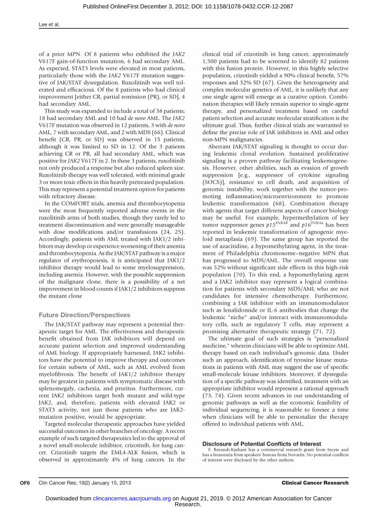
Figure 1. JAK receptor signaling and activation of STAT proteins. A and B, normal JAK/STAT signaling after specific binding of cytokine receptors by ligand.Cytokine binding to JAK2/JAK2 homodimers (A) or JAK1/JAK2 heterodimers (B) results in increased activation of STAT proteins, which leads tosubsequent transcriptional changes that induce cellular survival and proliferation. C, MPL receptor mutations can result in ligand-independent activationof the JAK/STAT pathway. D, mutations in JAK1 or JAK2, specifically JAK2 V617F, which is often observed in myelofibrosis, PV, or ET, can lead toconstitutively active JAK function independent of ligand binding. E, mutations in JAK3, which have been observed in patients with acute megakaryoblasticleukemia (AMKL) can lead to elevated STAT activity.
JAK in AML
www.aacrjournals.org Clin Cancer Res; 19(2) January 15, 2013 OF3
Research. on August 21, 2019. © 2012 American Association for Cancerclincancerres.aacrjournals.org Downloaded from
Published OnlineFirst December 3, 2012; DOI: 10.1158/1078-0432.CCR-12-2087

The prognostic implication of JAK2 V617F in thissetting remains unclear. One study reported a significantlyinferior survival in JAK-mutated AML,whereas other studieshave not detected any prognostic or predictive implications(48–50). Currently, no well-defined treatment is availablefor AML arising from a preexisting MPN, and such patientswould likely receive standard de novo AML therapies or bereferred to a clinical trial.
Rationale for Targeting JAK/STAT in AMLGiven the high occurrence of JAK dysregulation in MPN,
it may be suggested that JAK/STAT pathway dysregulationplays a role in the pathogenesis of secondary AML. Yet JAK/STAT dysregulationmay not be exclusive to secondary AML,as elevated JAK and STAT levels have been observed inpatients with de novo AML. As p-JAK levels are increased inpatients with AML, and the JAK2 V617Fmutation is presentin only a small percentage of these patients, this specificmutation cannot be exclusively responsible for JAK dysre-gulation. It is possible that the classic V617F mutation,when present, may function either as a driver mutation inthe initial stages of AML or as a passenger mutation in laterstages by virtue of increased genomic instability and muta-tion load. Besides the JAK2 V617F mutation, several novelJAK2 activation mutations have been observed in AMLpatient samples, including PCM1-JAK2, JAK2 K607N, andJAK2 T875N (Table 1; refs. 51–56).
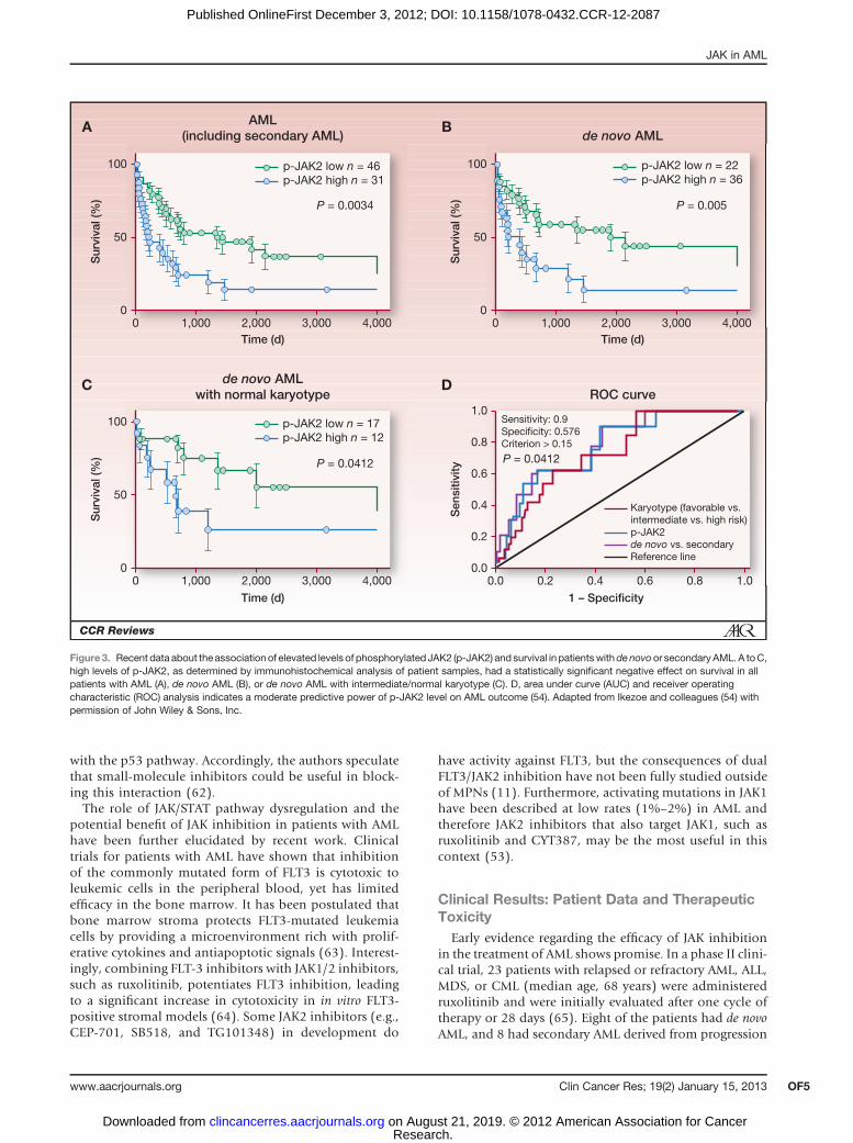
A recent study found elevated levels of activated p-JAK2in bone marrow samples from a large cohort of patients
with AML (n ¼ 77). Elevated p-JAK2 correlated with highwhite blood cell count, low platelet count, and shortersurvival in patients with either de novo or secondary AML(Fig. 3; ref. 54). Although elevated p-JAK2 levels wereclearly observed, JAK2 V617F mutation was noted in onlyone of the 77 patients with AML, indicating that JAK/STAT dysregulation occurs through alternative mechan-isms. Accordingly, treatment of AML cell lines withAZ960, a JAK2 inhibitor, reduced growth in AML celllines, and induced apoptosis. Furthermore, inhibition ofJAK/STAT by AZ960 resulted in increased apoptosis inCD34þ/CD38� cells, which are proposed to contain asubpopulation of dormant leukemia stem cells (58).
The role of JAK/STAT dysregulation in AML has beenfurther elucidated by several groups. Notably, inhibitionof IL-27R, an activator of JAK/STAT signaling and a cellsurface marker of AML, induced apoptosis and cell-cyclearrest in 32D myeloid cells (59). Furthermore, in vitroinhibition of JAK2-mediated phosphorylation of STAT3and STAT5 in AML cell lines inhibited cell growth andinduced apoptosis (60). More specifically, addressing acommon cytogenetic abnormality, JAK inhibition withTG101348 inhibited monosomy 7 MDS bone marrowcell proliferation in vitro, whereas diploid cell numbersremained stable (61). Whether the result of genetic orepigenetic JAK/STAT pathway modifications, JAK hyper-activation and pathway dysregulation in AML has beenshown, and inhibition of JAK/STAT in AML cell linesresulted in the inhibition of cell proliferation. Chou andcolleagues recently published an elegant study elucidatingone possible mechanism for this observation. Core-bind-ing factor leukemia cells were shown to evade p53-depen-dent apoptosis by the upregulation of Bcl-xL throughthrombopoietin and its receptor MPL, which interactsdirectly with p53 to regulate cell death. Thrombopoietinsignaling, previously associated with providing a prolif-erative advantage through the JAK/STAT pathway, isthought to provide a "survival signal" via its interaction
© 2012 American Association for Cancer Research
5%–15% per 10 y
• Plt ≥ 450 × 109/L
• 50% patients JAK2 V617F
<10% per 10 yrs
1.4% per 10 y
8%–23%
per 10 y• Megakaryocyte
proliferation/atypia
• 50% patients JAK2 V617F
10% per 10 yrs
• Hb > 18.5 g/dL (M)
> 16.5 g/dL (F)
• 95% patients JAK2 V617F
PV
MF
AML
ET
Figure 2. MPNs associated with the JAK2 V617F mutation. The JAK2V617F mutation occurs in 95% of patients with PV and 50% ofpatients with ET or PMF. The approximate transformation rates areindicated. Plt, platelet; Hb, hemoglbin; M, male; F, female; y, years.
Table 1. Clinical observed JAK mutations inAML
JAK familymember
Specific GOFmutation
Diseasesubtype Reference
JAK1 T478S AML 54V623A AML 54
JAK2 V617F AML 55K607N AML 57T875N AMKL 53PCM1-JAK2 AML 52
JAK3 V722I AMKL 56A572V AMKL 56P132T AMKL 56
Abbreviations: AMKL, acute megakaryoblastic leukemia;GOF, gain-of-function
Lee et al.
Clin Cancer Res; 19(2) January 15, 2013 Clinical Cancer ResearchOF4
Research. on August 21, 2019. © 2012 American Association for Cancerclincancerres.aacrjournals.org Downloaded from
Published OnlineFirst December 3, 2012; DOI: 10.1158/1078-0432.CCR-12-2087
with the p53 pathway. Accordingly, the authors speculatethat small-molecule inhibitors could be useful in block-ing this interaction (62).The role of JAK/STAT pathway dysregulation and the
potential benefit of JAK inhibition in patients with AMLhave been further elucidated by recent work. Clinicaltrials for patients with AML have shown that inhibitionof the commonly mutated form of FLT3 is cytotoxic toleukemic cells in the peripheral blood, yet has limitedefficacy in the bone marrow. It has been postulated thatbone marrow stroma protects FLT3-mutated leukemiacells by providing a microenvironment rich with prolif-erative cytokines and antiapoptotic signals (63). Interest-ingly, combining FLT-3 inhibitors with JAK1/2 inhibitors,such as ruxolitinib, potentiates FLT3 inhibition, leadingto a significant increase in cytotoxicity in in vitro FLT3-positive stromal models (64). Some JAK2 inhibitors (e.g.,CEP-701, SB518, and TG101348) in development do
have activity against FLT3, but the consequences of dualFLT3/JAK2 inhibition have not been fully studied outsideof MPNs (11). Furthermore, activating mutations in JAK1have been described at low rates (1%–2%) in AML andtherefore JAK2 inhibitors that also target JAK1, such asruxolitinib and CYT387, may be the most useful in thiscontext (53).
Clinical Results: Patient Data and TherapeuticToxicity
Early evidence regarding the efficacy of JAK inhibitionin the treatment of AML shows promise. In a phase II clini-cal trial, 23 patients with relapsed or refractory AML, ALL,MDS, or CML (median age, 68 years) were administeredruxolitinib and were initially evaluated after one cycle oftherapy or 28 days (65). Eight of the patients had de novoAML, and 8 had secondary AML derived from progression
AML
(including secondary AML)
Time (d)
100
50
0
p-JAK2 low n = 46
p-JAK2 high n = 31
P = 0.0034
P = 0.0412
P = 0.005
P = 0.0412
p-JAK2 low n = 22
p-JAK2 high n = 36
Sensitivity: 0.9
Specificity: 0.576
Criterion > 0.15
Karyotype (favorable vs.
intermediate vs. high risk)
p-JAK2
de novo vs. secondary
Reference line
p-JAK2 low n = 17
p-JAK2 high n = 12
0
0.00.0
0.2
0.4
0.6
0.8
1.0
0.2 0.4 0.6 0.8 1.0
1,000 2,000 3,000 4,000
100
50
00 1,000 2,000 3,000 4,000
100
50
00 1,000 2,000 3,000 4,000
Time (d)
Time (d) 1 – Specificity
Sen
sit
ivit
y
Su
rviv
al (%
)
Su
rviv
al (%
)
Su
rviv
al (%
)
de novo AML
ROC curve
de novo AML
with normal karyotype
A B
C D
Figure 3. Recent data about the association of elevated levels of phosphorylated JAK2 (p-JAK2) and survival in patientswithdenovoor secondaryAML. A toC,high levels of p-JAK2, as determined by immunohistochemical analysis of patient samples, had a statistically significant negative effect on survival in allpatients with AML (A), de novo AML (B), or de novo AML with intermediate/normal karyotype (C). D, area under curve (AUC) and receiver operatingcharacteristic (ROC) analysis indicates a moderate predictive power of p-JAK2 level on AML outcome (54). Adapted from Ikezoe and colleagues (54) withpermission of John Wiley & Sons, Inc.
JAK in AML
www.aacrjournals.org Clin Cancer Res; 19(2) January 15, 2013 OF5
Research. on August 21, 2019. © 2012 American Association for Cancerclincancerres.aacrjournals.org Downloaded from
Published OnlineFirst December 3, 2012; DOI: 10.1158/1078-0432.CCR-12-2087
of a prior MPN. Of 8 patients who exhibited the JAK2V617F gain-of-function mutation, 6 had secondary AML.As expected, STAT3 levels were elevated in most patients,particularly those with the JAK2 V617F mutation sugges-tive of JAK/STAT dysregulation. Ruxolitinib was well tol-erated and efficacious. Of the 8 patients who had clinicalimprovement [either CR, partial remission (PR), or SD], 4had secondary AML.
This study was expanded to include a total of 38 patients;18 had secondary AML and 10 had de novo AML. The JAK2V617Fmutation was observed in 12 patients, 3 with de novoAML, 7 with secondary AML, and 2 withMDS (66). Clinicalbenefit (CR, PR, or SD) was observed in 15 patients,although it was limited to SD in 12. Of the 3 patientsachieving CR or PR, all had secondary AML, which waspositive for JAK2 V617F in 2. In these 3 patients, ruxolitinibnot only produced a response but also reduced spleen size.Ruxolitinib therapy was well tolerated, with minimal grade3 ormore toxic effects in this heavily pretreated population.Thismay represent a potential treatment option for patientswith refractory disease.
In the COMFORT trials, anemia and thrombocytopeniawere the most frequently reported adverse events in theruxolitinib arms of both studies, though they rarely led totreatment discontinuation and were generally manageablewith dose modifications and/or transfusions (24, 25).Accordingly, patients with AML treated with JAK1/2 inhi-bitorsmay develop or experienceworsening of their anemiaand thrombocytopenia. As the JAK/STATpathway is amajorregulator of erythropoiesis, it is anticipated that JAK1/2inhibitor therapy would lead to some myelosuppression,including anemia. However, with the possible suppressionof the malignant clone, there is a possibility of a netimprovement in blood counts if JAK1/2 inhibitors suppressthe mutant clone
Future Direction/PerspectivesThe JAK/STAT pathway may represent a potential ther-
apeutic target for AML. The effectiveness and therapeuticbenefit obtained from JAK inhibitors will depend onaccurate patient selection and improved understandingof AML biology. If appropriately harnessed, JAK2 inhibi-tors have the potential to improve therapy and outcomesfor certain subsets of AML, such as AML evolved frommyelofibrosis. The benefit of JAK1/2 inhibitor therapymay be greatest in patients with symptomatic disease withsplenomegaly, cachexia, and pruritus. Furthermore, cur-rent JAK2 inhibitors target both mutant and wild-typeJAK2, and, therefore, patients with elevated JAK2 orSTAT3 activity, not just those patients who are JAK2-mutation positive, would be appropriate.
Targeted molecular therapeutic approaches have yieldedsuccessful outcomes in other branches of oncology. A recentexample of such targeted therapeutics led to the approval ofa novel small-molecule inhibitor, crizotinib, for lung can-cer. Crizotinib targets the EML4-ALK fusion, which isobserved in approximately 4% of lung cancers. In the
clinical trial of crizotinib in lung cancer, approximately1,500 patients had to be screened to identify 82 patientswith this fusion protein. However, in this highly selectivepopulation, crizotinib yielded a 90% clinical benefit, 57%responses and 32% SD (67). Given the heterogeneity andcomplex molecular genetics of AML, it is unlikely that anyone single agent will emerge as a curative option. Combi-nation therapies will likely remain superior to single-agenttherapy, and personalized treatment based on carefulpatient selection and accurate molecular stratification is theultimate goal. Thus, further clinical trials are warranted todefine the precise role of JAK inhibitors in AML and othernon-MPN malignancies.
Aberrant JAK/STAT signaling is thought to occur dur-ing leukemic clonal evolution. Sustained proliferativesignaling is a proven pathway facilitating leukemogene-sis. However, other abilities, such as evasion of growthsuppression [e.g., suppressor of cytokine signaling(SOCS)], resistance to cell death, and acquisition ofgenomic instability, work together with the tumor-pro-moting inflammation/microenvironment to promoteleukemic transformation (68). Combination therapywith agents that target different aspects of cancer biologymay be useful. For example, hypermethylation of keytumor suppressor genes p15INK4b and p16INK4a has beenreported in leukemic transformation of agnogenic mye-loid metaplasia (69). The same group has reported theuse of azacitidine, a hypomethylating agent, in the treat-ment of Philadelphia chromosome–negative MPN thathas progressed to MDS/AML. The overall response ratewas 52% without significant side effects in this high-riskpopulation (70). To this end, a hypomethylating agentand a JAK2 inhibitor may represent a logical combina-tion for patients with secondary MDS/AML who are notcandidates for intensive chemotherapy. Furthermore,combining a JAK inhibitor with an immunomodulatorsuch as lenalidomide or IL-6 antibodies that change theleukemic "niche" and/or interact with immunomodula-tory cells, such as regulatory T cells, may represent apromising alternative therapeutic strategy (71, 72).
The ultimate goal of such strategies is "personalizedmedicine," wherein clinicians will be able to optimize AMLtherapy based on each individual’s genomic data. Undersuch an approach, identification of tyrosine kinase muta-tions in patients with AML may suggest the use of specificsmall-molecule kinase inhibitors. Moreover, if dysregula-tion of a specific pathway was identified, treatment with anappropriate inhibitor would represent a rational approach(73, 74). Given recent advances in our understanding ofgenomic pathways as well as the economic feasibility ofindividual sequencing, it is reasonable to foresee a timewhen clinicians will be able to personalize the therapyoffered to individual patients with AML.
Disclosure of Potential Conflicts of InterestF. Ravandi-Kashani has a commercial research grant from Incyte and
has a honoraria from speakers’ bureau from Novartis. No potential conflictsof interest were disclosed by the other authors.
Lee et al.
Clin Cancer Res; 19(2) January 15, 2013 Clinical Cancer ResearchOF6
Research. on August 21, 2019. © 2012 American Association for Cancerclincancerres.aacrjournals.org Downloaded from
Published OnlineFirst December 3, 2012; DOI: 10.1158/1078-0432.CCR-12-2087

Authors' ContributionsConception and design: H.J. Lee, H.M. Kantarjian, F. Ravandi-KashaniDevelopment of methodology: H.J. Lee, N. Daver, S. VerstovsekAcquisitionofdata (provided animals, acquired andmanagedpatients,provided facilities, etc.): H.J. Lee, H.M. KantarjianAnalysis and interpretation of data (e.g., statistical analysis,biostatistics, computational analysis): H.J. Lee, N. Daver, H.M.KantarjianWriting, review, and/or revision of the manuscript: H.J. Lee, N. Daver,H.M. Kantarjian, S. Verstovsek, F. Ravandi-Kashani
Administrative, technical, or material support (i.e., reporting or orga-nizing data, constructing databases): H.J. Lee, H.M. Kantarjian
AcknowledgmentsThe authors thank Matthew Hoelzle, PhD, and Daniel Hutta, PhD, for
their assistance with this manuscript and Novartis Pharmaceuticals forfinancial support with medical editorial and graphic design.
Received June 27, 2012; revised September 28, 2012; acceptedOctober 21,2012; published OnlineFirst December 3, 2012.
References1. Vardiman JW, Thiele J, Arber DA, Brunning RD, Borowitz MJ, Porwit A,
et al. The 2008 revision of the world health organization (WHO)classification of myeloid neoplasms and acute leukemia: rationale andimportant changes. Blood 2009;114:937–51.
2. Shipley JL, Butera JN. Acute myelogenous leukemia. Exp Hematol2009;37:649–58.
3. Rowe JM. Optimal induction and post-remission therapy for AML infirst remission. Hematology Am Soc Hematol Educ Program 2009;2009:396–405.
4. Yamamoto JF, Goodman MT. Patterns of leukemia incidence in theunited states by subtype and demographic characteristics, 1997–2002. Cancer Causes Control 2008;19:379–90.
5. Haase D, Germing U, Schanz J, Pfeilstocker M, Nosslinger T, Hildeb-randt B, et al. New insights into the prognostic impact of the karyotypeinMDS and correlation with subtypes: evidence from a core dataset of2124 patients. Blood 2007;110:4385–95.
6. Kumar CC. Genetic abnormalities and challenges in the treatment ofacute myeloid leukemia. Genes Cancer 2011;2:95–107.
7. Robak T, Wierzbowska A. Current and emerging therapies for acutemyeloid leukemia. Clin Ther 2009;31:2349–70.
8. Lai SY, Johnson FM. Defining the role of the JAK-STAT pathwayin head and neck and thoracic malignancies: implications forfuture therapeutic approaches. Drug Resist Updat 2010;13:67–78.
9. Zhang F, Li C, Halfter H, Liu J. Delineating an oncostatin M-activatedSTAT3 signaling pathway that coordinates the expression of genesinvolved in cell cycle regulation and extracellular matrix deposition ofMCF-7 cells. Oncogene 2003;22:894–905.
10. Seavey MM, Dobrzanski P. The many faces of Janus kinase. BiochemPharmacol 2011;83:1136–45.
11. Lucia E, Recchia AG, Gentile M, Bossio S, Vigna E, Mazzone C, et al.Janus kinase 2 inhibitors in myeloproliferative disorders. Expert OpinInvestig Drugs 2011;20:41–59.
12. Aoki Y, Feldman GM, Tosato G. Inhibition of STAT3 signaling inducesapoptosis and decreases survivin expression in primary effusionlymphoma. Blood 2003;101:1535–42.
13. Yang E, Korsmeyer SJ. Molecular thanatopsis: a discourse on theBCL2 family and cell death. Blood 1996;88:386–401.
14. Levine RL, Pardanani A, Tefferi A, Gilliland DG. Role of JAK2 in thepathogenesis and therapy of myeloproliferative disorders. Nat RevCancer 2007;7:673–83.
15. VainchenkerW,DusaA,ConstantinescuSN. JAKs in pathology: role ofJanus kinases in hematopoietic malignancies and immunodeficien-cies. Semin Cell Dev Biol 2008;19:385–93.
16. James C, Ugo V, Le Couedic JP, Staerk J, Delhommeau F, Lacout C,et al. A unique clonal JAK2 mutation leading to constitutive signallingcauses polycythaemia vera. Nature 2005;434:1144–8.
17. Kralovics R, Passamonti F, Buser AS, Teo SS, Tiedt R, Passweg JR,et al. A gain-of-function mutation of JAK2 in myeloproliferative dis-orders. N Engl J Med 2005;352:1779–90.
18. Baxter EJ, Scott LM, Campbell PJ, East C, Fourouclas N, Swanton S,et al. Acquired mutation of the tyrosine kinase JAK2 in human mye-loproliferative disorders. Lancet 2005;365:1054–61.
19. Levine RL, Wadleigh M, Cools J, Ebert BL, Wernig G, Huntly BJ, et al.Activating mutation in the tyrosine kinase JAK2 in polycythemia vera,essential thrombocythemia, and myeloid metaplasia with myelofibro-sis. Cancer Cell 2005;7:387–97.
20. Tefferi A, Vaidya R, Caramazza D, Finke C, Lasho T, Pardanani A.Circulating interleukin (IL)-8, IL-2R, IL-12, and IL-15 levelsare independently prognostic in primary myelofibrosis: a com-prehensive cytokine profiling study. J Clin Oncol 2011;29:1356–63.
21. Hasselbalch HC. Perspectives on chronic inflammation in essentialthrombocythemia, polycythemia vera, and myelofibrosis: is chronicinflammation a trigger and driver of clonal evolution and developmentof accelerated atherosclerosis and second cancer? Blood 2012;119:3219–25.
22. Manshouri T, Estrov Z, Quintas-Cardama A, Burger J, Zhang Y,Livun A, et al. Bone marrow stroma-secreted cytokines protectJAK2(V617F)-mutatedcells from theeffectsof a JAK2 inhibitor. CancerRes 2011;71:3831–40.
23. Fleischman AG, Aichberger KJ, Luty SB, Bumm TG, Petersen CL,Doratotaj S, et al. TNFalpha facilitates clonal expansion of JAK2V617Fpositive cells in myeloproliferative neoplasms. Blood 2011;118:6392–8.
24. Verstovsek S, Mesa RA, Gotlib J, Levy RS, Gupta V, DiPersio J, et al. Adouble-blind, placebo-controlled trial of ruxolitinib for myelofibrosis. NEngl J Med 2012;366:799–807.
25. Harrison C, Kiladjian JJ, Al-Ali HK, Gisslinger H, Waltzman R,Stalbovskaya V, et al. JAK inhibition with ruxolitinib versus bestavailable therapy for myelofibrosis. N Engl J Med 2012;366:787–98.
26. Verstovsek S, Kantarjian HM, Estrov Z, Cortes JE, Thomas DA,Kadia T, et al. Long-term outcomes of 107 patients with myelo-fibrosis receiving JAK1/JAK2 inhibitor ruxolitinib: survival advan-tage in comparison to matched historical controls. Blood. Epub2012 Jun 20.
27. Ohanian M, Leventaki V, Verstovsek S, Estrov Z, Lin P, Yin C, et al.Acute lymphoblastic leukemia arising in post-polycythemic myelofi-brosis: a rare entity. Leuk Lymphoma 2012;53:1839–41.
28. Steinberg MH, McCarthy WF, Castro O, Ballas SK, Armstrong FD,Smith W, et al. The risks and benefits of long-term use of hydroxyureain sickle cell anemia: a 17.5 year follow-up. Am J Hematol 2010;85:403–8.
29. Campbell PJ, Green AR. The myeloproliferative disorders. N EnglJ Med 2006;355:2452–66.
30. Kundranda MN, Tibes R, Mesa RA. Transformation of a chronicmyeloproliferative neoplasm to acute myelogenous leukemia: doesanything work? Curr Hematol Malig Rep 2012;7:78–86.
31. Saberwal G, Horvath E, Hu L, Zhu C, Hjort E, Eklund EA. The interferonconsensus sequence binding protein (ICSBP/IRF8) activates tran-scription of the FANCF gene during myeloid differentiation. J BiolChem 2009;284:33242–54.
32. Konieczna I, Horvath E, Wang H, Lindsey S, Saberwal G, Bei L, et al.Constitutive activation of SHP2 in mice cooperates with ICSBP defi-ciency to accelerate progression to acute myeloid leukemia. J ClinInvest 2008;118:853–67.
33. Abdulkarim K, Ridell B, Johansson P, Kutti J, Safai-Kutti S, Andreas-sonB. The impact of peripheral blood values andbonemarrow findingson prognosis for patients with essential thrombocythemia and poly-cythemia vera. Eur J Haematol 2011;86:148–55.
34. Tefferi A, Lasho TL, Abdel-Wahab O, Guglielmelli P, Patel J,Caramazza D, et al. IDH1 and IDH2 mutation studies in 1473patients with chronic-, fibrotic- or blast-phase essential
JAK in AML
www.aacrjournals.org Clin Cancer Res; 19(2) January 15, 2013 OF7
Research. on August 21, 2019. © 2012 American Association for Cancerclincancerres.aacrjournals.org Downloaded from
Published OnlineFirst December 3, 2012; DOI: 10.1158/1078-0432.CCR-12-2087

thrombocythemia, polycythemia vera or myelofibrosis. Leukemia2010;24:1302–9.
35. Green A, Beer P. Somatic mutations of IDH1 and IDH2 in the leukemictransformation of myeloproliferative neoplasms. N Engl J Med 2010;362:369–70.
36. Jager R,Gisslinger H, Passamonti F, Rumi E, Berg T, Gisslinger B, et al.Deletions of the transcription factor ikaros in myeloproliferative neo-plasms. Leukemia 2010;24:1290–8.
37. DingY,HaradaY, ImagawaJ,KimuraA,HaradaH.AML1/RUNX1pointmutation possibly promotes leukemic transformation in myeloprolif-erative neoplasms. Blood 2009;114:5201–5.
38. Zhang SJ, Rampal R, Manshouri T, Patel J, Mensah N, Kayserian A,et al. Genetic analysis of patients with leukemic transformation ofmyeloproliferative neoplasms reveals recurrent SRSF2 mutationswhich are associated with adverse outcome. Blood 2012;119:4480–5.
39. Beer PA, Delhommeau F, LeCouedic JP, Dawson MA, Chen E, Bare-ford D, et al. Two routes to leukemic transformation after a JAK2mutation-positive myeloproliferative neoplasm. Blood 2010;115:2891–900.
40. Wolanskyj AP, Schwager SM, McClure RF, Larson DR, Tefferi A.Essential thrombocythemia beyond the first decade: life expectancy,long-term complication rates, and prognostic factors. Mayo Clin Proc2006;81:159–66.
41. Finazzi G, Caruso V, Marchioli R, Capnist G, Chisesi T, Finelli C, et al.Acute leukemia in polycythemia vera: an analysis of 1638 patientsenrolled in a prospective observational study. Blood 2005;105:2664–70.
42. Abdel-Wahab OI, Levine RL. Primary myelofibrosis: update ondefinition, pathogenesis, and treatment. Annu Rev Med 2009;60:233–45.
43. Mesa RA, Li CY, Ketterling RP, Schroeder GS, Knudson RA, TefferiA. Leukemic transformation in myelofibrosis with myeloid metapla-sia: a single-institution experience with 91 cases. Blood 2005;105:973–7.
44. Jelinek J, Oki Y, Gharibyan V, Bueso-Ramos C, Prchal JT, VerstovsekS, et al. JAK2 mutation 1849G>T is rare in acute leukemias but can befound in CMML, philadelphia chromosome-negative CML, and mega-karyocytic leukemia. Blood 2005;106:3370–3.
45. Levine RL, Loriaux M, Huntly BJ, Loh ML, Beran M, Stoffregen E, et al.The JAK2V617F activating mutation occurs in chronic myelomono-cytic leukemia and acute myeloid leukemia, but not in acute lympho-blastic leukemia or chronic lymphocytic leukemia. Blood 2005;106:3377–9.
46. Steensma DP, McClure RF, Karp JE, Tefferi A, Lasho TL, Powell HL,et al. JAK2 V617F is a rare finding in de novo acute myeloid leukemia,but STAT3 activation is common and remains unexplained. Leukemia2006;20:971–8.
47. Theocharides A, Boissinot M, Girodon F, Garand R, Teo SS, Lippert E,et al. Leukemic blasts in transformed JAK2-V617F-positive myelopro-liferative disorders are frequently negative for the JAK2-V617F muta-tion. Blood 2007;110:375–9.
48. Campbell PJ, Griesshammer M, Dohner K, Dohner H, Kusec R,Hasselbalch HC, et al. V617F mutation in JAK2 is associated withpoorer survival in idiopathic myelofibrosis. Blood 2006;107:2098–100.
49. Mesa RA, Powell H, Lasho T, Dewald G, McClure R, Tefferi A. JAK2(V617F) and leukemic transformation in myelofibrosis with myeloidmetaplasia. Leuk Res 2006;30:1457–60.
50. Tefferi A, LashoTL,SchwagerSM,SteensmaDP,MesaRA, LiCY, et al.The JAK2(V617F) tyrosine kinase mutation in myelofibrosis with mye-loid metaplasia: lineage specificity and clinical correlates. Br J Hae-matol 2005;131:320–8.
51. Nguyen MH, Ho JM, Beattie BK, Barber DL. TEL-JAK2 mediatesconstitutive activation of the phosphatidylinositol 30-kinase/proteinkinase B signaling pathway. J Biol Chem 2001;276:32704–13.
52. Mercher T, Wernig G, Moore SA, Levine RL, Gu TL, Frohling S, et al.JAK2T875N is a novel activating mutation that results in myelo-proliferative disease with features of megakaryoblastic leukemia ina murine bone marrow transplantation model. Blood 2006;108:2770–9.
53. Xiang Z, Zhao Y, Mitaksov V, Fremont DH, Kasai Y, Molitoris A, et al.Identification of somatic JAK1mutations in patientswith acutemyeloidleukemia. Blood 2008;111:4809–12.
54. Ikezoe T, Kojima S, Furihata M, Yang J, Nishioka C, Takeuchi A, et al.Expression of p-JAK2 predicts clinical outcome and is a potentialmolecular target of acute myelogenous leukemia. Int J Cancer2011;129:2512–21.
55. Walters DK, Mercher T, Gu TL, O'Hare T, Tyner JW, Loriaux M, et al.Activating alleles of JAK3 in acutemegakaryoblastic leukemia. CancerCell 2006;10:65–75.
56. Lee JW, KimYG, Soung YH, Han KJ, Kim SY, RhimHS, et al. The JAK2V617F mutation in de novo acute myelogenous leukemias. Oncogene2006;25:1434–6.
57. Griffiths EA, Gore SD, Hooker CM, Mohammad HP, McDevitt MA,Smith BD, et al. Epigenetic differences in cytogenetically normalversus abnormal acute myeloid leukemia. Epigenetics 2010;5:590–600.
58. Ikezoe T, Yang J, Nishioka C, Kojima S, Takeuchi A, Phillip Koeffler H,et al. Inhibition of signal transducer and activator of transcription 5 bythe inhibitor of Janus kinases stimulates dormant human leukemiaCD34þ/CD38- cells and sensitizes them to antileukemia agents. Int JCancer 2011;128:2317–25.
59. Pradhan A, Lambert QT, Reuther GW. Transformation of hematopoi-etic cells and activation of JAK2-V617F by IL-27R, a component of aheterodimeric type I cytokine receptor. Proc Natl Acad Sci U S A2007;104:18502–7.
60. Faderl S, Ferrajoli A, Harris D, Van Q, Kantarjian HM, Estrov Z.Atiprimod blocks phosphorylation of JAK-STAT and inhibits prolifer-ation of acute myeloid leukemia (AML) cells. Leuk Res 2007;31:91–5.
61. Olnes MJ, Poon A, Tucker Z, Young NS, Sloand EM. JAK2 inhibitionwith TG101348 inhibits monosomy 7 myelodysplastic syndromes(MDS) bone marrow cells in vitro: a potential targeted therapy formonosomy 7 MDS [abstract]. In: Proceedings of the 53rd ASH AnnualMeeting and Exposition; 2011 Dec 10–13; Orlando, FL. Washington,DC: ASH; 2011. Abstract nr 973.
62. Chou FS, Griesinger A,WunderlichM, Lin S, Link KA, ShresthaM, et al.The THPO/MPL/Bcl-xL pathway is essential for survival and self-renewal in human pre-leukemia induced by AML1-ETO. Blood2012;120:709–19.
63. Konopleva MY, Jordan CT. Leukemia stem cells and microenviron-ment: biology and therapeutic targeting. J Clin Oncol 2011;29:591–9.
64. Weisberg E, Liu Q, Nelson E, Kung AL, Christie AL, Bronson R, et al.Using combination therapy to override stromal-mediated chemore-sistance in mutant FLT3-positive AML: synergism between FLT3inhibitors, dasatinib/multi-targeted inhibitors, and JAK inhibitors. Leu-kemia 2012;26:2233–44.
65. Ravandi F, Verstovsek S, Estrov Z, Burger JA, GeorgeS, Bivins C, et al.Significant activity of the JAK2 inhibitor, INCB018424 in patients withsecondary, post-myeloproliferative disorder (MPD) acute myeloid leu-kemia (sAML): results of an exploratory phase II study [abstract]. In:Proceedings of the 51st ASH Annual Meeting and Exposition; 2009Dec 5–9; New Orleans, LA. Washington, DC: ASH; 2009. Abstract nr631.
66. Eghtedar A, Verstovsek S, Estrov Z, Burger J, Cortes J, Bivins C, et al.Phase II study of the JAK kinase inhibitor ruxolitinib in patients withrefractory leukemias, including post myeloproliferative neoplasms(MPN) acute myeloid leukemia (AML). Blood 2012;119:4614–8.
67. Kwak EL, Bang YJ, Camidge DR, Shaw AT, Solomon B,Maki RG, et al.Anaplastic lymphoma kinase inhibition in non–small-cell lung cancer.N Engl J Med 2010;363:1693–703.
68. Melzner I, Bucur AJ, Bruderlein S, Dorsch K, Hasel C, Barth TF, et al.Biallelic mutation of SOCS-1 impairs JAK2 degradation and sustainsphospho-JAK2 action in the MedB-1 mediastinal lymphoma line.Blood 2005;105:2535–42.
69. Wang JC, Chen W, Nallusamy S, Chen C, Novetsky AD. Hypermethy-lation of the P15INK4b and P16INK4a in agnogenic myeloid metapla-sia (AMM) and AMM in leukaemic transformation. Br J Haematol2002;116:582–6.
70. Thepot S, Itzykson R, Seegers V, Raffoux E, Quesnel B, Chait Y,et al. Treatment of progression of philadelphia-negative
Lee et al.
Clin Cancer Res; 19(2) January 15, 2013 Clinical Cancer ResearchOF8
Research. on August 21, 2019. © 2012 American Association for Cancerclincancerres.aacrjournals.org Downloaded from
Published OnlineFirst December 3, 2012; DOI: 10.1158/1078-0432.CCR-12-2087

myeloproliferative neoplasms to myelodysplastic syndrome oracute myeloid leukemia by azacitidine: a report on 54 cases onthe behalf of the groupe francophone des myelodysplasies (GFM).Blood 2010;116:3735–42.
71. Quintas-Cardama A, Kantarjian HM, Manshouri T, Thomas D,Cortes J, Ravandi F, et al. Lenalidomide plus prednisoneresults in durable clinical, histopathologic, and molecularresponses in patients with myelofibrosis. J Clin Oncol 2009;27:4760–6.
72. Lee H, Deng J, Kujawski M, Yang C, Liu Y, Herrmann A, et al. STAT3-induced S1PR1 expression is crucial for persistent STAT3 activation intumors. Nat Med 2010;16:1421–8.
73. Kojima K, Konopleva M, Samudio IJ, Shikami M, Cabreira-Hansen M,McQueen T, et al. MDM2 antagonists induce p53-dependent apopto-sis in AML: implications for leukemia therapy. Blood 2005;106:3150–9.
74. Fong PC, Boss DS, Yap TA, Tutt A, Wu P, Mergui-Roelvink M, et al.Inhibition of poly(ADP-ribose) polymerase in tumors fromBRCAmuta-tion carriers. N Engl J Med 2009;361:123–34.
www.aacrjournals.org Clin Cancer Res; 19(2) January 15, 2013 OF9
JAK in AML
Research. on August 21, 2019. © 2012 American Association for Cancerclincancerres.aacrjournals.org Downloaded from
Published OnlineFirst December 3, 2012; DOI: 10.1158/1078-0432.CCR-12-2087

Published OnlineFirst December 3, 2012.Clin Cancer Res Hun Ju Lee, Naval Daver, Hagop M. Kantarjian, et al. and Treatment of Acute Myeloid LeukemiaThe Role of JAK Pathway Dysregulation in the Pathogenesis
Updated version
10.1158/1078-0432.CCR-12-2087doi:
Access the most recent version of this article at:
E-mail alerts related to this article or journal.Sign up to receive free email-alerts
Subscriptions
Reprints and
To order reprints of this article or to subscribe to the journal, contact the AACR Publications
Permissions
Rightslink site. (CCC)Click on "Request Permissions" which will take you to the Copyright Clearance Center's
.http://clincancerres.aacrjournals.org/content/early/2013/01/07/1078-0432.CCR-12-2087To request permission to re-use all or part of this article, use this link
Research. on August 21, 2019. © 2012 American Association for Cancerclincancerres.aacrjournals.org Downloaded from
Published OnlineFirst December 3, 2012; DOI: 10.1158/1078-0432.CCR-12-2087