the role of cytochrome p450 1b1 and its associated mid
TRANSCRIPT
Vol.:(0123456789)1 3
Mol Cell Biochem (2017) 429:151–165 DOI 10.1007/s11010-017-2943-y
The role of cytochrome P450 1B1 and its associated mid-chain hydroxyeicosatetraenoic acid metabolites in the development of cardiac hypertrophy induced by isoproterenol
Zaid H. Maayah1 · Hassan N. Althurwi1,2 · Ahmed A. El-Sherbeni1 · Ghada Abdelhamid1,3 · Arno G. Siraki1 · Ayman O. S. El-Kadi1
Received: 1 September 2016 / Accepted: 17 January 2017 / Published online: 1 March 2017 © Springer Science+Business Media New York 2017
attenuates ISO-induced hypertrophy. Furthermore, overex-pression of CYP1B1 significantly induced cellular hyper-trophy and mid-chain HETEs metabolite. Mechanistically, the protective effect of TMS against cardiac hypertrophy was mediated through the modulation of superoxide anion, mitogen-activated protein kinases (MAPKs), and nuclear factor-κB (NF-κB). In conclusion, our study provides the first evidence that CYP1B1 and its associated mid-chain HETE metabolites are directly involved in the ISO-induced cardiac hypertrophy.
Keywords Hypertrophy · Cytochrome P450 1B1 · Mid-chain hydroxyeicosatetraenoic acids
Introduction
Cardiac hypertrophy is a major risk factor for heart dis-eases which frequently occur following acute events, such as myocardial infarction, or accompanying chronic insults such as hypertension [1]. At the early stage of pathologic cardiac hypertrophy, the heart walls thicken in an attempt to compensate for the increased stress [2]. However, pro-longed hypertrophy deteriorates the heart functions and eventually leads to heart failure. Heart failure is a serious cardiovascular disease that affects more than five million people in North America [3]. Despite the substantial pro-gress of heart research, heart diseases remain the leading cause of death in North America [4].
Many cytochrome P450 (CYP) enzyme families have been identified in the heart, endothelium, and smooth mus-cle of blood vessels [5]. One of the important physiologic roles of CYP enzymes is the oxidation of arachidonic acid (AA) into epoxyeicosatrienoic acids (EETs) and hydroxyei-cosatetraenoic acids (HETEs) which are known to play an
Abstract Numerous experimental studies have demon-strated the role of cytochrome P450 1B1 (CYP1B1) and its associated mid-chain hydroxyeicosatetraenoic acids (mid-chain HETEs) metabolite in the pathogenesis of cardiac hypertrophy. However, the ability of isoproterenol (ISO) to induce cardiac hypertrophy through mid-chain HETEs has not been investigated yet. Therefore, we hypothesized that ISO induces cardiac hypertrophy through the induction of CYP1B1 and its associated mid-chain HETE metabolites. To test our hypothesis, Sprague–Dawley rats were treated with ISO (5 mg/kg i.p.) for 12 and 72 h whereas, human ventricular cardiomyocytes RL-14 cells were exposed to 100 μM ISO in the presence and absence of 0.5 μM tetramethoxystilbene (TMS) a selective CYP1B1 inhibitor, or 25 nM CYP1B1-siRNA. Moreover, RL-14 cells were transiently transfected with the CRISPR-CYP1B1 plasmid. Thereafter, real-time PCR, western blot analysis, and liquid chromatography–electrospray ionization mass spectroscopy were used to determine the level of gene expression, pro-tein expression, and mid-chain HETEs, respectively. Our results showed that ISO induced CYP1B1 protein expres-sion and the level of cardiac mid-chain HETEs in vivo at pre-hypertrophic and hypertrophic stage. In vitro, inhibition of CYP1B1 using TMS or CYP1B1-siRNA significantly
* Ayman O. S. El-Kadi [email protected]
1 Faculty of Pharmacy & Pharmaceutical Sciences, 2142J Katz Group-Rexall Centre for Pharmacy and Health Research, University of Alberta, Edmonton T6G 2E1, Canada
2 Department of Pharmacology, College of Pharmacy, Prince Sattam Bin Abdulaziz University, Al kharj, Saudi Arabia
3 Department of Pharmacology and Toxicology, Faculty of Pharmacy, Helwan, University Helwan, Helwan, Egypt

152 Mol Cell Biochem (2017) 429:151–165
1 3
important role in the maintenance of cardiovascular health [6–8]. In particular, CYP1B1 has been reported to be con-tributed in the pathogenesis of cardiovascular diseases such as ischemic heart diseases, myocardial infarction, hyperten-sion, atherosclerosis, heart failure, and cardiac hypertrophy [9, 10]. In fact, the expression of CYP1B1 was shown to be increased during pressure overloads, isoproterenol (ISO)-, and angiotensin II (Ang-II)-induced cardiac hypertro-phy [11–14]. Furthermore, 2,3′,4,5′-tetramethoxystilbene (TMS), a selective CYP1B1 inhibitor, reversed deoxycor-ticosterone-induced hypertension and cardiac hypertrophy through the inhibition of reactive oxygen species (ROS) and mitogen-activated protein kinases (MAPKs) suggesting a possible role for the CYP1B1 in the development of car-diac hypertrophy [15].
The cardiac hypertrophic role of CYP1B1 is primarily mediated through the metabolism of AA into mid-chain HETEs and the generation of superoxide radicals during its uncoupled catalytic turnover [16, 17]. Several studies have established the role of mid-chain HETEs, typified by 5-, 8-, 12-, and 15-HETE, in the development of cardiovascu-lar diseases [18–20]. For example, we have demonstrated recently that mid-chain HETEs induce cellular hyper-trophy in the human ventricular cardiomyocytes, RL-14 cells, through MAPKs and nuclear factor-κB (NF-κB)-dependent mechanisms [21–23]. 5- and 12-HETEs possess a broad spectrum of biological actions with potent effects on recruitment and activation of inflammatory effectors and the induction of cellular hypertrophy [24–26]. 11- and 15-HETE have been shown to increase the sensitivity of the ISO-mediated β-adrenergic response in cardiomyocytes and has been proposed to be implicated in heart failure by the induction of cardiac fibrosis [27, 28].
ISO-induced cardiac hypertrophy is a reliable, repro-ducible, and well-characterized model as it mimics the sustained sympathetic activation during maladaptive car-diac hypertrophy [29–31]. The activation of β-adrenergic receptor induces several signaling pathways involved in the development of hypertrophic phenotype including phos-phatidylinositol-3 kinases (PI3K), mitogen-activated pro-tein kinases (MAPK), G protein-coupled receptor kinase (GRK) in addition to elevated oxidative stress and protein synthesis [32, 33].
Based on the information described above, the possibil-ity that the inhibition of CYP1B1 and its associated mid-chain HETE metabolites would be involved in cardiac hypertrophy induced by ISO has never been investigated before and could not be ruled out. Accordingly, we hypoth-esize that ISO causes cardiac hypertrophy, at least in part, by the induction of CYP1B1 and its associated mid-chain HETE metabolites. For this purpose, the current study was designed to: (1) examine the capacity of ISO to induce CYP1B1 and mid-chain HETE formation in vivo in rats
and in vitro in the human ventricular cardiomyocyte cell line RL-14, and (2) explore whether inhibiting CYP1B1 and its associated mid-chain HETE metabolites confers cardioprotection against ISO-induced cardiac hypertrophy. Our study provides the first evidence that ISO induces car-diac hypertrophy in vivo in rats and in RL-14 cells through a CYP1B1/mid-chain HETEs-dependent mechanism.
Materials and methods
Materials
TMS, as well as the deuterated metabolites (internal standards), were purchased from Cayman Chemical (Ann Arbor, MI). Dulbecco’s Modified Eagle’s Medium/F-12 (DMEM/F-12), goat IgG peroxidase secondary antibody, and 3-[4,5-dimethylthiazol-2-yl]-2,5-diphenyltetrazoli-umbromide (MTT) were purchased from Sigma Chemical Co. (St. Louis, MO). TRIzol reagent was purchased from Invitrogen Co. (Grand Island, NY). High-Capacity cDNA Reverse Transcription kit and SYBR® Green PCR Mas-ter Mix were purchased from Applied Biosystems (Fos-ter city, CA). Nitrocellulose was purchased from Bio-Rad Laboratories (Hercules, CA). CYP1B1 rabbit polyclonal (sc 32,882), 5-lipoxygenase (5-LOX) mouse monoclo-nal (sc-136,195), 12-LOX rabbit polyclonal (sc-32,939), 15-LOX mouse monoclonal (sc-133,085), cyclooxyge-nase-2 (COX-2) mouse monoclonal (sc-376,861), and glyceraldehyde-3-phosphate dehydrogenase (GAPDH) (sc 47,724) mouse monoclonal primary antibodies in addition to anti-rabbit IgG peroxidase secondary antibody were pur-chased from Santa Cruz Biotechnology, Inc. (Santa Cruz, CA). Anti-mouse IgG peroxidase secondary antibody was purchased from R&D Systems (Minneapolis, MN, USA). PhosphoTracer ERK1/2 (pT202/Y204) + p38 MAPK (pT180/Y182) + JNK1/2/3 (pT183/Y185) ELISA Kit was purchased from Abcam (Toronto, CA). NF-κB Family EZ-TFA Transcription Factor Assay Chemiluminescent Kit was purchased from Millipore (Millipore, Schwalbach/Ts., Germany, #70–660). ECL™ Chemiluminescence Western blot detection kits were obtained from GE Healthcare Life Sciences (Piscataway, NJ). All other chemicals were pur-chased from Fisher Scientific Co. (Toronto, ON).
Animals
The investigation follows the Guide for the Care and Use of Laboratory Animals published by the US National Insti-tutes of Health (Publication No. 85-23, revised 1996). All experimental animal procedures were approved by the University of Alberta Health Sciences Animal Policy and Welfare Committee. Male Sprague–Dawley rats weighing
153Mol Cell Biochem (2017) 429:151–165
1 3
200–250 g were obtained from Charles River Canada (St. Constant, QC, Canada). All animals were maintained on a 12-hour light/dark cycle with food and water available ad libitum.
Experimental design and treatment protocol
The rats were randomly segregated into three groups. The first group (n = 6) consisted of control rats that received an intraperitoneal (i.p.) injection of saline. The second group (n = 6) consisted of ISO-treated rats that received i.p. injec-tion of ISO (5 mg kg−1 for 12 h). The third group (n = 6) consisted of ISO-treated rats that received i.p. injection of ISO (5 mg kg−1 for 72 h). Thereafter, rats were dissected and hearts were quickly excised, washed with saline, blot-ted with filter paper, and then immediately frozen in liquid nitrogen, then stored at −80 °C until analysis.
Measurement of mid-chain HETEs in heart tissue
Measurement of heart tissue mid-chain HETEs was per-formed using a previously described method [34]. Briefly, 250 mg heart tissue was dissected and homogenized on ice with 1 mL of methanol containing the deuterated metabo-lites (internal standards), acetic acid, butylated hydroxy-toluene, and ethylenediaminetetraacetic acid. Thereafter, the homogenates were centrifuged at 10,000×g for 15 min at 0 °C. AA metabolites were extracted from the resulted supernatant by the solid-phase cartridge (Oasis®HLB). Conditioning and equilibration of the solid-phase cartridge were performed with 1 mL of each of methanol, ethyl ace-tate, 0.2% formic acid (v/v), and 10% methanol in 0.2% formic acid (v/v), in sequence. After sample application, cartridges were washed with 1 mL of each of 0.2% formic acid (v/v), and 10% methanol in 0.2% formic acid (v/v), in sequence. Finally, AA metabolites were eluted by 1 mL of each of 1% formic acid in acetonitrile (v/v) and ethyl ace-tate, in sequence. Sample eluents were dried using speed vacuum (Savant, Farmingdale, NY), reconstituted in 60 µL of 0.01% formic acid in acetonitrile to be analyzed by mass spectrometry.
Apparatus and chromatographic conditions
AA and HETEs were analyzed using liquid chromatogra-phy–electrospray ionization mass spectrometry (Waters Micromass ZQ 4000 spectrometer; Waters Corporation, Milford, MA) as previously described [35]. Briefly, a gra-dient separation was performed on a reverse-phase C18 column (Alltima HP, 150 × 2.1 mm; GRACE Davison, Lokeren, Belgium) at 35 °C. Mobile phase A consisted of water with 0.01% formic acid and 0.005% triethylamine (v/v), whereas mobile phase B consisted of 8% methanol,
8% isopropanol, and 84% acetonitrile with 0.01% formic acid and 0.005% triethylamine (v/v). Samples were sub-jected to linear gradient elution at a flow rate of 200 µl/min, as follows: 60 to 48% in 4 min, held isocratically at 48% for 24 min, 48 to 35% in 11 min, 35 to 0% in 11 min, and finally held isocratically at 0% for 7 min of mobile phase A. Injection volume of 40 µL was used, and the mass spectrometer was run under negative ionization mode with single ion monitoring of AA at m/z = 303, HETEs at m/z = 319, and internal standards at m/z = 311, m/z = 327, and m/z = 360 for AA-d8, 15-HETE-d8, and PGE2-d9, respectively.
Preparation of microsomal protein
Preparations of heart microsomal protein were prepared by differential centrifugation of homogenized tissues. Briefly, individual heart tissues were rapidly removed and washed in ice-cold potassium chloride [1.15% (w/v)]. Conse-quently, they were cut into pieces and homogenized in cold sucrose solution (1 g of tissue in 5 ml of 0.25 M sucrose). The homogenate was centrifuged at 10,000×g for 20 min, and the resulting supernatant was centrifuged again at 100,000×g for 60 min to obtain the microsomal pellet. The final pellets were reconstituted in cold sucrose and stored at −80 °C. Thereafter, microsomal protein concentrations were determined by the Lowry method using bovine serum albumin as a standard.
Western blot analysis
Western blot analysis under denaturing and reducing conditions was performed using a previously described method [36, 37]. Briefly, proteins from each treated group were diluted with same amount (1:1) of 2X loading buffer (0.1 M tris(hydroxymethyl)aminomethane (Tris)-HCl, pH 6.8, 4% sodium dodecyl sulfate (SDS), 1.5% bromophe-nol blue, 20% glycerol, 5% β-mercaptoethanol), boiled and loaded onto a 10% SDS–polyacrylamide gel. Samples were electrophoresed at 120 V for 2 h and separated pro-teins were transferred to Trans-Blot nitrocellulose mem-brane (0.45 μm) in a buffer containing 25 mM Tris–HCl, 192 mM glycine, and 20% (v/v) methanol. Protein blots were blocked overnight at 4 °C in a solution containing 5% skim milk powder, 2% BSA, and 0.5% Tween-20 in Tris-buffered saline (TBS) solution (0.15 M NaCl, 3 mM KCl, 25 mM Tris-base). After blocking, the blots were washed 6 times for 1 h with TBS–Tween-20 before being incubated with a primary polyclonal rabbit of CYP1B1 antibody (0.2 μg/ml) for 2 h at room temperature in TBS solution containing 0.05% (v/v) Tween-20 and 0.02% sodium azide. Incubation with a peroxidase conjugated anti-rabbit IgG secondary antibody was carried out in blocking solution for
154 Mol Cell Biochem (2017) 429:151–165
1 3
1 h at room temperature. The bands were visualized using the enhanced chemiluminescence method according to the manufacturer’s instructions (GE Healthcare, Mississauga, ON). The intensity of protein band was semi-quantified relative to the signals obtained for glyceraldehyde-3-phos-phate dehydrogenase (GAPDH) protein, using ImageJ® image processing program (National Institutes of Health, Bethesda, MD, http://rsb.info.nih.gov/ij).
Cell culture and treatments
Human cardiomyocyte RL-14 cells (American Type Cell Culture Patent Deposit Designation No. PTA-1499, Manas-sas, VA), were maintained in DMEM/F-12, with phenol red supplemented with 12.5% fetal bovine serum, 20 µM L-glutamine, 100 IU/ml penicillin G, and 100 µg/ml strep-tomycin. Cells were grown in 75 cm2 tissue culture flasks at 37 °C under a 5% CO2 humidified environment [38, 39].
The cells were seeded in 12- and 6-well cell culture plates in F12/DMEM culture media for RNA and protein assays, respectively. In all experiments, the cells were washed with phosphate-buffered saline (PBS) and then treated for the indicated time intervals in serum-free media with test compounds as indicated.
Effect of tested compounds on cell viability
The effect of 100 μM ISO in the presence and absence of 0.5 μM TMS or 25 nM CYP1B1-siRNA at 24 and 48 h on RL-14 cell viability was determined by measuring the capacity of reducing enzymes to convert MTT to colored formazan crystals as described previously and by measur-ing the level of LDH release as described previously [36, 40].
RNA extraction and cDNA synthesis
Total RNA from frozen tissues or treated cells was iso-lated using TRIzol reagent (Invitrogen®) according to the manufacturer’s instructions and quantified by measuring the absorbance at 260 nm. RNA purity was determined
by measuring the 260/280 ratio (>1.8). Thereafter, first, strand cDNA synthesis was performed using the High-Capacity cDNA reverse transcription kit (Applied Bio-systems), according to the manufacturer’s instructions as described previously [12]. Briefly, 1.5 µg of total RNA from each sample was added to a mixture of 2.0 µl of 10× reverse transcriptase buffer, 0.8 µl of 25× dNTP mix (100 mM), 2.0 µl of 10× reverse transcriptase random primers, 1.0 µl of MultiScribe reverse transcriptase, and 4.2 µl of nuclease-free water. The final reaction mixture was kept at 25 °C for 10 min, heated to 37 °C for 120 min, heated to 85 °C for 5 min, and finally cooled to 4 °C.
Quantification of mRNA expression by quantitative real-time polymerase chain reaction (real-time PCR)
Quantitative analysis of specific mRNA expression was performed by real-time PCR by subjecting the resulting 1.5 µg cDNA to PCR amplification using 96-well optical reaction plates in the ABI Prism 7500 System (Applied Biosystems) [41]. The 25 µl reaction mixture contained 0.1 µl of 10 µM forward primer and 0.1 µl of 10 µM reverse primers (40 nM final concentration of each primer), 12.5 µl of SYBR Green Universal Master mix, 11.05 µl of nuclease-free water, and 1.25 µl of cDNA sample. Human primer sequences and probes for CYP1B1, atrial natriuretic peptide (ANP), brain natriuretic peptide (BNP), α-myosin heavy chain (α-MHC), β-myosin heavy chain (β-MHC), and β-actin are listed in Table 1. These primers were pur-chased from Integrated DNA technologies (IDT, Coral-ville, IA). The ratio of β-MHC to α-MHC expression was quantitated by assessing relative cDNA levels of the genes compared with β-actin expression from the same sample. The real-time PCR data were analyzed using the relative gene expression (i.e., ΔΔ CT) method, as described and explained previously [42]. Briefly, the fold change in the level of target genes between treated and untreated cells, corrected for the level of β-actin, was determined using the following equation: fold change = 2− Δ(ΔCt), where ΔCt = Ct(target) − Ct(β − actin) and Δ(ΔCt) = ΔCt(treated) − ΔCt(untreated).
Table 1 Primers sequences used for RT-PCR reactions
Gene Forward primer Reverse primer
a-MHC CCA AAC GAG TTC CGG CCT TGC CCA AAC CAA AGA GAA TGAβ-MHC GAA TGG CTT CTA GTC CCA TCA TCT TCT CAC TAA GGG CTANP CAG AAG CTG CTG GAG CTG ATAAG TGT AGG GCC TTG GTC CTT TGBNP CAG AAG CTG CTG GAG CTG ATAAG TGT AGG GCC TTG GTC CTT TGCYP 1B1 CAG AAG CTG CTG GAG CTG ATAAG TGT AGG GCC TTG GTC CTT TGβ-actin CCA GAT CAT GTT TGA GAC CTT CAA GTG GTA CGA CCA GAG GCA TACA

155Mol Cell Biochem (2017) 429:151–165
1 3
Measurement of cell volume
Relative changes in cell volume, as an indicator for hyper-trophy in response to treatments, were measured using phase-contrast imaging, which was taken with Zeiss Axio Observer Z1 inverted microscope using 20X objective lens as described previously [43]. Briefly, RL-14 cells were treated with 100 μM ISO in the presence and absence of 0.5 μM TMS for 24 h; thereafter, phase-contrast images were taken with Zeiss Axio Observer Z1 inverted micro-scope using the 20× objective lens. Surface area was then quantified by imaging to the complete boundary of individ-ual cells with Zeiss AxioVision Software (Carl Zeiss Imag-ing Solutions). Five different images have been taken and fifty cells were counted for each treatment group.
Transfecting RL-14 cells with CYP1B1-siRNA
RL-14 cells were plated onto 6-well cell culture plates. Each well of cells was transfected with CYP1B1-siRNA at the concentration of 25 nM using INTERFERin reagent according to manufacturer’s instructions (Polyplus). siRNA against CYP1B1 (SC-44,546) was purchased from Santa Cruz Biotechnology, Inc. (Santa Cruz, CA). After 48 h of transfection, the experimental medium was added to the cells, followed by ISO treatment (100 μM).
Transfecting RL-14 cells with CYP1B1 CRISPR Activation Plasmid
Cells were transiently transfected with CRISPR-CYP1B1 (sc-437,275, Santa Cruz, CA) according to the manufac-turer’s protocol. Briefly, as the density of the cultured cells reached ∼50–70%, a complex of UltraCruz® Transfection Reagent and CYP1B1 CRISPR Activation Plasmid was prepared immediately prior to transfection and added to Plasmid Transfection Medium. After 48 h of transfection, the experimental medium was added to the cells.
Determination of superoxide radical
RL-14 cells grown to 90% confluence in 96-well cell cul-ture plates were treated with 100 µM ISO in the presence and absence of 0.5 μM TMS for 24 h. Thereafter, cells were washed with PBS before incubation for 30 min in fresh media containing 10 μM dihydroethidium (DHE). Fluores-cence measurements at excitation/emission (545/575 nm) of the wells were recorded using the Bio-Tek Synergy H1 Hybrid Multi-Mode Microplate Readers (Bio-Tek Instru-ments, Winooski, VT, USA).
Determination of MAPKs signaling pathway
RL-14 cells grown to 90% confluence in 6-well cell cul-ture plates were treated with 100 µM ISO in the presence and absence of 0.5 μM TMS for 24 h. Thereafter, MAPKs protein phosphorylation was determined in cytoplasmic protein extracts using the PhosphoTracer ERK1/2 (pT202/Y204) + p38 MAPK (pT180/Y182) + JNK1/2/3 (pT183/Y185) ELISA Kit (Abcam, Cambridge, UK). The kit detects ERK1 and 2, p38 and JNK1, 2, and 3 when phos-phorylated at the indicated conserved threonine or tyrosine sites of each protein and was used according to manufactur-er’s instructions. Fluorescence measurements at excitation/emission (545 /575 nm) of the wells were recorded using the Bio-Tek Synergy H1 Hybrid Multi-Mode Microplate Readers (Bio-Tek Instruments, Winooski, VT, USA). Fluo-rescence data were normalized against total protein concen-tration from the same sample.
Preparation of nuclear extract
Nuclear extracts from RL-14 cells were prepared accord-ing to a previously described procedure with minor modi-fications to determine NF-κB binding activity [44]. Briefly, RL-14 cells were grown on 100-mm Petri dishes and treated with 100 µM ISO in the presence and absence of 0.5 μM TMS for 24 h. Thereafter, cells were washed twice with cold PBS, pelleted and suspended in cold buffer A [10 mM Hepes-KOH, 1.5 mm MgCl2, 10 mM KC1, 0.5 mM dithiothreitol, and 0.5 mM phenylmethylsulfo-nyl fluoride (PMSF)] pH 7.9, at 4 °C. After 15 min on ice, the cells were centrifuged at 6500 g and the pellets were suspended again in high salt concentration cold buffer C (20 mM Hepes-KOH, pH 7.9, 25% glycerol, 420 mM NaCl, 1.5 mM MgCl2, 0.2 mM EDTA, 0.5 mM dithiothreitol, and 0.5 mM PMSF) to extract nuclear proteins. The cells were then incubated on ice with vigorous agitation for 60 min followed by centrifugation for 10 min at 12,000×g at 4 °C. The nuclear extracts (supernatant) were stored at −80 °C till further use.
Determination of NF-κB binding activity
The NF-κB Family EZ-TFA Transcription Factor Assay Chemiluminescent Kit (Millipore, Schwalbach/Ts., Ger-many, #70–660) was used according to the manufacturer’s protocol [45]. Briefly, the NFκB capture probe, a double-stranded biotinylated oligonucleotide containing the flanked DNA-binding consensus sequence for NFκB (5′-GGG ACT TTCC-3′), was mixed with cellular (nuclear) extract in the transcription factor assay buffer provided. When incubated together, the active form of NFκB contained in the nuclear extract binds to its consensus sequence. The extract/probe/
156 Mol Cell Biochem (2017) 429:151–165
1 3
buffer mixture was then directly transferred to the strepta-vidin-coated plate. The active NFκB protein was immobi-lized on the biotinylated double-stranded oligonucleotide capture probe bound to the streptavidin plate well, and any inactive, unbound material was washed away. The bound NFκB transcription factor subunits, P50 and P65, were detected with specific primary antibodies directed against them. A horseradish peroxidase (HRP)-conjugated second-ary antibody was then used for chemiluminescent detection using the Bio-Tek Synergy H1 Hybrid Multi-Mode Micro-plate Readers (Bio-Tek Instruments, Winooski, VT, USA).
Metabolism of AA by RL-14 cells
In an attempt to explore the role of CYP1B1 in the forma-tion of mid-chain HETE metabolites, human ventricular cardiomyocytes, RL-14 cells were transiently transfected with CRISPR-CYP1B1 or CYP1B1-siRNA for 48 h and then the cells were incubated with 50 μM AA for 3 h. AA metabolites were extracted with ethyl acetate and dried using speed vacuum (Savant, Farmingdale, NY). Extracted AA and metabolites were analyzed using liquid chro-matography–electrospray ionization mass spectrometry (LC–ESI–MS) (Waters Micromass ZQ 4000 spectrometer) method as described previously [46].
Statistical analysis
The comparative analysis of the results from various exper-imental groups with their corresponding controls was per-formed using SigmaPlot® for Windows (Systat Software, Inc, CA). All means are reported with SEM. One-way analysis of variance (ANOVA) followed by Tukey–Kramer multiple comparison tests or unpaired two-sided student t-tests were carried out to assess which treatment group(s) showed a significant difference from the control group. The differences were considered significant when p < 0.05.
Results
Effect of ISO on the expression of CYP1B1, LOXs, and COX protein and mid-chain HETEs level in rats
Recently, we have demonstrated that ISO induced cardiac hypertrophy in vivo in rats in a time-dependent manner [47]. Hypertrophy initiation induced by ISO was assessed and confirmed by heart weight/body weight ratio (HW/BW) and echocardiography [47]. Echocardiographic exam-ination of ISO-treated rats showed structural changes in left ventricular morphology such as intraventricular septum and left ventricular posterior wall thickness after 3 days of ISO treatment. The early onset of cardiac hypertrophy was
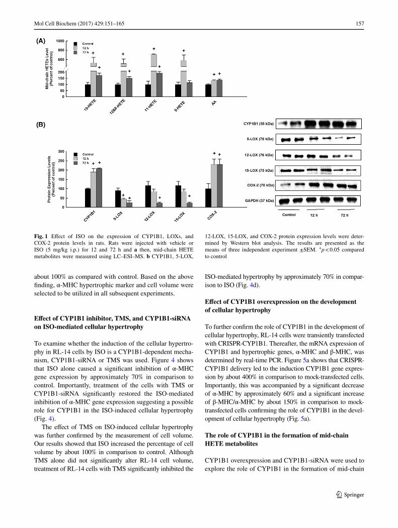
further confirmed by a significant increase in HW/BW in comparison to control [47]. Currently, our aim is to deter-mine the capacity of ISO to increase the level of cardiac mid-chain HETEs in vivo in rats at the pre-hypertrophic stage, 12 h, and at the hypertrophic stage, 72 h. For this purpose, mid-chain HETE metabolites were determined using LC–ESI–MS. Figure 1a shows that ISO significantly increases the level of cardiac mid-chain HETEs by about 500% and 200% at 12 and 72 h, respectively, in comparison to control.
Mid-chain HETEs are known to be formed by LOXs in addition to CYP1B1 and degraded by cyclooxygenase-2 (COX-2) enzyme. Therefore, we investigated whether the induction of mid-chain HETEs by ISO is attributed to the activation of LOXs and CYP1B1 or the inhibition of the COX-2 enzyme. For this purpose, CYP1B1, LOXs, and COX-2 protein expression levels were determined by West-ern blot analysis. Figure 1b shows that treatment of rats with ISO significantly induced CYP1B1 and COX-2 at 12 and 72 h by approximately 200% in comparison to control. On the other hand, ISO significantly inhibited the expres-sions of 5-LOX, 12-LOX, and 15-LOX at 72 h by approxi-mately 70% in comparison to control (Fig. 1b).
Effect of ISO, TMS, and CYP1B1-siRNA on RL-14 cell viability
To determine the effect of ISO in the presence and absence of TMS or CYP1B1-siRNA on RL-14 cell viability, MTT and LDH assays were used. Our results showed that the treatment of RL-14 cells with 100 μM ISO in the presence and absence of 0.5 μM TMS or 25 nM CYP1B1-siRNA at 24 and 48 h, respectively, did not significantly affect RL-14 cell viability using MTT and LDH assays (Fig. 2a, b). Therefore, the observed changes were not due to the decreased cell viability or toxicity.
Effect of ISO on hypertrophic genes and RL-14 cell volume
The effect of ISO on hypertrophic genes was determined by real-time PCR. Figure 3a shows that treatment of RL-14 cells for 24 h with 100 μM ISO significantly induced β-MHC by about 150% and inhibited α-MHC by approxi-mately 70% in comparison to control. However, treatment of RL-14 cells with ISO for 24 h did not significantly alter the expression of ANP and BNP mRNA levels (Fig. 3a).
To determine whether the ISO-induced hypertrophy markers at the mRNA (Fig. 3) were associated with cellu-lar hypertrophy and increase the cell volume, RL-14 cells were treated for 24 h with ISO 100 µM; thereafter, the cell surface area was determined. Figure 3b shows that ISO sig-nificantly increased the percentage of cell surface area by
157Mol Cell Biochem (2017) 429:151–165
1 3
about 100% as compared with control. Based on the above finding, α-MHC hypertrophic marker and cell volume were selected to be utilized in all subsequent experiments.
Effect of CYP1B1 inhibitor, TMS, and CYP1B1-siRNA on ISO-mediated cellular hypertrophy
To examine whether the induction of the cellular hypertro-phy in RL-14 cells by ISO is a CYP1B1-dependent mecha-nism, CYP1B1-siRNA or TMS was used. Figure 4 shows that ISO alone caused a significant inhibition of α-MHC gene expression by approximately 70% in comparison to control. Importantly, treatment of the cells with TMS or CYP1B1-siRNA significantly restored the ISO-mediated inhibition of α-MHC gene expression suggesting a possible role for CYP1B1 in the ISO-induced cellular hypertrophy (Fig. 4).
The effect of TMS on ISO-induced cellular hypertrophy was further confirmed by the measurement of cell volume. Our results showed that ISO increased the percentage of cell volume by about 100% in comparison to control. Although TMS alone did not significantly alter RL-14 cell volume, treatment of RL-14 cells with TMS significantly inhibited the
ISO-mediated hypertrophy by approximately 70% in compar-ison to ISO (Fig. 4d).
Effect of CYP1B1 overexpression on the development of cellular hypertrophy
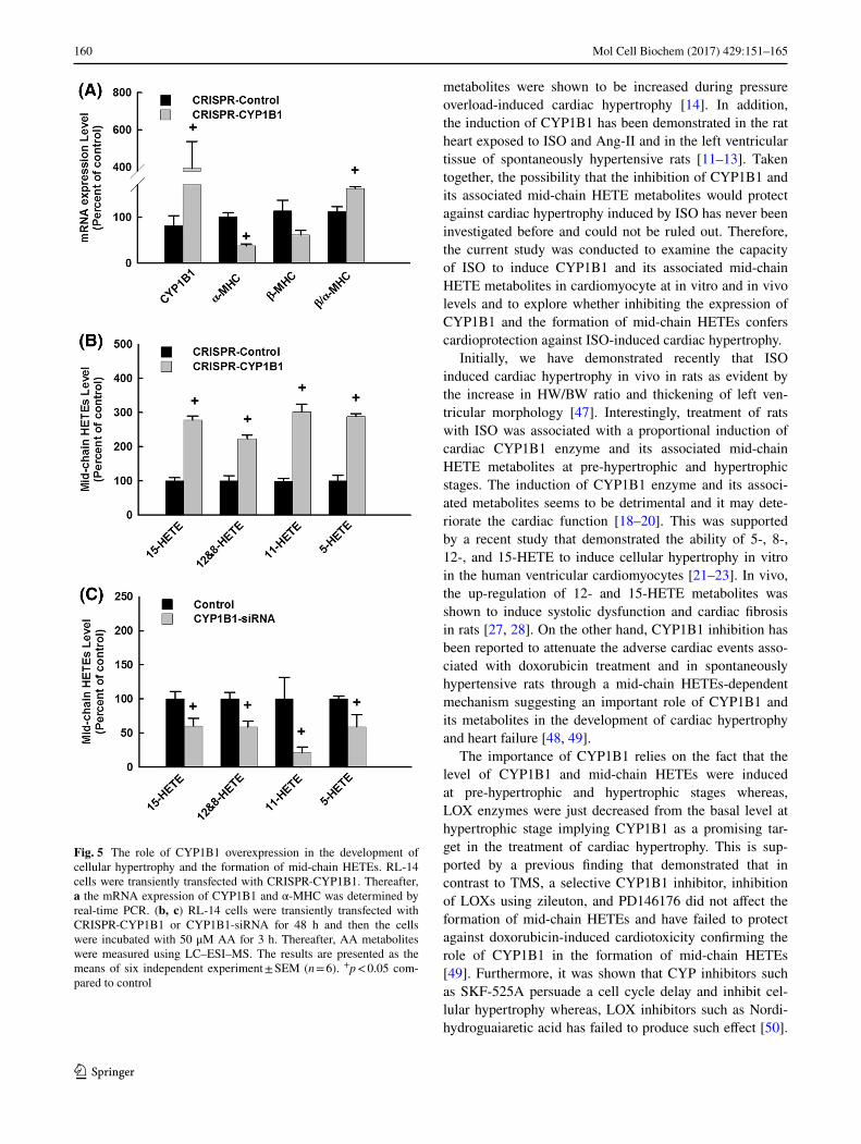
To further confirm the role of CYP1B1 in the development of cellular hypertrophy, RL-14 cells were transiently transfected with CRISPR-CYP1B1. Thereafter, the mRNA expression of CYP1B1 and hypertrophic genes, α-MHC and β-MHC, was determined by real-time PCR. Figure 5a shows that CRISPR-CYP1B1 delivery led to the induction CYP1B1 gene expres-sion by about 400% in comparison to mock-transfected cells. Importantly, this was accompanied by a significant decrease of α-MHC by approximately 60% and a significant increase of β-MHC/α-MHC by about 150% in comparison to mock-transfected cells confirming the role of CYP1B1 in the devel-opment of cellular hypertrophy (Fig. 5a).
The role of CYP1B1 in the formation of mid-chain HETE metabolites
CYP1B1 overexpression and CYP1B1-siRNA were used to explore the role of CYP1B1 in the formation of mid-chain
Fig. 1 Effect of ISO on the expression of CYP1B1, LOXs, and COX-2 protein levels in rats. Rats were injected with vehicle or ISO (5 mg/kg i.p.) for 12 and 72 h and a then, mid-chain HETE metabolites were measured using LC–ESI–MS. b CYP1B1, 5-LOX,
12-LOX, 15-LOX, and COX-2 protein expression levels were deter-mined by Western blot analysis. The results are presented as the means of three independent experiment ±SEM. +p < 0.05 compared to control

158 Mol Cell Biochem (2017) 429:151–165
1 3
HETE metabolites. Figure 5b shows that CRISPR-CYP1B1 delivery led to a significant increase in the formation of the formation of 15-, 12-, and 8-, 11-, and 5-HETE by approxi-mately 280, 220, 300, and 290%, respectively, in compari-son to control. On the other hand, treatment of cells with CYP1B1-siRNA significantly inhibited the formation of 15-, 12-, and 8-, 11-, and 5-HETE by about 40% in compar-ison to control (Fig. 5c) confirming the role of CYP1B1 in the formation of mid-chain HETE metabolites. The effect of CYP1B1-siRNA on mid-chain HETEs is consistent with its effect on CYP1B1 mRNA level (Fig. 4a).
Effect of TMS on ISO-mediated effect on superoxide radical and MAPK signaling pathway
The level of superoxide radical and MAPK signaling pathway were determined to explore the mechanism by
which TMS mediates its protective effect against ISO-induced cellular hypertrophy. Figure 5a shows that incu-bation of the cells with 100 µM of ISO significantly increased superoxide radical formation by approximately 180% and inhibited the phosphorylated ERK1/2 by about 70% in comparison to control. Importantly, treatment with TMS significantly reduced the increased formation of superoxide radical and restored the inhibition of phos-phorylated ERK1/2 in response to ISO by approximately 80% suggesting an important role of superoxide radical and phosphorylated ERK1/2 in TMS-mediated protec-tion against ISO-induced cardiac hypertrophy (Fig. 6). Treatment of cells with TMS did not significantly alter the phosphorylated level of p38 and JNK (Data not shown).
Fig. 2 Effect of ISO, TMS, and CYP1B1-siRNA on RL-14 cell via-bility. RL-14 cells were exposed to 100 μM ISO in the presence and absence of 0.5 μM TMS or 25 nM CYP1B1-siRNA at 24 and 48 h, respectively. Thereafter, RL-14 cell viability was determined using MTT and LDH assays. The results are presented as the means of six independent experiment ±SEM (n = 6). +p < 0.05 compared to con-trol. *p < 0.05 compared to ISO
Fig. 3 Effect of ISO on hypertrophic genes and RL-14 cell vol-ume. a RL-14 cells were treated for 24 h with 100 µM ISO. There-after, the mRNA level of ANP, BNP, α-MHC, β-MHC was quanti-fied using real-time PCR. b RL-14 cells were treated with 100 μM ISO for 24h. Thereafter, cell volume was analyzed by phase-contrast imaging. The results are presented as the means of six independent experiment ±SEM (n = 6). +p < 0.05 compared to control. *p < 0.05 compared to ISO

159Mol Cell Biochem (2017) 429:151–165
1 3
Effect of TMS on ISO-mediated induction of NF-κB signaling pathway
To investigate whether the effect of TMS on ISO-induced cellular hypertrophy is mediated through the inhibition of NF-κB, P50 and P65 binding activity was determined. Figure 6c shows that ISO alone was able to induce the binding activity of NF-κB to its respon-sive element to 148% for P50 in comparison to control. Importantly, treatment with TMS significantly reduced the P50 binding activity in response to ISO to 103% in comparison to control suggesting that inhibition of NF-κB is essential for TMS-mediated protection against ISO-induced cellular hypertrophy.
Discussion
The present work provides the first evidence that ISO induces cardiac hypertrophy in vivo in a rat model and in vitro in human ventricular cardiomyocytes, RL-14 cell line, through a CYP1B1-mediated mid-chain HETEs-dependent mechanism.
Mid-chain HETEs are biologically active eicosanoids resulting from the metabolism of AA by the CYP1B1-catalyzed bis-allylic oxidation reaction. Several lines of evidence support the role of CYP1B1 and its associated mid-chain HETE metabolites in the development of car-diac hypertrophy [9, 10]. For example, the expression of CYP1B1 protein and the formation of mid-chain HETE
Fig. 4 Effect of CYP1B1 inhibitor, TMS, and CYP1B1-siRNA on ISO-mediated cellular hypertrophy. a, b and c RL-14 cells were treated with 100 μM ISO in the presence and absence of 0.5 μM TMS or 25 nM CYP1B1-siRNA at 24 and 48 h, respectively. Thereafter, the mRNA level of CYP1B1 and α-MHC were quantified using real-
time PCR. d RL-14 cells were treated with 100 μM ISO in the pres-ence and absence of 0.5 μM TMS for 24. Thereafter, cell volume was analyzed by phase-contrast imaging. The results are presented as the means of six independent experiment ±SEM (n = 6). +p < 0.05 com-pared to control. *p < 0.05 compared to ISO
160 Mol Cell Biochem (2017) 429:151–165
1 3
metabolites were shown to be increased during pressure overload-induced cardiac hypertrophy [14]. In addition, the induction of CYP1B1 has been demonstrated in the rat heart exposed to ISO and Ang-II and in the left ventricular tissue of spontaneously hypertensive rats [11–13]. Taken together, the possibility that the inhibition of CYP1B1 and its associated mid-chain HETE metabolites would protect against cardiac hypertrophy induced by ISO has never been investigated before and could not be ruled out. Therefore, the current study was conducted to examine the capacity of ISO to induce CYP1B1 and its associated mid-chain HETE metabolites in cardiomyocyte at in vitro and in vivo levels and to explore whether inhibiting the expression of CYP1B1 and the formation of mid-chain HETEs confers cardioprotection against ISO-induced cardiac hypertrophy.
Initially, we have demonstrated recently that ISO induced cardiac hypertrophy in vivo in rats as evident by the increase in HW/BW ratio and thickening of left ven-tricular morphology [47]. Interestingly, treatment of rats with ISO was associated with a proportional induction of cardiac CYP1B1 enzyme and its associated mid-chain HETE metabolites at pre-hypertrophic and hypertrophic stages. The induction of CYP1B1 enzyme and its associ-ated metabolites seems to be detrimental and it may dete-riorate the cardiac function [18–20]. This was supported by a recent study that demonstrated the ability of 5-, 8-, 12-, and 15-HETE to induce cellular hypertrophy in vitro in the human ventricular cardiomyocytes [21–23]. In vivo, the up-regulation of 12- and 15-HETE metabolites was shown to induce systolic dysfunction and cardiac fibrosis in rats [27, 28]. On the other hand, CYP1B1 inhibition has been reported to attenuate the adverse cardiac events asso-ciated with doxorubicin treatment and in spontaneously hypertensive rats through a mid-chain HETEs-dependent mechanism suggesting an important role of CYP1B1 and its metabolites in the development of cardiac hypertrophy and heart failure [48, 49].
The importance of CYP1B1 relies on the fact that the level of CYP1B1 and mid-chain HETEs were induced at pre-hypertrophic and hypertrophic stages whereas, LOX enzymes were just decreased from the basal level at hypertrophic stage implying CYP1B1 as a promising tar-get in the treatment of cardiac hypertrophy. This is sup-ported by a previous finding that demonstrated that in contrast to TMS, a selective CYP1B1 inhibitor, inhibition of LOXs using zileuton, and PD146176 did not affect the formation of mid-chain HETEs and have failed to protect against doxorubicin-induced cardiotoxicity confirming the role of CYP1B1 in the formation of mid-chain HETEs [49]. Furthermore, it was shown that CYP inhibitors such as SKF-525A persuade a cell cycle delay and inhibit cel-lular hypertrophy whereas, LOX inhibitors such as Nordi-hydroguaiaretic acid has failed to produce such effect [50].
Fig. 5 The role of CYP1B1 overexpression in the development of cellular hypertrophy and the formation of mid-chain HETEs. RL-14 cells were transiently transfected with CRISPR-CYP1B1. Thereafter, a the mRNA expression of CYP1B1 and α-MHC was determined by real-time PCR. (b, c) RL-14 cells were transiently transfected with CRISPR-CYP1B1 or CYP1B1-siRNA for 48 h and then the cells were incubated with 50 μM AA for 3 h. Thereafter, AA metabolites were measured using LC–ESI–MS. The results are presented as the means of six independent experiment ± SEM (n = 6). +p < 0.05 com-pared to control

161Mol Cell Biochem (2017) 429:151–165
1 3
The inhibition of cellular growth in response to SKF-525A was associated with CYP inhibition and the subsequent impairment of mid-chain HETEs synthesis. Interestingly, exogenous addition of mid-chain HETEs reversed the effects of SKF-525A confirming an important role of CYP in the regulation of mid-chain HETEs [50].
In vitro, treatment of RL-14 cells with ISO caused cel-lular hypertrophy which is evidenced first by the induc-tion of β-MHC, inhibition of α-MHC, and the increase in cell volume. However, there was no significant change in the expression of ANP and BNP mRNA levels. The rela-tive ratio of β-MHC to α-MHC has been considered as a good predictor of ventricular dysfunction and was reported
Fig. 6 Effect of TMS on ISO-mediated effect on superoxide radical, MAPK, and NF-κB signaling pathway. RL-14 cells were treated for 24 h with 100 µM ISO in the presence and absence of 0.5 μM TMS. Thereafter, a superoxide anion was determined using DHE assay. b MAPKs protein phosphorylation was determined in cytoplasmic
protein extracts using PhosphoTracer Elisa Kit (Abcam, Cambridge, UK). c NF-κB binding activity was determined using commercially available kit. The results are presented as the means of six inde-pendent experiment ±SEM (n = 6). +p < 0.05 compared to control. *p < 0.05 compared to ISO

162 Mol Cell Biochem (2017) 429:151–165
1 3
to be altered during cardiac hypertrophy [51]. MHC iso-form switching from α-MHC to β-MHC decreases the myosin ATPase enzyme velocity and depletes the intracel-lular energy level, thereby disrupting myocardial twitch kinetics. This detrimentally affects the systolic function and decreases contractile performances of the cardiomyocytes that significantly contributes to the initiation and progres-sion of heart failure [52–54]. On the other hand, natriuretic peptides, such as ANP and BNP are considered as potent endogenous inhibitors of hypertrophy and they are released upon cardiomyocytes in response to increased ventricular wall stress [55]. BNP has been considered as a good pre-dictor of heart failure [56] whereas, ANP may serve as a marker of cardiac stress but not essentially as a hyper-trophic marker, especially on an organ level [57].
The direct evidence for the involvement of CYP1B1 in the development of cardiac hypertrophy induced by ISO was supported by the following observations: (a) inhibi-tion of CYP1B1 using TMS, a selective CYP1B1 inhibi-tor, or knockdown of CYP1B1 gene expression using CYP1B1-siRNA significantly restored the mRNA expres-sion of hypertrophic gene and the increase in cell volume in response to ISO to their normal level. In agreement with our results, it has been shown that disruption of a cyp1b1 gene in addition to TMS, a selective CYP1B1 inhibi-tor, provides a significant protective effect against deoxy-corticosterone salt-induced cardiac hypertrophy [15, 58]. The premise of this observation emerges from the finding that the levels of LOXs, Cyp4a, and Cyp4f protein were not changed in the cyp1b1-/- mice suggesting a CYP1B1-dependent mechanism. (b) Overexpression of CYP1B1 using CRISPR-CYP1B1 plasmid significantly induced cel-lular hypertrophy as evident by the inhibition of α-MHC gene expression. Low expression level of α-MHC sig-nificantly accelerates myocardial twitch kinetics, thereby enhancing systolic function in the large mammalian myo-cardium [54]. Similar to our observation, it has been pre-viously reported that induction of CYP1B1 by the AhR ligands, benzo(a)pyrene, and 3-methylcholanthrene, causes significant cardiac hypertrophy in vivo in rats [59]. In addi-tion, TCDD and β-naphthoflavone, CYP1 family inducers, cause hypertrophy in cardiac-derived H9c2 cells through the induction of CYP1B1 gene expression [60].
The above information suggests the role of CYP1B1 in cardiac hypertrophy and in the formation of mid-chain HETEs metabolite induced by ISO. This raises the ques-tion of whether or not CYP1B1 is directly involved in the formation of mid-chain HETE metabolites. Therefore, we examined the ability CYP1B1 overexpression to increase the formation of mid-chain HETEs in RL-14 cells. Perhaps the finding of greatest interest in the current study was the observation that CRISPR-CYP1B1 delivery led to a signifi-cant increase in the formation of 15-, 12-, and 8-, 11-, and
5-HETE, implying that CYP1B1 is directly involved in the formation of these metabolites. Consistent with our find-ings, it has been demonstrated previously that the recombi-nant CYP1B1 enzyme catalyzes the formation of mid-chain HETEs [14, 61]. Importantly, the above finding was further confirmed by the ability of CYP1B1-siRNA to inhibit the formation of mid-chain HETEs suggesting an important role of CYP1B1 in the formation of these metabolites.
Mechanistically, CYP1B1 has been reported to cause cardiac hypertrophy through the generation of superoxide radical [10, 16]. Superoxide anion was shown to be impli-cated in the hypertrophy process through the increase of proto-oncogenes factor, such as c-myc and c-fos, medi-ating the linkage of Na+/K+ ATPase to hypertrophy and modulation of the activity of MAPK [62, 63]. Treatment with antioxidants inhibited the hypertrophic response of cardiomyocytes [64, 65]. Currently, we have demonstrated that ISO treatment significantly increased the generation of superoxide anion. Of interest, the inhibition of CYP1B1 using TMS significantly inhibited the formation of super-oxide anion in response to ISO. Our results are supported by the previous observation showing that the induction of CYP1B1 by TCDD caused cardiac hypertrophy, which was attributed to the generation of superoxide anion and eleva-tion of blood pressure [66]. Furthermore, TMS displayed antihypertensive effect and inhibited its associated cardio-vascular events in spontaneously hypertensive rats, primar-ily by inhibiting the generation of superoxide anion and MAPKs signaling pathway [48].
MAPKs and NF-κB are intracellular signal transduction factors that are critically involved in the regulation of sign-aling pathways, ultimately leading to cardiac hypertrophy and heart failure [67]. Accumulating data provide convinc-ing evidence that persistent activation of NF-κB, p38, and JNK promotes apoptosis, resulting in cardiac hypertrophy [68–71]. ERK1/2 has been proposed to regulate smooth muscle contraction and to promote cellular hypertrophy [68]. Genetic inhibition of ERK1/2 has been reported to promote stress-induced apoptosis and heart failure [72]. Taken together, the possibility that the inhibition of CYP1B1 and its associated mid-chain HETEs and superox-ide anion by TMS would inhibit the modulation of MAPKs and NF-κB by ISO has never been investigated before and could not be ruled out. Thus, the third objective of the cur-rent study was to explore the role of TMS on ISO-mediated effect on NF-κB and MAPKs signaling pathways.
Our results showed that ISO treatment significantly inhibited the phosphorylated ERK1/2, whereas it increased the binding activity of P50 NF-κB to its responsive ele-ments. This was similar to a previous observation demon-strating that ISO dephosphorylates and inactivates phos-phorylated ERK in human cells through the activation of protein phosphatases [73]. Importantly, the inhibition of

163Mol Cell Biochem (2017) 429:151–165
1 3
CYP1B1 using TMS significantly abolished the effect of ISO on the phosphorylated MAPK and the binding activity of NF-κB, suggesting that MAPKs and NF-κB are essential for TMS-mediated protection against ISO-induced cardiac hypertrophy. In agreement with our results, resveratrol, a TMS analog, has been reported to possess a significant cardioprotective effect against Ang-II-induced cardiac hypertrophy through the inhibition of MAPKs [74]. NF-κB inhibition was shown to ameliorate myocardial hypertrophy in response to aortic banding, ISO, and chronic infusion of Ang-II [75, 76].
To reiterate, our findings may cast light on the role of CYP1B1 in the development of cardiac hypertrophy and indicate that CYP1B1 can serve as a novel target in the treatment of heart diseases. Such observation will raise the potential of having selective inhibitors of this enzyme to be used clinically in the treatment of cardiovascular diseases.
Acknowledgements This work was supported by a grant from the Canadian Institutes of Health Research [Grant 106665] to A.O.S.E. Z.H.M. is the recipient Izaak Walton Killam Memorial Scholarship and Alberta Innovates Health Solution Graduate Student Scholar-ship. AAE is the recipient of Egyptian Government Scholarship and Alberta Innovates-Health Solutions studentship.
Compliance with ethical standards
Conflict of interest There is no conflict of interest.
References
1. Vakili BA, Okin PM, Devereux RB (2001) Prognostic implica-tions of left ventricular hypertrophy. Am Heart J 141:334–341. doi:10.1067/mhj.2001.113218
2. Carreno JE, Apablaza F, Ocaranza MP, Jalil JE (2006) Cardiac hypertrophy: molecular and cellular events. Rev Esp Cardiol 59:473–486
3. Berenji K, Drazner MH, Rothermel BA, Hill JA (2005) Does load-induced ventricular hypertrophy progress to systolic heart failure? Am J Physiol Heart Circ Physiol 289:H8–H16. doi:10.1152/ajpheart.01303.2004
4. Levy D, Kenchaiah S, Larson MG, Benjamin EJ, Kupka MJ, Ho KK, Murabito JM, Vasan RS (2002) Long-term trends in the incidence of and survival with heart failure. N Engl J Med 347:1397–1402. doi:10.1056/NEJMoa020265
5. Roman RJ (2002) P-450 metabolites of arachidonic acid in the control of cardiovascular function. Physiol Rev 82:131–185. doi:10.1152/physrev.00021.2001
6. Zordoky BN, El-Kadi AO (2010) Effect of cytochrome P450 polymorphism on arachidonic acid metabolism and their impact on cardiovascular diseases. Pharmacol Ther 125:446–463. doi:10.1016/j.pharmthera.2009.12.002
7. Elshenawy OH, Anwar-Mohamed A, El-Kadi AO (2013) 20-Hydroxyeicosatetraenoic acid is a potential therapeutic target in cardiovascular diseases. Curr Drug Metab 14:706–719
8. Elshenawy OH, Anwar-Mohamed A, Abdelhamid G, El-Kadi AO (2013) Murine atrial HL-1 cell line is a reliable model to
study drug metabolizing enzymes in the heart. Vascul Pharmacol 58:326–333. doi:10.1016/j.vph.2012.12.002
9. Korashy HM, El-Kadi AO (2006) The role of aryl hydrocarbon receptor in the pathogenesis of cardiovascular diseases. Drug Metab Rev 38:411–450. doi:10.1080/03602530600632063
10. Malik KU, Jennings BL, Yaghini FA, Sahan-Firat S, Song CY, Estes AM, Fang XR (2012) Contribution of cytochrome P450 1B1 to hypertension and associated pathophysiology: a novel target for antihypertensive agents. Prostaglandins Other Lipid Mediat 98:69–74. doi:10.1016/j.prostaglandins.2011.12.003
11. Thum T, Borlak J (2002) Testosterone, cytochrome P450, and cardiac hypertrophy. FASEB J 16:1537–1549. doi:10.1096/fj.02-0138com
12. Zordoky BN, Aboutabl ME, El-Kadi AO (2008) Modulation of cytochrome P450 gene expression and arachidonic acid metabolism during isoproterenol-induced cardiac hypertro-phy in rats. Drug Metab Dispos 36:2277–2286. doi:10.1124/dmd.108.023077
13. Jennings BL, Anderson LJ, Estes AM, Yaghini FA, Fang XR, Porter J, Gonzalez FJ, Campbell WB, Malik KU (2012) Cytochrome P450 1B1 contributes to renal dysfunction and damage caused by angiotensin II in mice. Hypertension 59:348–354. doi:10.1161/HYPERTENSIONAHA.111.183301
14. El-Sherbeni AA, El-Kadi AO (2014) Alterations in cytochrome P450-derived arachidonic acid metabolism during pressure overload-induced cardiac hypertrophy. Biochem Pharmacol 87:456–466. doi:10.1016/j.bcp.2013.11.015
15. Jennings BL, Estes AM, Anderson LJ, Fang XR, Yaghini FA, Fan Z, Gonzalez FJ, Campbell WB, Malik KU (2012) Cytochrome P450 1B1 gene disruption mini-mizes deoxycorticosterone acetate-salt-induced hyperten-sion and associated cardiac dysfunction and renal dam-age in mice. Hypertension 60:1510–1516. doi:10.1161/HYPERTENSIONAHA.112.202606
16. Morgan ET (2001) Regulation of cytochrome p450 by inflamma-tory mediators: why and how? Drug Metab Dispos 29:207–212
17. Maayah ZH, El-Kadi AO (2016) The role of mid-chain hydrox-yeicosatetraenoic acids in the pathogenesis of hypertension and cardiac hypertrophy. Arch Toxicol 90:119–136. doi:10.1007/s00204-015-1620-8
18. Jenkins CM, Cedars A, Gross RW (2009) Eicosanoid signalling pathways in the heart. Cardiovasc Res 82:240–249. doi:10.1093/cvr/cvn346
19. Cyrus T, Witztum JL, Rader DJ, Tangirala R, Fazio S, Linton MF, Funk CD (1999) Disruption of the 12/15-lipoxygenase gene diminishes atherosclerosis in apo E-deficient mice. J Clin Invest 103:1597–1604. doi:10.1172/JCI5897
20. Nozawa K, Tuck ML, Golub M, Eggena P, Nadler JL, Stern N (1990) Inhibition of lipoxygenase pathway reduces blood pressure in renovascular hypertensive rats. Am J Physiol 259:H1774–H1780
21. Maayah ZH, El-Kadi AO (2015) 5-, 12- and 15-Hydroxyeicosa-tetraenoic acids induce cellular hypertrophy in the human ven-tricular cardiomyocyte, RL-14 cell line, through MAPK- and NF-kappaB-dependent mechanism. Arch Toxicol. doi:10.1007/s00204-014-1419-z
22. Maayah ZH, El-Kadi AO (2015) The role of mid-chain hydroxyeicosatetraenoic acids in the pathogenesis of hyper-tension and cardiac hypertrophy. Arch Toxicol. doi:10.1007/s00204-015-1620-8
23. Maayah ZH, Abdelhamid G, El-Kadi AO (2015) Development of cellular hypertrophy by 8-hydroxyeicosatetraenoic acid in the human ventricular cardiomyocyte, RL-14 cell line, is implicated by MAPK and NF-kappaB. Cell Biol Toxicol. doi:10.1007/s10565-015-9308-7

164 Mol Cell Biochem (2017) 429:151–165
1 3
24. Burhop KE, Selig WM, Malik AB (1988) Monohydroxyeico-satetraenoic acids (5-HETE and 15-HETE) induce pulmonary vasoconstriction and edema. Circ Res 62:687–698
25. Wen Y, Gu J, Peng X, Zhang G, Nadler J (2003) Overexpression of 12-lipoxygenase and cardiac fibroblast hypertrophy. Trends Cardiovasc Med 13:129–136
26. Wen Y, Gu J, Liu Y, Wang PH, Sun Y, Nadler JL (2001) Over-expression of 12-lipoxygenase causes cardiac fibroblast cell growth. Circ Res 88:70–76
27. Wallukat G, Morwinski R, Kuhn H (1994) Modulation of the beta-adrenergic response of cardiomyocytes by specific lipoxy-genase products involves their incorporation into phosphati-dylinositol and activation of protein kinase C. J Biol Chem 269:29055–29060
28. Kayama Y, Minamino T, Toko H, Sakamoto M, Shimizu I, Taka-hashi H, Okada S, Tateno K, Moriya J, Yokoyama M, Nojima A, Yoshimura M, Egashira K, Aburatani H, Komuro I (2009) Cardiac 12/15 lipoxygenase-induced inflammation is involved in heart failure. J Exp Med 206:1565–1574. doi:10.1084/jem.20082596
29. Molojavyi A, Lindecke A, Raupach A, Moellendorf S, Kohrer K, Godecke A (2010) Myoglobin-deficient mice activate a dis-tinct cardiac gene expression program in response to isopro-terenol-induced hypertrophy. Physiol Genomics 41:137–145. doi:10.1152/physiolgenomics.90297.2008
30. Osadchii OE (2007) Cardiac hypertrophy induced by sustained beta-adrenoreceptor activation: pathophysiological aspects. Heart Fail Rev 12:66–86. doi:10.1007/s10741-007-9007-4
31. Meszaros J, Levai G (1990) Ultrastructural and electrophysi-ological alterations during the development of catecholamine-induced cardiac hypertrophy and failure. Acta Biol Hung 41:289–307
32. Doggrell SA, Brown L (1998) Rat models of hypertension, car-diac hypertrophy and failure. Cardiovasc Res 39:89–105
33. Oyama N, Urasawa K, Kaneta S, Sakai H, Saito T, Takagi C, Yoshida I, Kitabatake A, Tsutsui H (2005) Chronic beta-adren-ergic receptor stimulation enhances the expression of G-Protein coupled receptor kinases, GRK2 and GRK5, in both the heart and peripheral lymphocytes. Circ J 69:987–990
34. Chun YJ, Kim S, Kim D, Lee SK, Guengerich FP (2001) A new selective and potent inhibitor of human cytochrome P450 1B1 and its application to antimutagenesis. Cancer Res 61:8164–8170
35. El-Sherbeni AA, El-Kadi AO (2014) Characterization of ara-chidonic acid metabolism by rat cytochrome P450 enzymes: the involvement of CYP1As. Drug Metab Dispos 42:1498–1507. doi:10.1124/dmd.114.057836
36. Liu Y, Peterson DA, Kimura H, Schubert D (1997) Mechanism of cellular 3-(4,5-dimethylthiazol-2-yl)-2,5-diphenyltetrazolium bromide (MTT) reduction. J Neurochem 69:581–593
37. Sambrook J, Fritsch EF and Maniatatis T (1989) In: Ford, N. (Ed.), Molecular Cloning. A Laboratory Manual. Cold Spring Harbour Laboratory Press, Plainview.
38. Davidson MM (2007) Immortalization of human post-mitotic cells. Google Patents
39. Maayah ZH, Elshenawy OH, Althurwi HN, Abdelhamid G, El-Kadi AO (2014) Human fetal ventricular cardiomyocyte, RL-14 cell line, is a promising model to study drug metaboliz-ing enzymes and their associated arachidonic acid metabolites. J Pharmacol Toxicol Methods. doi:10.1016/j.vascn.2014.11.005
40. Maayah ZH, El Gendy MA, El-Kadi AO, Korashy HM (2013) Sunitinib, a tyrosine kinase inhibitor, induces cytochrome P450 1A1 gene in human breast cancer MCF7 cells through ligand-independent aryl hydrocarbon receptor activation. Arch Toxicol 87:847–856. doi:10.1007/s00204-012-0996-y
41. Maayah ZH, Ansari MA, El Gendy MA, Al-Arifi MN, Korashy HM (2014) Development of cardiac hypertrophy by sunitinib
in vivo and in vitro rat cardiomyocytes is influenced by the aryl hydrocarbon receptor signaling pathway. Arch Toxicol 88:725–738. doi:10.1007/s00204-013-1159-5
42. Livak KJ, Schmittgen TD (2001) Analysis of relative gene expression data using real-time quantitative PCR and the 2(-Delta Delta C(T)) Method. Methods 25:402–408. doi:10.1006/meth.2001.1262
43. Tse MM, Aboutabl ME, Althurwi HN, Elshenawy OH, Abdel-hamid G, El-Kadi AO (2013) Cytochrome P450 epoxygenase metabolite, 14,15-EET, protects against isoproterenol-induced cellular hypertrophy in H9c2 rat cell line. Vascul Pharmacol 58:363–373. doi:10.1016/j.vph.2013.02.004
44. Andrews NC, Faller DV (1991) A rapid micropreparation tech-nique for extraction of DNA-binding proteins from limiting numbers of mammalian cells. Nucleic Acids Res 19:2499
45. Bhattacharya N, Sarno A, Idler IS, Fuhrer M, Zenz T, Dohner H, Stilgenbauer S, Mertens D (2010) High-throughput detec-tion of nuclear factor-kappaB activity using a sensitive oligo-based chemiluminescent enzyme-linked immunosorbent assay. Int J Cancer 127:404–411. doi:10.1002/ijc.25054
46. Zordoky BN, Anwar-Mohamed A, Aboutabl ME, El-Kadi AO (2010) Acute doxorubicin cardiotoxicity alters cardiac cytochrome P450 expression and arachidonic acid metabolism in rats. Toxicol Appl Pharmacol 242:38–46. doi:10.1016/j.taap.2009.09.012
47. Althurwi HN, Maayah ZH, Elshenawy OH, El-Kadi AO (2015) Early changes in cytochrome P450s and their associated ara-chidonic acid metabolites play a crucial role in the initiation of cardiac hypertrophy induced by isoproterenol. Drug Metab Dispos 43:1254–1266. doi:10.1124/dmd.115.063776
48. Jennings BL, Montanez DE, May ME Jr, Estes AM, Fang XR, Yaghini FA, Kanu A, Malik KU (2014) Cytochrome P450 1B1 contributes to increased blood pressure and cardiovascular and renal dysfunction in spontaneously hypertensive rats. Cardio-vasc Drugs Ther 28:145–161. doi:10.1007/s10557-014-6510-4
49. Maayah ZH, Althurwi HN, Abdelhamid G, Lesyk G, Jurasz P, El-Kadi AO (2016) CYP1B1 inhibition attenuates doxo-rubicin-induced cardiotoxicity through a mid-chain HETEs-dependent mechanism. Pharmacol Res. doi:10.1016/j.phrs.2015.12.016
50. Nieves D, Moreno JJ (2006) Hydroxyeicosatetraenoic acids released through the cytochrome P-450 pathway regulate 3T6 fibroblast growth. J Lipid Res 47:2681–2689. doi:10.1194/jlr.M600212-JLR200
51. Barry SP, Davidson SM, Townsend PA (2008) Molecular regula-tion of cardiac hypertrophy. Int J Biochem Cell Biol 40:2023–2039. doi:10.1016/j.biocel.2008.02.020
52. Fatkin D, McConnell BK, Mudd JO, Semsarian C, Moskow-itz IG, Schoen FJ, Giewat M, Seidman CE, Seidman JG (2000) An abnormal Ca(2+) response in mutant sarcomere protein-mediated familial hypertrophic cardiomyopathy. J Clin Invest 106:1351–1359. doi:10.1172/JCI11093
53. Kiriazis H, Kranias EG (2000) Genetically engineered models with alterations in cardiac membrane calcium-handling pro-teins. Annu Rev Physiol 62:321–351. doi:10.1146/annurev.physiol.62.1.321
54. Locher MR, Razumova MV, Stelzer JE, Norman HS, Moss RL (2011) Effects of low-level α-myosin heavy chain expression on contractile kinetics in porcine myocardium. Am J Physiol Heart Circ Physiol 300:H869–H878. doi:10.1152/ajpheart.00452.2010
55. de Lemos JA, McGuire DK, Drazner MH (2003) B-type natriu-retic peptide in cardiovascular disease. The Lancet 362:316–322. doi:10.1016/S0140-6736(03)13976-1
56. Latini R, Masson S, de Angelis N, Anand I (2002) Role of brain natriuretic peptide in the diagnosis and management of heart failure: current concepts. J Card Fail 8:288–299
165Mol Cell Biochem (2017) 429:151–165
1 3
57. Cox EJ, Marsh SA (2014) A systematic review of fetal genes as biomarkers of cardiac hypertrophy in rodent models of diabetes. PLoS One 9:e92903. doi:10.1371/journal.pone.0092903
58. Sahan-Firat S, Jennings BL, Yaghini FA, Song CY, Estes AM, Fang XR, Farjana N, Khan AI, Malik KU (2010) 2,3′,4,5′-Tetramethoxystilbene prevents deoxycorticosterone-salt-induced hypertension: contribution of cytochrome P-450 1B1. Am J Physiol Heart Circ Physiol 299:H1891–H1901. doi:10.1152/ajpheart.00655.2010
59. Aboutabl ME, Zordoky BN, El-Kadi AO (2009) 3-methylcho-lanthrene and benzo(a)pyrene modulate cardiac cytochrome P450 gene expression and arachidonic acid metabolism in male Sprague Dawley rats. Br J Pharmacol 158:1808–1819. doi:10.1111/j.1476-5381.2009.00461.x
60. Zordoky BN, El-Kadi AO (2010) 2,3,7,8-Tetrachlorodibenzo-p-dioxin and beta-naphthoflavone induce cellular hypertro-phy in H9c2 cells by an aryl hydrocarbon receptor-dependant mechanism. Toxicol In Vitro 24:863–871. doi:10.1016/j.tiv.2009.12.002
61. Choudhary D, Jansson I, Stoilov I, Sarfarazi M, Schenkman JB (2004) Metabolism of retinoids and arachidonic acid by human and mouse cytochrome P450 1b1. Drug Metab Dispos 32:840–847
62. Wassmann S, Laufs U, Baumer AT, Muller K, Konkol C, Sauer H, Bohm M, Nickenig G (2001) Inhibition of geranylgeranyla-tion reduces angiotensin II-mediated free radical production in vascular smooth muscle cells: involvement of angiotensin AT1 receptor expression and Rac1 GTPase. Mol Pharmacol 59:646–654
63. Xie Z, Kometiani P, Liu J, Li J, Shapiro JI, Askari A (1999) Intracellular reactive oxygen species mediate the linkage of Na+/K+-ATPase to hypertrophy and its marker genes in cardiac myo-cytes. J Biol Chem 274:19323–19328
64. Nakamura K, Fushimi K, Kouchi H, Mihara K, Miyazaki M, Ohe T, Namba M (1998) Inhibitory effects of antioxidants on neona-tal rat cardiac myocyte hypertrophy induced by tumor necrosis factor-alpha and angiotensin II. Circulation 98:794–799
65. Tanaka K, Honda M, Takabatake T (2001) Redox regulation of MAPK pathways and cardiac hypertrophy in adult rat cardiac myocyte. J Am Coll Cardiol 37:676–685
66. Kopf PG, Huwe JK, Walker MK (2008) Hypertension, car-diac hypertrophy, and impaired vascular relaxation induced by 2,3,7,8-tetrachlorodibenzo-p-dioxin are associated with increased superoxide. Cardiovasc Toxicol 8:181–193. doi:10.1007/s12012-008-9027-x
67. Zhang W, Elimban V, Nijjar MS, Gupta SK, Dhalla NS (2003) Role of mitogen-activated protein kinase in cardiac hypertrophy and heart failure. Exp. Clin Cardiol 8:173–183
68. Pearson G, Robinson F, Beers Gibson T, Xu BE, Karandikar M, Berman K, Cobb MH (2001) Mitogen-activated protein (MAP) kinase pathways: regulation and physiological functions. Endocr Rev 22:153–183. doi:10.1210/edrv.22.2.0428
69. Xu D, Li N, He Y, Timofeyev V, Lu L, Tsai HJ, Kim IH, Tuteja D, Mateo RK, Singapuri A, Davis BB, Low R, Ham-mock BD, Chiamvimonvat N (2006) Prevention and reversal of cardiac hypertrophy by soluble epoxide hydrolase inhibi-tors. Proc Natl Acad Sci U S A 103:18733–18738. doi:10.1073/pnas.0609158103
70. Li S EM, Yu B (2008) Adriamycin induces myocardium apopto-sis through activation of nuclear factor kappaB in rat. Mol Biol Rep 35:489–494. doi:10.1007/s11033-007-9112-4
71. Esposito G, Rapacciuolo A, Naga Prasad SV, Takaoka H, Thomas SA, Koch WJ, Rockman HA (2002) Genetic altera-tions that inhibit in vivo pressure-overload hypertrophy prevent cardiac dysfunction despite increased wall stress. Circulation 105:85–92
72. Purcell NH, Wilkins BJ, York A, Saba-El-Leil MK, Meloche S, Robbins J, Molkentin JD (2007) Genetic inhibition of cardiac ERK1/2 promotes stress-induced apoptosis and heart failure but has no effect on hypertrophy in vivo. Proc Natl Acad Sci U S A 104:14074–14079. doi:10.1073/pnas.0610906104
73. Chen J, Hoffman BB, Isseroff RR (2002) Beta-adrenergic recep-tor activation inhibits keratinocyte migration via a cyclic adeno-sine monophosphate-independent mechanism. J Invest Dermatol 119:1261–1268. doi:10.1046/j.1523-1747.2002.19611.x
74. Cheng TH, Liu JC, Lin H, Shih NL, Chen YL, Huang MT, Chan P, Cheng CF, Chen JJ (2004) Inhibitory effect of resveratrol on angiotensin II-induced cardiomyocyte hypertrophy. Naunyn Schmiedebergs Arch Pharmacol 369:239–244. doi:10.1007/s00210-003-0849-6
75. Kawano S, Kubota T, Monden Y, Kawamura N, Tsutsui H, Takeshita A, Sunagawa K (2005) Blockade of NF-kappaB ame-liorates myocardial hypertrophy in response to chronic infusion of angiotensin II. Cardiovasc Res 67:689–698. doi:10.1016/j.cardiores.2005.04.030
76. Hu X, Wang H, Lv X, Chu L, Liu Z, Wei X, Chen Q, Zhu L, Cui W (2015) Cardioprotective effects of tannic acid on isopro-terenol-induced myocardial injury in rats: further insight into ‘French Paradox’. Phytother Res. doi:10.1002/ptr.5376