the role of cymbopogon citratus extract in protecting the...
TRANSCRIPT

International Journal of Clinical and Developmental Anatomy 2015; 1(4): 89-95
Published online February 26, 2016 (http://www.sciencepublishinggroup.com/j/ijcda)
doi: 10.11648/j.ijcda.20150104.12
ISSN: 2469-7990 (Print); ISSN: 2469-8008 (Online)
The Role of Cymbopogon Citratus Extract in Protecting the Liver Against Injurious Effect of Dimethylnitrosamine in Rats
Naglaa Ali Saber Sarg1, Usama Fouad Ahmed
1, Ali Mohamed Ali, Saleh Sayed Idris
2, *
1Department of Anatomy, Faculty of Medicine, Benha University, Benha City, Egypt 2Department of Health Education & Health Promotion, Umm Al-Qura University, Makkah City, Saudi Arabia
Email address: [email protected] (S. S. Idris)
To cite this article: Naglaa Ali Saber Sarg, Usama Fouad Ahmed, Ali Mohamed Ali, Saleh Sayed Idris. The Role of Cymbopogon Citratus Extract in Protecting
the Liver Against Injurious Effect of Dimethylnitrosamine in Rats. International Journal of Clinical and Developmental Anatomy.
Vol. 1, No. 4, 2015, pp. 89-95. doi: 10.11648/j.ijcda.20150104.12
Abstract: Background: N-Nitrosodimethylamine (NDMA) is a potent hepatotoxin that induces fibrosis of the liver.
Cymbopogon citratus, a plant that is widely cultivated to be eaten either fresh with food or dried in tea or soft drink has been
reported to possess a number of medicinal and aromatic properties. Aim of the work: This work is aimed to study the protective
effect of Cymbopogon citratus ethanolic leaf extract against hepatic injury and fibrosis induced by repeated intermittent
administration of Dimethylnitrosamine (DMN) in rats. Materials and methods: A total of 30 rats divided into 3 groups were
used. Group 1 served as control, Group 2 was given intraperitoneal injection with DMA at a concentration of 10mg/kg body
weight on three consecutive days of each week over a period of three weeks. Group 3 was given DMA at a concentration of
10mg/kg body weight on three consecutive days of each week over a period of three weeks and 100 mg/kg body weight of
Cymbopogon citratus ethanolic aqueous leaf extract 5 hours after administration of DMN. Results: It was found that DMN
produces histopathological changes of the liver; including signs of severe hepatic injury. The cytoplasm of most hepatocytes
appeared vacuolated with pyknotic and karyolytic nuclei, congested blood vessels and focal necrotic areas with marked
increase of the collagen fibers deposition around the blood vessels. Ultrastructural changes showed Pyknotic nuclei, vacuolated
cytoplasm and reduction or complete loss of cristae in the mitochondria. These were reversed by simultaneous treatment with
Cymbopogon citratus. In conclusion: The results of this present study indicated that aqueous leaf extracts of Cymbopogon
citratus has an antihepatotoxic action against DMN induced hepatic oxidative damage in rats which might be ascribed to its
antioxidant and free radical scavenging property.
Keywords: Dimethylnitrosmine, Fibrosis, Liver, Rat, Cymbopogon Citratus
1. Introduction
The liver is the largest visceral organ in the human body
and the chief site for metabolism and detoxification. Liver
diseases often progress from sub-clinical icteric hepatitis to
necro-inflammatory hepatitis, fibrosis, cirrhosis, and
hepatocellular carcinoma [1]. Liver cirrhosis often results in
high mortality and is also a risk factor in the development of
hepatocellular carcinoma (HCC) [2], which rank the fifth of
the cancer incidence worldwide [3].
Several experimental and clinical evidences have shown a
common link between chronic liver injury, oxidative stress,
activation of hepatic stellate cells (HSCs) and their
transformation to myofibroblast- like cells, associated with
increased production of extracellular matrix proteins during
hepatic fibrosis [4].
Dimethylnitrosamine (DMN), is an industrial by-product
or waste product of several industrial processes. Of more
general concern, water treatment via chlorination or
chloramination of organic nitrogen-containing waste water
can lead to the production of DMA at potentially harmful
levels, [5]. DMA's contamination of drinking water is of
particular concern due to the minute concentrations at which
it is harmful, the difficulty in detecting it at these
concentrations, and to the difficulty in removing it from
drinking water, [6]. DMA may be formed during cooking
foods, especially cured meats and fish, that contain sodium
nitrite as a preservative, but is also found in several

90 Naglaa Ali Saber Sarg et al.: The Role of Cymbopogon Citratus Extract in Protecting the Liver Against
Injurious Effect of Dimethylnitrosamine in Rats
vegetables, cheeses, alcoholic beverages and fruits, as a
contaminant in rubber products and tobacco smoke, [7].
DMN is a potent hepatotoxin, carcinogen and mutagen
which has been demonstrated to induce bridging fibrosis,
necrosis and collapse of parenchymal framework of liver, [8].
Hepatic cells are involved in a variety of metabolic events;
therefore the establishment of liver protective/therapeutic
agents is of a paramount importance in the protection of the
liver from damage. Natural remedies from traditional plants
are seen as effective and safe alternative treatments for
hepatotoxicity, [9]. Cymbopogon citratus, commonly known
as lemon grass, is a tropical monocotyledonous hypogeal
perennial herb belonging to the family Poaceae. is widely
cultivated to be eaten either fresh with food or dried in tea or
soft drink has been reported to possess a number of medicinal
and aromatic properties, its oil is use of more general levels
as culinary flavoring, scent, and medicine, [10].
The present investigation was undertaken with the objective
of evaluating the anti-fibrotic, antioxidant and protective
efficacy of ethanolic leaf extract of Cymbopogon citratus
against repeated DMN-induced hepatic fibrosis in rats.
2. Materials and Method
2.1. Animals
Thirty adult male albino rats were used in this study. Their
weight ranged from 200-250 gm. Before commencing
experimentation, all the animals were subjected to one week
period of passive preliminaries in order to adapt themselves
to their new environment at animal bread house in Anatomy
department in Benha faculty of medicine. They were housed
in clean well ventilated cages under strict care and hygiene.
The rats were provided with standard laboratory diet and
water.
2.2. Chemicals
a N-Nitrosodimethylamine:
NDMA obtained from, faculty of science, Biochemistry
department, Benha university. 10mg of DMA dissolved in 10
ml of sodium hydrochloride, so every 1ml solution contains 1
mg of the drug.
b C-citratus leaves Extraction:
The collected C. citratus leave was cleaned and oven-dried
at 65ºC and coarsely ground. The powdered materials of C.
citratus (100 g) were extracted with 1000 mL of 70%
methanol for 3 days with occasional shake. The plant was
filtered through filter paper and this procedure was repeated
three times. The extracts were then evaporated to eliminate
the methanol. The extracts were then stored at −20ºC until
used. The yields were 12.34 g. The powdered extracts were
reconstituted with distilled water to the desired
concentrations prior to use.
2.3. Experimental Design
The animals were divided into three groups of ten rats
each:
The liver of the three groups were extracted. The
specimens of liver were divided into two parts. One part was
fixed in 10% formaldehyde for light microscopic study, while
the other part was fixed in 2% cacodylate buffered
gluteraldehyde solution for 2-4 hours at 4ºC for electron
microscopic study.
For light microscopy, the slides were stained by:
(1) Hematoxylin and Eosin stain [11].
(2) Periodic Acid Schiff’s stain (PAS) [12].
(3) Masson’s trichrome stain [13].
For electron microscopic examination, the ultrathin
sections were stained by uranyl acetate and lead citrate and
examined with Joel CX 100 transmission electron
microscope in laboratory of the histology and cell Biology
Department, Faculty of Medicine Benha University.
3. Results
Light microscopic examination of liver sections of an adult
control rat normal hepatic architecture where the hepatocytes
were arranged in cords radiating from the central vein
extending to the periphery of the lobule. The cords were
separated from each other by blood sinusoids which lined by
flat endothelial cells and few kuppfer cells. The hepatocytes
showed an acidophilic cytoplasm and rounded vesicular
nuclei with one or two prominent nucleoli [Fig. 1]. The PAS
preparations of the normal liver of rats revealed that the
glycogen granules were observed in the cytoplasm of the
hepatocytes as indicated by large number of magenta red fine
granules [Fig. 2]. In Masson's trichrome stain, the connective
tissue around the portal tract was scanty [Fig. 3].
The ultrastructural picture of the hepatic tissue in the same
group revealed, normal hepatic cell with rounded nucleus and
prominent nucleolus, tight junction and bile canaliculi
between adjacent hepatocytes [Fig. 4]. The cytoplasm was
crowded with cell organelles, the most numerous were
mitochondria with regularly arranged cristae in close
association appeared variable in sizes and shapes, glycogen
granules and the rough endoplasmic reticulum were located
near the nucleus [Fig. 5].
Fig. 1. A photomicrograph of liver section of control rat showing: Cords
(HC) of hepatocytes (H) radiating from the central vein (CV). Notice Kupffer
cells (black arrow) bulge in blood sinusoids (S). [Hx &E X200].

International Journal of Clinical and Developmental Anatomy 2015; 1(4): 89-95 91
Fig. 2. A photomicrograph of liver section of control rat showing: Positive
PAS reaction for glycogen granules (g) in the cytoplasm of hepatocytes.
Notice the central vein (CV). [PAS X 200].
Fig. 3. A photomicrograph of liver section of control rat showing: Scanty
perivascular connective tissue (arrow) around portal tract containing a
branch of hepatic artery (HA), a branch of portal vein (PV) and bile ducts
(BD). Notice cords of hepatocytes (H) and blood sinusoids (S) between
hepatic cords (HC). [Masson's Trichrome X400].
Fig. 4. An electron photomicrograph of ultrathin section in liver of control
rat showing: The hepatocyte (H) with rounded nucleus (N) and nucleolus
(nu). The cytoplasm contains many mitochondria (M), smooth endoplasmic
reticulum (SER). Notice the canaliculi (bc) and tight junction (TJ) in
between the cells. [E. M X 6000].
Fig. 5. An electron photomicrograph of ultrathin section in liver of control
rat showing: The cytoplasm contains mitochondria (M), glycogen granules
(g), rough endoplasmic reticulum (RER) and lysosomes (LY). Notice a part
of nucleus (N) of hepatocyte. [E.M X 10000].
The light microscopic examination of rats which were
exposed to nitrosodimethylamine for 3 weeks showed clear
signs of severe hepatic injury manifested by lose of normal
hepatic architecture, dilated and congested central vein. The
cytoplasm of most hepatocytes appeared vacuolated area
with pyknotic or, karyolytic nuclei (Fig. 6). Most
hepatocytes appeared vacuolated [Fig. 7]. After exposure of
rats to DMN, the glycogen granules of the liver cells
obviously were decreased [Fig. 8]. After Masson's
trichrome staining there was marked increase of the
collagen fibers deposition around the portal tirades
compared with the control one [Fig. 9].
The ultrastructural picture of the hepatic tissue in the
same group revealed that some cells showed the nuclei
frequently appeared with irregular shapes and chromatin
condensation. Other damaged cells appeared with pyknotic
nuclei, vacuolated cytoplasm [Fig. 10] and little or no
glycogen granules. The mitochondria showed a reduction or
complete loss of cristae and an amorphous or granular
matrix [Fig. 11].
Fig. 6. A photomicrograph of liver section of rat treated with DMN showing:
dilated central vein (CV), loss of normal hepatic architecture. The cytoplasm
of hepatocytes is vacuolated (VH). Notice the kupffer cell (kr). [Hx &E
X400].

92 Naglaa Ali Saber Sarg et al.: The Role of Cymbopogon Citratus Extract in Protecting the Liver Against
Injurious Effect of Dimethylnitrosamine in Rats
Fig. 7. A photomicrograph of liver section of rat treated with DMN showing:
Portal tract containing a branch of hepatic artery (HA), congested and
dilated portal vein (PV) and bile duct (BD)., Notice hepatocytes are
vacuolated (HV). [Hx& E X400].
Fig. 8. A photomicrograph of liver section of rat treated with DMN showing:
Vacuolations in liver cells (V) with depletion of glycogen granules (g).
Notice the central vein. [PAS X 200].
Fig. 9. A photomicrograph of liver section of rat treated with DMN showing:
Increase in connective tissue (arrow) around the portal tract. Notice the
congested portal vein (PV), hepatic artery (HA) and bile duct. Notice the
vacuolated hepatocytes (VH). [Masson's Trichrome X200].
Fig. 10. An electron photomicrograph of ultrathin section in liver of rat
treated with DMN shows the nucleus (N) with irregular shape and chromatin
condensation. The cytoplasm shows large vacuoles, mitochondria (M),
condensed rough endoplasmic reticulum (RER) around the nucleus and
lysosomes (L). Notice the tight junction (TJ) and bile canaliculi (bc). (E. M X
6000).
Fig. 11. An electron photomicrograph of ultrathin section in liver of rat
treated with DMN showing: The nuclei of hepatocytes (N) are vacuolated (V)
with collection of chromatin materials in the center of nucleus. The
cytoplasm contains vacuoles (V) of some degenerated mitochondria (M).
Notice the bile canaliculi (bc). [E. M X 6000].
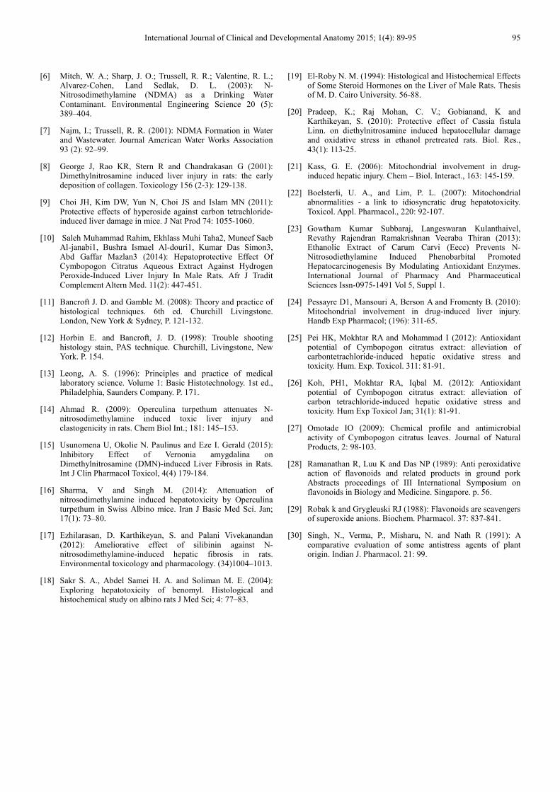
The light microscopic examination of rats which were
exposed to nitrosodimethylamine and C. citratus aqueous
extract showed signs of improvement through a well
arrangement of hepatocytes with normal cytoplasm.
Sinusoids were well preserved and some hepatocytes showed
vacuolated cytoplasm compared with rats treated with
nitrosodimethylamine only [Fig. 12]. In PAS staining, most
of hepatocytes had good positive PAS for glycogen granules
and few hepatocytes had poor PAS reaction [Fig. 13]. In
Masson’s trichrome stain, the connective tissue around the
central vein and portal tract was decreased when compared
with the nitrosodimethylamine group [Fig. 14].
The ultrastructural picture of the hepatic tissue in the same
group showed some improvement, the hepatocytes appear
normal with rounded nucleus and nucleolus. The cytoplasm
contains numerous organelles. Some mitochondria are
numerous and intact, with apparent cristae. Rough
endoplasmic reticulum was preserved. Lipid droplets,
glycogen granules, multivesicular bodies, and few vacuoles
International Journal of Clinical and Developmental Anatomy 2015; 1(4): 89-95 93
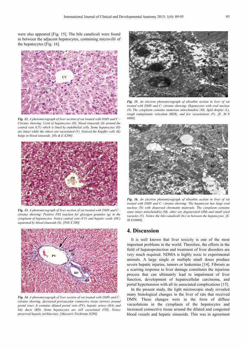
were also appeared [Fig. 15]. The bile canaliculi were found
in between the adjacent hepatocytes, containing microvilli of
the hepatocytes [Fig. 16].
Fig. 12. A photomicrograph of liver section of rat treated with DMN and C –
Citratus showing: Cord of hepatocytes (H), blood sinusoids (S) around the
central vein (CV) which is lined by endothelial cells. Some hepatocytes (H)
are intact while the others are vacuolated (V). Noticed the Kupffer cells (K)
bulge in blood sinusoids. [Hx & E X200].
Fig. 13. A photomicrograph of liver section of rat treated with DMN and C -
citratus showing: Positive PAS reaction for glycogen granules (g) in the
cytoplasm of hepatocytes. Notice central vein (CV) and hepatic cords (HC)
separated by blood sinusoids (S). [PAS X 200].
Fig. 14. A photomicrograph of liver section of rat treated with DMN and C–
citratus showing, decreased perivascular connective tissue (arrow) around
portal tract. It contains dilated portal vein (PV), hepatic artery (HA) and
bile ducts (BD). Some hepatocytes are still vacuolated (VH). Notice
preserved hepatic architecture. [Masson's Trichrome X200].
Fig. 15. An electron photomicrograph of ultrathin section in liver of rat
treated with DMN and C- citratus showing: Hepatocytes with oval nucleus
(N) The cytoplasm contains numerous mitochondria (M), lipid droplet (L),
rough endoplasmic reticulum (RER), and few vacuolations (V). [E. M X
6000].
Fig. 16. An electron photomicrograph of ultrathin section in liver of rat
treated with DMN and C- citratus showing: The hepatocyte has large oval
nucleus (N) with dispersed chromatin materials. The cytoplasm contains
some intact mitochondria (M), other are degenerated (dM) and small sized
vacuoles (V). Notice the bile canaliculi (bc) in between the hepatocytes. [E.
M X10000].
4. Discussion
It is well known that liver toxicity is one of the most
important problems in the world. Therefore, the efforts in the
field of hepatoprotection and treatment of liver disorders are
very much required. NDMA is highly toxic to experimental
animals. A large single or multiple small doses produce
severe hepatic injuries, tumors or leukemias [14]. Fibrosis as
a scarring response to liver damage constitutes the injurious
process that can ultimately lead to impairment of liver
function, development of hepatocellular carcinoma, and
portal hypertension with all its associated complications [15].
In the present study, the light microscopic study revealed
many histological changes in the liver of rats that received
DMN. These changes were in the form of diffuse
vacuolations in the cytoplasm of the hepatocytes and
increased connective tissue around the dilated and congested
blood vessels and hepatic sinusoids. This was in agreement

94 Naglaa Ali Saber Sarg et al.: The Role of Cymbopogon Citratus Extract in Protecting the Liver Against
Injurious Effect of Dimethylnitrosamine in Rats
with [16]; who found that animals treated with NDMA
exhibited perivenular necrosis and micro vesicular fatty
change in peripheral hepatocytes. There were severe
centrilobular congestion and marked dilatation of central vein
and sinusoids with massive necrosis and initiation of fibrosis.
Also, these results were similar to the study of [17] that
showed, in the Hx & E stained rat liver cells of DMN treated
rats, a high degree of centrilobular necrosis and bridging
fibrosis between the portal triad and the central vein.
In the current work, PAS-stained sections of the control
group showed normal content of glycogen granules
evidenced by a strong PAS reaction (in the form of
mahogany coloration). Also, these granules were evident
ultrastructurally in the control group. In contrast, the DMN-
treated group showed a weak PAS reaction indicating
depletion of glycogen. The reduction in carbohydrate
contents could be attributed to increased stress on the liver,
leading to the consumption of high energy in an attempt to
reduce or equalize the pressure exerted on it [18].
In the current study the ultrastructural changes showed
electron-lucent areas of the cytoplasm in hepatocytes in rats
that received DMN. Other degenerative changes were also
observed. These changes include lipid droplets deposition in
the cytoplasm of hepatocytes, decreased organelles, damaged
cristae and vacuoles in mitochondria, dilated endoplasmic
reticulum and decrease of glycogen granules. In this research,
there were variations in the size and shape of nuclei of the
liver cells. Most of hepatocytes had extended chromatin with
marginal displacement of their nucleoli and peripheral
condensation of their nuclear chromatin which might be
attributed to cellular hyperactivity. Some of the hepatocytes
had shrunken electron-dense nuclei. The variations of the
shape of nuclei were explained by [19], to be due to
compression of nuclei by vacuoles in the cytoplasm of
hepatocytes. However, the variations in size of nuclei may be
due to the increase in liver DNA content. The endoplasmic
reticulum was particularly liable to the free radical attack, not
only because it is considered as a site of radical production
but also because its membrane is rich in polyunsaturated fatty
acids which are susceptible to free radical attack. Therefore,
the dramatic ultrastructural changes observed in the present
study may be attributed to increased oxidative stress as
reported by [20].
The mitochondrion was the major target in drug-induced
hepatic damage [21]. Mitochondrial dysfunction was
generally accompanied with oxidative stress, which is a key
regulator of mitochondria-mediated cell death. Hence, drugs
cause cell death by inducing oxidative stress in hepatic
mitochondria. [22]. The cytoplasmic vacuolations could be
attributed to mitochondrial degeneration. The mitochondrial
damage probably occurs because of the electrolyte imbalance
or edema because of the inhibition of the membrane enzyme
of the sodium pump [23].
The increase in the lipid peroxidation of mitochondria
could lead to inhibition of respiratory enzymes which in turn
could inhibit ATP production. The decrease in intracellular
ATP leads to mitochondrial damage [24].
In the present investigation, amelioration of DMN-induced
hepatic fibrosis, inflammatory changes and hepatic necrosis
was well evidenced in rats treated with C. citratus aqueous
extract with DMN simultaneously. Light microscopic
examination of the liver sections showed a more or less similar
picture to those of the control group with relative restoration of
the general hepatic lobular architecture. The central vein was
surrounded by cords of relatively normally appearing
hepatocytes and blood sinusoids. Some hepatocytes were
binucleated and others still had a vacuolated cytoplasm and
small dark nuclei. However, still there was dilated central vein.
A strong PAS reaction could be observed in this group. And
this was in line with the results of [25], who found an
improvement of liver tissue in rats treated with. C. citratus
aqueous extract after DMN intoxication. [10] Found that the
liver tissue of rats treated with C. citratus aqueous extract
with oxidative stress induced by H2O2, exhibited a similar
liver cytoarchitecture compared with the control group. The
hepatoprotective effect of C. citratus might be ascribable to
its antioxidant and free radical scavenging property [26].
[27], reported that the leaves of C. citratus contained
saponins, sesquiterpenes, lactones, steroids, flavonoids.
Flavonoids were reported to exhibit antioxidant activity ([28]
and were effective scavengers of superoxide anions [29]. The
aqueous extract of C. citratus may have exhibited
hepatoprotective activity due to its possible antioxidant
content attributable to flavonoids. Interestingly, saponins
especially terpene glycosides were reported to enhance
natural resistance and recurative powers of the body [30].
5. Conclusion
The use of aqueous leaf extracts of Cymbopogon citratus
was found to have a protective action against DMN induced
hepatic oxidative damage in rats. This was due to its
antioxidant and free radical scavenging property.
References
[1] Lima CF, Fernandes-Ferreira M and Pereira-Wilson C (2007): Drinking of Salvia officinalis tea increases CCl4-induced hepatotoxicity in mice. Food Chem Toxicol 45 (3): 456-464.
[2] Bataller R and Brenner DA (2005): Liver fibrosis. J Clin Invest 115 (2): 209-218.
[3] Caldwell S and Park SH (2009): The epidemiology of hepatocellular cancer: from the perspectives of public health problem to tumor biology. J Gastroenterol 44 (suppl. 19): 96-101.
[4] Vendemiale G, Grattagliano I, Caruso ML, Serviddio G and Valentini AM (2001): Increased oxidative stress in dimethylnitrosamine-induced liver fibrosis in the rat: effect of N-acetylcysteine and interferon-alpha. Toxicol Appl Pharm acol 175 (2): 130-139.
[5] David L. Sedlak, Rula A. Deeb, Elisabeth L. Hawley, William A. Mitch, Timothy D. Durbin, Sam Mowbray and Steve Carr(2005): Sources and Fate of Nitrosodimethylamine and Its Precursors in Municipal Wastewater Treatment Plants Water Environment Research Vol. 77, No., pp. 32-39.
International Journal of Clinical and Developmental Anatomy 2015; 1(4): 89-95 95
[6] Mitch, W. A.; Sharp, J. O.; Trussell, R. R.; Valentine, R. L.; Alvarez-Cohen, Land Sedlak, D. L. (2003): N-Nitrosodimethylamine (NDMA) as a Drinking Water Contaminant. Environmental Engineering Science 20 (5): 389–404.
[7] Najm, I.; Trussell, R. R. (2001): NDMA Formation in Water and Wastewater. Journal American Water Works Association 93 (2): 92–99.
[8] George J, Rao KR, Stern R and Chandrakasan G (2001): Dimethylnitrosamine induced liver injury in rats: the early deposition of collagen. Toxicology 156 (2-3): 129-138.
[9] Choi JH, Kim DW, Yun N, Choi JS and Islam MN (2011): Protective effects of hyperoside against carbon tetrachloride-induced liver damage in mice. J Nat Prod 74: 1055-1060.
[10] Saleh Muhammad Rahim, Ekhlass Muhi Taha2, Muneef Saeb Al-janabi1, Bushra Ismael Al-douri1, Kumar Das Simon3, Abd Gaffar Mazlan3 (2014): Hepatoprotective Effect Of Cymbopogon Citratus Aqueous Extract Against Hydrogen Peroxide-Induced Liver Injury In Male Rats. Afr J Tradit Complement Altern Med. 11(2): 447-451.
[11] Bancroft J. D. and Gamble M. (2008): Theory and practice of histological techniques. 6th ed. Churchill Livingstone. London, New York & Sydney, P. 121-132.
[12] Horbin E. and Bancroft, J. D. (1998): Trouble shooting histology stain, PAS technique. Churchill, Livingstone, New York. P. 154.
[13] Leong, A. S. (1996): Principles and practice of medical laboratory science. Volume 1: Basic Histotechnology. 1st ed., Philadelphia, Saunders Company. P. 171.
[14] Ahmad R. (2009): Operculina turpethum attenuates N-nitrosodimethylamine induced toxic liver injury and clastogenicity in rats. Chem Biol Int.; 181: 145–153.
[15] Usunomena U, Okolie N. Paulinus and Eze I. Gerald (2015): Inhibitory Effect of Vernonia amygdalina on Dimethylnitrosamine (DMN)-induced Liver Fibrosis in Rats. Int J Clin Pharmacol Toxicol, 4(4) 179-184.
[16] Sharma, V and Singh M. (2014): Attenuation of nitrosodimethylamine induced hepatotoxicity by Operculina turpethum in Swiss Albino mice. Iran J Basic Med Sci. Jan; 17(1): 73–80.
[17] Ezhilarasan, D. Karthikeyan, S. and Palani Vivekanandan (2012): Ameliorative effect of silibinin against N-nitrosodimethylamine-induced hepatic fibrosis in rats. Environmental toxicology and pharmacology. (34)1004–1013.
[18] Sakr S. A., Abdel Samei H. A. and Soliman M. E. (2004): Exploring hepatotoxicity of benomyl. Histological and histochemical study on albino rats J Med Sci; 4: 77–83.
[19] El-Roby N. M. (1994): Histological and Histochemical Effects of Some Steroid Hormones on the Liver of Male Rats. Thesis of M. D. Cairo University. 56-88.
[20] Pradeep, K.; Raj Mohan, C. V.; Gobianand, K and Karthikeyan, S. (2010): Protective effect of Cassia fistula Linn. on diethylnitrosamine induced hepatocellular damage and oxidative stress in ethanol pretreated rats. Biol. Res., 43(1): 113-25.
[21] Kass, G. E. (2006): Mitochondrial involvement in drug-induced hepatic injury. Chem – Biol. Interact., 163: 145-159.
[22] Boelsterli, U. A., and Lim, P. L. (2007): Mitochondrial abnormalities - a link to idiosyncratic drug hepatotoxicity. Toxicol. Appl. Pharmacol., 220: 92-107.
[23] Gowtham Kumar Subbaraj, Langeswaran Kulanthaivel, Revathy Rajendran Ramakrishnan Veeraba Thiran (2013): Ethanolic Extract of Carum Carvi (Eecc) Prevents N-Nitrosodiethylamine Induced Phenobarbital Promoted Hepatocarcinogenesis By Modulating Antioxidant Enzymes. International Journal of Pharmacy And Pharmaceutical Sciences Issn-0975-1491 Vol 5, Suppl 1.
[24] Pessayre D1, Mansouri A, Berson A and Fromenty B. (2010): Mitochondrial involvement in drug-induced liver injury. Handb Exp Pharmacol; (196): 311-65.
[25] Pei HK, Mokhtar RA and Mohammad I (2012): Antioxidant potential of Cymbopogon citratus extract: alleviation of carbontetrachloride-induced hepatic oxidative stress and toxicity. Hum. Exp. Toxicol. 311: 81-91.
[26] Koh, PH1, Mokhtar RA, Iqbal M. (2012): Antioxidant potential of Cymbopogon citratus extract: alleviation of carbon tetrachloride-induced hepatic oxidative stress and toxicity. Hum Exp Toxicol Jan; 31(1): 81-91.
[27] Omotade IO (2009): Chemical profile and antimicrobial activity of Cymbopogon citratus leaves. Journal of Natural Products, 2: 98-103.
[28] Ramanathan R, Luu K and Das NP (1989): Anti peroxidative action of flavonoids and related products in ground pork Abstracts proceedings of III International Symposium on flavonoids in Biology and Medicine. Singapore. p. 56.
[29] Robak k and Grygleuski RJ (1988): Flavonoids are scavengers of superoxide anions. Biochem. Pharmacol. 37: 837-841.
[30] Singh, N., Verma, P., Misharu, N. and Nath R (1991): A comparative evaluation of some antistress agents of plant origin. Indian J. Pharmacol. 21: 99.