the role of casein micelles and their - wur e-depot home
TRANSCRIPT


The role of casein micelles and their
aggregates in foam stabilization
Min Chen

Thesis committee
Promotors
Prof. Dr E. van der Linden
Professor of Physics and Physical Chemistry of Foods
Wageningen University
Prof. Dr A.C.M. van Hooijdonk
Professor of Dairy Science
Wageningen University
Co-promotors
Dr M.B.J. Meinders
Senior Researcher
Wageningen Food and Biobased Research
Dr G. Sala
Researcher
Wageningen Food and Biobased Research
Other members
Prof. Dr J. van der Gucht, Wageningen University
Prof. Dr P. Fischer, ETH Zurich, Switzerland
Prof. Dr A. Kelly, University College Cork, Ireland
Dr P.A. Wierenga, Wageningen University
This research was conducted under the auspices of the Graduate School VLAG (Advanced
studies in Food Technology, Agrobiotechnology, Nutrition and Health Sciences).

The role of casein micelles and their
aggregates in foam stabilization
Min Chen
Thesis
submitted in fulfilment of the requirements for the degree of doctor
at Wageningen University
by the authority of the Rector Magnificus
Prof. Dr A.P.J. Mol,
in the presence of the
Thesis Committee appointed by the Academic Board
to be defended in public
on Monday 12th of December 2016
at 1:30 p.m. in the Aula.

Min Chen
The role of casein micelles and their aggregates in foam stabilization
140 pages.
PhD thesis, Wageningen University, Wageningen, NL (2016)
With references, with summary in English
ISBN 978-94-6257-984-2
DOI 10.18174/393613

Abstract
Many foam products derived from milk or specific dairy ingredients suffer from drainage,
coalescence and/or disproportionation. Previous studies indicated that foam properties of
milk are strongly influenced by the composition of the milk as well as by the processing
conditions during foam production. The aim of this research was to get a better
understanding of these two factors. Interestingly, the presence of aggregates of casein
micelles was found to result in very stable foams. The interfacial properties (adsorption
speed, adsorption energy, dynamical interfacial tension, interfacial dilatational moduli), thin
film stability (rupture time) and foam properties (foamability, drainage, coalescence) of
casein micelle dispersions were determined. Based on these data, the very stable foams
were concluded to result from properties of the thin films in the foam, which were affected
drastically by the presence of the large aggregates of casein micelles.

Learn & Grow 致我的父母

Table of Contents
Chapter 1 1
General introduction
Chapter 2 21
Particle size determines foam stability of casein micelle dispersions
Chapter 3 41
Interfacial properties, thin film stability and foam stability of casein micelle dispersions
Chapter 4 59
Mechanism of ultra-stabilization of foam by casein micelle aggregates
Chapter 5 75
Foam properties of β-casein and silica particle mixtures: the influence of particle size and
concentration
Chapter 6 95
General discussion
Summary 113
Acknowledgements 117
About the author 121

1
Chapter 1 General introduction

Chapter 1
2
1.1 Introduction
Consumers like aerated foods or foams. A problem of these products is that many are
unstable and suffer from drainage, coalescence and/or disproportionation. Despite the
enormous research that has been performed on the formation and stability of foams, there
are still important issues to be solved. This especially concerns the role of surface and bulk
components in complex food systems on the formation and stability of foams. Various
studies have been performed on the formation and stability of dairy foams [1-10]. Results
indicated that foam properties of milk are strongly influenced by the composition of the
milk as well as the processing conditions during foam production [8]. However, the role of
the different components in milk on the formation and stability of foams is still not well
understood. The aim of this research was to get better understanding of the role of the milk
components in milk foam behaviour and to use this understanding to better control dairy
aerated products. Hereto, milk was first decomposed into key ingredients that control the
formation and stability of milk foam. Special focus was on the influence of proteins, lipids
(phospholipid, diglycerides and free fatty acids) in milk. Casein micelles, the main protein
reservoir in milk, which is also a natural nanogel particle, stand out. As a preliminary
finding we observed that certain processing conditions improved the stability of milk foam
to a large extent. In particular, casein micelles dispersions were hypothesised as one of the
key factors in this observation. Therefore, casein micelle dispersions were taken as a model
system for the study. Research was conducted on casein micelle dispersions regarding
interfacial properties (adsorption speed, adsorption energy, dynamical interfacial tension,
interfacial dilatational moduli), thin film stability (rupture time) and foaming properties
(formability, drainage, coalescence) to uncover the stabilization mechanism. Finally these
findings were tested in a model system simulating casein micelle dispersions to get better
insights on foam stabilization with particles in complex system. In this chapter, we first
shortly address the theory of foam stabilization in general and then give a more detailed
review of foams stabilized with complex system of particles mixed with proteins or
surfactants. In the final part we present the outline of the thesis.

General introduction
3
1.2 Foams
Foams are metastable colloidal systems that consists of two phases, a continuous liquid
phase surrounding a disperse gas phase. The thin film separating two bubbles is called
lamella or film. The thicker channels where three lamella meet are Plateau borders (Figure
1-1). The basis of foam formation is the insertion of gas bubbles into a liquid phase [11].
Foams do not form spontaneously. Energy is required to disperse the gas in the liquid.
Foams can be prepared using various methods like sparging, gas injection, agitation, and
super saturation etc. [8]. When new bubbles are formed, energy is needed to create the
air/water surface. Foams are therefore instable and tend to coalesce and disproportionate to
minimize the air/water surface area [12] [13]. Depending on the liquid fraction, foams can
be divided into wet foams and dry foams (Фl ≤0.02) [14].
Figure 1-1. Schematic representation of foam structure redrawn after [15].
1.3 Foam instability
Foam instability is caused by liquid drainage, bubble coalescence and disproportionation.
After foam formation, foam consists of spherical air bubbles that are separated by the
continuous phase. Liquid drainage starts immediately due to gravity [2]. After a certain
time, sufficient amount of liquid drains out and bubbles come into contact, allowing
deformation of bubbles into polyhedral. Liquid will drain out from the lamella into the
Plateau borders due to capillary suction. At a certain thickness the film can rupture, leading
to bubble coalescence, which results in formation of larger bubbles, smaller total interfacial
area and reduction in bubble number [8]. As a matter of fact, bubble coalescence also
occurs during foam formation and this process is greatly affected by the adsorption of
surface active molecules. Faster adsorption of these molecules leads to the formation of
more stable films and thus minimizing coalescence during foam formation, creating foam
with smaller bubbles [16]. Together with drainage and coalescence, disproportionation
(Oswald ripening) also takes place after foam formation. This is a process of gas transfer
Chapter 1
4
from small to large bubbles (through the lamella) due to differences in Laplace pressure. As
a result, small bubbles disappear, while large ones become larger. Disproportionation can
be slowed down by minimizing the polydispersity of the bubbles, by using gas with low
solubility in the continuous phase (i.e. using N2 instead of CO2) [8], or by formation of an
elastic protein layer [17].
1.4 Liquid film rupture and coalescence
1.4.1 Interaction between two interfaces
During foaming, surfactants adsorb at the surface of air bubbles. When two bubbles meet,
the forces between the two interfaces are crucial to understand the stability of the thin liquid
film. The forces include the London-van der Waals forces and the electrostatic forces,
which results repulsion when the interfaces are equally charged. Besides, there may be
other forces involved like hydration forces, steric repulsion forces and supramolecular
forces due to the presence of organized structures in the film like micelles and their
confinement in the films. For proteins, adsorbed polymers and solid particles adsorbed at an
interface, steric repulsion is observed at a thickness of several hundreds of nanometers,
which could be either homogeneous or non-uniform. These forces are normally expressed
per unit area and called the disjoining pressure.
1.4.2 Film thinning and rupture
The curvature of the Plateau border is larger than that of the film so that the hydrodynamic
pressure in the film is larger than in the Plateau borders. This causes the suction of liquid
from the film into the adjacent Plateau borders, which is called capillary suction. Film
thinning occurs. In order for a flat film to exist at equilibrium, it is necessary that, in
addition to the liquid pressure, p, there is a positive disjoining pressure, Π, which balances
the gas pressures and resists the suction from the Plateau borders. At equilibrium, the
disjoining pressure is equal to the capillary pressure Pc, 𝛱 = 𝑃𝑐 =𝛾
𝑟 , where r is the radius
of curvature of the Plateau border, which depends on the bubble diameter and the liquid
fraction, and γ, the interfacial tension.
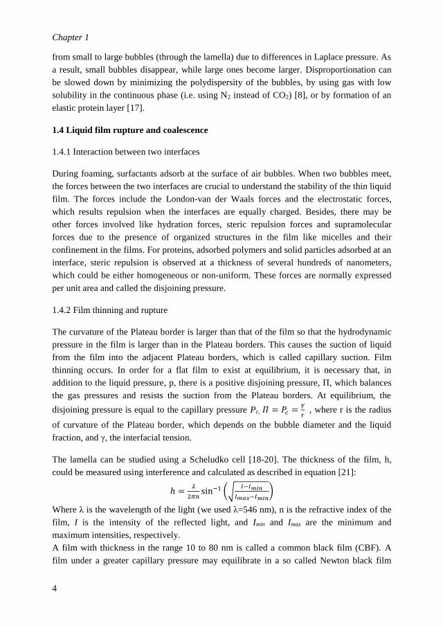
The lamella can be studied using a Scheludko cell [18-20]. The thickness of the film, h,
could be measured using interference and calculated as described in equation [21]:
ℎ =𝜆
2𝜋𝑛sin−1 (√
𝐼−𝐼𝑚𝑖𝑛
𝐼𝑚𝑎𝑥−𝐼𝑚𝑖𝑛)
Where λ is the wavelength of the light (we used λ=546 nm), n is the refractive index of the
film, I is the intensity of the reflected light, and Imin and Imax are the minimum and
maximum intensities, respectively.
A film with thickness in the range 10 to 80 nm is called a common black film (CBF). A
film under a greater capillary pressure may equilibrate in a so called Newton black film

General introduction
5
(NBF). Generally the film is approximated by a circular horizontal film of radius R. Its
thinning velocity is expressed as 𝜈𝑅𝑒 = −𝑑ℎ/𝑑𝑡. Besides film thinning, oscillations of the
interfaces of a film generated by mechanical or thermal perturbations sometimes cause film
rupture. Ivanov [22] and Vrij et al.[23] have shown that there is a critical film thickness for
film rupture.
1.4.3 Coalescence of foam
Foam can disappear via a succession of film ruptures: coalescence. The bubble films at the
top of foam are most susceptible to rupture: they are the thinnest due to drainage, the most
curved and they evaporated the most quickly and subject to external perturbations. Thus
foam often collapses from the top. The liquid fraction of foam depends on the height.
1.5 Foaming agents
Foaming agents includes all kinds of low molecular weight surfactants, proteins, colloidal
particles. Milk proteins are widely used in aerated products. Milk mainly contains caseins
(four kinds of phosphoproteins: αS1, αS2, β, κ), organized in casein micelles (colloidal
particles), and whey proteins. Amphiphilic surfactants like caseins [24], sodium caseinates
[25] and β-lactoglobulin [26] can adsorb fast and stabilize the air/water interface by
forming a relatively strong elastic interfacial layer. Casein micelles, natural food nano-gel
particles, function as the main emulsion stabilizing component of homogenized milk.
1.5.1 Physicochemical properties of casein micelles
Casein micelles is natural nanoparticles present in milk with a diameter of 50 to 500 nm
and average size of 150 nm, which account for about 2.8% of the dry matter content take up
10% of volume of milk. The dry matter of casein micelles contain up to 94% of protein and
the remaining 6% is composed of low molecular mass compounds, mainly inorganic salts
of calcium phosphates called colloidal calcium phosphate (CCP). Casein micelles are
highly hydrated and have a voluminosity of 4.4 cm3/g protein [27, 28]. The morphology of
casein micelles is shown in Figure 1-2. The structure of casein micelle have been
extensively studied and still under debate, and several models have been proposed for it: (i)
sub micelle model, (ii) nanocluster model, and (iii) dual binding model [29]. In a review
[30], it is proposed that casein micelles can be seen as large spherical complexes with a
sponge-like structure. The interior part consists of linked CCP/calcium nanocluster, while
the surface is covered with the hairy κ-casein layer, providing a steric stabilization to the
micelles and preventing them to approach each other.

Chapter 1
6
Figure 1-2. SEM image of casein micelles studied in this thesis.
1.5.2 Role of casein micelles in stabilizing milk foam
Casein micelles can be prepared from skim milk by ultracentrifugation. Considering the
molecular weight (around 108 Da) of casein micelles, centrifugation is sufficient to
sediment most of the casein micelles in milk (90-95%). The redispersion of casein micelle
pellets is temperature dependent [31]. Pellets can be redispersed in a milk permeate without
changing the original properties of the micelles [9, 32] or, depending on redispersion
conditions like temperatures and times, casein micelle dispersions (CMDs) with different
particle properties like size distribution of the casein micelles or their aggregates can be
obtained. Previous research studied casein micelles in skim milk samples [3, 4, 6] and the
influence of heat treatment, temperatures [9], Ca2+ chelating agents [33], pH [34] and ionic
strength [4] on their foaming properties. An interesting finding [9] is that micellar casein
fraction exhibited higher foam stability compared with low-heated skim milk. Casein
micelles seem to play an important role in the dairy foam stabilization. Microscopic images
of bubble ghosts of milk foam suggested that casein micelles can be adsorbed at the
air/water interface [35], possibly in a reversible way [1]. However, Borcherding et al. [6]
indicated that casein micelles are not likely to be present at the air/water interface. Due to
the complex composition of CMDs, which contain individual caseins, casein micelles,
possibly small amount of casein-derived peptides, it remains unclear whether the casein
micelles actually lead to changes in interfacial properties that result in better foaming
properties. Further research needs to be performed in order to establish a quantitative
correlation between interfacial properties, thin film characteristics and foam stability of
CMDs. Since it is reported that the presence of protein aggregates could either improve or
decrease the foam stability of protein solutions [36], inducing aggregates into CMDs makes
the system even more complex and therefore foaming behaviour more complicated.
1.6 Foams stabilized by mixed system of protein and aggregates

General introduction
7
Protein aggregates could be made by heat-treatment induced fibrillization, denaturation of
proteins and gelation [36] [37-39] [40]. Also, complexes can be formed by electrostatic
attraction of two ingredients [41]. Previous research has shown that the presence of
aggregates can have a large influence on foam stability of protein solutions [42]. Whey
protein aggregates [36, 37, 43], soy protein aggregates [44, 45] whey protein fibrils [38, 46]
and whey protein microgel [47, 48] were shown to be advantageous for foam stabilization,
while other studies indicated that protein aggregates can also decrease foam stability [41,
43]. The aggregate size resulting in higher foam stability varied for different aggregated
materials and their structure [41, 43]. There seems to be an optimal particle size for
aggregates to stabilize foam and this optimal size is system-related, ranging from tens of
nanometers to a few micrometers. A recent review reported improved foam stability in the
presence of protein aggregates without causing significant differences in the interfacial
properties of the air/water interface [49]. However, the dilatational properties were
determined only at small deformations and constant frequency, and no large amplitude
dilatations, which are more sensitive to subtle changes in the microstructure of the interface,
were performed. Ultrastabilization of foams with particles or protein aggregates is mostly
ascribed to a final jamming or gelled network formed in the lamella and/or plateau borders
[41, 42, 50-52]. A correlation between thin film stability and the foam stability has been
reported [50, 51]. According to Rullier et al. [53] [43, 50], the thin film stability is
dependent on the aggregate size and on the ratio between non-aggregated proteins and
protein aggregates. The mobility of aggregates at the film surface was found to be crucial
for film stability. A gel-like network formed within the foam film was interpreted from the
immobility of aggregates on the film surface. Saint-Jalmes et al. (2005) [51] investigated a
CMD with a particle size range between 50 nm and 300 nm. These casein aggregates
appeared as thick spot-regions of a few microns within the thin film. The mentioned group
also indicated that an increase in concentration of the casein aggregates yielded higher film
stability. However, the reason why casein micelle aggregates get trapped in the film
remains unknown. No explanation was given on formation of these casein aggregates with
normal casein micelles within size range of 300 nm in the initial sample. The effective
concentration of aggregates accounted for improved foam stability is not well quantified.
For large casein micelle aggregates, there is still no direct proof on the gel network
formation by these aggregates in the foam lamella. Besides, the optimum size of casein
aggregates for foam stabilization, the location of these aggregates regarding different
particle size and particle concentration is unknown and need further research. Table 1-1
give an overview of recent research on foams of systems containing aggregates.
1.7 Foams stabilized by particles
Besides protein aggregates, foams stabilized by food particles [41, 42, 46, 54-58] have
received considerable attention recently. Foam stabilization by solid particles is an
extensively studied topic [42, 59]. Compared to irregular protein aggregates, particles are

Chapter 1
8
often better designed and can be used as elements of complex systems for foam study. The
important physical properties of particles for foam stabilization include contact angle, size,
shape, and concentration [59]. Depending on surface properties, particles can be divided
into adsorbing particles and non-adsorbing particles. Completely hydrophilic particles do
not adsorb at air/water interface neither stabilize the foam [60]. Partially hydrophobic
particles with contact angle, θ, close to 90° can act as a foam stabiliser, whereas very
hydrophobic particles (θ>90°) act in the opposite way and are used as antifoams through a
bridging de-wetting mechanism [59]. The particle hydrophobicity could be modified by
appropriate chemical synthesis [61, 62] or after dispersing them in the aqueous phase and
adjusting the salt concentration [63]. Hydrophobic particles can be accomplished through
the adsorption of appropriate amphiphilic compounds on the surface of hydrophilic
particles [57]. The surface roughness of the particle could also influence their contact angle
since it could lead to a ‘non-equilibrium’ wetting characteristic of the particles. Kaptay
(2003) indicated that there is a maximum regarding particle size for foam stabilization with
particles [64] and explained the importance of particle size on film and foam stabilization.
The role of particles in thinning and stability of the aqueous films separating the bubbles is
crucial for the coalescence and foam collapse. Depending on the contact angle of particles,
two cases for particle location can be considered: (i) the particles are attached to the film
surfaces; (ii) the particles are present only inside the film but not at its surfaces [59].
In the first case, it is reported that the mobility of particles at the film surfaces plays a very
important role in the film stabilisation by solid particles. According to Vinaldini et al.
(2013), the interfacial stability of foam stabilized by particles is defined as:
𝐼𝑛𝑡𝑒𝑟𝑓𝑎𝑐𝑖𝑎𝑙 𝑆𝑡𝑎𝑏𝑖𝑙𝑖𝑦 =∆𝐺𝑎𝑑𝑠
𝑘𝐵𝑇=
𝐴𝑑𝑠𝑜𝑟𝑝𝑡𝑖𝑜𝑛 𝐸𝑛𝑒𝑟𝑔𝑦
𝑇ℎ𝑒𝑟𝑚𝑎𝑙 𝐸𝑛𝑒𝑟𝑔𝑦
Where ∆𝐺𝑎𝑑𝑠 is the adsorption energy of the particles, which could be calculated according
to equation:
∆𝐺𝑎𝑑𝑠 = 𝜋𝑅2𝛾𝐴𝑊(1 ± cos 𝜃)2
Where 𝛾𝐴𝑊is the air/water interfacial tension (mN/m) and R is the particle radius (m), θ is
the contact angle of the particles on the air/water interface. When the adsorption energy of
particles (∆𝐺𝑎𝑑𝑠) is lower or just above the thermal energy (𝑘𝐵𝑇), foam of particles cannot
be formed because of an unstable air/water interface. Other research also indicate that
surface irregularities (for instance the aggregation patterns of particles at the interface) can
also influence the stability of air/water interface depending on the surface coverage of the
particles [65] and the interaction between them [66-68]. For films with diluted particle at
the surfaces, there is a hydrodynamic liquid flow inside the film due to drainage. Particles
in dilute monolayers cannot resist the flow and are dragged away from the film centre,

General introduction
9
leaving the centre and thinnest part of the film unprotected and vulnerable to rupture [69].
However, films with close-packed particle monolayers at their surfaces behave differently
from those with dilute monolayers. Close packed particle monolayers at the film surfaces
can oppose the drag, thus slowing down the film thinning and preventing the film rupture.
The films of partially hydrophobic particles with contact angles around 65° and smaller
than 90° were found to be optimal for foam stabilization [70-72]. This is the so called
Pickering stabilization of foams by particles, which has been reported in several recent
studies [73-77].
In the second case, when the particles are present only inside the film, a stable bilayer of
particles or a bridging monolayer is formed at the final stage of thinning. Stratification of
particles in the films could be visualized in this case [78, 79]. Further suction of liquid out
of the meniscus becomes impossible. Alternatively, as shown in Figure 1-3, if the particle
rearrangement during bilayer-monolayer transition is difficult due to strong cohesion, a
void (crack) could be formed inside the bilayer, then the film breaks due to the rupture of
the unprotected region [80].
Figure 1-3. Possible mechanisms of rupture of a water film stabilized by a bilayer of
particles: (B-C) direct rupture without rearrangement of the particles, (B-M-C) via bilayer-
to monolayer transition and (B-V-C) via void formation redrawn after [59].
Besides the two cases described above, another mechanism of foam film stabilisation is due
to the presence of particle aggregates inside the films. It occurs either when the excess
particles in the bulk aqueous phase are flocculated and form three dimensional networks
(gel) or if a sufficient amount of particle aggregates with an appropriate size get trapped
and confined in the thin liquid films. This mechanism has been used to explain the high
stability of particle-stabilised foams and bubbles reported recently [52, 61, 81-85]. In
summary, the mechanism of foam stabilization by particles is different depending on the

Chapter 1
10
physical properties of studied particles and the interaction between particles and other
coexisting components in the system.
Silica (SiO2) is one of the widely used spherical inorganic particles [61]. Silica particles are
negatively charged at pH above isoelectric point (2 to 3) due to the dissociation of surface
silanol groups (Si-OH) [86]. At neutral pH, unmodified silica particles are completely
hydrophilic and cannot go to the air/water interface to stabilize foam [61]. Foaming
behaviour of silica can be controlled by surface modification of particles, i.e. with different
amount of SiOH on the particle surface. Table 1-2 gives an overview of foam studies of
modified silica particles. Foams of chemically modified silica particles have been
intensively studied. For pure particles, modified particles with intermediate hydrophobicity
exhibited the best foam stabilization. More recently research has been focussed on foams of
silica modified by adsorption of amphiphiles on particle surface, for instance silica particles
modified by SDS [87] and hexylamine [88]. In these particle-amphiphile systems, the
foaming behaviour becomes more complicated depending on the ratio between particles
and amphiphiles. Below a certain concentration of amphiphiles, bulk aggregation of
particles occurred [89]. It is reported that β-CN could attach to the surface of silica [90]. In
this project, three hydrophilic silica with three seizes were used, 200 nm, 1 µm (both non-
porous and well spherical) and 3 µm (porous and roughly spherical with some
irregularities). Particles modified with β-CN were expected to have a similar surface charge
and hydrophobicity compared to that of casein micelles. This is also why the foaming
behaviour of β-CN/silica system was investigated and compared to that of CMDs in this
project.

General introduction
11
Table 1-1. An overview of studies on foams of systems containing aggregates.
System Formation of
aggregates
Aggregates
size (RA)
Concentration
of aggregates
(CA)
Charge Foam
stability
Bulk
properties
Interfacial
properties
Thin film
stability
Ref
β-Lg
with/without
aggregates
Heat
treatment
30-200 nm 1-10 g/L pH 7.0 RA <100 nm (+)
When RA >100 nm,
CA: ≤90% (+)
RA =197 nm
(Π =0 mN/m)
Size RA,
ratio
between
A/NA
[43,
53,
91]
Aggregated β-
lactoglobulin
Heat
treatment
3-1000 nm 10 g /L pH 6.8-
8.0
I=0-130
mM
(+) RA=1000 nm
No link to
Interfacial
properties
Dynamic η Π , E’&E’’
Slower
protein
adsorption
with larger
RA
[37]
Whey protein
Aggregates
Heat
Treatment
50-330 nm 1 g/L pH 6.0-
7.0
I=0-120
mM
(+) pH>6.6, I>70
mM
Π, E’&E’’ [36]
Casein 50-300 nm 0.03-1 g/L pH 5.6 Π (t= 3 s) ≤20
mN/m
(+) CA
dependent
[51]
Casein micelles Redispersion
of
casein pellet
500-5000
nm
0.6-50 g/L pH 6.7 (+) CA dependent,
no link to Interfacial
properties
η slightly
increased with
CA
Π (t= 1 s & 60
s) ≤25 mN/m,
E’&E’’
[92]
Soy (11S)
fibril−peptide
system
Heat-induced
fibrillar
aggregates
Height: a
few
nanometers
Length: 2.3
μm
1.0 g/L pH 7.0 (+) no link to
Interfacial
properties
Nonlinear
Surface
Dilatational
Rheology
[38]
Whey proteins
(globular
aggregates and
fibre)
Heat-induced
denaturation
and
fibrillization
Height: 2–3
nm
Length: 15
µm
20 g/L pH 7.0
pH 2.0
(+) Slower drainage [39]
Protein/pectin
complexes
Electrostatic
attraction
200 -500 nm
or > 1500
nm
0.1 -5 g/L pH 7.0
I=25mM
& 148
mM
CA, ionic strength,
RA:
200 -500 nm (+);
1500 nm (-)
Free proteins
or RA
Π [41]
Whey protein
micro gels
Heat
treatment
270-6000
nm
50 g/L pH 5.0 (+) Slower drainage
& less
disproportionation
Intrinsic η (10
mL/g)
Self-
aggregation at
IEP
[40]
Whey protein
fluid gels
Heat induced
gelation
103-106 nm 50 g/L pH 5.0 &
8.0
(+) larger RA,
link to bulk an
interfacial properties
pH5.0→8.0,
higher local
bulk viscosity
pH5.0→8.0,
higher E’&E’’
[47]
A: aggregates;
NAP: non-aggregated protein
I: Ionic strength;
IEP: isoelectronic pH

Chapter 1
12
Table 1-2. An overview of studies on foams of hydrophilic/hydrophobic particles.
System Surface
Modification
Particle
Size (Rp)
Cp θ (Surface
Charge)
Foam
ability
Foam
stability
Interfacial
properties
Bulk
properties
Ref
Silica
pH 5–40 nm 10–
150
g/L
pH 2-12 (+) at
pHpzc,
(-)
pH↑,
(-)
Rp↑,
(+)
Cp↑
(+) Cp↑ till
160 g/L
Π ↑Partially
modified,
especially at
pHpzc
[93, 94]
Silica
Silanization 20–50 nm 30
g/L
SiOH:
14-100%
θ: 13º-84º
(+)
Max:
32%
SiOH,
(-)
≥42%
SiOH
32%, SiOH Adsorbed
particle
layer,
γ and
E’dependent
on Cp
[61, 74]
Silica Dichloro-
dimethylsilane
20 nm 10
g/L
SiOR:
30-55%
θ: 30-110º
(-) Rp↑ (+) 33%
SiOR
NaCl
A weak gel
work with
salts present
[63, 83]
Inorganic
colloidal
particles
Adsorption of
amphiphiles
on particle
surface
50-1800
nm
35%,
v/v
pH 10.6
(-) θ↑ Bubble
size: 20 to
80 µm,
Foam
life>1 day,
(+) Cp↑
Adsorbed
particle
layer
η<2 Pa s
[81, 82]
[84]
Silica Adsorption of
Surfactant
CTAB on
particle
surface
20 nm 2 g/L pH 6.5-8.0 (+)
CCTAB
↑
(+) 1.0-
3.0mmol/L
CTAB
Π<45
mN/m
dependent
on CCTAB
[95]
Silica Adsorption of
Surfactant on
particle
surface
25
g/L
pH 10.3
Cs ↑
first
(+),
then
(-)
(+) Bulk
aggregation
of particles
Aggregation
of particles,
dependent
on Cs
[89]
Hydrophobic
silica
SiOH replaced by
CH3-, then mixed with
silicone oil
20
nm
42
g/L
Hydrophobic Defoamer
by bridging
dewetting
[96]
Catalyst
particles
oxidised with
nitric acid
20
µm
0-
6.0%,
v/v
81.2º Defoamer
by bridging
dewetting
[97]
pHpzc: the pH when at point of zero charge of the particles
Cp: concentration of particles
Cs: concentration of surfactants

General introduction
13
1.8 Aim and outline of this thesis
The aim of this project was to get better insights of the role of milk components in milk
foam behaviour and to use these insights to better control milk foams. One of the key
components identified in a preliminary study was that of casein micelles. Therefore, we
distinguished two main systems
System 1: casein micelle dispersions (CMDs)
System 2: silica particle + β-CN (β-CN-Silica)
The research aim for system 1 was to uncover the mechanism of foam stabilization by
dispersions of casein micelle and casein micelle aggregates: interfacial properties
(adsorption speed, adsorption energy, dynamical interfacial tension, interfacial dilatational
and shear moduli), thin film stability (rupture time, disjoining pressure) and foaming
properties (formability, drainage, coalescence) were investigated with this regard. The
research aim for system 2 was to determine the mechanisms of foam (de-)stabilisation of
the hydrophilic casein micelle aggregates (CMAs) particles using silica particles as a better
defined model system and to compare these with those of system 1.
The outline of this thesis is presented as follows. In the first study which is described in
Chapter 2, CMDs with different particle size distributions were obtained by controlling the
dispersing temperature of casein micelle pellets. The influence of particle size and protein
concertation and composition on the foam stability of casein micelle dispersions (CMDs)
was investigated. In addition, the foaming behaviour of CMDs was compared to that of the
skim milk. The second study is described Chapter 3, different systems including sodium
caseinates, CMDs without and with CMAs were studied. Linear and non-linear surface
rheology was conducted to reveal the mechanical properties of the air/water interface
stabilized by casein micelles. The stability and morphology of thin films stabilized by
CMDs were studied with microscope equipped with a Scheludko cell. The foaming
behaviour of all studied samples was characterized and link to their interfacial properties as
well as thin film stability. In the third study of this project (Chapter 4), ultra-stable foam
was obtained with CMDs which contained large CMAs. The amount of CMAs in the bulk
and in the foam lamella were quantified and related to the corresponding foam stability and
thin film stability. The mechanism of the ultra-stabilization of foams by CMDs with CMAs
present is discussed regarding whether a gel network formed in the foam lamellae. In the
last study which is described in Chapter 5, the versatility of foam stabilization mechanism
with CMDs was checked particularly on the role of particle size and concentration in foam
stabilization. A model system for CMDs was build up by adding hydrophilic silica particles
with three sizes (200 nm, 1 µm and 3 µm) into β-CN solution at varying weight ratios. The
foaming and interfacial properties of β-CN/silica system as well as its thin film properties
were investigated. Finally, a general discussion is given in Chapter 6. The role of casein
micelles and CMAs in foams of CMDs was evaluated and compared to that of silica

Chapter 1
14
particle by linking their foaming behaviour to corresponding interfacial structure and thin
film morphology.

General introduction
15
References
1. Brooker, B.E., Observations on the air-serum interface of milk foams. Food Structure, 1985. 4(2): p. 12.
2. Dickinson, E., et al., Adsorption at interfaces in dairy systems. International Journal of Dairy
Technology, 1989. 42(1): p. 18-22.
3. Ward, B.R., et al., EDTA-induced dissociation of casein micelles and its effect on foaming properties of
milk. Journal of Dairy Research, 1997. 64(4): p. 495-504.
4. Zhang, Z., Dalgleish, D.G. and Goff, H.D., Effect of pH and ionic strength on competitive protein
adsorption to air/water interfaces in aqueous foams made with mixed milk proteins. Colloids and
Surfaces B: Biointerfaces, 2004. 34(2): p. 113-121.
5. Rouimi, S., et al., Foam stability and interfacial properties of milk protein–surfactant systems. Food
Hydrocolloids, 2005. 19(3): p. 467-478.
6. Borcherding, K., et al., Effect of foaming temperature and varying time/temperature-conditions of pre-
heating on the foaming properties of skimmed milk. International Dairy Journal, 2008. 18(4): p. 349-
358.
7. Kamath, S., et al., The influence of temperature on the foaming of milk. International Dairy Journal,
2008. 18(10): p. 994-1002.
8. Huppertz, T., Foaming properties of milk: A review of the influence of composition and processing.
International Journal of Dairy Technology, 2010. 63(4): p. 477-488.
9. Kamath, S., Webb, R.E. and Deeth,H.C., The composition of interfacial material from skim milk foams.
Journal of Dairy Science, 2011. 94(6): p. 2707-2718.
10. Oetjen, K., et al., Temperature effect on foamability, foam stability, and foam structure of milk.
Colloids and Surfaces A: Physicochemical and Engineering Aspects, 2014. 460: p. 280-285.
11. Hailing, P.J. and Walstra, P., Protein-stabilized foams and emulsions. CRC Critical Reviews in Food
Science and Nutrition, 1981. 15(2): p. 155-203.
12. Kinsella, J.E., Functional properties of proteins: possible relationship between structure and function
in foams. Food Chemistry, 1981. 7(4), p. 273-288.
13. Langevin, D., Aqueous foams: a field of investigation at the frontier between chemistry and physics.
ChemPhysChem, 2008. 9(4): p. 510-522.
14. Cantat, I., et al., Foams: Structure and Dynamics. 2013: OUP Oxford.
15. Schramm, L.L. and Wassmuth, F., Foams: Fundamentals and Applications in the Petroleum Industry.
American Chemical Society, 1994. 242.
16. Marinova, K.G., et al., Physico-chemical factors controlling the foamability and foam stability of milk
proteins: Sodium caseinate and whey protein concentrates. Food Hydrocolloids, 2009. 23: p. 1864-
1876.
17. Kloek, W., Vliet,T.v. and Meinders, M., Effect of Bulk and Interfacial Rheological Properties on
Bubble Dissolution. Journal of Colloid and Interface Science, 2001. 237(2): p. 158-166.
18. Scheludko, A., Thin liquid films. Advances in Colloid and Interface Science, 1967. 1(4): p. 391-464.
19. Bergeron, V., Waltermo, Å. and Claesson, P.M., Disjoining pressure measurements for foam films
stabilized by a nonionic sugar-based surfactant. Langmuir, 1996. 12(5): p. 1336-1342.
20. Lech, F.J., et al., Stability Properties of Surfactant-Free Thin Films at Different Ionic Strengths:
Measurements and Modeling. Langmuir, 2015. 31(9): p. 2777-2782.
21. Scheludko, A. and Platikanowa, D., Untersuchung dunner flussiger schichten auf quecksiber.Kolloid-
zertschrift and zeitschrift fur polymere, 1961. 175(2): p. 150.
22. Ivanov, I., Thin liquid films. Vol. 29. 1988: CRC Press.
23. Vrij, A. and Overbeek, J.T.G., Rupture of thin liquid films due to spontaneous fluctuations in thickness.
Journal of the American Chemical Society, 1968. 90(12): p. 3074-3078.
24. Danov, K.D., et al., Surface shear rheology of hydrophobin adsorption layers: laws of viscoelastic
behaviour with applications to long-term foam stability. Faraday Discussions, 2012. 158: p. 195-221.
Chapter 1
16
25. Antipova, A.S., Semenova,M.G. and Belyakova, L.E., The effect of sucrose on the thermodynamic
properties of ovalbumin and sodium caseinate in bulk solution and at air–water interface. Colloids and
Surfaces B: Biointerfaces, 1999. 12(3): p. 261-270.
26. Jiménez-Castaño, L., et al., Study on β-lactoglobulin glycosylation with dextran: effect on solubility
and heat stability. Food Chemistry, 2005. 93(4): p. 689-695.
27. Fox, P.F. and McSweeney, P.L.H., Advanced Dairy Chemistry. New York: kluwer Academic/Plenum
Publishers, 2003. 1: p. 1-48.
28. Morris, G., Foster, T. and Harding, S., Further observations on the size, shape, and hydration of casein
micelles from novel analytical ultracentrifuge and capillary viscometry approaches.
Biomacromolecules, 2000. 1(4): p. 764-767.
29. Horne, D.S., Casien micelle structure and stability, in Milk porteins: from expression to food. Elsevier,
Inc.: New York, 2009. p. 133-162.
30. Dalgleish, D.G. and Corredig,M., The Structure of the Casein Micelles of Milk and Its Changes During
Processing. Annual Reviews of Food Science and Technology, 2012. 3: p. 449-467.
31. Horne, D.S., Casein interactions: casting light on the black boxes, the structure in dairy products.
International Dairy Journal, 1998. 8(3): p. 171-177.
32. Huppertz, T. and de Kruif, C.G., Rennet-induced coagulation of enzymatically cross-linked casein
micelles. International dairy journal, 2007. 17(5): p. 442-447.
33. Augustin, M.A. and Clarke, P.T., Skim milk powders with enhanced foaming and steam-frothing
properties. Dairy Science and Technology, 2008. 88(1): p. 149-161.
34. Zhang, Z., Goff, H.D. and Tharp, B., Studying the composition of the air interface in aqueous milk
protein foam and ice cream. In Ice cream II. Proceedings of the Second IDF International Symposium
on Ice Cream, Thessaloniki, Greece. International Dairy Federation. 2004. 209-222.
35. Mulder, H. and P. Walstra, The Milk Fat Globule: Emulsion Science as Applied to Milk Products and
Comparable Foods. No. 4. Wageningen, Netherlands, Centre for Agricultural Publishing and
Documentation., 1974.
36. Schmitt, C., et al., Whey protein soluble aggregates from heating with NaCl: Physicochemical,
interfacial, and foaming properties. Langmuir, 2007. 23(8): p. 4155-4166.
37. Dombrowski, J., et al., Correlation between bulk characteristics of aggregated β-lactoglobulin and its
surface and foaming properties. Food Hydrocolloids, 2016. 61: p. 318-328.
38. Wan, Z., Yang, X. and Sagis, L.M., Nonlinear Surface Dilatational Rheology and Foaming Behavior
of Protein and Protein Fibrillar Aggregates in the Presence of Natural Surfactant. Langmuir, 2016.
32(15): p. 3679-3690.
39. Oboroceanu, D., et al., Fibrillization of whey proteins improves foaming capacity and foam stability at
low protein concentrations. Journal of Food Engineering, 2014. 121: p. 102-111.
40. Schmitt, C., Bovay,C. and Rouvet,M., Bulk self-aggregation drives foam stabilization properties of
whey protein microgels. Food Hydrocolloids, 2014. 42: p. 139-148.
41. Schmidt, I., et al., Foaming properties of protein/pectin electrostatic complexes and foam structure at
nanoscale. Journal of Colloid and Interface Science, 2010. 345(2): p. 316-324.
42. Fameau, A.L. and Salonen, A., Effect of particles and aggregated structures on the foam stability and
aging. Comptes Rendus Physique, 2014. 15(8-9): p. 748-760.
43. Rullier, B., Novales, B. and Axelos, M.A.V., Effect of protein aggregates on foaming properties of β-
lactoglobulin. Colloids and Surfaces A: Physicochemical and Engineering Aspects, 2008. 330(2-3): p.
96-102.
44. Guo, F., et al., Surface Properties of Heat‐Induced Soluble Soy Protein Aggregates of Different
Molecular Masses. Journal of food science, 2015. 80(2): p. C279-C287.
45. Morales, R., et al., Modification of foaming properties of soy protein isolate by high ultrasound
intensity: particle size effect. Ultrasonics sonochemistry, 2015. 26: p. 48-55.
46. Oboroceanu, D., et al., Fibrillization of whey proteins improves foaming capacity and foam stability at
low protein concentrations. Journal of Food Engineering, 2014. 121: p. 102-111.

General introduction
17
47. Lazidis, A., et al., Whey protein fluid gels for the stabilisation of foams. Food Hydrocolloids, 2016. 53:
p. 209-217.
48. Nicolai, T., Formation and functionality of self-assembled whey protein microgels. Colloids and
Surfaces B: Biointerfaces, 2016. 137: p. 32-38.
49. Wierenga, P.A., van Norél, L. and Basheva, E.S., Reconsidering the importance of interfacial
properties in foam stability. Colloids and Surfaces A: Physicochemical and Engineering Aspects, 2009.
344(1-3): p. 72-78.
50. Rullier, B., et al., β-Lactoglobulin aggregates in foam films: Effect of the concentration and size of the
protein aggregates. Journal of colloid and interface science, 2010. 343(1): p. 330-337.
51. Saint-Jalmes, A., et al., Differences between protein and surfactant foams: microscopic properties,
stability and coarsening. Colloids and Surfaces A: Physicochemical and Engineering Aspects, 2005.
263(1): p. 219-225.
52. Rio, E., et al., Unusually stable liquid foams. Advances in Colloid and Interface Science, 2014. 205: p.
74-86.
53. Rullier, B., et al., β-Lactoglobulin aggregates in foam films: Correlation between foam films and
foaming properties. Journal of Colloid and Interface Science, 2009. 336(2): p. 750-755.
54. Murray, B.S., et al., Stabilization of foams and emulsions by mixtures of surface active food-grade
particles and proteins. Food Hydrocolloids, 2011. 25(4): p. 627-638.
55. Nicolai, T., Britten, M. and Schmitt, C., β-Lactoglobulin and WPI aggregates: Formation, structure
and applications. Food Hydrocolloids, 2011. 25(8): p. 1945-1962.
56. Lazidis, A., et al., Whey protein fluid gels for the stabilisation of foams. Food Hydrocolloids, 2016. 53:
p. 209-217.
57. Binks, B.P., et al., Dispersion Behavior and Aqueous Foams in Mixtures of a Vesicle-Forming
Surfactant and Edible Nanoparticles. Langmuir, 2015. 31(10): p. 2967-2978.
58. Asghari, A.K., et al., Interfacial and foaming characterisation of mixed protein-starch particle systems
for food-foam applications. Food Hydrocolloids, 2016. 53: p. 311-319.
59. Horozov, T.S., Foams and foam films stabilised by solid particles. Current Opinion in Colloid &
Interface Science, 2008. 13(3): p. 134-140.
60. Vivaldini, D.O., et al., Why foams containing colloidal hydrophilic particles are unstable? Ceramics
International, 2013. 39(5): p. 6005-6008.
61. Binks, B.P. and Horozov, T.S., Aqueous foams stabilized solely by silica nanoparticles. Angewandte
Chemie, 2005. 117(24): p. 3788-3791.
62. Safouane, M., Langevin, D. and Binks, B., Effect of particle hydrophobicity on the properties of silica
particle layers at the air-water interface. Langmuir, 2007. 23(23): p. 11546-11553.
63. Dickinson, E., et al., Factors controlling the formation and stability of air bubbles stabilized by
partially hydrophobic silica nanoparticles. Langmuir, 2004. 20(20): p. 8517-8525.
64. Kaptay, G., Interfacial criteria for stabilization of liquid foams by solid particles. Colloids and
Surfaces A: Physicochemical and Engineering Aspects, 2003. 230(1): p. 67-80.
65. Hunter, T.N., et al., The role of particles in stabilising foams and emulsions. Advances in Colloid and
Interface Science, 2008. 137(2): p. 57-81.
66. Stamou, D., Duschl, C. and Johannsmann, D., Long-range attraction between colloidal spheres at the
air-water interface: The consequence of an irregular meniscus. Physical Review E, 2000. 62(4): p.
5263.
67. Kralchevsky, P.A., Denkov, N.D. and Danov, K.D. Particles with an undulated contact line at a fluid
interface: interaction between capillary quadrupoles and rheology of particulate monolayers.
Langmuir, 2001. 17(24): p. 7694-7705.
68. Ghezzi, F., et al., Pattern formation in colloidal monolayers at the air–water interface. Journal of
colloid and interface science, 2001. 238(2): p. 433-446.
69. Narsimhan, G., Drainage of particle stabilized foam film. Colloids and Surfaces A: Physicochemical
and Engineering Aspects, 2016. 495: p. 20-29.
Chapter 1
18
70. Ata, S., Ahmed, N. and Jameson, G.J., Collection of hydrophobic particles in the froth phase.
International Journal of Mineral Processing, 2002. 64(2): p. 101-122.
71. Ata, S., N. Ahmed, and Jameson, G., The effect of hydrophobicity on the drainage of gangue minerals
in flotation froths. Minerals engineering, 2004. 17(7): p. 897-901.
72. Schwarz, S. and Grano, S., Effect of particle hydrophobicity on particle and water transport across a
flotation froth. Colloids and Surfaces A: Physicochemical and Engineering Aspects, 2005. 256(2): p.
157-164.
73. Alargova, R.G., et al., Foam superstabilization by polymer microrods. Langmuir, 2004. 20(24): p.
10371-10374.
74. Stocco, A., et al., Aqueous foams stabilized solely by particles. Soft Matter, 2011. 7(4): p. 1260-1267.
75. Cervin, N.T., et al., Lightweight and strong cellulose materials made from aqueous foams stabilized by
nanofibrillated cellulose. Biomacromolecules, 2013. 14(2): p. 503-511.
76. Guevara, J.S., et al., Stabilization of Pickering foams by high-aspect-ratio nano-sheets. Soft Matter,
2013. 9(4): p. 1327-1336.
77. Wasan, D., Nikolov,A. and Shah, A., Foaming-antifoaming in boiling suspensions. Industrial &
engineering chemistry research, 2004. 43(14): p. 3812-3816.
78. Sethumadhavan, G.N., Nikolov, A.D. and Wasan, D.T., Stability of liquid films containing
monodisperse colloidal particles. Journal of colloid and interface science, 2001. 240(1): p. 105-112.
79. Sethumadhavan, G., Nikolov, A. and Wasan, D., Stability of films with nanoparticles. Journal of
colloid and interface science, 2004. 272(1): p. 167-171.
80. Nushtaeva, A. and Kruglyakov, P., Investigation of model emulsion films stabilized by solid particles:
thickness of films, their stability, and interfacial tension. Colloid Journal, 2004. 66(4): p. 456-465.
81. Gonzenbach, U.T., et al., Ultrastable Particle‐Stabilized Foams. Angewandte Chemie International
Edition, 2006. 45(21): p. 3526-3530.
82. Gonzenbach, U.T., et al., Stabilization of foams with inorganic colloidal particles. Langmuir, 2006.
22(26): p. 10983-10988.
83. Kostakis, T., Ettelaie, R. and Murray,B.S., Effect of high salt concentrations on the stabilization of
bubbles by silica particles. Langmuir, 2006. 22(3): p. 1273-1280.
84. Gonzenbach, U.T., et al., Tailoring the microstructure of particle-stabilized wet foams. Langmuir, 2007.
23(3): p. 1025-1032.
85. Schmitt, C., Bovay, C. and Rouvet, M., Bulk self-aggregation drives foam stabilization properties of
whey protein microgels. Food Hydrocolloids, 2014. 42: p. 139-148.
86. Binks, B.P., Kirkland, M. and Rodrigues, J.A., Origin of stabilisation of aqueous foams in
nanoparticle–surfactant mixtures. Soft Matter, 2008. 4(12): p. 2373.
87. Karakashev, S.I., et al., Formation and stability of foams stabilized by fine particles with similar size,
contact angle and different shapes. Colloids and Surfaces A: Physicochemical and Engineering
Aspects, 2011. 382(1–3): p. 132-138.
88. Kruglyakov, P.M., Elaneva, S.I. and Vilkova, N.G., About mechanism of foam stabilization by solid
particles. Advances in colloid and interface science, 2011. 165(2): p. 108-116.
89. Carl, A., Bannuscher, A. and von Klitzing, R., Particle stabilized aqueous foams at different length
scales: synergy between silica particles and alkylamines. Langmuir, 2015. 31(5): p. 1615-1622.
90. Krisdhasima, Vinaraphong, V., P. and McGuire, J., Adsorption Kinetics and Elutability of α-
Lactalbumin, β-Casein, β-Lactoglobulin, and Bovine Serum Albumin at Hydrophobic and Hydrophilic
Interfaces. Journal of Colloid and Interface Science, 1993. 161(2): p. 325-334.
91. Rullier, B., et al., β-Lactoglobulin aggregates in foam films: Effect of the concentration and size of the
protein aggregates. Journal of Colloid and Interface Science, 2010. 343(1): p. 330-337.
92. Chen, M., et al., Particle size determines foam stability of casein micelle dispersions. International
Dairy Journal, 2016. 56: p. 151-158.
93. Blute, I., et al., Silica nanoparticle sols: 1. Surface chemical characterization and evaluation of the
foam generation (foamability). Journal of colloid and interface science, 2007. 313(2): p. 645-655.
General introduction
19
94. Blute, I., et al., Industrial manufactured silica nanoparticle sols. 2: Surface tension, particle
concentration, foam generation and stability. Colloids and Surfaces A: Physicochemical and
Engineering Aspects, 2009. 337(1): p. 127-135.
95. Dong, X., et al., Aqueous foam stabilized by hydrophobically modified silica particles and liquid
paraffin droplets. Colloids and Surfaces A: Physicochemical and Engineering Aspects, 2010. 353(2): p.
181-188.
96. Marinova, K.G., et al., Optimal hydrophobicity of silica in mixed oil-silica antifoams. Langmuir, 2002.
18(9): p. 3399-3403.
97. Van der Zon, M., et al., Coalescence of freely moving bubbles in water by the action of suspended
hydrophobic particles. Chemical engineering science, 2002. 57(22): p. 4845-4853.

20

21
Chapter 2 Particle size determines foam stability of casein micelle
dispersions
Abstract
This study examined the role of interfacial properties and size of casein micelles aggregates
on foam stability of casein micelle dispersions (CMDs). CMDs were prepared by
redispersing casein micelles pellets obtained by ultracentrifugation. The size of colloidal
particles could be controlled by differences in redispersing temperature. CMD redispersed
at 20°C (CMD20°C) and 4°C (CMD4°C) had average particle sizes of around 200 nm
(micelles) and 500 nm (micelles and aggregates), respectively. The foaming properties of
CMD20°C and CMD4°C with different total protein concentrations, cp, were studied. The
foam half-life, t½, of CMD4°C (t½≈1d for cp≥3%) was significantly higher than that of
CMD20°C and skim milk (t½≈4h for cp≥3%). No correlation between foam stability and
surface rheological properties or protein composition could be observed. Foam stability was
strongly related to the size of colloidal particles present in CMD. This was confirmed by
the observation that the foam stability of CMD4°C decreased to that of CMD20°C when the
aggregates were broken down by homogenization.
Published as:
M. Chen, R. Bleeker, G. Sala, M.B.J. Meinders, H.J.F. van Valenberg, A.C.M.
vanHooijdonk, E. van der Linden. Particle size determines foam stability of casein micelle
dispersions. International Dairy Journal (2016), 56, 151-158.

Chapter 2
22
2.1 Introduction
Previous research has shown that the foaming properties of milk are strongly influenced by
its composition. However, the foaming properties of milk are still not well understood [1,
2]. Several studies have been conducted on the foaming properties of skim milk [2-4].
These studies indicate that casein micelles, which account for 80% of milk proteins, play an
important role in the stability of milk foams. Kamath et al. (2011)[5] reported that a
micellar casein fraction showed far better foam stability than skim milk. The reasons
suggested for this higher foam stability were the absence of foam-inhibiting compounds,
such as fat, glycerides, free fatty acids and phospholipids, and higher protein coverage at
the air/water interface. This would imply that changes in interfacial properties would be the
main reason for the higher foam stability. Indeed, foam stabilization is commonly linked to
the properties of the adsorbed interfacial layer between the air and liquid phase. For
example, an adsorbed protein layer with high surface viscosity and elasticity was claimed to
reduce the rate of drainage [6], retard disproportionation [7, 8] and prevent rupture of the
film between two adjacent bubbles [9]. Only a few studies have been published on the
relationship between interfacial properties of casein micelle dispersions and their foaming
properties. Microscopic images of bubble ghosts of milk foam suggested that casein
micelles can be adsorbed at the air/water interface [10], possibly in a reversible way [11].
However, Borcherding et al. (2008) [3] indicated that casein micelles are not likely to be
present at the air/liquid interface. Whether the casein micelles actually lead to changes in
interfacial properties that result in better foaming properties remains unclear. In fact,
whether the increased foam stability observed by Kamath et al. (2011) [5] can be attributed
at all to interfacial phenomena remains unproven at this point.
A parameter that might also explain the stability of foam prepared with colloidal
dispersions is the presence of aggregated proteins, which would affect the stabilisation of
the thin films in the foam. Although not much research has been performed on this, it has
been reported that the presence of protein aggregates could either increase or decrease the
foam stability of protein solutions [12]. Thus, different particle size distributions of casein
micelle dispersions (CMDs) might lead to different foaming properties. Indeed, the average
diameter of particles in the samples prepared by Kamath et al. [5] ranged from 400 to 600
nm, and foams prepared from dispersions with larger particles were more stable. The size
of CMDs used to prepare foams was approximately twice the average diameter of casein
micelles in regular milk samples (200 to 250 nm). This difference might be caused by the
fact that Kamath et al. (2011) prepared the CMDs by first separating casein micelles by
ultracentrifugation and then redispersing the casein micelle pellets in milk permeate at 4ºC
for 24 h.
In this paper the key factors for foam stability of CMDs and skimmed milk were addressed.
Therefore, to what extend interfacial properties or the presence of large casein micelle
aggregates control foam stability was studied. Variation in the size of colloidal particles

Particle size and foam stability
23
present in the dispersions was obtained by varying the redispersion temperature. Casein
micelle pellets obtained by ultracentrifugation were redispersed in milk permeate at 20°C
and 4°C. Skim milk was prepared by reconstitution of Nilac powder at 20°C and 4°C.
Foaming properties, including foam half-life and mean bubble diameter, were related to
interfacial properties, particle size distribution (micelles and/or aggregates), total protein
concentration, viscosity, protein and fat content.
2.2 Material and Methods
2.2.1 Materials
Low-heat skim milk powder NILAC was obtained from NIZO (Ede, The Netherlands).
Acetonitrile was bought from Biosolve-Chemicals (HPLC Ultra–Gradient, Eindhoven,
Netherlands). Tri-sodium citrate dihydrate and trifluoroacetic acid were purchased from
Merck (Haarlem, The Netherlands). Bis-Tris buffer, DL-dithiothreitol (DTT), guanidine
hydrochloride (GdnHCl) and sodium azide were purchased from Sigma Aldrich
(Zwijndrecht, The Netherlands). Ultra-pure water (MilliQ Purelab Ultra, Darmstadt,
Germany), free of surface active contaminants, was used in all experiments (>18.2 MΩ-cm,
surface tension of 72.26 ± 0.4 mN m-1 at 20°C).
2.2.2 Preparation of casein micelle dispersions (CMD)
Skim milk was reconstituted (10%, w/w) by dissolving NILAC milk powder in MilliQ
water and stirring overnight at room temperature (RT). Sodium azide (0.02%, w/w) was
added as preservative. The reconstituted skim milk was ultracentrifuged (L-60 Beckman
Ultracentrifuge, rotor type 70 Ti, Krefeld, Germany) at 100,000 g for 90 min at 20°C, as
described by Huppertz et al.(2007) [13]. The obtained casein micelle pellets were separated
from the serum phase and ground using a Mixer Mill MM 400 (Retsch GmbH, Haan,
Germany) at a frequency of 30 Hz for 10 min at RT. Subsequently, the obtained casein
micelle paste were redispersed in milk permeate either at 20°C or at 4°C for 60 hours to
obtain casein micelle dispersions (CMDs) with 3% and 5% (w/w). CMDs with 0.06% and
0.6% were made by dilution of CMD3% in milk permeates. In this paper, CMD20°C and
CMD4°C are used to indicate CMDs redispersed at 20°C and 4°C, respectively. The milk
permeate was prepared by reconstitution of milk permeate powder in MilliQ water for 30
min. Milk permeate powder was prepared by ultrafiltration of the reconstituted skim milk
(10%, w/w) using a polysulfone membrane with a pore size of 10 kDa and surface area of
0.48 m2. The length and outer diameter of the membrane were 73.5 cm and 3.2 cm,
respectively. The skim milk was stirred and cooled at 4°C for one hour before being poured
into the cold trap (8°C). Subsequently, it was filtered through the membrane under pressure
from 0.3 bar to 1.7 bar at maximum. The milk permeate was collected, freeze-dried and
stored at -20°C. The dry matter content of the milk permeate was 5.76% (w/w). The

Chapter 2
24
calcium activity of milk permeates was 4.76E-04 (i.e. comparable to that of skim milk,
4.88E-04).
For a specific experiment, CMD4°C samples with protein concentration of 3% (w/w) were
homogenized at 200 bar for 10 min with a homogenizer (Delta Instruments, Drachten, the
Netherlands) at RT. CMD20°C with 0.02 mg kg−1 of the plasmin inhibitor aprotinin (Sigma–
Aldrich, Steinheim, Germany) (CMD20°CPI) was prepared to check the effect of the
proteolytic activity of plasmin during redispersion on the foaming properties of CMDs.
2.2.3 Characterization of samples
Viscosity and pH
The shear viscosity of the samples was measured with a controlled stress rheometer MCR
301 (Anton Paar, Graz, Austria) equipped with a Double Gap geometry DG 26.7/T1 and a
measuring cell C-DG 26.7/T200/Ti. Sample (3.8 mL) was placed into the measuring cell,
and shear rate sweep tests from 1 s-1 to 1000 s-1 at 20°C were carried out. Each sample was
measured in triplicate. The pH of the samples was measured with Advanced
ISE/pH/mV/ORP apparatus (Thermo Electron Corporation, Massachusetts, USA).
Particle size
The size distribution of the colloidal particles (casein micelles and/or aggregates) present in
milk and CMDs was measured using a Malvern Zetasizer Nano-ZS (Malvern Instruments
Ltd, Worcestershire, United Kingdom). The samples were diluted to a protein concentration
of 0.03% (w/w) with milk permeate and subsequently transferred into a cuvette (DTS0012)
using a syringe. A single measurement consisted of 11 runs and the duration of each run
was 10 s. The refractive indices used for the calculation of the sizes were 1.341 [14] for the
milk permeate and 1.57 [15] for the casein micelles, respectively. The measurement angle
was set to 173º backscatter (NBS default) with automatic measurement duration. Three
measurements of each sample without pausing were performed at 20ºC.
Supernatant preparation
CMDs and skim milk samples were ultracentrifuged for a second time according to the
procedure described in Section 2.2. The fat content of the supernatant of the studied
samples was analysed by MilkoScan FT 120 (FOSS Benelux BV, IJsselstein, the
Netherlands). Their protein content and composition were further analysed as described
below.
Protein content
Particle size and foam stability
25
Total nitrogen content was determined using the Dumas method [16]. Samples of 200 μL
were dried in an oven at 60ºC overnight. A factor of 6.38 was used for the conversion of the
nitrogen content into total protein content.
Protein composition
The protein composition was measured by Reversed Phase High Pressure Liquid
Chromatography (RP-HPLC) according to the method of Bonfatti et al.(2008) [17] with
some modifications. This procedure was used for the quantification and identification of the
casein fractions (κ-caseins; αS1-casein; αS2-casein; β-casein) in the CMDs and their
supernatants. Samples were mixed with 0.1 M Bis-Tris buffer (pH adjusted to 6.8), 6 M
guanidine hydrochloride (GdnHCL), 5.37 mM sodium citrate and 19.5 mM Dithiothreitol
(DTT) (pH 7) at a ratio of 1:1. The mixture was kept at RT for one hour and centrifuged for
5 min at 16000 g to remove the fat. Subsequently the samples were diluted 1:1 (v/v) with a
solution containing 4.5 mM GndHCL in solvent A (pH= 2.0), which consisted of 0.1 %
(v/v) trifluoroacetic acid in water. Separations were performed on a reversed-phase
analytical column C18 (Aeris Widepore 3.6 µm XB-C18 RP, Phenomenex, Utrecht, The
Netherlands) with a silica-based packing (3.6 µm, 300 Å, 250 x 4.6 I.D.). The temperature
of the column was set to 45ºC. A Security Guard Ultra Cartridge System (Phenomenex,
Utrecht, the Netherlands) was used as pre-column (wide-pore C18 for 4.6 mm I.D.). The
sample vials were kept at constant low temperature (4ºC) inside the auto-sampling unit, and
an injection loop of 50 µL was used. The UV detection wavelength was 214 nm. And the
flow rate was 0.25 mL min-1 to 24 min, after which it was increased to 0.4 mL min-1 over 3
min, leading to a total analysis time of 41 min per sample.
2.2.4 Interfacial properties
Interfacial properties were measured using the SINTERFACE PAT 1-M (SINTERFACE
Technologies, Berlin, Germany). Data were obtained using the pendent drop method and
SINTERFACE software (Profile Analysis Tensiometer PAT 1-M version 1.4.0.685),
according to Wüstneck et al.(2012) [18] with some modifications. The area of the droplet
was 25 mm2. Dynamic surface dilatational elastic modulus, E’, and surface viscous
modulus, E’’, were determined after t=1000 s from the interfacial pressure response to an
oscillatory change of the interfacial area. The frequency and relative amplitude of the
oscillations were set to 0.1 Hz and 6%, respectively. There were 5 oscillations in one sweep,
which was repeated 10 times with 10 s pause in between.
2.2.5 Foaming properties
Foaming properties were assessed with a FoamScan (Teclis IT-Concept, Longessaigne,
France). Foam was generated by sparging air through a porous frit in 40 mL solution. The
gas flow rate was set at 200 mL min-1 until the volume of the foam reached 120 mL. After

Chapter 2
26
reaching this volume, the gas flow rate was set to 0. All experiments were carried out at
20ºC. All solutions were stirred for one hour prior to foaming at RT. The foam volume was
estimated from light intensity of tube images based on calibration of pixels and the
black/white coefficient was set to 55%. Foam volume was recorded as a function of time.
The measurement stopped when the foam reached half of its initial value. The
corresponding time, t½ (foam half-life), was used as a measure for foam stability.
Bubble size distribution
Two dimensional images of the foam at the wall of the tube were recorded using a charge-
coupled device (CCD) camera. The image covers an area of 1.0 cm2. From the images,
analysed using Matlab V.2013a (Mathworks) and the Dip Image software (Quantitative
Imaging Group, Faculty of Applied Sciences, Delft University of Technology, Delft, The
Netherlands), the mean bubble diameter, D, was obtained.
2.2.6 Statistical analysis
All tests were conducted in triplicate. The results obtained were subjected to a one-way
analysis of variance (ANOVA). Duncan's new multiple range test was performed using
SPSS 22.0 software (SPSS Inc., Chicago, IL, USA) to determine the significance of
difference between samples using a significance level of p<0.05.
2.3 Results
2.3.1 Foam properties

Particle size and foam stability
27
Figure 2-1. Foam half-life, t½, [min] of foams made with CMD redispersed at 20°C and 4°C
as a function of protein concentration, Cp [%, w/w]. t½, values of skimmed milk (Cp around
3%) redispersed at 20°C and 4°C, homogenised CMD4°C3% and CMD 20°CPI (CMD 20°C with
plasmin inhibitor added) are also shown. Data with different letters indicate statistically
significant differences.
All measured samples reached the set foam volume of 120 mL after 36 s, with the set gas
flow rate of 200 mL min-1. Figure 2-1 shows the foam half-life, t½, of CMDs and skimmed
milk as a function of total protein concentration. CMD4°C showed a significantly higher
foam half-life than CMD20°C, for all protein concentrations. For both types of CMD, t½
increased with protein concentration up to a concentration of 3% (w/w). Further increases
in protein concentration did not improve the foam stability of the CMDs. To rule out the
effect of protein concentration, t½ of the skimmed milk was compared to that of CMD with
equivalent protein concentration. The total protein concentration of skimmed milk was
3.27% (w/w). On comparing t½ values of the samples with a protein concentration of 3%
(w/w) (Fig. 2-1), it can be deduced that the foam stability of CMD4°C was considerably
higher than that of the other samples. t½ of CMD4°C was higher than 25 h, while that of
CMD20°C, was about 230 min, i.e., slightly higher than that of skimmed milk redispersed at
20°C (t1/2=120 min) and 4°C (t1/2=85 min). After homogenization at 20 MPa for 10 min, the
foam stability of CMD4°C decreased to around 4 h. The effect of the proteolytic activity of
plasmin on foam stability of CMD was investigated. There was no significant difference
between t½ of CMD20°C3% with or without plasmin inhibitor added and without.

Chapter 2
28
Figure 2-2. Images of bubbles located at the wall of the foaming tube of foams made with
CMD and skim milk, for different redispersing temperatures and protein concentrations, at
different times.
Bubble images were taken automatically every minute with a CCD camera located at the
middle part of the foaming tube (roughly just underneath half of the original foam height).
Images taken at 1, 5, 30, 60, 120, 180, 240, 600 and 1200 min after foaming were selected
(Figure 2-2). Bubbles were all spherical in the beginning and became polygonal with
drainage. Coalescence of bubbles could be clearly visualized already within 30 min for
CMDs with the lowest protein concentration (0.06%, w/w). For the other samples, bubble
coalescence was observed after 30 min. For CMD4°C with protein concentrations of 3% and
5% (w/w), the images hardly changed after 120 min. The averaged bubble diameter, D,
determined from the first image after sparging and taken as a measure of the initial mean
bubble diameter, was 0.3 mm for the CMDs with the lowest protein concentration (0.06%,
w/w) while, for all other samples, with higher protein concentration, it was about 0.15 mm.
2.3.2 Sample characterization
CM 0.06 % CM 0.6 % CM 3 % CM 5 % Skim milk
20 °C 4 °C 20 °C 4 °C 20 °C 4 °C 20 °C 4 °C 20 °C 4 °C
1 min
5 min
30min
60min
120 min
180 min
240 min
600 min
1200 min
Particle size and foam stability
29
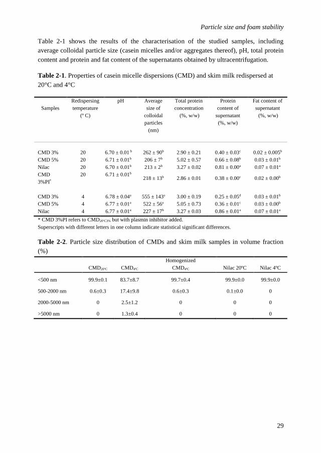
Table 2-1 shows the results of the characterisation of the studied samples, including
average colloidal particle size (casein micelles and/or aggregates thereof), pH, total protein
content and protein and fat content of the supernatants obtained by ultracentrifugation.
Table 2-1. Properties of casein micelle dispersions (CMD) and skim milk redispersed at
20°C and 4°C
Samples
Redispersing
temperature
(º C)
pH Average
size of
colloidal
particles
(nm)
Total protein
concentration
(%, w/w)
Protein
content of
supernatant
(%, w/w)
Fat content of
supernatant
(%, w/w)
CMD 3% 20 6.70 ± 0.01 b 262 ± 90b 2.90 ± 0.21 0.40 ± 0.03c 0.02 ± 0.005b
CMD 5% 20 6.71 ± 0.01b 206 ± 7b 5.02 ± 0.57 0.66 ± 0.08b 0.03 ± 0.01b
Nilac 20 6.70 ± 0.01b 213 ± 2b 3.27 ± 0.02 0.81 ± 0.00a 0.07 ± 0.01a
CMD
3%PI*
20 6.71 ± 0.01b 218 ± 13b 2.86 ± 0.01 0.38 ± 0.00c 0.02 ± 0.00b
CMD 3% 4 6.78 ± 0.04a 555 ± 143a 3.00 ± 0.19 0.25 ± 0.05d 0.03 ± 0.01b
CMD 5% 4 6.77 ± 0.01a 522 ± 56a 5.05 ± 0.73 0.36 ± 0.01c 0.03 ± 0.00b
Nilac 4 6.77 ± 0.01a 227 ± 17b 3.27 ± 0.03 0.86 ± 0.01a 0.07 ± 0.01a
* CMD 3%PI refers to CMD20ºC3% but with plasmin inhibitor added.
Superscripts with different letters in one column indicate statistical significant differences.
Table 2-2. Particle size distribution of CMDs and skim milk samples in volume fraction
(%)
CMD20ºC CMD4ºC
Homogenized
CMD4ºC Nilac 20ºC Nilac 4ºC
<500 nm 99.9±0.1 83.7±8.7 99.7±0.4 99.9±0.0 99.9±0.0
500-2000 nm 0.6±0.3 17.4±9.8 0.6±0.3 0.1±0.0 0
2000-5000 nm 0 2.5±1.2 0 0 0
>5000 nm 0 1.3±0.4 0 0 0
Chapter 2
30
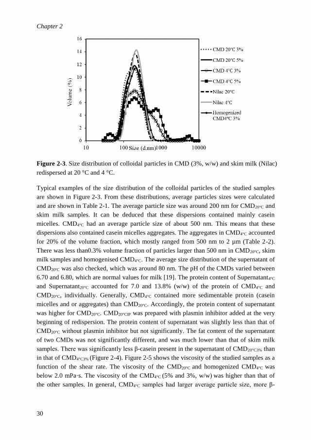
Figure 2-3. Size distribution of colloidal particles in CMD (3%, w/w) and skim milk (Nilac)
redispersed at 20 °C and 4 °C.
Typical examples of the size distribution of the colloidal particles of the studied samples
are shown in Figure 2-3. From these distributions, average particles sizes were calculated
and are shown in Table 2-1. The average particle size was around 200 nm for CMD20°C and
skim milk samples. It can be deduced that these dispersions contained mainly casein
micelles. CMD4°C had an average particle size of about 500 nm. This means that these
dispersions also contained casein micelles aggregates. The aggregates in CMD4°C accounted
for 20% of the volume fraction, which mostly ranged from 500 nm to 2 µm (Table 2-2).
There was less than0.3% volume fraction of particles larger than 500 nm in CMD20°C, skim
milk samples and homogenised CMD4°C. The average size distribution of the supernatant of
CMD20ºC was also checked, which was around 80 nm. The pH of the CMDs varied between
6.70 and 6.80, which are normal values for milk [19]. The protein content of Supernatant4°C
and Supernatant20°C accounted for 7.0 and 13.8% (w/w) of the protein of CMD4°C and
CMD20°C, individually. Generally, CMD4°C contained more sedimentable protein (casein
micelles and or aggregates) than CMD20°C. Accordingly, the protein content of supernatant
was higher for CMD20°C. CMD20°CIP was prepared with plasmin inhibitor added at the very
beginning of redispersion. The protein content of supernatant was slightly less than that of
CMD20°C without plasmin inhibitor but not significantly. The fat content of the supernatant
of two CMDs was not significantly different, and was much lower than that of skim milk
samples. There was significantly less β-casein present in the supernatant of CMD20°C3% than
in that of CMD4°C3% (Figure 2-4). Figure 2-5 shows the viscosity of the studied samples as a
function of the shear rate. The viscosity of the CMD20°C and homogenized CMD4°C was
below 2.0 mPa·s. The viscosity of the CMD4°C (5% and 3%, w/w) was higher than that of
the other samples. In general, CMD4°C samples had larger average particle size, more β-

Particle size and foam stability
31
casein casein in supernatant, less total protein in supernatant, and higher viscosity
compared to CMD20°C.
Figure 2-4. Casein composition of CMDs (3%, w/w) and their supernatants measured by
RP-HPLC.
Figure 2-5. Viscosity as a function of shear rate of CMDs (3% and 5%, w/w) redispersed at
20°C and 4°C as well as of homogenized CMD4°C3%.

Chapter 2
32
2.3.3 Interfacial properties
The dynamic surface tension was followed for 60 s in order to consider a time range
comparable with that of the foam formation, which took about 40 s. Figure 2-6 shows the
surface pressure (Π) of CMD20°C and CMD4°C as a function of different total protein
concentrations at 1 s and 60 s. There was a clear difference between the adsorption kinetics
of the CMDs with 0.06% (w/w) protein and that of the other CMDs with higher protein
concentrations. In general, the surface pressure of CMD20°C at 1 min was slightly higher
than that of CMD4°C. This might be attributed to the protein content of the supernatant,
which was higher for CMD20°C than for CMD4°C. The surface pressure of skim milk samples
was comparable to that of CMDs with 3% protein concentration.
Figure 2-6. Surface pressure Π (mN m-1) of CMD20°C and CMD4°C with different protein
concentrations (A) Π (t=1 s) (B) Π (t=60 s).

Particle size and foam stability
33
Fig. 2-7. Surface elasticity E’ (mN m-1) and surface viscosity E’’ (mN m-1) of CMD20°C and
CMD4°C as a function of protein concentrations
Figure 2-7 shows the dilatational elastic and viscous modulus of the air/liquid interface of
studied samples. This was determined at 1000 s after bubble formation, which represents a
situation that can be considered as equilibrium. The surface elasticity of CMD increased
with increasing protein concentration. CMD4°C showed a higher surface elasticity at
equilibrium compared to CMD20°C for protein concentrations lower than 3% (w/w). At 3%
(w/w) protein, 0.1 Hz and 6% deformation, no significant difference was observed between
CMD4°C and CMD20°C in terms of both surface elasticity and viscous modulus. The surface
elastic and viscous modulus of skim milk samples were not significantly different from
those of CMDs with 3% protein concentration. It could be seen from Figure 2-7 (B) that,
for each CMD, t½ increased with increasing surface elasticity. However, when CMD4°C
was compared to CMD20°C, there was no correlation between surface elasticity and t½. The
same was observed when CMDs were compared to skim milk samples. Foam stability were
t½≈1.5 h for skim milk samples, t½≈4 h for CMD20°C3% and t½≈1 d for CMD4°C3%.
However, these samples had similar surface elasticity.
2.4 Discussion
2.4.1 Role of interfacial properties on foaming properties
In literature, the formation and stability of foams, including those made with individual
milk proteins, have often been related to the adsorption rate of surface active compounds at
the air/liquid interface and to the rheological properties of the formed interfacial layer,
respectively [20-22]. The adsorption kinetics of proteins are mainly studied by measuring
the dynamic surface tension in time [23], which is generally claimed to give information
about the stabilizing properties shortly after bubble formation. In this study, all measured
samples reached the set foam volume of 120 mL after 36 s according to the set gas flow
rate of 200 mL min-1. This means that, during foam formation, no gas left from the foam,
i.e., no coalescence of generated bubbles with the environment occurred. This can be
attributed to the fact that the interface at the top of the liquid was generated 15 min before
foam formation started, and had higher protein coverage and better stabilizing properties
than the interface of the newly generated bubbles. Therefore, for the systems studied here,
the adsorption kinetics of the surface-active components of the CMDs was not a limiting
factor for foamability. However, the mean bubble size at 1 min after foam formation was
significantly larger (0.3 mm) for CMD0.06% than for the other samples (0.15 mm). The effect
of the differences in surface pressure at short times (t=1 s) which, for CMD0.06% and the
other samples was about 9 and 20 mN m-1, respectively, on the difference in bubble size
directly after snap off at the orifice is negligible [24]. The same holds for the difference in
Laplace pressure, which can only explain a factor of about 1.2. The larger bubble size of
CMD0.06% can be attributed to the lower coverage and therefore lower stabilizing properties

Chapter 2
34
of CMD0.06% compared to the other samples, which resulted in rapid bubble coalesce, within
1 min after foam formation.
Dilatational interfacial rheology of the air/liquid interface was conducted to explain the
long-term foam stability [22]. As can be seen from Figure 2-7 (A), E’ (elastic modulus) was
positively correlated with protein concentration. Furthermore, E’ of CMD4°C was slightly
higher than that of CMD20°C. This is in line with the difference in foam half-life, t½, which
increased with increasing protein concentration for samples with equal ingredients and
which was also higher for CMD4°C than for CMD20°C. However, at a protein concentration
of 3% (w/w), at which the difference between the foam stability of CMD4°C and CMD20°C
was largest, the difference in elastic modulus was negligibly small and not significant.
When CMDs were compared to skim milk samples, they showed better foam stability with
similar surface modulus. Similar reasoning holds for E” and the relaxation rate τ=ωE”/E’.
Therefore, based on interfacial rheology, it may be concluded that the elastic modulus
cannot explain the differences observed in foam stability. This supports recent findings by
other researchers who did not find a correlation between foam stability and surface elastic
modulus [25].
Another possibility for the worse foam stability of CMD20°C compared to CMD4°C could be
the higher amount of peptides present due to proteolytic action during redispersion at 20ºC.
The protein content of its supernatant also indicated that more small molecules could be
present in CMD20°C. This would have led to differences in interfacial properties between
two CMDs, which were however not found in this study. Also, no difference in foam half-
life after addition of plasmin inhibitor at the beginning of preparation of CMD20°C was
observed. Thus the proteolytic activity of plasmin did not have influence on the foam
stability of CMD20ºC in this study.
According to previous research, more β-casein would dissociate from the casein micelles
into the liquid phase (serum) compared to the other casein fractions when milk is held at
4ºC, and this was supposed to result in better interfacial properties, which could contribute
to improved foaming properties [26, 27]. In this study, CMD4°C was prepared at 4ºC. Indeed,
the casein composition of the supernatants confirmed that there was more β-casein present
in the supernatant of CMD4°C compared to that of CMD20°C. However, the protein
composition did not result in a difference in interfacial rheological properties. However, the
elastic properties shown, which are generally used to explain foam stability, were measured
at only one frequency and time (at semi equilibrium). The CMDs and skim milk samples
contained peptides, different forms of caseins, a small amount of milk fat and possibly
other milk proteins. It might also be a coincidence that CMDs and skim milk exhibited a
similar interfacial modulus but had different interfacial composition. It is not yet clear to
what extent interfacial rheological parameters of protein-stabilized interfaces are directly
related to the foam stability.

Particle size and foam stability
35
2.4.2 Effect of the size of colloidal particles on foam stability
A significant difference in particle size distribution was observed between samples
redispersed at 20°C and 4°C. The particle size distribution of skimmed milk samples as
well as that of all CMD20°C samples appears as a single peak, with an average around
200 nm. These values are comparable to the values measured for natural casein micelles
present in milk [14, 19, 28, 29]. Furthermore, Martin et al. [29] reported a value of 215 nm
for casein micelles present in low-heated skim milk reconstituted at 4°C, which is close to
the values we measured. However, the particle size distribution of the CMDs redispersed at
4°C was significantly different from that of the other samples, with an additional peak
appearing near 1 µm. Most probably, this peak corresponds to aggregates of casein micelles
that accounted for 20% volume fraction mostly ranging from 500 nm to 2 µm. The
relationship between particle size distribution and foam stability can lead to the conclusion
that the presence of larger colloidal particles is advantageous for foam stability in this case.
In order to check this, CMD4°C3% samples were also homogenized at 200 bar for 10 min, to
dissociate the aggregates. After homogenization, the particle size distribution of the
CMD4°C3% appeared similar to that of CMD20°C3% and skimmed milk samples (about
200 nm). Furthermore, foaming experiments showed a significant decrease in foam stability
of the homogenized sample to similar to that of the other samples. One might argue that a
lower amount of phospholipids could be released from phospholipid-rich fractions
sedimentable by ultracentrifugation, like the milk fat globule membrane MFGM [30],
during the limited resuspension of casein micelle pellets at low temperature compared to
room temperature. This would result in less phospholipids present in the Supernatant4°C
compared to Supernatant20°C, and thereby less components able to destabilize the foam of
CMD4°C. One might also argue that, after homogenization the unreleased phospholipids
within the casein micelle aggregates might be released, with a consequent destabilisation of
the foam of CMD4°C. To check this possibility, the total fat content of the Supernatant4°C
and Supernatant20°C was measured as an indicator of the content of phospholipids. The
amount of fat content in these two supernatants was the same. Therefore, it is unlikely that
phospholipids can play a role in the differences in foam stability observed in our research.
It can be concluded that the size of the colloidal particles or the presence of larger
aggregates determine the foam stability of CMDs observed.
The precise reason for this is at the moment not completely clear. The viscosity of CMDs
was slightly higher when casein micelles aggregates were present. The viscosity is an
important parameter for foam stability because it is inversely proportional to the drainage
rate. A higher viscosity will slow down plateau border drainage and drainage from the thin
film to the plateau border. However, viscosity alone cannot explain the large difference in
foam stability between CMD4°C3% and CMD20°C3%. Casein micelles and their aggregates
might be confined in the thin film lamellae between bubbles. These trapped colloidal
particles can decrease the thinning rate of the film. Furthermore, when colloidal particles
have good wetting properties, they can also slow down film drainage and prevent contact

Chapter 2
36
between the two air/liquid interfaces, with subsequent rupture. In previous studies, a similar
role of protein aggregates in foam stability has been proposed. The presence of small β-
lactoglobulin aggregates with size of tens of nm decelerated foam drainage rate, while
aggregates with size around 200 nm were too large to give a stable foam [31, 32]. Similar
observations are also described by Nicolai et al. [33], whey protein aggregates induced by
heating were claimed to associate and build a network within thin films to enhance foam
stability. Recently, a study has shown that protein particles/aggregates slow down foam
drainage and disproportionation [34]. The observations that the size of the aggregates
primarily control the stability of CMDs are of great importance for a better prediction of the
foaming properties of complex systems, such as food foams made from commercial protein
concentrates, which often contain both native protein molecules and various amounts of
protein aggregates.
In summary, even though a correlation between interfacial properties and foam stability
was not found in this study, further research on interfacial properties needs to be conducted
as a function of amplitudes of deformation and frequency. This way, more info about
mechanical properties of air/liquid interface of two CMDs made at different temperatures
can hopefully be uncovered. The presence of casein micelle aggregates appears to be the
key factor for the foam stability of CMD. Thin film stability should also be measured to
further understand the mechanism by which casein micelle aggregates improve foam
stability, by being trapped and confined in the lamella or by formation of a network that
slows down the drainage.

Particle size and foam stability
37
2.5 Conclusion
Casein micelle dispersions (CMDs) with different average size of the colloidal particles
(casein micelles and/or aggregates thereof) could be obtained by redispersion of casein
micelle pellets at different temperatures. CMDs redispersed at 4°C (CMD4°) exhibited
significantly higher foam stability than those redispersed at 20ºC (CMD20°), almost by a
factor of 6 for foam half-life. These differences could not be explained by surface
rheological properties and/or protein composition, but could be ascribed to the average size
of the colloidal particles present in dispersion. CMD4°C showing the highest foam stability
contained colloidal particles with an average size of about 500 nm (micelles and/or
aggregates), while that of CMD20°C was about 200 nm. Homogenisation of CMD containing
large casein micelle aggregates reduced the average particle size and the foam stability to
those of CMD and skimmed milk samples with average particle size of 200 nm. Therefore,
the presence of casein micelle aggregates and thus the size of the colloidal particles
determine the foam stability of casein micelle dispersions.

Chapter 2
38
References
1. Huppertz, T., Foaming properties of milk: A review of the influence of composition and processing.
International Journal of Dairy Technology, 2010. 63(4): p. 477-488.
2. Ward, B.R., et al., EDTA-induced dissociation of casein micelles and its effect on foaming properties of
milk. Journal of Dairy Research, 1997. 64(4): p. 495-504.
3. Borcherding, K., et al., Effect of foaming temperature and varying time/temperature-conditions of pre-
heating on the foaming properties of skimmed milk. International Dairy Journal, 2008. 18(4): p. 349-
358.
4. Zhang, Z., Dalgleish, D.G. and Goff, H.D., Effect of pH and ionic strength on competitive protein
adsorption to air/water interfaces in aqueous foams made with mixed milk proteins. Colloids and
Surfaces B: Biointerfaces, 2004. 34(2): p. 113-121.
5. Kamath, S., Webb, R.E. and Deeth, H.C., The composition of interfacial material from skim milk foams.
Journal of Dairy Science, 2011. 94(6): p. 2707-2718.
6. Dickinson, E., et al., Adsorption at interfaces in dairy systems. International Journal of Dairy
Technology, 1989. 42(1): p. 18-22.
7. Murray, B.S., Stabilization of bubbles and foams. Current Opinion in Colloid & Interface Science,
2007. 12(4): p. 232-241.
8. Meinders, M.B.J. and van Vliet, T., The role of interfacial rheological properties on Ostwald ripening
in emulsions. Advances in Colloid and Interface Science, 2004. 108: p. 119-126.
9. Hilgenfeldt, S., S.A. Koehler, and H.A. Stone, Dynamics of Coarsening Foams: Accelerated and Self-
Limiting Drainage. Physical Review Letters, 2001. 86(20): p. 4704-4707.
10. Mulder, H. and P. Walstra, The Milk Fat Globule: Emulsion Science as Applied to Milk Products and
Comparable Foods. No. 4. Wageningen, Netherlands, Centre for Agricultural Publishing and
Documentation., 1974.
11. Brooker, B.E., Observations on the air-serum interface of milk foams. Food Structure, 1985. 4(2): p. 12.
12. Schmitt, C., et al., Whey protein soluble aggregates from heating with NaCl: Physicochemical,
interfacial, and foaming properties. Langmuir, 2007. 23(8): p. 4155-4166.
13. Huppertz, T. and de Kruif, C.G. Rennet-induced coagulation of enzymatically cross-linked casein
micelles. International dairy journal, 2007. 17(5): p. 442-447.
14. Beliciu, C.M. and Moraru, C.I., Effect of solvent and temperature on the size distribution of casein
micelles measured by dynamic light scattering. Journal of Dairy Science, 2009. 92(5): p. 1829-1839.
15. Ambrose Griffin, M.C. and Griffin, W.G., A simple turbidimetric method for the determination of the
refractive index of large colloidal particles applied to casein micelles. Journal of Colloid and Interface
Science, 1985. 104(2): p. 409-415.
16. Ribadeau-Dumas, B. and Grappin, R., Milk protein analysis. Le Lait, 1989. 69(5): p. 357-416.
17. Bonfatti, V., et al., Validation of a new reversed-phase high-performance liquid chromatography
method for separation and quantification of bovine milk protein genetic variants. Journal of
Chromatography A, 2008. 1195(1): p. 101-106.
18. Wüstneck, R., et al., Surface dilatational behavior of β-casein at the solution/air interface at different
pH values. Colloids and Surfaces A: Physicochemical and Engineering Aspects, 2012. 404: p. 17-24.
19. De Kruif, C. and Holt, C., Casein micelle structure, functions and interactions. Advanced Dairy
Chemistry—1 Proteins. 2003, p. 233-276. Springer US.
20. Sánchez, C.C. and Patino, J.M.R., Interfacial, foaming and emulsifying characteristics of sodium
caseinate as influenced by protein concentration in solution. Food Hydrocolloids, 2005. 19(3): p. 407-
416.
21. Rouimi, S., et al., Foam stability and interfacial properties of milk protein–surfactant systems. Food
Hydrocolloids, 2005. 19(3): p. 467-478.
22. Wierenga, P. and Gruppen, H., New views on foams from protein solutions. Current Opinion in Colloid
& Interface Science, 2010. 15(5): p. 365-373.

Particle size and foam stability
39
23. Bos, M.A. and van Vliet, T., Interfacial rheological properties of adsorbed protein layers and
surfactants: a review. Advances in Colloid and Interface Science, 2001. 91(3): p. 437-471.
24. Davidson, J.F. and Schüler, B.O.G., Bubble formation at an orifice in a viscous liquid. Chemical
Engineering Research and Design, 1997. 75: p. S105-S115.
25. Lech, F.J., et al., Identifying changes in chemical, interfacial and foam properties of β-lactoglobulin–
sodium dodecyl sulphate mixtures. Colloids and Surfaces A: Physicochemical and Engineering Aspects,
2014. 462: p. 34-44.
26. Dalgleish, D.G., On the structural models of bovine casein micelles—review and possible
improvements. Soft Matter, 2011. 7(6): p. 2265.
27. Reimerdes, E. and H. Klostermeyer, Temperature-dependent changes in milk and milk products. I
Change in the ratio of micelle protein to serum protein during refrigeration of milk. 1976. Kiel.
Milchwirtsch. Forschungsber, 1976. 28: p. 17-25.
28. Bijl, E., et al., Factors influencing casein micelle size in milk of individual cows: Genetic variants and
glycosylation of κ-casein. International Dairy Journal, 2014. 34(1): p. 135-141.
29. Martin, G.J.O., Williams, R.P.W. and Dunstan, D.E., Comparison of Casein Micelles in Raw and
Reconstituted Skim Milk. Journal of Dairy Science, 2007. 90(10): p. 4543-4551.
30. Singh, H., The milk fat globule membrane—A biophysical system for food applications. Current
Opinion in Colloid & Interface Science, 2006. 11(2): p. 154-163.
31. Rullier, B., Novales, B. and Axelos, M.A., Effect of protein aggregates on foaming properties of β-
lactoglobulin. Colloids and Surfaces A: Physicochemical and Engineering Aspects, 2008. 330(2): p.
96-102.
32. Rullier, B., et al., β-Lactoglobulin aggregates in foam films: Effect of the concentration and size of the
protein aggregates. Journal of colloid and interface science, 2010. 343(1): p. 330-337.
33. Nicolai, T., M. Britten, and C. Schmitt, β-Lactoglobulin and WPI aggregates: Formation, structure
and applications. Food Hydrocolloids, 2011. 25(8): p. 1945-1962.
34. Schmitt, C., Bovay, C. and Rouvet, M., Bulk self-aggregation drives foam stabilization properties of
whey protein microgels. Food Hydrocolloids, 2014. 42: p. 139-148.

40

41
Chapter 3 Interfacial properties, thin film stability and foam stability of
casein micelle dispersions
Abstract
Foam stability of casein micelle dispersions (CMDs) strongly depends on aggregate size.
To elucidate the underlying mechanism, the role of interfacial and thin film properties was
investigated. CMDs were prepared at 4°C and 20°C, designated as CMD4ºC and CMD20ºC.
At equal protein concentrations, foam stability of CMD4ºC (with casein micelle aggregates)
was markedly higher than CMD20ºC (without aggregates). Although the elastic modulus of
CMD4ºC was twice as that of CMD20ºC at 0.005Hz, the protein adsorbed amount was slightly
higher for CMD20ºC than for CMD4ºC, which indicated a slight difference in interfacial
composition of the air/water interface. Non-linear surface dilatational rheology showed
minor differences between mechanical properties of air/water interfaces stabilized by two
CMDs. These differences in interfacial properties could not explain the large difference in
foam stability between two CMDs. The thin film analysis showed that films made with
CMD20ºC drained to a more homogeneous film compared to films stabilized by CMD4ºC.
Large casein micelle aggregates trapped in the thin film of CMD4ºC made the film more
heterogeneous. The rupture time of thin films was significantly longer for CMD4ºC (>1 h)
than for CMD20ºC (<600 s) at equal protein concentration. After homogenization, which
broke down the aggregates, the thin films of CMD4ºC became much more homogeneous,
and both the rupture time of thin films and foam stability decreased significantly. In
conclusion, the increased stability of foam prepared with CMD4ºC appears to be the result of
entrapment of casein micelle aggregates in the liquid films of the foam.
Published as:
Chen, M., Sala, G., Meinders, M. B., van Valenberg, H. J., van der Linden, E., & Sagis, L.
M. (2017). Interfacial properties, thin film stability and foam stability of casein micelle
dispersions. Colloids and Surfaces B: Biointerfaces, 149, 56-63.

Chapter 3
42
3.1 Introduction
Milk proteins are widely used to produce aerated products in food industry, like e.g. aerated
desserts, whipped cream, and cappuccino foam. Caseins, which make up about 80% of cow
milk protein and are predominantly present in the form of colloidal particles, termed casein
micelles, exhibit good foaming properties[1]. Kamath et al. (2011)[2] discovered that
isolated micellar casein fractions could produce foams with much higher stability than skim
milk. The improved foam stability of micellar casein fractions was ascribed to improved
interfacial properties. In a recent study, we found that casein micelle dispersions prepared at
4ºC (CMD4ºC), with an average particle size of 500 nm and which contained aggregates of
casein micelles, formed much more stable foams compared to foams made from CMD
prepared at 20ºC (CMD20ºC). The latter CMD had a smaller average particle size (200 nm),
and consisted predominantly of non-aggregated micelles. The aggregates present in
CMD4ºC, which appeared to play a crucial role in the increased foam stability, caused only
marginal changes in linear dilatational properties of the air/water interface, and these minor
differences could not explain the large difference in foam stability between the two CMDs.
There are several factors that can influence the stability of aqueous foams. Among these are
the properties of the adsorbed interfacial layer between air and liquid phase (surface
tension, dilatation modulus, surface shear modulus), and the bulk properties of the liquid
films that separate the bubbles[3]. For relatively simple systems, like foams stabilized by
LMW-surfactants, the interfacial properties of the air/water interface are often found to be
dominant for foam stabilization[4, 5], [6]. For more complex systems, stabilized by
(mixtures of) proteins or colloidal particles, it is still unclear whether interfacial or bulk
film properties are dominant for the foam stability. Previous studies have reported that
protein aggregates can have a large influence on foam stability. The presence of whey
protein soluble aggregates [7], β-Lactoglobulin aggregates [8-10] and whey protein fibrils
[11] was shown to improve foam stability, but other studies indicated that their presence
can also decrease foam stability [12]. Rullier et al.[8-10] studied films and foams made
from mixtures of non-aggregated β-Lactoglobulin and their aggregates, and found that film
stability was dependent on the aggregate size and on the ratio between non-aggregated
proteins and protein aggregates. For aggregate fractions ranging from 1 to 90% of the total
protein concentration (1 g/L), gel formation in the thin film was observed. For higher
fractions of aggregates, it was shown that the amounts of non-aggregated proteins were no
longer sufficient to completely cover the interface, and as the larger aggregates did not
reduce the surface tension at short times as much as the non-aggregated protein, the
corresponding films and foams were less stable. For the fractions between 1 and 90%, the
rheological properties of the interface were not determined. It is therefore hard to establish
whether the immobilization of the films is the result of in-plane (surface) gel formation,
cross-film (bulk) gel formation, or a combination of both.

Interfacial properties, thin film stability and foam stability
43
A recent review reported improved foam stability in the presence of protein aggregates
without causing significant differences in the interfacial properties of the air/water
interface[13], which is in line with our previous research [14]. In our previous study the
minor differences in linear surface rheology of air/water interfaces stabilized by CMDs
could not explain the large difference in foam stability. However, we determined the
dilatational properties only at small deformations and constant frequency, and did not
perform large amplitude dilatations, which are more sensitive to subtle changes in the
microstructure of the interface. Particularly when coarsening of the foam is predominantly
driven by disproportionation, interfaces are subjected to large deformations, and dilatational
moduli data determined at small deformations are therefore not a good indicator to establish
whether surface properties play a role in foam stability.
The composition of air/water interfaces stabilized by different CMDs is still largely
unknown. CMD is a mixture of four kinds of caseins, peptides, small micelles, casein
micelles and casein micelle aggregates. The adsorption of casein micelles on the interface is
still under debate. Borcherding et al. (2008 )[15] indicated that casein micelles are not
likely to be present at the air/water interface. In another study, microscopic images of
bubble ghosts of milk foam suggested that casein micelles were adsorbed at the air/ liquid
interface, possibly reversibly[16].
In the current study, foaming properties, interfacial properties and thin film stability of
CMDs with different particle size distribution and of their supernatants were investigated.
Samples were characterised for size distribution of the casein micelles (aggregates) and
protein concentration. Sodium-caseinate was studied as a control. Frequency and strain
amplitude dependence of the surface dilatational modulus of the CMDs were checked in
large amplitude oscillatory dilatation. The absorbed amount of protein at the air/water
interface for the different samples was determined by ellipsometry. Thin film properties
including rupture time and morphology of the thin liquid film were studied by a microscope
equipped with a Scheludko cell. Foam properties such as the foam half-life (t1/2) and mean
bubble diameter were obtained from bubble image analysis. By combining the above
measurements, the relation between interfacial properties and foam stability as well as
relationship between thin film stability and foam stability was examined to establish the
mechanism behind the significantly improved foam stability of CMDs prepared at low
temperature.
3.2 Material and Methods
3.2.1 Materials
Low-heated skim milk powder NILAC was obtained from NIZO food research (Ede,
Netherland). Sodium caseinate (EM7-A9040445) was obtained from DMV International
(Veghel, The Netherlands). Sodium azide was purchased from Sigma Aldrich (Zwijndrecht,
The Netherlands). Ultra-pure water (MilliQ Purelab Ultra, Darmstadt, Germany), free of

Chapter 3
44
surface active contaminants, was used in all experiments (>18.2 MΩ-cm, surface tension of
72.26 ± 0.4 mN/m at 20°C).
3.2.2 Preparation of casein micelle dispersions (CMDs) and their supernatants
Skim milk was reconstituted (10%, w/w) by dissolving NILAC milk powder in MilliQ
water and stirring overnight at room temperature. Sodium azide (0.02%, w/w) was added as
a preservative. The reconstituted skim milk was ultracentrifuged (L-60 Beckman
Ultracentrifuge, rotor type 70 Ti, Krefeld, Germany) at 100,000 g for 90 minutes at 20°C
according to Huppertz and de Kruif[17]. The obtained casein micelle pellets were separated
from the serum phase and ground using a Mixer Mill MM 400 (Retsch GmbH, Haan,
Germany) at a frequency of 30 Hz for 10 minutes at room temperature. Subsequently,
different amounts of the obtained casein micelle paste were redispersed in milk permeate
either at 20°C or at 4°C for 60 hours to obtain casein micelle dispersions (CMD) with 2.5%
(w/w) protein concentrations. CMD20°C denotes the CMD redispersed at 20°C and CMD4°C
is a CMD made at 4°C. The milk permeate was prepared by reconstitution of milk permeate
powder (5.76%, w/w) in MilliQ water for 30 minutes. Milk permeate powder was prepared
by ultrafiltration of the reconstituted skim milk (10%, w/w) using a polysulfone membrane
with a pore size of 10 kDa and surface area of 0.48 m2. The dimensions of the membrane
were 73.5 L x 3.2 cm o.d. The skim milk was stirred and cooled at 4°C for one hour before
being poured into the cold trap (8°C). Subsequently it was filtered through the membrane
under pressure. The milk permeate was collected, freeze-dried and stored at -20°C. The
calcium activity of the permeate is 4.76E-04, which is comparable to that of skim milk
(4.88E-04).
For the thin film studies with homogenized CMD4°C, CMD4°C samples with protein
concentration of 2.5% (w/w) were homogenized at 20 MPa for 10 min with a homogenizer
(Delta Instruments, Drachten, the Netherlands).
For the preparation of the supernatants, CMDs and skim milk samples were
ultracentrifuged for a second time according to the procedure described above. Their
protein content was further analysed as described below.
3.2.3 Characterization of samples
Particle size
The size distribution of the colloidal particles (casein micelles and/or aggregates) present in
milk and CMDs was measured using a Malvern Zetasizer Nano-ZS (Malvern Instruments
Ltd, Worcestershire, United Kingdom). The samples were diluted with milk permeate to a
protein concentration of 0.03% (w/w) and subsequently transferred into a cuvette
(DTS0012) using a syringe. A single measurement consisted of 11 runs and the duration of
each run was 10 s. The refractive indices used for the calculation of the sizes were
1.341[18] for the milk permeate and 1.57[19] for the casein micelles, respectively. The
measurement angle was set to 173º backscatter (NBS default) with automatic measurement
duration. Three measurements of each sample without pausing were performed at 20ºC.

Interfacial properties, thin film stability and foam stability
45
Protein content
The total nitrogen content of the samples was determined using the Dumas method[20].
Specimens of 200 μL were dried in an oven at 60ºC overnight. A factor of 6.38 was used
for the conversion of the nitrogen content into total protein content.
3.2.4 Foaming properties
The foaming properties were assessed with a FoamScan (Teclis IT-Concept, Longessaigne,
France). Foam was generated by sparging air through a porous frit in 40 mL solution. The
gas flow rate was set at 200 mL min-1 until the volume of the foam reached 120 mL. After
reaching this volume, the gas flow rate was set to 0. All experiments were carried out at
20ºC. All solutions were stirred for one hour prior to foaming at room temperature. The
foam volume was estimated from light intensity of tube images based on calibration of
pixels and the black/white coefficient (greyscale level) was set to 55%. Foam volume was
recorded as a function of time. The measurement stopped when the foam volume reached
half of its initial value. The corresponding time t½ (foam half-life) was used as a measure
for foam stability. For CMD4°C, the foam stability was also checked using a foaming tube
(2.0 cm in diameter, 34.0 cm in height, glass grid at the bottom) and sparging.
Bubble size distribution
2D images of the bubbles of the foam at the wall of the FoamScan tubes were recorded
using a CCD camera. The image covered an area of 1.0 cm2. From the images, analysed
using Matlab V.2013a (Mathworks) and the Dip Image software (Quantitative Imaging
Group, Faculty of Applied Sciences, Delft University of Technology, Delft, The
Netherlands), the mean bubble diameter, D, was calculated.
3.2.5 Determination of the interfacial properties with Profile Analysis Tensiometer (PAT)
The surface properties were measured using a SINTERFACE PAT 1-M (SINTERFACE
Technologies, Berlin, Germany) and data were analysed using SINTERFACE Profile
Analysis Tensiometer PAT 1-M version 1.4.0.685 software according to van Kempen et al.
[21] with some modifications. The pendant drop method was used and the area of the
droplet was 25 mm2, with volume ranging from 13-14 mm3. The surface tension of ultra-
pure water used in this experiment was 72.26 ± 0.4 mN/m. Sinusoidal oscillation was
applied after reaching equilibrium (t=1000 s) with 5 oscillations in one sweep to obtain the
surface elastic modulus E’ and surface viscous modulus E’’. There were 10 cycles of
sweeps with 10 s pause in between for each measurement. The dependence of E’ on
different deformation amplitudes (7.5%, 10%, 15%, 20% and 30%) was assessed at
frequency of 0.1 Hz. Results of non-linear dilatational rheology were analysed by Lissajous
plots of surface pressure versus strain. Besides, the frequency dependence of E’ was
determined with amplitude of 7.5% at various frequencies (0.005Hz, 0.01Hz, 0.05Hz,
0.1Hz and 0.2Hz). The slope of a double logarithmic plot of E’ versus frequency was
determined using linear regression. The protein concentrations of the studied samples were
the same as those shown in Table 3-1.

Chapter 3
46
3.2.6 Ellipsometry
The adsorption to the air-liquid interfaces of the samples was monitored using a Multiskop
ellipsometer (Optrell, Germany). Surface load (Г, mg/m2) in time was measured. 21.0 mL
of each sample (0.1%, w/w) was placed in a plastic plate with a diameter of 5.0 cm. The
rate of adsorption from 1.0 mg/mL solutions was measured at least in duplicate at 20°C.
The wavelength of the laser was set to be 6328 Å. The values for the ellipsometric angles ∆
and φ were used to calculate Г, using software from the supplier (Optrell). To do this, the
refractive index and thickness of the adsorbed protein layer were fitted in a model based on
two bulk phases (air and water) and one adsorbed layer, with parameters: nair=1.000,
npermeate=1.333, nproteinsolution=1.456, dn/dc= 0.185. The angle of incidence was 50°. Control
experiments with ultra-pure water between measurements confirmed the right ellipsometric
angles ∆ and φ.
3.2.7 Thin film stability
The thin film stability was measured by micro interferometry using a microscope (Axio
plan 01, Zeiss, Jena, Germany) according to Lech et al.[22] with some modifications. Thin
liquid films (diameter = 200 μm) were made in a Scheludko cell (diameter = 0.3 cm) closed
with a glass cover. The casing had a reservoir of milk permeate at the bottom to ensure a
relative humidity of 100% in the cell during measurements. Samples had initial
concentrations equal to those given in Table 3-1, and were diluted by a factor of 2000 prior
to measurement. All samples were equilibrated to room temperature (20°C) for 1 h before
measurements. Liquid was drawn into the capillary with a syringe (500 μL, Hamilton,
Reno, NV, USA) and the whole setup was left to equilibrate for 10 min before a thin film
was made by drawing sample solution into the capillary. The microscope was in reflected
light mode, equipped with a five mega pixel CCD camera (Mightex Systems, Pleasanton,
CA, USA). If a film did not break after 60 min, it was considered stable. In this case, the
lifetime was expressed as >3600 s. The rupture time was the time from reaching a diameter
of 200 μm to the time of rupture. In case of stable films, samples were measured for 5
times. In the case of rupturing films, experiments were repeated 10 times to calculate the
average and standard deviation of the rupture time and film thickness. Images of the thin
films were collected with a software script (developed at the Laboratory of Food
Chemistry, Wageningen University) for Matlab (Software version 2013b, MathWorks,
Natick). The magnification was ×100.
3.2.8 Statistical analysis
All the tests were conducted at least in triplicate. The results obtained were subjected to
one-way analysis of variance (ANOVA). Duncan's new multiple range test was performed
using SPSS 22.0 software (SPSS Inc., Chicago, IL, USA) to determine the difference
between samples which is significant when p<0.05.

Interfacial properties, thin film stability and foam stability
47
3.3 Results and discussion
Table 3-1. Particle size and protein content of casein micelle dispersions (CMDs), their
supernatants, and sodium caseinate.
Samples
Average size of the
casein micelles
(aggregates)
Protein content
(%, w/w)
CMD20º C 262 ± 90 2.52 ± 0.13
CMD4º C 555 ± 143 2.64 ± 0.03
SupernatantCDM20ºC 88 ± 4 0.22 ± 0.01
SupernatantCMD4ºC 144 ± 6 0.15 ± 0.01
Sodium caseinate ------ 2.59 ± 0.00
Figure 3-1. Particle size distribution of CMDs and supernatants.
As shown in Table 3-1, the average particle size of the CMD20ºC was around 200 nm and
that of CMD4ºC around 500 nm. CMD4ºC had some aggregates of casein micelles ranging
from 500 nm to a few µm (Figure 3-1). SupernatantCMD20ºC and SupernatantCMD4ºC indicate
the supernatants of CMD20ºC and CMD4ºC, which contained the small molecular weight
fractions of the two CMDs, respectively. There was still some small casein micelles present

Chapter 3
48
in these supernatants. The particle size was 88 nm for SupernatantCMD20ºC and 144 nm for
SupernatantCMD4ºC. The protein content of SupernatantCMD20ºC was slightly higher than that
of SupernatantCMD4ºC, indicating that CMD20°C contained more non-sedimentable protein.
CMD4ºC had a larger particle size than CMD20ºC probably because the paste of casein
micelles, which contained mostly pieces of casein pellet, had a worse dispersibility at low
temperature. Since the pH and ionic strength of the two dispersions were the same, we
assumed that the temperature-dependent dispersibility of the casein micelles pastes was
controlled by the temperature dependence of electrostatic and hydrophobic interactions
between casein micelles as described by Horne (1998)[23].
Figure 3-2. Foam volume decay curve of CMDs, supernatants and sodium caseinate till
half of the initial foam volume.
All measured samples reached the set foam volume of 120 mL after 36 s, with a gas flow
rate of 200 mL min-1. The decay of the foam volume as a function of time was measured,
and the foam half-life (t1/2, the time at which the foam volume had decreased to half of its
initial value) was determined. As shown in Figure 3-2, t1/2 was similar for
SupernatantCMD20ºC, SupernatantCMD4ºC and sodium caseinate, equal to about 1.5 h, in spite of
the fact that the sodium caseinate sample had a much higher protein concentration than the
supernatants. For samples with the same protein concentration of 2.5% (w/w), the t1/2 was
3.5 h for CMD20ºC but more than 24 h for CMD4ºC. When CMD20ºC was compared with
SupernatantCMD20ºC, t1/2 of the supernatant was lower by a factor of 2, while its protein
concentration was lower by a factor of 11.5. For CMD4ºC and SupernatantCMD4ºC, the foam
half-life of the CMD was higher by a factor of 16, while its protein concentration was
higher by a factor of about 17. This shows that removing casein micelle aggregates from
CMD4ºC dramatically decreased the foam stability. For CMD4ºC, a slight increase in foam
volume with time was observed. This increase was a result of the slow leakage of a small
amount of gas form the sparger. The foam stability of CMD4ºC was also determined with a

Interfacial properties, thin film stability and foam stability
49
glass foaming tube, also by sparging: the foam volume did not increase in time, and the
foam stability was even longer than that determined with the FoamScan. To check for any
influence of proteolytic action during redispersion of CMD20ºC on foam stability, a plasmin
inhibitor was added to the samples. No difference in foam half-life was observed in samples
prepared with or without plasmin inhibitor added. One minute after foam formation there
was no significant difference in the mean bubble size of the studied samples (results not
shown). However, 60 min after foam formation bubble coalescence clearly had occurred in
the foam made with sodium caseinate and with the two supernatants. The mean bubble size
increased by a factor of 2, while the mean bubble size of CMD20ºC and CMD4ºC increased
by a factor of 1.5. After 240 min, the foam made with CMD4ºC turned into a kind of solid
foam and its structure did not change any more.
Figure 3-3. Lissajous plots of CMD4°C at different amplitudes of deformation at 0.1 Hz: 7.5%
(solid line), 10% (dotted line), 15% (solid line with cross), 20% (solid line with blank circle)
and 30% (solid line with blank square).
Figure 3-3 shows the Lissajous plots of the liquid/air interface stabilized by CMD4°C at
different amplitudes of deformation (7.5%, 10%, 15%, 20% and 30%) at a frequency of 0.1
Hz. The Lissajous curves have asymmetric shapes even at the lowest deformation of 7.5%,
which indicates that the maximum linear strain for this interface was below 7.5%. With
increasing amplitude of deformation, the response of the air/water interface became more
viscous. The asymmetric shape of the Lissajous curves indicates that the air/liquid
interfaces were strain softening during extension and strain hardening during compression.
Interestingly, for the highest amplitude, when maximum compression is reached (∆A/A =
30%), and the interface is subsequently expanded again, the slope of the Lissajous curve at
the start of the extension within the cycle is very steep, which is indicative of an initially

Chapter 3
50
strong resistance against extension. This indicates that compression leads to increased
cohesion between interfacial structural elements. This is a response typical of interfaces
stabilized by components that assemble into a gelled or jammed state after adsorption to the
interface (for example, particles, proteins, or certain types of oligosaccharide esters [11]).
Both the asymmetry in the Lissajous curves and the strain amplitude dependence of the
response are indications that in-plane deviatoric stresses contribute to the response to
dilatation [11],[24], [25], [26].
Figure 3-4. Lissajous plots of CMDs, supernatants and sodium caseinate at 30%
deformation at 0.1 Hz.
In Figure 3-4, the Lissajous plots at 30% deformation and 0.1 Hz of air/water interfaces
stabilized by CMDs, supernatants and sodium caseinate are shown. Compared to the two
CMDs, sodium caseinate-stabilized interfaces had a significantly lower resistance against
dilation, and did not show a strong increase in cohesion upon compression, which indicates
the air/water interface formed by pure sodium caseinate is quite different in microstructure
from interfaces stabilized by CMDs. The presence of casein micelles in the sub phase is
hence affecting the microstructure of the interface, either by adsorption of micelles into the
interface or by attachment to a primary layer formed by individual protein molecules. When
supernatants were compared with CMDs, the response of the air/water interfaces showed no
significant difference to dilation in the extension part of the cycle (upper right quadrant of
the Lissajous curve). However, the supernatant-stabilized interfaces displayed stronger
strain hardening during compression, and had a higher modulus in this part of the cycle.
According to the size distributions in Figure 3-1, the supernatants contained significantly
smaller micelles, and in spite of a much lower protein concentration than the CMDs, these

Interfacial properties, thin film stability and foam stability
51
smaller micelles may lead to a denser and more homogeneous interfacial structure, with a
higher resistance to compression.
Figure 3-5. Surface elastic modulus (E’) of air/water interface stabilized with CMD20°C and
CMD4°C as a function of frequency.
At a frequency of 0.1 Hz and 30% deformation amplitude, there was no significant
difference between the mechanical properties of CMD20ºC and CMD4ºC. To check if the
structures of the interfaces were indeed similar, we determined the frequency dependence
of the elastic modulus (E’). The slope of a double logarithmic plot of complex surface
dilatational modulus as a function of frequency was determined. From Figure 3-5, we can
see that at the lowest frequency of 0.005Hz CMD4ºC-stabilized interfaces had a significantly
higher E’ than those stabilized by CMD20ºC, approximately a factor of 2. The slope n of the
scaling relation 𝐸𝑑′ ~𝜔𝑛 was equal to 0.35 for CMD20ºC and 0.25 for CMD4ºC, which
indicated that CMD20°C with its smaller particle size was showing a behaviour more similar
to that of a surfactant system (which according to the Lucassen van den Tempel model[27]
have an exponent n=0.5). This is consistent with the fact that there was more non-
sedimentable protein (small peptides, free caseins, and small micelles) in CMD20°C
compared to CMD4ºC.
CMDs contained small peptides, individual caseins, casein micelles and casein micelle
aggregates, which all differ significantly in size. Small surface-active agents diffuse
significantly faster than large particles[28]. Based on the surface rheology results, one
possible structure for the CMD interface could be a heterogeneous structure consisting of
patches of peptides and individual caseins. Aggregates could either be randomly

Chapter 3
52
incorporated in this structure or randomly attached to a primary layer of peptides, caseins
and small micelles. Upon compression, small peptides and caseins, which are often
reversibly adsorbed, can be pushed out of the interface, leading to an increase in surface
fraction of by micelles and aggregates. The observed strain hardening is then a result of
jamming of these larger structures. This would also explain the increased cohesion after full
compression.
Figure 3-6. Adsorbed amount Г (mg/m2) of CMDs, supernatants and sodium caseinate at
Cp=0.1%, w/w. (A) Г t=60s (B) Г t=60 min
The adsorbed amount of protein at a single air/water interface of all samples at t= 60 s and
60 min is shown in Figure 3-6. The protein concentration in the sub phase was 0.1% (w/w)
for all studied samples. When the CMDs were compared to their respective supernatants,
there was no significant difference between the adsorbed amounts. In the surface
dilatational rheology experiments we saw that the responses of the CMD- and supernatant-
0.0
1.0
2.0
3.0
4.0
5.0
6.0
7.0
Гt=
60 s
(m
g/m
2)
0.0
1.0
2.0
3.0
4.0
5.0
6.0
7.0
Гt=
60 m
in(m
g/m
2)
A
B

Interfacial properties, thin film stability and foam stability
53
stabilized interfaces to extension was also identical. In spite of the equal protein loading of
CMD- and supernatant-stabilized interfaces, the latter displayed a higher degree of strain
hardening in compression. As we postulated before, the smaller micelles in the supernatant
may form a somewhat more homogeneous microstructure, with a slightly higher resistance
to compression. The adsorbed amount of protein for CMD20ºC was slightly higher than that
for CMD4ºC, yet the dilatational modulus of interfaces stabilized by the latter was slightly
higher, particularly at low frequencies. So the presence of large aggregates in the sub phase
was affecting the surface rheology of the air/water interface, albeit to a minor extent.
Summarising, interfaces stabilized by these CMDs differed only slightly in their surface
rheology, even though the half-life of their foams was remarkably different. Supernatants
showed a stronger strain hardening effect during compression than CMD stabilized
interfaces, in spite of the fact that former had much lower protein concentrations, and
contained smaller casein micelles and no aggregates of micelles. However, the stability of
foams prepared from the supernatants was much worse than that of foam prepared from
either CMD. We conclude from the above that there is no direct correlation between the
surface rheological properties and foam stability of CMDs. Several recent articles also
reported that aggregates had large effects on foam stability, without inducing differences in
surface rheological properties [13, 29, 30]. However, this does not mean that interfacial
properties are not important for foam stability, but the thin film properties are probably
more dominant. Rouimi et al. (2005)[31], who studied milk protein–surfactant systems,
found a correlation between elastic component of the dilatational modulus of the air–water
interface and foam stability. However, Georgieva et al. (2009)[32], who studied non-ionic
surfactants and pluronics copolymers, and Fruhner et al. (2000)[5], who investigated
different types of surfactants and surface active substances like sodium dodecyl sulfate
(SDS), cetyltrimethylammonium bromide (CTAB) and Triton-X-100, reported that a direct
relation between surface dilatational elasticity values and foam stability could not be
detected. Instead of surface elasticity, the surface viscosity of protein films was implicated
in stabilizing foams against drainage[32]. Wierenga et al. (2010)[33] suggested that
researchers should reconsider the relationship between foam stability and interfacial
properties, especially for systems with protein aggregates or surfactant and particle
mixtures.

Chapter 3
54
Figure 3-7. Microscopic images at different time points and rupture times of thin films
made with (A) CMD20ºC, (B) CMD4ºC and (C) homogenized CMD4ºC at Cp=0.00125%, w/w.
The number at the left side is the rupture time of the film. The red number on each image is
the time scale at which the image was collected. The magnification is ×100.
The thin film balance technique was used to examine the effects of particle size on drainage
from the lamellae to the plateau borders as well as on the thin film stability. Films with a
diameter of 200 µm were generated in a Scheludko cell with a diameter of 0.3 cm. The film
thickness could not be measured because the films stabilized by both CMDs were
inhomogeneous and scattered the light so that no clear interference pattern with varying
film thickness could be observed. As shown in Figure 3-7, the film of CMD20ºC was more
homogenous than that of CMD4ºC, with no large patches or aggregates present. Before the
CMD20ºC-stabilized film ruptured, at around 600 s, some dark domains appeared in time,
corresponding to local thinning of the film due to drainage, in line with the stepwise
stratification of casein submicelles film observed by Koczo et al. (1995)[34]. The behaviour
of CMD4ºC-stabilized films was markedly different. The film was more heterogeneous, with
large casein micelle aggregates stuck in the thin film. There was no obvious thinning of the
film, and aggregates were not moving. After 3600 s, the film of CMD4ºC was still stable and
in the same state as it was at the start of the measurement, although a minor degree of
thinning in regions between aggregates could be observed. After homogenization, the film
of the CMD4ºC sample was more homogeneous. Casein micelle aggregates were no longer
present in this sample and casein micelles gradually drained out from one side of the cell.

Interfacial properties, thin film stability and foam stability
55
After about 150 to 200 s, the film ruptured, compared to a rupture time of more than 1 h for
the non-homogenized sample. The morphology of thin film made with homogenized
CMD4ºC was similar to that of CMD20ºC. For the studied samples, the rupture time of the
thin films was in the same order as of the foam half-life. This indicates that the much higher
foam stability of CMD4ºC was not due to a surface effect, but rather the result of entrapment
of large aggregates in the lamellae and plateau borders of the foam, which slowed (and
even stopped) drainage of liquid from the foam. The mechanism by which the aggregates
contribute to film stabilization and to more stable foam cannot be deduced from these
measurements with complete certainty. Our study of the interfacial properties strongly
suggests that aggregates are not adsorbed in the interface, and are either attached to it as a
sublayer or remain in the bulk phase. Both of these options can lead to a pinning of the thin
film, and a slowing down of the drainage of liquid from the film (and foam). Saint-Jalmes
et al. (2005)[4], also found that during aging thin liquid films became static, with casein
micelle aggregates confined and trapped in them, which slowed down the film thinning.
Other studies also suggest that aggregates that do not adsorb at the air/water interface might
be confined inside the lamella or entrapped inside the plateau borders, as observed in foam
stabilized by hydrophilic non-absorbing nanoparticles[35]. Nicolai et al. (2011)[36] claimed
that whey protein aggregates formed by heating associated and built a network within thin
films, leading to enhanced foam stability. Whether this also occurs for the casein micelle
dispersions above sufficiently high concentrations is not yet clear.
Chapter 3
56
3.4 Conclusions
Foam prepared from casein micelle dispersions (CMDs) with different preparation histories
exhibited vastly different foam stability, with half-life times around 24h for dispersions
prepared at 4oC, and 3.5 h for dispersions prepared at 20oC. The dilatational properties of
air/liquid interfaces stabilized by both CMDs and their supernatants were determined in
large amplitude oscillatory dilatation as a function of frequency and deformation amplitude
and were compared with the properties of interfaces between air and aqueous sodium
caseinate dispersions. The CMD-stabilized interfaces had clearly different interfacial
mechanical properties compared to caseinate-stabilized air/water interfaces. Nevertheless,
both CMDs were fairly similar in their response and no correlation between the surface
rheology and foam stability of CMDs was found. Thin films stabilized with CMD without
casein micelle aggregates were more homogeneous compared to those of CMD containing
large casein micelle aggregates. The latter films had a much longer rupture time. There was
a strong link between thin film properties and foam stability of CMDs with different
particle size distribution. We conclude that rather than changing the surface rheology,
micellar aggregates become trapped in the lamellae and plateau borders of the foam, and
subsequently slow down (and even stop) drainage of liquid from the foam.
Interfacial properties, thin film stability and foam stability
57
References
1. Fox, P.F. and McSweeney, P.L.H., Advanced Dairy Chemistry. New York: kluwer Academic/Plenum
Publishers, 2003. 1: p. 1-48.
2. Kamath, S., Webb, R.E. and Deeth, H.C., The composition of interfacial material from skim milk
foams. Journal of Dairy Science, 2011. 94(6): p. 2707-2718.
3. Cantat, I., et al., Foams: Structure and Dynamics. 2013: OUP Oxford.
4. Saint-Jalmes, A., et al., Differences between protein and surfactant foams: microscopic properties,
stability and coarsening. Colloids and Surfaces A: Physicochemical and Engineering Aspects, 2005.
263(1): p. 219-225.
5. Fruhner, H., Wantke, K. D. and Lunkenheimer, K., Relationship between surface dilational properties
and foam stability. Colloids and Surfaces A: Physicochemical and Engineering Aspects, 2000. 162(1):
p. 193-202.
6. Pugh, R., Foaming, foam films, antifoaming and defoaming. Advances in Colloid and Interface
Science, 1996. 64: p. 67-142.
7. Schmitt, C., et al., Whey protein soluble aggregates from heating with NaCl: Physicochemical,
interfacial, and foaming properties. Langmuir, 2007. 23(8): p. 4155-4166.
8. Rullier, B., Novales, B. and Axelos, M.A.V., Effect of protein aggregates on foaming properties of β-
lactoglobulin. Colloids and Surfaces A: Physicochemical and Engineering Aspects, 2008. 330(2): p.
96-102.
9. Rullier, B., et al., β-Lactoglobulin aggregates in foam films: Correlation between foam films and
foaming properties. Journal of Colloid and Interface Science, 2009. 336(2): p. 750-755.
10. Rullier, B., et al., β-Lactoglobulin aggregates in foam films: Effect of the concentration and size of the
protein aggregates. Journal of colloid and interface science, 2010. 343(1): p. 330-337.
11. Oboroceanu, D., et al., Fibrillization of whey proteins improves foaming capacity and foam stability at
low protein concentrations. Journal of Food Engineering, 2014. 121: p. 102-111.
12. Schmidt, I., et al., Foaming properties of protein/pectin electrostatic complexes and foam structure at
nanoscale. Journal of Colloid and Interface Science, 2010. 345(2): p. 316-324.
13. Wierenga, P.A., van Norél, L. and Basheva, E.S., Reconsidering the importance of interfacial
properties in foam stability. Colloids and Surfaces A: Physicochemical and Engineering Aspects, 2009.
344(1-3): p. 72-78.
14. Chen, M., et al., Particle size determines foam stability of casein micelle dispersions. International
Dairy Journal, 2016. 56: p. 151-158.
15. Borcherding, K., et al., Effect of foaming temperature and varying time/temperature-conditions of pre-
heating on the foaming properties of skimmed milk. International Dairy Journal, 2008. 18(4): p. 349-
358.
16. Brooker, B.E., Observations on the air-serum interface of milk foams. Food Structure, 1985. 4(2): p.
12.
17. Huppertz, T. and de Kruif, C.G., Rennet-induced coagulation of enzymatically cross-linked casein
micelles. International dairy journal, 2007. 17(5): p. 442-447.
18. Beliciu, C.M. and Moraru, C.I., Effect of solvent and temperature on the size distribution of casein
micelles measured by dynamic light scattering. Journal of Dairy Science, 2009. 92(5): p. 1829-1839.
19. Ambrose Griffin, M.C. and W.G. Griffin, A simple turbidimetric method for the determination of the
refractive index of large colloidal particles applied to casein micelles. Journal of Colloid and Interface
Science, 1985. 104(2): p. 409-415.
20. Ribadeau-Dumas, B. and Grappin, R., Milk protein analysis. Le Lait, 1989. 69(5): p. 357-416.
21. van Kempen, S.E., et al., Non-linear surface dilatational rheology as a tool for understanding
microstructures of air/water interfaces stabilized by oligofructose fatty acid esters. Soft Matter, 2013.
9(40): p. 9579-9592.
22. Lech, F.J., et al., Stability Properties of Surfactant-Free Thin Films at Different Ionic Strengths:
Measurements and Modeling. Langmuir, 2015. 31(9): p. 2777-2782.

Chapter 3
58
23. Horne, D.S., Casein Interactions: Casting Light on the Black Boxes, the Structure in Dairy Products.
International Dairy Journal, 1998. 8(3): p. 171-177.
24. Sagis, L., Humblet-Hua, K. and van Kempen, S., Nonlinear stress deformation behavior of interfaces
stabilized by food-based ingredients. Journal of Physics: Condensed Matter, 2014. 26(46): p. 464105.
25. Sagis, L., Generalized surface momentum balances for the analysis of surface dilatational data. The
European Physical Journal Special Topics, 2013. 222(1): p. 31-38.
26. Sagis, L.M., Dynamic properties of interfaces in soft matter: Experiments and theory. Reviews of
Modern Physics, 2011. 83(4): p. 1367.
27. Lucassen, J. and Van Den Tempel, M., Dynamic measurements of dilational properties of a liquid
interface. Chemical Engineering Science, 1972. 27(6): p. 1283-1291.
28. Bos, M.A. and van Vliet, T., Interfacial rheological properties of adsorbed protein layers and
surfactants: a review. Advances in Colloid and Interface Science, 2001. 91(3): p. 437-471.
29. Bals, A. and Kulozik, U., Effect of pre-heating on the foaming properties of whey protein isolate using
a membrane foaming apparatus. International Dairy Journal, 2003. 13(11): p. 903-908.
30. Davis, J.P. and Foegeding, E.A., Foaming and Interfacial Properties of Polymerized Whey Protein
Isolate. Journal of Food Science, 2004. 69(5): p. C404-C410.
31. Rouimi, S., et al., Foam stability and interfacial properties of milk protein–surfactant systems. Food
Hydrocolloids, 2005. 19(3): p. 467-478.
32. Georgieva, D., Cagna, A. and Langevin, D. Link between surface elasticity and foam stability. Soft
Matter, 2009. 5(10): p. 2063-2071.
33. Wierenga, P. and Gruppen, H., New views on foams from protein solutions. Current Opinion in Colloid
& Interface Science, 2010. 15(5): p. 365-373.
34. Koczo, K., et al., Layering of Sodium Caseinate Submicelles in Thin Liquid Films— A New Stability
Mechanism for Food Dispersions. Journal of Colloid and Interface Science, 1996. 178(2): p. 694-702.
35. Fameau, A.L. and Salonen, A., Effect of particles and aggregated structures on the foam stability and
aging. Comptes Rendus Physique, 2014. 15(8): p. 748-760.
36. Nicolai, T., Britten, M. and Schmitt, C., β-Lactoglobulin and WPI aggregates: Formation, structure
and applications. Food Hydrocolloids, 2011. 25(8): p. 1945-1962.

59
Chapter 4 Mechanism of ultra-stabilization of foam by casein micelle
aggregates
Abstract
The effect of concentration and size of casein micelle aggregates (CMAs) on foam stability
and thin film stability was investigated. The majority of the aggregates were spherical
particles, which were visualized in the foam lamella by optical microscopy. Foam and thin
film properties were investigated of mixtures with varying ratios of two casein micelle
dispersions being a casein micelle aggregate dispersion (CMAD) with average
aggregate/particle size of 6.7±0.5 µm and a casein micelle dispersion (CMD) with average
micelle/particle size of 0.13±0.02 µm. Foam stability increased with increasing CMAD
concentration. Foam half-life correlated well with the aggregate concentration in the
dispersions as well as to the number of aggregates present in the lamella, measured using
optical microscopy. Thin film properties of diluted dispersions were studied showing also
good correlation between aggregate concentration in the thin films and film rupture times.
At the protein concentration studied (2%), the aggregates did not form a gel network in the
lamella but were more or less randomly distributed over the film and not only located at the
outer region of the film. We suggest to ascribe the film and foam stabilization by CMAs to
the fact that they effectively divided the whole film into film elements with smaller radius,
resulting in a smaller critical film thickness for film rupture, in combination with slower
film drainage due to the wetting of the film of hydrophilic particles and resistance of fluid
flow by the presence of the aggregated particles.
Submitted as:
M. Chen, S. Feijen, G. Sala, M.B.J. Meinders, H.J.F. van Valenberg, A.C.M. van
Hooijdonk, E. van der Linden. Mechanism of ultra-stabilisation of foam by casein micelle
aggregates.

Chapter 4
60
4.1 Introduction
Previous research has shown that protein aggregates or particles can have a large influence
on foam stability. Whey protein aggregates [1], β-Lactoglobulin aggregates [2] and whey
protein fibrils [3] were shown to be advantageous for foam stabilization, while other studies
indicated that protein aggregates can also decrease foam stability [2, 4]. [5] published a
review on the effect of protein aggregates on foaming properties. The aggregate size
resulting in high foam stability varied for different materials; for example, the maximum
particle size to improve the foam stability was around 70 nm for β-lactoglobulin, but a few
hundred nanometers for napin/pectin complexes [2, 4]. There seems to be an optimal
particle size for aggregates to stabilize foam and this optimal size is system-related, ranging
from tens of nanometers to a few micrometers [6-10].
The relation between aggregate properties and foam stabilization is still not clear. Foam
stabilization by solid particles like silica particles is an extensively studied topic [5, 11].
Partially hydrophobic silica particles with contact angle, θ, close to 90° can act as a foam
stabiliser, whereas very hydrophobic particles (θ>90°) are used as antifoams through a
bridging/de-wetting mechanism [11]. The partially hydrophobic particles with contact
angles around 65° and smaller than 90° were found to be optimal for foam stabilization [12-
14]: these particles will adsorb at the air/water interface, while hydrophilic particles that
will remain in the continuous phase of the foam. The adsorption of partially hydrophobic
particles at the interface has been found to reduce disproportionation [15, 16], or induce
structural reinforcement of the film against coalescences [17]. However, the presence of
hydrophilic particles in the continuous phase of a foam has been related to retarded
drainage: these particles are suggested to act as a liquid-trapping structural element or to
create a jammed structure in the Plateau borders [18, 19]. Aggregation of particles in the
foam network (lamella and Plateau borders) could also occur depending on the bulk
concertation and interaction among particles[20], which would further improve foam
stability. These phenomena were observed even at concentrations that were initially much
lower than closed packing density (3%, w/w) by [20, 21]. Previous research demonstrated
that casein micelles and presumably their aggregates (unpublished data in review) are not
an integral part of the air/water interface [8, 22-24], which is in line with research on other
types of protein aggregates that also did not find an effect on the interfacial properties of
aggregate dispersions [25-27]. Therefore, protein aggregates probably stay in the
continuous phase, i.e. in the lamella or Plateau borders, similarly to hydrophilic solid
particles. However, whether there is a jammed or gelled network formed in the lamella
and/or Plateau borders probably depends on the type of protein or is even system dependent
for the same type of protein, for instance the size and concentration of protein aggregates or
the ratio between these protein aggregates and other components.
Rullier et al. [28] [2, 29] studied the influence of concentration of whey protein aggregates
on their foam stability and thin film stability. They showed a correlation between thin film

Quantification of aggregates in foam lamellae and foam stability
61
stability and the foam stability, which was in line with the results of Saint-Jalmes et al. [30].
More important, the thin film stability was dependent on the mobility of aggregates at the
film surface which was influenced by the aggregate size and on the ratio between non-
aggregated proteins and protein aggregates. In particular, the observed immobility of
aggregates (around 200 nm) on the film surface was interpreted as formation of a gel-like
network within the foam film, which was thought to be responsible for the improved foam
stability. Saint-Jalmes et al. [30] investigated a casein micelle dispersion (CMD) with a
particle size range between 50 nm and 300 nm. The stabilization mechanism of the casein
thin films was related by these authors to the confinement and percolation of so-called
“casein aggregates” with a size not larger than 300 nm. The sizes of thick spot-regions were
respectively in the order of a few microns and hundreds of nanometers in the thin film.
These thick spots were interpreted as confined casein aggregates containing probably many
casein micelles. They also indicated that an increase in concentration of the casein
aggregates yielded higher film stability. However, there is no further information on
whether these “casein aggregates” in the thin film were formed by normal casein micelles
within the size range of 300 nm and how many of these aggregates actually contributed to
the final foam stability. According to our previous research [8], the half-life of a foam
prepared with a dispersion of non-aggregated micelles (CMD) (3% protein) was only a few
hours. Compared to this, the foam half-life of a CMD with certain amount of micrometer-
sized casein micelle aggregates (CMAs) was markedly longer (24 h). Homogenization was
conducted to disrupt the CMAs, which resulted in a reduction of the foam half-life, back
again to values typical to those of CMD. A long-term stability of foams made with particles
or protein aggregates was mostly ascribed to a final jamming of the particles or to the
formation of a gelled network in the lamella and/or Plateau borders [4, 5, 29-31]. For
CMAs, it is still unproven whether these aggregates form a gel network in the foam lamella
or not.
To establish the mechanism leading to ultra-stable foams as made using CMAs, a series of
samples was prepared by mixing two well-defined systems, i.e. CMD and casein micelle
aggregates dispersion (CMAD), in different ratios. Varying the mixing ratio between a
dispersion with aggregates and a dispersion with only micelles allowed us to investigate the
influence of size distribution and concentration of casein micelle aggregates on foam
stability and thin film stability. Samples were characterized for particle size distribution,
and the morphologies of the colloidal particles present in CMD and CMAD were studied by
SEM. Foam was produced in two ways: by shaking and by sparging. The microstructure of
the foams was visualized using light microscopy and the thin film stability was measured
by the thin film balance technique using a Scheludko cell. Correlations between the
concentration of CMAs in the bulk and in the lamella with foam stability and thin film
stability were determined to uncover the mechanism of ultra-stabilization of foams by
CMAs in the dispersions.

Chapter 4
62
4.2 Materials and Methods
4.2.1 Materials
Low-heated skim milk powder NILAC was obtained from NIZO (Ede, The Netherlands).
Ultra-pure water (MilliQ Purelab Ultra, Darmstadt, Germany), free of surface active
contaminants, was used in all experiments (resistivity>18.2 MΩ-cm, surface tension is
72.26 ± 0.4 mN m-1 at 20°C). Poly-L-lysine hydrobromide 0.01% and Glutaraldehyde 50%,
w/w were purchased from Sigma–Aldrich (Steinheim, Germany). Other chemicals were of
analytical grade and purchased from Sigma Aldrich (Steinheim, Germany).
4.2.2 Preparation of casein micelle dispersion (CMD) and casein micelle aggregates
dispersion (CMAD)
Skim milk was reconstituted by dissolving NILAC milk powder in MilliQ water (10%, w/w)
and stirring overnight at room temperature (RT). Sodium azide (0.02%, w/w) was added as
preservative. The reconstituted skim milk was ultracentrifuged (L-60 Beckman
Ultracentrifuge, rotor type 70 Ti, Krefeld, Germany) at 100,000 g for 90 min at 20°C, as
described by [32]. Subsequently the pellets were milled at 30 Hz at 20oC using a Mixer Mill
MM400 (Retch GmbH, Haan, Germany) for different times depending on the samples to be
obtained. The ground casein pellets were re-dispersed in milk permeate (2%, w/w). Milk
permeate powder was prepared as described by Chen et al. (2016) and reconstituted in
MilliQ water (5.76%, w/w) for 30 minutes. CMD refers to casein micelle dispersion. This
dispersion was prepared by milling for 20 minutes, re-dispersing in milk permeate for 60
hours and subsequent homogenization at 40 Pa with a Labhoscop Homogenizer HU 3.0
(Delta Instruments, Drachten, the Netherlands). A dispersion of casein micelle aggregates
(CMAD) was prepared by milling pellets for 30 minutes and redispersing in milk permeate
for 3 hours at room temperature. The CMD and CMAD were mixed in varying ratios and
mixtures containing 25%, 50%, 75% and 100% CMAD were obtained. Sodium azide
(0.02%w/w) was added to all samples to prevent microbial growth.
4.2.3 Characterization of samples
Particle size distribution
The size distribution of all samples was measured using a Mastersizer 2000 (Malvern
instrument Ltd, Worcestershire, United Kingdom). The refractive index was set to 1.341 for
milk permeate [33] and 1.57 for casein micelles [34]. Samples were placed in the
Mastersizer 2000 undiluted and measured in three runs.
Scanning Electron Microscopy (SEM)
Clean circular cover slips of 8 mm (Menzel, Brauschweig, Germany) were coated with
Poly-L-lysine hydrobromide (Sigma), 0.2% in water, dried overnight and stored in the

Quantification of aggregates in foam lamellae and foam stability
63
fridge. 150 µL of the CMD and CMAD were put on these coated cover slips for 30
minutes. After gently rinsing in water, the samples were fixed in 3% glutaraldehyde in
phosphate buffer (pH 7.4), rinsed in water and dehydrated in series of acetone (30, 50, 70,
100%, 10 minutes per step). The samples were subsequently critical point dried with carbon
dioxide and visualized with a magnification between 2000 and 200000.
4.2.4 Foam preparation and determination of the foaming properties
Shaking
Two specimens of 10 mL of each sample were shaken by hand for 30 seconds at RT at the
same time. Photos of the foams were taken every 30 seconds with a webcam (Logitech
C310, Lausanne, Switzerland) until complete foam had collapse. With these images the
foam half-life (t1/2) was calculated by the time for foam height decreased to half of the
initial foam height.
Sparging
A FoamScan (Teclis IT-Concept, Longessaigne, France) was used to assess the stability of
the CMD/CMAD foams prepared by sparging according to [8]. The corresponding time
foam half-life (t½) was used as a measure for foam stability. All experiments were carried
out at 20oC and all samples were stirred for 30 minutes prior to foaming.
4.2.5 Light microscopy of foam
For all samples, an Axioskop 50 (Carl Zeiss, Oberkochen, Germany) was used with an
Axiocam HRc (Carl Zeiss, Oberkochen, Germany) to investigate and quantify the presence
of aggregated particles in the lamellae of the foam. Axiovision SE64 software was used for
image analysis (Carl Zeiss, Oberkochen, Germany). Foam samples were prepared by
manually shaking 5 mL of samples in a test tube for 30s. A small part of the foam was
transferred to a concave glass slide (with depth of 1.20-1.50 mm, Thermo Fisher Scientific
Gerhard Menzel B.V. & Co., Braunschweig, Germany) and covered with a cover glass for
further observation. 20 images from 20 different bubbles with a magnification of 100x were
taken to quantify the number of aggregated particles in the foam lamellae for all samples.
The number of particles per image was counted manually. The size of each image
represented an area of A=889µm×660 µm.
4.2.6 Film stability
The thin film stability was measured by micro-interferometry using a microscope (Axio
plan 01, Zeiss, Jena, Germany) according to [35], with some modifications. Thin liquid
films (diameter = 200 μm) were made in a Scheludko cell (diameter = 0.3 cm) closed with a
glass cover. The casing had a reservoir of milk permeate at the bottom to ensure a relative

Chapter 4
64
humidity of 100% in the cell during measurements. CMD and CMAD with an initial
protein concentration of 2.20% and 2.18% (w/w) respectively were diluted by a factor of
2000 and mixed in different ratios prior to measurement. Liquid was drawn into the
capillary with a syringe (500 μL, Hamilton, Reno, NV, USA) and the whole setup was left
to equilibrate for 5 min before a thin film was made by drawing sample solution into the
capillary. The microscope was in reflected light mode, equipped with a five mega pixel
CCD camera (Mightex Systems, Pleasanton, CA, USA). The rupture time was the time
from reaching a diameter of 200 μm to the time of rupture. If a film did not break after 60
min, it was considered stable. In this case, the lifetime was expressed as 3600 s. In case of
stable films, samples were measured for 6 times. In the case of rupturing films, experiments
were repeated 10 times to calculate the average and standard deviation of the rupture time.
4.2.7 Statistical analysis
All tests were conducted at least in triplicate. The results obtained were subjected to a one-
way analysis of variance (ANOVA). Duncan's new multiple range test was performed using
SPSS 22.0 software (SPSS Inc., Chicago, IL, USA) to determine the significance of
difference between samples using a significance level of p<0.05.
4.3 Results and discussion
4.3.1 Particle size of CMD and CMAD
Figure 4-1. Particle size distribution of the mixtures of casein micelle dispersion (CMD)
and casein micelle aggregates dispersion (CMAD) with different ratios.
Figure 4-1 shows the particle size distribution of a casein micelle dispersion (CMD), a
dispersion of casein micelle aggregates, (CMAD) and of their mixtures containing 25%,
50%, 75% and 100% CMAD. The size of colloidal particles present in the CMD ranged

Quantification of aggregates in foam lamellae and foam stability
65
from 30 to 300 nm which was the same as research by Saint-Jalmes, et al. (2005), with an
average of 0.13±0.02 µm, which was a bit smaller compared to the average particle size of
casein micelles in skim milk (200 nm) [8, 36]. The average particle size of CMAD was
6.7±0.5 µm with a volume percentage of 80% >10 µm and 98% >5 µm. As expected, when
more CMAD was present in the mixture, the peak of the smaller particles decreased and the
peak of the large aggregates increased. The measurement of the particle size distribution by
dynamic light scattering is based on a mathematical model that assumes the particles to be
spherical. SEM and light microscopy images were taken to further investigate the size and
shape of the aggregates present in the dispersions (Figure 4-2). It was confirmed that the
homogenization of CMD did not affect the morphology of the micelles, as shown in Figure
4-2A. The morphology of the casein micelles before (data not shown) and after
homogenization was comparable, and similar to that reported in previous studies on the
structure of casein micelles [37, 38]. For CMAD, circular shaped flat particles were
observed using optical microscopy (Figure 4-2B). Interesting to note is that all these
particles had a very similar shape (Figure 4-2C and 2D). No spherical-shaped CMAs were
found in the SEM images, only collapsed ones. Based on the SEM image of the paste that
was used to make the CMAD (Supplementary materials), these collapsed shell particles
might originate from air-filled casein micelle stabilized microbubbles formed during
milling. Although the mechanism responsible for the formation of these particles is
certainly very interesting, it is outside the scope of this research and was therefore not
further investigated.

Chapter 4
66
Figure 4-2. (A) SEM image of CMD with a magnification of 200000x. (B) The optical
microscopy image of CMAD with a magnification of 100x. (C) and (D) SEM image of
CMAD with a magnification of 2000x and 20000x, respectively.
4.3.2 Foam stability
To study the effect of the concentration of CMAs on the stability of foam, foams were
made by both manual shaking and sparging. In Figure 4-3, foam stability, expressed as half-
life t1/2, is plotted versus the volume fraction of CMAD in the mixture. It was observed that
foam half-life increased with CMAD volume fraction both for foams made by shaking and
by sparging. It increased from about 3 h for CMD to 24 h for CMAD in the case of shaking
and from about 9 h to 27 h in the case of sparging.
Figure 4-3. Foam half-life (t1/2) as a function of volume percentage of CMAD in CMD-
CMAD mixtures.
4.3.3 Foam visualization and film stability

Quantification of aggregates in foam lamellae and foam stability
67
Figure 4-4. Bubbles visualized with optical microscopy at a magnification of 100x (A)
Casein micelle aggregates visualized in foam of CMAD. (B) Foam of CMD.
Figure 4-5. Lamellae in foam of CMD-CMAD mixtures with different ratios visualized
with optical microscopy at a magnification of 100x. (A) 0% CMAD (B) 25% CMAD (C)
50% CMAD (D) 75% CMAD (E) 100% CMAD.

Chapter 4
68
Figure 4-6. Number of casein micelle aggregates (CMAs) present in the lamellas observed
by optical microscopy as a function of volume percentage of CMAD in CMD-CMAD
mixtures. The green line is the linear trend line.
Microscopy was performed to visualize the aggregates in the different structural elements
of the foam. As shown in Figure 4-4A, spherical particles of around 10 µm were clearly
visualized in the lamellae of a foam made with CMAD. As expected, no large particles
were observed for foams prepared with CMD (Figure 4-4B). For foams prepared with
mixtures of CMD and CMAD, the presence of 10 µm particles increased with increasing
the CMAD volume fraction (Figure 4-5). A roughly linear correlation was observed
between the number of aggregates in the lamellae per image and the volume fraction of
CMA in the mixtures (Figure 4-6). The combination of these findings with the results
depicted in Figure 4-3 suggests a strong correlation between the number of aggregates
present in lamella and the stability of foams. In order to further check this fact, we also
measured the stability of thin films prepared with mixtures of CMD and CMAD in a dilute
regime with a microscope in reflected light mode and equipped with a Scheludko cell. Thin
film images of CMD-CMAD mixtures (2000 times diluted) with increasing volume fraction
of CMAD are shown in Figure 4-7. As can be seen, with increasing CMA concentration,
the films became more heterogeneous, indicating the presence of larger aggregates. The
stability of these thin films is shown in Figure 4-8. The rupture time of the film was less
than 100 s for CMD, but more than one hour for CMAD. A clear trend was observed for the
mixtures: thin film stability improved as the CMAD volume fraction in the mixtures
increased. This is in line with our findings reported above.
Figure 4-7. Thin film images of CMD-CMAD mixtures containing different amounts of
CMAD (from left to right: 0%, 25%, 50%, 75% and 100%). The size of the film was
monitored by the distance between two red lines which is 200 µm.

Quantification of aggregates in foam lamellae and foam stability
69
Figure 4-8. Thin film stability as a function of volume percentage of CMAD in CMD-
CMAD mixtures.
The mechanism responsible for the increased foam stability upon addition of aggregates is
not yet clear. Our previous research indicated that the thin film stability in a diluted regime
corresponds well with the foam stability of CMDs, both for the systems with and without
aggregates. The current study confirms the strong correlation between foam stability and
thin film stability, at dilution. Furthermore, we found that the thin film stability increased
with an increased number of aggregates in the film. For highly stable foams, the bulk
concentration of aggregates was in line with the corresponding number of CMAs in the
foam lamellae. This makes the correlation between lamella stability and foam stability even
stronger. As far as we know, this direct relation between number of aggregates present in
the foam lamella and foam stability is discovered for the first time for foams made with
protein particles or aggregates. It was hypothesized by [29] that a good foam stability with
presence of aggregates was due to a higher film stability caused by a slower film drainage
and a gel network formation in the thin liquid films. However, there was no direct proof of
this gel network formation in the thin film. It was inferred from an apparent immobility of
aggregates on the film surface [28-30]. In our microscopic visualization of the foam,
separated individual aggregate particles were observed and there were no signs that these
particles aggregated in the lamellae. Therefore, we conclude that the improved foam
stability is not due to the formation of a gel network in the foam lamella. Most probably the
observed aggregates are effectively entrapped within the lamellae by interaction with the
immobile interfacial layer with adsorbed caseins with attached casein micelles in the sub
phase. These entrapped particles are well wetted because of hydrophilicity and will keep
the two air/water interfaces better apart, thereby slowing down film drainage and film
rupture. Furthermore, the particles divide the whole lamella into smaller thin film regions

Chapter 4
70
that contain no aggregates. These regions may eventually rupture by thinning. The more
aggregates are present in the lamella, the smaller these regions will be. According to Vrij
[39], the critical film thickness below rupture of the film can occur is inversely proportional
to the film area. This effect may also add to the relation that more aggregates in the film
lead to higher film stability. The driving force for the drainage of the film is the difference
between the Laplace pressure in the Plateau border and the disjoining pressure in the film.
The wettability of the particles in the thin film will result in an additional force that
effectively reduces this drainage driving force, thereby slowing down the drainage. We
hypothesize that the pinning of the aggregate particles to the interface is the main factor
leading to the delay of film rupture.

Quantification of aggregates in foam lamellae and foam stability
71
4.4 Conclusions
In this study we investigated the mechanism of ultra-stabilization of foams by large casein
micelle aggregates with a size of about 5-10 µm. Directly visualization of these aggregates
indicated that these particles are more or less randomly distributed in the thin films and not
moved to the outer periphery, most probably due to the immobile character of the adsorbed
casein interfaces with casein micelles attached in the sub phase. Furthermore, quantification
of the number of aggregates in the foam lamella showed that it increased with increasing
concentration of aggregates in the bulk. A clear relation between the number of aggregates
effectively entrapped in the foam lamella and foam stability was established: more
aggregates lead to a more stable foam. With a protein concentration of 2% (w/w), we found
no evidence that the improved foam stability was due to the formation of a gel network.
The picture arising from the foam stability data was further strengthened by thin film
stability studies on diluted systems.

Chapter 4
72
References
1. Schmitt, C., et al., Whey protein soluble aggregates from heating with NaCl: Physicochemical,
interfacial, and foaming properties. Langmuir, 2007. 23(8): p. 4155-4166.
2. Rullier, B., Novales, B. and Axelos, M.A.V., Effect of protein aggregates on foaming properties of β-
lactoglobulin. Colloids and Surfaces A: Physicochemical and Engineering Aspects, 2008. 330(2): p.
96-102.
3. Oboroceanu, D., et al., Fibrillization of whey proteins improves foaming capacity and foam stability at
low protein concentrations. Journal of Food Engineering, 2014. 121: p. 102-111.
4. Schmidt, I., et al., Foaming properties of protein/pectin electrostatic complexes and foam structure at
nanoscale. Journal of Colloid and Interface Science, 2010. 345(2): p. 316-324.
5. Fameau, A.L. and Salonen, A., Effect of particles and aggregated structures on the foam stability and
aging. Comptes Rendus Physique, 2014. 15(8): p. 748-760.
6. Oboroceanu, D., et al., Fibrillization of whey proteins improves foaming capacity and foam stability at
low protein concentrations. Journal of Food Engineering, 2014. 121: p. 102-111.
7. Schmitt, C., Bovay, C. and Rouvet, M., Bulk self-aggregation drives foam stabilization properties of
whey protein microgels. Food Hydrocolloids, 2014. 42: p. 139-148.
8. Chen, M., et al., Particle size determines foam stability of casein micelle dispersions. International
Dairy Journal, 2016. 56: p. 151-158.
9. Lazidis, A., et al., Whey protein fluid gels for the stabilisation of foams. Food Hydrocolloids, 2016. 53:
p. 209-217.
10. Wan, Z., Yang, X. and Sagis, L.M., Nonlinear Surface Dilatational Rheology and Foaming Behavior
of Protein and Protein Fibrillar Aggregates in the Presence of Natural Surfactant. Langmuir, 2016.
32(15): p. 3679-3690.
11. Horozov, T.S., Foams and foam films stabilised by solid particles. Current Opinion in Colloid &
Interface Science, 2008. 13(3): p. 134-140.
12. Ata, S., Ahmed, N. and Jameson, G.J., Collection of hydrophobic particles in the froth phase.
International Journal of Mineral Processing, 2002. 64(2): p. 101-122.
13. Ata, S., Ahmed, N. and Jameson, G., The effect of hydrophobicity on the drainage of gangue minerals
in flotation froths. Minerals engineering, 2004. 17(7): p. 897-901.
14. Schwarz, S. and Grano, S., Effect of particle hydrophobicity on particle and water transport across a
flotation froth. Colloids and Surfaces A: Physicochemical and Engineering Aspects, 2005. 256(2): p.
157-164.
15. Subramanian, R.S., Larsen, R.J. and Stone, H.A., Stability of a Flat Gas−Liquid Interface Containing
Nonidentical Spheres to Gas Transport: Toward an Explanation of Particle Stabilization of Gas
Bubbles. Langmuir, 2005. 21(10): p. 4526-4531.
16. Stocco, A., et al., Aqueous foams stabilized solely by particles. Soft Matter, 2011. 7(4): p. 1260-1267.
17. Kaptay, G., Interfacial criteria for stabilization of liquid foams by solid particles. Colloids and
Surfaces A: Physicochemical and Engineering Aspects, 2003. 230(1): p. 67-80.
18. Murray, B.S. and Ettelaie, R., Foam stability: proteins and nanoparticles. Current Opinion in Colloid
& Interface Science, 2004. 9(5): p. 314-320.
19. Guignot, S., et al., Liquid and particles retention in foamed suspensions. Chemical Engineering
Science, 2010. 65(8): p. 2579-2585.
20. Carl, A., Bannuscher, A. and von Klitzing, R., Particle stabilized aqueous foams at different length
scales: synergy between silica particles and alkylamines. Langmuir, 2015. 31(5): p. 1615-1622.
21. Carn, F., et al., Foam Drainage in the Presence of Nanoparticle−Surfactant Mixtures. Langmuir, 2009.
25(14): p. 7847-7856.
22. Brooker, B., Observations on the air-serum interface of milk foams. Food microstructure (USA), 1985.
23. Zhang, Z., Dalgleish, D.G. and Goff, H.D., Effect of pH and ionic strength on competitive protein
adsorption to air/water interfaces in aqueous foams made with mixed milk proteins. Colloids and
Surfaces B: Biointerfaces, 2004. 34(2): p. 113-121.

Quantification of aggregates in foam lamellae and foam stability
73
24. Borcherding, K., et al., Effect of foaming temperature and varying time/temperature-conditions of pre-
heating on the foaming properties of skimmed milk. International Dairy Journal, 2008. 18(4): p. 349-
358.
25. Bals, A. and Kulozik, U., Effect of pre-heating on the foaming properties of whey protein isolate using
a membrane foaming apparatus. International Dairy Journal, 2003. 13(11): p. 903-908.
26. Davis, J.P. and Foegeding, E.A., Foaming and Interfacial Properties of Polymerized Whey Protein
Isolate. Journal of Food Science, 2004. 69(5): p. C404-C410.
27. Wierenga, P.A., van Norél, L. and Basheva, E.S., Reconsidering the importance of interfacial
properties in foam stability. Colloids and Surfaces A: Physicochemical and Engineering Aspects, 2009.
344(1-3): p. 72-78.
28. Rullier, B., et al., β-Lactoglobulin aggregates in foam films: Correlation between foam films and
foaming properties. Journal of Colloid and Interface Science, 2009. 336(2): p. 750-755.
29. Rullier, B., et al., β-Lactoglobulin aggregates in foam films: Effect of the concentration and size of the
protein aggregates. Journal of colloid and interface science, 2010. 343(1): p. 330-337.
30. Saint-Jalmes, A., et al., Differences between protein and surfactant foams: microscopic properties,
stability and coarsening. Colloids and Surfaces A: Physicochemical and Engineering Aspects, 2005.
263(1): p. 219-225.
31. Rio, E., et al., Unusually stable liquid foams. Advances in Colloid and Interface Science, 2014. 205: p.
74-86.
32. Huppertz, T. and de Kruif, C.G., Rennet-induced coagulation of enzymatically cross-linked casein
micelles. International dairy journal, 2007. 17(5): p. 442-447.
33. Beliciu, C.M. and Moraru, C.I., Effect of solvent and temperature on the size distribution of casein
micelles measured by dynamic light scattering. Journal of Dairy Science, 2009. 92(5): p. 1829-1839.
34. Ambrose Griffin, M.C. and Griffin, W.G., A simple turbidimetric method for the determination of the
refractive index of large colloidal particles applied to casein micelles. Journal of Colloid and Interface
Science, 1985. 104(2): p. 409-415.
35. Lech, F.J., et al., Stability Properties of Surfactant-Free Thin Films at Different Ionic Strengths:
Measurements and Modeling. Langmuir, 2015. 31(9): p. 2777-2782.
36. Glantz, M., et al., Importance of casein micelle size and milk composition for milk gelation. Journal of
Dairy Science, 2010. 93(4): p. 1444-1451.
37. McMahon, D.J. and McManus, W.R., Rethinking Casein Micelle Structure Using Electron
Microscopy1. Journal of Dairy Science, 1998. 81(11): p. 2985-2993.
38. Dalgleish, D.G., Spagnuolo, P.A. and Douglas Goff, H., A possible structure of the casein micelle
based on high-resolution field-emission scanning electron microscopy. International Dairy Journal,
2004. 14(12): p. 1025-1031.
39. Vrij, A., Possible mechanism for the spontaneous rupture of thin, free liquid films. Discussions of the
Faraday Society, 1966. 42: p. 23-33.

Chapter 4
74
Supplementary information
Supplementary Figure 4.1 SEM image of the paste of casein micelle pellets made by
milling
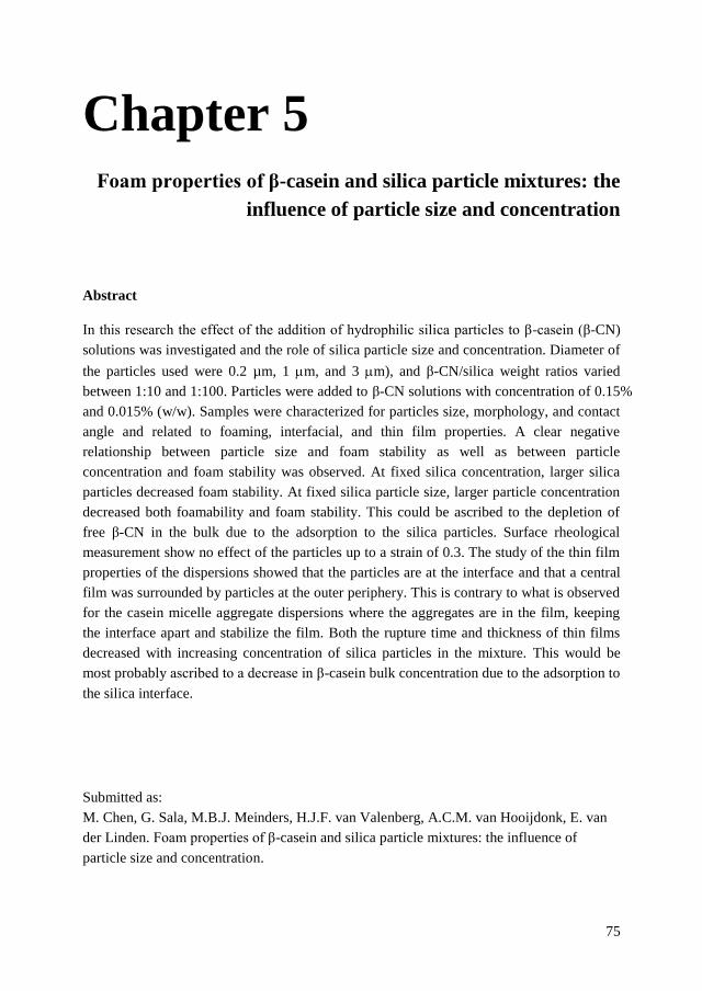
75
Chapter 5 Foam properties of β-casein and silica particle mixtures: the
influence of particle size and concentration
Abstract
In this research the effect of the addition of hydrophilic silica particles to β-casein (β-CN)
solutions was investigated and the role of silica particle size and concentration. Diameter of
the particles used were 0.2 µm, 1 m, and 3 m), and β-CN/silica weight ratios varied
between 1:10 and 1:100. Particles were added to β-CN solutions with concentration of 0.15%
and 0.015% (w/w). Samples were characterized for particles size, morphology, and contact
angle and related to foaming, interfacial, and thin film properties. A clear negative
relationship between particle size and foam stability as well as between particle
concentration and foam stability was observed. At fixed silica concentration, larger silica
particles decreased foam stability. At fixed silica particle size, larger particle concentration
decreased both foamability and foam stability. This could be ascribed to the depletion of
free β-CN in the bulk due to the adsorption to the silica particles. Surface rheological
measurement show no effect of the particles up to a strain of 0.3. The study of the thin film
properties of the dispersions showed that the particles are at the interface and that a central
film was surrounded by particles at the outer periphery. This is contrary to what is observed
for the casein micelle aggregate dispersions where the aggregates are in the film, keeping
the interface apart and stabilize the film. Both the rupture time and thickness of thin films
decreased with increasing concentration of silica particles in the mixture. This would be
most probably ascribed to a decrease in β-casein bulk concentration due to the adsorption to
the silica interface.
Submitted as:
M. Chen, G. Sala, M.B.J. Meinders, H.J.F. van Valenberg, A.C.M. van Hooijdonk, E. van
der Linden. Foam properties of β-casein and silica particle mixtures: the influence of
particle size and concentration.

Chapter 5
76
5.1 Introduction
In food science, foams stabilized by particles, like protein particles, aggregates, fluid gel
particles, and modified starch particles have received lots of attention [1-8]. Casein micelles,
the natural food nano-gel particle present in milk, function as the main emulsion stabilizing
component of homogenized milk and exhibit good foaming properties [9]. In previous
research we studied the foaming properties of casein micelle dispersions (CMDs) and found
that the foam stability of CMDs that contained casein micelle aggregates (CMAs)
significantly improved [10]. The size of CMAs in our studies ranged from 0.5 µm till a few
µm. This study indicated that the type of protein as well as particle size, shape and
concentration play an important role in the stabilization of foams made by CMDs. We
found that aggregates of casein micelles were entrapped in the lamellae between bubbles
without forming a gel. These entrapped particles keep the two interfaces apart. This
entrapment was suggested to be the main factor leading to the delay of film rupture and
stabilization of the foams made from CMA’s.
The location of particles in a foam structure is influenced by the wetting properties of
particles [11-13], which also will determine whether particles stabilize or destabilize foam
[14, 15]. The wettability of particles depends on their hydrophobicity. In general,
completely hydrophilic particles do not adsorb onto the air/water interface [16]. Partially
hydrophobic particles with contact angle θ close to 90° can act as a foam stabiliser, whereas
hydrophobic particles (θ>90°) destabilize a foam and are often used as antifoaming agents
[17]. The particle hydrophobicity could be modified by appropriate chemical reaction [18,
19], by adjusting the salt concentration [20], or through the adsorption of amphiphiles to the
particle surface [7]. For foams made of particles, partially hydrophobic particles were found
to be optimum for foam stabilization [21-23]. Foaming behaviour becomes more
complicated for mixed systems of particles and amphiphiles.
Silica (SiO2) is one of the widely chosen spherical inorganic particles for foam studies due
to their well characterized physicochemical properties [18] [24]. Previous studies have
investigated foaming properties of silica particles hydrophobized by SDS [25] or
hexylamine [26]. The former had a contact angle of 66º and the latter ranged between 26 to
56°. In the study of Karakashev et al. (2011) [25], silica particles were segregated and
trapped in both foam lamellas and Plateau borders. Foams with silica particles get less
stable than foams without. According to Kruglyakov et al. (2011) [26], aggregation of
particles and formation of the thixotropic gels in the dispersion medium of the foam
enabled a ultra-stable foam. Thus, modification of silica particles by coverage with of
amphiphiles on the particle surface may lead to stable foams depending on the type and
concentration of amphiphiles used.
β-casein (β-CN) is an abundant and surface active amphiphilic protein in milk. Above the
critical micellization concentration it forms micelles, which at 25°C is about 24 µmol/L

Foam of β-casein/silica mixtures
77
[27]. β-CN adopts flexible disordered configurations with little ordered secondary structure
in aqueous phase because of the preponderance of proline residues and the lack of cysteine
residues in protein primary structure [28]. As far as we know, β-CN could attach to the
surface of silica [29], which could possibly modify the surface charge, roughness and
hydrophobicity of the particles. It may be expected that after adsorption of casein to the
particle surface, silica could have a similar surface charge and hydrophobicity as casein
micelles. This would make β-CN/silica a simplified model system for casein micelle
dispersions where the model systems allows for testing the effect of particle size and
thereby mimic the casein micelle aggregates.
To be able to control foam stabilization using hydrophilic particles, more insight in the
stabilization mechanism is needed. Therefore, we investigated the effect of silica particle
size and concentration on foaming behaviour of β-CN/silica mixed systems. Hydrophilic
silica particles with different sizes (Small: 0.2 µm; Medium: 1 m and Large: 3 m) were
added to β-CN solutions at varying ratios. Samples were characterized for particle size and
morphology. The contact angle of silica particles was estimated by measuring an air-liquid
contact angle of β-CN on smooth hydrophilic glass surface. The foaming properties were
measured using a FoamScan. Linear and non-linear surface rheology was applied to study
the interfacial properties. Thin film stability was measured with thin film balance technique.
These physical properties were related to the foaming and interfacial properties of the
mixtures, as well as to their thin film properties.
5.2 Materials and methods
5.2.1 Materials
β-casein was generously provided by NIZO food research BV (Ede, Netherlands). The
silica particles (sicastar) were purchased from Micromod Partikeltechnologie GmbH
(Rostock, Germany), and were received as 50 mg/mL (5.0%, w/w) aqueous dispersions.
The surface of silica particles presented hydrophilic Si-OH groups, without any
modification. In this work, three particular silica samples were used: one with an average
diameter of 0.2 µm (Small), one with an average diameter of 1 m (Medium) and one with
an average diameter of 3 m (Large). The water used in all the experiments was first passed
through a PURELAB Ultra Milli-Q (Elga, Darmstadt, Germany) water system. Its surface
tension at 25°C was 71.99 mN/m. All measurements were repeated at least twice and the
average values were used.
5.2.2 Preparation of β-casein/silica particles mixtures
All β-casein/silica particles mixtures (β-CN/silica) were prepared at room temperature in a
phosphate buffer (PB). PB was prepared by first mixing appropriate stock solutions of 0.1
M Na2HPO4 and 0.1 M NaH2PO4 with a further dilution to 10 mM. The buffer solution had
a final pH of 7.0 and an ionic strength of 21.5 mM. Fresh PB was prepared before each

Chapter 5
78
measurement. 0.02% (w/w) sodium azide was added to prevent bacterial contamination. A
0.015% (w/w) β-casein solution was mixed with silica particles (5.0%, w/w) at varying
ratios (1:5, 1:10, 1:50, 1:100, weight of β-CN: weight of particles) and stirred overnight.
Correspondingly, the concentrations of silica particles in β-CN/silica mixtures were 0.075%,
0.15%, 0.75% and 1.5% (w/w). β-CN/silica mixtures were defined as: β-CN/S-L for large
silica (3 µm), β-CN/S-M for medium silica (1 µm) and β-CN/S-S for small size silica (0.2
µm) For the ratio 1:10, the β-CN/silica mixtures were ultracentrifuged at 100,000 g for 90
min at 20ºC to remove the particles. The supernatants were collected for further research.
5.2.3 Particle size distribution
The size distribution of silica particles was determined with multi angle light scattering [30]
(Mastersizer 2000 Malvern Instruments, Worcestershire, U.K.). The refractive index used
for silica was 1.54 [31]. The particle size distribution was also checked using Malvern
Zetasizer Nano-ZS (Malvern, Worcestershire, UK) by dynamic light scattering [32] with
the same refractive index mentioned above.
5.2.4 Scanning electron microscope (SEM)
150 µL of silica particle dispersions were carefully dripped on poly-L-lysine coated glasses.
Excess of the sample was removed by dipping in MilliQ-water. The glass with sample
attached was immersed in 3% (v/v) glutaraldehyde for fixation and MilliQ-water was used
to wash the excess of glutaraldehyde. After that, the MilliQ-water was subsequently
replaced by ethanol (10%, 30%, 50%, 70%, 90%, and 100%) for dehydration. Samples in
ethanol attached on sample holders with CCC Carbon Adhesive (Electron Microscopy
Sciences, Washington, USA) were further dried by critical point drying (CPD). After that,
the samples were sputter coated with a layer of tungsten in SCD 500 (Leica EM VCT 100,
Leica, Vienna, Austria) before imaging. All samples were analysed with a scanning
electron microscope (Magellan XHR 400L FE-SEM, FEI, Eindhoven, the Netherlands) at a
working distance of 4 mm with SE detection at 2.00 kV.
5.2.5 Static contact angle measurement
The contact angle of β-CN on glass (hydrophilic silica) surface was measured with VCA
2500XE Video contact angle system (Advanced Surface Technology Inc., Billerica, MA).
The samples analysed in this study were MilliQ water, phosphate buffer (pH=7.0, 10 mM)
and β-casein (0.015%, w/w) in the phosphate buffer. A droplet of the mentioned liquids
was dispensed manually at the tip of a micropipette to a smooth glass plate (7 cm×2 cm), as
the stage raised the sample surface into contact with the liquid. Upon creating the droplet
on the surface of the sample, the VCA software program captured the image on a CCD
camera. The contact angle was calculated manually with the VCA Optima XE. At least 6
measurements were conducted for each sample. Two numbers were displayed: the left and
right contact angles with variance within 1-2º. The mean contact angle was selected.

Foam of β-casein/silica mixtures
79
5.2.6 Foaming properties
Foaming properties of β-CN solution, β-CN/silica mixtures and supernatants of CN/S-L
1:10 were assessed with a FoamScan (Teclis IT-Concept, Longessaigne, France) according
to Chen et al. (2016) [10]. Foam was produced using sparging air through a frit for 40 s,
corresponding to a total air volume of 120 mL. The foam volume after sparging for 40 s
was used to assess the foamability of the samples. The foam half-life, the time when the
foam reached half of its initial height (t½), was used as a measure for foam stability. Two
dimensional images of the foam at the wall of the tube were recorded using a charge-
coupled device (CCD) camera. The image covered an area of 1.0 cm2.
5.2.7 Interfacial properties measured with Profile Analysis Tensiometer (PAT)
The interfacial properties of β-CN solution, silica in PB and β-CN-/silica mixtures were
measured using a SINTERFACE PAT 1-M (SINTERFACE Technologies, Berlin,
Germany) according to Chen et al. (2016). Data were analysed using SINTERFACE Profile
Analysis Tensiometer PAT 1-M version 1.4.0.685 software according to van Kempen et al.
[33] with some modifications. The surface tension was 72.2 ± 0.7 mN/m for MilliQ-water
and 70.1±0.3 mN/m for PB. First, the surface tension was measured for 1000 s with the
area (A) of the droplet kept constant at 25 mm2. A sinusoidal oscillation was applied after
reaching equilibrium (t=1000 s) to obtain the dilatational modulus with a deformation of
7% and 30% at 0.02 Hz. Results of non-linear dilatational rheology were analysed by
Lissajous plots of surface pressure (Π ) versus strain (ΔA/A).
5.2.8 Thin film stability
The thin film stability of β-CN solution, silica in PB and β-CN/S-L (3 µm) mixtures was
measured by micro interferometry using a microscope (Axio plan 01, Zeiss, Jena, Germany)
according to Chen et al. (2016) with some modifications. The studied samples were silica in
PB (Large: 3 µm) with a concentration from 0.00015% to 0.15% (w/w), β-CN (0.015%,
w/w) and β-CN/silica mixtures with varying ratios (1:10, 1:20, 1:30, 1:50, 1:100, w/w).
Thin liquid films (diameter = 200 μm) were made in a Scheludko cell (diameter = 0.3 cm)
covered with a glass cover. The casing had a reservoir of PB (10 mM, pH 7.0) at the bottom
to ensure a relative humidity of 100% in the cell during measurements. Liquid was drawn
into the capillary with a syringe (500 μL, Hamilton, Reno, NV, USA) and the whole setup
was left to equilibrate for 5 min before a thin film was made by drawing sample solution
into the capillary. If a film did not break after 30 min, it was considered stable. In this case,
the lifetime was expressed as 1800 s. In case of stable films, samples were measured for 5
times. In the case of rupturing films, experiments were repeated 10 times to calculate the
average and standard deviation of the rupture time and film thickness (h). Images of the
thin films were collected with a software script (developed at the Laboratory of Food
Chemistry, Wageningen University) for Matlab (Software version 2013b, MathWorks,
Natick). The magnification was ×100. The equilibrium film thickness (h) was calculated
using the intensity of the reflected monochromatic light (λ= 546 nm). The average intensity

Chapter 5
80
of light (pixels) was recorded in the centre of the film in a circle of radius of around 25 µm.
The thickness of the film was calculated as described in reference [34] using:
ℎ =𝜆
2𝜋𝑛sin−1 (√
𝐼−𝐼𝑚𝑖𝑛
𝐼𝑚𝑎𝑥−𝐼𝑚𝑖𝑛)
(1)
Where λ is the wavelength of the light (λ=546 nm), n is the refractive index of the film, I is
the intensity of the reflected light, and Imin and Imax are the minimum and maximum
intensities, respectively.
5.3 Results and discussion
5.3.1 Sample characterization
Figure 5-1. Particle size distribution of silica dispersed in buffer and β-CN/silica mixtures
in volume fraction (%).The β-CN/silica mixtures contained 0.075%, 0.15%, 0.75% and 1.5%
(w/w) silica particles and 0.015% (w/w) β-CN. Silica particles with different sizes were
used. (A): 200 nm; (B) 1 µm; (C) 3 µm. (D) Morphology of silica particles with different
sizes: (S) 200 nm; (M) 1 µm; (L) 3 µm.
The silica particles and the β-CN/silica mixtures were characterized for size distribution
and particle morphology. According to the supplier, the particle size distribution of so-
called small silica is 0.2 µm, so-called medium silica is 1 m and so-called large silica is 3
m. Particle size was checked with mastersizer, nanosizer and SEM. Results indicated that
nanosizer was more accurate for size measurement of small particles while the mastersizer
was more accurate for size measurement of large particles. All methods worked well for
medium size particles. Thus, the average particle size of β-CN/S-S mixtures containing
D

Foam of β-casein/silica mixtures
81
small silica was taken from nanosizer results (Figure 5-1A). The particle size of β-CN/S-M
and β-CN/S-L mixtures containing medium and large silica respectively was taken from the
mastersizer results (Figure 5-1 B and C). The average particle size was 0.2 µm for (S), 1.05
µm for (M) and 3.59 µm for (L). These data are in line with the specifications provided by
the supplier. The particle size determined in β-CN/silica mixtures was similar to that found
for the bare silica particles, indicating that the particles did not aggregate in the β-CN
solution. Samples with the large silica particles were more polydisperse than the samples of
the other two sizes.
To check the morphology of the particles, SEM was conducted. The obtained images are
shown in Figure 5-1D. The small and medium sized silica particles were well spherical and
monodisperse with a smooth surface. However, the large size silica particles showed a
higher polydispersity, confirming the findings of the Mastersizer determination, and a
rougher surface. The higher surface roughness is because the large silica particles were
porous. The medium and small silica were both nonporous. As expected, no differences
were observed between the morphology of the silica particles dispersed in water or
dispersed in β-CN (data not shown).
Figure 5-2. Contact angle of water, phosphate buffer (pH=7.0, 10 mM) and β-CN (0.015%,
w/w in phosphate buffer) on a glass plate.
The size distribution values measured by SEM were in line with those obtained with
Mastersizer. Also here, no sign of aggregation of the particles in the β-CN/silica mixtures
used in this study was observed. According to F. Tiberg et al.(2001), β-CN forms an
asymmetric-bilayer when adsorbed to hydrophilic silica [35], which indicated that the

Chapter 5
82
surface of hydrophilic silica with adsorbed β-CN becomes more hydrophobic. To check this
we estimated the contact angle of the particles after attachment of β-CN. Therefore, we
used a hydrophilic glass surface to mimic the surface of hydrophilic silica particles. Results
are shown in Figure 5-2. The hydrophilic glass surface had a water contact angle of 11±1º
which increased to 21±2º when water was replaced by β-CN solution. This indicates that
the adsorption of β-CN at the surface of silica indeed makes the glass surface more
hydrophobic. For the contact angle of silica particles with adsorbed β-CN, we expect that it
will be close to 21±2º. Other research also reported an hydrophobicity of particle changed
by binding of amphiphilic molecules to the surface of hydrophilic inorganic particles [36,
37]. However, besides material, the surface roughness of the particle could also influence
the contact angle since it could lead to a ‘non-equilibrium’ wetting characteristic of the
particles.
5.3.2 Foam properties
Foamability
No foam could be formed with silica particles dispersed in buffer solution only, i.e. without
β-CN. This is the case for all particle sizes. This is in line with previous results of Binks et
al. [18] who found that hydrophilic silica particles with a surface coverage of Si-OH larger
than 70% (θ<13º) does not foam. The silica particles used in this work were hydrophilic
with a Si-OH surface coverage of about 100%. The isoelectric point of silica is between 2-3
[24] so that the silica particles are negatively charged at pH=7.0.
Figure 5-3. Foamability of β-CN/silica mixtures with different size and concentration of
silica particles. The concentration of β-CN in all samples was kept at 0.015% (w/w).
Aqueous foams were prepared with β-CN/silica mixtures at varying ratios (1:0, 1:5, 1:10,
1:50, 1:100, w/w). As shown in Figure 5-3, samples could reach a final foam volume of 120

Foam of β-casein/silica mixtures
83
mL after 40 s of sparging, except β-CN/S-L 1:50 and β-CN/S-L 1:100. As the concentration
of large silica particles increased, a dramatic decrease in foam height was observed: β-
CN/S-L 1:100 hardly foamed.
Foam stability
For all investigated β-CN/silica mixtures, the results of samples containing 0.015% (w/w)
β-CN are shown in Figure 5-4A and those containing the ten times higher concentration of
0.15% (w/w) are shown in Figure 5-4B. The t1/2 of the 0.015% (w/w) β-CN solution was
26020 min while that of the 0.15% (w/w) solution was 4557 min, being significantly
higher than that of any other β-CN/silica mixtures. For mixtures with the same silica
particle size but different amounts of silica particles, foam stability decreased with
increasing concentration of silica particles at two β-CN concentrations. It is noted that,
compared to β-CN without any particles, foam stability decreased to 0 min at 0.015% (w/w)
upon addition of 1.5% large silica particles, while it decreased by factor of 6 at 0.15% (w/w)
upon addition of the same amount of large silica particles. This indicates that the decrease
in foam stability upon addition of particles is relatively stronger at lower β-CN
concentrations.
Figure 5-4. Foam half-life (t1/2) of β-CN/silica mixtures with different size and
concentration of silica particles. Csilica=0% represents a β-CN solution with a protein
concentration of 0.015% (w/w) in (A) and 0.15% (w/w) in (B). Black bar: β-CN.
The role of particle size in this decrease in foam stability by adding particles was also
checked, at the lower β-CN concentration of 0.015% (w/w). For equal weight ratio in β-
CN/silica, mixtures show a clear negative relation between particle size and foam stability.
We also measured the foam properties of the supernatant of the β-CN/silica 1:10 solution
for all three particle sizes and results are shown in Figure 5-5. It was found that foams of
the supernatants (containing no silica particles) were markedly lower in height than those of

Chapter 5
84
the initial 0.015%, w/w β-CN solution. This effect was more pronounced for the larger
silica particle samples and their supernatants. The supernatant of β-CN/S-S 1:10, which had
the most stable foam among supernatants, showed a decrease in foam height of almost a
factor of two compared to that of initial β-CN solution. One possible explanation for the
above behaviour could be the decrease of β-CN in solution due to its adsorption to the silica
particles, i.e. the silica depriving the solution from its β-CN. Based on this assumption, the
reason why foam stability of β-CN/silica decreased less at the higher β-CN concentration of
0.15% is most probably due to the fact that there is more β-CN left free in the bulk and the
effect therefore is relatively smaller because there is still enough β-CN left in solution to
sufficiently stabilise the foam. The adsorbed amount (Γ) of β-casein on hydrophilic silica
surface was reported to be 0.26 µg/cm2 for a bulk concentration of 27 µM [29]. From this,
the according estimated amount of β-CN adsorbed to the particle surface on basis of the
total area of the silica spheres in solution was calculated to be 100% for 200 nm, 25.3% for
1 µm and 6.5% for 3 µm sized silica particles in a 0.015% (w/w) β-CN solution that
contains 1.5% (w/w) silica particles. For the 0.15% solution, approximately 10%, 3%, and 1%
of the β-CN is estimated to adsorb to the silica particles. This estimate also implies that a
considerable amount of β-CN could indeed be adsorbed at the silica particle surface and be
deprived from the solution. The high porosity and surface roughness of large silica (3 µm)
probably resulted in more adsorption of β-CN to the particle surfaces compared to smaller
particles. To get more insights in the possible mechanisms of foam destabilization of β-CN
solutions by these silica particles, the properties of the air/water interfaces and the thin film
stability were studied as below.
Figure 5-5. Foam half-life (t1/2) of β-CN/Silica (L: 3 µm; M: 1 µm; S: 200 nm) mixtures
with a ratio of 1:10 (w/w) and their supernatants. Dark bars represent corresponding
supernatant of the β-CN/silica mixture.
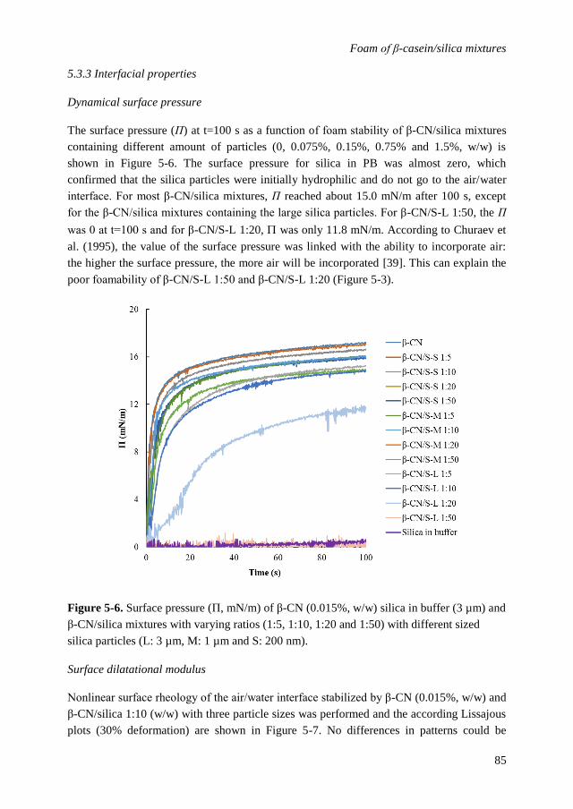
Foam of β-casein/silica mixtures
85
5.3.3 Interfacial properties
Dynamical surface pressure
The surface pressure (Π) at t=100 s as a function of foam stability of β-CN/silica mixtures
containing different amount of particles (0, 0.075%, 0.15%, 0.75% and 1.5%, w/w) is
shown in Figure 5-6. The surface pressure for silica in PB was almost zero, which
confirmed that the silica particles were initially hydrophilic and do not go to the air/water
interface. For most β-CN/silica mixtures, Π reached about 15.0 mN/m after 100 s, except
for the β-CN/silica mixtures containing the large silica particles. For β-CN/S-L 1:50, the Π
was 0 at t=100 s and for β-CN/S-L 1:20, was only 11.8 mN/m. According to Churaev et
al. (1995), the value of the surface pressure was linked with the ability to incorporate air:
the higher the surface pressure, the more air will be incorporated [39]. This can explain the
poor foamability of β-CN/S-L 1:50 and β-CN/S-L 1:20 (Figure 5-3).
Figure 5-6. Surface pressure (Π, mN/m) of β-CN (0.015%, w/w) silica in buffer (3 µm) and
β-CN/silica mixtures with varying ratios (1:5, 1:10, 1:20 and 1:50) with different sized
silica particles (L: 3 µm, M: 1 µm and S: 200 nm).
Surface dilatational modulus
Nonlinear surface rheology of the air/water interface stabilized by β-CN (0.015%, w/w) and
β-CN/silica 1:10 (w/w) with three particle sizes was performed and the according Lissajous
plots (30% deformation) are shown in Figure 5-7. No differences in patterns could be

Chapter 5
86
observed between the β-CN and β-CN/silica mixtures containing small and medium sized
particles. Their air/water interfaces showed a strong increase in cohesion in the end of
compression, which was probably caused by the entanglement of β-CN. However the
air/water interface stabilized by β-CN/S-L appeared like that of a diluted surfactant layer
without cohesion in the end of compression. This may be simply due to the fact that there is
a lower bulk concentration of free β-CN in β-CN/S-L. From these experiments, no
conclusions can be drawn whether the particles adsorb at the air/water interface or not.
However, we can conclude that if the particles adsorb, they do not significantly change the
interfacial properties indicating that they are diluted and do not interact up to a strain of 0.3.
Figure 5-7. Nonlinear surface rheology (30% deformation) of β-CN and β-CN/silica
mixtures with ratio of 1:10 (w/w). Blue line: β-CN; dash line: β-CN/S-L; solid black line:
β-CN/S-M; solid grey line: β-CN/S-S.
5.3.4 Thin film properties
The morphology and stability of thin liquid films made from β-CN solutions containing
silica particles of 3 µm with varying β-CN concentration (ranging from 0.00015% to 0.15%,
w/w) as well as varying β-CN/silica weight ratios (1:10, 1:20, 1:30, 1:50, 1:100, w/w) were
studied with the thin film balance technique. Figure 5-8 shows thin films of silica (L) in PB
for β-CN/S-L 1:10. For films of a solution of only silica particles in PB, the morphology
and stability were both similar to that of films made with only PB. Their rupture times were
around 20 s. This is in line with the observation that PB solutions containing only
hydrophilic silica do not foam. We note that in films made from β-CN/silica mixtures, the
silica particles seem located at the interface and are depleted from the film centre and are

Foam of β-casein/silica mixtures
87
more concentrated around the periphery of the film in a band of about ten times the silica
particle diameter. This concentrated band of particles seemingly touching or even bridging
the film surfaces at the outer periphery is depicted in Fig. 5-8B and 8C. The depletion of
particles from the centre of the film observed in our experiments is in line with the work of
Horozov (2008) [17] and of Narsimhan (2016) [15], who report that particles in dilute
monolayers in the film cannot resist the hydrodynamic flow inside the film and will be
dragged away from the film centre into its periphery.
Figure 5-8. Images of thin films of silica (L: 3 µm) in PB (A), β-CN/S-L 1:10 (B) and (C)
Zoomed in image of the area within green cube in image (B).
Figure 5-9 shows the evolution of the morphology of films of β-CN/silica mixtures in time.
For all films of β-CN/silica mixtures, the thicker central part of the films gradually flattened,
thinned and eventually ruptured. Only for β-CN/S-L 1:10, the thinning rate of the film (data
not shown), did not change much compared to that of β-CN without silica particles. From
β-CN/S-L 1:20 to β-CN/S-L 1:100, the films exhibited a faster thinning process compared
to the β-CN solution without particles. With increasing particle concentration, the gradually
decreasing light intensity at equilibrium also indicated a lower final inner film thickness
shortly before rupture.

Chapter 5
88

Foam of β-casein/silica mixtures
89
Figure 5-9. Thin liquid film images of silica, β-CN and β-CN/silica mixtures with varying
ratios (from 1:10 to 1:100, w/w). The size of silica particle for thin film study was 3 µm.
The number on the images is the different time points (s) and “E” represent equilibrium.
Figure 5-10. Rupture time of thin liquid films of (A) Large silica particles (from 0.00015%
to 0.15%, w/w) in buffer and (B) β-CN/silica mixtures containing 0% to 0.15% silica
particles. The concentration of β-CN was kept constant at 0.0015% (w/w).
Figure 5-11. Thickness just before rupture (h) of liquid films of β-CN/silica mixtures
containing from 0% to 0.15% silica particles with a size of 3 µm.
According to Figure 5-10A, for silica in PB (10 mM, pH 7.0), the thin film stability was
similar to that of only PB, with an average rupture time of around 20 s. Figure 5-10B shows
the stability of β-CN solution and of β-CN/silica mixtures containing from 0.0%-0.15%

Chapter 5
90
silica with silica particle size of 3 µm. The concentration of β-CN in the samples used for
the study of the thin film properties was kept constant at 0.0015% (w/w). Note that this is
10 times lower than that used for foam experiments). The film of the β-CN solution was
relatively stable (rupture time: 1800 s). After adding silica particles to the β-CN solution,
the rupture time decreased. The more particles added to the mixture, the smaller the rupture
time and the lower the stability of the liquid film. For instance, with 0.045% (w/w) or more
silica particles in the mixtures, the thin film stability of β-CN/S-L dramatically decreased to
less than 400 s. Besides the rupture time, the thickness h of the central part of films of β-
CN/silica mixtures at the equilibrium before rupture was estimated and is shown in Figure
5-11. Interestingly, this thickness decreased gradually with increasing particle concentration
in the β-CN/silica mixtures, from 47.7±4.6 nm for the β-CN solution (0.0015%, w/w) to
20.0±0.9 nm for β-CN/S-L 1:100.
Research on mixtures of protein aggregates and proteins showed foam stabilization
improved with the presence of protein aggregate particles that range in size between tens of
nanometers to a few microns [10, 41-45]. We hypothesized that adding hydrophilic silica to
β-CN solution would lead to β-CN coated particles that could mimic, depending on the size,
casein micelles and their aggregates. In the previous chapter it was shown that these
aggregates increased foam stability (Chapter 4). In contrast to this hypothesis our results
presented in this chapter show the opposite; a clear destabilization effect when adding
hydrophilic silica particles. Based on the results above, it is most likely that the silica
particles deplete the solution from its β-CN molecules. In line with our research,
Karakashev et al. (2011) [25] found that addition of silica particles to SDS solutions
increased the rate of foam decay. The mechanism behind their results was not worked out
in detail. Our suggested mechanism of β-CN depletion from the solution could be proven
by chemical analysis of the silica particles centrifuged from the β-CN solution and is also
supported by a theoretical estimation of the amount of β-CN adsorbed at the particles
interface. In addition we observed that the silica particles are at the air/water interface and
located in the outer film region, most probably transported their due to the drag of the liquid
during film drainage. This is (also) contrary to what is observed in the case of casein
micelle aggregates, which are more or less randomly distributed over the film.

Foam of β-casein/silica mixtures
91
5.4 Conclusion
In this research the effect of size and concentration of hydrophilic silica particles on the
properties of foams from solutions containing β-casein (β-CN) and silica particles was
investigated. Foam stability of the β-CN/silica mixture decreased with increasing size and
concentration of the silica particles. This destabilizing effect is suggested to be caused the
depletion of β-CN from solution by means of its adsorption to the silica particles. Thin film
study shows that the β-CN/silica particles are at the interface. The thin foam films arising
from β-CN silica dispersions exhibit a central region without silica particles and a band of
more concentrated solution of silica particles. Both the rupture time and thickness of the
thin films just before rupture (inner region) decreased with increasing particle concentration
in the mixtures. The presence of the particles at the outer side of the lamellae would add to
the stability of the film because of a reduced pressure difference across the film. However,
the decrease in foam and film stability would be ascribed to a decrease in β-casein bulk
concentration due to the adsorption to the silica interface.

Chapter 5
92
References
1. Murray, B.S., et al., Stabilization of foams and emulsions by mixtures of surface active food-grade
particles and proteins. Food Hydrocolloids, 2011. 25(4): p. 627-638.
2. Schmidt, I., et al., Foaming properties of protein/pectin electrostatic complexes and foam structure at
nanoscale. Journal of Colloid and Interface Science, 2010. 345(2): p. 316-324.
3. Nicolai, T., Britten, M. and Schmitt, C., β-Lactoglobulin and WPI aggregates: Formation, structure
and applications. Food Hydrocolloids, 2011. 25(8): p. 1945-1962.
4. Fameau, A.L. and Salonen, A., Effect of particles and aggregated structures on the foam stability and
aging. Comptes Rendus Physique, 2014. 15(8): p. 748-760.
5. Lazidis, A., et al., Whey protein fluid gels for the stabilisation of foams. Food Hydrocolloids,
2016. 53: p. 209-217.
6. Oboroceanu, D., et al., Fibrillization of whey proteins improves foaming capacity and foam stability at
low protein concentrations. Journal of Food Engineering, 2014. 121: p. 102-111.
7. Binks, B.P., et al., Dispersion Behavior and Aqueous Foams in Mixtures of a Vesicle-Forming
Surfactant and Edible Nanoparticles. Langmuir, 2015. 31(10): p. 2967-2978.
8. Asghari, A.K., et al., Interfacial and foaming characterisation of mixed protein-starch particle systems
for food-foam applications. Food Hydrocolloids, 2016. 53: p. 311-319.
9. De Kruif, C. and Holt, C., Casein micelle structure, functions and interactions. Advanced Dairy
Chemistry—1 Proteins. 2003, p. 233-276. Springer US.
10. Chen, M., et al., Particle size determines foam stability of casein micelle dispersions. International
Dairy Journal, 2016. 56: p. 151-158.
11. Stamou, D., Duschl, C. and Johannsmann, D., Long-range attraction between colloidal spheres at the
air-water interface: The consequence of an irregular meniscus. Physical Review E, 2000. 62(4): p.
5263.
12. Kralchevsky, P.A., Denkov, N.D. and Danov, K.D., Particles with an undulated contact line at a fluid
interface: interaction between capillary quadrupoles and rheology of particulate monolayers.
Langmuir, 2001. 17(24): p. 7694-7705.
13. Ghezzi, F., et al., Pattern formation in colloidal monolayers at the air–water interface. Journal of
colloid and interface science, 2001. 238(2): p. 433-446.
14. Hunter, T.N., et al., The role of particles in stabilising foams and emulsions. Advances in Colloid and
Interface Science, 2008. 137(2): p. 57-81.
15. Narsimhan, G., Drainage of particle stabilized foam film. Colloids and Surfaces A: Physicochemical
and Engineering Aspects, 2016. 495: p. 20-29.
16. Vivaldini, D.O., et al., Why foams containing colloidal hydrophilic particles are unstable? Ceramics
International, 2013. 39(5): p. 6005-6008.
17. Horozov, T.S., Foams and foam films stabilised by solid particles. Current Opinion in Colloid &
Interface Science, 2008. 13(3): p. 134-140.
18. Binks, B.P. and Horozov, T.S., Aqueous Foams Stabilized Solely by Silica Nanoparticles. Angewandte
Chemie, 2005. 117(24): p. 3788-3791.
19. Safouane, M., Langevin, D. and Binks, B., Effect of particle hydrophobicity on the properties of silica
particle layers at the air-water interface. Langmuir, 2007. 23(23): p. 11546-11553.
20. Dickinson, E., et al., Factors controlling the formation and stability of air bubbles stabilized by
partially hydrophobic silica nanoparticles. Langmuir, 2004. 20(20): p. 8517-8525.
21. Ata, S., Ahmed, N. and Jameson, G.J., Collection of hydrophobic particles in the froth phase.
International Journal of Mineral Processing, 2002. 64(2): p. 101-122.
22. Ata, S., Ahmed, N. and Jameson, G., The effect of hydrophobicity on the drainage of gangue minerals
in flotation froths. Minerals engineering, 2004. 17(7): p. 897-901.
23. Schwarz, S. and Grano, S., Effect of particle hydrophobicity on particle and water transport across a
flotation froth. Colloids and Surfaces A: Physicochemical and Engineering Aspects, 2005. 256(2): p.
157-164.

Foam of β-casein/silica mixtures
93
24. Binks, B.P., Kirkland, M. and Rodrigues, J.A., Origin of stabilisation of aqueous foams in
nanoparticle–surfactant mixtures. Soft Matter, 2008. 4(12): p. 2373.
25. Karakashev, S.I., et al., Formation and stability of foams stabilized by fine particles with similar size,
contact angle and different shapes. Colloids and Surfaces A: Physicochemical and Engineering
Aspects, 2011. 382(1): p. 132-138.
26. Kruglyakov, P.M., Elaneva, S.I. and Vilkova, N.G., About mechanism of foam stabilization by solid
particles. Advances in colloid and interface science, 2011. 165(2): p. 108-116.
27. Esmaili, M., et al., Beta casein-micelle as a nano vehicle for solubility enhancement of curcumin; food
industry application. LWT - Food Science and Technology, 2011. 44(10): p. 2166-2172.
28. Dickinson, E., Caseins in emulsions: interfacial properties and interactions. International Dairy
Journal, 1999. 9(3): p. 305-312.
29. Krisdhasima, V., Vinaraphong, P. and McGuire, J., Adsorption Kinetics and Elutability of α-
Lactalbumin, β-Casein, β-Lactoglobulin, and Bovine Serum Albumin at Hydrophobic and Hydrophilic
Interfaces. Journal of Colloid and Interface Science, 1993. 161(2): p. 325-334.
30. Ince Coskun, A.E., et al., Preparation, structure and stability of sodium caseinate and gelatin micro-
particles. Food Hydrocolloids, 2015. 45: p. 291-300.
31. Ghosh, G., Dispersion-equation coefficients for the refractive index and birefringence of calcite and
quartz crystals. Optics Communications, 1999. 163(1): p. 95-102.
32. Berghout, J.A.M., et al., Comparing functional properties of concentrated protein isolates with freeze-
dried protein isolates from lupin seeds. Food Hydrocolloids, 2015. 51: p. 346-354.
33. van Kempen, S.E., et al., Non-linear surface dilatational rheology as a tool for understanding
microstructures of air/water interfaces stabilized by oligofructose fatty acid esters. Soft Matter, 2013.
9(40): p. 9579-9592.
34. Scheludko, A. and Platikanowa, D., Untersuchung dunner flussiger schichten auf
quecksiber.Kolloid-zertschrift and zeitschrift fur polymere, 1961. 175(2): p. 150.
35. Tiberg, F., et al., β-Casein Adsorption at the Silicon Oxide−Aqueous Solution Interface.
Biomacromolecules, 2001. 2(3): p. 844-850.
36. Gonzenbach, U.T., et al., Stabilization of Foams with Inorganic Colloidal Particles. Langmuir, 2006.
22(26): p. 10983-10988.
37. Kostakis, T., Ettelaie, R. and Murray, B.S., Enhancement of stability of bubbles to disproportionation
using hydrophilic silica particles mixed with surfactants or proteins. Food Colloids, 2007. p. 357-368.
38. van Kempen, S.E.H.J., et al., Effect of Variations in the Fatty Acid Chain of Oligofructose Fatty Acid
Esters on Their Foaming Functionality. Food Biophysics, 2013. 9(2), p. 114-124.
39. Churaev, N.V., Contact angles and surface forces. Advances in Colloid and Interface Science, 1995.
58(2): p. 87-118.
40. Gergely, V., Jones, R. and Clyne, T., The effect of capillarity-driven melt flow and size of particles in
cell faces on metal foam structure evolution. Transactions-JWRI, 2001. 30(SPI): p. 371-376.
41. Schmitt, C., et al., Whey protein soluble aggregates from heating with NaCl: Physicochemical,
interfacial, and foaming properties. Langmuir, 2007. 23(8): p. 4155-4166.
42. Rullier, B., et al., β-Lactoglobulin aggregates in foam films: Effect of the concentration and size of the
protein aggregates. Journal of Colloid and Interface Science, 2010. 343(1): p. 330-337.
43. Lech, F., et al., Role of complex formation on foam stability of protein surfactant mixtures. 2014.
44. Morales, R., et al., Modification of foaming properties of soy protein isolate by high ultrasound
intensity: particle size effect. Ultrasonics sonochemistry, 2015. 26: p. 48-55.
45. Wan, Z., Yang, X. and Sagis, L.M., Nonlinear Surface Dilatational Rheology and Foaming Behavior
of Protein and Protein Fibrillar Aggregates in the Presence of Natural Surfactant. Langmuir, 2016.
32(15): p. 3679-3690.

94

95
Chapter 6 General discussion

Chapter 6
96
6.1 Introduction
The aim of the research described in this thesis is to better understand the role of milk
proteins on milk foam behaviour. Milk is composed of different protein fractions and the
main focus in this thesis is on casein micelles. The influence of composition, particle size
(size of aggregates of the casein micelles) and concentration on the foaming behaviour of
dispersions of casein micelles (i.e. casein micelle dispersions or, in short, CMDs) is
described in Chapter 2-4. In order to compare the findings on the behaviour of foams
stabilised by CMDs to that of other particle stabilised foams, the β-casein/silica system is
investigated (Chapter 5). In the current chapter, we discuss how the results described in the
different chapters can be combined to lead to a more refined overall emerging picture of the
importance of casein micelle particles and their aggregation for milk foam behaviour and
discuss implications for applications and future work. In the next section we first describe
the physico-chemical properties of the various systems studied in this thesis.

General discussion
97
6.2 Physicochemical properties of the systems studied in this thesis
Chapters 2-4 of this thesis are focused on the foam, thin film and air/water interface
stabilized by casein micelle dispersions (CMDs) that vary in the number of aggregates of
casein micelles (CMAs). The CMDs that practically do not contain any aggregated casein
micelles, i.e. as in the case in regular milk, mainly contain single casein micelles with a
small amount of casein molecules and casein-derived peptides. The CMDs that contain
aggregates of casein micelles are referred to as CMADs. Chapter 5 describes a foam study
of a mixture of β-casein and hydrophilic silica (β-CN/silica). The initial idea of the work in
Chapter 5 was to build a model system to check the role of particles in foam stabilization
and the role of β-casein as a possible adsorbent on the surface of the silica particles, thereby
possibly modifying the contact angle between the silica particles and air/water interface [1].
For both the casein dispersion systems and the silica system we have considered
concentration, size, contact angle, surface charge, shape and surface roughness and density
of the dispersed casein micelles and its aggregates, and silica particles, respectively. These
properties can be summarized as follows.
The concentration of particles in the two systems investigated (CMDs/CMADs and β-
CN/silica) is in the range of 0%-5.0 % (w/w). The average particle size of CMDs without
aggregates (i.e. casein micelles) ranges between 130 and 200 nm. The average particle size
for the CMADs containing aggregates ranges between 500 nm and 10 µm. The contact
angle of hydrophilic silica particles with adsorbed β-casein was estimated from our
experiments to be around 20º, which is comparable to that of hydrated casein micelles at
neutral pH [2, 3]. The zeta potential of casein micelles measured in milk permeate is around
-20 mV [4]. The bare silica particles used in this study have a surface charge density
according to the supplier of 30 mM/g for 200 nm, 7 mM/g for 1.0 µm and 1.2 mM/g for 3
µm. The zeta potential with adsorbed β-casein is between -20 and -30 mV, similar to that of
casein micelles. The pH of the two particle systems was comparable, i.e. 6.7-7.0. The ionic
strength is 125.4 mM for CMDs, which is calculated according to [5] based on the recipe of
SMUF in [6] whereas it is 21.5 mM for the β-CN/silica dispersions. The shape of casein
micelles is roughly spherical, however, that of the CMAs is either irregular-shaped or flat-
spherical. As derived from Figure 6-1, the surface roughness of casein micelles is around
15-20 nm. Because CMAs are composed of casein micelles, the large scale apparent surface
roughness of the casein micelle aggregates is of the order of the length scale of size of
casein micelles while at the shorter length scale the apparent roughness is given by that of a
single casein micelle. In β-CN/silica systems, the shape and surface roughness depends on
the size of particles (Figure 6-2). Silica particles of 200 nm and 1 µm are non-porous, well
spherical and have very smooth surface. Silica particles of 3 µm are roughly spherical with
some irregularities. Their surface is rougher than that of smaller silica. In general, the
morphology of silica particles is not affected by adsorption of β-casein. Casein micelles
have a density of 1.0632 g/cm3 and a voluminosity of 4.4 cm3/g, they are porous and highly
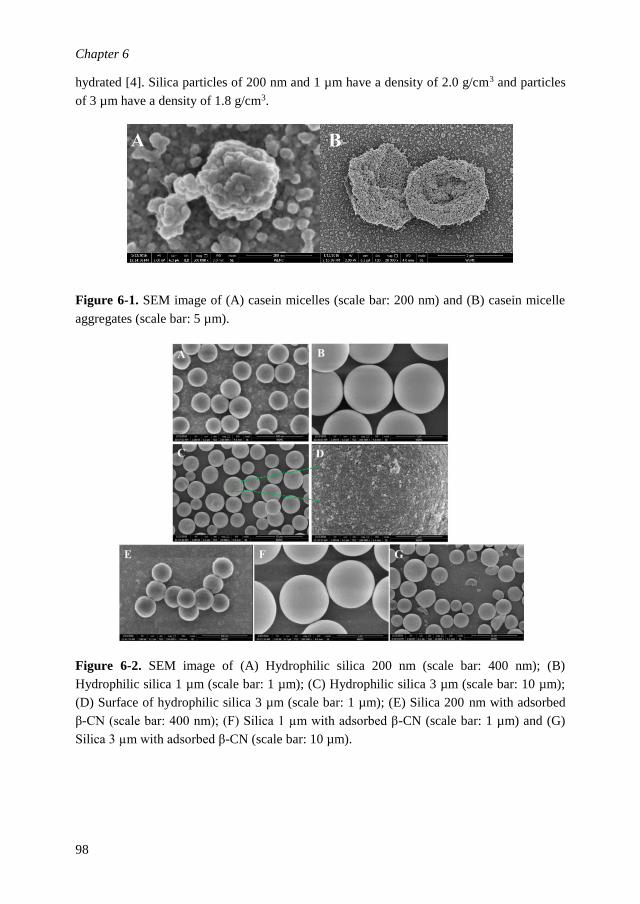
Chapter 6
98
hydrated [4]. Silica particles of 200 nm and 1 µm have a density of 2.0 g/cm3 and particles
of 3 µm have a density of 1.8 g/cm3.
Figure 6-1. SEM image of (A) casein micelles (scale bar: 200 nm) and (B) casein micelle
aggregates (scale bar: 5 µm).
Figure 6-2. SEM image of (A) Hydrophilic silica 200 nm (scale bar: 400 nm); (B)
Hydrophilic silica 1 µm (scale bar: 1 µm); (C) Hydrophilic silica 3 µm (scale bar: 10 µm);
(D) Surface of hydrophilic silica 3 µm (scale bar: 1 µm); (E) Silica 200 nm with adsorbed
β-CN (scale bar: 400 nm); (F) Silica 1 µm with adsorbed β-CN (scale bar: 1 µm) and (G)
Silica 3 µm with adsorbed β-CN (scale bar: 10 µm).

General discussion
99
6.3 Structures and properties at different length scales
In this study, system structures and properties at different length scales are characterized. A
typical example of the different structures and corresponding length scales that exist in
foams stabilized by CMDs is given in Figure 6-3.
Figure 6-3. Structures at different length scales in foams of casein micelle dispersions
6.3.1 Interfaces
To create a foam, newly formed bubbles need to be covered with foaming agents as quickly
as possible, which require sufficient amounts of the surface active agents to rapidly adsorb
to the air/water interface. To get insight in foaming behavior, we therefore, studied the
dynamics of adsorption of CMDs and β-CN/silica systems, which is described in Chapter
2-3 and Chapter 5, respectively. The dynamics of adsorption is relevant for the initial
stages of foam formation [7]. It is usually assessed by measuring, the surface pressure (Π)
as a function of time [8] and its dependence on the surfactant concentration in the bulk, C,
and at the surface, Γ [9, 10]. In this study, the rheological properties of interfaces are
investigated to obtain the mechanical properties of the interfacial film [11] [12, 13]
(Chapter 2, 3 & 5). Surface dilatation is conducted which measures surface pressure (Π) as
a function of the change in area (ΔA) of the droplet while maintaining a constant shape of
the air/water interface. The surface dilatational modulus consists of an elastic part E’ that
presents the recoverable energy stored in the interface and a viscous part E’’ that presents
the loss of energy through relaxation processes [14]. Both linear and nonlinear surface
rheology [15] is conducted. The amplitude of deformation ranges from 6% to 30% in this
study. Frequency sweeps, where the speed of interfacial deformation will relate to the time
scale of the transport of molecules from bulk to interface and vice versa, are also conducted
and used to deduce insights of the interfacial properties on the microstructural scale.
6.3.2 Thin liquid film
At the scale of thin liquid films, direct observation of morphology of the lamella is
conducted with optical microscopy. CMAs are directly visualized in the foam lamella under
transmission light mode (Chapter 4). This enables a better quantification of the
concentration of particles in foam lamella. In the thin film balance technique, a horizontal

Chapter 6
100
thin liquid film is generated with a Scheludko cell to mimic the foam lamella. Under a
reflected light mode, the stability of thin films including film thinning rate, rupture time and
film thickness h [16-18] is evaluated through light interference (Chapter 3 & 5). The thin
film stability measured with this technique is found to be more stable compared to that of a
thin film in a real foam, implying this parameter will only serve as an indication of stability
of foam lamellae.
6.3.3 Bubble and macroscopic foam
At the scale of a single bubble, the bubble size distribution is observed by microscopic
imaging in 2D linked with image analysis technique (Chapter 2 & 5). The bubble size
shortly after foam formation is measured as an indication of foam stability against early
stage coalescence, which is closely related to the adsorption kinetics and concentration of
foaming agents in bulk. At the macroscopic level, foam is generated mainly by sparging.
Shaking is performed in Chapter 4 for CMDAs with large CMAs as well. Foam decay is
evaluated by foam half-life (t1/2), which is the time required for the foam to reach half of its
initial volume [19]. Drainage of liquid is assessed by the liquid volume accumulated at the
bottom of the foam [20] and the liquid fraction in foam measured by electric conductivity.
The pattern of foam collapse is different for CMDs and β-CN/silica system.

General discussion
101
Ch
ap
ter 2
Chapter 3-4
Foa
m sta
biliz
atio
n
by
CM
Ds/C
MA
Ds
Ch
ap
ter 5 silica
→β
-casein
F
oam
desta
biliza
tion
Foa
ma
bility
an
d sta
bility
Figure 6
-4 Schematic o
verv
iew of th
e general discu
ssion in this th
esis

Chapter 6
102
6.4 Main results and discussion
Figure 6-4 depicts a schematic overview of the general discussion in this thesis. As
introduced above, several particle systems are studied, of which the results are
described in this thesis: casein micelle dispersions without aggregates (CMDs) and
with aggregates (CMADs) (Chapter 2-4), and β-CN/silica systems (Chapter 5).
CMDs and CMADs exhibited foam stabilization whereas addition of silica
particles into β-CN solution results in foam destabilization. A comparison is made
on the bulk properties, interfacial structure and thin film morphology of the
systems and how these properties relate to differences in foaming behaviour.
6.4.1 Bulk rheology and drainage
Colloidal properties of casein micelles dispersions depend on the concentration and
polydispersity of particles [21, 22]. According to [21], at concentrations below 100 g/L,
CMDs behaved as Newtonian hard-sphere fluids. In our study, the bulk concentration of
CMDs and sodium caseinate is smaller than 50 g/L, the shear viscosity is smaller than 10
mPa.s (Chapter 2-4), and the behaviour is Newtonian. The bulk rheological properties and
flow behaviour of silica particles is mainly dependent on the volume fraction of particles in
dispersions [23]. In diluted dispersions [24], a solution of silica particles behaves as a
Newtonian hard sphere fluid. The maximum concentration in this thesis study used for
silica and β-CN (Chapter 5) is 1.5% (w/w) and 0.015% (w/w), respectively. The shear
viscosity of β-CN/silica systems is measured to be close to the viscosity of water
confirming the diluted regime. While Kruglyakov et al. reported [25] that amphiphiles
could induce aggregation of particles, resulting in a marked increase in bulk viscosity, this
is apparently not the case for our system. High viscosity of the continuous phase could slow
down liquid drainage [26]. However, the shear viscosity of two studied systems in this
thesis is below 10 mPa.s, and therefore we assume that for our systems there is no large
influence of bulk viscosity on foam drainage.
6.4.2 Influence of physicochemical properties on interfacial structure and thin film
morphology
6.4.2.1 Contact angle & location of particles

General discussion
103
Figure 6-5. Location of particles with increasing contact angle at the air/water interface
redrawn after [27]
The contact angle of particles determines the location of particles in foam. As illustrated in
Figure 6-5, completely hydrophilic particles do not adsorb at the air/water interface, which
is also reported by [28], whereas partially hydrophobic particles can [29]. With increase in
particle contact angle, more particle area will be exposed in the air. The particle
hydrophobicity could be modified by means of appropriate modification [30-33]. Large
CMAs are most probably completely wetted and present in the film. The bare silica
particles used in this study are completely hydrophilic with a measured contact angle
between particle and water-air interface of about 10 º. In a β-CN solution of 0.015wt% the
contact angle between particle and liquid-air interface is measured to be close to 21º
(Chapter 5) and partially more hydrophobic due to the adsorption of β-CN onto the particle
surface.
6.4.2.2 Structure of the air/water interface in various systems studied in this thesis
In this study, interfacial properties are thoroughly investigated for CMDs/CMADs and
sodium caseinate (Chapter 3) and the β-CN/silica system (Chapter 5). The main aspects to
quantify are the surface composition and mechanical properties of the air/water interface.
CMDs mainly contain individual caseins, casein micelles, and possibly a small fraction of
casein-derived peptides. The difference in composition of CMDs and CMADs is that
CMADs contains CMAs (aggregates of casein micelles). Small surface-active agents
diffuse significantly faster than large particles [34]. From the surface rheology results we
conclude that a possible structure for the CMDs-stabilized interface could be a
heterogeneous structure consisting of patches of peptides and individual caseins. Casein
micelles could either be randomly incorporated in this structure or attached to a primary
adsorbed layer which contains peptides, caseins and small micelles. The observed strain
hardening is due to jamming of these larger structures, which explains the increased
cohesion after full compression. Sodium caseinate-stabilized interfaces do not show this
strong increase in cohesion upon compression and exhibit a significantly lower resistance
against dilation, which indicates a microstructure of the interface quite different from that
of an interface stabilized by CMDs. No difference is found between the surface structure of
CMDs and CMADs regarding the presence of aggregates, indicating the aggregates are
completely wetted and stay in the film and therefore they do not show influence on the
interfacial properties, which is in line with other studies [35-37].
For interfaces stabilized by β-CN/silica system, β-CN will be adsorbed at the interface
much faster than the silica particles. We showed that β-CN covered silica particles are at
the interface, but do hardly interact up to an interfacial strain of 0.3, so their concentration
at the interface is low. Furthermore, the particles at the air/water interface are mobile, and
seem to have been transported to the outer region of the thin film due to the film drainage

Chapter 6
104
flow that dragged the particles along. Most probably this results in an inhomogeneous
interfacial structure. A slightly increased cohesion after full compression is found for only
β-CN solution and the mixture of β-CN with the smaller silica particles. That is probably
due to the association and entanglement of β-CN with a sufficient high surface coverage
under full compression.
6.4.2.3 Morphology of thin liquid films of the systems studied
The morphology of foam lamellae and thin liquid films of CMDs/CMADs (Chapter 3-4)
and of the β-CN/silica system (Chapter 5) is investigated using light interference and
transmission. As shown in Figure 6-6, films of CMDs without aggregates are very
homogeneous. Local thinning of the film due to drainage is observed in this study, in line
with the stepwise stratification of casein submicelles film reported by Koczo et al. (1995)
[38]. Films of CMADs appear more heterogeneous, with large CMAs entrapped in the thin
film, supporting the result of Rullier et al. [39] and Saint-Jalmes et al. [40]. Only a minor
degree of thinning in regions between these aggregates is observed. After homogenization
of the samples, aggregates are no longer present in this sample and drainage of casein
micelles is observed again. It was found that the individual large CMAs (5-10 µm) are
evenly distributed in the foam lamellae, and not connected with each other (Chapter 4).
The whole film may be seen as effectively divided into many small film elements by these
aggregates. As for silica dispersions, particles are completely wetted in the buffer and the
whole film is like a water film with salts.

General discussion
105
Figure 6-6. Morphology of thin liquid films of (A) CMDs without aggregates (B) CMADs
with aggregates (C) Silica in buffer (D) β-CN/silica (Large: 3 µm). The magnification is
100x with film diameter of 200 µm.
Thin liquid films of β-CN/silica system with large silica particles (3 µm) exhibit a very
different morphology. The silica particles are expelled from the film centre toward its
periphery, giving a thin film in the centre that is surrounded by a thick film with a high
interfacial concentration of particles, similar as described by Narsimhan et al. (2016) [41].
A schematic drawing is shown in Figure 6-7.
6.4.3 Comparing foam stabilisation by different particles
6.4.3.1 CMDs with/without CMAs
A positive relation is found between thin film stability and number of aggregates in the film
and foam lamellae (Chapter 4): film stability is proportional to the number of aggregates in
the film. As far as we know, this direct relation between number of aggregates present in
the foam lamellae and foam stability is discovered for the first time for foams made with
protein particles or aggregates. Previously, [40, 42, 43], the presence of aggregates was also
ascribed to a high foam stability by means of a slower film drainage, and a gel network
formation in the thin liquid films. We note that these conclusions were inferred from an
apparent immobility of aggregates on the film surface. In this study, individual aggregate
particles are still found to exist separately and we found no evidence for formation a gel
network in the lamellae (Chapter 4). Therefore, we think that the improved foam stability
in our study (protein concentration, Cp<5% for casein micelle system) is most probably
caused by the fact that the originally present aggregates are effectively entrapped within the
lamellae. These entrapped particles keep the two air/water interfaces apart and thus stabilize
the film. These particles also divide the whole lamellae into small thin films elements. With
more aggregates present in the lamellae, the area of these thin film elements gets smaller.
According to Vrij [44], a shorter thin film element will lead to smaller critical film
thickness for film rupture. As another contributing factor, the wettability of the particles in
the thin film can result in an additional force that effectively reduces drainage. In
conclusion, it is hypothesized that the presence itself of aggregates/particles in a thin film
already can be facilitating film stability and that a network formation is not a prerequisite
sine qua non.
6.4.3.2 β-CN/silica systems
Foam stability of β-CN/silica dispersions decreases with increasing concentration of the
hydrophilic silica particles, contrary to what we initially expected based on the CMA study.
For the concentrations used in our study we could attribute this for a large extent to the
adsorption of β-CN to the silica particle surface. This caused a decrease in the bulk

Chapter 6
106
concentration of β-CN/silica accordingly. This decreased β-CN concentration lead to a lack
of foaming agent that adsorbs quickly to the air/water interface, lowering the stabilization
of the newly formed bubbles. Besides the effect of decreased β-CN concentration on foam
stability, we observed that the β-CN/silica particles are mobile at the film surfaces, and are
transported to the outer region of the thin film, as described by Horozov (2008) [29] and
Narsimhan (2016) [41]. The particles are diluted at the interface in the film and cannot
resist the hydrodynamic flow inside the film due to thin film drainage and will be dragged
away from the film centre into the periphery, thus leaving the inner part of the film
unprotected and vulnerable to rupture. This would also have a stabilizing effect with respect
to a film without particles because the wetting properties of the particles in the outer ring
causes that the pressure difference across the film due to the difference in curvature will be
lower than in the case the thick outer ring would not be there. However, the decrease in
bulk-β-casein concentration is dominant causing a decrease in stability. In addition it is
hypothesized here that the movement of particles from the central of film to the periphery
might break the structure of initially adsorbed interfacial layers, thereby adding to the
destabilization of the thin film.
Figure 6-7. Schematic representation of foam film structure for (A) CMADs and (B) β-
CN/silica

General discussion
107
6.5 Conclusions and outlook
Based on the findings reported in this thesis, the role of particles in foams has become
clearer. Casein micelle aggregates in model systems of milk seem to be responsible for a
stabilisation effect due to their entrapment in foam lamellae, without influencing the
interfacial rheological properties of the concomitant air-water interfaces. If only casein
micelles are present, this entrapment stabilisation mechanism becomes less important and
the stability drops. In β-casein solutions containing silica particles, which do not form
aggregates, the entrapment mechanism for stabilisation does not seem to occur. The
particles are attached to or adsorbed at the interface and are dragged toward the outer region
of the lamellae due to the drainage flow. The presence of the particles at the outer side of
the lamellae would add to the stability of the film because of a reduced pressure difference
across the film. In contrast, a decrease in foam stability is observed upon increasing silica
concentration, which we however would ascribe to a decrease in β-casein bulk
concentration due to the adsorption to the silica interface.
The findings on the extra stabilisation mechanism by entrapment of larger particles in the
case of casein micelle aggregate dispersions can be used by manufacturers while producing
foams, mousses, etc.. In particular when these processes take place close to 0ºC,
incorporation of casein micelle aggregates as obtained from a separate processing line can
lead to high foam stability that will act as long as the product remains at low temperature.
Alternatively, when high foam stability is only needed for a couple of hours, one may
consider using a micelle aggregate dispersion kept at low temperature to make the aerated
product. This product is likely to remain stable for the period of time that the aggregates
remain, i.e. a couple of hours. If the product is consumed before that time the advantages of
the high stability due to the presence of the micelle aggregates can still be used. This may
be mostly applicable to culinary applications.
To better understand the role of particles in foams of complex systems, we recommend that
more species of better defined particles with more controlled variables (particularly surface
properties and shape) should be applied for further foam study. Complexity of the mixed
system could be increased by either a wider concentration regime, or more varying ratio
between different components in the system. More insights are needed to better control and
apply particles and aggregates in foam stabilisation of complex mixtures. For instance, the
incorporation of gas bubbles as a full or partial replacement for dispersed fat particles can
be very helpful in the design of healthier food products which is also a matter of great
technological and commercial significance. Particles prepared from natural resources are
recommended, since they are sustainable and biocompatible. Recently particles including
hydrogel protein particle, capsules, protein aggregates and polymer fibres etc. have
received considerable attention [45-49], which could be promising in application of foods
and drug delivery mediums. These particles would greatly extend the types of particles that

Chapter 6
108
can influence the thin film properties which are so important to the foam stabilisation
mechanisms.

General discussion
109
References
1. Krisdhasima, V., Vinaraphong, P. and McGuire, J., Adsorption Kinetics and Elutability of α-
Lactalbumin, β-Casein, β-Lactoglobulin, and Bovine Serum Albumin at Hydrophobic and Hydrophilic
Interfaces. Journal of Colloid and Interface Science, 1993. 161(2): p. 325-334.
2. Kühnl, W., et al., Impact of colloidal interactions on the flux in cross-flow microfiltration of milk at
different pH values: a surface energy approach. Journal of Membrane Science, 2010. 352(1): p. 107-
115.
3. Britten, M., Boulet, M. and Paquin, P., Estimation of casein micelles' surface energy by means of
contact angle measurements. Journal of Dairy Science, 1989. 56: p. 223-234.
4. De Kruif, C. and Holt, C., Casein micelle structure, functions and interactions. Advanced Dairy
Chemistry—1 Proteins. 2003, p. 233-276. Springer US.
5. Ellis, K.J., Morrison, J.F. and Daniel, L.P. [23] Buffers of constant ionic strength for studying pH-
dependent processes. Methods in Enzymology, 1982p. 405-426.
6. Jenness, R. and Koops, J., Preparation and properties of a salt solution which simulates milk
ultrafiltrate. The Netherlands Milt und Dairy Journal, 1962. 16(3): 153-164.
7. Javadi, A., et al., Fast dynamic interfacial tension measurements and dilational rheology of interfacial
layers by using the capillary pressure technique. Colloids and Surfaces A: Physicochemical and
Engineering Aspects, 2012. 407: p. 159-168.
8. Ganzevles, R.A., Protein/polysaccharide complexes at air/water interfaces. 2007, Wageningen
University: Wageningen.
9. Binks, B.P., Particles as surfactants—similarities and differences. Current Opinion in Colloid &
Interface Science, 2002. 7(1): p. 21-41.
10. Chang, C. H. and Franses, E.I., Adsorption dynamics of surfactants at the air/water interface: a critical
review of mathematical models, data, and mechanisms. Colloids and Surfaces A: Physicochemical and
Engineering Aspects, 1995. 100: p. 1-45.
11. Richards, T., Interfacial rheology: principle, applications, and instrumentation. American laboratory,
33(13), 38-39.
12. Sagis, L.M., Dynamic properties of interfaces in soft matter: Experiments and theory. Reviews of
Modern Physics, 2011. 83(4): p. 1367.
13. Sagis, L., Generalized surface momentum balances for the analysis of surface dilatational data. The
European Physical Journal Special Topics, 2013. 222(1): p. 31-38.
14. Kazakov, V.N., et al., Dilational rheology of serum albumin and blood serum solutions as studied by
oscillating drop tensiometry. Colloids and Surfaces B: Biointerfaces, 2008. 62(1): p. 77-82.
15. Sagis, L., Humblet-Hua, K. and van Kempen, S., Nonlinear stress deformation behavior of interfaces
stabilized by food-based ingredients. Journal of Physics: Condensed Matter, 2014. 26(46): p. 464105.
16. Wasan, D. and Nikolov, A., Thin liquid films containing micelles or nanoparticles. Current Opinion in
Colloid and Interface Science, 2008. 13(3): p. 128-133.
17. Gochev, G., Thin liquid films stabilized by polymers and polymer/surfactant mixtures. Current Opinion
in Colloid and Interface Science, 2015. 20(2): p. 115-123.
18. Morris, G., Hadler, K. and Cilliers, J., Particles in thin liquid films and at interfaces. Current Opinion
in Colloid and Interface Science, 2015. 20(2): p. 98-104.
19. Huppertz, T., Foaming Properties of Milk: A Review of the Influence of Composition and Processing.
International Journal of Dairy Technology, 2010. 63(4): p. 477-488.
20. Silva, S., et al., Formation and stability of milk foams, Bubbles in food, 2008. 2: p. 153-161.
21. Bouchoux, A., et al., Rheology and phase behavior of dense casein micelle dispersions. The Journal of
chemical physics, 2009. 131(16): p. 165106.
22. Krishnankutty Nair, P., et al., Physico-chemical properties of casein micelles in unheated skim milk
concentrated by osmotic stressing: Interactions and changes in the composition of the serum phase.
Food Hydrocolloids, 2014. 34(0): p. 46-53.
23. de Kruif, C.d., et al., Hard sphere colloidal dispersions: Viscosity as a function of shear rate and
volume fraction. The Journal of chemical physics, 1985. 83(9): p. 4717-4725.

Chapter 6
110
24. Lee, J.D., So, J.H. and Yang, S.M., Rheological behavior and stability of concentrated silica
suspensions. Journal of Rheology, 1999. 43(5): p. 1117-1140.
25. Kruglyakov, P.M., Elaneva, S.I. and Vilkova, N.G., About mechanism of foam stabilization by solid
particles. Advances in colloid and interface science, 2011. 165(2): p. 108-116.
26. Dickinson, E., et al., Adsorption at interfaces in dairy systems*. International Journal of Dairy
Technology, 1989. 42(1): p. 18-22.
27. Maestro, A., et al., Contact angle of micro-and nanoparticles at fluid interfaces. Current Opinion in
Colloid & Interface Science, 2014. 19(4): p. 355-367.
28. Vivaldini, D.O., et al., Why foams containing colloidal hydrophilic particles are unstable? Ceramics
International, 2013. 39(5): p. 6005-6008.
29. Horozov, T.S., Foams and foam films stabilised by solid particles. Current Opinion in Colloid &
Interface Science, 2008. 13(3): p. 134-140.
30. Binks, B.P. and Horozov, T.S., Aqueous foams stabilized solely by silica nanoparticles. Angewandte
Chemie, 2005. 117(24): p. 3788-3791.
31. Safouane, M., Langevin, D. and Binks, B., Effect of particle hydrophobicity on the properties of silica
particle layers at the air-water interface. Langmuir, 2007. 23(23): p. 11546-11553.
32. Dickinson, E., et al., Factors controlling the formation and stability of air bubbles stabilized by
partially hydrophobic silica nanoparticles. Langmuir, 2004. 20(20): p. 8517-8525.
33. Binks, B.P., et al., Dispersion Behavior and Aqueous Foams in Mixtures of a Vesicle-Forming
Surfactant and Edible Nanoparticles. Langmuir, 2015. 31(10): p. 2967-2978.
34. Bos, M.A. and van Vliet, T., Interfacial rheological properties of adsorbed protein layers and
surfactants: a review. Advances in Colloid and Interface Science, 2001. 91(3): p. 437-471.
35. Bals, A. and Kulozik, U., Effect of pre-heating on the foaming properties of whey protein isolate using
a membrane foaming apparatus. International Dairy Journal, 2003. 13(11): p. 903-908.
36. Davis, J.P. and Foegeding, E.A., Foaming and Interfacial Properties of Polymerized Whey Protein
Isolate. Journal of Food Science, 2004. 69(5): p. C404-C410.
37. Wierenga, P.A., van Norél, L. and Basheva, E.S., Reconsidering the importance of interfacial
properties in foam stability. Colloids and Surfaces A: Physicochemical and Engineering Aspects, 2009.
344(1-3): p. 72-78.
38. Koczo, K., et al., Layering of Sodium Caseinate Submicelles in Thin Liquid Films— A New Stability
Mechanism for Food Dispersions. Journal of Colloid and Interface Science, 1996. 178(2): p. 694-702.
39. Rullier, B., et al., β-Lactoglobulin aggregates in foam films: Effect of the concentration and size of the
protein aggregates. Journal of Colloid and Interface Science, 2010. 343(1): p. 330-337.
40. Saint-Jalmes, A., et al., Differences between protein and surfactant foams: microscopic properties,
stability and coarsening. Colloids and Surfaces A: Physicochemical and Engineering Aspects, 2005.
263(1): p. 219-225.
41. Narsimhan, G., Drainage of particle stabilized foam film. Colloids and Surfaces A: Physicochemical
and Engineering Aspects, 2016. 495: p. 20-29.
42. Rullier, B., et al., β-Lactoglobulin aggregates in foam films: Correlation between foam films and
foaming properties. Journal of Colloid and Interface Science, 2009. 336(2): p. 750-755.
43. Rullier, B., et al., β-Lactoglobulin aggregates in foam films: Effect of the concentration and size of the
protein aggregates. Journal of colloid and interface science, 2010. 343(1): p. 330-337.
44. Vrij, A., Possible mechanism for the spontaneous rupture of thin, free liquid films. Discussions of the
Faraday Society, 1966. 42: p. 23-33.
45. Fameau, A.L. and Salonen, A. Effect of particles and aggregated structures on the foam stability and
aging. Comptes Rendus Physique, 2014. 15(8–9): p. 748-760.
46. Lam, S., Velikov, K.P. and Velev, O.D., Pickering stabilization of foams and emulsions with particles
of biological origin. Current Opinion in Colloid and Interface Science, 2014. 19(5): p. 490-500.
47. Dickinson, E., Structuring of colloidal particles at interfaces and the relationship to food emulsion and
foam stability. Journal of Colloid and Interface Science, 2015. 449: p. 38-45.

General discussion
111
48. Morales, R., et al., Modification of foaming properties of soy protein isolate by high ultrasound
intensity: Particle size effect. Ultrasonics Sonochemistry, 2015. 26: p. 48-55.
49. Rodríguez, S.D., von Staszewski, M. and Pilosof, A.M.R., Green tea polyphenols-whey proteins
nanoparticles: Bulk, interfacial and foaming behavior. Food Hydrocolloids, 2015. 50: p. 108-115.

112

113
Summary

114
Summary
This thesis describes the foaming behaviour of casein micelle systems (Chapter 2-4) and a
β-casein/silica system (Chapter 5) at different length scales. In the following, the term
casein micelle dispersions (CMDs) refer to all dispersions without aggregates of casein
micelles. Furthermore, casein micelle aggregates are abbreviated as CMAs. The CMDs that
do contain casein micelle aggregates are referred to as CMADs. If we refer to a single
dispersion, of any kind, we use the abbreviations CMD or CMAD.
We examined the role of composition and particle size in foaming properties of CMDs
compared to that of skim milk. These studies are described in Chapter 2. CMDs with
different composition and particle sizes were obtained by dispersing casein pellets at
different temperatures in milk permeate. CMDs yielded an average particle size of around
200 nm and CMADs yielded an average particle size of 500 nm. The foam half-life, t½, was
increased from 4 h for CMDs to 24 h for CMADs, at the same protein concentration. This
difference could be ascribed to the average size of the colloidal particles present in
dispersions due to the fact that the foam half-life of CMADs decreased back to that of
CMDs when the aggregates were broken down by homogenization. To elucidate the
underlying mechanism, the role of interfacial and thin film properties in foam stabilization
by CMDs/CMADs were investigated (Chapter 3). The dilatational properties of air/water
interfaces stabilized by CMD or CMAD and their supernatants were determined under large
amplitude oscillatory dilatation as a function of frequency and deformation amplitude and
were compared with that of sodium caseinate. The elastic modulus of the interface
stabilized with CMAD was twice that of CMD, at 0.005Hz, which indicates a difference in
interfacial composition. The CMD-stabilized interfaces exhibited an increased cohesion
after full compression compared to the sodium caseinate stabilized interfaces. The
differences in interfacial properties could not fully explain the differences in foam stability.
Also there were differences in thin film behaviour for example. Thin films stabilized with
CMDs were more homogeneous and had a much shorter rupture time compared to those of
CMADs. After homogenization of CMADs, i.e. breaking down the aggregates, effectively
changing the CMADs to CMDs, the resulting thin films became much more homogeneous
again, and both the rupture time of thin films and foam stability decreased significantly. A
strong relation was found between thin film stability (of diluted CMADs and CMDs), and
foam stability (of CMADs and CMDs).
In Chapter 4, the mechanism is addressed of ultra-stabilization of foams containing large
CMAs of a size of about 5-10 µm. The size of the CMAs was tuned by varying the milling
time of casein micelle pellets. Microscopic visualization indicated that the large aggregates
were randomly distributed in the thin films and had not moved to the outer periphery of the
thin film. This may be due to the immobile character of the adsorbed casein interfaces with
casein micelles attached in the sub phase. Nonetheless, the number of aggregates in the

Summary
115
foam lamella increased with increasing concentration of aggregates in the initial solution
before foaming. Most interestingly, the more aggregates in the initial solution (and thus in
the lamellae), the longer the foam remained stable. We found no evidence that the
improved foam stability was due to the formation of a gel network in the film (at a protein
concentration of 2%, w/w). The relation between foam stability and number of aggregates
present in the thin film without forming a gel network is further supported by thin film
stability studies on the according diluted systems. We suggest to ascribe the film and foam
stabilization by CMAs to the fact that they are effectively entrapped in the film without
being part of a gelled network. The aggregates keep the two air/water interfaces well apart
and markedly delay film drainage. The mechanism may be related to the fact that CMAs
effectively divide the entire film into smaller elements, i.e. effectively decreasing the
critical film thickness for rupture.
To better understand the role of particles in foaming behavior, the effect of particle size and
concentration on foam properties of β-casein/silica (β-CN/silica) mixtures was investigated,
and described in Chapter 5. Hydrophilic Silica particles of different size (Small: 200 nm;
Medium: 1 m and Large: 3 m) were added to a β-CN solution (0.015%, w/w), at varying
ratios. Both foamability and foam stability of the β-CN/silica mixtures decreased with
increasing size and concentration of the silica particles. The attachment of β-CN to silica
affected the contact angle of silica particles. No correlation was found between foam
stability and surface dilatational modulus. Foamability could be correlated to the surface
pressure. The morphology of films of β-CN/silica dispersions showed that a central dimple
was surrounded by particles being pushed to the outer periphery of the film. This resulted in
an inner film and an outer film. Both the rupture time and thickness of thin films (inner film)
decreased with increasing particle concentration in the mixture. This destabilizing effect
was ascribed to the decreased bulk concentration of β-CN in mixed systems and a high
mobility of particles.

116

117
Acknowledgments
Doing a PhD is far more than only doing research. I could not describe it better than a short
bloody unique journey full of different flavours. I tasted it, swallowed it, and digested it.
Looking back, I appreciate it, in the meantime, I would like to express my sincere gratitude
to all the people involved in this process.
First, I would like to give my most sincere gratitude to all my five supervisors. I would like
to thank one of my promoters Prof. Erik van der Linden, the leader of my supervisor-group
and the chair of Physics and Physical Chemistry of Foods. As a mentor, he tries to think
from my side and guides me be aware of the crucial moments to make critical decisions
through my PhD study. Besides, it’s so kind of him to offer me an extension contract. Many
thanks go to my co-promoter Dr. Marcel. Meinders for his solid expertise and contribution
to this thesis. He brought me into foam research, sent me to courses, workshops and
conferences. His response to my papers was very efficient and critical with a lot of
constructive comments and suggestions. He sometimes also let me survive by myself and
be more independent. I could have not finished my thesis in a good quality without his
contribution. A lot of thanks go to Dr. Guido Sala, everytime I want to discuss something
that I can approach him easily. He was very nice and patient in listening. He understands
much better with the practical problems that I had in my research and many times
introduced me to the right experts. Besides, my second promoter Prof. Toon C.M. van
Hooijdonk, the expert of Dairy Science, has also contributed a lot to my PhD research. He
tried to provide me feedback on how to improve my papers and thesis. His passion and
contribution to the progress of dairy science and technology is greatly admired. The last but
not the least, Ir. Hein van Valenberg, he is the first contact person before I came to
Wageningen. Thanks for picking me up from the airport right after I arrived in the
Netherlands. Special thanks go to Dr. Leonard M.C. Sagis, although officially he is not my
supervisor, but I’ve learnt a lot from him about interfaces studies. Many times when I had a
scientific question, he has always been very patient and gave a very clear explanation. My
PhD study was partially founded by a CSC scholarship from China. I would like to
sincerely acknowledge the support from Chinese Scholarship Council and China
Agriculture University. I would not be granted with a Doctor’s title without the evaluation
from my thesis committee members. Special thanks go to Prof. Dr. P. Fischer from ETH
Zurich and Prof. Dr. A. Kelly from University College Cork, Prof. Dr. J van der Gucht and
Dr. P.A. Wierenga from Wageningen University. Thanks a lot for their efforts in reading
my thesis and travelling all the way to my defence.
Many thanks go to Els, the secretary of Physics and Physical Chemistry of Foods. Els was
so kind and helpful. I would like to thank Harry and Miranda for their great support in the
lab of Physics and Physical Chemistry of Foods. Besides, I would like to thank Frans and
Xandra who helped me when I worked in the lab of Food Quality and design. Thanks Tiny
for the SEM experiments.

Acknowledgments
118
I would like to acknowledge all the collaboration involved in the TIFN Foam project.
Specially, the laboratory of Food Chemistry where the foamscan and thin film equipment
are located. Frederik, I cannot remember how many times that I went to you with questions
regarding the equipment. Thank you very much for sharing your knowledge and solving all
kinds of problems together with me. Christian, thanks for your openness and nice chats,
thanks for giving me a lift to the fantastic foam course in Paris. Hans, Erik, Tijs and Ruud,
thank you very much for the lively discussions and the critical suggestions. Kees and
Arnoud, thank you for preparing the milk permeate powder for my research. Franklin
thanks for the milk powder from NIZO. It’s a pleasure to work with all of you.
Part of my thesis was contributed by my students: Cherisa, Rianne, Yuezi, Saskia, Xue and
Marit. All their hard work is greatly acknowledged. It was a nice experience to work with
them and I wish all of them a bright future.
I would like also to express my sincere amplitude to all colleagues and friends from Food
Physics and Physical Chemistry of Foods and Food Quality and Design. There are a lot of
unforgettable moments during the past four years. When I arrived in the Netherlands in
2012, I was more associated with the Dairy group. Etske Bijl, Elsa Antunes Fernandes, Jing
Lu, Dylan Tzompa Sosa, Fahui Liu, Lina Zhang, Chunyue Zhang, Kasper Hettinga and
Ruben de Vires, I really enjoyed the discussion with you during the regular DST meeting.
Thanks for all your help. The great hiking weekend in Monschau and visit to the chocolate
factory we had together will always stay in my mind. Dylan, the “Wadenlopen” day was
tough but also cheerful, we survived together with Lina, Ita and Alfred after walking for 11
km. Liya, Lina, Rian, Jialu and Yuzheng, it was a great time with you in the sports centre.
Jing Yan, Ningjing Liu, Marine, Sara, Geraldine, Grace, Mary Luz, Irmela, Radhika and
Klementina, I will keep the warm memories of the lab trips and all the hot-pot, Christmas
dinners and coffee breaks we had together. It was a great pleasure to have you around.
I feel so lucky to be involved in two groups that I get to know a lot more colleagues. From
the second year of my study, most part of my PhD I spent was in Food Physics and
Physical Chemistry of Foods group. Of course, there were also many cheerful moments that
I shared with my Physics colleagues. I really enjoyed the fantastic PhD trip in US and
Canada with Munialo Claire, Carsten Ersch, Auke de Vires, Tijs Rovers, Vaida Urbonaite
Kun Liu, Anika Oppermann and many others. All the nice presentations, discussions and
visits to different universities and companies were an incredible precious experience. The
Otterlo group meeting was also impressive with lots of discussions, nice food and games.
Zhili Wan and Yuan Zou, it was a great pleasure to have you around during the last two
years of my PhD. Thanks for your openness in discussions on both science and life. Special
thanks go to Zhili, thank you for faithfully listening to me when I need support. Philipp
Fuhrmann, Monica Aguaya Mendoza and Auke de Vires, thank you for the nice chats
which gave me a lot of inspirations on my propositions. Elke Scholten, it was nice to have
you around teasing me once in a while although that was not always funny. However, the
“Efteling” day was really great, a lot of fun with many others. Thanks for organizing it.

Acknowledgments
119
Belinda Dewi, thank you for your nice words and support during my intensive thesis
writing. Leen Sturtewagen and Claudine Diedericks, it was a very nice evening with you in
Amsterdam during the light festival. Vaida Urbonaite, Maria Tzoumaki, Laura Oliver
Hernandez, Jissy Jose and Anja Oechsle, it was very nice to have you in the group, thank
you for your openness and friendliness, also for sharing your learning and working
experience. Elisabete Silva, Yuan Zou, Alve Ince, Pauline van Leusden and Jacob Bouman,
I am very happy to share the office with you. Besides, I would like to thank also many other
colleagues, Paul Venema, Lenka Tonneijck-Srpova, Marco Santagiuliana and Arianne van
Eck, May, Pouvreau Laurice, Harmen de Jongh and Anneke Martin. It was very nice to
work with you.
Many thanks go to my paranymphs: Philipp Fuhrmann and Chunyue Zhang. Philipp, it was
such a pleasure to have you around, thanks a lot for your kindness and help, all the best
with your research and you are very welcome to visit me in China. Chunyue, it has been
very nice to have you as both a colleague and friend, especially sitting by my side in the
final defence. I will always keep the beautiful memories we had together, all kinds of
dinners, Karaoke party, Walibi-day and the Waltz dancing course...... Special thanks go to
Fahui Liu, for designing thesis cover for me.
Thanks many other nice friends and colleagues in food science being around, Lingmin,
Fangjie, Bin, Yongfu, Ya, Yuxi, Red, Zhaojun, qiu, Qi, thanks for the time we had together.
Dear Yujie (He), it has been always so nice to have you around. Thanks for sharing the
vegetables you grow by yourself, celebrating my birthdays, doing sports and travelling
together with me, as well as gently offering me your room whenever I want to sing Karaoke,
many nice dinners and constant support I cannot list here......Thank you! Yuanyuan (Zhang),
Guiling (Ren), we came to Wageningen together, I feel very lucky to have you in my life.
You, thank you for your deep love and constant support during my study in the Netherlands.
Your efforts in keeping our relationship are greatly appreciated! Thank you for bringing me
surprises and visiting me abroad with my favourite snacks. It is always fantastic to learn
poem from you. It is a great fortune to have you love me.
The last but not the least, I would like to give my greatest amplitude to my dear family: 亲
爱的爸爸妈妈,谢谢你们用最无私的爱温暖我,引导我,鼓励我! 教会我为人真诚善
良,懂得包容谦让,慎于言敏于行,知进退识大体,倾心付出,感恩收获,不争不
妒,自立自强,但求问心无愧,做温暖有用的人。还有我最亲爱的老果,谢谢来自
你无微不至的疼爱,永远爱你们!
完 陈敏
2016-11-12

120

About the Author
121
Curriculum Vitae
Min Chen was born on August 9th, 1987 in Shandong in China. In
2005, she started her study in biology in Shandong Normal University.
After one year, she moved to specialization of Food Science and
acquired a Bachelor degree in Food Science in 2009. In the same year,
she started a Master degree program in Food Science in College of Food Science and
Nutritional Engineering of China Agricultural University. Her master project was about
digestibility of antioxidant peptides. In the meantime, she worked as a master-management
assistant in the same group for one year. After obtaining her MSc degree in 2012, she was
granted with a CSC scholarship and started to work as a PhD candidate in Physics and
Physical chemistry of Foods (FPH) and Dairy Science and Technology (DST) in
Wageningen UR. Her PhD project was “The role of casein micelles and their aggregates in
foam stabilization” which was financed by TI Food and Nutrition. She will defend her PhD
thesis on December 12th 2016. The results of her research are presented in this thesis.
Contact email: [email protected].

About the author
122
List of publications
M. Chen, G. Sala, M.B.J. Meinders, H.J.F. van Valenberg, A.C.M. van Hooijdonk, E. van
der Linden. (2017). The influence of silica particle size and concentration on foams of beta-
casein and silica mixtures. (Submitted)
M. Chen, S. Feijen, G. Sala, M.B.J. Meinders, H.J.F. van Valenberg, A.C.M. van
Hooijdonk, E. van der Linden. (2017). Mechanism of ultra-stabilization of foam by casein
micelle aggregates. (Submitted)
M. Chen, G. Sala, M.B.J. Meinders, H.J.F. van Valenberg, E. van der Linden, L.M.C.
Sagis. (2017) Interfacial properties, thin film stability and foam stability of casein micelle
dispersions. Colloids and Surfaces B: Biointerfaces 149: 56-63.
Chen, M., Bleeker, R., Sala, G., Meinders, M. B. J., van Valenberg, H. J. F., van
Hooijdonk, A. C. M., & van der Linden, E. (2016). Particle size determines foam stability
of casein micelle dispersions. International Dairy Journal, 56, 151-158.
Chen, M., & Li, B. (2012). The effect of molecular weights on the survivability of casein-
derived antioxidant peptides after the simulated gastrointestinal digestion. Innovative Food
Science & Emerging Technologies, 16, 341-348.

About the Author
123
Overview of completed training activities
Discipline specific activities
Courses and Workshops
Bubbles and Foams joint workshop (FPE-FPH-PCC). Wageningen, NL, 2015
Soft matter at aqueous interface. Berlin, DE, 2014
Structure and dynamics of liquid foams and their applications. Paris, FR, 2013
Workshop on Foam Coarsening. Paris, FR, 2013
Conferences and Symposium
(Oral presentation) The 16th Food colloids. Wageningen, NL, 2016
(Oral presentation) The 7th International Symposium on Food Rheology and
Structure – ISFRS. Zurich, CH, 2015
(Poster) 15th Food Colloids Conference. Karlsruhe, DE, 2014
Structure design in emulsions and foams. Wageningen, NL, 2012
General courses
Career Perspectives (CCP). Wageningen, NL, 2016
Scientific Writing. Wageningen, NL, 2014
Applied Statistics. Wageningen, NL, 2014
Techniques for Writing and Presenting a Scientific Paper. Wageningen, NL, 2013
Project and Time Management. Wageningen, NL, 2013
PhD week. Wageningen, NL, 2012
Additional activities
Organize and participate in PhD trip in US and Canada, 2014. Oral presentation in
University of Massachusetts Amherst, Rutgers University and University of
Guelph.
Science meeting of FPH, weekly
Dairy science and technology meeting, biweekly
Foam Project (TIFN FS003) meeting, biweekly
Annual TIFN meeting

124
The research described in this thesis was financially supported by Top Institute Food &
Nutrition and Chinese scholarship council.
Financial support from Wageningen University and Top Institute Food & Nutrition for
printing this thesis is gratefully acknowledged.
Cover design: Fahui Liu, PhD candidate in Wageningen University.
This thesis was printed by GilderPrint, Enschede, The Netherlands.
Min Chen, 2016
