the role of angiogenesis in neuroendocrine tumors · the role of angiogenesis in neuroendocrine...
TRANSCRIPT

The Role ofAngiogenesis inNeuroendocrine Tumors
John Lyons III, MD, Catherine T. Anthony, PhD,Eugene A. Woltering, MD*
KEYWORDS
� Angiogenesis � i-MTOR inhibitors� Vascular endothelial growth factor (VEGF)� Platelet-derived growth factor (PDGF)� Human in vitro modeling � Somatostatin receptors
Angiogenesis is the process by which new capillaries develop from previously formedvenules. Adult angiogenesis exists in a few normal (physiologic) settings, such asmenstruation. Angiogenesis is rare in adults except in pathologic settings, such asrheumatoid arthritis. This size represents proliferative retinopathies, psoriasis, andtumor growth. Both benign and malignant tumors must develop their own bloodsupply to grow larger than 2 mm in size, the limits of effective simple diffusion ofoxygen and nutrients and the removal of metabolic waste products.The angiogenesis response is triggered by an “angiogenic switch” that occurs when
the concentration or actions of proangiogenic agents exceeds those of antiangiogenicagents.1 The concept of an angiogenic switch and its key role in the modulation of newblood vessel growth has led researchers to search for more than three decades foragents responsible for inducing and for inhibiting angiogenesis.Somatostatin, or somatomedin release-inhibiting factor (SRIF), was originally
described in 1968.2 It is an endogenenous peptide known for its ubiquitous inhibitorycapacity on gastrointestinal function. SRIF inhibits gastrointestinal motility, aminerelease, peptide release, growth factor synthesis and release, and the secretion ofa variety of fluids. Although this inhibitory capacity was attractive to clinicians wishingto inhibit bowel fluid secretion and to decrease bowel motility, SRIF’s short half-lifelimited the compound’s clinical usefulness. The limited half-life of the nativepeptide led to the development of longer lasting, more potent analogs or congenersof somatostatin. These novel peptides have been used since the 1980s in a varietyof clinical situations, including limiting the peptide hypersecretion from tumors of
Department of Surgery, Louisiana State University, Health Sciences Center, New Orleans, LA70112, USA* Corresponding author.E-mail address: [email protected]
Endocrinol Metab Clin N Am 39 (2010) 839–852doi:10.1016/j.ecl.2010.08.006 endo.theclinics.com0889-8529/10/$ – see front matter � 2010 Elsevier Inc. All rights reserved.

Lyons et al840
gastroenteropancreatic axis.3 In the late 1980s, reports began to surface that somato-statin analogs could produce not only the relief of clinical symptoms due to amine orpeptide excess but also an antitumor effect.4 Some researchers hypothesized that thisantitumor effect was the result of inhibition of angiogenesis. This seemed like a logicaltheory considering the nearly universal inhibitory abilities of these somatostatinanalogs. Fassler and colleagues5 were the first to test the antiangiogenic effects ofoctreotide. In 1988, they presented preliminary data supporting the antiangiogeniceffects of octreotide acetate in a few chicken eggs using the chicken chorioallantoicmembrane (CAM) model. These investigators demonstrated that octreotide acetatecould inhibit blood vessel growth. This theory was further investigated by Wolteringand colleagues6 using the CAM. The CAM is an assay popularized by Dr JudahFolkman,7 the father of the angiogenesis concept. In the CAM model, fertilized chickeggs are placed in a 37�C incubator. On day 2 or 3 of development, the embryos areremoved from their shell and placed in a plastic wrap hammock and reincubated. Onday 6 or 7, a methylcelluose disc containing a test substance is placed on the outerthird of the CAM. The radius of the zone of inhibition of blood vessels is visuallyassessed 24 to 48 hours after disc implantation. Woltering and colleagues testedthe angiogenic potential of both SMS 201-995 (octreotide) and RC-160 (vapreotide).Both somatostatin analogs inhibited angiogenesis, but RC-160 demonstrated a slightlyhigher percentage of eggs exhibiting inhibition of angiogenesis and a higher degree ofoverall growth inhibition. These effects were dose dependent. Another finding fromthese studies was that the degree of angiogenic inhibition was similar to that of thepositive (inhibitory) control, a combination of heparin and a steroid, hydrocortisone21-phosphate. The Folkman group used this steroid along with a heparin facilitatorto inhibit capillary growth and had proposed this mixture as a potential chemothera-peutic.8 The observation that Folkman’s steroid required the presence of heparin toinhibit capillary growth,9 whereas the somatostatin analogs inhibited angiogenesiswithout such a facilitator, suggested that the effect of somatostatin analogs may occurdirectly on the cell’s membrane, acting through specific somatostatin receptors.In an effort to better understand the ability of somatostatin to inhibit angiogenesis,
Barrie and colleagues10 compared the angiogenic potential of native somastatin-14and eight novel somatostatin analogs in the CAM model. These investigators foundthat the inhibitory ability of these molecules varied greatly. This variation in potencydepended on the structure of the analog and its specific amino acid sequence(Fig. 1 and Table 1). This finding implied that certain analogs bound to specificsomatostatin receptor subtypes (sst) with varying degrees of affinity. The most potentdrugs in this study were cyclic octapeptides that retained a lysine in position 5 anda cysteine at positions 2 and 7 (forming a cysteine-cysteine bridge). The substitutionof the position 5 lysine for ornithine, an amino acid not found in mammalian biosyn-thesis, rendered the analog biologically inactive. These data also suggested thata specific ligand/receptor interaction was required in order for somatostatin analogsto confer different biologic responses, including their antiangiogenic effects. Further-more, these studies demonstrated that there was a direct correlation between ananalog’s effectiveness as antiangiogenic agent and its affinity for sst 2. Thoseanalogs with better growth hormone inhibitory ability also bound to sst 2 with greateraffinity and were more potent antiangiogenic agents.11
In an attempt to determine the specific signal transduction mechanisms responsiblefor somatostatin-induced angiogenic inhibition, Patel and colleagues12 tested octreo-tide alone and in combination with blockers of specific postreceptor signal transduc-tion pathways.12 These investigators found that octreotide’s inhibition of angiogenesiswas G-protein dependent and adenylate cyclase dependent. Octreotide was also

Fig. 1. Amino acid homology of peptide sequences of various somatostatin analogs withnative somatostatin-14 (SRIF). (From Barrie R, Woltering EA, Hajarizadeh H, et al. Inhibitionof angiogenesis by somatostatin and somatostatin-like compounds is structurally depen-dent. J Surg Res 1993;55(4):446; with permission.)
Table 1Angiogenesis inhibition somatostatin analogs compared with native somatostatin-14
Test Substance % Inhibition RPR
SRIF 26 1
RC-160 68a,b 2.6
SMS 201-995 61a,b 2.3
BIM 23,014C 46a,b 1.8
RC 121 35 1.3
BIM 23,034c 25 1.0
SDZ 204-354 12 0.5
BIM 23,068 10 0.4
BIM 23,030 0 0
Pos. control 71 2.7
Lyoph buffer 14 0.5
Buffer 3 0–1
Relative potency ratios are the percentage of inhibition induced by an analogue divided by thepercent inhibition of angiogenesis induced by native somatostatin. SMS 201–995 is octreotideacetate. The positive control is hydrocortisone 21-phosphate.
Abbreviations: Lyoph buffer, lypholysed buffer; NR, not reported; Pos. control, positive control;RPR, relative potency ratio; SRIF, somadomedin release inhibiting factor.
a Different from SRIF (P<.05).b Different from buffer (P� .05).c Opaque disks.Data from Woltering EA, Watson JC, Alperin-Lea RC, et al. Somatostatin analogs: angiogenesis
inhibitors with novel mechanisms of action. Invest New Drugs 1997;15:77–86.
Angiogenesis and NETS 841
Lyons et al842
found to act along calcium-dependent pathways. Octreotide’s angiogenic inhibitorypotency was significantly decreased when this analog was combined with either bra-dykinin, an agent that drives calcium across the cell membrane, or extracellular hyper-calcemia. Verapamil, an L-calcium channel blocker, was able to reverse the effects ofbradykinin and hypercalcemia and to restore octreotide’s ability to inhibitangiogenesis.Unfortunately, the CAM model has significant limitations. The most obvious limita-
tion of this model is that it uses animal, not human, tissues as its target. Additionally,the blood vessel response that is assessed in this growing chick embryo is more akinto vasculogenesis and embryogenesis than to true angiogenesis. To overcome theissue of embryogenesis, somatostatin analogs were tested against porcine endothe-lial cells and smooth muscle cells.13 Octreotide inhibited cell proliferation of both celltypes. Danesi and colleagues14 demonstrated similar results using human umbilicalvein endothelial cells (HUVECs). This group found that octreotide at 10�9 M reducedthe proliferation of HUVECs by 45% versus untreated controls.Some of the limitations of the CAM model were overcome with the development of
a model that used full-thickness, 3-D, intact mammalian tissues. One of the first suchmodels was a murine model of ex vivo angiogenesis developed by Nicosia and Otti-netti.15 Briefly, thoracic aortas were harvested from Fischer 344 male rats, sectionedinto rings of 1-mm length, and placed into either fibrin or collagen gels. The investiga-tors found that the aortic rings generated branching microvessels using serum-freemedia in both fibrin and collagen gels. These microvessels were inhibited by the addi-tion of hydrocortisone and upregulated with the addition of medium conditioned withsarcoma 180 cells.15 Although this model overcame the issue of embryogenesis aswas observed in the CAM, it was limited due to the use of animal tissues as itsmedium.To counter the limitations imposed by animal-based cell or organ culture systems,
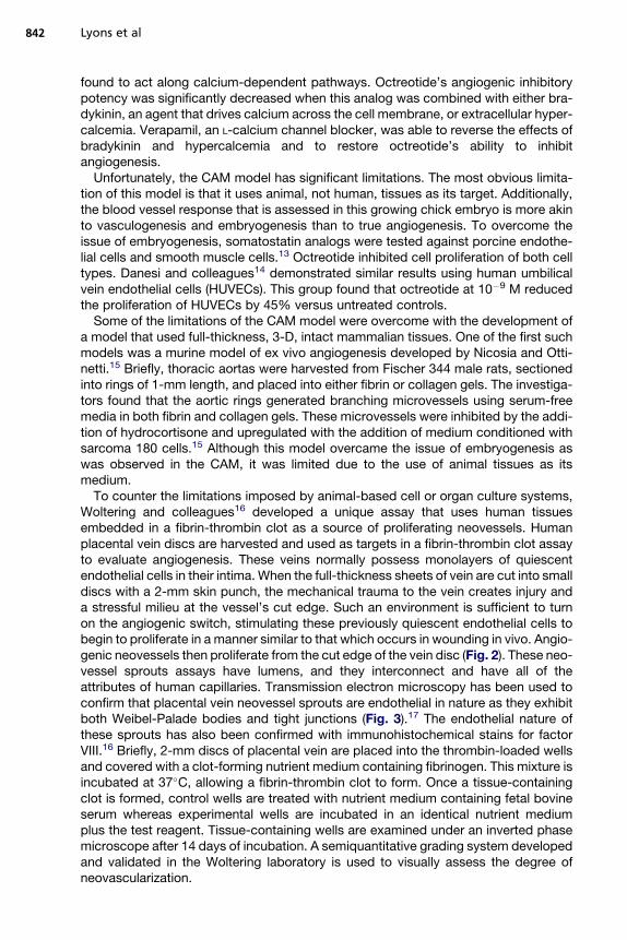
Woltering and colleagues16 developed a unique assay that uses human tissuesembedded in a fibrin-thrombin clot as a source of proliferating neovessels. Humanplacental vein discs are harvested and used as targets in a fibrin-thrombin clot assayto evaluate angiogenesis. These veins normally possess monolayers of quiescentendothelial cells in their intima. When the full-thickness sheets of vein are cut into smalldiscs with a 2-mm skin punch, the mechanical trauma to the vein creates injury anda stressful milieu at the vessel’s cut edge. Such an environment is sufficient to turnon the angiogenic switch, stimulating these previously quiescent endothelial cells tobegin to proliferate in a manner similar to that which occurs in wounding in vivo. Angio-genic neovessels then proliferate from the cut edge of the vein disc (Fig. 2). These neo-vessel sprouts assays have lumens, and they interconnect and have all of theattributes of human capillaries. Transmission electron microscopy has been used toconfirm that placental vein neovessel sprouts are endothelial in nature as they exhibitboth Weibel-Palade bodies and tight junctions (Fig. 3).17 The endothelial nature ofthese sprouts has also been confirmed with immunohistochemical stains for factorVIII.16 Briefly, 2-mm discs of placental vein are placed into the thrombin-loaded wellsand covered with a clot-forming nutrient medium containing fibrinogen. This mixture isincubated at 37�C, allowing a fibrin-thrombin clot to form. Once a tissue-containingclot is formed, control wells are treated with nutrient medium containing fetal bovineserum whereas experimental wells are incubated in an identical nutrient mediumplus the test reagent. Tissue-containing wells are examined under an inverted phasemicroscope after 14 days of incubation. A semiquantitative grading system developedand validated in the Woltering laboratory is used to visually assess the degree ofneovascularization.

Fig. 2. Evaluation of angiogenesis. Tissue disks were divided into four quadrants with eachquadrant given a numeric score from 0 to 4 based on neovessel length, density, andpercentage of the quadrant’s circumference involved with the angiogenic response.Numeric results from the four quadrants were summed and expressed as a semiquantativeAI (AI, 0–16). Panels A–D depict tissue disks with quadrant AI values of 1–4, respectively.(From Stafford SJ, Schwimer J, Anthony CT, et al. Colchicine and 2-methoxyestradiol inhibithuman angiogenesis. J Surg Res 2005;125(1):104–8; with permission.)
Angiogenesis and NETS 843
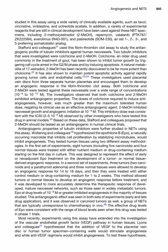
The knowledge that a somatostatin analog’s antiangiogenic effectiveness is directlyproportional to its sst 2–binding affinity11 led Watson and colleagues18 to hypothesizethat proliferating human vascular endothelial cells express sst 2 whereas quiescentones do not. To test this hypothesis, they used human tissues in the previouslydescribed fibrin-thrombin clot assay. They embedded placental vein discs from sixanonymous donors into fibrin-thrombin clots and assessed their angiogenic responseon day 15. Those discs that demonstrated endothelial cell growth from the cut edge ofthe disc were deemed to be proliferating and those viable discs that lacked endothelialcell growth were deemed to have remained quiescent. These investigators demon-strated for the first time that sst 2 gene expression was universally present in prolifer-ating vascular endothelium but sst 2 expression was uniquely absent in quiescentendothelium derived from tissue-matched placental vein samples.To confirm these reverse transcriptase–polymerase chain reaction (RT-PCR)
results, immunohistochemical staining using anti–sst 2 antiserum was performed inproliferating and nonproliferating discs. These selective stains revealed that prolifer-ating endothelium stained positive for sst 2 receptors whereas quiescent endotheliumdid not. These observations were extended into an animal model. Nude mice wereimplanted with neuroblastoma tumor cells that lacked the sst 2 receptor as measuredby RT-PCR and in vitro assays. When the tumors were approximately 2 cm, mice wereinjected with 125I-WOC4a, a radiolabeled, sst 2–preferring somatostatin analog.Nuclear medicine scans and radiographs were performed in register. Significant

Fig. 3. Electron microscopic examination of microvessel outgrowth from the cut edge ofa human placental vein disc. Capillary cross-sections: proximal (original magnification�4200), mid (original magnification �2000), and distal (original magnification �2700).(FromWatson JC, Redmann JG, Meyers MO, et al. Breast cancer increases initiation of angio-genesis without accelerating neovessel growth rate. Surgery 1997;122:509–14; withpermission.)
Lyons et al844
uptake was observed in the tumor whereas non–tumor-bearing sites did not accumu-late radioligand (except for the liver, which was the normal route of excretion for theradiolabeled peptide). The binding seen in the tumors was thought to be the resultof sst 2 receptor expression on angiogenesis blood vessels supplying the tumor.An additional advantage of the fibrin-thrombin clot assay is its ability to test human
tumor specimens against a variety of antiangiogenic agents over a wide range ofconcentrations. Similar to the method used to plate vein discs, fresh human tumorsharvested at operation can be made into 1-mm3 fragments and embedded in thefibrin-thrombin clots.19 As neovessels sprout from the tumor’s cut edge, the angio-genic response can then be quantified by the percent of specimens that begin togrow (percent initiation) or the degree of neovessel growth (angiogenic index [AI]) orthe overall effect of the drug (overall drug effect). Unlike vein discs, which possessonly resting endothelial cells, tumor fragments harbor actively proliferating endothelialcells in their existing angiogenic vessels. This tumor-based fibrin-thrombin clot assaywas used to further investigate the presence of sst 2 receptors on angiogenic bloodvessels.20 Two different tumor types, one whose tumor cells were sst 2 positive andthe other whose tumor cells were sst 2 negative, were implanted in the fibrin-thrombinclot assay. Both were allowed to develop an angiogenic response (presumably sst 2positive). Then both tumor models were treated with an sst 2–favoring radiolabeledsomatostatin analog. Tumoricidal effects were seen only in the sst 2–positive tumorcells, whereas antiangiogenic effects were seen in both tumor types. The investigatorsconcluded that although sst 2s were present on the tumor cells of only one of thetumors, sst 2s were present on the neovessels of both tumors.To date, many tumor types have been studied in this fibrin-thrombin clot assay.
These include breast, colon, and ovarian cancers as well as malignant neuroendocrinetumors (NETs). Carcinoid tumors and islet cell tumors (ICTs) have been extensively
Angiogenesis and NETS 845
studied in this assay using a wide variety of clinically available agents, such as taxol,vincristine, vinblastine, and octreotide acetate. In addition, a variety of experimentalreagents that are still in clinical development have been used against these NET spec-imens, including 2-methoxyestradiol (2-MeOH), rapamycin, vatalanib (PTK787/ZK222584), everolimus (RAD-001), and pasireotide (SOM-230), an sst 1-, 2-, 3-, and5-preferring somatostatin analog.Stafford and colleagues21 used this fibrin-thrombin clot assay to study the antian-
giogenic profile of tubulin inhibitors against human neovessels. Two tubulin inhibitorsthat were investigated were colchicine and 2-MeOH. Colchicine, an older drug usedcommonly in the treatment of gout, has been shown to inhibit tumor growth by trig-gering cell cycle arrest in the G2/M phase and by inducing apoptosis. A natural metab-olite of 17-estradiol, 2-MeOH has been recently discovered to be far more potent thancholcicine.22 It has also shown to maintain potent apoptotic activity against rapidlygrowing tumor cells and endothelial cells.23,24 These investigators used placentalvein discs from three separate human placentas and encouraged them to developan angiogenic response in the fibrin-thrombin clot assay. Both colchicine and2-MeOH were tested against these neovessels over a wide range of concentrations(10�6 to 10�12 M). The investigators observed that both colchicine and 2-MeOHexhibited antiangiogenic properties. The doses of colchicine required to induce anti-angiogenesis, however, was much greater than the maximum tolerated humandose, negating its clinical use as an effective antiangiogenic agent. 2-MeOH inhibitedneovessel growth and angiogenic initiation at 10�6 M. This inhibitory dose was consis-tent with the IC50 (2–5 10�6 M) observed by other investigators who have tested thisdrug in animal models.25 Based on these data, Stafford and colleagues proposed that2-MeOH should be tested as an antiangiogenic in human clinical trials.Antiangiogenic properties of tubulin inhibitors were further studied in NETs using
this assay.Woltering and colleagues16 hypothesized the epothilone B (Epo), a naturallyoccurring macrolide that inhibits cell proliferation by stabilizing microtubules, wouldinhibit angiogenesis. They tested human tissues using two different assay methodol-ogies. In the first set of experiments, eight tumors (including five carcinoids) and fournormal tissues were treated with either nutrient medium or drug-containing mediumstarting on the first day in culture. This was designed to represent the effect of earlyor neoadjuvant Epo treatment on the development of a tumor- or normal tissue–derived angiogenic response. In a second set of experiments, three tumors (two carci-noids and a parathyroid adenoma) and three normal tissues were allowed to developan angiogenic response for 14 to 18 days, and then they were treated with eithercontrol medium or drug-containing medium for 1 to 2 weeks. This method allowedtumors or normal tissues to develop an angiogenic response before treatment, andit was developed to more accurately determine the therapeutic response of devel-oped, mature neovessel networks, such as those seen in widely metastatic tumors.Epo at drug levels of 10�8 M or greater inhibited angiogenesis in the majority of tissuesstudied. This antiangiogenic effect was seen in both sets of experiments (early and latedrug application), and it was observed in carcinoid tumors as well, a group of NETsthat are typically unresponsive to chemotherapy in vivo.26 The effective drug levelsof Epo were consistent with the range of blood levels seen when this drug was testedin phase 1 trials.Most recently, experiments using this assay have extended into the investigation
of the vascular endothelial growth factor (VEGF) pathway in human tissues. Lyonsand colleagues27 hypothesized that the addition of VEGF to the placental veindisc or human tumor specimen–containing wells would stimulate angiogenesisand while anti-VEGF regimens would inhibit angiogenesis. To test these hypotheses,

Lyons et al846
discs of human placental veins (physiologic model) and fragments of human tumors(pathologic model) were embedded in fibrin-thrombin clots and treated with eitherVEGFA165 or anti-VEGF pathway reagents. The anti-VEGF drugs included bevaci-zumab, a humanized monoclonal antibody to VEGF; IMC-18F1 and IMC-1121,human monoclonal antibodies directed against VEGF R1 and VEGF R2, respec-tively; and vatalanib (PTK787/ZK222584; PTK/ZK), a tyrosine kinase inhibitor of allthree primary VEGF receptors (VEGF R1, R2, and R3) and several non-VEGF recep-tors, such as platelet-derived growth factor receptor. VEGF was tested against thevein discs of five separate human placentas and the fragments of eight separatemalignant tumors, four of which were carcinoids. To the investigators’ surprise,VEGF did not consistently stimulate neovascularization in human, vein-containingwells or in malignant tumor fragments. These experiments were repeated testingplacental veins and malignant tumors (including carcinoids) with anti-VEGFreagents. Antibodies targeting VEGF R1 and VEGF R2 did not inhibit neovasculari-zation in human tissues. Bevacizumab, although not affecting placental veins orgynecologic tumors, moderately inhibited angiogenesis in a subset of NETS (carci-noid tumors). In contrast, PTK/ZK significantly inhibited angiogenesis in every tissuetype tested at multiple different concentrations. The inhibitory capacity of eachmolecule was proportional to the number of downstream targets that it affected.Antibodies to VEGF R1 and VEGF R2 affected only one target and had no inhibitoryability. Bevacizumab, an antibody that targets the VEGF-A ligand, affects all thereceptors influenced by VEGF-A. This molecule had a modest inhibitory affect,but one that was greater than the antibodies to VEGF R1 and VEGF R2. PTK/ZKdirectly targets three primary VEGF receptors (R1, R2, and R3) as well as PDGFreceptors and other non-VEGF receptors. PTK/ZK’s inhibitory capacity wasprofound across all tissues and tumor types. This led the investigators to concludethat simply stimulating or blocking the VEGF pathway alone does not consistentlyalter neovascularization in human tissues. Manipulation of human neovessels wasmore consistently achieved when multiple growth factor pathways were affectedsimultaneously.The fibrin-thrombin clot angiogenesis assay has been used extensively to study
carcinoid tumors and ICTs. The ability to harvest an individual patient’s tumor andto visually assess the angiogenic response of that tumor to a variety of agents poten-tially offers clinicians several unique opportunities to gauge a tumors’ responsivenessto specific agents or pathways. These assays have the potential to make statementsabout the aggressiveness of a tumor. These assays may also enable clinicians toscreen a variety of potentially usefully medications and to design a customized drugregimen with the best antiangiogenic potential for that individual.Methods to screen the effects of chemotherapeutic agents against tumor cells
have been widely reported for several decades. These assays were first reportedby Hamburger and Salmon,28 who described their in vitro soft agar culture systemin 1977. The ability of these assays to choose effective chemotherapeutic agentswas clinically evaluated when Von Hoff and colleagues29 compared the clinicalresponses of patients who received a single reagent that was either picked by a clini-cian or picked by the assay. Patients who received an assay-guided drug enjoyeda 25% response rate whereas those given a clinician-selected reagent experienceda 15% response rate.29 In contrast to chemosensitivity assays, Kern and Weisenthal30
described a chemoresistance assay that exposes tumor specimens to supra-pharmacologic concentrations of reagents. They have shown that drugs failing tosuppress tumor growth at these extreme concentrations are likely to clinicallyfail 90% of the time. None of these chemosensitivity or chemoresistance assays

0
1
2
3
4
5
6
7
8
9
10
(+) Control
Gallic Acid 1 mM
Sweet Leaf Tea 1 mg/ml
Black Raspberry 1 mg/ml
PTK 20 uM
RAD001 10 nM
SOM230 10 nM
IntronA 2.5 IU/ml
CP Avastin 168 pM
2MeOH 1 uM
Taxol 10 nM
Epo 10 nM
Gleevec 2.5 uM
Vincristine 54 nM
VP-16 2.5 uM
Colchicine 100 pM
5FU 2.5 uM
Treatments
)MES2
±(
xednI
cinegoign
Anae
M
Fig. 4. One human carcinoid liver metastasis was tested against the 17 potential antiangio-genic drugs. Drugs with a lower angiogenic response are the better antiangiogenics in vitro.Avastin, bevacizumab; VP-16, etoposide; Gleevec, imantinib.
Angiogenesis and NETS 847
considers the blood vessel compartment of the tumor growth, however. The abilityof fibrin-thrombin clot assay to assess the effect of therapy on neovessel growthmakes this assay unique.Figs. 4 and 5 outline the types of data that can be obtained from this fibrin-
thrombin clot angiogenesis assay. Fig. 4 illustrates data from one patient witha carcinoid. It outlines the angiogenic response in the fibrin-thrombin clot assaywhen tested with a panel of 17 different drugs. Drugs with a lower angiogenicresponse are the better antiangiogenics in vitro. Based on this data, it can besurmised that the most effective antiangiogenics in this carcinoid patient are gallicacid, sweet leaf tea, taxol, Epo, and vincristine, because they all significantly
Fig. 5. Effect of 1mM gallic acid on overall angiogenic response. Fifty-two different humanNETx were tested against the antiangiogenic drug, gallic acid. These different tumors arerepresented on the X axis as Specimens. Data were arranged in ascending order accordingto the control group’s overall angiogenic response (0–16). Gallic acid consistently inhibitedthe overall angiogenic response, even in those specimens that had a robust control angio-genic response.

Table 2Selected antiangiogenic agents tested in clinical trials
Agent
Carcinoid Noncarcinoid NET
Complete Response Partial Response Stable Disease Complete Response Partial Response Stable Disease
Bevacizumab 1 FOLFOX34 0/5 (0%) 1/5 (20%) 4/5 (80%) 0/7 (0%) 2/7 (29%) 5/9 (71%)
Bevacizumab 1 2methoxyestradiol35 0/31 (0%) 0/31 (0%) 27/31 (93%) NR NR NR
Bevacizumab 1 temozolamide36 0/12 (0%) 0/12 (0%) 11/12 (92%) 0/17 (0%) 4/17 (24%) 12/17 (70%)
Bevacizumab 1 Interferon31 0/22 (0%) 4/22 (18%) 17/22 (77%) NR NR NR
Imantinib37 0/27 (0%) 1/27 (4%) 17/27 (63%) NR NR NR
Imantinib38 0/2 (0%) 0/2 (0%) 1/2 (50%) 0/13 (0%) 0/13 (0%) 2/13 (15%)
Endostatin39 NR NR NR 0/40 (0%)a 0/40 (0%)a 32/40 (80%)a
Vatalanib40 NR NR NR 0/10 (0%)a 0/10 (0%)a 1/10 (10%)a
Atiprimod41 0/23 (0%) 0/23 (0%) 21/23 (91%) NR NR NR
Gefitinib42 0/22 (0%) 0/22 (0%) 14/22 (64%) 0/15 (0%) 0/15 (0%) 2/15 (13%)
Sunitinib33 0/41 (0%) 1/41 (0%) 34/41 (83%) 0/66 (0%) 11/66 (17%) 45/66 (68%)
Sunitinib32 NR NR NR 2/86 (2%) 6/86 (7%) NR
Sorafenib43 0/41 (0%) 4/41 (10%) NR 0/41 (0%) 4/41 (10%) NR
Everolimus 1 temozolamide44 NR NR NR 0/17 (0%) 6/17 (35%) 9/17 (53%)
Everolimus 1 octreotide45 0/30 (0%) 5/30 (17%) 24/30 (80%) 0/30 (0%) 8/30 (27%) 18/30 (60%)
Pasireotide46 0/11 (0%) 0/11 (0%) 9/11 (82%) NR NR NR
Lanreotide47 NR NR NR 0/25 (0%) 1/25 (4%) 7/25 (28%)
Intron A47 NR NR NR 0/27 (0%) 1/27 (4%) 7/27 (26%)
Lanreotide 1 intron A47 NR NR NR 0/28 (0%) 2/28 (7%) 5/28 (18%)
Octreotide48 NR NR NR 0/42 (0%)a 1/42 (2%)a 28/42 (66%)a
5-fluorouracil 1 octreotide49 NR NR NR 0/29 (0%) 7/29 (24%) 20/29 (68%)
Thalidomide 1 temozolamide50 0/14 (0%) 1/14 (7%) NR 1/14 (7%) 6/14 (43%) 19/28 (68%)a
Abbreviation: NR, not reported.a This includes patients with carcinoid and noncarcinoid NETs because there was no clear distinction.
Lyonsetal
848

Angiogenesis and NETS 849
inhibited the overall angiogenic response. Fig. 5 depicts the results of severalpatients (specimens 1–52) whose tumors were tested in vitro with the same antian-giogenic drug, gallic acid. In all 52 patients studied, gallic acid consistently inhibitedneovascularization, even in those tumors with a robust control angiogenic response.Another notable point is that the specimen depicted in Fig. 4 was harvested froma patient’s liver metastasis. It has been occasionally observed that the same drugyields different antiangiogenic responses within the same patient depending onthe harvest location of the specimen. In other words, a patient liver metastasismay significantly respond to gallic acid in vitro whereas his lymph node metastasismay not (data not shown). The potential for such intrapatient variability furtherunderscores the importance of pretreatment drug screening.Several investigators have tested antiangiogenic agents against carcinoids and
other NETs in clinical trials. Table 2 outlines selected clinical data that have beenreported to date. Although complete responses to these reagents have been rare,many patients have experienced disease stabilization and some have enjoyedimproved progression-free survival (PFS). Yao and colleagues31 randomly assigned44 patients with advanced carcinoid to receive 18 weeks of bevacizumab or pegylatedinterferon alfa-2b (intron A). Partial responses were observed in four (18%) patientsreceiving bevacizumab and zero (0%) receiving intron A. Stable and progressivedisease was observed in 17 (77%) patients and 1 (5%) patient receiving bevacizumab,respectively, and 15 (68%) and 6 (27%) patients receiving intron A. The PFS rate after18 weeks was 95% in the bevacizumab arm and 68% in the intron A the arm. Ray-mond and colleagues32 evaluated another anti-VEGF reagent, sunitinib, versusplacebo in patients with pancreatic ICTs. They also observed prolonged PFS inpatients receiving antiangiogenic therapy (median PFS was 11 months after sunitinibvs 5 after placebo). Kulke and colleagues33 evaluated this reagent in patients with bothcarcinoids and ICTs, and they found more objective tumor responses in patients withICTs (16% vs 2%) than in patients with carcinoids. The median time to tumor progres-sion was not significantly different between ICT patients and carcinoid patients (7 vs10 months, respectively) nor was the 1-year survival rate (81% vs 83%, respectively).The optimal drug regimen for patients with NETs has not yet been identified, but
pretreatment in vitro drug screening to assess those drugs that are most likely togenerate antiangiogenesis may hasten the ability to identify the most clinically effica-cious drugs. Further investigations need to be made to clinically validate the angio-genic response observed with the fibrin-thrombin clot assay in vitro (ie, betterunderstanding is needed of how a patient’s in vitro response translates into a clinicaloutcome). Such information could enable avoiding futile medications in the future andcustomizing patients’ therapy to drugs that are most likely to be affective againsta NET.31–50
REFERENCES
1. Folkman J. Role of angiogenesis in tumor growth and metastasis. Semin Oncol2002;29(6 Suppl 16):15–8.
2. Krulich L, Dhariwal AP, McCann SM. Stimulatory and inhibitory effects of purifiedhypothalamic extracts on growth hormone release from rat pituitary in vitro. Endo-crinology 1968;83(4):783–90.
3. Pless J, Bauer W, Briner U, et al. Chemistry and pharmacology of SMS 201–995,a long-acting octapeptide analogue of somatostatin. Scand J GastroenterolSuppl 1986;119:54–64.

Lyons et al850
4. Gorden P, Comi RJ, Maton PN, et al. NIH Conference. Somatostatin and somato-statin analogue (SMS 201–995) in treatment of hormone-secreting tumors of thepituitary and gastrointestinal tract and non-neoplastic diseases of the gut. AnnIntern Med 1989;110(1):35–50.
5. Fassler JA, O’Dorisio TM, Stevens RE, et al. Are somatostatin analogues anti-angiogenic? Clin Res 1988;36:869A.
6. Woltering EA, Barrie R, O’Dorisio TM, et al. Somatostatin analogues inhibit angio-genesis in the chick chorioallantoic membrane. J Surg Res 1991;50(3):245–51.
7. Folkman J. Proceedings: tumor angiogenesis factor. Cancer Res 1974;34(8):2109–13.
8. Crum R, Szabo S, Folkman J. A new class of steroids inhibits angiogenesis in thepresence of heparin or a heparin fragment. Science 1985;230(4732):1375–8.
9. Ingber DE, Madri JA, Folkman J. A possible mechanism for inhibition of angio-genesis by angiostatic steroids: induction of capillary basement membranedissolution. Endocrinology 1986;119(4):1768–75.
10. Barrie R, Woltering EA, Hajarizadeh H, et al. Inhibition of angiogenesis bysomatostatin and somatostatin-like compounds is structurally dependent.J Surg Res 1993;55(4):446–50.
11. WolteringEA,WatsonJC,Alperin-LeaRC, et al. Somatostatin analogs: angiogenesisinhibitors with novel mechanisms of action. Invest New Drugs 1997;15(1):77–86.
12. Patel PC, Barrie R, Hill N, et al. Postreceptor signal transduction mechanismsinvolved in octreotide-induced inhibition of angiogenesis. Surgery 1994;116(6):1148–52.
13. Sharma C, Alperin-Lea RC, Johnson MA, et al. Inhibition of octreotide acetate(o–a) on proliferation of porcine coronary artery smooth muscle, endotheliumand fibroblasts cells [abstract]. FASEB J 1995;9(3).
14. Danesi R, Agen C, Benelli U, et al. Inhibition of experimental angiogenesis by thesomatostatin analogue octreotide acetate (SMS 201–995). Clin Cancer Res 1997;3(2):265–72.
15. Nicosia RF, Ottinetti A. Growth of microvessels in serum-free matrix culture of rataorta. A quantitative assay of angiogenesis in vitro. Lab Invest 1990;63(1):115–22.
16. Woltering EA, Lewis JM, Maxwell PJ 4th, et al. Development of a novel in vitrohuman tissue-based angiogenesis assay to evaluate the effect of antiangiogenicdrugs. Ann Surg 2003;237(6):790–8 [discussion: 798–800].
17. Watson JC, Redmann JG, Meyers MO, et al. Breast cancer increases initiation ofangiogenesis without accelerating neovessel growth rate. Surgery 1997;122:509–14.
18. Watson JC, Balster DA, Gebhardt BM, et al. Growing vascular endothelial cellsexpress somatostatin subtype 2 receptors. Br J Cancer 2001;85(2):266–72.
19. Gulec SA, Woltering EA. A new in vitro assay for human tumor angiogenesis:three-dimensional human tumor angiogenesis assay. Ann Surg Oncol 2004;11(1):99–104.
20. Gulec SA, Drouant GJ, Fuselier J, et al. Antitumor and antiangiogenic effects ofsomatostatin receptor-targeted in situ radiation with 111In-DTPA-JIC 2DL.J Surg Res 2001;97(2):131–7.
21. Stafford SJ, Schwimer J, Anthony CT, et al. Colchicine and 2-methoxyestradiolinhibit human angiogenesis. J Surg Res 2005;125(1):104–8.
22. LaValee TM, Zhan XH, Herbstritt CJ, et al. 2-Methoxyestradiol inhibits proliferationand induces apoptosis independently of estrogen receptors a and b. Cancer Res2002;62(13):3691–7.

Angiogenesis and NETS 851
23. Seegers JC, Lottering ML, Grobler CJS, et al. The mammalian metabolite 2-me-thoxyestradiol affects p53 levels and apoptosis induction in transformed cells butnot normal cells. J Steroid Biochem Mol Biol 1997;62(4):253–67.
24. Fotsis T, Zhang Y, Pepper MS, et al. The endogenous oestrogen metabolite 2-me-thoxyestradiol inhibits angiogenesis and suppresses tumor growth. Nature 1994;368(6468):237–9.
25. Schumacher G, Kataoka M, Roth JA, et al. Potent antitumor activity of 2-methox-yestradiol in human pancreatic cancer cell lines. Clin Cancer Res 1999;5(3):493–9.
26. Kvols LK, Reubi JC. Metastatic carcinoid tumors and the malignant carcinoidsyndrome. Acta Oncol 1993;32:197–201.
27. Lyons JM 3rd, Schwimer JE, Anthony CT, et al. The role of VEGF pathways inhuman physiologic and pathologic angiogenesis. J Surg Res 2010;159(1):517–27.
28. Hamburger AW, Salmon SE. Primary bioassay of human tumor stem cells.Science 1977;197(4302):461–3.
29. Von Hoff DD, Clark GM, Stogdill BJ, et al. Prospective clinical trial of a humantumor cloning system. Cancer Res 1983;43:1926–31.
30. Kern DH, Weisenthal LM. Highly specific prediction of antineoplastic drug resis-tance with an in vitro assay using suprapharmacologic drug exposures. J NatlCancer Inst 1990;82:582.
31. Yao JC, Phan A, Hoff PM, et al. Targeting vascular endothelial growth factor inadvanced carcinoid tumor: a random assignment phase II study of depot octreo-tide with bevacizumab and pegylated interferon alpha-2b. J Clin Oncol 2008;26(8):1316–23.
32. Raymond E, Niccoli-Sire P, Bang Y, et al. Updated results of the phase III trial ofsunitinib (SU) versus placebo (PBO) for treatment of advanced pancreaticneuroendocrine tumors (NET). ASCO Gastrointestinal Cancers Symposium2010 [abstract]. Available at: http://www.asco.org/ASCOv2/Meetings/Abstracts?&vmview5abst_detail_view&confID572&abstractID51797. Accessed April 20,2010.
33. Kulke MH, Lenz HJ, Meropol NJ, et al. Activity of sunitinib in patients withadvanced neuroendocrine tumors. J Clin Oncol 2008;26(20):3403–10.
34. Venook AP, Ko AH, Tempero MA, et al. Phase II trial of FOLFOX plus bevacizu-mab in advanced, progressive neuroendocrine tumors [abstract]. J Clin Oncol2008;26(15S):15545.
35. Kulke M, Chan JA, Meyerhardt JA, et al. Phase II study of 2-methoxyestradiol(2ME2) administered in combination with bevacizumab, in patients (Pts)with advanced carcinoid tumors. ASCO Gastrointestinal Cancers Symposium2008 [abstract]. Available at: http://www.asco.org/ascov2/Meetings/Abstracts?&vmview5abst_detail_view&confID553&abstractID510528. Accessed April 20,2010.
36. Kulke MH, Stuart K, Enzinger PC, et al. Phase II study of temozolomide andthalidomide in patients with metastatic neuroendocrine tumors. J Clin Oncol2006;24(3):401–6.
37. Yao JC, Zhang YX, Rashid A, et al. Clinical and in vitro studies of imatinib inadvanced carcinoid tumors. Clin Cancer Res 2007;13(1):234–40.
38. Gross DJ, Munter G, Bitan M, et al. Israel glivec in solid tumors study group. Therole of imatinib mesylate (Glivec) for treatment of patients with malignant endo-crine tumors positive for c-kit or PDGF-R. Endocr Relat Cancer 2006;13(2):535–40.

Lyons et al852
39. Kulke MH, Bergsland EK, Ryan DP, et al. Phase II study of recombinant humanendostatin in patients with advanced neuroendocrine tumors. J Clin Oncol2006;24(22):3555–61.
40. Anthony L, Chester M, Michael S, et al. Phase II open-label clinical trial of vata-lanib (PTK/ZK) in patients with progressive neuroendocrine cancer [abstract].J Clin Oncol 2008;26(Suppl 20):14624.
41. Sung MW, Kvols L, Wolin E, et al. PhaseII proof-of-concept study of atiprimod inpatients with advanced low-to intermediate-grade neuroendocrine carcinoma[abstract]. J Clin Oncol 2008;26(Suppl 20):4611.
42. Hobday TJ, Holen K, Donehower R, et al. A phase II trial of gefitinib in patients(pts) with progressive metastatic neuroendocrine tumors (NET): a phase IIconsortium (P2C) study [abstract]. J Clin Oncol 2006;24(18S):4043.
43. Hobday TJ, Rubin J, Holen K, et al. MC044h, a phase II trial of sorafenib inpatients (pts) with metastatic neuroendocrine tumors (NET): a phase II consor-tium (P2C) study [abstract]. J Clin Oncol 2007;25(18S):4504.
44. Kulke M, Blaszkowski LS, Zhu AX, et al. PhaseI/II study of everolimus (RAD001)in combination with temozolomide (TMZ) in patients (pts) with advanced pancre-atic neuroendocrine tumors (NET). ASCO Gastrointestinal Cancers Symposium2010 [abstract]. Available at: http://www.asco.org/ASCOv2/Meetings/Abstracts?&vmview5abst_detail_view&confID572&abstractID52350. Accessed April 20,2010.
45. Yao JC, Phan A, Chang DZ, et al. Phase II study of RAD001 (everolimus) anddepot octreotide (sandostatin LAR) in advanced low grade neuroendocrine carci-noma (LGNET) [abstract]. J Clin Oncol 2007;25(18S):4503.
46. Kvols L, Wiedenmann B, Oberg K, et al. Safety and efficacy of pasireotide(SOM230) in patients with metastatic carcinoid tumors refractory or resistant tooctreotide LAR: results of a phase II study [abstract]. J Clin Oncol 2006;24(18S):4082.
47. Faiss S, Pape UF, Bohmig M, et al. International lanreotide and interferon alfastudy group. Prospective, randomized, multicenter trial on the antiproliferativeeffect of lanreotide, interferon alfa, and their combination for therapy of metastaticneuroendocrine gastroenteropancreatic tumors–the international lanreotide andinterferon alfa study group. J Clin Oncol 2003;21(14):2689–96.
48. Rinke A, Muller HH, Schade-Brittinger C, et al. Placebo-controlled, double-blind,prospective, randomized study on the effect of octreotide LAR in the control oftumor growth in patients with metastatic neuroendocrine midgut tumors: a reportfrom the PROMID study group. J Clin Oncol 2009;27(28):4656–63.
49. Brizzi MP, Berruti A, Ferrero A, et al. Continuous 5-fluorouracil infusion plus longacting octreotide in advanced well-differentiated neuroendocrine carcinomas.A phase II trial of the piemonte oncology network. BMC Cancer 2009;9:388.
50. Kulke MH, Stuart K, Enzinger PC, et al. Phase II study of temozolomide andthalidomide in patients with metastatic neuroendocrine tumors. J Clin Oncol2006;24(3):401–6.