the rano criteria - icon plc brochures/imi_rano_critieria... · 3 r•ano basics the revised...
TRANSCRIPT

The RANO Criteria
Table of Contents
RANO Basics/References ..............................................................................................3Image Acquisition ............................................................................................................4Definitions........................................................................................................................5Baseline Algorithm ..........................................................................................................7Target Lesions ................................................................................................................8Follow-Up Visit Algorithm ..............................................................................................10Target Lesion Response Definitions ..............................................................................11Non-Target Lesions ......................................................................................................12Enhancing Non-Target Lesion Response ......................................................................13T2/FLAIR Lesion Response ..........................................................................................14Pseudoresponse ..........................................................................................................15Pseudoprogression ......................................................................................................16From Lesions to Timepoint ............................................................................................17
3
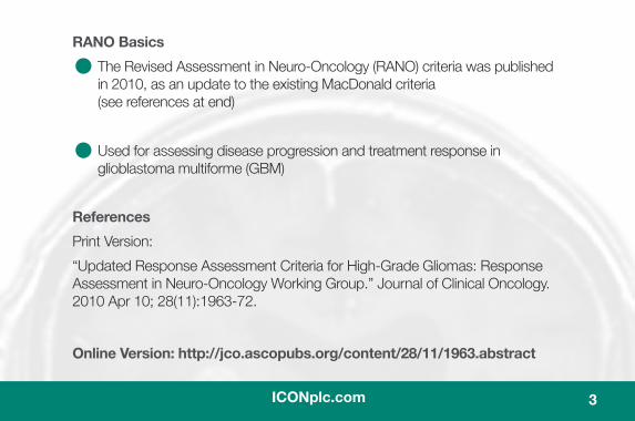
RANO Basics
•The Revised Assessment in Neuro-Oncology (RANO) criteria was publishedin 2010, as an update to the existing MacDonald criteria(see references at end)
•Used for assessing disease progression and treatment response inglioblastoma multiforme (GBM)
ReferencesPrint Version:“Updated Response Assessment Criteria for High-Grade Gliomas: ResponseAssessment in Neuro-Oncology Working Group.” Journal of Clinical Oncology.2010 Apr 10; 28(11):1963-72.
Online Version: http://jco.ascopubs.org/content/28/11/1963.abstract
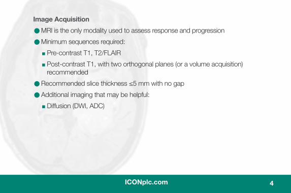
Image Acquisition
•MRI is the only modality used to assess response and progression
•Minimum sequences required:Pre-contrast T1, T2/FLAIRPost-contrast T1, with two orthogonal planes (or a volume acquisition)recommended
•Recommended slice thickness ≤5 mm with no gap
•Additional imaging that may be helpful:Diffusion (DWI, ADC)
4

Definitions
• Measurable lesionsContrast enhancing lesionsMinimum size: two perpendicular diameters ≥10 mm
If slice + gap thickness >5 mm, minimum size is 2 times the totalDo not include cavity, cyst, or necrosis in the measurement
• Non-measurable lesionsLesions that are too small (e.g. 12 x 8 mm)Lesions that do not enhance (seen only on T2/FLAIR)Lesions with a poorly defined margin
5
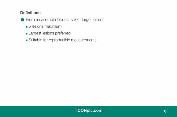
Definitions
• From measurable lesions, select target lesions:5 lesions maximumLargest lesions preferredSuitable for reproducible measurements
6
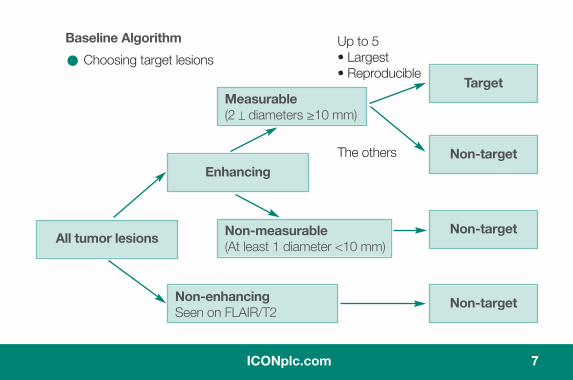
Baseline Algorithm
• Choosing target lesionsUp to 5• Largest• Reproducible
The others
7
Enhancing
All tumor lesions
Non-enhancingSeen on FLAIR/T2
Target
Non-target
Non-target
Non-target
Measurable(2 _ diameters ≥10 mm)
Non-measurable(At least 1 diameter <10 mm)

Target Lesions
• Calculate products of maximal diameters and add them together to yield thesum of products of diameters (SPD)
A1 x B1 + A2 x B2 + … = SPD
8

Follow-Up Visits
9
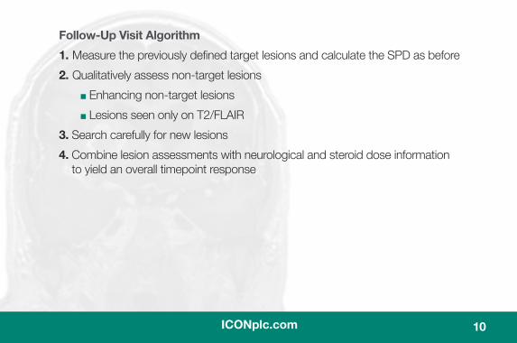
Follow-Up Visit Algorithm1. Measure the previously defined target lesions and calculate the SPD as before2. Qualitatively assess non-target lesions
Enhancing non-target lesionsLesions seen only on T2/FLAIR
3. Search carefully for new lesions4. Combine lesion assessments with neurological and steroid dose information
to yield an overall timepoint response
10
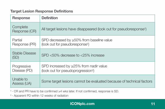
Target Lesion Response Definitions
11
Response Definition
CompleteResponse (CR) All target lesions have disappeared (look out for pseudoresponse†)
PartialResponse (PR)
SPD decreased by ≥50% from baseline value(look out for pseudoresponse†)
Stable Disease(SD) SPD <50% decrease to <25% increase
ProgressiveDisease (PD)
SPD increased by ≥25% from nadir value(look out for pseudoprogression‡)
Unable toAssess (UA) Some target lesions cannot be evaluated because of technical factors
† - CR and PR have to be confirmed ≥4 wks later. If not confirmed, response is SD.‡ - Apparent PD within 12 weeks of radiation
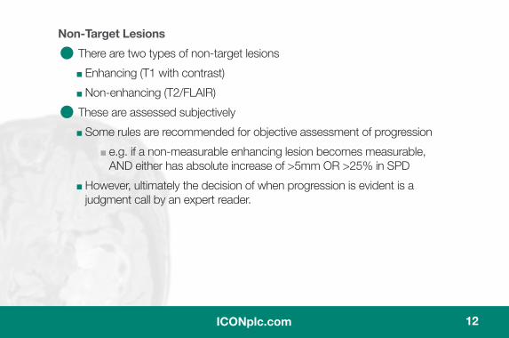
Non-Target Lesions
•There are two types of non-target lesionsEnhancing (T1 with contrast)Non-enhancing (T2/FLAIR)
•These are assessed subjectivelySome rules are recommended for objective assessment of progression
e.g. if a non-measurable enhancing lesion becomes measurable,AND either has absolute increase of >5mm OR >25% in SPD
However, ultimately the decision of when progression is evident is ajudgment call by an expert reader.
12
13
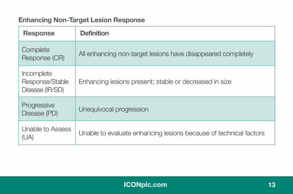
Enhancing Non-Target Lesion Response
Response Definition
CompleteResponse (CR) All enhancing non-target lesions have disappeared completely
IncompleteResponse/StableDisease (IR/SD)
Enhancing lesions present; stable or decreased in size
ProgressiveDisease (PD) Unequivocal progression
Unable to Assess(UA) Unable to evaluate enhancing lesions because of technical factors
14
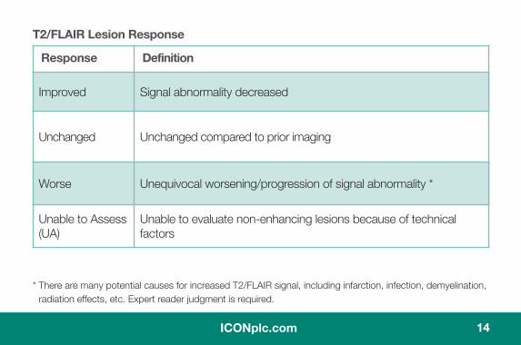
T2/FLAIR Lesion Response
Response Definition
Improved Signal abnormality decreased
Unchanged Unchanged compared to prior imaging
Worse Unequivocal worsening/progression of signal abnormality *
Unable to Assess(UA)
Unable to evaluate non-enhancing lesions because of technicalfactors
* There are many potential causes for increased T2/FLAIR signal, including infarction, infection, demyelination,radiation effects, etc. Expert reader judgment is required.
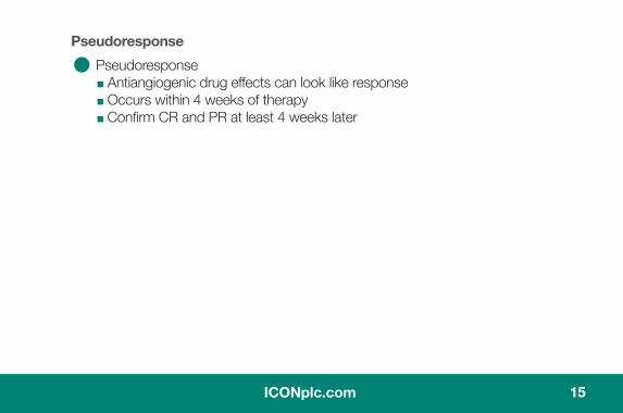
Pseudoresponse
•PseudoresponseAntiangiogenic drug effects can look like responseOccurs within 4 weeks of therapyConfirm CR and PR at least 4 weeks later
15
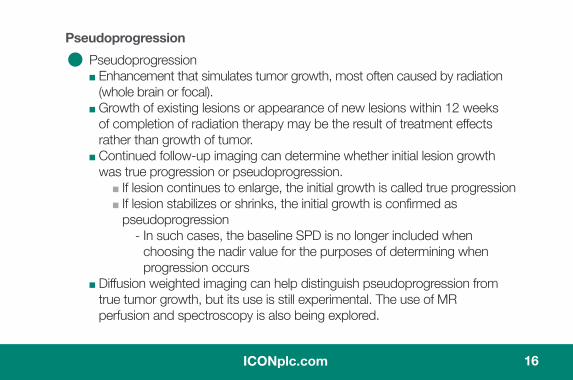
Pseudoprogression
• PseudoprogressionEnhancement that simulates tumor growth, most often caused by radiation(whole brain or focal).Growth of existing lesions or appearance of new lesions within 12 weeksof completion of radiation therapy may be the result of treatment effectsrather than growth of tumor.Continued follow-up imaging can determine whether initial lesion growthwas true progression or pseudoprogression.
If lesion continues to enlarge, the initial growth is called true progressionIf lesion stabilizes or shrinks, the initial growth is confirmed aspseudoprogression
- In such cases, the baseline SPD is no longer included whenchoosing the nadir value for the purposes of determining whenprogression occurs
Diffusion weighted imaging can help distinguish pseudoprogression fromtrue tumor growth, but its use is still experimental. The use of MRperfusion and spectroscopy is also being explored.
16
17
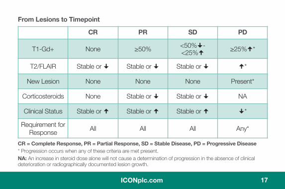
From Lesions to Timepoint
CR = Complete Response, PR = Partial Response, SD = Stable Disease, PD = Progressive Disease* Progression occurs when any of these criteria are met present.NA: An increase in steroid dose alone will not cause a determination of progression in the absence of clinicaldeterioration or radiographically documented lesion growth.
CR PR SD PD
T1-Gd+ None ≥50% <50% -<25% ≥25% *
T2/FLAIR Stable or Stable or Stable or *
New Lesion None None None Present*
Corticosteroids None Stable or Stable or NA
Clinical Status Stable or Stable or Stable or *
Requirement forResponse All All All Any*

To learn more about how ICON Medical Imagingwill benefit your clinical trial, contact us:
ICON US Headquarters2100 Pennbrook Parkway
North Wales, PA 19454, USAT: +1 215 616 3000F: +1 215 699 6288
ICON Medical Imaging2800 Kelly Road, Suite 200Warrington, PA 18976, USA
T: +1 267 482 6300
Email: [email protected]
ICONplc.com