the pterional-transsylvian approach for tumor in the ... · each approach has its advantages and...
TRANSCRIPT

118
INTRODUCTION
Although several surgical approaches to temporal horn tu-mors of the lateral ventricle have been introduced, two surgical routes to the temporal horn are commonly performed. These two approaches are the pterional-transsylvian approach and the subtemporal approach through the occipitotemporal sul-cus. Neurosurgeons are familiar with the pterional-transsylvi-an approach but it is a less-established route for accessing temporal horn tumors. It is also a valid alternative for the treat-ment of intracranial aneurysm and spontaneous intracere-bral hemorrhage. Each approach has its advantages and dis-advantages. The pterional-transsylvian approach enables safe entry into the temporal horn without injuring the optic radi-ation and the uncinate fasciculus [1].
The occipitotemporal sulcus approach is also very effective at visual field preservation. However, it does involve a signifi-cant degree of retraction and can lead to contusions or injury
The Pterional-Transsylvian Approach for Tumor in the Temporal Horn: A Case ReportJung-Hyun Park, Hyok-Rae Cho, Won-Bae Seung, Sung Hun Lee, Yong-Seok ParkDepartment of Neurosurgery, Kosin University Gospel Hospital, Busan, Korea
Received December 29, 2014Revised April 29, 2015Accepted June 9, 2015
CorrespondenceHyok-Rae ChoDepartment of Neurosurgery, Kosin University Gospel Hospital, 262 Gamcheon-ro, Seo-gu, Busan 49267, KoreaTel: +82-51-990-6124Fax: +82-51-990-3005E-mail: [email protected]
A variety of surgical approaches to temporal horn tumors of the lateral ventricle have been described. Magnetic resonance imaging (MRI) and angiography are the preferred modalities for preoperative eval-uation and provide important information for the choice of surgical approach. A 59-year-old man was referred to our hospital due to confusion and gait disturbance. On enhanced MRI, a homogeneous en-hanced solitary mass was observed within the temporal horn of the left lateral ventricle with transepen-dymal extension. The lesion was accompanied by increased hypervascular tumor blush on preopera-tive cerebral angiography. Subtotal removal of the temporal horn tumor was performed because the lesion was identified as lymphoma during surgery. The postoperative course was un-eventful. The pa-tient was referred to the oncology department for conventional chemotherapy. Adjuvant chemotherapy improved the clinical outcome. The pterional-transsylvian approach was beneficial for partial removal of the tumor and tissue diagnosis in this case.
Key Words Lateral ventricle.
to the vein of Labbé [2]. From our experience, the transsylvian approach is an appro-
priate surgical procedure for the diagnosis and management of tumors in the temporal horn.
CASE REPORT
A 59-year-old man presented with a several-day history of confusion and gait disturbance. His past history included an operation for a cerebellar hemangioblastoma at an outside hospital more than 10 years prior to the current presentation. On examination, the patient was confused and had no cranial nerve deficits. He had an unsteady gait. His motor strength was normal.
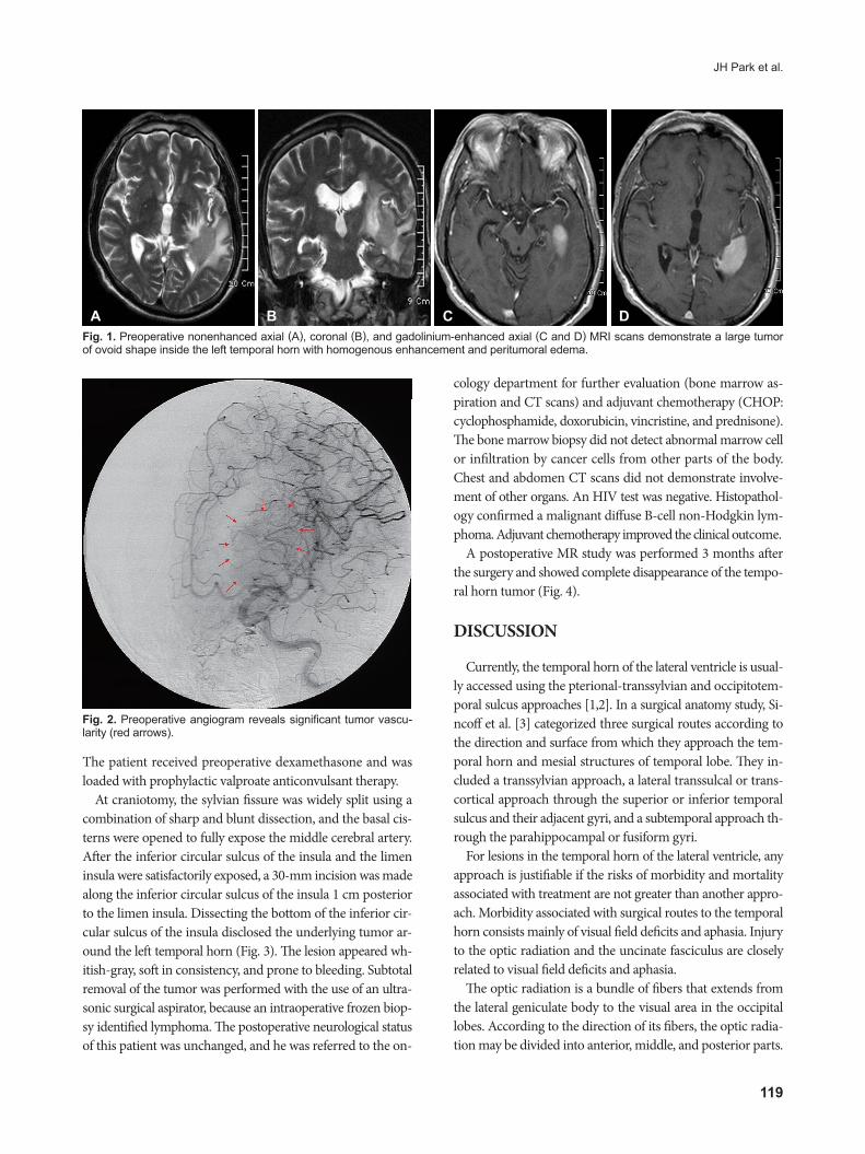
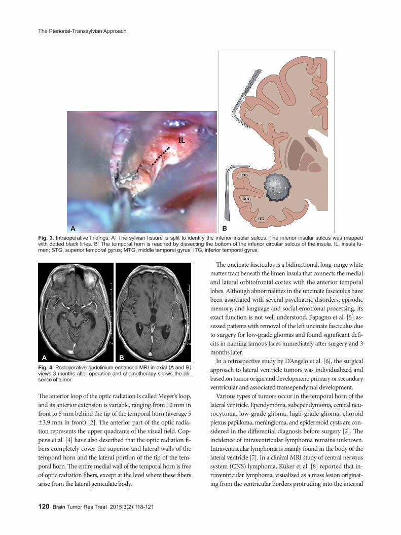
MRI revealed a 3 cm, oval-shaped mass in the left temporal horn on T1-weighted sequences, with homogeneous contrast enhancement after gadolinium administration. T2-weighted imaging highlighted edema surrounding the tumor (Fig. 1). Tumor vascularity was assessed preoperatively using cerebral angiography (Fig. 2). Ependymoma and meningioma were considered in the preoperative differential diagnosis. This pa-tient was not a candidate for stereotactic biopsy due to promi-nent vascular blush of the tumor on preoperative angiography.
CASE REPORT Brain Tumor Res Treat 2015;3(2):118-121 / pISSN 2288-2405 / eISSN 2288-2413http://dx.doi.org/10.14791/btrt.2015.3.2.118
This is an Open Access article distributed under the terms of the Creative Commons Attribution Non-Commercial License (http://creativecommons.org/licenses/by-nc/3.0) which permits unrestricted non-commercial use, distribution, and reproduction in any medium, provided the original work is properly cited.Copyright © 2015 The Korean Brain Tumor Society, The Korean Society for Neuro-Oncology, and The Korean Society for Pediatric Neuro-Oncology
JH Park et al.
119
The patient received preoperative dexamethasone and was loaded with prophylactic valproate anticonvulsant therapy.
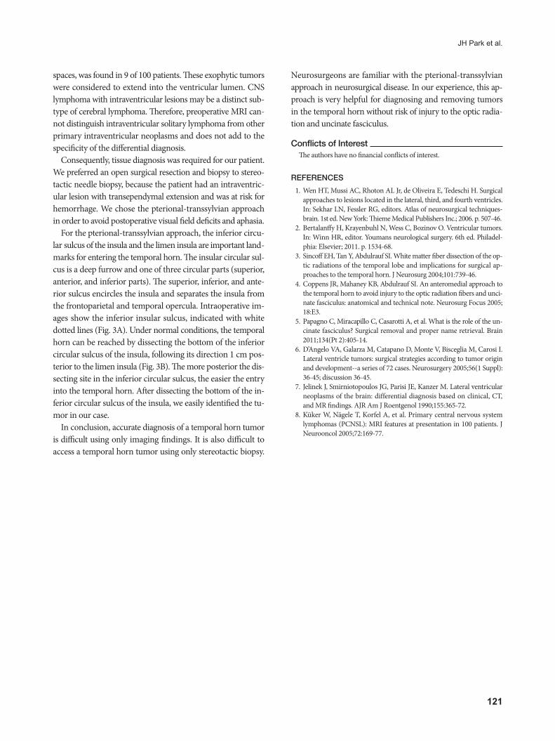
At craniotomy, the sylvian fissure was widely split using a combination of sharp and blunt dissection, and the basal cis-terns were opened to fully expose the middle cerebral artery. After the inferior circular sulcus of the insula and the limen insula were satisfactorily exposed, a 30-mm incision was made along the inferior circular sulcus of the insula 1 cm posterior to the limen insula. Dissecting the bottom of the inferior cir-cular sulcus of the insula disclosed the underlying tumor ar-ound the left temporal horn (Fig. 3). The lesion appeared wh-itish-gray, soft in consistency, and prone to bleeding. Subtotal removal of the tumor was performed with the use of an ultra-sonic surgical aspirator, because an intraoperative frozen biop-sy identified lymphoma. The postoperative neurological status of this patient was unchanged, and he was referred to the on-
cology department for further evaluation (bone marrow as-piration and CT scans) and adjuvant chemotherapy (CHOP: cyclophosphamide, doxorubicin, vincristine, and prednisone). The bone marrow biopsy did not detect abnormal marrow cell or infiltration by cancer cells from other parts of the body. Chest and abdomen CT scans did not demonstrate involve-ment of other organs. An HIV test was negative. Histopathol-ogy confirmed a malignant diffuse B-cell non-Hodgkin lym-phoma. Adjuvant chemotherapy improved the clinical outcome.
A postoperative MR study was performed 3 months after the surgery and showed complete disappearance of the tempo-ral horn tumor (Fig. 4).
DISCUSSION
Currently, the temporal horn of the lateral ventricle is usual-ly accessed using the pterional-transsylvian and occipitotem-poral sulcus approaches [1,2]. In a surgical anatomy study, Si-ncoff et al. [3] categorized three surgical routes according to the direction and surface from which they approach the tem-poral horn and mesial structures of temporal lobe. They in-cluded a transsylvian approach, a lateral transsulcal or trans-cortical approach through the superior or inferior temporal sulcus and their adjacent gyri, and a subtemporal approach th-rough the parahippocampal or fusiform gyri.
For lesions in the temporal horn of the lateral ventricle, any approach is justifiable if the risks of morbidity and mortality associated with treatment are not greater than another appro-ach. Morbidity associated with surgical routes to the temporal horn consists mainly of visual field deficits and aphasia. Injury to the optic radiation and the uncinate fasciculus are closely related to visual field deficits and aphasia.
The optic radiation is a bundle of fibers that extends from the lateral geniculate body to the visual area in the occipital lobes. According to the direction of its fibers, the optic radia-tion may be divided into anterior, middle, and posterior parts.
A B C DFig. 1. Preoperative nonenhanced axial (A), coronal (B), and gadolinium-enhanced axial (C and D) MRI scans demonstrate a large tumor of ovoid shape inside the left temporal horn with homogenous enhancement and peritumoral edema.
Fig. 2. Preoperative angiogram reveals significant tumor vascu-larity (red arrows).
120 Brain Tumor Res Treat 2015;3(2):118-121
The Pterional-Transsylvian Approach
The anterior loop of the optic radiation is called Meyer’s loop, and its anterior extension is variable, ranging from 10 mm in front to 5 mm behind the tip of the temporal horn (average 5 ±3.9 mm in front) [2]. The anterior part of the optic radia-tion represents the upper quadrants of the visual field. Cop-pens et al. [4] have also described that the optic radiation fi-bers completely cover the superior and lateral walls of the temporal horn and the lateral portion of the tip of the tem-poral horn. The entire medial wall of the temporal horn is free of optic radiation fibers, except at the level where these fibers arise from the lateral geniculate body.
The uncinate fasciculus is a bidirectional, long-range white matter tract beneath the limen insula that connects the medial and lateral orbitofrontal cortex with the anterior temporal lobes. Although abnormalities in the uncinate fasciculus have been associated with several psychiatric disorders, episodic memory, and language and social emotional processing, its exact function is not well understood. Papagno et al. [5] as-sessed patients with removal of the left uncinate fasciculus due to surgery for low-grade gliomas and found significant defi-cits in naming famous faces immediately after surgery and 3 months later.
In a retrospective study by D’Angelo et al. [6], the surgical approach to lateral ventricle tumors was individualized and based on tumor origin and development: primary or secondary ventricular and associated transependymal development.
Various types of tumors occur in the temporal horn of the lateral ventricle. Ependymoma, subependymoma, central neu-rocytoma, low-grade glioma, high-grade glioma, choroid plexus papilloma, meningioma, and epidermoid cysts are con-sidered in the differential diagnosis before surgery [2]. The incidence of intraventricular lymphoma remains unknown. Intraventricular lymphoma is mainly found in the body of the lateral ventricle [7]. In a clinical MRI study of central nervous system (CNS) lymphoma, Küker et al. [8] reported that in-traventricular lymphoma, visualized as a mass lesion originat-ing from the ventricular borders protruding into the internal
A BFig. 3. Intraoperative findings. A: The sylvian fissure is split to identify the inferior insular sulcus. The inferior insular sulcus was mapped with dotted black lines. B: The temporal horn is reached by dissecting the bottom of the inferior circular sulcus of the insula. IL, insula lu-men; STG, superior temporal gyrus; MTG, middle temporal gyrus; ITG, inferior temporal gyrus.
Fig. 4. Postoperative gadolinium-enhanced MRI in axial (A and B) views 3 months after operation and chemotherapy shows the ab-sence of tumor.
A B
JH Park et al.
121
spaces, was found in 9 of 100 patients. These exophytic tumors were considered to extend into the ventricular lumen. CNS lymphoma with intraventricular lesions may be a distinct sub-type of cerebral lymphoma. Therefore, preoperative MRI can-not distinguish intraventricular solitary lymphoma from other primary intraventricular neoplasms and does not add to the specificity of the differential diagnosis.
Consequently, tissue diagnosis was required for our patient. We preferred an open surgical resection and biopsy to stereo-tactic needle biopsy, because the patient had an intraventric-ular lesion with transependymal extension and was at risk for hemorrhage. We chose the pterional-transsylvian approach in order to avoid postoperative visual field deficits and aphasia.
For the pterional-transsylvian approach, the inferior circu-lar sulcus of the insula and the limen insula are important land-marks for entering the temporal horn. The insular circular sul-cus is a deep furrow and one of three circular parts (superior, anterior, and inferior parts). The superior, inferior, and ante-rior sulcus encircles the insula and separates the insula from the frontoparietal and temporal opercula. Intraoperative im-ages show the inferior insular sulcus, indicated with white dotted lines (Fig. 3A). Under normal conditions, the temporal horn can be reached by dissecting the bottom of the inferior circular sulcus of the insula, following its direction 1 cm pos-terior to the limen insula (Fig. 3B). The more posterior the dis-secting site in the inferior circular sulcus, the easier the entry into the temporal horn. After dissecting the bottom of the in-ferior circular sulcus of the insula, we easily identified the tu-mor in our case.
In conclusion, accurate diagnosis of a temporal horn tumor is difficult using only imaging findings. It is also difficult to access a temporal horn tumor using only stereotactic biopsy.
Neurosurgeons are familiar with the pterional-transsylvian approach in neurosurgical disease. In our experience, this ap-proach is very helpful for diagnosing and removing tumors in the temporal horn without risk of injury to the optic radia-tion and uncinate fasciculus.
Conflicts of InterestThe authors have no financial conflicts of interest.
REFERENCES
1. Wen HT, Mussi AC, Rhoton AL Jr, de Oliveira E, Tedeschi H. Surgical approaches to lesions located in the lateral, third, and fourth ventricles. In: Sekhar LN, Fessler RG, editors. Atlas of neurosurgical techniques-brain. 1st ed. New York: Thieme Medical Publishers Inc.; 2006. p. 507-46.
2. Bertalanffy H, Krayenbuhl N, Wess C, Bozinov O. Ventricular tumors. In: Winn HR, editor. Youmans neurological surgery. 6th ed. Philadel-phia: Elsevier; 2011. p. 1534-68.
3. Sincoff EH, Tan Y, Abdulrauf SI. White matter fiber dissection of the op-tic radiations of the temporal lobe and implications for surgical ap-proaches to the temporal horn. J Neurosurg 2004;101:739-46.
4. Coppens JR, Mahaney KB, Abdulrauf SI. An anteromedial approach to the temporal horn to avoid injury to the optic radiation fibers and unci-nate fasciculus: anatomical and technical note. Neurosurg Focus 2005; 18:E3.
5. Papagno C, Miracapillo C, Casarotti A, et al. What is the role of the un-cinate fasciculus? Surgical removal and proper name retrieval. Brain 2011;134(Pt 2):405-14.
6. D’Angelo VA, Galarza M, Catapano D, Monte V, Bisceglia M, Carosi I. Lateral ventricle tumors: surgical strategies according to tumor origin and development--a series of 72 cases. Neurosurgery 2005;56(1 Suppl): 36-45; discussion 36-45.
7. Jelinek J, Smirniotopoulos JG, Parisi JE, Kanzer M. Lateral ventricular neoplasms of the brain: differential diagnosis based on clinical, CT, and MR findings. AJR Am J Roentgenol 1990;155:365-72.
8. Küker W, Nägele T, Korfel A, et al. Primary central nervous system lymphomas (PCNSL): MRI features at presentation in 100 patients. J Neurooncol 2005;72:169-77.