the protective effect of orange juice on glyphosate ... · the protective effect of orange juice on...
TRANSCRIPT
www.jocpr.comAvailable online
Journal of Chemical and Pharmaceutical Research, 2016, 8(3):13-28
Research Article ISSN : 0975-7384
CODEN(USA) : JCPRC5
13
The protective effect of orange juice on glyphosate toxicity in adult male mice
Eman R. Youness1, Fatma E. Agha2, Safinaz E. El-Toukhy1, Sabah M. M. El-Naggar3,
Amal A. I. Selim4 and Amr M. M. Ibrahim 1
1Department of Medical Biochemistry, National Research Centre, Cairo, Egypt 2Department of Forensic Medicine and Clinical Toxicology, Faculty of Medicine for Girls, Al- Azhar University,
Cairo, Egypt 3Department of Biology –Ibn Sina National College for Medical Studies, Jeddah
4Department of Biology, Faculty of Applied Sciences, Umm Al-Qura University, Makkah _____________________________________________________________________________________________
ABSTRACT Glyphosate is a broad spectrum herbicide. Fruits and vegetables improve health and a diminish chronic degenerative processes. This study aims to investigate orange juice protective role against the toxicity of glyphosate induced in mice. Fifty six adult male albino mice divided into seven groups. Oral administration of glyphosate caused a significant increase in MDA levels, serum AST, ALT, BUN, creatinine, CEA and DNA damage and decrease in serum testosterone levels. Treatment with orange juice combined with glyophosate for 2 and 4 weeks decrease the incidence of hepatotoxicity, nephrotoxicity, lipid peroxidation, genotoxicity and serum CEA in concomitant with significant elevation in serum testosterone levels compared to glyphosate- treated groups. Results proved that orange juice is a potent protector against glyphosate-induced toxicity, and its protective role is time-dependent.
Key words: Orange juice, glyophosate, lipid peroxidation, biochemical parameters, testicular pathology and mice. _____________________________________________________________________________________________
INTRODUCTION
Humans are exposed daily to many of xenobiotics and their metabolites, which are present as pollutants [1]. They have compensatory, multiplicative, or synergistic effects [2]. Universally, Glyphosate is one of the major pollutants of rivers and surface waters [3]. They have the ability to contaminate organisms, including humans, and ecosystems [4,5 &6]. Liver exposure to glyphosate and metabolism may lead to malondialdehyde elevation and oxidative stress by production of free radicals. They affect integrity of the cells by damage of lipids, proteins and DNA [7 & 8]. The clinical outcomes of ingesting glyphosate herbicides include multi-organ toxicity, with nephrotoxicity, hepatotoxicity, gastrointestinal, cardiovascular effects [9,10 &11]. Death from glyphosate herbicides ingestion develops acute kidney injury. The kidney may also be an organ for excretion of glyphosate components [12]. So, early verification of kidney injury could be important as a risk of a fatal result in glyphosate herbicides toxicity. Plasma or serum creatinine and blood urea nitrogen have usually been used as indicators, or biomarkers, for kidney injury in human glyphosate herbicides intoxication [13 & 14]. There was a decrease in production of sperms to half in experimental animals exposed to glyphosate [15]. McDuffie and his colleagues (2001) revealed that glyphosate causes liver damage in rats, because of intracellular liver

Eman R. Youness et al J. Chem. Pharm. Res., 2016, 8(3):13-28 ______________________________________________________________________________
14
enzymes leakage [16]. There is a relation between glyphosate administration and non-Hodgkin lymphoma risk [17,18&19]. Moreover, it was revealed that glyphosate inhibits RNA transcription [20]. Diets rich in fresh fruit and vegetables decrease incidence of some diseases like cardiovascular and cancer. These protective effects have been associated with compounds naturally present in juices as phenolic compounds, carotenoids and vitamin C [21&22]. Constituents of Orange juice exert various biological effects [23,24&25]. OJ provides considerable amounts of vitamins and minerals that can decrease DNA damage and cancer risk [26&27]. OJ also provides significant amounts of phenolic substances, mainly of flavonoids, that can also have chemopreventive action [28]. A single intake of OJ (600 mL) increases the antioxidant capacity for more than 3 hours [29]. Two-week treatments of healthy persons (up to 500 ml OJ/day ) decrease the levels of lipid peroxidation [30]. Dosages equal or greater than 600 mL daily for more than two weeks have been shown to induce lipid peroxidation decrease and reduction in DNA oxidation [31&32]. This study aims to evaluate the orange juice protective effect against glyphosate induced toxicity in adult male mice.
EXPERIMENTAL SECTION Chemicals Pesticide Glyphosate (N-(phosphonomethyl)glycine),commonly known by its original trade name Roundup™ (manufactured by Monsanto), was obtained from Sigma Chemical Company-St Louis, MO, USA. Orange Juice was supplied by Juhayna and beverages company, Giza, Egypt. Animals Fifty six adult male albino mice aged 8-12 weeks (at the beginning of the work) with an average body weight of 21± 3 gm, obtained from the animal house colony of the National Research Center, Dokki, Cairo, Egypt, were used in the study. The animals were housed in polypropylene cages, given water ad libitum and fed standard pellets diet for two weeks for adaptation. Mice were exposed to a 12:12 light/ dark cycle, at a room temperature of 18-22°C. Treatment The animals were organized into seven groups (8 animals in each group). The animals of group I were used as a control group and received distilled water. The animals of group II & III were treated with orange juice (30 ml/kg) orally by gavages once daily for two and four weeks respectively. The animals of groups IV&V were treated with 500 mg/kg body weight of glyphosate diluted in distilled water orally by gavages once daily for two and four weeks respectively. While The animals of groups VI&VII treated with orange juice combined with glyphosate in a dose which mentioned before. Blood collection and tissue homogenate: Mice were anaesthetized by diethyl ether and samples were taken from retro orbital plexus using glass capillaries. Blood samples were collected at the end of the 2nd and the 4th week of treatment for determining DNA damage using the comet assay. The blood was first centrifuged at 1500 × g for 10 min at ambient temperature. Then serum was separated and used for the assay of aspartate aminotransferase (AST), alanine aminotransferase (ALT), urea, creatinine, total testosterone and carcino-embryonic antigen. The mice were sacrificed by cervical dislocation and the liver and kidney of each animal were dissected and weighted, then immediately homogenized in 50 mM ice-cold phosphate buffer (pH 7.4) to give 20% homogenate (w/v). The homogenate was centrifuged at 1700 rpm and 4°C and the supernatant (20%) was used for the determination of hepatic and renal lipid peroxidation . The study was approved by the ethics committee of the National Research Centre and all subjects gave their informed consent prior to entering this study. II Methods Biochemical analyses: Serum ALT and AST activities were measured by the dinitophenylhydrazene (DNPH) method according to [33] using commercial kits (Biodiagnostic CO., Egypt). Urea and creatinine levels also were measured using commercial kits (Biodiagnostic CO., Egypt). Hepatic and renal lipid peroxidation was assayed by the measurement of malondialdehyde (MDA) by spectrophotometric method [34] using commercial kits (Biodiagnostic reagent kits, Egypt). The level of lipid peroxidation was expressed as nmol/g liver and kidney tissue. Testosterone level was estimated in serum by Elisa (Micro-well method) according to [35]. Serum CEA levels were determined by an enzyme immunoassay test kit (DPC Diagnostic Product Co., Los Angeles, CA, USA) and results were given as ng ⁄ mL. Comet assay

Eman R. Youness et al J. Chem. Pharm. Res., 2016, 8(3):13-28 ______________________________________________________________________________
15
Isolated blood cells of all groups of male mice were subjected to the modified single-cell gel electrophoresis or comet assay [36]. To obtain the cells, the pellet of blood cells was washed with an excess of ice-cold Hank's balanced salt solution (HBSS) and minced quickly into approximately 1 mm3 pieces while immersed in HBSS, with a pair of stainless steel scissors. After several washings with cold phosphate-buffered saline (to remove red blood cells), the blood cells were dispersed into single cells using a pipette. In brief, the protocol for electrophoresis involved embedding of the isolated cells in agarose gel on microscopic slides and lysing them with detergent at high salt concentrations overnight (in the cold). The cells were treated with alkali for 20 min to denature the DNA and subjected to a 30 min electrophoresis under alkaline conditions at 300 ma, 25 V. The slides were stained with ethidium bromide and examined using a fluorescence microscope (Olympus BX60 F-3) with a green filter at 40X magnification lens (N.A.=1.3). For each experimental condition, about 100 cells (about 25 cells per animal) were examined to determine the percentage of cells with DNA damage that appear like comets. The non-overlapping cells were randomly selected and were visually assigned a score on an arbitrary scale of 0–3 (i.e., class 0 = no detectable DNA damage and no tail; class 1 = tail with a length less than the diameter of the nucleus; class 2 = tail with length between 1× and 2× the nuclear diameter; and class 3 = tail longer than 2× the diameter of the nucleus) based on perceived comet tail length migration and relative proportion of DNA in the nucleus [37]. A total damage score for each slide was derived by multiplying the number of cells assigned to each class of damage by the numeric value of the class and summing up the values. Slides were analyzed by one observer to minimize the scoring variability. Histopathological Study: For histological and histochemical studies routine paraffin sections 7mm thick and blood films were prepared and stained by: heamatoxylin and eosin for the histological investigations, PAS periodic acid Schiff for polysaccharides (Drury and wilington1980 ) and bromophenol blue procedure for general proteins [38]. Statistical analysis Data were expressed as means ± S.E. and analyzed statistically using Student’s t test. Results were considered to be statistically significant when P < 0.05.
RESULTS AND DISCUSSION The effect of orange juice and/or glyphosate on (MDA): The changes in MDA levels in the liver and kidney tissues of all the groups after treatment with orange juice and glyphosate are shown in table(1) . There was no significant differences between MDA levels in groups treated with orange juice when compared to their normal counterparts. were observed (P > 0.05). Whereas MDA levels in the liver and kidney tissues of the glyphosate-treated group significantly increased, compared with the control group (P <0.001 ). However, oral administration of orange juice for 2 and 4weeks prior to glyophosate statistically significant decrease the MDA levels in the tissues as compared to glyphosate- treated groups(table 1).
Table(1):Effect of treatment with orange juice combined with glyphosate for 2 and 4 weeks on liver and kidney MDA levels
Kidney malondialdehyde (µmol/g) Liver malondialdehyde (µmol/g) Groups 1.60±0.31a 2.99±0.145a Control 1.59±0.29a 2.591±0.189a Orange juice for 2weeks 1.61±0.4a 2.688±0.201a Orange juice for one month
7.1±0.41***b 18.98±0.91***b Glyophosate for 2weeks 10.1±0.89***b 27.24±0.376***b Glyophosate for 4 weeks
6.1±0.11*c 12.94±0.91***c Orange juice+ Glyophosate for 2weeks 5.6±0.91**c 10.9±0.45***c Orange juice+ Glyophosate for 4 weeks
Data are presented as mean ± SE of the eight animals. * P<0.05,**P<0.01 and***P<0.001 (b) Significantly different from control group.
(c) Significantly different from glyophosate treated group.
Effect of orange juice and/or glyphosate on liver enzymes activities and blood urea creatinine : The activity of AST, ALT, serum creatinine and urea concentrations showed an increase in glyphosate-treated groups for 2 and 4 weeks when compared to control group. This indicates the severity of hepatic and renal injury caused by glyophosate. However, no changes in the activity of liver enzymes, serum creatinine & urea concentrations in mice - treated with orange juice alone for 2 and 4 weeks as compared to the control group. There was a decrease in the activity of the above enzymes(p <0.01) and the levels of the serum creatinine and urea (p <0.001) in the groups treated with orange juice combined with glyophosate for 2 and 4 weeks as compared to glyphosate- treated groups (table 2).

Eman R. Youness et al J. Chem. Pharm. Res., 2016, 8(3):13-28 ______________________________________________________________________________
16
Table(2):Effect of treatment with orange juice combined with glyphosate for 2 and 4 weeks on serum alanine aminotransferase, aspartate aminotransferase, blood urea nitrogen and serum creatinine levels
BUN
(mg/dl) Serum creatinine
(mg/dl) ALP
(IU/L) AST
(IU/L) Groups
40.1±1.8 a 0.82±0.024a 36.0±0.54a 54.8±0.86a Control 41.3±1.7 a 0.81±0.047 a 36.8±0.40a 53.4±1.08a Orange juice for 2weeks 39.8±1.9 a 0.83±0.043 a 35.6±0.4a 52.6±0.51a Orange juice for one month
77.0±2.24***b 1.43±0.034***b 211.2±2.9***b 304.6±2.4***b Glyophosate for 2weeks 80.1±2.1***b 1.48±0.011***b 221.6±3.03***b 355.4±4.2***b Glyophosate for 4 week 66.1±1.3**c 1.18±0.06**c 195.8±3.1**c 285.2±3.32**c Orange juice+ Glyophosate for 2weeks
60.3±1.49***c 1.06±0.04***c 191.0±2.2***c 270.0±4.4***c Orange juice+ Glyophosate for 4weeks Data are presented as mean ± SE of the eight animals. **P<0.01 and***P<0.001
(b) Significantly different from control group. (c) Significantly different from glyophosate treated group.
Effect of orange juice and/or glyphosate on serum testosterone levels: The levels of testosterone were decreased by 32.6, 45.3 % in groups treated with glyphosate as compared with control group. Serum testosterone levels were no significantly changed in orange juice-administered mice for 2 and 4weeks compared to control group. A significant increase in testosterone levels were seen in the groups treated with orange juice combined with glyophosate for 2 and 4 weeks as compared to glyphosate- treated groups. The effect of orange juice and glyphosate on this parameter was time-dependent (table3).
Table(3):Effect of treatment with orange juice combined with glyphosate for 2 and 4 weeks on serum total testosterone (ng/dl)
Weeks4 2weeks Groups 154.5±2.9 a 154.5±2.9a Control 151.2±3.1 a 148.9±2.2 a Orange juice
84.5±1.9***b 104.1±1.2***b Glyphosate 129.8±2.4***c 118.9±1.6***c Orange juice+ Glyphosate
Data are presented as mean ± SE of the eight animals. ***P<0.001 (b) Significantly different from control group.
(c) Significantly different from glyophosate treated group.
Effect of orange juice and/or glyphosate on DNA damage using comet assay: There was a statistically significant increase (p< 0.001) in DNA dmage in blood cells of mice treated with glyphosate for 2 and 4 weeks as compared to those of control, that clearly indicates a genotoxic effect of glyphosate. While the values of DNA damage were significantly decreased in the groups treated with orange juice combined with glyophosate for 2 and 4 weeks compared to groups treated with glyphosate. The effect of orange juice and glyphosate on this parameter was time-dependent, which indicates the protective effect of orange juice against DNA damage induced by glyphosate (table4).
Table (4):Effect of treatment with orange juice combined with glyphosate for 2 and 4 weeks on DNA damage frequency in mice leucocytes
Weeks4 2weeks No. of animals Groups
1.8±0.75 a 1.8±0.75a 10 Control 1.8±0.441 a 1.8±0.44 a 10 Orange juice .8±0.435***b39 27.8±0.438***b 10 Glyphosate
22.9±0.437***c 4.8±0.436**c2 10 Orange juice+ Glyphosate Data are presented as mean ± SE of the eight animals. **P<0.01 and ***P<0.001
(b) Significantly different from control group. (c) Significantly different from glyophosate treated group.
Effect of orange juice and/or glyphosate on serum carcino-embryonic antigen levels(CEA): As shown in (table5), levels of carcino-embryonic antigen (CEA) in control group were 2.52 and 2.45 ng/mL at 2 and 4 weeks, respectively. The levels of CEA in glyphosate-treated group were significantly increased (7.6 ng/mL, p < 0.001) after 2 weeks of treatment as compared to those of control. There were direct relationship between increasing the time of the treatment for 4weeks and increasing the levels of CEA in group treated with glyphosate (12.4 ng/mL , p < 0.001). However, oral administration of orange juice combined with glyophosate for 2 and 4weeks statistically significant (p < 0.001) decrease the CEA levels as compared to glyphosate- treated groups.
Eman R. Youness et al J. Chem. Pharm. Res., 2016, 8(3):13-28 ______________________________________________________________________________
17
Table(5): Effect of treatment with orange juice combined with glyphosate for 2 and 4 weeks on carcino-embryonic antigen(ng/mL)
Weeks4 2weeks Groups ±0.21a 2.52 ±0.21a 2.52 Control
2.9±0.66a 3.0±0.19a Orange juice 12.4±0.18***b 7.6±0.16***b Glyphosate 4.2±0.081***c 4.16±0.093***c Orange juice+ Glyphosate
Data are presented as mean ± SE of the eight animals. ***P<0.001 (b) Significantly different from control group.
(c) Significantly different from glyophosate treated group.
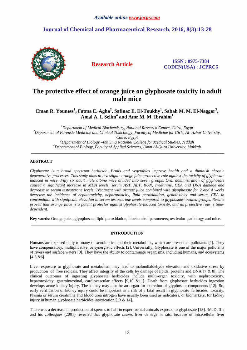
Pathological findings; (A) Light microscopic results: Light microscopic examination of testis of control group showed rounded seminiferous tubules bounded by a fibrous capsule formed of two layers; an inner visceral layer (tunica vasculosa) and tunica albuginea consisted of an outer parietal collagenous layer. The outer layer of the seminiferous tubules appeared enclosed by a fibrous connective tissue consisting of several layers of fibroblasts and well-defined basal lamina (fig 1). The seminiferous tubules was shown to be lined with spermatogenic layer that is formed of spermatogonia appeared relatively small situated next to the basal lamina of the epithelium. Spermatogonia was followed by the primary spermatocytes which is the largest cells of the spermatogenic layer and containing chromosomes in various stages. The spermatids can be distinguished by their small size and their nuclei with areas of condensed chromatin. Large numbers were transformed into spermatozoa released into the lumen of the seminifirous tubule. Sertoli cells are clear as triangular in shape with vesicular nuclei. The space between the tubules contained connective tissue, blood, lymphatic vessels and Leydig cells are arranged into small clumps (fig 1).
Fig. 1: A photomicrograph of a section in the testis of a control mouse showing a seminiferous tubule appeared lined with basal lamina (Bl), spermatogonia (Sg), Sertoli cells (S), peritubular myoid cells (My), primary spermatocytes (SI), secondary spermatocytes (SII),
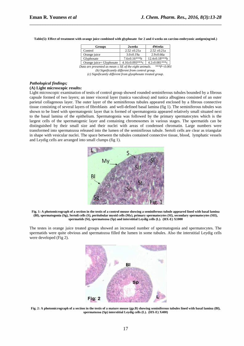
spermatids (St), spermatozoa (Sp) and interstitial Leydig cells (L). (HX-E) X1000 The testes in orange juice treated groups showed an increased number of spermatogonia and spermatocytes. The spermatids were quite obvious and spermatozoa filled the lumen in some tubules. Also the interstitial Leydig cells were developed (Fig 2).
Fig. 2: A photomicrograph of a section in the testis of a mature mouse (gp.B) showing seminiferous tubules lined with basal lamina (Bl), spermatozoa (Sp) interstitial Leydig cells (L). (HX-E) X400)

Eman R. Youness et al J. Chem. Pharm. Res., 2016, 8(3):13-28 ______________________________________________________________________________
18
The testicular sections from glyphosate treated groups revealed some lesions in the seminiferous tubules as shrinking, distortion, acidophilic hyalinization, depletion of the germinal cells, and atresia in the spermatogenic cells (Figs3&4).
Fig. 3: A photomicrograph of a section in the testis of a mature mouse from group (C) showing a seminiferous tubule appeared lined with basal lamina (Bl), primary spermatocytes (SI), secondary spermatocytes (SII), spermatozoa (Sp), vacuolations (V) and Leydig cells (L).
(HX-E) X400
Fig. 4: A photomicrograph of a section in the testis of a mouse showing hyalinization (H) in the lumin of a seminiferous tubule appeared lined with basal lamina (Bl), vacuolations (V) spermatozoa (Sp) and Leydig cells (L). (HX-E) X400
Also, obvious signs of carcinogenation, fragmentation, necrosis and vacuolation in some seminiferous tubules were spotted. Additionally, clear signs of nuclear pyknosis, depletion in the interstitial Leydig cells and loss of sperms was observed (Figs.5, 6 & 7).
Fig. 5: A photomicrograph of a section in the testis of a mature mouse (gp.C) illustrating a seminiferous tubule containing; spermatogonia (Sg), primary spermatocytes (SI), spermatids (St), interstitial Leydig cells (L), interstitial and some vacuolated areas.
(HX-E) X400)

Eman R. Youness et al J. Chem. Pharm. Res., 2016, 8(3):13-28 ______________________________________________________________________________
19
Fig. 6: A photomicrograph of a section in the testis of a mature mouse (gp. C) illustrating a seminiferous tubule appeared lined with basal lamina (Bl), Sertoli cells (S) and containing, secondary spermatocytes (SII), spermatozoa (Sp) interstitial Leydig cells interstitial
Leydig cells (L), some vacuolated areas (V) and a blood capillary (Bc) (HX-E) X1000)
Fig. 7: A photomicrograph of a section in the testis of a mature mouse (gp. C) illustrating a seminiferous tubule appeared containing some vacuolated (V) and necrotic areas (N), primary spermatocytes (SI). (HX-E) X1000)
The testicular sections of mice treated with orange juice combined with glyphosate herbicide showed less defects in the testicular tissue in comparison with those observed in sections of glyphosate treated groups. The shape of some seminiferous tubules appeared distorted, displayed atresia, vacuolation, fragmentation and few necrotic areas (Figs, 8 & 9). Though these defects observed in these groups, yet a marked improvement in most of the seminiferous tubules, increased number of germ cells, the occurrence of different stages of meiotic division and the formation of spermatozoa was observed in sections of testes from orange juice combined with glyphosate treated groups.
Fig. 8: A photomicrograph of a section in the testis of a mature mouse (gp. D) illustrating a seminiferous tubules containing, spermatozoa (Sp), interstitial Leydig cells (L) and a vacuolated area (V). (HX-E) X400)

Eman R. Youness et al J. Chem. Pharm. Res., 2016, 8(3):13-28 ______________________________________________________________________________
20
Fig. 9: A photomicrograph of a section in the testis of a mature mouse (gp. D) illustrating a seminiferous tubule containing, Sertoli cells (S), primary spermatocytes (SI), Sertoli cells (S), secondary spermatocytes (SII) and interstitial Leydig cells (L) (HX-E) X1000
(B) Histochemical Results: PAS periodic acid Schiff for polysaccharides
Fig. 10: A photomicrograph of a section in the testis of a control mouse (gp. A) showing the total polysaccharides contents in the basal lamina (Bl), spermatozoa (Sp), and the walls of blood capillary appeared more intensely stained than the germ cells. (PAS) X 1000
Controversly, stain quality of the interstitial Leydig cells and all spermatogenic cells stages in sections from orange juice treated groups, were nearly equal as the control group (Fig 11).
Fig. 11: A photomicrograph of a section in the testis of a mature mouse (gp. B) showing the total polysaccharides contents, the basal lamina (Bl), spermatozoa (Sp), and blood capillary appeared more intensely stained than the spermatocyte
(PAS) X 1000 The contents of polysaccharides in the testicular tissues of glyphosate-treated groups and orange juice combined with glyphosate treated groups were lower than that of the control (Figs 12 &13 ).

Eman R. Youness et al J. Chem. Pharm. Res., 2016, 8(3):13-28 ______________________________________________________________________________
21
Fig. 12: A photomicrograph of a section in the testis of a mature mouse (gp. C) showing the total polysaccharides contents showing less staining quality than control sections in the basal lamina (Bl),vacuolated (V) and necrotic areas (N), spermatozoa (Sp), Leydig cells (L)
and blood capillary (Bc). (PAS) X 400
Fig. 13: A photomicrograph of a section in the testis of a mouse (gp. D) showing the total polysaccharides contents in the basal lamina (Bl), spermatozoa (Sp), and vacuolation (V). The tissues appeared more intensely stained than the testicular tissues in group (C). (PAS) X
400 Bromophenol blue procedure for general proteins The testicular sections from the control mice stained by the bromophenol blue stain illustrated normal distribution of general proteins (Fig 14).
Fig. 14: A photomicrograph of a section in the testis of a mature mouse (gp.A) showing strong stainability and the interstitial Leydig cells (L). They appeared more intensely stained than the spermatocytes and the Leydig cells. The tails of the spermatozoa (Sp) appeared
darkly stained. (B ph. B)X400 Also, general protein contents are the same in sections from orange juice treated groups and control group. Tunica albuginea, the interstitial tissue and the basal laminae around the seminiferous tubules become stained more intensely while the spermatogenic cells appeared faintly stained (Fig. 15).
Eman R. Youness et al J. Chem. Pharm. Res., 2016, 8(3):13-28 ______________________________________________________________________________
22
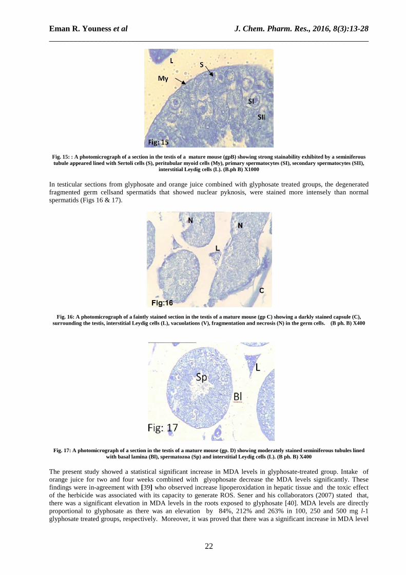
Fig. 15: : A photomicrograph of a section in the testis of a mature mouse (gpB) showing strong stainability exhibited by a seminiferous tubule appeared lined with Sertoli cells (S), peritubular myoid cells (My), primary spermatocytes (SI), secondary spermatocytes (SII),
interstitial Leydig cells (L). (B.ph B) X1000
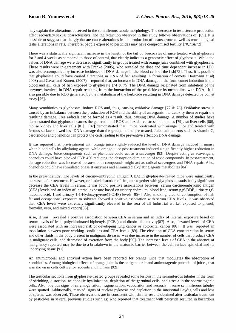
In testicular sections from glyphosate and orange juice combined with glyphosate treated groups, the degenerated fragmented germ cellsand spermatids that showed nuclear pyknosis, were stained more intensely than normal spermatids (Figs 16 & 17).
Fig. 16: A photomicrograph of a faintly stained section in the testis of a mature mouse (gp C) showing a darkly stained capsule (C), surrounding the testis, interstitial Leydig cells (L), vacuolations (V), fragmentation and necrosis (N) in the germ cells. (B ph. B) X400
Fig. 17: A photomicrograph of a section in the testis of a mature mouse (gp. D) showing moderately stained seminiferous tubules lined with basal lamina (Bl), spermatozoa (Sp) and interstitial Leydig cells (L). (B ph. B) X400
The present study showed a statistical significant increase in MDA levels in glyphosate-treated group. Intake of orange juice for two and four weeks combined with glyophosate decrease the MDA levels significantly. These findings were in-agreement with [39] who observed increase lipoperoxidation in hepatic tissue and the toxic effect of the herbicide was associated with its capacity to generate ROS. Sener and his collaborators (2007) stated that, there was a significant elevation in MDA levels in the roots exposed to glyphosate [40]. MDA levels are directly proportional to glyphosate as there was an elevation by 84%, 212% and 263% in 100, 250 and 500 mg l-1 glyphosate treated groups, respectively. Moreover, it was proved that there was a significant increase in MDA level

Eman R. Youness et al J. Chem. Pharm. Res., 2016, 8(3):13-28 ______________________________________________________________________________
23
in plants treated with different doses of glyphosate [41&42]. Additionally, it was revealed that glyphosate has a direct effect on the lipid peroxidation [43]. MDA is the most significant indicator for lipid peroxidation, due to the oxidative decomposition of certain macromolecules such as lipids [44&45]. Since ,membrane’s lipid are highly susceptible to peroxidation, the free radicals easily peroxidized the membranes lipids, thus MDA was produced [46]. It was reported that elevation in MDA levels of both liver and kidney tissues enhanced lipid peroxidation which lead to tissue damage and failure of antioxidant scavenging mechanisms[47&48]. Also it was showed that Ginkgo biloba L. Leaf extract treatment significantly inhibited MDA production, reduce cellular injury and lipid peroxidation that protect the tissues against glyphosate-induced oxidative damage. It was found that, lipoperoxidation can occur through the direct interaction of organophosphorus compounds with the cytoplasmic membrane, leading to damage of the systems[49&50]. This is the principal mechanism associated with the toxicity of several pesticides. The association of Roundup with antioxidants, such as N-acetyl-L-cysteine, vitamins C and E, could reduce toxic damage. It was stated that drinking orange juice enhanced concentration of vitamin C and reduced oxidative stress in vivo through lowering the concentration of F2-isoprostanes, revealing a new evidence of the health benefits of eating fruit[51]. Results of the current study revealed that enzymes activity levels, serum creatinine and urea concentrations were time-dependent statistically significant increase in glyphosate treated groups for 2 and 4 weeks. The activity of the above enzymes and the levels of the serum creatinine and urea were time-dependent statistically significant decrease in the groups treated with orange juice combined with glyophosate. These results were found to be in-accordance with Bishop LM, Bishop LM,et al., who found that the levels of ALT and AST were significantly increased by the exposure to the various concentrations of Roundups during the sampling days[52]. It was revealed that, administration a single intraperitoneal dose of glyphosate- Roundup to albino Swiss mice[53]. After fifteen days, significant hepatic damage will be developed in mice, with changes in serum levels of ALT and AST, as well as altered concentrations of urea and creatinine, indicative of hepatic and renal damage, these alterations could have been related to decreased glutathione level and increased lipoperoxidation. It can be explained by the enzyme leakage from liver cytosol into the bloodstream. Elevations of liver enzymes are the most useful indicators of hepatic dysfunction and hepatocellular damage [54]. As reported by Sanchez-Moreno and his colleagues who found subchronic exposure to glyphosate caused liver damage[55]. Conversely, the increase in the level of AST due to exposure to glyphosate was attenuated due to supplementation with zinc , thus indicating the antioxidant activity of zinc which brought about restoration of the AST activity in rats. Clair revealed that, the effect of glyphosate on liver enzymes activity in Oreochromis niloticus and reported that glyphosate caused significant increase in the their levels[56]. Serum creatinine and blood urea nitrogen (BUN) were used as indicators, or biomarkers, for kidney injury in human glyphosate-surfactant herbicides intoxication. [57&58]. Dallegrave et al showed that creatinine levels increased as early as 8 h after Roundup treatment[59]. Walsh et al revealed serum urea levels were significantly increased after 2 weeks of glyphosate treatment [60]. The level of blood urea is a good indicator for kidney dysfunction. Urea is the principal end product of protein catabolism. Enhanced protein catabolism interpret the elevated levels of urea [61]. It was reported a significant increase in BUN and creatinine levels after exposure pesticides [62 & 63]. There was a statistically significantly decrease in the levels of AST, ALT, BUN, and creatinine after the administration of two different doses of Ginkgo biloba L. Leaf extract together with glyphosate. As the liver is the main organ responsible for elimination of xenobiotics, it is the site of multiple oxidative reactions. As a result the liver tissue shows high antioxidant activity, although such activity does not appear to have been sufficient to avoid the damage promoted by glyphosate [64]. Orange juices have antioxidant and phenolic compounds and vitamin C. However, other antioxidants present in oranges, as vitamin E, carotenoids and minerals as well as the synergistic effects among all antioxidants can increase the total antioxidant potential of the juices [65&66]. Testosterone level were decreased by 32.6, 45.3 % in glyphosate-treated rats for two and four weeks, respectively. While the levels of testosterone were significantly increased in the groups treated with orange juice combined with glyophosate. These results were in-agreement with Guilherme (2012) who showed , lower non toxic concentrations of glyphosate, testosterone decrease by 35% [67]. It was found that, the concentration of testosterone in serum was significantly different among the control and the treated groups, being reduced by 30, 45 and 50% in the groups received 5, 50 and 250 mg/kg, respectively[68]. This
Eman R. Youness et al J. Chem. Pharm. Res., 2016, 8(3):13-28 ______________________________________________________________________________
24
may explain the alterations observed in the somniferous tubule morphology. The decrease in testosterone production affect secondary sexual characteristics. and the reduction observed in this study follows observations of [69]. It is possible to suggest that the glyphosate cause alterations in the production of testosterone as well as morphological testis alterations in rats. Therefore, people exposed to pesticides may have compromised fertility [70,71&72]. There was a statistically significant increase in the length of the tail of leucocytes of mice treated with glyphosate for 2 and 4 weeks as compared to those of control, that clearly indicates a genotoxic effect of glyphosate. While the values of DNA damage were decreased significantly in groups treated with orange juice combined with glyophosate. These results were in-agreement with Franke (2005), who revealed the dose and time dependent increase in LPO was also accompanied by increase incidence of DNA damage in the blood cells of the fish[73]. Thus, it is possible that glyphosate could have caused alterations in DNA of fish resulting in formation of comets. Hartmann et al( 2003) and Cavas and Konen, (2007) reported that, an increase in DNA damage in the form comet induction in the blood and gill cells of fish exposed to glyphosate [74 & 75].The DNA damage originated from inhibition of the enzymes involved in DNA repair resulting from the interaction of the pesticide or its metabolites with DNA. It is also possible due to ROS generated by the metabolism of the herbicide resulting in DNA damage detected by comet assay [76]. Many xenobiotics,as glyphosate, induce ROS and, thus, causing oxidative damage [77 & 78]. Oxidative stress is caused by an imbalance between the production of ROS and the ability of an organism to detoxify them or repair the resulting damage. Free radicals can be formed as a result, thus, causing DNA damage. A number of studies have demonstrated that glyphosate causes the generation of ROS and oxidative stress in tadpoles [79], rat liver cells [80], mouse kidney and liver cells [81]. [82] demonstrated that, mice pre-treated with orange juice and treated with ferrous sulfate showed less DNA damage than the groups not so pre-treated. Juice components such as vitamin C, carotenoids and phenolics can protect the cells leading to the preventive effect on DNA damage. It was reported that, pre-treatment with orange juice slightly reduced the level of DNA damage induced in mouse white blood cells by alkylating agents. while orange juice post-treatment induced a significantly higher reduction in DNA damage. Juice components such as phenolics could act as a scavenger [83]. Despite acting as scavengers, phenolics could have blocked CYP 450 reducing the absorption/elimination of toxic compounds. In post-treatment, damage reduction was increased because both compounds might act as radical scavengers and DNA repair. Also, phenolics could have stimulated phase II enzymes and eliminated alkylating agents metabolites [84]. In the present study, The levels of carcino-embryonic antigen (CEA) in glyphosate-treated mice were significantly increased after treatment. However, oral administration of the juice together with glyophosate statistically significant decrease the CEA levels in serum. It was found positive associations between serum carcinoembryonic antigen (CEA) levels and an index of internal exposure based on urinary cadmium, blood lead, serum p,p'-DDE, urinary t,t'-muconic acid, 1,and urinary 1-1-Hydroxypyrene(1-OHP) levels [85+]. Also smoking, alcohol consumption of fish fat and occupational exposure to solvents showed a positive association with serum CEA levels. It was observed that, CEA levels were extremely significantly elevated in the sera of all Industrial worker exposed to phenol, formalin, urea, and mixed vapors[86]. Also, It was revealed a positive association between CEA in serum and an index of internal exposure based on serum levels of lead, polychlorinated biphenyls (PCBs) and dioxin like activity[87]. Also, elevated levels of CEA were associated with an increased risk of developing lung cancer or colorectal cancer [88]. It was reported an association between poor working conditions and CEA levels [89]. The elevation of CEA concentration in serum and other fluids in the body present in malignant diseases was due increase in the number of cells that produce CEA in malignant cells, and decreased of excretion from the body [90]. The increased levels of CEA in the absence of malignancy reported may be due to a breakdown in the anatomic barrier between the cell surface epithelial and its underlying tissue [91]. An antimicrobial and antiviral action have been reported for orange juice that modulates the absorption of xenobiotics. Among biological effects of orange juice is the antigenotoxic and antimutagenic potential of juices, that was shown in cells culture for rodents and humans [92]. The testicular sections from glyphosate-treated groups revealed some lesions in the seminiferous tubules in the form of shrinking, distortion, acidophilic hyalinization, depletion of the germinal cells, and atresia in the spermatogenic cells. Also, obvious signs of carcinogenation, fragmentation, vacuolation and necrosis in some seminiferous tubules were spotted. Additionally, marked, signs of nuclear pyknosis and depletion in the interstitial Leydig cells and loss of sperms was observed. These observations are in consistent with similar results obtained after testicular treatment by pesticides in several previous studies such as; who reported that treatment with pesticide resulted in hazardous

Eman R. Youness et al J. Chem. Pharm. Res., 2016, 8(3):13-28 ______________________________________________________________________________
25
effects on sperm and semen quality was dose‐dependent. [93] found that Roundup herbicide altered the structure of the testis and epididymal region therefore, Roundup cause disorder in the morphophysiology of male genital system of animals. Exposure the herbicide glyphosate alters testosterone levels, leading to describe the herbicide as “a potent endocrine disruptor [94]. Rayes 2008, investigated that glyphosate-based herbicide induced necrosis and apoptosis in the testicular cells of mature rat in vitro, and testosterone decrease at lower levels [95]. It was found that Roundup and glyphosate at low concentrations produced a decrease in the testosterone level by 35%. exposure to glyphosate during pregnancy disturbed the masculinization process and promoted behavioral and histological changes and endocrine problems in reproductive parameters [96]. Another study, made by [97] on male rats treated by glyphosate with another pesticide, produce oxidative stress in male testes leads to inhibited production of testosterone and disrupted gonadotropin levels. The testicular sections of mice treated with orange juice combined with glyphosate herbicide showed less defects in the testicular tissue in comparison with those observed in sections of glyphosate treated groups. Some seminiferous tubules appeared distorted in shape, displaying atresia, vacuolation, fragmentation and showed few necrotic areas. Though these defects observed in these groups, yet a marked improvement in most of the seminiferous tubules, increased number of germ cells, the occurrence of different stages of meiotic division and the formation of spermatozoa was observed in sections of testes from orange juice combined with glyphosate treated groups. These testicular lesions have been attributed mainly to the toxic effects of the herbicide which was mediated by administration of the orange juice. These results confirm the protective benefits of the regular consumption of orange juice which may be attributed to that orange juice is a rich source of antioxidants; vitamin C, folate, and flavonoids such as hesperidin. Consistent with the results obtained from the present study, Worldwide studies confirmed that use of the herbal medicine has positive effect on treating infertility. Several studies confirmed the protective effects of the phytochemicals included in the orange juice on testicular tissues. [98] investigated that the inhibition effect of cyclosporine to hypothalamic-pituitary-testicular axis of the intact adult rat is completely reversible after administration of orange juice for 4 weeks. It was found that antioxidants including most vitamins in diet may protect sperm DNA from free radicals[85]. A study showed that consumption of orange juice caused increase in sperm viability, motility and population so it has positive effects on male infertility. Evidences suggest certain, cardiovascular protection, neuro-protection, bone health promotion and anti-inflammatory protection. The testicular tissues of polysaccharides contents of glyphosate treated groups and orange juice combined with glyphosate treated groups were less than that in the testicular preparations in the control group . In testicular sections from glyphosate and orange juice combined with glyphosate treated groups stained by the bromophenol blue stain showed degenerated and fragmented germ cells, the degenerating spermatids which showed nuclear pyknosis, were more stained than the normal ones.
CONCLUSION
The results obtained in this study clearly confirmed that glyphosate toxicity induces biochemical changes, oxidative stress, DNA damage and altered the structure of the testis which inhibited and disturbed the process of spermatogenesis in adult male mice. However, supplementation with orange juice has a protective effect against glyphosate toxicity, by reduction effects of free radicals and counteract the oxidative stress through its antioxidant properties. Therefore, the antioxidant role of orange juice may be used as a ‘‘toxicity-limiting agent’’ to reduce effects on human health of pesticides in the near future. Acknowledgement The authors wish to appreciate the department of clinical biochemistry National Research Centre for making use of their facilities to carry out this work.
REFERENCES
[1] VJ Feron; FR Cassee; JP Groten; PW van Vliet; and JA van Zorge, Environ. Health Perspect., 2002, 110: 893–899. [2] N Benachour; H Sipahutar; S Moslemi; C Gasnier; C Travert; and GE Seralini, Environ. Contam. Toxicol., 2007, 53: 126–133.

Eman R. Youness et al J. Chem. Pharm. Res., 2016, 8(3):13-28 ______________________________________________________________________________
26
[3] J Cox, Pest. Reform., 1998,18: 3–17. [4] M Takahashi; M Horie; and N Aoba, Shokuhin Eiseigaku Zasshi., 2001,42: 304–308. [5] JF Acquavella; H Bruce; BH Alexander; JS Mandel; C Gustin; B Baker; P Champan;and M Bleeke,Environ. Health Perspect., 2004,112:321–326. [6] V Contardo-Jara; E Klingelmann; and C Wiegand, Environ. Pollut., 2008, 157: 57–63. [7] E Catalıa; and A Cerruti, Int. J. Biochem. Cell. Biol., 1997, 29: 541–546. [8] CH Lee; CP Shih; KH Hsu; DZ Hung; and Lin CC, Am. J. Emerg. Med.;2008, 26: 275–281. [9] HL Lee; CD Kan; CL Tsai; MJ Liou; and HR Guo, Clin. Toxicol. (Phila.); 2009,47: 651–658. [10] DM Roberts; NA Buckley; F Mohamed; M Eddleston; DA Goldstein; A Mehrsheikh; MS Bleeke; and AH Dawson, Clin. Toxicol. (Phila.); 2010,48: 129–136. [11] P Sribanditmongkol; P Jutavijittum; P Pongraveevongsa; K Wunnapuk; V Durongkadech, Am. J. Forensic Med. Pathol.; 2012, 33:234–237. [12] NS El-Shenawy, Toxicol. Pharmacol.,2009, 28: 379–385. [13] JM Moon; and BJ Chun, Clin. Toxicol. (Phila.) ;2010, 48: 718–724. [14] MI Yousef; K Bertheussen; HZ Ibrahim; S Helmi; MA Seehy and MH Salem,The Journal of Environmental Science and Health Part B; 1996,31: 99-115. [15] A Benedetti; C De Lourdes Vituri; AG Trentin; MAC Dominguesc and Alvarez-Silva M, Toxicology Letters, 2004,153: 227-232. [16] H McDuffie; P Pahwa; JR McLaughlin; JJ Spinelli; S Fincham; JA Dosman; D Robson; LF Skinnider and NW Choi, Cancer Epidemiology Biomarkers and Prevention ; 2001, 10: 1155-1163. [17] L Hardell; M Eriksson and M Nordstrom, Leukemia Lymphoma,2002, 43: 1043–1049. [18] AJ De Roos; SH Zahm; KP Cantor; DD Weisenburger; FF Holmes;Burmeister LF;A Blair,Occup Environ Med ,2003,60:11. [19] J Marc; M Le Breton; P Cormier; J Morales; R Belle and O Mulner-Lorillo, Toxicology and Applied Pharmacology,2005, 203: 1-8. [20 ] V Kabasakalis; D Siopidou, and E Moshatou. Food Chem., 2000, 70: 325-328. [21] B Halliwell, Mutat Res., 2001,475: 29–35. [22 ] SIR Franke; K Ckless; JD Silveira; G Rubensam; and M Brendel Food Chem; 2004, 88: 45–55. [23] AA Franke; RV Cooney; SM Henning; and LJ Custer. 2005a ,53: 5170–5178. [24] SM Vieira; KH Theodoro; and MBA Gloria, Food Chemistry,2007, 100: 895–903. [25] M Fenech and JW Crott, Mutat Res; 2002,504: 131–136. [26] E Middleton; C Kandaswami; and TC Theoharides, Pharmacol Rev; 2000,52: 673–751. [27] H Ghanim; P Mohanty; R Pathak; A Chaudhuri; and CL Sia, Diabetes Care, 2007,30: 1406–1411. [28] C Sanchez-Moreno; MP Cano; B de Ancos; L Plaza; B Olmedilla, J Nutr, 2003,133: 2204–2209. [29] P Riso; F Visioli; C Gardana; S Grande, and Brusamolino J Agric Food Chem., 2005, 53: 941–94. [30] S Retiman; and S. A Frankel, Am. J. clin. Patho.;1957, 28: 56. [31] k Satoh; Clinica Chimica Acta.; 1978, 90: 37. [32] Parker F. In: Text Book of Endocrinology, William, RH. ( Hresg ). W.B. saunders Co., Philadelphia, 1981,1080 – 1098. [33]Mazia, D.,Brewe P. and Affert M. J.Biol. Bull.; 1953,104: 57-64. [34] R Jasper; GO Locatelli; C Pilati; C Locatelli, Interdiscip Toxicol., 2012,5(3): 133–140. [35] K Çavusoglu; Z Yalcin E;Turkmen; K Kürşad Yapar; K Çavusoglu; F Cicek,Journal of Agricultural Sciences;2011, 17:131-142. [36] IG Sergiev; VS Alexieva; SV Ivanov; II Moskova; EN Karanov, Pesticide Biochemistry and Physiology; 2006, 85:139–146. [37] LPE Miteva; SV Ivanov and VS Alexieva, 2010, 57: 131-136. [38] G McLaren; and R Don, ISTA Seed Symposium, Budapest.2004. [39] A Ogutcu; M Uzunhisarcikli; S Kalender; Durak; D Bayrakdar; and Y Kalender, Pestic Biochem Phys ;2006, 6:93–98. [40] G Sener; O Sehirli; A Tozan; A Velioglu-Ovunc; N Gedik; and GZ Omurtag, Food Chem Toxicol ;2007, 45:543–550. [41] D Essiz; L Altintas; and YK Das, Bull Vet Inst Pulawy ,2006,50:585–5. [42] I Altuntas; N Delibas; and R Sutcu, Hum Exp Toxicol ; 2002, 21:681–685. [43] CJ Beuret; F Zirulnik; and MS Gimenez, Reprod Toxicol ;2005,19:501–504. [44] A Hazarika; SN Sarkar; S Hajare; M Kataria; and JK Malik,Toxicol.; 2003,185: 1-8. [45] P Kavitha; and V Rao, Pest. Biochem. Physiol;. 2007,87: 182–188. [46] C Sánchez-Moreno; MP Cano; B de Ancos; L Plaza; B Olmedilla; F Granado; and A Martín, Am J Clin Nutr ; 2003,78:454–60. [47] SJ Gholami-Seyedkolaei ; A Mirvaghefi; H Farahmand ; and AA Kosari, Ecotoxicology and Environmental Safety;2013, 98:135–141.

Eman R. Youness et al J. Chem. Pharm. Res., 2016, 8(3):13-28 ______________________________________________________________________________
27
[48] A Sloss. Aust Prescr., 2009,;32:32–35. [49] W Jiraungkoorskul; ES Upatham; M Kruatrachue; S Sahaphong,Vichasri-Grams,and P Pokethitiyook, Environ Toxicol Water Qual .,2003, 18: 260–267. [50] K Wunnapuk; G Gobe; Z Endre; P Peake; JE Grice; MS Roberts; NA Buckley; and Liu,Toxicology Letters; 225,2014, 192– 200. [51] S Caglar; and D Kolankaya, Toxicol. Pharmacol., 2008,25 (1): 57–62. [52] LM Bishop; PE Fody; and HL Schoe, 5th ed. Lippincott Williams and Wilkins, Philadelphia/Hong Kong, 2005, 220-253. [53] M Kerem; N Bedirli; N Gurbuz; O Ekinci; A Bedirli; T Akaya; O Sakrak; and H Pasaoglu, Turk J Med Sci., 2007,37:281–288. [54] AM Attia; and HM Nasr, J Slovak, Anim Sci ; 2009, 42: 87–94. [55] C Sanchez-Moreno; JA Larrauri; and F Saura-Calixto, Food Research International,1999, 32: 407-412. [56] E Clair; R Mesnage; C Travert; and G Séralini , A Toxicology in Vitro; 2012, 26: 269–279. [57] RM Romano ; M A Romano ; MM Bernardi ; PV Furtado ; and CA Oliveira, Arch Toxicol; 2010, 84:309–317. [58] AG Oliveira; LF Telles; RA Hess; GAB Mahecha; and CA Oliveira, Reprod Toxicol ; 2007, 23:182–191. [59] E Dallegrave; FD Mantese; RT Oliveira; Andrade;Dalsenter PR, Arch Toxicol; 2007, 81(9):665- 673. [60] LP Walsh; C McCormick; C Martin; and DM Stocco, Environ Health Perspect; 2000, 108:769–776. [61] M Clementi; GM Tiboni;R Causin;CL Rocca;F Maranghi; F RaVagnato, Tenconi R Reprod Toxicol;2008, 26:13-18. [62] WG Foster; MS Neal; MS Han; and MM Dominguez, J Toxicol Environ Health; 2008, 11:162–176. [63] N Roeleveld; and R Bretveld, The impact of pesticides on male fertility. Curr Opin Obstet Gynecol; 2008, 20:229–233. [64] CD Nwani; NS Nagpure; R Kumar; B Kushwaha; and WS Lakra, DNA Environmental toxicology and pharmacology; 2013, 36: 539–547. [65] DGM Cavalcante; CBR Martinez; and SH Sofia, Mutat. Res; 2008,655: 41–46. [66] T Cavas; and S Konen, Mutagenesis; 2007,22: 263–268. [67] S Guilherme; MA Santos; C Barroso; I Gaivo; and M Pacheco, Ecotoxicology; 2012,21: 1381–1390. [68] S Parvez; S Raisuddin, Environ.Toxicol. Pharmacol ;2005, 20: 112–117. [69] ME Goetz; and A Luch, Cancer Lett., 2008,266: 73–83. [70] MJ Costa; Monteiro; AL Oliveira-Neto; FT Rantin; and AL Kalinin, Ecotoxicology; 2008,17: 153–163. [71] C Bolognesi; S Bonatti; P Degan; E Gallerani; Peluso M; R Rabboni; P Roggieri, and A Abbondandolo, J. Agric. Food Chem., 1997, 45: 1957-1962. [72] SIR Franke; D Pra; R Giulian; JF Dias; ML Yoneama; J da Silva; B Erdtmann and JAP Henriques, Food and Chemical Toxicology;2006, 44: 425–435. [73] SIR Franke; D Pra; B Erdtmann; JAP Henriques and J da Silva, Mutagenesis; 2005b, 20: 279–283. [74] Hartmann A, Agurell E, Beevers C, Brendler-Schwaab S, Burlinson B. Clay P, Collins A, Smith A, Speit G, Thybaud V, and Tice RR. 4th International Comet Assay Workshop. Mutagenesis;2003, 18: 45–51. [75] Cavas T, and Konen S. 2007: Mutagenesis; 22:263–268. [76] El Far M, El Naggar M, and Elkhawaga OAY. Carcinoembryonic Inhalation Toxicology; 2007,18:1041–1046. [77] van Larebeke NA, Bracke ME, Nelen V, Koppen G, Schoeters G, Van Loon H, and Vlietinck R. Environ Health Perspect ; 2006,114:887-892. [78] Palmqvist R, Engaras B, Lindmark G, Hallmans G, Tavelin B, Nilsson O, Hammarstrom S, and Hafstrom L. Dis Colon Rectum ;2003, 46:1538-1544. [79] Herbeth B, and Bagrel A. A Oncodev Biol Med; 1980,1:191-198. [80] Sawabata N, Maeda H, Yokota S, Takeda, Koma SI, Tokunaga MT, and Ito M. Cancer;2004, 101(4):803–809. [81] Harison S. Principles of internal medicine, 12th ed., 1991, 11– 1322. [82] Franke SIR , Guecheva TN, Henriques JAP and Prá D. Nutrition and Cancer; 2013, 65(7) :943-953. [83] Astiz M, Catalfo GEH , García MN , Galletti SM, Errecalde AI, de Alaniz MJT, and Marra CA. Ecotoxicol Environ Saf. ; 2013,91:129-38. [84] Romano MR, Romano RM, Santos LD, Wisniewski P, Campos DA, de Souza PB, Viau P, Bernardi MM, Nunes MT, de Oliveira CA. Arch Toxicol. Nov 26. Epub. PMID:2011, 22120950. [85] Sikka SC, Koyle MA, Swerdloff RS, and Rajfer J. Transplantation; 1988,45(4):784-7. [86] Narra VR, Harapanhalli RS, Howell RW, Sastry KSR, and Rao DV. Radiation Research; 137( 3) :1994, 394-399. [87] Yang HS, Han DK, Kim JR and Sim JC. J Korean Med Sci.; 2006,21:445-451. [88] Khaki A, Fathiazad F, Nouri M, Khaki AA, Jabbari-kh H and Hammadeh M. lia Morphologica.; 2009,68(1): 45-51. [89] Ghanim H, Sia CL, Upadhyay M , Korzeniewski K, Viswanathan P, Abuaysheh S, Mohanty P and Dandona P. Am J Clin Nutr.;2010, 91:940–9.

Eman R. Youness et al J. Chem. Pharm. Res., 2016, 8(3):13-28 ______________________________________________________________________________
28
[90] Fathiazad F, khak A, Nouri M and Khaki A.A. IJWHR.; 2013, 1(1): 29-35. [91]Atessahin A, Bucak MN, Tuncer PB and Kizil M. Res.; 2008,77:38-44. [92] Dkhil MAl, Quraishy SA, and Abdel Moneim AE. Pakistan J. Zool.; 2013,45(5): 1343-5. [93] Abd El-Magid S, El-Gharib R, and Kawkab A. J. Egypt. Ger.Soc.Zool. (45c) Histology and Histochemistry 2004,1-13. [94] Yousif JMA. A thesis of (Ph.D) in science Girl's collage of Education, Jeddah-K.S.A. 2005. [95] Rayes AAH. Journal of Applied Sciences Research;2008, 4(7): 803-813. [96] Rayes AAH, El-Naggar MMS, and Mehanna SHM. J.AL Azhar Medical Faculty; 2008,27 (2): 2028-2046. [97] Mutawie HH and Hegazi AM. The Journal of American Science;7(10): 2011, 479-488. [98] Mutawie HH and El-Naggar SMM. The Life Science Journal; 2013,10(4): 2474- 82.