the production of antibodies to coumarin...
TRANSCRIPT

THE PRODUCTION OF ANTIBODIES TO COUMARIN AND ITS
MAJOR HUMAN METABOLITES
A thesis submitted for the degree of Ph.D.
by
Anthony Killard B.A. (Mod).
July 1998
Based on research carried out at
School of Biological Sciences
Dublin City University
Dublin 9
Ireland
Under the supervision of
Prof. Richard O’Kennedy

To Mum, Dad, Jackie and Kieran.
"An expert is a man who has made all the mistakes which can be made in a very
narrow field."
Niels Henrik Bohr
"I think I must be an expert by now."
Anthony J. Killard
ii

I hereby certify that this material, which I now submit for assessment on the programme
of study leading to the award of Ph.D. is entirely my own work and has not been taken
from the work of others save and to the extent that such work has been cited and
acknowledged within the text of my own work.
iii

ACKNOWLEDGMENTS
Although the previous page states that this work is all my own, no Ph.D. would be
possible without the help and support of so many people; family, friends and colleagues.
Many thanks to Richard for his patience and constant positive attitude. Thanks to my
family for giving me all the support I needed to get through this. To all the lab gang, past
(especially Mary, Teresa, Rob and Declan), and present (Stephen, Paul, Gary, Ciaran,
Deirdre, Brian and John). You have all been an inspiration. Special thanks to Mike who
helped me out of a few difficult patches. Also to the technical staff for putting up with
my all too frequent visits for so long. Thank you everybody.
iv

CONTENTS
Publications and Presentations xvi
Abstract xviii
Abbreviations and symbols xix
1.0 INTRODUCTION 1
1.1 THE IMMUNE SYSTEM 1
1.1.1 The Lymphoid system 1
1.1.2 The humoral immune response 3
1.2 ANTIBODY STRUCTURE 8
1.3 ANTIBODY CHAIN GENE RECOMBINATION 11
1.4 MONOCLONAL ANTIBODY TECHNOLOGY 13
1.5 ANTIBODY ENGINEERING 15
1.6 COMBINATORIAL PHAGE DISPLAY LIBRARIES 21
1.6.1 The pComb3 System 24
1.6.2 The Nissim Library 30
1.6.3 Bacterial surface display 30
1.7 ANTIBODY CONJUGATES AND CHIMAERAS 33
1.8 HUMANISED ANTIBODIES 36
1.9 AFFINITY MATURATION 39
1.10 CATALYTIC ANTIBODIES 40
1.11 ENGINEERED ANTIBODIES IN THE TREATMENT OF DISEASE 44
1.12 INTRACELLULAR IMMUNISATION 45
1.13 BISPECIFIC ANTIBODIES 56
1.13.1 Bispecific antibody therapy in association with
chemotherapeutic drugs 50
1.14 COUMARIN 50
1.14.1 The metabolism and pharmacokinetics of coumarin in man 51
1.14.2 Pharmacological role of coumarin and its metabolites 55
1.14.3 The analysis of coumarin and its metabolites 57
1.14.3.1 Physical methods of coumarin analysis 57
v

1.14.3.2 lmmunoanalytical techniques for coumarin analysis 57
1.14.4 Engineered antibodies for coumarin analysis 59
1.15 AIMS 61
2.0 MATERIALS AND METHODS 62
2.1 MATERIALS 63
2.2 EQUIPMENT 64
2.3 METHODS 66
2.3.1 General protocols 66
2.3.1.1 Culture media formulations 66
2.3.1.2 Standard molecular biology protocols 67
2.3.1.3 Sodium dodecyl sulphate polyacrylamide gel electrophoresis
(SDS-PAGE) 67
2.3.1.4 Coomassie blue staining 67
2.3.1.5 Silver staining 68
2.3.1.6 Western blotting 68
2.3.1.7 Dot blotting 68
2.3.1.8 Bicinchoninic acid (BCA) assay 69
2.3.1.9 Production of anti-VC SMI 3 antibody in rabbit 69
2.3.1.10 Production and purification of anti-c-myc antibody from
mycl-9E10 70
2.3.1.11 Standard enzyme immunoassay protocol using polyclonal
antibodies 70
2.3.1.11.1 Non-competitive enzyme immunoassay 70
2.3.1.11.2 Competitive enzyme immunoassay 71
2.3.1.12 Standard enzyme immunoassay protocol using single chain
Fv antibodies 71
2.3.1.12.1 Non-competitive enzyme immunoassay 71
2.3.1.12.2 Competitive enzyme immunoassay 72
2.3.1.13 Agarose gel electrophoresis 72
vi

2.3.2 Production of murine Fab antibody libraries to coumarin and 7-
bydroxycoumarin 73
2.3.2.1 Production of a coumarin-thyroglobuttn conjugate 73
2.3.2.2 Characterisation of the coumarin-thyroglobulin conjugate 74
2.32.3 Spectrophotometric estimation of drug-protein coupling ratios 74
2.3.2.4 Immunisation of mice with coumarin-thyroglobulin and 7-
hydroxycoumarin-thyroglobulin 74
2.3.2.5 Preparation of mouse spleen messenger RNA 75
2.3.2.6 Reverse transcription of mouse spleen mRNA 76
2.3.2.7 Amplification of antibody light and heavy chain genes using
polymerase chain reaction 77
2.3.2.7.1 PCR Primers 77
2.3.2.7.2 PCR reaction 78
2.3.2.7.3 Purification o f PCR reaction products 79
2.3.2.8 Insertion of antibody heavy or light chain genes into the
pGEM-T plasmid vector 79
2.3.2.9 Preparation of electrocompetent Escherichia coli XLl-Blue cells 79
2.3.2.10 Measurement o f transformation efficiency of
electrocompetent E. coli XLl-Blue cells 80
2.3.2.11 Transformation of E. coli XLl-Blue with pGEM-T plasmid
vector containing light and heavy chain gene inserts 80
2.3.2.12 Isolation of light and heavy chain gene inserts from pGEM-T 81
2.3.2.13 Preparation of pComb3 phagemid vector for insert ligation
of antibody heavy chain genes 81
2.3.2.14 Transformation of E. coli XLl-Blue with pComb3 plasmid
vector containing light and heavy chain gene inserts 82
2.3.3 Production of single chain Fv antibodies to coumarin and 7-
hydroxycoumarin 83
2.3.3.1 The Nissim library 83
2.3.3.2 Growth of the Nissim Library 83
2.3.3.3 Preparation of phagemid particles 83
vii

2.3.3.4 Affinity selection of scFv antibodies using the Nissim library 84
2.3.3.5 Affinity selection of antibodies using BlAcore™ 85
2.3.3.6 Screening of scFv antibodies by Phage ELISA 86
2.3.3.6.1 Preparation o f clones for phage ELISA 86
2.3.3.6.2 Phage ELISA 86
2.3.3.7 Transfer of pHENl to HB2151 for screening of soluble scFv
antibody expression 87
2.3.3.8 Screening of scFv isolates for soluble antibody expression 87
2.3.3.9 Analysis of the cellular distribution ofscFv antibodies 88
2.3.3.10 Large scale scFv antibody production 88
2.3.3.11 Production ofscFv antibody from intracellular compartments 89
2.3.3.12 Large scale phage antibody production 89
2.3.3.13 BlAcore™ kinetic analysis of anti-coumarin scFv antibody
clones 89
2.3.4 The in vitro production of 7-hydroxycoumarin glucuronide 90
2.3.4.1 Preparation of porcine and bovine UDP-glucuronyl transferase 90
2.3.4.2 Initial analysis of the in vitro production of 7-hydroxy coumarin-
glucuronide by HPLC 91
2.3.4.3 Repeat analysis of the in vitro production of 7-hydroxy coumarin-
glucuronide by HPLC 92
2.3.4.4 Analysis of the in vitro production of 7-hydroxy coumarin-
glucuronide by capillary electrophoresis (CE) 94
2.3.5 Purification of 7-hydroxycoumarin-glucuronide 95
2.3.5.1 Chromatographic separation of 7-hydroxycoumarin and 7-
hydroxycoumarin-glucuronide 95
2.3.5.2 Chromatographic purification of 7-hydroxycoumarin-glucuronide
from crude enzymatic reaction mixtures 95
2.3.5.3 Chromatographic purification of 7-hydroxycoumarin-glucuronide
from coumarin-treated urine 95
viii

2.3.5.4 Development of a methodology for the extraction of 7-
hydroxycoumarin-glucuronide from aqueous solutions 96
2.3.5.5 Extraction of 7-hydroxycoumarin-glucuronide from enzymatic
reaction mixtures and coumarin-treated patient urine 96
2.3.5.6 Assessment of the thermal stability of 7-hydroxy coumarin-
glucuronide at 6(fC 96
2.3.5.7 Assessment of the stability of 7-hydroxy coumarin-glucuronide to
a range of different pH conditions 96
2.3.5.8 In vitro enzymatic production of 3-amino-7-hydroxycoumarin-
glucuronide 97
2.3.5.8.1 Preparation of 3-amino-7-hydroxycoumarin 97
2.3.5.8.2 In vitro enzymatic production o f 3-amino-7-
hydroxy coumarin-glucuronide 97
2.3.5.9 Production of a 7-hydroxycoumarin-glucuronide-bovine serum
albumin conjugate 97
3.0 PRODUCTION OF MURINE Fab ANTIBODY LIBRARIES TO
COUMARIN AND 7-HYDROXYCOUMARIN USING THE pComb3
SYSTEM 99
3.1 INTRODUCTION 100
3.1.1 Antibody phage display systems 100
3.1.2 Source and quality of material for library construction 100
3.1.3 Production of hapten-protein conjugates 101
3.1.4 T/A cloning vectors 101
3.2 RESULTS 103
3.2.1 Preparation of coumarin and 7-hydroxycoumarin protein conjugates 103
3.2.2 Production of coumarin-thyroglobulin (coumarin-THG) conjugate 103
3.2.3 Immunisation of mice with coumarin-THG and 7-hydroxycoumarin-
THG 111
ix

3.2.4 Preparation of splenomic RNA 113
3.2.5 Selection of murine PCR primers based on homology plots 113
3.2.6 Murine PCR Primers 114
3.2.7 PCR optimisation 117
3.2.8 Preparation of pComb3 for heavy chain insert ligation 119
3.2.9 Measurement of the Electrocompetence of Escherichia coli XLl-Blue 119
3.2.10 Insertion of mouse 5 heavy chain genes into pGEM-T 119
3.2.11 Insertion of mouse 5 variable light chain genes into pGEM-T 120
3.3 DISCUSSION 124
3.3.1 Preparation of protein conjugates for immunisation 124
3.3.2 Characterisation of coumarin-thyroglobulin (coumarin-THG) 125
3.3.3 Mouse serum antibody titres following immunisation 125
3.3.4 PCR primer production 126
3.3.5 PCR optimisation 127
3.3.6 PCR fragment purification 128
3.3.7 Construction of light and heavy chain gene libraries in pGEM-T 128
3.3.8 Heavy and light chain gene insertion into pComb3 128
4.0 THE PRODUCTION OF SINGLE CHAIN Fv (scFv) ANTIBODIES TO
COUMARIN AND 7-HYDROXYCOUMARIN USING THE N1SSIM
LIBRARY 130
4.1 INTRODUCTION 131
4.1.1 Use of the Nissim library in the isolation of single chain scFv (scFv)
antibodies 131
4.1.2 Affinity selection strategies 131
4.1.3 Affinity selection of antibodies from libraries using BIAcore™ 134
4.1.4 Induction of protein expression from phagemid pHENl 135
4.1.5 Screening of antibody clones from phage display libraries 135
x

4.1.6 Isolation and refolding of scFv antibody protein from insoluble
inclusion bodies 137
4.1.7 Affinity purification of antibodies 138
4.1.8 Composition of affinity matrices 139
4.1.9 Concentration of purified antibodies 139
4.1.10 Immunoassay strategies for the measurement of drug-hapten
concentrations 140
4.1.11 Antibody kinetics and equilibria 143
4.1.12 Analysis of antibody kinetics using BIAcore™ 145
RESULTS 146
4.2 PRODUCTION OF ANTI-COUMARIN ANTIBODIES FROM THE
NISSIM LIBRARY 146
4.2.1 Phage ELISA of anti-coumarin phage antibodies 146
4.2.2 Phage ELISA of anti-coumarin phage antibodies raised against 6-
amino-coumarin coupled via N-oxysuccinimide ester 146
4.2.3 Screening of anti-coumarin positive clones for soluble scFv antibody
production 146
4.2.4 Optimisation of affinity purification of anti-coumarin scFv clones 150
4.2.4.1 Effect of high pH on OrC14 elution from coumarin-BSA 150
4.2.4.2 Batch purification of a -C5 and OrC13 150
4.2.4.3 Optimisation of OrC13 affinity purification 154
4.2.4.4 Comparison of phosphate-buffered saline (PBS) and Tris as
buffers in ELISA of U-C5 scFv 154
4.2.4.5 Purification of a-C5 and oc-C13 using NaCl gradient 158
4.2.4.6 Purification of c/j-C14 using NaCl gradient 158
4.2.4.7 Optimised purification of anti-coumarin scFv antibodies from
culture supernatants and periplasmic lysates 159
4.2.4.8 Modification of scFv ELISA using anti-human F(ab’)2 antibody 159
4.2.5 Characterisation of anti-coumarin scFv antibodies 172
xi

4.2.5.1 Optimisation o f competition ELISA for the detection o f free
coumarin 172
4.2.5.1a Selection of optimal antibody concentration 172
4.2.5.1b Selection of optimal coumarin-BSA coating concentrations 172
4.2.5.1c Competition ELISA analysis o f free coumarin using anti-
coumarin scFv 172
4.2.6 Assessment of the effect of methanol and NaCl concentration on
competition ELISA with a-C5, a-C13 and a-C14 172
4.2.7 The effect of NaCl concentration on anti-coumarin scFv binding to
coumarin-BSA 174
4.2.8 Competition of anti-coumarin-BSA scFv antibodies against coumarin-
BSA 174
4.2.9 Competition of a-C5 scFv with different coumarin-protein conjugates 174
4.2.10 Assessment of the multi-specificity of the anti-coumarin scFv
clones to different coumarin derivative-BSA conjugates 175
4.2.11 Use of anti-coumarin-BSA scFv antibodies as western blot reagents 175
4.2.12 Characterisation and kinetic analysis of anti-coumarin-BSA scFv
antibodies using BIAcore™ 185
4.3 PRODUCTION OF ANTI-7-HYDROXYCOUMARIN ANTIBODIES
FROM THE NISSIM LIBRARY 187
4.3.1 Phage ELISA screening of first anti-7-hydroxycoumarin library 187
4.3.2 Phage ELISA analysis of second anti-7-hydroxycoumarin library 187
4.3.3 Analysis of supernatant and periplasmic lysates of anti-7-
hydroxycoumarin clones for the presence of scFv 187
4.3.4 Use of BIAcore for the affinity selection of anti-7-hydroxycoumarin
clones from the Nissim library 188
4.3.5 Comparison of lysis methods in the isolation of scFv from intracellular
compartments 193
4.3.6 Analysis of denatured and refolded anti-7-hydroxycoumarin clones 193
4.3.7 Induction and affinity purification of (X-70HC10 193

4.3.8 ELISA of purified OC-70HC10 199
4.3.9 Affinity purification of a-70HC9 199
4.3.10 Induction optimisation of (X-70HC10 204
4.3.11 Precipitation of purified and concentrated scFv preparations 204
4.3.12 Characterisation of anti-7-hydroxycoumarin phage antibodies 207
4.3.13 Reselection, screening and isolation of a-7-hydroxy coumarin scFv
clones 207
DISCUSSION 212
4.4 PRODUCTION OF ANTI-COUMARIN scFv ANTIBODIES USING
THE NISSIM LIBRARY 212
4.4.1 Selection of scFv antibodies to coumarin 212
4.4.2 Screening of scFv antibodies to coumarin 212
4.4.3 Purification of scFv antibodies to coumarin 213
4.4.4 Characterisation of scFv antibodies to coumarin 216
4.4.5 Summary 222
4.5 PRODUCTION OF ANTI-7-HYDROXYCOUMARIN scFv
ANTIBODIES USING THE NISSIM LIBRARY 223
4.5.1 Selection and screening of scFv antibodies to 7-hydroxycoumarin 223
4.5.2 Isolation of scFv antibodies to 7-hydroxycoumarin from intracellular
lysates 225
4.5.3 Production of phage antibodies to 7-hydroxycoumarin 227
4.5.4 Affinity selection of scFv antibodies to 7-hydroxycoumarin using
BIAcore 227
4.5.5 Optimised affinity selection and screening of scFv antibodies to 7-
hydroxycoumarin 228
4.5.6 Characterisation of scFv antibodies to 7-hydroxycoumarin 228
xiii

4.6 THEORIES AS TO WHY scFv ANTIBODY PRODUCTION
STRATEGIES DID NOT RESULT IN ANTIBODIES WHICH
SIGNIFICANTLY RECOGNISE FREE DRUG 229
4.7 CONCLUSION 232
5.0 7-HYDROXYCOUMARIN-GLUCURONIDE: PRODUCTION,
PURIFICATION, ANALYSIS AND CONJUGATION FOR THE
PRODUCTION OF POLYCLONAL ANTIBODIES 233
5.1 INTRODUCTION 234
5.1.1 In vitro metabolic studies of 7-hydroxycoumarin 234
5.1.2 The use of HPLC for the analysis of coumarin metabolism 235
5.1.3 Reverse phase chromatography 236
5.1.4 Capillary electrophoresis (CE) 236
5.1.5 Liquid/liquid extraction 237
5.1.6 Conjugation strategies 237
RESULTS 240
5.2 The in vitro production of 7-hydroxycoumarin-glucuronide 240
5.2.1 Initial analysis of the in vitro production of 7-hydroxycoumarin
glucuronide by HPLC 240
5.2.2 Repeat analysis of the in vitro production of 7-hydroxycoumarin-
glucuronide by HPLC 245
5.2.3 Analysis of the in vitro production of 7-hydroxycoumarin-glucuronide
by capillary electrophoresis (CE) 248
5.3 Purification of 7-hydroxycoumarin-glucuronide 251
5.3.1 Separation of 7-hydroxycoumarin and 7-hydroxycoumarin-glucuronide
by reverse phase column chromatography 251
5.3.2 Purification of 7-hydroxycoumarin-glucuronide from crude enzymatic
reaction mixtures by reverse phase column chromatography 255
xiv

5.3.3 Purification of 7-hydroxycoumarin from coumarin-treated patient
urine using reverse phase column chromatography 258
5.3.4 Extraction of 7-hydroxycoumarin-glucuronide from urine in
dichloromethane (DCM)rmethanol 258
5.3.5 Purification of 7-hydroxycoumarin-glucuronide from coumarin-treated
patient urine and enzymatic reaction mixtures by reverse phase
chromatography following extraction in dichloromethane
(DCM): methanol (70:30 v/v) 261
5.4 Characterisation of 7-hydroxycoumarin-glucuronide 264
5.4.1 Thermal stability of 7-hydroxycoumarin-glucuronide at 60°C 264
5.4.2 Stability of 7-hydroxycoumarin-glucuronide with respect to pH 265
5.5 In vitro enzymatic production of 3-amino-7-hydroxycoumarin-
glucuronide 266
5.5.1 Production of 3-amino-7-hydroxycoumarin 266
5.5.2 In vitro enzymatic production of 3-amino-7-hydroxycoumarin-
glucuronide 266
5.6 Production and analysis of a 7-hydroxycoumarin-glucuronide-bovine
serum albumin conjugate prepared by EDC/NHS coupling chemistry 272
DISCUSSION 275
5.8 In vitro production of 7-hydroxycoumarin-glucuronide and analysis by
HPLC 275
5.9 In vitro production of 7-hydroxycoumarin-glucuronide and analysis by
CE 277
5.10 Purification of 7-hydroxycoumarin-glucuronide 278
5.11 Physical analysis of 7-hydroxycoumarin-glucuronide 282
5.12 Conjugate production 282
6.0 OVERALL CONCLUSIONS 286
7.0 REFERENCES 290
APPENDIX Prize winning essays in Biochemistry 319
xv

PUBLICATIONS AND PRESENTATIONS
Killard, A.J., O’Kennedy, R., and Bogan, D.P. (1996). Analysis of the glucuronidation
of 7-hydroxycoumarin by HPLC. J. Pharm. Biomed. Anal., 14:1585-1590.
Bogan, D.P., Killard, A.J., and O’Kennedy, R. (1995). The use of capillary
electrophoresis for studying the in vitro glucuronidation of 7-hydroxycoumarin. J.
Capillary Electrophoresis, 2(5):241-245.
Killard, A.J., Deasey, B., O’Kennedy, R., and Smyth, M.R. (1995). Antibodies:
Production, functions and applications in biosensors. Trends in Analytical Chemistry,
14(6):257-266.
McCormack, T., Keating, G.J., Killard, A.J., and O’Kennedy, R. Biomaterials for
Biosensors in "Sensors and Signals", John Wiley and Sons, New York, 1998.
Roben, P., Killard, A.J., and O’Kennedy, R. Immunoanalytical techniques: Concepts and
applications in "Bioanalysis", Ellis Horwood, In Press.
Killard, A.J., Keating, G.J. and O’Kennedy, R. (1997). Production and characterization
of anti-coumarin scFv antibodies. Biochem. Soc. Trans. 26: S33.
Killard, A., How the antibody army kills what could kill us. The Irish Times, June 3rd
1996, p.2.
Killard, A. PCR and its uses in genetic engineering of antibodies, Modem Techniques in
Molecular Life Sciences. An Occasional Symposium Series for Researchers. Royal Irish
Academy National Committee for Biochemistry, Trinity College, Dublin, May 24th 1995.
xvi

Killard, A.J., Keating, GJ. and O’Kennedy, R , Production and characterization of anti-
coumarin scFv antibodies, Biochemical Society, 663rd Meeting, University College
Galway, 3-4 Sep. 1997.
Daly, S.J., Killard, AJ., Dillon, P.P., Smyth, M.R. and O’Kennedy, R., Single chain
antibodies to 7-hydroxycoumarin and towards a bispecific scFv, Biochemical Society,
663rd Meeting, University College Galway, 3-4 Sep. 1997.
Killard, A.J., Smyth, M.R. and O’Kennedy, R., The production of single chain Fv
antibodies to small, hydrophobic haptens, Biochemical Society Meeting, Dublin City
University, 9-10 Sep. 1998.

ABSTRACT
Two strategies were applied to the production of antibodies to coumarin and its phase I
metabolite, 7-hydroxycoumarin. One system was the production of Fab antibody fragments
using the combinatorial phage display library, employing the phagemid vector, pComb3.
Although a library of murine heavy and light chain genes was established following
immunisations with coumarin and 7-hydroxycoumarin conjugates, preparation of the
library in pComb3 was not possible due to associated problems with this vector.
An alternative antibody library system was obtained. This was the naive semi-synthetic
human scFv library of Nissim et al. (1994). The production of antibodies to both
coumarin and 7-hydroxycoumarin was investigated. Various production and isolation
techniques were explored. Best results were obtained by isolating correctly folded scFv
protein from the periplasm or culture supernatants of E. coli. In the case of coumarin,
three soluble antibody clones were isolated and characterised. These clones showed the
characteristics of poor affinity for free coumarin, but did bind well to coumarin-protein
conjugates. Isolated anti-7-hydroxycoumarin clones also showed the same characteristics
of no binding to free drug. It was theorised that, due to the structural simplicity of these
molecules, affinity selection and screening is biased against the detection of clones with
high affinity for free drug. This conclusion was in broad agreement with other findings
(Danilova, 1994). Recommendations are made as to how the selection and screening
procedures can be improved to isolate antibodies to such molecules.
Techniques for the in vitro production of the phase II metabolite 7-hydroxycoumarin-
glucuronide were developed. These were accompanied by the development of analytical
methods (capillary electrophoresis and HPLC) for the glucuronide. Purification
methodologies using 7-hydroxycoumarin present in these metabolic mixtures and in urine
were also developed. The coupling of 7-hydroxycoumarin-glucuronide to proteins for the
production of antibodies was also explored. A 7-hydroxycoumarin-glucuronide-bovine
serum albumin conjugate was successfully synthesised for use in further antibody
production strategies.

ABBREVIATIONS AND SYMBOLS
a Alpha - denoting anti, i.e. anti-7-hydroxycoumarin (a-7-hydroxycoumarin)Ab AntibodyAg AntigenBSA Bovine serum albuminbp base pairsCb CarbenicillincDNA copy (complementary) DNACDR Complementarity determining regionCE Capillary electrophoresisCEA Carcinoembryonic antigencfu colony forming unitsCIP Calf intestinal phosphataseCMV CytomegalovirusDCM DichloromethaneDEAE Diethyl-aminoethylDEPC Diethyl-pyrocarbonateDMSO Dimethyl sulphoxidedNTP deoxynucleotidyl triphosphatesdsFv disulphide FvDTT DithiothreitolEBV Epstein-Barr virusEDC 1-ethyl-3 (3-dimethylaminopropyl)carbodiimideELISA Enzyme-linked immunosorbent assayER Endoplasmic reticulumFab Antigen binding fragmentFCA Freund’s Complete AdjuvantFv Variable fragmentGdn GuanidineHAMA Human anti-mouse antibodyHEL Hen egg lysozymeHEV High endothelial venulehGH human growth hormoneHTV Human immunodeficiency virusHPLC High performance liquid chromatographyHRP Horseradish peroxidaseHSV Herpes simplex virusIFN InterferonIG Intergenic regionIg ImmunoglobulinIPTG Isopropyl-P-D-thiogalactopyranosideIR InfraredITAM Immunoreceptor tyrosine-based activation motifKLH Keyhole limpet haemocyaninKn KanamycinLOD Limit of detection
xix

LPS LipopolysaccharideMeOH MethanolMHC Major histocompatibility complexmlg membrane IgmRNA messenger RNANHS N-hydroxysuccinimideOD Optical density70HC 7-hydroxycoumarin7OHCG 7-hydroxyc oumarin-glucuronide OPDA o-phenylene diamined> Phi - denoting attachment of antibody to phagePAGE Polyacrylamide gel electrophoresisPBL Peripheral blood lymphocytePBS Phosphate-buffered salinePCR Polymerase chain reactionPDA Photodiode arraypfu plaque forming unitPMSF Phenyl methyl sulphonylfluoridePVDF Polyvinylidene difluorideRSV Respiratory syncitial virusRT Room temperatureRU Response unitscFv Single chain FvSDS Sodium dodecyl sulphateSPR Surface plasmon resonanceTc TetracyclineTCR T cell receptortfu titre forming unitTGF T cell growth factorTHG ThyroglobulinTLC Thin layer chromatographyTNF Tumour necrosis factorTris Tris(hydroxymethyl)aminomethaneUDP Uridine diphosphateUDPGT UDP-glucuronyl transferaseVSV Varicella Zoster virusX-Gal 5-bromo-4-chloro-3-indolyl-p-D-galactoside
xx

1. INTRODUCTION

1.1 THE IMMUNE SYSTEM
The mammalian immune system is composed of a group of organs distributed throughout
the organism called the lymphoid system. These structures give rise to, and harbour
specialised cells which produce polypeptide signals called cytokines that control the
immune response. These cells, associated proteins and cytokines orchestrate the
surveillance, detection and removal of all foreign materials from the organism (Kuby,
1997; Roitt et al., 1998).
1.1.1 The Lymphoid system
The lymphoid system is a collection of organs scattered throughout the body. The primary
lymphoid organs comprise the bone marrow and the thymus. Bone marrow is the initial
source of pluripotent haemopoietic stem cells which differentiate into lymphoid or
myeloid progenitor cells. Lymphoid cells that mature in the bone marrow are called B
cells. These develop the property of antibody production. Other lymphoid cells migrate
to the thymus gland where they develop into various classes of T cell which possess the
ability to recognise foreign antigenic epitopes via a surface immunoglobulin structure
known as the T cell receptor (TCR). These cells are considered part of the adaptive
immune response because they selectively recognise foreign structures in the body. Other
cells of the immune system which develop from myeloid precursors go on to form
phagocytic cells such as monocytes and macrophages as well as lytic cells such as
neutrophils, basophils and eosinophils. These have no ability to differentiate between self
and non-self, but respond non-specifically to damage and injury. These, in conjunction
with soluble immune components such as complement and interferons comprise the innate
immune system.
Many organs possess a secondary lymphoid function. The most important organ in this
regard is the spleen. Also important are the lymph nodes which are scattered along the
median line of the body and are connected to each other and the spleen via a network of
lymphatic vessels. Although differing in structure, both organs act as sites at which T and
B cells can interact with one another at high concentrations to bring about effective T
2

cell-dependent B cell activation. Other important secondary lymphoid organs are the
mucosa-associated lymphoid tissues (MALT) present in the g astro-intestinal, urinogenital
and respiratory tracts and the skin-associated lymphoid tissues (SALT). These all show
similar structures as those found in the spleen and lymph nodes comprising areas of high
B and T cell density. Lymphoid cells can circulate freely between the cardiovascular
system and the lymphatic system either through the lymphoid organs themselves or across
high endothelial venules (HEV).
1.1.2 The humoral immune response
Immune responses are normally initiated at a site of damage or injury, as a result of skin
abrasions, for example, or tissue necrosis brought about by microbial infection.
Polymorphonuclear cells are attracted to the site of damage via chemotaxis, either via
complement C5a release by damaged cells or via the alternative complement cascade
whereby bacteria bring about the release of complement C3. This infiltration is rapidly
followed by other phagocytic cells that digest damaged tissues, necrotic cells and other
foreign components such as bacteria. This infiltration of immune cells in a localised area
leads to the symptoms of inflammation: heat (calor); pain (dolor); redness (rubor); and
swelling (rigor). Opsonization is triggered by attachment of bacteria to non-specific
receptors or via complement C3b receptors. Opsonization can also be carried out by other
antigen presenting cells such as monocytes and macrophages. Normally for an effective
adaptive response, both B and T cells must become stimulated via antigen presentation.
T cell-independent activation of B cells, however, can be brought about by the cross-
linking of surface Ig with a polyvalent antigen, or via a mitogenic antigen such as
lipopolysaccharide (LPS), which cross-links surface Ig with a B cell mitogen receptor. The
response brought about by T cell-independent antigens is poorer than T cell-dependent
responses, resulting in limited class switching and affinity maturation, yielding
predominantly low affinity IgM responses. Antigen presentation primarily takes place in
the secondary lymphoid tissues, whether in the skin via Langerhan’s cells or dendritic
cells, in Peyer’s patches in the gastrointestinal mucosa and other mucosal surfaces, or via
circulating monocytes in the lymph nodes or spleen. These macrophages, or naive B cells
- which recognise antigen via surface expressed mlgM and IgD - endocytose antigen.
3

Macrophages are attracted to the site of damage or infection by chemotactic factors such
as complement components, blood clotting factors and cytokines. Large organisms and
dead cells adhere to the surface of the macrophages and are phagocytosed. Naive B cells
endocytose in response to the attachment of antigen to surface immunoglobulins. Once
internalised, the process of phagocytosis takes place. Here, highly hydrolytic enzymes,
peroxidases and lysozyme are secreted into the phagolysosome contaning the endocytosed
antigen. Most of the antigen is then expelled from the cell by exocytosis. Some parts of
the antigen such as short peptides are presented at the cell surface in association with
class IIMHC (Fig. 1.1). Antigen cross-linking of mlg on naive B cells also results in the
first of the competence signals which drives the naive B cell from resting G0 into Gt.
Signal transduction is facilitated by the B cell receptor complex comprising mlg and two
signal transducing Ig-oc/Ig-P heterodimers. Immunoreceptor tyrosine-based activation
motifs (ITAMs) present on the cytoplasmic side of Ig-a and Ig-p interact with members
of the Src family of tyrosine kinases and Syk kinase, activating them. These phosphorylate
tyrosine residues on the cytoplasmic tails of Ig-oc/Ig-P, activating three pathways that
bring about transcriptional modifications.
Although antigen presenting cells are involved in activation of TH cells, B cells recognise
and internalise antigen specifically and present antigen at concentrations of hundreds to
thousands of times lower than other antigen presenting cells, and so are probably more
important at lower antigen concentrations. The other cell types principally involved in
antigen processing and presentation are dendritic cells and macrophages. All three cell
types express MHC class n , but dendritic cells and B cells do so constitutively.
Macrophages are activated by the phagocytosis of large antigens such as microorganisms,
before MHC class II expression is induced.
TH cells recognise antigen in the presence of the B cell or macrophage in association with
MHC class II via their T cell receptor complex. When this occurs, the T and B cell form
a complex. This causes the expression of CD40L on the surface of the activated TH cell.
CD40L interacts with the B cell surface glycoprotein CD40, which triggers the second B
cell competence signal, which in turn triggers the B cell to express receptors for various
cytokines. TH cells also undergo internal rearrangements which result in the targeted
4

secretion of cytokines towards the B cell. TH cells produce IL-2, IL-4 and IL-5 which
drive the B cells into S phase, mitosis and B cell proliferation. These activated B cells are
referred to as centroblasts. TH cells form two distinct populations based on their cytokine
secretion characteristics and are referred to as TH1 and TH2. TH2 cells act most effectively
as helper cells in B cell activation. TH1 cells are primarily responsible for the activation
of cytotoxic T cells.
Activated centroblasts now undergo a process of affinity maturation which involves
somatic hypermutation and high affinity selection. Somatic mutation involves the
introduction of point mutations, deletions and insertions in the rearranged VHJ segments
of genes, most of which occur in the CDR regions. This mutation rate is estimated at 1
in 1000 base pairs per division and is 106 fold greater than normal mutation rates. This
occurs late in the primary response. This mutation rate will bring about a single mutation
in each antibody clone. Most mutations will result in decreased affinities. The few that
are of higher affinity are positively selected by binding to antigen complexes on the
surface of follicular dendritic cells. This interaction brings about a signal which induces
up regulation of Bcl-2, so preventing apoptosis. Those that have decreased affinity and
do not bind to antigen on follicular dendritic cells undergo apoptosis.
Many mutations are required to bring about the large increases in affinity seen in a mature
immune response. Following a period of cellular rest, the centrocyte undergoes cycles of
proliferation and mutation, followed by further rounds of positive selection via antigen
binding on follicular dendritic cells.
Another important part of the differentiation of B cells is class switching. These events
are also TH cell-dependent and require the competence signals triggered by CD40/CD40L
interactions, as well as cytokine induction. The proliferating, high affinity centrocyte will
be triggered to form different isotypes as a result of the type of cytokine signalling it
receives, which is, in turn, dictated by its local environment. B cells maturing in MALT
will form IgA antibodies under the influence of TGF-(3. B cells from non-mucosal
lymphoid tissues such as the spleen, tonsils and lymph nodes will produce IgG subtypes
5

under the influence of IFN-y or IL-4. Exposure to these various cytokines make certain
switch sites on the CH domains of genes accessible to recombinase enzymes.
High level soluble antibody production in plasma cells is triggered by IL-1 and CD23
secretion from the follicular dendritic cell. CD23 binds to CR2 on the plasma cell and
switches off production of mlg with up-regulation of soluble antibody production. For
memory B cell production, the centrocyte internalizes antigen and displays it in
association with MHC class II to TH cells, resulting in CD40/CD40L interactions
necessary for memory cell formation. This high affinity antibody producing, fully
differentiated cell circulates for long periods and is readily induced to form antibody-
producing plasma cells following subsequent rounds of antigenic challenge.
6

Antigen in form of iccosomes Plasma cell
Figure 1.1. Tn cell-dependent B cell maturation in the humoral immune response.
Antigen presentation via the B cell, in combination with surface cross-linking of
antibodies at the B cell surface and interaction between the B and Th cell via
CD40/CD40L brings about the competence signals which trigger the B cell to enter the
cell cycle. Expression of cytokine receptors by the B cell and production of cytokines
by the T„ cell drives the proliferation of the B cell. The activated B cell (centroblast)
producing antibody of good affinity interacts with antigen released from the surface of
follicular dendritic cells in the form of immune complexes called iccosomes. Cytokine
signals from the follicular dendritic cell drives the B cell into rapid division and somatic
mutation. High affinity variants go on to interact with follicular dendritic cells, while
low affinity B cells are apoptosed. After many rounds of affinity maturation, high
affinity antibody producing B cells either become antibody-producing plasma cells or
circulating memory B cells.
7

1.2 ANTIBODY STRUCTURE
Antibody molecules have come to symbolise the immense power of the mammalian
immune system. Their power comes primarily from three characteristics; specificity,
affinity and their ability to recruit effector functions. These characteristics are a result of
their structure and ontogeny.
The structure of antibody molecules varies depending on their isotype, but are typified by
the immunoglobulin G (IgG) molecule. (Roitt et al., 1998) (Fig. 1.2). The basic four
polypeptide chain model was proposed by Rodney Porter (Porter et al., 1966). IgG
consists of two identical heavy (H) and two identical light (L) chains. The H chain is
some 450 amino acids (approximately 50 kDa) and the L chain is 212 amino acids
(approximately 25 kDa) in length. The two H chains are held together by interchain
disulphide linkages at the hinge region. The number and arrangement of these disulphide
bonds varies between classes and subclasses (Edelmann and Poulik, 1961). L and H chains
are also divided into distinct structural domains possessing intrachain disulphide bonds.
Antibodies are glycoproteins. All classes are glycosylated at the CH2 domain and
additional glycosylation occurs at the CH1, hinge region and CH2 domains, again
depending on isotype. Glycosylation may also occur in the V region. The H chain constant
domains are class-specific. IgG molecules possess three C domains. IgM has an additional
constant domain which, in combination with a cross-linking component, the J chain,
results in cross-linking between adjacent constant domains, yielding the pentameric IgM
structure. IgA has only three C domains with a distinctive c-terminal octapeptide
sequence. IgA can form dimers with the presence of a secretory piece and J chain
component anchoring adjacent Ca2 and Ca3 domains. IgE has an additional C domain
which results in the oblation of the hinge region. There is a single constant L chain
domain which can be of two isotypes, either k or X.
Isotypic variation primarily results in changes in effector function of the various antibody
types. The properties of antigenic recognition, specificity and affinity are as a result of the
structure of the variable (V) domains present on both the L and H chains. Denoted by
8

their high sequence variability, these variable domains are further subdivided into
framework regions and complementarity determining regions based on levels of sequence
variability. The four framework (FR) regions possess relatively low levels of sequence
variation. These flank the three complementarity determining regions (CDRs) which
possess high levels of sequence variability, with most variability being found in the third
CDR region, CDR3. The CDRs of both L and H chain form six hypervariable loop
structures on a beta sheet framework resulting in a three dimensional structure that is the
antigen binding site. It is the sum total of hydrophobic and electrostatic interactions, van
der Waals forces and hydrogen bonding between the amino acid side chains of the CDR
regions and the antigen molecule that results in the high affinity interaction between the
9

ANTIGEN BINDING
SITE
L chainCDRs
CARBOHYDRATE
ANTIGENBINDING
SITE
INTERCHAINDISULPHIDE
BRIDGES
INTRACHAINDISULPHIDE
BRIDGES
Figure 1.2. The immunoglobulin IgGl molecule. The antibody is composed of four
peptide chains: two identical light and two identical heavy chains. H chains possess five
distinct domains, variable (V), three constant (C) and a hinge (H) region. Light chains
possess a single V and C domain. H chains and adjacent L and H chains are held
together by interchain disulphide bridges. Two of these connect H domains of IgGl.
Intrachain disulphide bridges also exist in V and C regions, resulting in the folded
globular structure characteristic of antibodies. Areas of high sequence variability termed
complementarity determining regions (CDR) form six hypervariable loop structures on
a series of sequence frameworks, known as framework regions (FR). This is known as
the antigen binding site and is the site of antigen attachment to the antibody.
10

1.3 ANTIBODY CHAIN GENE RECOMBINATION
The variability present in the V regions of both L and H chains, and the differences in C
region structure are a result of the genetic nature of antibody L and H chain germline
genes (Tizard, 1995). Variable H chain regions are composed of sequences from three
distinct loci, known as V, D and J regions. In L chain variable genes there are only V and
J regions. The V region forms the 5’ end of the variable region, the D sequence, a central
region and the J region, the 3’ end of the sequence. These genes are also found in this
sequence on the germ line genes. V region sequences have evolved from common
sequence ancestors to contribute to diversity. These V sequences have been grouped into
families based on their 5’ sequence similarity. Different groupings were originally
proposed, based both on DNA and protein sequence data (Brodeur and Riblet, 1984;
Dildrop, 1984).
Variability results primarily from the presence of multiple V, D and J region sequences
on the germline gene, of which there are several hundred VH, twelve DH and four JH
segments. It is the recombination that occurs between these three regions that contributes
further to variability. As a consequence of B cell maturation, recombination of VDJ (H
chain) and VJ (L chain) sequences takes place. For a D region to become adjacent to a
V region, an intervening sequence of other V and D regions is removed. This is done by
a process of looping out. Here, intervening heptameric and nonameric complementary
sequences form stem-loop structures and are removed (Fig. 1.3). This recombination is
essentially random, with any V region recombining with any D and J region. The resulting
VDJ or VJ sequence is now functional at the transcriptional level. Further diversity results
from base shifts during recombination, somatic mutation and assortment between different
H and L chain genes. Not surprisingly, the interface between V and D genes results in the
CDR3 region in the VH chain as a result of these factors contributing to diversity.
Antibody class is also determined by recombination as a result of B cell ontogeny.
Constant H region genes are found in sequence downstream from the variable region
genes. These are in a specific order; |i, 8, y, e, and a. At the 5’ end of each of these
sequences, except 8, there is a switching sequence. In response to B cell maturation, C
region genes are looped out at these switching sequences. IgM- and IgD-producing B cells
11

V region germline DNA
Figure 1.3. Antibody variable heavy chain gene recombination. Multiple VH, VD and Vj
regions present on the variable heavy region of the antibody germline gene recombine
with one another in an essentially random fashion by the formation of stem-loop
structures of intervening sequences, created by areas of complementarity in distinctive
heptameric and nonameric stretches. Recombinase enzymes remove the intervening
stem-loop, introducing further variability due to variations at the site of cleavage. The
adjacent VDJ region now forms a functional B cell variable heavy chain gene.
12

go on to produce IgG, IgA and IgE, depending on the individual environmental conditions
to which that particular B cell is exposed. The resulting functional H chain transcripts now
comprise VDJ and C region sequences in the case of H chains and VJC sequences in light
chains, which result in the translation and assembly of intact immunoglobulin molecules.
1.4 MONOCLONAL ANTIBODY TECHNOLOGY
Prior to the advent of monoclonal antibody technology, antibody sera was mainly
polyclonal in nature. These were made up of many antibody isotypes with various
specificities to epitopes on the antigen of interest. Although such heterogeneous
populations were useful in many serological applications, this heterogeneity also limited
their potential in many areas. The development of monoclonal antibodies described by
Kohler and Milstein in 1975 meant that a permanent source of homogeneous antibody
with defined specificity and affinity could be produced. The homogeneity of the resulting
monoclonal antibodies also allowed the elucidation of antibody structure by
crystallographic techniques. The versatility of the antibody-antigen interaction has resulted
in the use of monoclonal antibodies permeating almost every area of the biological
sciences and beyond.
One area in which monoclonal antibodies have found widespread application is in the
detection and therapy of neoplastic disease (von Mehren and Weiner, 1996; Bodey et a i,
1996; Karius and Marriott, 1997) where their properties of specificity and high affinity
have led to them being described as ’magic bullets’, with the ability to selectively target
and destroy cancer cells. Many monoclonal antibodies have been developed with the aim
of targeting tumour cells via tumour-specific or tumour-associated antigens. These cell
surface markers assist in the differentiation between healthy and abnormal cells. In
therapy, having selectively targeted the tumour, various means can be employed to destroy
the cells. Antibodies alone can recruit natural effector functions. However, most
monoclonal antibodies for clinical application are murine in origin, whose Fc portions do
not effectively recruit human effector functions. Responses have been improved by the use
13

of mouse/human chimaeras which possess human Fc portions fused to murine variable
regions to improve effector activity (Motmans et al., 1996).
The enhancement of traditional radiotherapy and chemotherapy has been brought about
by the use of monoclonal antibodies conjugated to radionuclides, toxins and other
chemotherapeutic agents. Linkage of these components to an antibody increases their
concentration at the site of action, resulting in reduced damage to healthy tissues while
increasing the efficacy of the applied agent. Combining antibody and radionuclide also
facilitates tumour imaging (Britton, 1996). Cancer detection and diagnosis are also
facilitated by the use of monoclonal antibodies in immunocytochemistry (Beaty et al,
1997).
Further improvements in the recruitment of cytotoxic cells for tumour destruction have
been brought about by the linkage of two monoclonal antibodies to one another in a
bispecific antibody. Here, one portion of the bispecific construct targets the tumour cell
and the other targets cytotoxic cells via a suitable surface marker (Lindhofer et al., 1996;
Clark et al., 1997). Prior to this, chemotherapeutic drugs had to be covalently linked to
antibodies to target them effectively, which could affect the performance of the drug.
Bispecific antibodies allow drug targeting without the requirement to conjugate the
chemotherapeutic agent to the antibody.
In a further advance on combined antibody-chemotherapeutic treatment, antibodies are
being coupled to enzymes which catalyse the breakdown of a non-toxic prodrug to a toxic
component at the site of action in a system referred to as antibody-dependent enzyme
prodrug therapy (ADEPT). A development of this technique utilises antibody catalysts to
convert prodrug to drug. This catalytic antibody has been referred to as an ’abzyme’ and
the technique labelled antibody-dependent ’abzyme’ prodrug therapy (ADAPT)
(Wentworth et al., 1996). The use of monoclonal antibodies is also widespread in other
therapeutic fields, such as the control of sepsis (Wheeler and Bernard, 1996), treatment
of viral disease and control of allergic responses (Lebecque et al., 1997).
14
Although there is an abundance of in vitro and animal model data for the benefit of
antibody-based reagents in the treatment of cancer, only limited numbers of treatments
have proved successful enough to reach full scale human clinical trials.
The use of monoclonal antibodies as a therapeutic tool in humans has, however, been
severely limited because the vast majority are murine in origin. The in vivo use of such
antibodies in humans leads to a human anti-murine antibody (HAMA) response. This
limits the number of times the antibody can be used in a single patient. The human
response reduces the antibody serum half-life and can, in the worst cases, lead to
anaphylaxis.
Attempts have been made to produce human monoclonal antibodies in the same manner
as murine hybridomas. However, there are fewer suitable myeloma fusion partners of
human origin, some of which secrete antibody fragments which results in mixed antibody
populations. Also, immunization is difficult and normally relies upon accidental
immunization via infection or disease. In vitro immunization protocols for human
lymphocytes have improved in recent times but are still fraught with difficulties. The
source of B lymphocytes is also a problem. Although tonsils and peripheral blood
lymphocytes are quite readily available, they do not equal the spleen in terms of the
quality and quantity of lymphocytes (Ohlin and Borrebaeck, 1996). Attempts have also
been made to immortalize specific B lymphocytes using the Epstein-Barr Virus (EBV),
again with only limited success (Kozbor and Roder, 1983). In an attempt to overcome
these obstacles, various molecular genetic techniques have been applied to make murine
antibodies more ’human’ and so avoid anti-species responses when administered in man.
1.5 ANTIBODY ENGINEERING
Improved therapeutic and analytical applications of antibodies depended upon the
development of techniques to genetically produce and manipulate antibody proteins
through the field of study which is now broadly termed antibody engineering. Traditional
molecular genetic techniques did not allow for the correct functional expression of whole
antibody molecules. Most expression vectors are derived from plasmids that propagate
15

in Escherichia coli (E. coli), and although correct expression of recombined light (L) and
heavy (H) chain genes is possible, glycosylation does not take place. This affects the
serum half-life and other characteristics of the antibody molecule. Full glycosylation
requires the use of eukaryotic expression systems (Wright and Morrison, 1997). Antibody
fragments are, however, easily manipulated using the expression vectors available. Work
has thus concentrated on those parts of the antibody involved in the antibody-antigen
interaction, namely the Fab fragment and its subcomponents. Various portions of this
region have been manipulated in numerous protocols. These include the whole Fab
(antigen-binding fragment) region, F(ab')2 (two antigen-binding fragments joined via the
hinge region), Fd (VH and CH1 domains), Fv (variable fragment) and CDRs, the Fv region
being the smallest fraction required to result in the complete antigen-antibody interaction
(Fig. 1.4). It is believed that these small fragments may be more useful in many
applications than whole antibody molecules as they possess improved tumour penetration
characteristics, better pharmacokinetic properties (they are removed from the body more
rapidly) and have reduced immunogenicity.
As stated, the absence of a reliable system to produce human antibodies was a significant
problem for monoclonal antibody technology. The developing field of molecular genetics
started to produce tools that could be applied to the problems encountered by those
involved in antibody technology. An understanding of the processing of antibody
messenger RNA (mRNA) (Schibler et al., 1978) was fundamental to being able to
manipulate these species in vitro. Early work also established the familial nature of
antibody genes (Brodeur and Riblet, 1984; Dildrop, 1984). With the advent of the
polymerase chain reaction (PCR), systems for amplifying specific DNA or RNA
sequences soon became available. (Saiki et al., 1985). It had been noted that large areas
of antibody chain genes were conserved. Even in the variable regions, conserved
sequences can be established when genes are divided into their respective families. These
sequences proved ideal for the production of oligonucleotide primers that could flank the
nucleic acid sequence of interest which could then be amplified and used for cloning
(Orlandi et al., 1989). In early work, systems for the expression of these amplified genes
in prokaryotes were inadequate. Initial studies involved transfection of antibody genes into
myeloma cells using viral vectors. For example, an Abelson murine leukaemia virus
16

CDR
Fab
\ /
Figure 1.4. Fragments of the antibody molecule. Various fractions of the whole
antibody molecule may he generated either through genetic or enzymatic and chemical
manipulation. Digestion with enzymes such as papain and pepsin yield either Fab
(antigen-binding fragment) or F(ab,)2 (two antigen binding fragments linked by a hinge
region) fragments and Fc (crystalline fragment) regions. Antibody chain gene
manipulation has resulted in whole Fab fragments, Fv (variable fragment) moieties,
some stabilised using a synthetic linker (scFv - single chain Fv), VH and CH1 chains
(Fd) or equivalent VL chain sequences, as well as single CDR regions which have all
been shown to be capable of binding antigen.
17

(AMuLV)-transformed lymphoid cell line resulted in the stable expression of a kappa (k )
L chain gene from a plasmid using an SV40 early promoter with selection via
mycophenolic acid resistance (Rice and Baltimore, 1982). Full antibody production was
restored in a non-L chain-secreting mutant hybridoma cell line when transfected with a
trinitrophenyl-specific k chain gene, again using the SV40 promoter and with selection
using neomycin resistance (Ochi et al., 1983). An IgGjiq antibody was also secreted
intact in a similar system in a J558L myeloma cell line (Oi, et a l, 1983). In a later
development, Briiggemann et a l (1989) used a human H chain gene DNA to produce a
transgenic mouse. DNA rearrangements had been found to occur at a high frequency in
the thymus and spleen when compared to other cells demonstrating that the cloned genes
were under the same cellular control as the mouse’s wild type antibodies. Briiggemann
and Neuberger (1996) have developed this transgenic mouse to possess near complete
human germline genes which undergo normal rearrangement and hypermutation. Such
animals could be used to generate human monoclonal antibodies via mouse immunization.
Parallel improvements in the understanding of processing and transport systems in
prokaryotes, especially E. coli gradually allowed the more efficient expression of foreign
genes in prokaryotes using phage and plasmid vectors. The smallest antibody fragment
required for complete antigen binding is the Fv fragment. Thus, for ease of cloning, much
work concentrated on this portion. Skerra and Pliickthun (1988) expressed the VH and the
VL domains of the murine anti-phosphorylcholine IgA antibody McPC603 in E. coli. To
target VH and VL chain genes to the periplasm, ompA and phoA leader sequences were
used, respectively. Expression of the genes could be controlled via the isopropyl-(3-D-
thiogalactopyranoside (IPTG)-inducible lac promoter/operator. This system did appear to
result in functional Fv assembly. Skerra and Pliickthun saw the translocation of protein
to the E. coli periplasm as functionally equivalent to eukaryotic protein transport to the
lumen of the endoplasmic reticulum. Others, however, had found the stable expression
of Fv fragments a little more difficult (Glockshuber et al., 1990). Fv fragments
dissociated at low protein concentrations and were unstable under physiological
conditions. This was because the interchain disulphide bonds present in the CH1 and the
CL regions were missing. Various strategies have been explored to improve this situation.
Three methods have been investigated. The first was glutaraldehyde cross-linking of VH
18
and VL while in the presence of antigen. The second method was the incorporation of
cysteine residues into the VH and VL regions to yield a disulphide linkage (dsFv). The
third method involved construction of a single chain fragment (scFv) using a 15 amino
acid linker (Gly-Gly-Gly-Gly-Ser)3. These systems did not improve yields of Fv but did
improve the stability of the resulting product. It was established that the yield is
determined by the correct folding of Fv and not V chain association. Brinkmann et al.
(1993) also found a disulphide-stabilized Fv-toxin chimaera to be as stable as the amino
acid-linked scFv.
The scFv is a popular means of producing stable Fv fragments as this does not require
any additional steps once the VH and VL chain genes have been cloned into a vector with
a linker present. It also avoids the necessity to insert cysteine residues into the V
sequences which may affect the conformation of the binding site and also requires correct
redox conditions for disulphide bond formation. The scFv fragment can also be purified
in a single step. The presence of the linker may still, however, affect the conformation
of the antigen-binding site and the function of the antibody molecule. Tang et al. (1996)
demonstrated that randomisation of an 18 amino acid linker led to the selection of higher
affinity scFvs and restored catalysis to a scFv version of a catalytic monoclonal antibody.
The ability to produce scFv fragments by recombinant methods has improved significantly
in recent times and has allowed the system to move beyond E. coli as an expression host.
Tavladoraki et al. (1993) successfully produced a transgenic artichoke that produces a
scFv antibody directed against the artichoke mottled crinkle virus. The scFv is
constitutively expressed and leads to reduced infection rates and a slower onset of disease.
Streptomyces is used for the production of many extracellular components. It would be
of more use than E. coli for antibody production as Gram negatives only secrete into the
periplasm and require lysis of the outer membrane. Streptomyces lividans was used as a
host for the extracellular production of VH and VL chains to hen egg-white lysozyme
(HEL) (Ueda et al., 1993) using suitable codon preferences. The cloned genes were
placed in a Streptomyces multi-copy plasmid vector pIJ702 using the signal for the
subtilisin inhibitor (SPssi), a major extracellular protein. Yields of 1 (ig/ml of V chains
resulted. In the absence of HEL, VH and VL dissociated. It is clear that this system would
19

have benefit greatly by scFv stabilisation. Chesnut et al. (1996) have developed systems
which transiently express scFv antibody anchored to the eukaryotic cell surface which can
be used to select cells expressing antibody from the rest of the cell population. The
importance of eukaryotic expression is growing as the desire to produce whole, correctly
expressed human antibodies continues. Persic et al. (1997) have developed several
eukaryotic expression vectors that allow both transient and stable expression of either Fab
fragments, or whole human antibodies, with specificity selected by insertion of V region
genes. The use of E.coli cell-free translation systems was also exploited by Ryabova et
al. (1997) which showed the requirement of protein disulphide isomerase and chaperones
for correct disulphide bond formation and protein folding for improved levels of soluble
scFv.
A potentially useful therapeutic scFv was produced by Savage et al. (1993). This murine
scFv recognizes placental alkaline phosphatase (PLAP) which is expressed on many
tumours and is used in the screening of ovarian and testicular cancer. The scFv was
specific for PLAP and could be more useful in diagnosis if it were of human origin as this
would minimise or totally remove the associated HAMA response.
Other fragments of the antibody molecule have been assessed for their possible uses. It
was noted early in the study of antibody-antigen interactions that the H chain was the
most important part of the antibody-antigen binding interaction and that the binding
affinities of L chains are about 100-fold lower than H chains (Utsumi and Karush, 1964).
However, there are instances where the light chain has been found to be dominant in the
antibody-antigen interaction (Sun et al., 1994). The use of the VH/CH1 (Fd) fragment
provides a molecule with good binding activity without the difficulties associated with the
expression of two antibody peptide chains (Kouki et al., 1997). Other work has
concentrated on larger fragments; the Fab or even F(ab’)2 portions of the antibody
molecule. These fragments are still much smaller than the entire antibody, and so still
possess advantages relating to tissue penetration and pharmacokinetics. These fragments
can also be efficiently expressed in prokaryotic vectors and do not have the difficulties
relating to stability experienced with Fv fragments. The absence of any interference in
the structure of the antigen-binding site ensures that the original binding characteristics
20

of the antibody are retained. Many Fab antibodies have been cloned from whole antibody
molecules. One such example is a human anti-rabies Fab antibody that was expressed in
a bacteriophage lambda (X) vector system using the E. coli pelB (pectate lyase) leader.
The Fab was capable of binding to the rabies virus coat glycoprotein in a manner similar
to whole antibody (Cheung et al., 1992).
It is clear that, due to the ease in which they can be cloned and expressed in various
hosts, and due to their improved characteristics, antibody fragments have a large role to
play in various areas of therapy and diagnosis. Every useful fraction of the antibody
molecule has been explored to assess its individual characteristics. Even the CDR region
alone has now been explored as an antigen-binding tool (Levi et al., 1993). In this
system, the CDR-H3 region from a murine anti-HIV-1 V3 loop antibody was found to be
capable of competing with whole antibody for the V3 loop. When the peptide was
cyclized, it was also found to be capable of inhibiting HIV-1 replication and syncytium
formation in infected cells. A single CDR is probably the limit in size reduction that
useful antibody fragments can reach.
1.6 COMBINATORIAL PHAGE DISPLAY LIBRARIES
The development of the technique known as phage display - a system capable of the
production of human antibody fragments - began back in 1985 when Smith (1985)
demonstrated that a foreign peptide could be expressed on the surface of a virion by
fusing it to a coat protein gene. The phage he used was a filamentous one, a member of
the M13 family. These possess two distinct coat proteins, pin and pVIII derived from
corresponding coat protein genes gill and gVIII, respectively. The pVHI coat protein is
the major component expressing many thousands of copies. The pill protein is a minor
component of the virion coat expressed at only 5 copies per virion but with the important
function of binding to the E. coli F pilus via its N terminus. Initial attempts were made
to insert a peptide between the C and N termini of pill to prevent disruption of terminal
functions. This still severely reduced the infectivity and, thus, the efficiency of the virion.
Parmley and Smith (1988) improved the methodology slightly by placing the inserted
peptide at the very N terminus of the pin protein. This interfered with function to a
21

lesser degree than central insertion but efficiency was still poor. Parmley and Smith also
improved vector design by using a tetracycline resistance fd phage (fd-tet) that could
propagate as a plasmid and did not need to reinfect to generate high titres. They also
demonstrated that the phage particles could be affinity-purified in a process now known
as panning. In their early version, phage was selected using biotinylated antibody to the
foreign virion epitope attaching to a streptavidin-coated plate. Subsequent rounds of this
enrichment procedure dilutes out weaker affinities and enriches higher affinity ligands.
Ward et al. (1989) were the first to use an M13 vector system to produce antibody chains
in E. coli. To achieve this, mouse spleen VH mRNA was amplified using PCR. These
early attempts only assessed the ability to clone a single chain in vectors of poor
efficiency.
Some early work concentrated on expressing antibody fragments using a X vector as this
phage was well understood and several suitable vectors were available. Huse et al (1989)
were the first to produce a combinatorial Fab antibody library using just such a vector.
The A.zapII vector allowed in vitro packaging of the cloned H and L chain genes following
amplification using PCR. H and L chains were cloned separately in vectors ?iHc2 and
A,Lcl, respectively, before being combined randomly. A library of 2.5 x 10⁷ pfu was
established, 1 x 10⁶ of which were screened using plaque lifts - a far less efficient
screening procedure than that allowed by filamentous phage display. Even so, 100
plaques were found that bound the KLH-/?-nitrophenyl phosphonamidate antigen with
affinities of 10-100 x 10'9 M.
Some workers persisted with the X phage system. Persson et al. (1991) generated a
combinatorial library to tetanus toxoid using human lymphocytes from peripheral blood
(PBLs), 5 litres of which had undergone leukapheresis to produce 1 x 10⁹ lymphocytes.
They assessed the effect stimulation of the B cells with antigen in vitro prior to cloning
and the effect of panning would have on generating greater yields of specific antibody.
Unstimulated cells produced a library of 1 x 10⁵, stimulated cells 6 x 10⁵ demonstrating
little improvement from antigen stimulation. This may be due to the inefficiency of in
vitro stimulation.
22

As methods based on X could only produce comparatively small libraries (106) and the
screening procedures were poor, work progressed on systems based on filamentous phage
display. Several groups looked at the ability of phage to display non-antibody proteins
such as hormones and random peptides. Bass et al. (1990) improved the M13 system in
several ways to efficiently display human growth hormone (hGH) as a pIH fusion. They
introduced the M13 intragenic (IG) region into the M13 plasmid to produce a ’phagemid’,
an entity capable of propagating as both a plasmid (using E. coli ori and tetracycline
resistance) or as a phage by becoming packaged via the IG region. The problem with the
protein pm being inactivated resulting in a phage incapable of F pilus infection was
circumvented by the introduction of a helper phage M13K07. This phage possesses an
in tact copy of gill, but has a non-functional IG region and so cannot transcribe its genes
unless in the presence of the phagemid. Thus, following cloning of the hormone gene,
coinfection of phagemid and helper results in the production of phage particles expressing
cloned and intact pin and so are still capable of reinfecting E. coli efficiently. Virions
expressing a single cloned pin would be particularly desirable when cloning antibody
genes as this would rule out avidity effects due to the presence of more than one antigen
binding site per virion and would allow for the selection of high affinity antibodies
amongst lower affinity molecules.
Several papers also reported similar phage display systems for the expression of random
peptide sequences (generated by oligonucleotide synthesizers) on the surface of phage to
identify important epitopes in antibody-antigen binding (Cwirla et al., 1990; Devlin et al.,
1990; Scott and Smith, 1990).
The antibody Fv domain was first cloned and expressed in a filamentous phage by
McCafferty et al. (1990) who used the VH and VL fragment genes from murine anti-
lysozyme antibody and linked them via the (Gly-Gly-Gly-Gly-Ser)3 linker to the gill of
fd phage.
Some attempts were made to exploit a similar technique based on the pVffl protein
instead of pill (Kang et al., 1991a). This also used a helper phage designated VCSM13
and a phagemid vector pComb8. Still better than X vectors in generating larger libraries,
23

this system makes it difficult to separate high affinity binders from lower affinity
molecules due to avidity effects and has been superseded by the p in system in most
applications.
1.6.1 The pComb3 System
A single phagemid vector system capable of efficiently expressing intact Fab fragments
in E. coli has now been established (Barbas et al., 1991) (Fig. 1.5). This vector is known
as phagemid pComb3 and contains a (3-lactamase resistance gene (bid) and origin of
replication for E. coli from plasmid pBluescript. It contains the IG region of the M l3
phage and has directional cloning sites for PCR-amplified H and L chain genes. Both of
these are placed downstream of pelB leader sequences for efficient E. coli periplasmic
transport and the dicistronic message is under the control of the IPTG-inducible lacZ
promoter. The H chain gene is placed C terminal to the gill gene sequence where, upon
expression it forms a fusion protein. This system has the potential to generate libraries
that are capable of cloning the entire human immunological repertoire which is estimated
to be in the region of 108 different specificities. With the helper phage VCSM13, the
phagemid can propagate as a phage particle. By removing the gill gene and in the absence
of helper phage, it propagates as a plasmid and soluble Fab antibody fragments can be
generated by induction with IPTG.
An outline of the pComb3 system is shown in Fig. 1.6. The first step in cloning the
human immunological repertoire is the extraction of mRNA from approximately 108 B
lymphocytes. This should theoretically contain the full repertoire of antigen specificities.
Having done this, complementary DNA (cDNA) can be synthesized from the mRNA using
oligo(dT) primers which complement the 5' end of the RNA messenger and using reverse
transcriptase to convert mRNA to its cDNA strand. This DNA is then used as the template
for the PCR amplification of the antibody L and H chain genes. The 5' ends of the CH1
region and the CL region are conserved and a single primer is required for each of these.
Several primers are required to complement the 3' ends of both the L and H chains as
24

bla
Figure 1.5. The pComb3 phagemid vector. Replication in E. coli is via the Col E l on.
Resistance is via the (5-lactamase resistance gene, bla. For phage packaging, pComb3
carries the intragenic region from f l phage. Directional insertion of light and heavy
chain genes is via Sacl/Xbal and XhoIISvel sites, respectively, each of which is
downstream from a LacZ promoter/operator and a velB leader sequence. Downstream
of the heavy chain gene insertion is g/ZZ. for phage surface expression of the
heavy /light chain Fab fragment.
25

these fall into specific groupings based on their DNA homologies. For PCR amplification,
aliquots of cDNA are mixed with a single 5’ and 3’ primer of either the L or H chain
which then undergoes thermal cycling to amplify these fragments. All the L chain
aliquots and all the H chain aliquots are then pooled. The PCR primers are so designed
that they also introduce restriction sites at the ends of the amplified L and H genes so
they can be ligated into the vector. H chain genes are cloned into the vector first. The
ends of the amplified sequences are cut with restriction enzymes and ligated into the
phagemid. This is transformed into E. coli and propagated as a plasmid. The plasmid is
repurified and then the L chain genes are inserted in a similar fashion. The phage is then
used to coinfect E. coli along with the helper phage. The IG region on the phagemid
allows the expression of phage genes, which assemble as a virion. The gene fusion of
pin and H chain is incorporated into the phage coat along with functionally correct pin. Due to its non-functioning IG region, phage DNA cannot be packaged into the virion and
so only phagemid is packaged. This results in an intact virion particle displaying wild
type and recombinant pffl protein on its surface. During periplasmic transport, the L
chain is able to reassociate with the H chain to form an intact Fab fragment expressed on
the virion surface, while within the virion are the genes coding for the antibody L and H
chains. The linkage of phenotype and genotype has been achieved. As E. coli is
propagated, virions are secreted into the culture medium. When separated from whole
cells by centrifugation, these can be used for panning. The antigen of interest is coated
to a microtitre plate either chemically or as a protein conjugate. Phage is then added.
Washing is used to remove those phage that are not expressing Fab capable of binding
with antigen leaving the high affinity binders still attached (Fig. 1.7). These are then
removed by more vigorous treatment and can then be propagated again in E. coli.
Normally about three rounds of panning are performed to select Fabs of suitable affinities.
Following purification of individual phage particles, soluble Fab antibody fragments can
then be prepared by plasmid propagation of the phagemid and induction using IPTG. This
generates H and L chains that are transported to the periplasm and can be recovered by
lysis of the bacterial outer membrane or culture supernatant. The soluble antibody
fragment can then be purified by affinity chromatography.
26

With the advent of a methodology powerful enough to potentially clone the complete
immunological repertoire of humans, it has been used to generate human antibody
fragments of clinical benefit. The system has also been used as a means of dissecting the
immune response but not without some controversy over its ability to truly mimic a
natural immune response. The generation of a phage display library must start with B
lymphocytic material. For the generation of human antibodies, obtaining this material still
remains the main difficulty. To generate murine antibodies, spleen cells are available. The
most readily available source of B lymphocytes in humans is from peripheral blood. This
has been used on several occasions to generate libraries (Persson et al., 1991; Walker et
al., 1992). Peripheral blood has, however, only a low concentration of B lymphocytes
(15-20% of cells) so large volumes of blood (approximately 0.5 1) are required to produce
sufficient numbers of B cells to clone the entire repertoire. Various strategies have been
applied to enriching the B cell concentration of peripheral blood. Most steps involve
separation of the whole blood on a suitable gradient (Persson et al., 1991). Also the
blood contains only a small population of differentiated B cells reacting to recent
antigenic challenge. Such a population could be greatly biased towards particular antigens
making it extremely difficult to select antibody chains against the antigen of interest.
27

H CHAIN GENES11111111 1113Z—
L, ^XheA Spa
VECTOR WITH H INSERT
TRANSFORM INTO£ coli
TRANSFORM INTOE coli
© ®©
B LYMPHOCYTES
PCR AMPIFIED H AND L CHAIN GENES
pComb3Xbd
PREPAREPLASMID
Sad
L CHAIN GENESn 11 i l 111 irr
Sad Xba
HELPERPHAGE
H AND L INSERTS
TRANSFORM INTOE. coli
■*
AFFINITYSELECTION'PANNING'
SOLUBLEFab
28

Figure 1.6. (previous page). Production of antibody Fab fragments using the pComb3
system. Antibody chain RNA is isolated from a suitable source, normally B lymphocytes.
The RNA undergoes reverse transcription and PCR amplification using families of
variable 5 ’ primers and constant 3 ’ primers, encoding restriction sites. Vector and PCR
product are digested, ligated and transformed into E. coli. The heavy chain library is
composed first and then recombined with light chains. Phage expression is brought
about by infection with helper phage, and affinity selection takes place against the
desired antigen immobilised to a solid support. When specific clones have been isolated,
soluble antibody may be expressed by removing s i l l and inducing high level expression
from the LacZ promoter.
Figure 1.7. Affinity selection or panning. Polyclonal phage is allowed to bind to antigen
on a solid support. Specific phage bind to the surface, and the rest of the material is
removed by vigorous washing. Specific phage is then eluted from the antigen and this
enriched material used to retransfect E. coli for screening or further rounds of affinity
selection.
29

1.6.2 The Nissim Library
Nissim et al. (1994) produced a scFv phage display library of human origin from the
peripheral blood lymphocytes of an unimmunised donor. To further enhance the diversity
of the library, they inserted random nucleotide sequences from 4-12 residues in length at
the CDRH3 site. This library also used a single L chain sequence (VA3), so diversity was
only as a result of the H chain. This semi-synthetic human library was combined with
libraries established by Hoogenboom and Winter (1992), resulting in a ’single-pot’ library
with very large diversity (>108). The Nissim scFv system is based on the phagemid vector
pHENl. This bears similarities to pComb3 in terms of resistance mechanism, M13 IG
region, E.coli ori, as well as the presence of lacZ promoter/operators and pelB leader
sequences. Significant differences exist at the site of antibody chain gene insertion. As the
scFv is a single sequence, insertion at only one site is required, upstream from pill.
Soluble and phage surface expression are controlled by means of an amber mutation at
the 3’ of p ill which can either be suppressed or expressed depending on the E.coli host
strain employed. Detection of the scFv is facilitated by the presence of a c-myc tag 5' to
the scFv.
The Nissim library offers the potential to produce human scFv antibodies against an
antigen of interest without the need for prior in vivo immunisation, or the establishment
of a combinatorial antibody library. Such a system exploits the concept that high affinity
antibody species can be derived from so-called ’naive’ repertoires if the diversity is great
enough. The fact that the antibodies are of human origin is also an important consideration
for their use in any form of in vivo therapy. The Nissim library has been shown to be
capable of generating high affinity scFvs to antigens such as foreign or self proteins and
various haptens.
1.6.3 Bacterial surface display
As well as phage and eukaryotic display systems, the bacterial cell surface has also been
put forward as a means of displaying antibody libraries (Little et al., 1993) with fusion
to several cell surface components, e.g., lipoproteins, outer membrane proteins, secretory
30

proteins, as well as flagellae and fimbriae. Robert et al. (1996) used Staphylococcal
species to assess their ability to display a viral coat protein via fusion to several surface
components. Such a system would find it hard to match the numbers of phage particles
that can be generated by bacteriophage infection, but might be adequate for the display
of previously established monoclonal antibodies.
31

Figure 1.8. The Nissim library. The library is a semi-synthetic human scFv library. It
consists of a phagemid backbone, with f l IG region and bla resistance gene (not
shown). Induction is via the lacZ promoter/operator (P/O) and secretion via the velB
leader sequence. The insert consists of a library of human variable heavy chain genes
possessing a synthetic CDR3 region. The variable heavy chain is linked to a Vx3
variable light chain gene via a (Gly-Gly-Gly-Gly-Ser)3 synthetic linker. Downstream of
the variable light chain gene is an amber codon which allows production of scFv with
or without p i l l by expression in suppressor and non-suppressor strains of E. coli.

1.7 ANTIBODY CONJUGATES AND CHIMAERAS
Chimaeric antibodies can be formed by linkage of two antibody species, such as human
and mouse. Antibodies have also been fused with proteins (toxins, enzymes, etc.) and
solid supports (Fig. 1.9). Such constructs have a myriad of uses. The most obvious use
that presents itself is in the area of antibody therapy of cancer. Examples of antibody
conjugates in areas of imaging, radiotherapy, chemotherapy and toxin therapy are
widespread (Reading et al., 1989; Adair, 1992; LoBuglio and Saleh, 1992).
A bacterial toxin that has proven popular as an antibody conjugate component is the
Pseudomonas aeruginosa exoenzyme S. A truncated protein derived from this known as
PE40 lacks the binding domain of the toxin. This has been genetically fused to a scFv
antibody directed against Tac, the p55 subunit of the human IL-2 receptor (Chaudhary
et al., 1989) and was found to be selectively cytotoxic to cells bearing the IL-2 receptor.
Such a conjugate could have application in immunosuppressive therapy. The same toxin
fragment was also linked to a scFv fragment constructed from the PR1 monoclonal
antibody which targets normal prostate or adenocarcinoma prostate cells (Brinkmann et
al., 1993). Such a targeting system might be useful post-prostectomy to destroy remaining
carcinoma cells.
A novel strategy using an antibody conjugate has been developed to remove blood clots
(Holvoet et al., 1992). Here, an scFv to fibrin is linked to a urokinase-type plasminogen
activator. This activates the declotting cascade concentrating it to the area of the clot and
was found to be 6-15 times more effective than plasminogen activator alone in an in vitro
clotting system.
A metallothionein-antibody conjugate has also been established (Sawyer et al., 1992) that
could be used to deliver radioactive metal isotopes to tumours for therapy or imaging. A
novel luminescent probe has been developed based on an antibody fusion with a
photoprotein called apoaequorin (Casadei et al., 1990). Such a construct was specifically
targeted to hapten and exhibited Ca2+-dependent bioluminescence.
33

As all the above examples demonstrate, antibody conjugates are extremely versatile and
their uses are as widespread as there are examples of a need to target one substance to
another.
34

Chemical linkage
Figure 1.9. Antibody conjugates and chimaeras. Antibody conjugates may be produced
by the joining of antibodies with proteins, enzymes and toxins by chemical or genetic
methods. Chimaeras may be formed by the joining of variable regions from a mouse
antibody with the constant regions of a human antibody.

1.8 HUMANISED ANTIBODIES
As has been stated, the production of human monoclonal antibodies has proved difficult.
The relative potential of the applications that have been described above for therapies are
drastically reduced because most of these are of murine or rat origin. In an attempt to
find a path around the problems of human antibody production, the antibody engineer has
turned to molecular genetic techniques.
It was postulated that most of the HAMA response was directed at the constant regions
of the antibody molecule. Much of this part of the molecule can be discarded in some
applications, e.g., removal of the Fc region. This cannot be done if antibody effector
functions are required. Also, removal of Fc alone still leaves CH1 and CL regions. If the
constant regions of a murine antibody could be replaced with those of a human antibody
while retaining the variable regions for antigen binding, much of the HAMA response
against the antibody would be lost. The body would see the molecule as less foreign.
Also, human antibody effector functions are more efficient than murine functions when
administered in man. Surfus et al (1996) produced a human/mouse chimaera composed
of mouse V regions from an anti-renal-cell carcinoma antibody fused to a human IgGl
antibody that was shown in vitro to enhance the antibody-dependent cellular cytotoxicity
of renal cell carcinoma cells with IL-2 treated peripheral blood monocytes, showing
possible in vivo therapeutic potential against renal cell carcinoma.
The resulting mouse/human chimaeras generated like that described above were better
than completely murine antibodies with respect to immunogenicity. These chimaeras still
carried long stretches of murine-specific sequences in the V regions, however, and so are
still potentially immunogenic. It had long been established that most of the antigen
binding of the antibody molecule was due to the three hypervariable regions, the CDRs
present in the L and H chain V regions. The next stage in the ’humanization’ process was
to remove the murine CDRs and graft them into a human framework (FR) region. Such
chimaeric antibodies have been termed ’humanized’ antibodies. This approach appears
to be proving popular. Early attempts used CDR sequences from established hybridomas
like the Bl-8 anti-4-hydroxy-3-nitrophenacetyl caproic acid antibody (Jones et a l, 1986).
36

This system dealt with the H chain CDRs only. This did not appear to bring about any
reduction in hapten binding affinity in this instance. Fiorentini et al. (1997) produced a
humanized Fab antibody using overlap PCR that recognises a surface marker over-exposed
on most breast tumours. This was derived from a previously established murine
monoclonal antibody, H23 with specificity to the Muc-1 peptide.
In a further example of the usefulness of humanized antibodies, blocking of CD11/CD18
on the surface of leucocytes using an antibody chimaera has been accomplished (Sims et
al., 1993). CD11/CD18 normally promotes the interaction of leucocytes with one another
and with endothelial cells and may play a role in disorders such as adult respiratory
distress syndrome and allograft rejection. Blocking these interactions without cell
destruction may lead to treatments in these cases by preventing the associated tissue
destruction characteristic of these disorders.
A diagnostic antibody already showing clinical benefits is a humanized version of the
anti-placental alkaline phosphatase single chain antibody used in the detection of ovarian
and testicular cancer (Savage et al., 1993). Humanization will allow it to be administered
a greater number of times in a single patient without the onset of a HAMA response.
With all these examples of clinically useful humanized antibodies, it might be assumed
that there is little problem with them in terms of design and application. This is not the
case. Problems with humanized antibodies stem from two incorrect assumptions. The
first is that the FR regions do not have a role to play in the shaping of the antigen binding
site. The second is that murine CDRs, when grafted onto a human FR will not be
immunogenic in humans.
Taking the first assumption: although the CDRs play the most important role in the
antibody’s interaction with the antigen, the FR region does provide critical residues that
place the CDRs in a correct conformation. Although good affinities may be obtained
without recourse to the structure of the FR region, this is a hit and miss approach and may
lead to unpredictable results. Several approaches have been taken for improving the
affinity of humanized antibodies after CDR grafting. One system relies on the
37

introduction of FR amino acids adjacent to the CDRs in an iterative fashion (Co and
Queen, 1991) until the binding affinity of the original antibody is restored. This is a
thorough but time-consuming approach. A less thorough but less lengthy method is to test
a suite of antibodies with different FR regions against the particular CDRs of interest
(Kao and Sharon, 1993). Although not guaranteed to restore the full affinity of the parent
antibody, it may considerably reduce the time taken to establish a useful chimaera of at
least moderately high affinity. Molecular modelling of the antigen binding site is reducing
the guess work involved in CDR grafting still further by allowing the identification of
specific framework residues crucial to the restoration of full binding in a humanized
antibody (Mateo et a l, 1997). They also found that residues selected by modelling
restored original mouse sequences in the framework region. Baca et al. (1997) have used
random mutagenesis of the framework regions, followed by selection of high affinity
antibodies via phage display and have produced antibodies with affinities greatly in excess
of the parent molecule.
The problem of the second assumption is, however, more difficult to circumvent.
Roguska et al. (1996) have compared the use of CDR grafting to variable domain
resurfacing in which only the crucial surface residues of the antigen-binding site are
replaced, instead of whole CDR regions. This method could be shown to restore full
binding of an antibody with the use of fewer murine residues than can be achieved with
CDR grafting. The epitopes generated by CDR grafting onto a human framework will be
many and varied. It is by no means certain whether the body will see these epitopes as
foreign or not. If they resemble ’self’ antigens then they will escape immunological
attack. If they do not, however, the immune system may raise an anti-idiotypic response
against them. Thus, to assess the fate of a particular humanized antibody in a particular
patient would be impossible. For this reason, the holy grail of antibody engineering still
remains the production of completely human antibody molecules.

1.9 AFFINITY MATURATION
It is accepted that one of the best sources of material for the generation of human libraries
is bone marrow. Here, the whole spectrum of B cells should be present, awaiting
triggering and maturation. Obtaining such material is not as simple as for blood, so, in
obtaining the marrow, there should be some degree of certainty that the clones of interest
can be derived from it. A debate has arisen as to whether combinatorial phage display
is capable of selecting antibody fragments of the required specificity and affinity without
prior stimulation of the immune system with the antigen in what has come to be known
as a ’naive’ library. (This is probably a misnomer as no immune system is ever truly
naive.) Such a process is the ultimate aim of many interested in the generation of human
monoclonal antibodies. There is a desire to be able to begin each round of selection by
using the same library that should potentially have all combinations of specificity present,
e.g., the Nissim library (Nissim et al., 1994). From this it would then be possible to
select high affinity binders. With this system, however, one encounters the problems of
antibody affinity maturation.
In a natural immune response, the initial first line of antibody defence is via IgM
antibodies. These are of relatively low affinity compared to their IgG siblings but their
decavalency increases their potential. Individual B cells undergo a process of affinity
maturation to convert a low affinity antibody to a high affinity one as a result of antigenic
challenge in vivo. Such fine tuning involves somatic mutation and gene rearrangements
from which high affinity molecules are selected and expanded. Without this maturation
process, naive libraries will result in mainly low affinity antibodies. Many have
concentrated on developing in vitro methods to further mimic antibody maturation. Further
L and H chain recombination in a process called ’chain shuffling’ has been used to
improve affinities (Kang et al., 1991b; Ohlin et al., 1996). Most maturation processes
involve the mutation of the V region followed by rounds of panning in an iterative
process to select higher affinity molecules. To increase affinity to the V3 loop of the HIV
gpl20 coat protein, Thompson et al. (1996) used the sequential replacement of L and H
chains in an scFv construct. L chain variations did not result in significant improvements
in affinity, but H chain variations did, the mutations mapping to the CDR2 and CDR3
39

regions. The highly mutatable mutD strain of E. coli has a defective proofreading enzyme
which results in increased rates of single base substitutions (Fowler et al., 1986). Such
a mutation system lacks specificity and would be an extremely inefficient way of
generating mutations. Mutations would only rarely occur in the H and L chain genes.
A more specific random mutagenic method is that of error-prone PCR (Gram et al., 1992).
Here, primers can be used to amplify the areas of concern, i.e., the V region, or more
precisely the CDRs alone. By manipulating the conditions for PCR, the cycling can be
induced to introduce errors. Such a technique has the potential to generate large numbers
of mutations at the antigen-binding site. PCR is also employed in random oligonucleotide
synthesis (Barbas et al., 1992b), again targeting important areas such as the CDR region,
with the ability to generate as many as 1020 sequence combinations.
The more precise method of site-directed mutagenesis has also been applied to the
maturation of an antibody in vitro (Sharon, 1990). This demonstrated that most mutations
leading to higher affinity were in the H chain and the majority of these were in CDR
regions. This strengthens the idea that the H chain is the major component in the antigen
binding interaction, the CDRs being of greatest importance. This would be useful for
studying the effect individual amino acid changes may have on affinity leading to detailed
information about the residues involved but would have limited use for generating large
numbers of recombinants for affinity maturation and selection.
1.10 CATALYTIC ANTIBODIES
In chemical synthesis, rarely can the chemist match the ability of biological processes to
produce a desired product in the enantiomeric form in which it is required. A case in
point is the famous thalidomide drug controversy where one racemate suppressed nausea,
the other caused teratogenesis in developing foetuses. Chemists also have to search long
and hard for an organic or inorganic compound that will promote or catalyse a particular
chemical reaction. Attempts have been made to turn to natural enzymes, but these have
a tendency to be unwilling to catalyse reactions other than those for which they have been
strictly evolved (Danishefsky, 1993). Consequently, chemists have turned their attention
to nature’s ability to synthesize vast combinations of peptides that interact with an antigen
40

in a specific way. In this manner, many millions of possible catalysts could be generated,
screened and selected in a rapid process.
The area of catalytic antibodies has been extensively reviewed (Schultz et al., 1990;
Lemer et a l, 1991; Paul, 1996; Wade and Scanlan, 1997). For a chemical reaction to
occur, a certain amount of free energy is required (entropy). The higher the entropic
barrier, the less likely the reaction is to take place. Catalysts can lower this entropic
barrier by increasing the stability of the reactive intermediate in the reaction. These
chemical species are usually very short-lived. The concept of catalytic antibodies revolves
around the theme that an antibody specific for this unstable intermediate could reduce the
amount of free energy required for the reaction and, thus, accelerate it. Due to the short
lived nature and rarity of this intermediate, this brings about obvious difficulties in
developing immunization protocols to raise a monoclonal antibody to such a substance as
well as difficulties with screening. Attempts have been made to screen for catalytic
antibodies raised against the substrate, but this approach has had limited success. Kohen
et al (1980) used an antibody to 2,4-dinitrophenyl hapten to enhance the ester hydrolysis
of a dinitrophenyl aminohexanoxyl-coumarin conjugate. Although with only limited
improvements in hydrolytic rates, it clearly demonstrated the potential of antibodies as
catalysts.
Most work has concentrated on raising monoclonal antibodies to stable transition-state
analogues of the unstable intermediate (Partridge et al, 1993). Antibodies raised using
such analogues can increase reaction rates 103-fold over the uncatalysed reaction.
Initial attempts at the production of catalytic antibodies were based on chemical reactions
that were already well understood. The Diels-Alder reaction is a very useful
transformation that allows the production of complex natural compounds. It involves the
concerted addition of a conjugated diene to an olefin resulting in a cyclohexene derivative.
This reaction has a high entropic barrier but has a highly ordered bicyclic adduct
intermediate. A suitable analogue to the intermediate was produced and was used to raise
monoclonal antibodies (Hilver et al., 1989). A single catalytic monoclonal antibody was
derived that increased the bicyclisation of the two compounds. Meekel et al. (1996) also
41

showed that antibodies to transition state analogues for the Diels-Alder cycloadditions of
trans- and cis- piperylenes to 4-nitroso-N-propylbenzamide were greater than 95% in
favour of a single regioisomer.
Phosphoryl transfer reactions involving the amino acids threonine, tyrosine and serine are
important in biological regulation and signalling and catalytic phosphorylation or
dephosphorylation of these amino acids would be useful as an investigative tool. A first
step to generating such catalytic antibodies is to produce antibodies capable of aryl
phosphate monoester hydrolysis (Scanlan et al., 1991; Tawfik et al., 1997) as in the
conversion of ¿»-nitrophenyl phosphate to nitrophenol and phosphate. Monoclonal
antibodies were raised against a suitable intermediate analogue, a-hydroxyphosphonate
conjugated to keyhole limpet haemocyanin (KLH) via a diazo linkage or nitrobenzyl-
phosphonate. Several antibodies isolated from both transition state analogues proved to
be catalytic for the hydrolysis of p-nitrophenyl phosphate.
In their search for catalytic antibodies that catalyse acyl transfer reactions, Jandra et al.
(1991) describe a technique called ’Bait and Switch’. This is concerned with inducing the
inclusion of certain amino acids in the antigen binding pocket. Using charged groups on
the analogue of the reactive intermediate that match those on the natural intermediate
results in the inclusion of amino acids at the antigen binding site that possess a
complementary charge. Such charge complementarity was found to bring about catalytic
antibodies. When neutral analogues were used, catalysis was not induced (Kikuchi and
Hilvert, 1996).
A further target for those investigating the uses of catalytic antibodies has been the
peptide bond. Some peptide bonds are extremely stable. The Gly-Phe peptide bond has
a half-life of about 7 years (turnover of 3 x 10‘9.s-1). Iverson and Lemer (1989) have
produced a metal-binding antibody that catalyses this reaction. The antibody was
produced using a Co(m) triethylenetetraamine (trien)-Gly-Phe as antigen. The resulting
metalloantibody was capable of binding cobalt and other metals and considerably
increased the turnover of this reaction to 6 x lO^.s'1.
42

The importance of metal-binding groups in enzymatic catalysis was further illustrated by
Roben (1993) and Roben et al. (1993), who produced ’metalloantibodies’ by combining
light chains which were capable of binding metal ions with gp 120-specific heavy chains,
producing a Fab fragment with enhanced viral neutralisation capabilities, demonstrating
a possible therapeutic use of catalytic antibodies in the treatment of HIV and AIDS.
Blackburn and Deng (1993) have used the pComb3 system to derive metalloantibodies
mimicking the action of zinc pro teases.
In an attempt to deactivate proteins in vivo, catalytic antibodies have also been produced
that partake in the two-step conversion of asparaginyl-glycyl to isoaspartyl-glycyl via an
aspartyl-glycyl intermediate (Gibbs et al., 1992). Wagner et al. (1996) have focused on
the design of haptens for transition-state analogues of peptide cleavage sites to develop
several catalytic antibodies to selectively target peptide bonds.
In a rather more unusual application, catalytic antibodies have been produced that are
capable of the enzymatic degradation of cocaine to the non-narcotic ecgonine methyl ester
and benzoic acid (Landry et al., 1993). These were produced using phosphonate
monoester transition state analogues and could be useful in the treatment of drug
addiction.
The strength of catalytic antibody production relies on the efficiency with which the
immunological repertoire can be exploited to yield as great a variety of antibody
combinations as is possible. Cloning the immunological repertoire in E. coli can simplify
the selection procedure greatly. Early work combining the search for catalytic antibodies
with phage display expression systems in E. coli concentrated on the VH chain gene alone
(Sastry et al., 1989) as it has been shown that the H chain still has significant binding
affinities in the absence of the L chain (Utsumi and Karush, 1964). Chen et al. (1993)
used both L and H chains to clone a Fab library to phosphonamidate in a X vector. Three
Fab antibodies were isolated that could hydrolyse a p-nitrophenyl substrate.
These examples clearly illustrate the ability of correctly selected antibody molecules to
catalyse unfavourable reactions by stabilizing the reaction transition state. Catalytic
43

antibodies become a more powerful concept when combined with advances in
combinatorial phage display libraries.
1.11 ENGINEERED ANTIBODIES IN THE TREATMENT OF DISEASE
Engineered antibody fragments are replacing monoclonal antibodies in many areas, most
notably in cancer treatment and other forms of potential in vivo therapy, where
developments such as antibody chimaerisation and humanisation, as well as other
characteristics of antibodies, such as their size, tissue penetration and pharmacokinetic
characteristics can be of greatest benefit. Hu et al. (1996) produced a disulphide-linked
bivalent VH-VL-CH3 construct against the carcinoembryonic antigen (CEA), a popular
tumour target that is found on colon neoplasms. These have been shown to be effective
in targeting human colon cancer xenografts in athymic mice, allowing effective tumour
imaging when labelled with 123I. An scFv to the testicular and ovarian cancer marker,
PLAP was derived from a monoclonal antibody (Deonarain et a l, 1997). However,
problems with solubility and folding serve to illustrate some of the difficulties in
converting established monoclonals to scFv, which can result in misfolding and
insolubility.
Pereira et a l (1997) have exploited the full potential of combinatorial phage display
technology to isolate a library of anti-melanoma Fab antibodies from the PBLs of a
remissive melanoma patient treated with allogenic melanoma vaccinations. This resulted
in Fab capable of binding to melanoma as well as some other tumour and normal cell
types.
In a search for new therapies for viral disease, human combinatorial libraries are being
greatly exploited. Research has concentrated on the most serious and chronic forms of
viral disease such as AIDS (Burton et a l, 1991; Barbas et al., 1992a), and hepatitis B
(Zebedee et al., 1992) and secondary viral infections closely related to HIV, respiratory
syncytial virus (RSV) and herpes simplex virus (HSV) (Ping et al., 1996; Barbas and
Burton, 1996). Other viruses to receive serious attention have been influenza virus (Caton,
1990), cytomegalovirus (CMV) and varicella zoster virus (VSV) (Williamson et a l, 1993).
44

Many human high affinity antibodies have been generated using phage display
technologies that show high affinity viral neutralisation properties that require clinical
evaluation in the fight against AIDS, hepatitis and other viral diseases.
1.12 INTRACELLULAR IMMUNISATION
Another interesting application to emerge from the eukaryotic expression of antibody
fragments is that of intracellular targeting. Here, the antibody molecule is produced inside
a host cell using a suitable vector, probably viral. Intracellular production of the antibody
could allow it to target within the cell. An IgM antibody to the 4-hydroxy-3-
nitrophenacetyl hapten was targeted to the nucleus of a mammalian cell using the SV40
large T nuclear antigen as the L chain leader sequence (Biocca et al., 1990). The
plasmids for L and H chain genes were based on human cytomegalovirus (CMV) and the
Drosophila heat shock protein hsp70, respectively, using a murine secretory piece and
eukaryotic selectable marker genes gpt (guanine phosphoribosyl transferase) and neo
(neomycin acetyl transferase). This system resulted in assembled and nuclear targeted
molecules. Such a system could have great therapeutic potential for the destruction of
intracellular pathogens such as viruses by producing antibody from within the cell where
these organisms are normally hidden from the immune system. Such a system has been
described for a scFv antibody against the gpl20 coat protein of HIV-1 (Marasco et al.,
1993). The scFv was stably expressed and retained in the endoplasmic reticulum. It
bound to HTV-1 gpl20 inside the cell and inhibited processing of envelope precursor and
syncytium formation. Thus, HIV-1 infectivity from these cells could be reduced and its
spread to neighbouring cells slowed.
Intracellular targeting can also be used to block and deactivate oncoproteins with the
potential to arrest malignant growth (Chen and Marasco, 1996). Wright et al. (1997)
exploited this potential using an anti-erb-2 scFv antibody targeted to several breast
carcinoma cell lines via recombinant adenovirus. This achieved high efficiency down-
regulation of erb-2 and resulted in apoptosis in cell lines overexpressing the protein.
Transfection of carcinoma cells with such antibodies may be extremely effective in
arresting cancerous growth where erb-2 plays a regulatory function.
45

McMahon and O’Kennedy (1997) have developed a vector system for the intracellular
expression of antibody libraries within eukaryotic cells. Clones conferring cytotoxic effects
on the cell can be identified and selected from this population. Such intracellular cytotoxic
antibodies may have many potential applications such as in therapies for cancer and viral
infections such as HIV.
1.13 BISPECIFIC ANTIBODIES
Antibodies are bivalent (bind at two sites, and monospecific (bind a single species). This
is for an important reason. Binding of the two Fab arms of the antibody molecule brings
about conformational changes in the Fc portion. It is this signal that triggers the effector
functions of the antibody molecule, e.g., complement activation or recognition by
macrophages. Bivalency also results in the formation of immune complexes. For the
antibody engineer, however, who tends only to be interested in the antibody-antigen
interaction, such duplication sometimes seems wasteful and serves no additional purpose.
It was envisaged that, rather than having a monospecific bivalent molecule, combining
Fabs from two heterologous antibodies could result in a bispecific molecule capable of
attaching to two different epitopes simultaneously. Such molecules have been termed
’bispecific’ antibodies.
The production of bispecific antibodies has been reviewed (Nolan and O’Kennedy, 1990;
Nolan and O’Kennedy, 1992). Production can be via either biological, chemical or
genetic processes. Two distinct methods exist for biological production of monoclonal
bispecific antibodies. The first involves the fusion of two hybridomas expressing
antibodies of different idiotypes. Upon fusion, chromosomal mixing occurs and a further
hybrid may result which produces H and L chains of both antibody types. This cell has
been termed a quadroma because it descends from 4 original parents (two myelomas and
two splenocytes). Because of this, yields of bispecific antibody can be low. Such cells
also have poor chromosomal stability and may loose the ability to produce some chains.
A second method for the biological production of bispecific monoclonal antibodies uses
the fusion of a hybridoma raised against one antigen with splenocytes from an animal
46

immunized with the second antigen. Such cells are called triomas as they are descended
from three parent cells (one myeloma and two splenocytes). The quadroma system is
more popular as it is easier to work with, although it does require unpredictable back
selection procedures.
Chemical methods have also been developed for bispecific antibody production. Various
strategies have been developed with varying degrees of success. An efficient and popular
system involves the use of the cross-linking reagent o-phenylene dimaleimide, which,
under conditions that prevent intersulphide bond formation between SH groups can be
linked to one Fab type and then cross-linked to a second Fab when introduced. This
system tends to produce yields of up to 50-70% bispecific antibody.
The nature of bispecific antibodies endows them with potential therapeutic efficacy (Nolan
and O’Kennedy, 1990). Before bispecific antibodies, chemotherapeutic agents could only
be targeted using antibodies via conjugation of the effector molecule directly to the
antibody. Such conjugation could have detrimental effects on the antibody or effector
substance by altering the therapeutic agent or the antigen-binding site. With bispecific
antibodies, one Fab could be directed to the target, the other to the effector molecule.
Such a system has several advantages over alternative therapies. As well as having no
deleterious effects on the antibody or the drug this system also allows the bispecific
antibody and the therapeutic agent to be administered separately. Administering bispecific
antibody alone would allow it to target specific areas and non-specific interactions could
be allowed to clear before administration of the therapeutic drug. The drug would be
delivered more specifically and more safely to the target area.
Antibody engineering has facilitated the production of bispecific antibodies by genetic
methods (Holliger and Winter, 1993; Pack et al., 1992; Pliickthun and Pack, 1997). Some
researchers have found spontaneous assembly of bivalent scFv fragments (McGregor et
al., 1994). However, this is a result of dimérisation of non-adjacent L and H chain pairs
and is not a reliable means of linking heterologous antibody fragments. Two strategies
have exploited natural heterodimeric proteins or amphipathic helices with helix-tum-helix
motifs (Pack and Plückthun, 1992). Both methodologies result in the production of stable
47

heterodimers. One popular heterodimeric protein pair are the leucine zippers, Jun and Fos
(Kostelny et al., 1992). The two proteins have a series of leucine residues down one side
of an alpha helix which can interdigitate with each other forming a stable protein
heterodimer. De Kruif and Logtenberg (1996) developed a bispecific antibody from scFv
derived from the Nissim library (Nissim et al., 1994) by mixing separately expressed
scFv-Jun or scFv-Fos fusions following expression of the individual fusion products.
Bispecific constructs have also been created from single chain Fvs by the fusing the VH
and VL domains of two distinct scFvs and expressing them in a single host (Holliger et
al., 1993). This results in the association of the original VH and VL pairings, forming a
bispecific ’diabody’. Other less successful linking methods have employed the direct
linkage of two antibodies through another protein, e.g. cellulase (Mallender and Voss,
1994).
Many effector substances can be used and have been proposed for use in conjunction with
bispecific antibodies. Bacterial and plant toxins are extremely potent cytotoxic agents.
Nature has created them in a particular way. Most toxins are divided into two fragments
or functional domains: the A (activity) fragment and B (binding) fragment. Only in
combination can the toxin bring about its cytotoxic effects. The binding fragment
mediates the cellular uptake of the A fragment normally either by mediated endocytosis
or by forming a pore in the cell membrane. The A fragment then targets specific cell
functions to bring about the destruction of the cell. Without the B moiety, the A chain
is completely innocuous. Bispecific antibodies, however, also appear to mediate
endocytosis of the A chain. This allows the toxin to be precisely targeted via an anti-
tumour Fab arm present as one half of the antibody and the toxin held by the other Fab
arm. Such a system has been applied using ricin A chain in prostatic carcinoma (Webb
et al., 1985).
Such bispecific antibody systems can also be used to retarget the cytotoxic functions of
the immune system itself to tumours rather than using foreign toxins. Just such a
bispecific antibody was produced with specificity for the c-erbB-2 protooncogene product
and the Fcylll receptor (FeyRIII) (CD16) (Hsieh-Ma et al., 1992). The c-erbB-2
protooncogene product is found on human breast cancer cells such as the SK-Br-3 cell
48

line. FcyRIII is found on mononuclear cells and stimulates cytotoxic activity. The
bispecific antibody brought about significant lysis of SK-Br-3 cells in the presence of
human monocytes. No significant lysis occurred with mixtures of the two parent
antibodies demonstrating the bispecificity of the antibody was key in the cytolytic process.
Bispecific F(ab’)2 fragments derived from the anti-cerfcB-2 protooncogene anti-FcyRIII
bispecific antibody were also as effective as whole antibody.
Clearly, the most popular bispecific construct for tumour therapy combines an anti-tumour
marker antibody with an anti-CD3 antibody to bring about T cell lysis of the tumour. This
has been done with the tumour antigen HER2 (Shalaby et al., 1992), the multidrug-
resistant glycoprotein (van Duk et al., 1989) and uncharacterised antitumour antibodies
(Weiner et al., 1994). Other cytotoxic components of the immune system have also been
redirected to tumours such as tumour necrosis factor a (TNFa) in combination with anti-
CEA in human colon carcinoma Robert et al., 1996). All these treatments show improved
targeting of cytotoxic agents over monoclonal antibodies alone.
Some, not content with the bispecificity of antibodies took the system a step further and
produced ’trispecific’ antibodies composed of three heterologous Fab moieties (Tutt, et
al., 1991). F(ab’)3 molecules were assembled, all of which had an anti-CD37 arm (used
to target to CD37+ tumour cells) and an anti-CD3 arm (to target T cells). The third arm
was to a series of cluster designation factors such as CD2, CD4 or CD8. Only trispecific
antibodies containing anti-CD2 demonstrated strong cytotoxic targeting to CD37+ tumour
cells in the presence of resting T cells. Being able to bring about the targeting and
stimulation of resting T cells to tumour cells gives such a trispecific molecule therapeutic
potential. Tetraspecific moieties have also been put forward as viable constructs (Adair
and Bright, 1993; Pack et al., 1995), with the possibility of targeting more than one
tumour marker for greater target specificity, or several cluster designation factors to more
effectively recruit immune effector cells.
49

1.13.1 Bispedfic antibody therapy in association with chemotherapeutic drugs
It is becoming clear that for more effective cancer therapies, several strategies must be
combined. Antibodies directed to tumours alone are only poorly effective. Bispecific
antibodies that target chemotherapeutic and other agents to tumour cells are more effective
and reduced the side effects associated with such treatments. Such therapies can be further
improved by using engineered antibody fragments from human origin and of smaller size
to remove anti-species responses and result in improved pharmacodynamics of the
antibody molecule. An engineered human bispecific antibody targeting chemotherapeutic
drugs to tumour cells is a tool with great potential for therapeutic success. According to
the presently available literature, however, most bispecific therapeutic reagents are still
being derived from murine monoclonal antibodies with fusion systems based on quadroma
or chemical linkage technology. Either work to date in this area is too young to yield
results, has produced data too poor to publish, or is successful enough to warrant secrecy
in the interests of patenting such therapies. Indeed, methodologies based on leucine zipper
production of bispecific scFv and Fab antibodies have already been patented (Tso et al.,
1993; Curtis, 1992). This will limit, or at least slow the development and application of
such systems until non-patented strategies can be developed.
1.14 COUMARIN
Coumarin is a ubiquitous secondary plant metabolite found in abundance in plants of the
Rutaceae and Umbelliferaceae genus (Keating and O’Kennedy, 1997). In the past, it has
had several industrial applications, mostly as a result of its odorous properties, such as a
flavouring enhancer and odour stabiliser (Egan et al., 1990). Coumarin has also been
found to have several therapeutically useful applications, such as in the treatment of high
protein lymphoedema (Casley-Smith and Casley-Smith, 1997), the treatment of bum
injuries (Piller, 1997), and the treatment of chronic infections associated with immune
suppression, e.g., brucellosis (Egan et al., 1990). However, most research interest in
coumarin and its metabolites has focused on their apparent immunomodulatory and anti-
carcinogenic effects. To this end, most experimental cancer therapy using coumarin and
its derivatives has been concentrated on neoplasias that were traditionally unresponsive
50

to existing treatments and would normally result in a high percentage of mortality. Three
such cancers are prostate cancer (Mohler et al., 1997), renal cell carcinoma (Ebbinghaus
et al., 1997) and malignant melanoma (Mohler et al., 1997). The high mortality of these
cancers is generally associated with their metastatic properties. Even following radical
surgery in all three cases, either by nephrectomy, prostectomy, or skin grafting, the
patients are at high risk of developing metastatic lesions which respond poorly to
chemotherapeutic intervention or radiotherapy.
Coumarin and its metabolites (principally 7-hydroxycoumarin) have been found to have
pleiotropic effects in such treatments (Seliger, 1997). They have been shown to have
immunostimulatory properties, such as increasing the proteolytic activity of macrophages,
interference with prostaglandin synthesis and their ability to stimulate cytokine production
(Zlabinger, 1997), as well as having more direct effects on tumour cell growth and
development (Seliger, 1997). Such properties have made the coumarins an important target
for anti-cancer chemotherapy research.
Coumarin has other research applications such as its use as a measure of cytochrome P450
activity in liver samples for the monitoring of liver activity and liver damage (Cooke et
al., 1997). Coumarin metabolism via the cytochrome P450-dependent pathway can be
induced in the presence of other xenobiotic agents such as phénobarbital, cocaine, carbon
tetrachloride, caesium and cobalt chlorides and others (Pelkonen, et al., 1997). Use of
coumarins in cytochrome P450 assays provides a sensitive fluorimetric substrate.
Coumarin metabolism itself has been used in conjunction with lignocaine metabolism as
a measure of overall liver function with analysis of the phase I metabolites, 7-
hydroxycoumarin and monoethylglycinexilyde in urine (Sotaniemi et a i, 1995).
1.14.1 The metabolism and pharmacokinetics of coumarin in man
With its widespread presence in the natural environment and occurrence in foodstuffs, as
well as its potential in vivo therapeutic applications, the metabolism of coumarin in man
and other species has been extensively studied (Pelkonen et al., 1997). Coumarin is
metabolised in man primarily via a cytochrome P450-dependent monooxygenase system,
51

CYP2A6 to the phase I metabolite, 7-hydroxycoumarin (Fig. 1.9). However, this is not
the only pathway that exists in man and 3-hydroxylation may take place (Fentem and Fry,
1993). It is believed that at low coumarin concentrations, 7 hydroxylation is most
important, but at high doses, the 3 hydroxylation pathway may predominate. The extent
and rate of this phase I metabolism in humans is dependent on several factors such as
genetics, tissue-type, sex, hormonal activity, chemical induction and the presence of liver
disease and damage. This is important because 3-hydroxylation is linked to hepatotoxicity
which takes place as a result of coumarin administration. Hepatotoxicity as a result of
coumarin administration is rare (0.2-0.4%) (Cox et al, 1989). 3-hydroxylation is the
predominant phase I pathway in the rat, where administration is linked to hepatotoxicity
(Fentem and Fry, 1993). Although 7-hydroxylation is the predominant pathway in man
differences in its metabolism due to the factors outlined above may vary the degree of 7-
hydroxylation taking place. In the absence of this pathway, 3-hydroxylation may become
a significant metabolic route, leading to hepatotoxic symptoms in such individuals (Van
Iersel et al., 1994). Phase I metabolism is rapid and extensive following first pass through
the liver. Coumarin has a half-life of 1 h following peroral or intravenous administration
and as little as 4% reaches the circulatory system following oral administration.
7-hydroxycoumarin is metabolised to the phase II metabolite 7-hydroxycoumarin-P-D-
glucuronide via a UDP-glucuronyl transferase (Ritschel et al., 1977) (Fig. 1.10). This
highly hydrophilic metabolite is rapidly excreted in the urine. The other more minor
secondary metabolic pathway is o-sulphation via sulphotransferase enzymes which may
predominate if the glucuronidation pathway is blocked or absent (Clarke and Burchell,
1994). Due to the combination of rapid phase I and phase II metabolism, both coumarin
and 7-hydroxycoumarin have low levels of bioavailability with near 100% excretion of
a range of oral or intravenous doses taking place within 24 h.
52

Coumarin
HO
7-hydroxycoumarin
Phase ICytochrome P450-dependent
I monooxygenase CYP2A6
Phase IIUDP-glucuronyl transferase/ Beta glucuronidase
7-hydroxy coumarin-ß-D-glucuronide
Figure 1.10. The major metabolic pathway of coumarin in man. Phase I metabolism is
via a cytochrome P450-dependent monooxygenase liver enzyme which rapidly converts
coumarin to 7-hydroxycoumarin. This is then metabolised to 7-hydroxycoumarin- fkD-
glucuronide by an endoplasmic reticulum enzyme system, UDP-glucuronyl transferase,
with the cosubstrate UDP-glucuronic acid.
53

Cytoplasm
AGLYCON BINDING «*>** DOMAIN J-b
AGLYCONE foox GLUCURONIDEo k D
Figure 1.11. Phase II metabolism of coumarin via UDP-glucuronyl transferase. 7-
hydroxycoumarin enters the lumen of the endoplasmic reticulum across the
phospholipid bilayer. UDP glucuronic acid enters via a specific transporter. Conjugation
takes place at the lumenal surface of the ER as the UDP-glucuronyl transferase is
anchored in the membrane. The glucuronidated 7-hydroxycoumarin is transported via
an aglycone glucuronide transporter for excretion.
54

1.14.2 Pharmacological role of coumarin and its metabolites
Due to its rapid metabolism to 7-hydroxycoumarin and its low bioavailability it has been
suggested that coumarin is a prodrug. Although it has been shown to possess
immunomodulatory and anti-proliferative effects on cells (Ebbinghaus et al, 1997), its
non-availability has led many to suspect that it may not play a significant pharmacological
role (Weinmann, 1997). 7-hydroxycoumarin has been shown to have greater cytostatic
properties than coumarin and it is suggested by many to be the active form of the drug
(Seliger, 1997).
However, it could be equally well argued that, due to its rapid phase II metabolism, the
bioavailability of 7-hydroxycoumarin is also quite low. Its ability to cross membranes via
passive diffusion and enter cells is comparable to coumarin, so this does not offer 7-
hydroxycoumarin an advantage over the parent molecule. Further investigation is still
required to show the relative contribution of these components in the in vivo situation.
Glucuronidation is a major detoxification pathway for many xenobiotics. The increase in
hydrophilicity normally leads to properties of decreased intracellular localisation due to
the inability to traverse membranes and stick to other hydrophobic components such as
lipids and proteins. It is thus mostly assumed that glucuronide metabolites do not play any
pharmacologically significant roles. However, some glucuronides have been shown to have
some pharmaceutical relevance, namely ibuprofen glucuronide (Niemeijer et al., 1989),
morphine glucuronide (Salem and Hope, 1997) and ferullic acid glucuronide (Ohta et al.
1997) and retinoid glucuronides (Abou-Issa et al., 1997; Biesalski and Schaffer, 1997),
and although no direct role for 7-hydroxycoumarin glucuronide has yet been shown to
exist, it may exert its effect indirectly via a conjugation-deconjugation cycling mechanism
(Casley-Smith and Casley-Smith, 1986, Kauffman, 1994) (Fig. 1.11). 7-hydroxycoumarin
glucuronide may enter cells, possibly via a specific transport mechanism, where it
becomes deconjugated intracellularly via p-glucuronidase and exerts its cytostatic or
cytotoxic effects before being removed from the cells and reconjugated via the liver.
Indeed, all body tissues have been shown to possess such P-glucuronidase activity (Bogan
et al., 1997). It is clear that
55

7-hydroxycoumarin Cellular interactionsHO_ a a ,0
7-hydroxyooumarin-p-D-glucuronide
Extracellularmatrix
Figure 1.12. Proposed hypothetical cycling mechanism for 7-hydroxycoumarin-
glucuronide. 7-hydroxycoumarin-glucuronide circulating in the system prior to excretion
enters cells via a hypothetical glucuronide transport mechanism. On entering the
cytoplasm, it is deconjugated by the ubiquitous (3-glucuronidase enzyme, whereupon, the
released 7-hydroxycoumarin exerts its pharmacological effects, before leaving the cell
probably by passive diffusion.
56

further investigation of coumarin, 7-hydroxycoumarin and its glucuronide is still necessary
for a full understanding of the mechanisms at work in their effects on cells. To study the
use of coumarin as a chemotherapy, monitoring of its metabolism and elucidation of its
mode of action, analytical methods must be available to monitor and measure these
metabolites, both in vitro and in vivo, in various matrices, such as blood, urine, faeces,
tissues and enzyme preparations.
1.14.3 The analysis of coumarin and its metabolites
1.14.3.1 Physical methods of coumarin analysis
As with most analyses, the techniques available to analyse coumarins has evolved with
the technologies available (Bogan et al., 1997). For qualitative analyses, early
methodologies used paper chromatography (Swain, 1953; Berlingozzi, 1954), and then thin
layer chromatography based on the fluorescent characteristics of coumarin and 7-
hydroxycoumarin (Cholerton et al., 1992). Quantitative methods applied have been
spectrofluorimetry (Egan and O’Kennedy, 1993a), high performance liquid
chromatography (with detection using UV absorbance and limits of detection for 7-
hydroxycoumarin as low as 0.3 ng/ml (Bogan and O’Kennedy, 1996) and coumarin as low
as 0.6 ng/ml) (Archer, 1988), gas chromatography/mass spectroscopy of coumarin (7.7
ng/ml LOD) (Hawthorne et al., 1988) and capillary electrophoresis using UV detection
with LOD of 1 (ig/ml (Bogan et al., 1996). Traditionally, 7-hydroxycoumarin glucuronide
was measured indirectly via deconjugation of the glucuronide via either chemical or
enzymatic treatment to 7-hydroxycoumarin which was then analysed by HPLC (Egan and
O’Kennedy, 1992).
1.14.3.2 Immunoanalytical techniques for coumarin analysis
As an alternative to solid state physical methods of analysis, several immunoanalytical
procedures have been developed for the analysis of coumarin and its metabolites.
Although the immunoanalytical systems so far developed are not yet comparable with
physical methods in terms of limit of detection and precision and accuracy, they still have
57

the potential to be significantly improved. They also offer other advantages such as not
requiring large amounts of capital equipment. These can be prepared cheaply, easily and
relatively quickly in most laboratories with only a limited amount of technical skill as
compared to that required to run HPLC and CE systems. Several enzyme immunoassay
strategies have been developed using polyclonal antibodies capable of detecting free
coumarin and 7-hydroxycoumarin. These assays are heterogeneous competition ELISAs
in which the antibody competes for bound protein-hapten conjugate and free drug. Best
limits of detection have been achieved using alkaline phosphatase assay systems with
LOD as low as 40 ng/ml (Egan, 1993; Egan and O’Kennedy, 1993; Bogan et al., 1997).
A bispecific antibody was also developed to measure 7-hydroxycoumarin using polyclonal
antibodies to 7-hydroxycoumarin and alkaline phosphatase in a chemically cross-linked
construct (Reinartz et al., 1996). This measured 7-hydroxycoumarin in the linear range of
0.1 to 1 ng/ml with a LOD of 30 ng/ml and illustrates the usefulness of a bispecific
antibody in the immunoanalysis of coumarins.
Polyclonal anti-sera have also been used to measure 7-hydroxycoumarin concentrations
using immunosensors. In these electroanalytical systems, protein-hapten conjugates of 7-
hydroxycoumarin can be immobilised onto an electrode surface (Deasey et al., 1994).
Anti-7-hydroxycoumarin antibody labelled with HRP competes with the immobilised 7-
hydroxycoumarin and free 7-hydroxycoumarin. After washing away unbound material,
HRP-labelled anti-7-hydroxycoumarin antibodies are left bound to the electrode surface
at a concentration inversely proportional to the quantity of free drug. In the presence of
hydrogen peroxide, HRP becomes oxidised. This becomes reduced again at the expense
of an electron from the mediator, hydroquinone, which itself becomes oxidised and is
again reduced by drawing electrons from the electrode surface. Binding of antibody to the
immobilised antigen thus results in an increase in the steady state current which can be
measured amperometrically.
Another widely used immunosensor technology is bispecific interaction analysis using the
Pharmacia Biosensor BIAcore™ system, based on the concept of surface plasmon
resonance. Here, a narrow wedge of light is focused onto a fine gold surface at a range
58

of incident angles. The reflected light is detected by a series of photodetectors. At a
certain critical angle known as the SPR angle, a component of the incident light is
coupled through the gold film as an evanescent wave. The intensity of the reflected
component at this critical angle is greatly reduced and can be measured by the detector.
The angle at which SPR takes place, amongst other things is dependent on the mass of
the gold film. Increasing the mass of the gold layer will change this SPR angle. Change
in mass is proportional to change in SPR angle which is measured in response units. A
reactive dextran matrix coupled to the sensor surface facilitates attachment of antibody or
antigen components whos mass interaction may be monitored. This system has also been
used in the design of a competitive immunoassay for 7-hydroxycoumarin (Bogan et al.,
1997). Here, 7-hydroxycoumarin-BSA was immobilised to the sensor surface via
EDC/NHS coupling chemistry (Hermanson, 1997). A rabbit polyclonal anti-7-
hydroxycoumarin antibody was mixed with a range of free 7-hydroxycoumarin
concentrations and was found to be linear and quantifiable from 0.5 to 80 ng/ml. Sensor
surfaces could be reused many times with surface regeneration in 5mM NaC)H-20mM
HC1-5 mM NaOH.
The bispecific antibody of Reinartz et al. (1996) was also evaluated using BIAcore™,
which confirmed that the antibody was bispecific in nature.
1.14.4 Engineered antibodies for coumarin analysis
As is clear, many physical and immunological methods have been developed for the
analysis of coumarin and its metabolites, principally 7-hydroxycoumarin.
Immunoanalytical techniques have proved to be cheaper, and simpler, without the
associated problems of sample preparation required for HPLC. The success of any
immunoassay procedure relies predominantly on the quality of the antibodies being used.
Polyclonal antibody sera are cheap and relatively easy to prepare. However, it is the
variation in the quality of polyclonal sera that is normally the problem in immunoassay
development. Hybridoma production has given rise to the availability of homogeneous
antibody preparations of fixed and measurable properties, but time and cost often limit
59

their use, and the outcome can often be a limited number of antibody species with
unpredictable characteristics that may not be suitable for the analytical system in which
it will be used.
Production and selection of antibodies through phage display libraries offers the potential
of producing a broader range of monoclonal reagents than hybridoma technology in less
time and with less cost. Their genetic nature means that they can also be further
manipulated to improve their characteristics, either improving antibody affinity, or by the
production of bispecific antibody reagents. If isolated from human origin, their potential
clinical use is of great importance, along with their improved pharmacodynamics resulting
from their smaller size. As bispecific reagents, novel analytical and therapeutic
applications also become possible.
When applied to coumarin research, such benefits from antibody engineering are clear.
The availability of reagents that recognise coumarin and its metabolites, 7-
hydroxycoumarin and 7-hydroxycoumarin glucuronide would be very useful in their direct
analysis without the need for sample preparation and deconjugation etc. As bispecific
antibodies with both drug and enzyme specificities, more rapid immunochemical systems
could be developed, either for enzyme immunoassays or for use in conjunction with
immunosensor systems. Such reagents may also offer therapeutic potential. Bispecific
antibodies utilising anti-tumour and anti-coumarin specificities may more effectively target
these drugs to the site of action, combatting their low bioavailability. Development of an
ADAPT system capable of 70HCG deconjugaton might also show clinical benefit if
conjugation-deconjugation cycling mechanisms are important in the pharmacology of these
drugs. In all, recombinant antibodies in coumarin research have the prospect of delivering
improved reagents with both analytical and clinical potential.
60

1.15 AIMS
The specific aims of this thesis were:
1. The development of combinatorial phage display systems for the production of
antibodies that detect coumarin and 7-hydroxycoumarin, having the potential for the
analysis of these drugs, either alone, or as bispecific antibodies, or in combination with
other detection systems. Such bispecific antibodies also have the potential to be used as
chemotherapeutic agents.
2. The study of 7-hydroxycoumarin glucuronide: its in vitro and in vivo metabolism,
purification and development of drug conjugates for antibody production. Such antibodies
could be applied to the development of immunoassays to 7-hydroxycoumarin-glucuronide
to aid in the investigation of its relative contribution in the pharmacokinetics of coumarin
and 7-hydroxycoumarin administration.
61

2. MATERIALS AND METHODS

2.1 MATERIALS
Unless otherwise stated, all standard laboratory reagents were purchased from Sigma
Chemical Co., Poole, Dorset, England.
Solvents were purchased from Labscan, Unit T26, Stillorgan Industrial Park, Co. Dublin.
Restriction enzymes were purchased from Boehringer Mannheim UK Ltd, Bell Lane, GB-
Lewes, East Sussex England.
Enzyme immunoassay microtitre plates (96 well) were purchased either from Nunc, PO
Box 280-Kamstrup DK, Roskilde, Denmark, or Costar, 1 Alewife Ctr, Cambridge MA
02140, USA.
Antibodies were purchased either from Sigma Chemical Co. or Pierce and Warriner (UK
Ltd, 44 Upper Northgate Street, Chester, Cheshire, England.
HPLC columns were supplied by Phenomenex, Melville House, Queens Avenue,
Hurdsfield Industrial Estate, Macclesfield, Cheshire, SK10 2BW, England.
Molecular biology reagents were purchased from Promega Corporation (2800 Wood
Hollow Road, Madison WI 53711-5399), Strategene, Qiagen Ltd (Unit 1, Tillingboume
Court, Dorking Business Park, Dorking, Surrey, RH4 1HJ, England), Gibco BRL Life
Technologies Inc. (8400, Helgerman Court, Gaithersburg, MD 20877), Genosys
Biotechnologies (Europe) (London Road, Pamisford, Cambridgeshire, CB2 4EF, England),
or Molecular Biologische GmbH (Wagenstieg 5, D-37077, Gottingen, Germany) as stated.
Bacterial growth media was supplied by Oxoid, unless otherwise stated.
Cell lines were obtained from the European Collection of Animal Cell Cultures,
Department of Animal Cell Resources, Centre for Applied Biology, Porton Down,
Salisbury SP4 OJG, England.
63

BRIclone was supplied by Bioresearch Ireland Ltd, Dublin City University, Glasnevin,
Dublin 9.
Coumarin, 7-hydroxycoumarin, and 7-hydroxycoumarin glucuronide were supplied by
Schaper and Brummer, Salzgitter, Germany. 7-hydroxycoumarin-BSA and 7-
hydroxycoumarin-THG conjugates were both gifts of Declan Bogan (Dublin City
University), and coumarin-BSA and 3-acetylamino-7-acetocoumarin were supplied by
Denise Egan (Dublin City University). Anti-7-hydroxycoumarin antibody was a gift of
Gary Keating (Dublin City University).
The plasmid vector pComb3 was supplied by The Scripps Institute, Department of
Immunology, 10666 North Torrey Pines Road, La Jolla, CA 92037, USA.
The Nissim library was supplied by The Medical Research Council, Centre for Protein
Engineering, MRC Centre, Hills Road, Cambridge CB2 2QH, England.
The plasmid vector pJuFo was supplied by the Swiss Institute of Allergy and Asthma
Research, Obere Strasse 22, CH-7270, Davos, Switzerland
2.2 EQUIPMENT
Standard spectrophotometric measurements were taken using a Shimadzu UV160A
(Shimadzu Corp., 1 Nishinokyo-Kwabaracho, Nakagyo-Ku, Kyoto 604, Japan).
Ultracentrifugation was performed on a Beckman L8-70M ultracentrifuge (Beckman
Instruments Inc., Bioindustrial Business Unit, Fullerton, CA 92634-3100, USA). High
speed centrifugation was carried out on a Sorvall RC-5B (DuPont Instruments, Newton,
CT, USA). Bench centrifugation was performed using a Heraeus Sepatech (Heraeus
Instruments Inc., 111-A Corporate Blvd, South Plainfield, NJ 07080, USA) and
centrifugation of 1.5 ml test tubes was performed using a Heraeus Biofuge 13.
64

All pH measurements were taken using a Jenway 3015 pH meter (Jenway Ltd, Gransmore
Green, Felsted, Dunmow, Essex CM6 3LB, England.
Agarose gel electrophoresis and polyacrylamide gel electrophoresis were carried out using
the Atto AE-6100 horizontal gel electrophoresis and AE-6450 vertical gel electrophoresis
systems, respectively (Atto Corp., 2-3 Hongo 7-chome, Bunkyo-Kui, Tokyo 113, Japan).
Infrared analyses were performed using a Nicolet infrared spectrophotometer (The Nicolet
Instrument Corp., 525-1 Verona Road, Madison, WI 53711, USA).
Freeze drying was performed using Hetosicc, Hetofrig and Hetovac (Heto Laboratory
Equipment, A/S Gyderang 17-19 DK 3450, Allerod, Denmark.
High performance liquid chromatographic (HPLC) analysis was performed using either
Beckman System Gold 507 autosampler, 126 pump, 166 UV detector and 168 photo diode
array (PDA) detector, or Waters Corp. PDA detector (Waters Corp., 34 Maple Street,
Milford, MA 01757, USA).
Analysis of microtitre plates was performed using the Titertek Multiscan Twinreader
(Flow Laboratories Ltd., Woodcock Hill, Harefield Road, Rickmans worth, Hertfordshire,
WD3 IPG, England).
Electrotransformations were performed using a BioRad Gene Pulser (BioRad Laboratories,
BioRad House, Maylands Avenue, Hemel Hempstead, Hertfordshire HP2 7TD, England).
65

2.3 METHODS
2.3.1 General protocols
2.3.1.1 Culture media formulations
LB medium SOC medium
For the growth of E. coli XLl-Blue For the recovery of cells following
electrotransformation
Tryptone 10 g/1 Tryptone 20 g/1
NaCl 10 g/1 Yeast extract 5 g/1
Yeast extract 5 g/1 NaCl 0.5 g/1
Bacteriological agar 1% (w/v) KC1 2.5 mM
MgCl2 20 mM
Glucose 20 mM
pH 7.0
Glucose and MgCl2 added separately
following autoclaving.
2xTY medium
For the growth of E. coli HB2151 and TGI
Tryptone 16g/l
Yeast extract 10 g/1
NaCl 5g/1
TYE medium
For the growth of HB2151 and TGI
Tryptone 10 g/1
Yeast extract 5 g/1
NaCl 8 g/1
Bacteriological agar 15 g/1
Terrific Broth
For the growth of E. coli XLl-Blue.
Prepared according to manufacturer’s instructions with 0.8 % (v/v) glycerol.
66

Phosphate-buffered saline
All phosphate-buffered saline (PBS) used was according to Dulbecco ‘A’ formulation
(Oxoid), as follows:
Potassium chloride
Sodium chloride 8 g/1
0.2 g/1
Disodium hydrogen phosphate 1.15 g/l
Potassium dihydrogen phosphate 0.2 g/l
PH 7.3
2.3.1.2 Standard molecular biology protocols
Standard molecular biology protocols were performed as described in Sambrook et al.,
2.3.1.3 Sodium dodecyl sulphate polyacrylamide gel electrophoresis (SDS-PAGE)
Polyacrylamide separating gels from 10% to 20% (w/v) were prepared from 30%
acrylamide (29.2% (w/v) acrylamide, 0.8% (w/v) bis-acrylamide) in separating buffer from
a 4X stock (1.5 M Tris-HCl, pH 8.8, 0.4% (w/v) SDS). Stacking gels were prepared at
5% (w/v) polyacrylamide in stacking gel buffer from a 4 X stock (0.5 M Tris-HCl, pH
6.8, 0.4% (w/v) SDS). Samples were prepared in 5 X sample buffer (60 mM Tris-HCl,
pH 6.8, 25% (w/v) glycerol, 2% (w/v) SDS, 14.4 mM 2-mercaptoethanol, 0.1% (w/v)
bromophenol blue) by boiling for 5 min. Samples were run on an Atto AE-6450 minigel
gel until samples had reached the base of the stacking gel and then at 20 mA per gel until
the dye front had reached the base of the gel. Gels were stained with Coomassie blue, or
by silver staining, or were used for western blotting.
2.3.1.4 Coomassie blue staining
Gels were stained in Coomassie blue staining solution (0.2% (w/v) Coomassie blue R250
in 30:10:60 (v/v/v) methanol:acetic acid:water) for 30 min and destained overnight in
destaining solution (10:7:53 (v/v/v) methanol:acetic acid:water).
(1989).
67

2.3.1.5 Silver staining
Gels were fixed for 30 min at room temp, or overnight at 4°C in 40:10:60 (v/v/v)
methanol:acetic. acid:water. Protein was oxidised on 0.1% (w/v) potassium dichromate in
100 ml water acidified with 27 (xl of 65% (w/v) nitric acid for 5 min while shaking.
Following three 5 min washes in water, 0.2% (w/v) silver nitrate was added to the gel for
20 min under shaking, washed for 1 min with water and developed in 29.65% (w/v)
sodium carbonate, 0.5% (v/v) formaldehyde, by adding and removing stain as desired. The
staining was stopped using fixative solution.
2.3.1.6 Western blotting
Proteins were transferred from acrylamide gels to nitrocellulose using a BioRad semi-dry
blotter in the same running buffer for 25 min at 15 V and blocked with blocking solution
(5% (w/v) non-fat milk powder, 0.01% (v/v) antifoam A, 0.02% (w/v) sodium azide in
PBS for 2 h at room temperature or overnight at 4°C. Intermediate, non-enzyme-labelled
antibodies were added in blocking solution for 1.5 h, shaking at room temperature,
followed by three 10 min washes in PBS. The nitrocellulose was washed with phosphate-
free, azide-free blocking solution (150 mM NaCl, 50 mM Tris-HCl, pH 7.5) three times
for 10 min. Alkaline phosphatase-labelled anti-mouse IgG antibody was added to the filter
in 5% (w/v) fat-free milk powder in phosphate-free, azide-free blocking solution for 1.5
h, shaking at room temperature. Final washing was four 10 min washes in phosphate-free,
azide-free blocking solution, prior to development with 5-bromo-4-chloro-3’-
indolylphosphate (BCIP) and nitro blue tetrazolium chloride (NBT) reagents (Promega)
in alkaline phosphatase buffer (100 mM NaCl, 5 mM MgCl2, 100 mM Tris-HCl, pH 9.5)
according to manufacturer’s instructions. Colour development was stopped by the addition
of 0.002M EDTA in PBS.
2.3.1.7 Dot blotting
Samples of 3 1 were dotted on to nitrocellulose filters, marked off in 0.9 cm x 0.9 cm
squares and blotted as for western blots (section 2.3.1.6).
68

2.3.1.8 Bicinchoninic acid (BCA) assay
10 1 of protein sample were placed in wells of a 96 well microtitre plate (Nunc). 190 jol
of BCA assay reagent (Pierce) (50 parts reagent A to 1 part reagent B) were added and
incubated at 60°C for 1 h. The absorbance was read on a Titretek Twinreader Plus at 562
nm and compared to a set of protein standards from 0 to 2 mg/ml.
2.3.1.9 Production of anti-VCSM13 antibody in rabbit
VCSM13 phage was used to infect TGI cells and phage particles isolated according to
section 2.3.3.3. 0.5 ml of VCSM13 phage, at lmg/ml, was mixed with 0.5 ml Freund’s
Complete Adjuvant and used to inoculate a New Zealand White rabbit at five sites i.d.
Three similar boosts were performed in Freund’s Incomplete adjuvant at 22, 42 and 124
days. The rabbit was sacrificed by exanguination and the serum prepared according to
section 2.3.2.4. 40% (v/v) saturated ammonium sulphate was added to the serum and
antibody allowed to precipitate. The precipitate was centrifuged at 4,000 x g for 10 min.
and resuspended in PBS. Saturated ammonium sulphate was again added to 40% (v/v)
saturation and antibody allowed to precipitate. This was again centrifuged at 4,000 x g for
10 min. The precipitate was finally redissolved in PBS and dialysed overnight at 4°C
against 50 volumes of PBS. The sample was applied to a 1 ml protein G column
equilibrated using PBS. The column was washed using 5 ml of PBS and the antibody was
eluted in 0.1 M formate buffer, pH 2.8. One millilitre fractions were collected into 1.5 ml
tubes containing 100 jol of 0.1 M Tris-HCl, pH 8.7. The fractions were analysed for the
presence of antibody by measuring the absorbance at 280 nm. Fractions containing protein
were pooled and dialysed against 50 volumes of PBS overnight at 4°C.
Rabbit serum was analysed five days subsequent to each immunisation by ELISA with
10 ng/ml VCSM13 phage and 1 in 10,000 horseradish peroxidase-conjugated anti-rabbit
IgG antibody according to Campbell (1986).
69

2.3.1.10 Production and purification of anti-c-myc antibody from mycl-9E10
Mycl-9E10 hybridoma (ECACC) was cultured in Dulbecco’s Modification of Eagle’s
Medium with 10% (v/v) foetal calf serum and 5% (v/v) BRIClone with 100 U/ml
penicillin, 0.1 mg/ml streptomycin and 50 |ig/ml gentamycin. Culture supernatants were
precipitated with 40% (v/v) saturated ammonium sulphate, centrifuged at 2,500 x g for
30 min, resuspended in 10 mM phosphate buffer, pH 7.2 and dialysed against 50 volumes
of the same buffer. Antibody was either used directly in enzyme immunoassays, or
purified further by DEAE-cellulose chromatography. For DEAE-cellulose chromatography,
a column was prepared. 4 g DEAE-cellulose were prepared in 10 mM phosphate buffer,
pH 7.2 and used to pour a 15 ml column (3 cm x 2.5 cm diameter). Antibody was applied
to the column in 1 ml fractions and washed with 10 column volumes of buffer. Antibody
was eluted in 10 mM phosphate buffer, pH 7.2, with a linear gradient from 0 to 0.2 M
NaCl. Fractions were collected, assayed, pooled and dialysed against 50 volumes of 10
mM phosphate buffer, pH 7.2.
The titre of anti-c-myc antibody was determined using chequerboard ELISA (Campbell,
1986) with doubling dilutions of antibody starting at 1 in 100 dilution, against doubling
dilutions of horseradish peroxidase-labelled anti-mouse IgG beginning at 1 in 100 dilution
with 1 in 100 anti-human IgG as control.
2.3.1.11 Standard enzyme immunoassay protocol using polyclonal antibodies
2.3.1.11.1 Non-competitive enzyme immunoassay
Protein-drug conjugates of appropriate type (coumarin-thyroglobulin, coumarin-bovine
serum albulin, 7-hydroxycoumarin-bovine serum albumin, 7-hydroxycoumarin-ovalbumin)
and concentration (up to 500 (a.g/ml) were coated onto immunoassay microtitre plate
(Nunc) in 100 fil volumes for 2 h at room temperature. Wells were emptied and rinsed
once with PBS, dried and blocked overnight with 1% (w/v) BSA at 4°C. The plates were
washed once with PBS and dried. Dilutions of antibody were added to wells in 100 (ol
volumes and allowed to bind for 90 min at room temperature. Plates were washed three
70
times with PBS/Tween 20 (0.05 %, v/v) and once with PBS and tapped dry. Anti-species
antibody conjugated to horseradish peroxidase, in PBS, was added to wells in 100 (ul
volumes and allowed to incubate at room temperature for 90 min. Plates were again
washed with three time PBS/Tween and once with PBS. 0.04 mg/ml o-phenylenediamine
in 50 mM sodium citrate, 0.15 M sodium phosphate buffer, pH 6.0 with 0.32 |al/ml H20 2
was added to each well and incubated at room temperature for 30 min. Substrate reactions
were terminated by the addition of 50 ul 4 M H2S04 per well. Absorbance was read at
492 nm.
2.3.1.11.2 Competitive enzyme immunoassay
Competitive enzyme immunoassays were performed as described in section 2.3.1.11.1,
except that concentrations of free analyte (coumarin or 7-hydroxycoumarin) of up to 500
Hg/ml were mixed with coumarin-protein or 7-hydroxycoumarin-protein conjugate as
appropriate at a concentration predetermined by enzyme immunoassay and added to
microtitre plate wells following coating and blocking of wells with drug-protein conjugate.
Addition of primary and secondary antibodies was also as described in section 2.3.1.11.1.
2.3.1.12 Standard enzyme immunoassay protocol using single chain Fv antibodies
2.3.1.12.1 Non-competitive enzyme immunoassay
Protein-drug conjugates of appropriate type and concentration as described in section
2.3.1.11.1 were coated onto immunoassay microtitre plate (Nunc) in 100 |il aliquots for
2 h at room temperature. Wells were emptied and rinsed once with PBS, dried and
blocked overnight at 4°C with 1% (w/v) BSA, ovalbumin or 5% (w/v) non-fat milk in
PBS as appropriate. The plates were washed once with PBS and dried. Appropriate
dilutions of scFv antibody in either 50 mM Tris, pH 7.4 or PBS were added to wells in
100 (il volumes and allowed to bind for 90 min at room temperature. (All washes and
dilutions of anti-coumarin scFv clones took place in 50 mM Tris, pH 7.4 and all anti-7-
hydroxycoumarin scFv clones took place in PBS. Wells were washed three times with
either 50 mM Tris, pH 7.4 or PBS and 100 fil of a 1 in 400 dilution of anti-c-myc
71

antibody was added to each well for 90 min at room temperature. The wells were again
washed and dried as before and 100 |al of a 1 in 5,000 dilution of horseradish peroxidase-
conjugated goat anti-mouse IgG antibody added to each well and incubated for 90 min
at room temperature. Following washing, substrate was added as described in section
2.3.1.11.1.
2.3.1.12.2 Competitive enzyme immunoassay
Competitive enzyme immunoassay was performed as section 2.3.1.12.1, except that free
analyte was mixed with antibody following coating and blocking of wells with drug-
protein conjugate.
2.3.1.13. Agarose gel electrophoresis
Samples from 10 to 20 nl were prepared in 6x loading buffer (0.25% (w/v) bromophenol
blue, 40% (w/v) sucrose). Samples were loaded into wells of agarose gels, typically
between 0.8 and 1.2%. Gels were prepared using 50x TAE (242g/l Tris, 57.1 ml/1 glacial
acetic acid, 100 ml/10.5 M EDTA, pH 8.0) and SeaKem GTG agarose (FMC BioPoducts).
Ethidium bromide was added to gels at 0.25 (J.g/ml. Samples were electrophoresed at 8
V/cm for 0.5 to 1.5 h.
72
2.3.2 Production of murine Fab antibody libraries to coumarin and 7-
hydroxycoumarin
2.3.2.1 Production of a coumarin-thyroglobuUn conjugate
Production of the coumarin-thyroglobulin (THG) conjugate was performed according to
Egan (1993), and involved the production of 6-aminocoumarin diazonium salt from
coumarin via the intermediates, 6-nitrocoumarin and 6-aminocoumarin. Briefly, 6-
nitrocoumarin was prepared by adding 25 ml concentrated nitric acid to 25 g of coumarin
dissolved in 50 ml concentrated sulphuric acid, mixed for one hour and precipitated on
ice, and the precipitate filtered and dried at 37°C. The 6-nitrocoumarin was analysed by
melting point, thin layer chromatography (TLC) (silica stationary phase, diethyl ether :
petroleum ether (90:10 v/v) mobile phase, and infrared (IR) spectral analysis.
6-aminocoumarin was prepared by suspending 4 g of 6-nitrocoumarin in 100 ml hot
distilled water, acidified with 4 ml concentrated hydrochloric acid, and reduced by the
addition of 8 g reduced iron filings (May and Baker Ltd), maintained at 70°C with mixing
for three hours and made alkaline by the addition of solid sodium hydrogen carbonate (pH
8). The hot mixture was filtered and precipitated on ice and dried at 37°C. Melting point,
TLC and IR analyses were performed as above.
To conjugate 6-aminocoumarin to THG, 0.25g 6-aminocoumarin was mixed in 25 ml
concentrated hydrochloric acid, followed by the addition of 2 ml distilled water, and
cooling to 4°C. 2.5 ml of a cold 10% (w/v) sodium nitrite solution was added dropwise
and mixed. This was then added dropwise to a solution of 0.2 g THG in 5 ml water,
maintaining the pH at 7.0 with the addition of 0.5 M sodium hydroxide and stored at 4°C
for two hours. The resulting coumarin-THG conjugate was dialysed extensively against
distilled water at 4°C. A control reaction, containing no 6-aminocoumarin, was also
performed with THG.
73

2.3.2.2 Characterisation of the coumarin-thyroglobulin conjugate
The resulting coumarin-THG conjugate was dissolved in dimethyl sulphoxide
(DMSO) rmethanol:water (1:1:1) and analyzed spectrophotometrically. The conjugate was
also analyzed by size exclusion chromatography using a Phenomenex SEC 4000 size
exclusion column on a System Gold HPLC system using an ultra pure water mobile phase
at a flow rate of 0.5 ml/min. Detection was via PDA. The THG control was analyzed on
a Waters HPLC with PDA under identical conditions. The conjugate was also analyzed
by sodium dodecyl sulphate polyacrylamide gel electrophoresis (SDS-PAGE) (section
2.3.1.3) on a 10% (w/v) reducing gel. The conjugate was also analyzed by enzyme-linked
immunosorbent assay (ELISA) as follows: coumarin-THG was coated onto a microtitre
plate (Nunc) at 50 ng/ml PBS, overnight at 4°C, washed with PBS, blocked with 10 jig/ml
bovine serum albumin (BSA) in PBS. Detection of coumarin was via a rabbit anti-
coumarin-BSA antibody (Egan, 1993), 1 in 100 dilution, and a horseradish peroxidase
(HRP) conjugated anti-rabbit immunoglobulin G (IgG) (1 in 10,000). The ELISA format
followed was that of Campbell (1986).
2.3.2.3 Spectrophotometric estimation of drug-protein coupling ratios
Spectrophotometric scans were taken of drug-protein conjugates of known concentration.
Protein concentration was estimated at 280 nm as 1 AU = 0.741 mg/ml. A standard curve
of drug concentration from 1 to 10 (ag/ml was prepared and measured at 320 nm. The
concentration of coumarin or 7-hydroxycoumarin in the drug-protein conjugates was
calculated from the standard curve. These concentrations were converted to molarities and
the molar ratios of drug to protein calculated.
2.3.2.4 Immunisation of mice with coumarin-thyroglobulin and 7-hydroxycoumarin-
thyroglobulin
Balb/c mice were immunised with either coumarin-THG or 7-hydroxy coumarin-THG
(70HC-THG). Conjugates were prepared at 50|ig in 50|il in sterile PBS. Initially,
conjugate was mixed with 100 jol Freund’s Complete Adjuvant (FCA). Mice were
74

immunised at several sites intradermally (i.d.). Mice were boosted after three weeks with
50(xg of conjugate in 50^1 sterile PBS and 100|xl FCA intraperitoneally (i.p.). A final boost
three weeks later was made using 250|ag of immunogen in 250^1 sterile PBS four days
prior to the removal of the spleen. A control mouse was not immunised with immunogen.
0.2 ml blood samples were taken five days prior to each immunisation and serum
prepared. Blood was allowed to clot for 3 h at room temperature (RT), and the clot
allowed to shrink overnight at 4°C. The blood samples were centrifuged (Heraeus Biofuge
13) at 2,000 rpm for 30 minutes and the sera stored at -20°C.
Serum titre was analyzed by ELISA. Nunc microtitre plates were coated with doubling
dilutions of either coumarin-bovine serum albumin (BSA) or 70HC-BSA, starting at
50^g/ml in PBS and coated overnight at 4°C. Doubling dilutions of mouse sera, beginning
at 1 in 100 were prepared in PBS, after blocking with BSA. Finally, HRP-conjugated anti
mouse IgG was added at 1 in 5,000 in PBS/BSA. A 1 in 100 dilution of serum from the
control mouse was used as the control. Following o-phenylenediamine (OPDA) substrate
addition and development, plates were read at 405 nm (Titretek Twinreader Multiscan)
according to Campbell (1986).
2.3.2.5 Preparation of mouse spleen messenger RNA
Following immunisation, mice were sacrificed by exsanguination and blood serum stored
as previously described. Mouse spleens were removed and transferred to a 5 ml chilled
sterile 0.9% (w/v) NaCl solution. Messenger RNA (mRNA) was isolated from the spleens
according to the method of Chomezynski and Saachi (1987) as follows. All glassware and
reagents were prepared using diethyl-pyrocarbonate (DEPC)-treated ultrapure water and
autoclaved at 121°C, 15 p.s.i. for 15 min. This was required to remove all contamination
resulting from RNAse enzymes.
Spleens were placed in a glass hand homogeniser in 10 ml denaturing solution (4.0 M
guanidine isothiocyanate, 0.25 M sodium citrate, pH 7.0, 0.1 M 2-mercaptoethanol) and
gently homogenised. This was followed by the addition of 1 ml 2 M sodium acetate, pH
4.0 and mixed thoroughly, followed by 1 ml saturated phenol and 2 ml
75

chloroform/isoamyl alcohol (24:1), mixed vigorously for 10 seconds and placed on ice for
15 min.
The homogenate was transferred to a polypropylene tube and centrifuged at 10,000 x g
for 20 min at 4°C. The upper layer was transferred to a fresh tube and mixed with an
equal volume of isopropanol and incubated for 1 h at -20°C. The precipitated RNA was
centrifuged at 10,000 x g for 20 min at 4°C and the pellet resuspended in 3 ml denaturing
solution. The RNA was reprecipitated with 3 ml isopropanol at -20°C for 1 h and
centrifuged at 10,000 x g for 20 min at 4°C. The pellet was washed in 70% (v/v) ethanol
and dried under vacuum for 15 min. Finally, the pellet was resuspended in 3 ml sterile,
ultrapure water.
2.3.2.6 Reverse transcription of mouse spleen mRNA
Reverse transcription was performed using the Promega PCR-related Reverse
Transcription System. 10(al mRNA preparations were incubated with 5|jg (10|al) oligo(dT)
primer, and 25 U of rRNAsin ribonuclease inhibitor in a total volume of 40(il at 70°C for
10 min and then cooled on ice. This was added to a mixture of 50fjl 10 X reverse
transcription buffer, 50^1 deoxynucleotide triphosphates (dNTPs) mix, (10 mM each
dATP, dCTP, dGTP, dTTP), lOOfol MgCl2 (25 mM) and 6pJ AMV reverse transcriptase
(25 U/|iJ) in a total of 500|al, incubated at RT for 10 min, 42°C for 2 h and 90°C for 5
min. 1 1 RNase H was added and incubated for 2 h at 37°C.
76
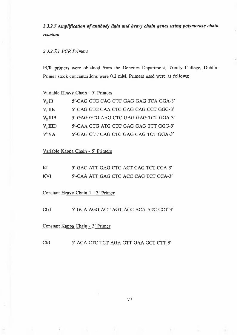
23.2.7 Amplification of antibody light and heavy chain genes using polymerase chain
reaction
2.3.2.7.1 PCR Primers
PCR primers were obtained from the Genetics Department, Trinity College, Dublin.
Primer stock concentrations were 0.2 mM. Primers used were as follows:
Variable Heavy Chain - 5' Primers
VhIB 5'-CAG GTG CAG CTC GAG GAG TCA GGA-3'
VhIIB 5'-CAG GTC CAA CTC GAG CAG CCT GGG-3'
VHIIIB 5'-GAG GTG AAG CTC GAG GAG TCT GGA-3'
V„IIID 5'-GAA GTG ATG CTC GAG GAG TCT GGG-3'
VhVA 5'-GAG GTT CAG CTC GAG CAG TCT GGA-3'
Variable Kappa Chain - 5' Primers
KI 5'-GAC ATT GAG CTC ACT CAG TCT CCA-3'
KVI 5'-CAA ATT GAG CTC ACC CAG TCT CCA-3'
Constant Heavy Chain 1 - 3 ' Primer
CGI 5'-GCA AGG ACT AGT ACC ACA ATC CCT-3'
Constant Kappa Chain - 3' Primer
Ckl 5'-ACA CTC TCT AGA GTT GAA GCT CTT-3'
77

2.3.2.7.2 PCR reaction
A stock PCR reaction mixture was prepared as follows:
5^1 each 10 mM dNTP stock (dATP, dCTP, dGTP, dTTP)
60m1 25 mM MgCl2
100|al 10 X Taq polymerase reaction buffer (Promega)
Sterile ultrapure water to 1 ml
Standard PCR reaction mixture was as follows:
lpl mouse spleen cDNA (0.5-ljj.g)
l^il each primer stock (20 iM)
2.5 U (0.5 pi) Taq polymerase (Promega)
97(il stock PCR reaction mixture
PCR conditions varied depending on individual DNA and primer conditions (Innis and
Gelfind, 1990), but typical thermal cycling conditions were as follows:
1. 95°C 5 min
2. 58°C 1.5 min
3. 72°C 1.5 min
4. 95°C 1 min
Steps 2-4 repeated 30 times
5. 58°C 2 min
6. 72°C 5 min
All ramping rates were at 2 s/°C.
78

23.2.7.3 Purification o f PCR reaction products
PCR reaction products were precipitated with sodium acetate and isopropanol according
to Sambrook, et al. (1989), resuspended in 20^1 sterile ultrapure water and run on a 1.5%
(w/v) agarose gel (SeaKem GTG, FMC BioProducts, 191 Thomaston Street, Rockland,
ME 04841, USA) containing 0.5|ig/ml ethidium bromide for 1 h at 80 V. The 700 base
pair (bp) band was cut from the gel with a sterile scalpel and the DNA purified from the
agarose using a Qiaex II purification System (Qiagen), according to manufacturer’s
instructions.
23.2.8 Insertion of antibody heavy or light chain genes into the pGEM-T plasmid vector
Gel purified light or heavy chain genes were ligated into the pGEM-T plasmid vector
(Promega) as follows:
50 ng (1 1) pGEM-T vector
2^1 gel purified antibody chain genes
l(jl 10 X reaction buffer
l|il ligase (Promega)
5|al sterile ultrapure water
Incubation was for 3 h at 15°C and overnight at 4°C.
23.2.9 Preparation of electrocompetent Escherichia coli XLl-Blue cells
Escherichia coli (E. coli) XLl-Blue (Stratagene) was streaked onto LB medium with
10(j.g/ml tetracycline (Tc) and grown for 24 h at 37°C. Single colonies were used to
inoculate 10 ml of Terrific Broth (TB) (Sigma), with 2% (w/v) glucose and 10|j.g/ml Tc
and grown overnight at 37°C, 200 rpm. The 10 ml was overnight culture was used to
inoculate 1000 ml (2 x 500 ml) Terrific Broth, 2% glucose and 10 |ag/ml Tc and grown
at 37°C, 200 rpm to an optical density (OD) at 550 nm of 0.8 absorbance units (AU). The
culture was chilled on ice for 15 min and centrifuged at 2,500 x g for 15 min at 4°C. The
79

media was thoroughly decanted and the cells resuspended in an equal volume of ice-cold
10% (v/v) glycerol in sterile ultrapure water, and centrifuged at 2,500 rpm for 15 min at
4°C. This was repeated using half a volume of 10% (v/v) glycerol (500 ml), and the cells
were resuspended in a final volume of 20 ml 10% (v/v) glycerol. Cells were aliquoted in
300^1 lots, snap frozen in liquid nitrogen and stored at -80°C.
2.3.2.10 Measurement of transformation efficiency of electrocompetent E. coli XLl-Blue
cells
300|il of electrocompetent E. coli XLl-Blue cells were thawed on ice for 5 min. 2 jil of
supercoiled pUC18 plasmid DNA (Stralagene) (100 ng/ml) was added, mixed gently by
pipetting and allowed to incubate on ice for 1 min. Cells were pulsed in a BioRad Gene
Pulser at 2.5 kV, 200 Q, 25 (iF in a 0.2 cm electroporation cuvette. 1 ml of prewarmed
(37°C) SOC medium was added to the cells and pipetted gently, before transfer to a
further 2 ml SOC medium and shaking at 200 rpm, 37°C for 1 h. Transformants were
plated on LB agar supplemented with 10 (ig/ml Tc and 100 ng/ml carbenicilin (Cb) and
grown for 24 h at 37°C.
Electrocompetence was calculated as the number of positive transformants per microgram
of pUC18 supercoiled plasmid DNA.
2.3.2.11 Transformation of E. coli XLl-Blue with pGEM-T plasmid vector containing
light and heavy chain gene inserts
An appropriate volume of ligation mixture from 232.1 (2, 5 or 10 jol) was added to a
300|jl aliquot of electrocompetent XLl-Blue cells and transformed according to section
2.3.2.10. Following 1 h incubation at 37°C, 200 rpm, the material was centrifuged at 2,500
x g, cells were resuspended in a minimal volume of liquid and spread on plates of LB
containing 10 ng/ml Tc and 100 ng/ml Cb. Self-ligated pGEM-T vector was distinguished
from insert ligation by the addition of 20 nl of 50 mg/ml 5-bromo-4-chloro-3-indolyl-P-D-
galactoside (X-Gal) (Promega) with 100 nl 100 mM isopropyl-p-D-galactoyranoside
(IPTG) to plates of LB agar 30 minutes prior to use. Plates were incubated for 24 h at 37°C.
80

2.3.2.12 Isolation of light and heavy chain gene inserts from pGEM-T
Transformed E. coli XLl-Blue carrying light or heavy chain inserts were resuspended
from LB agar plates in 1 ml sterile distilled water and transferred to 25 ml sterile vials
(Sterilin). Cells were pelleted at 2,500 x g for 20 min and plasmid DNA minipreps carried
out using Wizard miniprep (Promega) according to manufacturer’s instructions. pGEM-T
plasmid vector was digested with restriction enzymes, either for the removal of heavy or
light chain gene inserts as follows (figures given for the digestion of 1 \ig plasmid DNA
in 10 fil reaction volumes):
Heavy Chain Light Chain
Samples were incubated for 2 h at 37°C. The digested plasmid DNA was electrophoresed
and the 700 bp band excised and purified according to section 2.3.2.7.3.
2.3.2.13 Preparation of pComb3 phagemid vector for insert ligation of antibody heavy
chain genes
pComb3 (Scripps Institute) was transformed into E. coli XLl-Blue according to section
2.3.2.10. Individual colonies were used to inoculate TB with 2% glucose, 10 (ag/ml Tc,
100 ng/ml Cb, and cultures grown overnight at 200 rpm, 37°C. Plasmid minipreps were
prepared using Wizard Plus Minipreps (Promega). Plasmid DNA was digested as follows
(figures given for the digestion of 1 |ig of plasmid DNA in 10 (il reaction volumes):
Heavy chain Light chain
10 U Xhol
3 U SpeI
1 Ml 10 X Buffer H
10 U Xbal
5 U Sac I
1 Ml 10 X Buffer A
10 U Xhol
3 U Spe I
1 Ml 10 X Buffer
5 U Sacl
10 U Xbal
1 Ml 10 X Buffer
81

Samples were incubated for 2 h at 37°C. The digested plasmid DNA was electrophoresed
and the 700 bp band excised and purified according to section 2.3.2.7.3.
Ligation of heavy and light chain genes into pComb3 was performed using the Rapid
DNA Ligation Kit (Boehringer Mannheim) according to manufacturer’s instructions,
2.3.2.14 Transformation of E. coli XLl-Blue with pComb3 plasmid vector containing
light and heavy chain gene inserts
Transformation was performed according to section 2.3.2.11.
82

2.3.3 Production of single chain Fv antibodies to coumarin and 7-hydroxycoumarin
2.3.3.1 The Nissim library
The human synthetic phage antibody single chain Fv (scFv) library (Nissim library) was
obtained from the Medical Research Council, Cambridge (Nissim et a l, 1994). This
consisted of the synthetic scFv library in E. coli TGI cells. All bacterial stocks were
maintained at -80°C as 10% glycerol stocks.
2.3.3.2 Growth of the Nissim Library
50 ml 2xTY with 100 ng/ml Cb was inoculated with 50 pi of library cells (approx. 108
cells). The culture was grown in a shaking incubator (at 200 rpm) at 37°C, until the OD
at 600 nm had reached 0.5 AU. 10 |al VCSM13 helper phage (Stratagene) (1011 pfu/ml),
were added to 10 ml of culture and incubated at 37°C, static for 30 min, centrifuged at
2,500 x g for 10 min and the cells transferred to 300 ml 2xTY with 100 ng/ml Cb and 25
Mg/ml kanamycin (Kn) and incubated overnight at 30°C, 200 rpm.
2.3.3.3 Preparation of phagemid particles
The overnight culture from section 2.3.3.2 above was centrifuged at 5,000 x g for 10 min.
The culture supernatant was transferred to l/5th volume of PEG/NaCl (20% (w/v)
polyethylene glycol 8000 in 2.5 M NaCl), mixed and incubated at 4°C for 1 h. This was
centrifuged at 5,000 x g for 30 min (Heraeus Sepatech) and the pellet resuspended in 40
ml water. The phage particles were again precipitated with l/5th volume of PEG/NaCl for
20 min at 4°C and centrifuged at 2,500 x g for 30 min. The supernatant was thoroughly
removed and the pellet resuspended in 2.5 ml water, centrifuged at 2,500 x g for 10 min
and the supernatant stored at -20°C.
Phage preparations were titred by adding 50 1 serial dilutions of phage preparation to 50
|il TGI cells grown to an OD at 600 nm of 0.5 AU in 2xTY, 1% (w/v) glucose, incubated
for 30 min at 37°C and plated on TYE with 100 |ag/ml Cb. Phage titre was calculated as
83

titre-forming units (TFU) per ml.
2.3.3.4 Affinity selection of scFv antibodies using the Nissim library
16 wells of a Costar microtitre plate were coated with antigen at the appropriate
concentration. For 7-hydroxycoumarin, 10 ng/ml 7-hydroxycoumarin-bovine serum
albumin, for coumarin, 10 ng/ml coumarin-bovine serum albumin. All antigen was coated
in PBS. An N-oxysuccinimide-activated binding plate (Nunc) was coated directly with 10
mg/ml 6-amino coumarin in PBS. All plates were coated overnight at 4°C. The plates
were washed three times with PBS and blocked with 1% (w/v) BSA in PBS for 2 h at
37°C. Plates were then washed three times in PBS and 0.5 ml of library (approx. 1012
TFU) was placed in 3.5 ml 1% (w/v) bovine serum albumin in PBS and dispensed into
16 wells. The plates were shaken gently at RT for 30 min, followed by static incubation
for 90 min.
Unbound phage was removed from wells by shaking the plate over a sink and the plates
were washed 10 times with PBS-Tween (0.05 mg/ml Tween 20 in PBS) and 10 times with
PBS.
250 [ol of sterile elution buffer (1.6 ml 12 M HC1 in 150 ml water, adjusted to pH 2.2
with solid glycine and made up to 250 ml) were added to each well and left for 10 min.
Eluted phage was transferred to an equal volume of 2 M Tris-HCl pH 8.0 for
neutralisation and stored at -20°C. Eluted, neutralised phage was used to transfect 10 ml
cultures of TGI (grown to an OD at 600 nm of 0.5 AU in 2xTY, 1% (w/v) glucose), for
30 min at 37°C. This was transferred to 100 ml of 2xTY, 1% (w/v) glucose and 100 (ig/ml
Cb overnight at 37°C. Cells were pelleted by centrifugation at 2,500 x g for 30 min and
resuspended in 1 ml of 10% (v/v) glycerol and stored at -20°C. This material was used
in repeated rounds of growth and selection as described (section 2.3.3.4).
Second and third affinity selection strategies were performed for the production of
antibodies to 7-hydroxycoumarin. Three rounds of affinity selection were performed as
above using 70HC-BSA, 70HC-0VA and 70HC-BSA in rounds 1 to 3 of affinity
84

selection, respectively. The blocking solutions in rounds 1 to 3 were BSA, ovalbumin and
non-fat milk respectively.
2.3.3.S Affinity selection of antibodies using BIAcore™
Phage antibody particles from section 2.3.3.3 at a concentration of 5.5 x 10⁹ tfu/ml were
passed across a sensor chip surface immobilised with 7-hydroxycoumarin-BSA for 5 min
at a flow rate of 10 ^l/min. The sensor chip surface was washed with HEPES-0.5 M
NaCl, pH 7.4 at 100 jd/min for 13.4 min. Glycine buffer, pH 2.2, was passed across the
sensor surface at 10 |ol/min for 20 min and the eluent was neutralised with an equal
volume of 2 M Tris, pH 8.0. Aliquots of eluent were analysed by phage titre at various
time periods and the phage counts performed according to section 2.3.3.3.
85

2.3.3.6 Screening of scFv antibodies by Phage ELISA
Glycerol stocks of affinity-selected scFv antibody were serially diluted in 2xTY and plated
onto TYE with 100 |ig/ml Cb and grown overnight at 37°C. Viable counts were
determined as the number of cfu/ml of stock culture.
2.3.3.6.1 Preparation of clones for phage ELISA
Single colonies were selected from plates and used to inoculate individual wells of a 96
well cell culture dish containing 200 (xl 2xTY, 1% (w/v) glucose and 100 ng/ml Cb. The
plate was shaken (150 rpm) overnight at 37°C and used as a master plate to inoculate an
identical plate. 30 |xl of 60% (w/v) glycerol was added to each well of the master plate
and stored at -20°C. The second plate was grown (shaken at 150 rpm) for 8 h at 37°C. 25
1 of 2xTY containing 1% (w/v) glucose, 100 |xg/ml Cb and 109 VCSM13 helper phage
per ml was added to each well and allowed to stand at 37°C for 30 min followed by
shaking (150 rpm) for 1 h at 37°C. Plates were centrifuged at 2,000 x g for 10 min. The
supernatant was discarded and replaced with a mixture of 200 (ol 2xTY, 100 (ig/ml Cb and
25 (ig/ml Kn and grown at 150 rpm overnight at 30°C. The plates were centrifuged at
2,000 x g for 10 min and 75 1 of supernatant was used in the following phage ELISA.
An anti-NIP clone (Nissim et a l, 1994) was used as a positive and negative control for
the phage ELISA as outlined in section 2.3.3.6.2.
2.3.3.62 Phage EUSA
Enzyme immunoassay plates (Costar) were coated with 100 (il of either 10 ng/ml 7-
hydroxycoumarin-bovine serum albumin, 7-hydroxycoumarin-OVA or 10 |ag/ml coumarin-
bovine serum albumin, as appropriate and left overnight at 4°C. A positive control well
was coated with 10 fig/ml 3-nitro-4-hydroxy-iodophenylacetic acid (NlP)-bovine serum
albumin in PBS. A negative control well was coated with 7-hydroxycoumarin-bovine
serum albumin or coumarin-bovine serum albumin as appropriate. Plates were washed
once with PBS. Plates were blocked with 100 (jJ 1% (w/v) bovine serum albumin in PBS
86

(or ovalbumin in the case of the second anti-7-hydroxycoumarin library) for 1 h at RT.
The plate was washed three times with PBS-Tween. 75 (ol supernatant and 25 pi 4% (w/v)
bovine serum albumin (or ovalbumin) in PBS were added to each well for 2 h at RT.
Plates were washed three times with PBS-Tween and incubated with 100 pi per well of
1 in 400 dilution of anti-M13 antibody (sheep or rabbit IgG, Genosys, Sigma or raised in
rabbits (section 2.3.1.9)). This was followed by a 1 in 800 dilution of HRP-labelled anti
sheep or anti-rabbit IgG. This was incubated for 2 h at RT and plates were washed three
times in PBS-Tween. 100 pi substrate (0.04 mg/ml o-phenylenediamine in 50 mM sodium
citrate, 0.15 M sodium phosphate buffer, pH 6.0 with 0.32 pl/ml H20 2) was added to each
well and incubated at RT for 30 min. Substrate reactions were stopped by addition of 50
pi of 4 M H2S04 per well. Absorbance was read at 492 nm.
2.3.3.7 Transfer of pHENl to HB2151 for screening of soluble scFv antibody
expression
Positive isolates from phage ELISA were identified and 100 pi of the phage supernatant
was used to transfect 10 ml cultures of HB2151 grown in 2xTY with 1% (w/v) glucose
and 100 pg/ml Cb. Transfection was performed according to section 2.3.3.2.
2.3.3.8 Screening of scFv isolates for soluble antibody expression
Glycerol stocks of HB2151 clones from section 2.3.3.7 were plated onto TYE with 100
pg/ml Cb, 1% (w/v) glucose in a 96 well tissue culture plate, and grown shaking (150
rpm) overnight at 37°C. This was used as a master plate to inoculate a second plate
containing 200 pi 2xTY per well, 0.1% (w/v) glucose and 100 pg/ml Cb. This was grown,
while shaking, until the absorbance was approximately 0.9 AU. 25 pi 2xTY with 100
pg/ml Cb and 9 mM IPTG were added and the plates grown for 16 h at 37°C and 150
rpm to induce soluble antibody production.
Enzyme immunoassay plates were coated with antigen as appropriate (see section
2.3.3.5.2), blocked and washed. 75 pi culture supernatant and 25 pi 4% (w/v) bovine
87

serum albumin in PBS were added to each well, mixed and incubated at RT for 90 min.
Plates were washed three times with PBS-Tween and three times with PBS. 100 (ol of a
1 in 400 dilution of anti-c-myc antibody in PBS was added to each well at RT for 90 min.
The plates were washed again, three time with PBS-Tween and three times with PBS. 100
|il of a 1 in 100 dilution of a horseradish-peroxidase labelled anti-mouse IgG antibody was
added and incubated for 90 min at RT. The plates were washed again as before and
developed with o-phenylenediamine substrate as in section 2.3.3.5.2.
2.3.3.9 Analysis of the cellular distribution of scFv antibodies
Cultures of HB2151 selected for expression via phage ELISA were grown in 2xTY, 1%
(w/v) glucose, 100 (ig/ml Cb and induced as described in 2.3.3.8. Cell pellets were
analysed for periplasmic expression of scFv by resuspending in 100 (jl of PBS, 0.1% (w/v)
phenyl methyl sulphonylfluoride (PMSF) and 1.5% (w/v) streptomycin sulphate. They
were then subjected to three freeze-thaw cycles and centrifuged at 2,000 x g for 10 min
(Heraeus Sepatech). Lysates were analysed by dot blotting (section 2.3.1.7).
2.3.3.10 Large scale scFv antibody production
E. coli HB2151 expressing appropriate antibody clones were inoculated into 5 ml cultures
of 2xTY, 1% (w/v) glucose and 100 (ig/ml Cb and shaken (200 rpm), overnight at 37°C.
These were used to inoculate 500 ml cultures of the same media which were grown to an
OD of 0.8 at 600 nm, centrifuged at 2,500 x g for 20 min at RT and resuspended in an
equal volume of 2xTY, warmed to 30°C. The culture was again centrifuged at 2,500 x g
for 2 min and resuspended in an equal volume of 2xTY, 1 mM IPTG and incubated at
200 rpm overnight at 30°C. Cells were harvested by centrifugation at 10,000 x g for 10
min. Antibody was isolated from culture supernatant, periplasm or intracellular
compartments, as appropriate, and this is described in section 2.3.3.11.
88

2.3.3.11 Production of scFv antibody from intracellular compartments
The centrifuged bacterial pellet from above was resuspended in ice cold 50 mM Tris, pH
8.0, 1 mM EDTA, 100 mM KC1, 0.1 mM PMSF. Cells were disrupted, either by
sonication or use of a beadbeater with 50% (v/v) 0.1 mm glass beads and three 1 min
pulses followed by 1 min cooling time, on ice. The lysate was centrifuged at 25,000 x g
for 20 min at 4°C and the pellet resuspended in either 7.5 M guanidine hydrochloride
(Gdn HC1) or 8 M urea with gentle mixing for 2 h at RT. The denatured lysate was
centrifuged at 25,000 x g for 20 min at 4°C and dialysed against several changes of 0.1
M Tris, 0.4 M arginine, 2 mM EDTA, pH 8.0 at 4°C over periods of time ranging from
2 days to 2 weeks. The refolded lysate was finally centrifuged at 25,000 x g for 20 min
at 4°C.
2.3.3.12 Large scale phage antibody production
E. coli TGI cells expressing appropriate antibody clones were prepared according to
sections 2.3.3.2 and 2.3.3.3.
2.3.3.13 BIAcore™ kinetic analysis of anti-coumarin scFv antibody clones
A 5 min injection at 20 (xL/min of scFv antibody at a concentration of 1 nM was passed
over both a coumarin-BSA sensor chip surface (1,600 RU) and a BSA control surface
(6,000 RU) in HEPES buffer, pH 7.4. After 10 min dissociation phase, the surface was
regenerated using HEPES, 0.15 M NaCl, pH 7.4. Observed association and dissociation
rates were calculated using BIAevaluation 2.1™ software (Pharmacia Biosensor).
89

2.3.4 The in vitro production of 7-hydroxycoumarin glucuronide
2.3.4.1 Preparation of porcine and bovine UDP-glucuronyl transferase
Porcine or bovine UDP-glucuronyl transferase (UDPGT) was prepared according to
Tegtmeier et al. (1988). Fresh liver was cut into small pieces and washed in ice cold 0.9%
(w/v) NaCl until the supernatant was clear. The supernatant was discarded and the liver
was mixed with four times its mass (4 x) of 0.25 M sucrose (20%, w/v). The liver was
homogenised (Ultra-Turrax) and centrifuged at 10,000 x g at 4°C for 15 min (Sorvall RC
5B). The collected supernatant was centrifuged at 190,000 x g, 4°C for 90 min (Beckman
L8-70M). The resulting microsomal pellet was resuspended in 4 x the mass of ice cold
0.1 M Tris-HCl, pH 8.0, 0.1 mM dithiothreitol (DTT), 0.75% (w/v) sodium cholate, and
shaken gently for 30 min. The homogenate was centrifuged at 190,000 x g for 1 h.
Saturated ammonium sulphate to a final concentration of 40% (v/v) of saturation, was
added gradually to the resulting clear supernatant while stirring. The resultant turbid
solution was centrifuged at 10,000 x g at 4°C for 10 min, and saturated ammonium
sulphate added to bring it to a final concentration of 70% (v/v) of saturation. The turbid
solution was again centrifuged at 10,000 x g, for 10 min at 4°C and the precipitate
resolubilised in 4 x its mass of 50 mM Tris-HCl, pH 7.4. It was dialysed against 50
volumes of the same buffer and stored at -20°C. Protein concentrations were measured
using the BCA assay (section 2.3.1.8).
90

2.3.4.2 Initial analysis of the in vitro production of 7-hydroxycoumarin-glucuronide by
HPLC
4 x 20 ml reaction mixtures, labelled A to D were prepared according to Table 2.1.
Table 2.1. Composition of reaction mixtures for the initial in vitro production of 7- hydroxycoumarin-glucuronide.
Component A B C D
UDP glucuronyl transferase 1.2 mg/ml 1.2 mg/ml 1.2 mg/ml 0 mg/ml
UDP glucuronic acid 2 mM 0 mM 2 mM 2 mM
Tris-HCl, pH 7.4 0.1 M 0.1 M 0.1 M 0.1 M
7-hydroxycoumarin 140 mM 140 mM 0 mM 140 mM
Ethanol 4% (v/v) 4% (v/v) 4% (v/v) 4% (v/v)
Saccharolactone 5 mM 5 mM 5 mM 5 mM
Magnesium chloride 5 mM 5 mM 5 mM 5 mM
Water To 20 ml To 20 ml To 20 ml To 20 ml
Sample A was the test sample, containing enzyme and both substrates, 7-hydroxycoumarin
and UDP glucuronic acid. Sample B lacked UDP glucuronic acid, sample C lacked 7-
hydroxycoumarin and sample D lacked UDP glucuronyl transferase. Saccharolactone was
present as a (3-glucuronidase inhibitor and magnesium ions was act as a cofactor for UDP
glucuronyl transferase.
The samples were placed at 37°C revolving at 80 cycles per min, and 100 fil samples
removed at various time intervals, precipitated with 20% (w/v) trichloroacetic acid and
centrifuged at 13,000 rpm for 10 min at room temperature.
HPLC analysis conditions
HPLC analysis was performed on a System Gold chromatography station with System
Gold software, using a Phenomenex Bondclone 10 C-18 reverse phase column (25 cm)
91

with a flow rate of 2 ml/min and a mobile phase of 700:300:2 water:methanol:acetic acid.
Samples of 20 fil were injected with detection at 324 nm. Samples were compared against
glucuronide standards from 0 to 100 |ig/ml prepared in the same matrix.
2.3A.3 Repeat analysis of the in vitro production of 7-hydroxycoumarin-glucuronide
by HPLC
For the repeat analysis of the in vitro production of 7-hydroxycoumarin glucuronide,
bovine UDP glucuronyl transferase was prepared according to section 2.3.4.1. A reaction
mixture was prepared in triplicate according to Table 2.2.
Table 2.2. Composition of the reaction mixture for the repeat in vitro production of 7-hydroxycoumarin glucuronide.
Component Concentration
7-hydroxycoumarin 0.77 mM
UDP glucuronyl transferase 1 mg/ml
Magnesium chloride 6.25 mM
Saccharolactone 6.25 mM
UDP glucuronic acid 1.25 mM
Ethanol 4% (v/v)
Tris-HCl, pH 7.4 125 mM
Water To 4 ml
The reactions were carried out at 37°C in open 10 ml glass tubes. 200 (ol samples were
removed and 40 |il 20% (w/v) trichloroacetic acid added, mixed and centrifuged at 13,000
rpm for 10 min. Standards were also prepared (n=4) containing 0 to 1000 pg/ml 7-
hydroxycoumarin glucuronide in the reaction components after treatment with
trichloroacetic acid. 190 jol of sample was then added to 10 pi of a 1 mg/ml 4-
hydroxycoumarin internal standard. These were analysed by HPLC.
92

HPLC conditions
Chromatography was performed on a System Gold chromatography station using System
Gold software, with a Phenomenex Bondclone 10 C-18 column. This analysis used a
gradient method to increase the retention time of 7-hydroxycoumarin-glucuronide, and
allow for the simultaneous analysis of 7-hydroxycoumarin, 7-hydroxycoumarin-
glucuronide and the internal standard 4-hydroxycoumarin. Solvent A was 950:50:2
water:methanol acetic acid (v/v/v). Solvent B was 100% methanol. The flow rate was 1
ml/min with detection at 320 nm. The gradient was as follows (all percentages are v/v):
0-14 min: Solvent A (100%) to Solvent A (50%)/Solvent B (50%)
14-22 min: Solvent A (50%)/Solvent B (50%)
22-23 min: Solvent A (50%)/Solvent B (50%) to Solvent A (100%)
23-32 min: Solvent A (100%)
7-hydroxycoumarin-glucronide concentration was calculated by calculating the peak height
ratios of 7-hydroxycoumarin-glucuronide and the internal standard, 4-hydroxycoumarin.
93

2.3.4.4 Analysis of the in vitro production of 7-hydroxycoumarin-glucuronide by
capillary electrophoresis (CE)
An enzymatic reaction mixture was prepared according to Table 2.2 to a total volume of
1.6 ml. Reactions were carried out in open 2.0 ml test tubes. Fractions were removed and
analysed by CE. A standard curve of 7-hydroxycoumarin-glucuronide was prepared (n=3)
in the same matrix as the reaction mixture, except that the enzyme solution was denatured
prior to addition to the reaction mixture by boiling for 5 min.
CE separation conditions
Separation was carried out on a Beckman capillary electrophoresis instrument, P/ACE
System 5500 using System Gold software according to the method developed by Bogan
et al. (1996) for the analysis of 7-hydroxycoumarin and 7-hydroxycoumarin-glucuronide.
The capillary used was a 57 cm x 50 |im i.d. untreated silica capillary with a capillary-to-
detector distance of 50 cm. The capillary was conditioned each day by 10 min rinses with
0.1 M hydrochloric acid, followed by 0.1 M sodium hydroxide, then deionised water and
finally electrolyte buffer. Between each run, the capillary was rinsed for 0.8 min with 0.1
M NaOH, then a 2.5 min rinse with electrolyte solution. Samples were applied by 10
second pressurised injections at 0.5 p.s.i. Separation was achieved at 30 kV (rise time 0.2
min) at 25°C with detection at 320 nm. Typical running current was 170 (jA.
94

2.3.5 Purification of 7-hydroxycoumarin-glucuronide
2.3.5.1 Chromatographic separation of 7-hydroxycoumarin and 7-hydroxycoumarin-
glucuronide
A series of chromatographic experiments were performed using a Lichroprep RP-18
reverse phase silica column, 15 cm x 1 cm i.d. prepared in methanol. Buffer flow was
maintained via a peristaltic pump with flow rates in the order of 0.2 to 0.4 ml/min.
Various mobile phases, pump speeds and fraction intervals were assessed for the effective
separation of pure mixtures of 7-hydroxycoumarin and 7-hydroxycoumarin-glucuronide.
Fractions were analysed spectrophotometrically at 320 nm.
2.3.5.2 Chromatographic purification of 7-hydroxycoumarin-glucuronide from crude
enzymatic reaction mixtures
Chromatographic conditions utilising a mobile phase of 50:50 methanol:water (v/v) was
selected for the separation of 7-hydroxycoumarin and 7-hydroxycoumarin-glucuronide in
crude enzymatic reaction mixtures derived from section 2.3.4, with flow rates typically
in the order of 0.2 ml/min. Various sample volumes were assessed for their ability to be
effectively separated under the given conditions. Relevant fractions of purified 7-
hydroxycoumarin-glucuronide were analysed by HPLC according to section 2.3.4.2.
2.3.5.3 Chromatographic purification of 7-hydroxycoumarin-glucuronide from
coumarin-treated urine
A healthy human volunteer was administered a 1 g p.o. dose of coumarin in 10 x 100 mg
tablet doses subsequent to provision of a control urine sample. Urine from the treated
volunteer was collected at 2 h. Control and treated urine samples were stored in aliquots
at -20°C until use. Thawed samples were centrifuged and filtered through 0.45 (am sterile
acetate filters.
Chromatographic separation of 100 pi aliquots of control urine, control urine spiked with
95

7-hydroxycoumarin-glucuronide and treated urine samples were performed as described
in section 2.3.5.2.
2.3.5.4 Development of a methodology for the extraction of 7-hydroxycoumarin-
glucuronide from aqueous solutions
4 ml of coumarin-treated urine was extracted with 2 ml of DCM:methanol at varying
ratios from 10:90 to 95:5 (v/v) in 10 ml glass tubes. Tubes were rotated on a blood tube
mixer for 30 min at room temperature. The organic phase was removed and extracted into
a further 1 ml of 1 M KC1. The organic layer was dried and resuspended in 400 |il water
and analysed spectrophotometrically at 320 nm.
2.3.5.5 Extraction of 7-hydroxycoumarin-glucuronide from enzymatic reaction mixtures
and coumarin treated patient urine
Further extractions of either urine samples or enzymatic reaction mixtures were performed
using a DCM:methanol ratio of 70:30 (v/v). Extracted samples underwent chromatography
according to section 2.3.5.2.
2.3.5.6 Assessment of the thermal stability of 7-hydroxycoumarin-glucuronide at 60°C
A 1 mg/ml solution of purified 7-hydroxycoumarin-glucuronide was prepared in distilled
water and maintained at 60°C in a water bath. Aliquots were taken and analysed by HPLC
according to section 2.3.4.2. The 7-hydroxycoumarin-glucuronide peak area was monitored
over time.
2.3.5.7 Assessment of the stability of 7-hydroxycoumarin-glucuronide to a range of
different pH conditions
Samples of purified 7-hydroxycoumarin-glucuronide were prepared in aqueous solution
over a range of pH values from 2 to 12.
96

2.3.5.8 In vitro enzymatic production of 3-amino-7-hydroxycoumarin-glucuronide
2.3.5.8.1 Preparation o f 3-amino-7-hydroxycoumarin
3-amino-7-hydroxycoumarin was prepared from 3.5 g 3-acetylamino-7-acetocoumarin
according to Bogan (1996). Briefly, 3.5 g 3-acetylamino-7-acetocoumarin was refluxed
in 250 ml of 3 M hydrochloric acid for 30 min until all the solid present had reacted and
turned orange. The pH was neutralised with solid NaHC03. The resultant yellow
compound was filtered under vacuum, dissolved in a minimal volume of boiling water and
an equal volume of methanol, added and the whole solution placed on ice. The
recrystallised orange-yellow compound was filtered and tested by HPLC (section 2.3.4.2)
employing photodiode array detection and infrared spectral analysis.
2.3.5.8.2 In vitro enzymatic production of 3-amino-7-hydroxycoumarin-glucuronide
0.1 g of 3-amino-7-hydroxycoumarin was dissolved in 1 ml water and placed in the
reaction mixture described in Table 2.1. The mixture was incubated for 4 h at 37°C,
precipitated with 1 ml 20% (w/v) trichloroacetic acid and centrifuged to precipitate
protein. The sample was analysed by HPLC according to section 2.3.4.2 using photodiode
array detection.
2.3.5.9 Production of a 7-hydroxycoumarin-glucuronide-bovine serum albumin
conjugate
42 pmoles of 7-hydroxycoumarin-glucuronide (14.2 mg), 52 (¿moles N-
hydroxysuccinimide (1.87 mg) and 42 pmoles EDC (19.17 mg) were added to 270 mg
bovine serum albumin (20:1 molar excess of 7-hydroxycoumarin-glucuronide to BSA) in
3 ml carbonate buffer, pH 9.6 and 1 ml dimethyl sulphoxide. The reaction mixture was
stirred overnight at room temperature and dialysed against ultrapure water and freeze
dried. The resulting conjugate was analysed by SDS-PAGE (see section 2.3.1.3) and
HPLC size exclusion chromatography according to section 2.3.2.2 with photodiode array
analysis. The reaction was also performed without 7-hydroxycoumarin-glucuronide.
97

Further examples of 7-hydroxycoumarin-glucuronide-bovine serum albumin conjugates
were synthesised. A second conjugate was synthesised using borate buffer at pH 8.5 with
a 200:1 molar excess of 7-hydroxycoumarin-glucuronide and a third was also prepared at
this molar ratio, at pH 7.0
98

3. PRODUCTION OF MURINE Fab ANTIBODY LIBRARIES TO COUMARIN
AND 7-HYDROXY COUMARIN USING THE pComb3 SYSTEM
3.1 INTRODUCTION
3.1.1 Antibody phage display systems
Combinatorial antibody phage display technology, such as that developed by the Scripps
Institute. La Jolla, San Diego, CA, has the potential to produce and screen large libraries
of monoclonal antibodies to an antigen of interest. The pComb3 system utilises the
technique of phage display, to mimic antibody presentation on the surface of B cells, with
the linkage of genotype and phenotype resulting from the presence of only a single light
and heavy antibody chain combination present on a phagemid particle within that phage.
The enrichment technique of affinity selection, or panning, allows the selective
amplification of phage particles carrying antibody specificities of interest from the rest of
the population. Association of a single light/heavy chain gene combination with the
correct protein expression is guaranteed by the characteristics of filamentous phage
infection of E. coli, in which only a single phage particle can infect a cell, ensuring the
correct linkage of phenotype and genotype (section 1.6.1).
3.1.2 Source and quality of material for library construction
Antibody chain genes have, in the past been isolated from several sources, notably from
areas containing high B cell populations such as lymphoid tissues and blood. For the
production of human antibody libraries, this has posed greater difficulties, as the best
organ for B cell isolation in terms of quality and quantity of B cells is bone marrow.
Although donation of bone marrow for such methodologies has been arranged, such
procedures if performed routinely would be difficult to justify on ethical grounds. This
would apply to other lymphoid tissues also. The second problem encountered with human
monoclonal antibody production is lack of immunisation. Although naive, unimmunised
libraries have been created and used effectively, it is still thought that the most reliable
means of isolating high affinity light and heavy chain recombinations from such libraries
is facilitated by immunisation. During this process, the events giving rise to large numbers
of B cells expressing antibodies of high affinity occur. Placed in the context of antibody
library production, such immunisations may result in the more reliable selection of high
100

affinity light and heavy chain recombinants. For the production of murine antibody
libraries, the problems associated with immunisation are overcome. For the most effective
library production from mice, immunisation would be recommended, followed by
splenectomy, and isolation of B cell mRNA from this source.
3.1.3 Production of hapten-protein conjugates
For the development of an immune response to haptens such as coumarin and 7-
hydroxycoumarin, the haptens must be coupled to a large, immunogenic component,
normally a protein (see section 4.1.1). Traditionally, several proteins are used in this
regard: BSA, THG, keyhole limpet haemocyanin (KLH), and ovalbumin (OVA). Each of
these exhibit different characteristics, making them more or less suited to various
applications. Traditionally, BSA has been reserved for use during screening because of
its low cost, whereas THG is used for immunisations, having the advantage of creating
slightly less soluble conjugates, which characteristically, provoke a better immune
response (Hermanson, 1997).
3.1.4 T/A cloning vectors
T/A cloning vectors such as pGEM-T (Promega) have been developed for the
simplification of the insertion of products from polymerase chain reaction into vectors.
Polymerase chain reaction (PCR) of DNA fragments using Taq polymerase results in the
characteristic 5' adénylation of the amplified fragments. This characteristic has been
employed for PCR fragment cloning using T/A cloning vectors (Promega). The pGEM-T
vector is a modification of pGEM, which has been linearised and blunt ended followed
by thymidylation of both 5' blunt ends. Such vectors allow the direct insertion of PCR
fragments without the need for restriction digestion of the PCR fragments prior to
insertion. T/A cloning is generally less efficient than restriction/ligation using standard six-
cutter restriction endonucleases, which result in four nucleotide overhangs. It does,
however, circumvent problems associated with PCR fragment ligation in that effective
restriction digestion at sites introduced close to both the 3' and 5' ends of the PCR
fragment can not be easily validated due to the very small size change in the PCR
101

fragment, and the small size of the cut ends. Insertion into pGEM-T, followed by
restriction digestion should yield a PCR fragment, which has now been correctly digested
and should be ideal for further cloning.
102
3.2 RESULTS
3.2.1 Preparation of coumarin and 7-hydroxycoumarin protein conjugates
Diazo-coupled conjugates of coumarin and 7-hydroxycoumarin to BSA and 7-
hydroxycoumarin coupled to THG were donated by Egan (1993). These conjugates had
all been previously characterised by ETPLC size exclusion chromatography and photodiode
array analysis. Estimation of the coupling ratios of drug to protein were performed.
Coumarin-BSA was estimated to possess approximately 12 moles coumarin per mole of
BSA. This compared excellently with the estimates made by Egan (1993) of 12 molecules
coumarin per molecule of BSA. 7-hydroxycoumarin-BSA was estimated to have
approximately 3 moles of 7-hydroxycoumarin per mole of BSA.
BSA conjugates of coumarin and 70HC were further analysed by ELISA using rabbit
anti-coumarin and rabbit anti-70HC antibodies (Egan, 1993). Both showed specific
responses for the drug-protein conjugate alone, but not to protein (Figs. 3.1 A and B)
3.2.2 Production of coumarin-thyroglobulin (coumarin-THG) conjugate
Coumarin-THG was prepared according to Egan (1993). This required the production of
6-nitro-coumarin (6-N02-coumarin), which was then converted to 6-amino-coumarin (6-
NH2-coumarin). These were analysed by infrared spectrophotometric, TLC and melting
point analysis and identified as the correct species. The IR spectrum of 6-N02-coumarin
(Fig. 3.2) shows a stretch at 1,736 cm'1 (-C=0), and 1,350 and 1,526 cm'1 (-N02
symmetrical and asymmetrical stretch). The IR spectrum of 6-NH2-coumarin (Fig. 3.3)
shows a stretch at 1,699 (-C=0) and 3,322 and 3,402 cm'1 (-NH2 asymmetrical and
symmetrical stretch, respectively)
6-amino-coumarin was coupled to THG according to Egan (1993). Following freeze
drying, the resulting conjugate exhibited particularly poor solubility in water and other
aqueous buffers as well as in organic solvents such as methanol, ethanol and DMSO. This
lack of solubility would make it difficult to analyse by HPLC. Eventually, an effective
103

solute was found in the combined use of dimethyl sulphoxide (DMSO), methanol and
water. C-THG was dissolved in 1:1:1 DMSO/MeOH/water (v/v/v), adding DMSO first,
then MeOH and then water. A spectrophotometric scan showed characteristic peaks at 280
and 320 nm (Fig. 3.4), indicating the presence of coumarin and protein in the same
sample. In HPLC analysis and photodiode array detection, two major peaks occurred, one
at approximately 13 minutes - as expected for THG, which has a molecular weight of
approximately 300 kDa (Fig. 3.5). The PDA scans of both these peaks give similar
profiles characteristic of protein conjugated to coumarin (Fig. 3.6). This is manifest in the
shoulder at approximately 320 nm. which is not present in THG alone.
Analysis of the coumarin-THG conjugate was also performed using ELISA, again using
a rabbit anti-coumarin antibody (Egan. 1993). This also showed specific response for the
antibody to the conjugate, and not to protein alone (Fig. 3.7).
104

0.5
0.4
« 0.3
oa| 0.2
0.1
0.0101 102 103 104 10s
1/Antibody dilution
10°
Figure. 3.1 A and B. Analysis o f coumarin-BSA and 7-hydroxycoumarin-BSA (70HC-
BSA) conjugates by ELISA. The response o f the rabbit anti-coumarin antibody (A) was
specific for the coumarin-BSA conjugate (50 fig/ml) (open circles), and did not
recognise BSA alone (filled circles). The response o f the rabbit anti-7OHC antibody (B)
was specific for the 70HC-BSA conjugate (50 fig/ml) (open circles), and did not
recognise BSA alone (closed circles).
105

XTna
naml
ttan
ce
Oo
Figure 3.2. Infrared spectrum o f 6-N02-coumarin from 600 to 4000 cm 1. This shows
a stretch at 1,736 cm 1 (-C=0), and 1,350 and 1,526 cm'1 (-N02 symmetrical and
asymmetrical stretch), indicating the presence o f an N 02 present on the coumarin
molecule.
106

Figure 3.3. Infrared spectrum o f 6-NH2-coumarin from 600 to 4000 cm 1. This shows
a stretch at 1,699 (-C-O) and 3,322 and 3,402 cm 1 (-NH2 asymmetrical and symmetrical
stretch, respectively).
107

Figure 3.4. Spectrophotometric scan o f thyroglobulin (THG) (A), coumarin-THG (B)
and coumarin (C) from 400 to 200 nm. Comparison o f the coumarin-THG with THG
shows a distinct shoulder between 320 and 350 nm on the coumarin-THG sample that
was not present on THG. This was indicative o f the linkage o f coumarin to THG.
108

0.05
A
Time (min)
Figure 3.5. HPLC analysis o f coumarin-thyroglobulin by size exclusion
chromatography. The chromatogram shows predominant 280 nm peaks at 9.967 (peak
A) and 12.167 (peak B) minutes. The retention time o f 10 minutes corresponds to the
retention o f a large molecular weight protein such as thyroglobulin (300 kDa).
W avelength (n m )
Figure 3.6. Photodiode array analysis o f coumarin-THG. Protein peaks at 9.967 (peak
A, Fig. 3.5) and 12.167 minutes (peak B, Fig. 3.5) show an additional absorbance
shoulder at 320 nm. This was not present on the thyroglobulin control, indicating
attachment o f a coumarin-like molecule to the protein.
109

1/Antibody Dilution
Figure 3.7. Analysis o f coumarin-THG conjugate by ELISA with rabbit anti-coumarin
antibody. The graph shows recognition o f the coumarin-THG conjugate (50 fig/ml)
(open circles), but not THG alone (filled circles).
110

3.2.3 Immunisation of mice with coumarin-THG and 7-hydroxycoumarin-THG
Titres of mouse serum were performed following initial and secondary immunization.
Responses to coumarin and 70HC were made using the appropriate BSA conjugate. The
two mice immunised with coumarin-THG, mice 2 and 3, showed titres in excess of 1 in
10,000 following primary immunisation and 1 in 20,000 following secondary
immunisation, as compared to a 1 in 100 dilution of control mouse serum (mouse 1)
(Figs. 3.8A, B, C and D). Mice 4 and 5, immunised with 70HC-THG showed titres in
excess of 1 in 200 following secondary immunisation, as compared with a 1 in 100
dilution of control serum. These serum titrations were deemed high enough responses
from which to prepare splenomic RNA following a final intravenous boost, as according
to Campbell (1986).
I l l

1/A ntibody Dilution 1/A ntibody Dilution
1/Antibody Dilution 1/Antibody Dilution
Figure 3.8 A, B, C and D. Mouse serum titres following primary (open circles) and
secondary (closed circles) immunisation with either coumarin-THG (A and B), or
70HC-THG (C and D). Both mice immunised with coumarin-THG gave similar titres
o f 1 in 12,800 as compared to 1 in 100 dilution o f mouse control serum (solid line)
following first immunisation and rising to 1 in 25,600 as compared to 1 in 100 dilution
o f mouse 1 control serum (dotted line) following secondary immunisation. In the case
of mice 4 and 5 immunised with 70HC-THG, titres following primary immunisation
were 1 in 400 as compared to 1 in 100 control serum (solid line) and between 1 in 200
and 1 in 400 as compared to 1 in 100 control serum (dotted line) following secondary
immunisation.
112

3.2.4 Preparation of splenomic RNA
Splenomic RNA was isolated from spleens from mouse 2 (coumarin-THG) and mouse 5
(70HC-THG), yielding mRNA concentrations of 924 (ig/ml and 720 pg/ml, respectively.
Spleens from mice 3 and 4 were placed in storage at -80°C.
3.2.5 Selection of murine PCR primers based on homology plots
Homology plots of available murine 5' heavy and light chain gene family sequences
(Kabat and Wu, 1986) (Figure 3.9) were prepared (Figure 3.10). Primers were selected
according to levels of sequence homology. A sequence already closely represented by a
homolog was excluded, as mismatching during annealing may give rise to amplification
of these species also. Sequences with no similar homologs were included. Using heavy
chain primer sequences VHIB, VHIIB, VHHIB, VHIIID and VHVA resulted in at least 83%
homology with primers. For light chains, inclusion of primers VkI and VkVI gave at least
83% homology with known sequences, except for VkII with only 75% homology. The
ability of primers to amplify non-specific sequences was not tested further. This rationale
was merely chosen to reduce the numbers of primers required for library preparation.
113

Murine Heavy Chain 5' Primers:
v hia 5'-GAG c t g c a g c t c g a g GAG TCA GGA-3'
VhIB 5'-CAG GTG CAG CTG GAG GAG TCA GGA-3'
VhIIA 5'GAG GTC CAG CTC GAG CAG TCT GGA-3'
VHnB 5'-CAG GTC CAA CTC GAG CAG CCT GGG-3'
VHnC 5'-GAG GTT CAG CTC GAG CAG TCT GGG-3'VhIIIA 5'GAG GTG AAG CTC GAG GAA TCT GGA-3'
VhIIIB 5'GAG GTG AAG CTC GAG GAG TCT GGA-3'
VHIIIC 5'-GAA GTG AAG CTC GAG GAG TCT GGA-3'
VhIIID 5'-GAA GTG ATG CTC GAG GAG TCT GGG-3'
VhVA 5' GAG GTT CAG CTC GAG CAG TCT GGA-3'
Murine Light Chain 5' Primers
VkI 5'-GAC ATT GAG CTC ACT CAG TCT CCA-3'
vkn 5'-GAT GTT GAG CTC ACC CAA ACT CCA-3'
Vkin 5'-GAC ATT GAG CTC ACC CAG TCT CCA-3'
VkIV 5'-GAA AAT GAG CTC ACC CAG TCT CCA-3'
VkV 5'-GAC ATC GAG CTC ACC CAG TCT CCA-3'
VkVI 5'-CAA ATT GAG CTC ACC CAG TCT CCA-3'
Figure 3.9. Murine heavy chain and light chain gene family-specific 5' PCR primers,
incorporating the restriction sites (underlined) for Xhol and Xbal in the heavy and light
chain genes, respectively.
114

Murine Variable Heavy Chain
IA
IA 100 IB
IB 83 100 IIA
HA 83 79 100 HB
HB 62.5 71 83 100 IIC
n c 75 75 92 71 100 I II A
ILLA 71 75 75 58 71 100 IHB
TUB 79 71 75 58 71 83 100 m e
m e 79 75 75 58 71 83 83 100 HID
IHD 62.5 71 67 58 71 83 75 83 100
VA 92 75 92 75 92 71 72 79 62.5
VA
100
Murine Variable Light Chain
kl
kl 100 kH
kn 75 100 k m
k m 92 79 100 klV
klV 79 67 87.5 100 kV
kV 83 71 83 71 100
kVI 75 62.5 83 87.5 67
Figure 3.10. Homology plots o f murine variable heavy and variable light chain gene
family-specific 5' primers, as described in Rabat and Wu (1986).
115
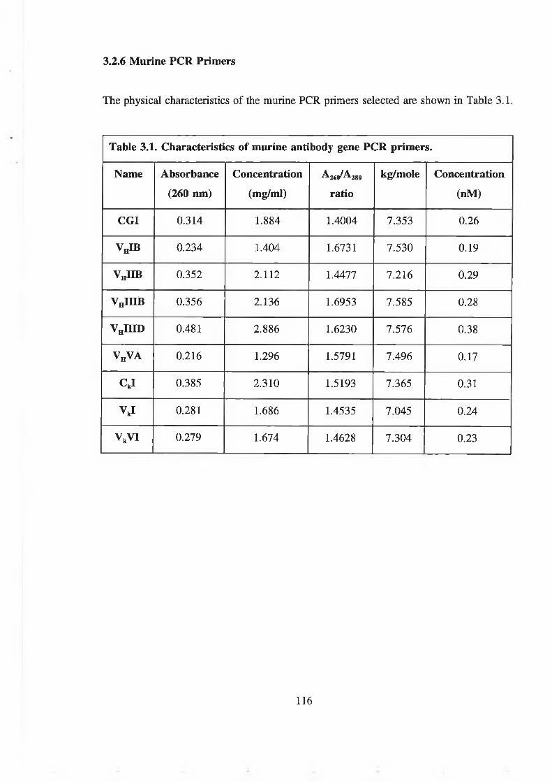
The physical characteristics of the murine PCR primers selected are shown in Table 3.1.
3.2.6 Murine PCR Primers
Table 3.1. Characteristics of murine antibody gene PCR primers.
Name Absorbance
(260 nm)
Concentration
(mg/ml)260 280ratio
kg/mole Concentration
(nM)
CGI 0.314 1.884 1.4004 7.353 0.26
VhIB 0.234 1.404 1.6731 7.530 0.19
VhIIB 0.352 2.112 1.4477 7.216 0.29
VnIIlB 0.356 2.136 1.6953 7.585 0.28
VhIIID 0.481 2.886 1.6230 7.576 0.38
v hv a 0.216 1.296 1.5791 7.496 0.17
CkI 0.385 2.310 1.5193 7.365 0.31
VkI 0.281 1.686 1.4535 7.045 0.24
VkVI 0.279 1.674 1.4628 7.304. . .
0.23
116

3.2.7 PCR optimization
Using the standard PCR protocol (section 2.3.2.7), single amplified bands of
approximately 700 bp, corresponding to amplified variable region genes were achieved
using the following specific PCR conditions:
VhIB/CG1 Annealing temp. 58°C
V„IIIB/CGl Annealing temp. 59°C
VhIIID/CG1 Annealing temp. 58°C, 10 |iM concentration of PCR primers
V„VA/CG1 Annealing temp. 58°C, 10 |iM concentration of PCR primers
VkI/Ckl Annealing temp. 58°C
VkII/Ckl Annealing temp. 58°C
Primers VHIIB/CG1 failed to amplify under a variety of conditions which involved the
variation of annealing temperature, adjustment of MgClj concentration and variation of
primer concentration (Fig. 3.11), so was excluded from the group.
117

1 2 3 4 5 6 7 8
2,645 ►
1,605^ 1,198►
676► 517^
Figure 3.11. Optimisation ofPCR conditions for the amplification o f murine heavy and
light chain genes from mouse 5 immunised with 7-hydroxycoumarin-THG. Lanes: 1,5:
pGEM molecular weight markers (bp); 2: VHIB; 3: VH1IID; 4: VHVA; 6: VnllIB; 7:
VJ; 8: VJI. The gel shows specific amplification o f 700 bp fragments using primer
combinations CG1/VJB, CG1/VHIIIB, CG1.VJ1ID, VnVA, C J /V J and Ckl/V kVI under
the conditions described in sections 2. and 3 ., but failed to amplify CG1/VHIIB over a
variety o f conditions (not shown).
118

3.2.8 Preparation of pComb3 for heavy chain insert ligation
Plasmid pComb3 was digested with either XhoVSpel or XbaUSacl restriction enzymes for
either heavy and light chain insertion, respectively. Digestion with XbaUSacl is shown in
Fig. 3.12. This resulted in a predominant fragment of approximately 4,000 bp, as expected
for a linear fragment of pComb3. No difference in molecular weight could be detected
between single digested (Xbal) and double digested (XbaUSacl) pComb3. This was also
the case for digestion for heavy chain insertion and digestion with Xhol and SpeI (data
not shown).
Restriction-ligation controls were performed on pComb3 using Xhol and Xbal/Spel (Fig.
3.13). Digestion with these two enzymes resulted in two distinct fragments of 3,073 and
956 bp. When the stuffer fragment was religated with the major fragment, significant
ligation was observed, with the production of various multimers. When the fragments had
been treated with calf intestinal phosphatase, however, there were only minor levels of
ligation. This indicated both efficient ligation and dephosphorylation of plasmid DNA.
3.2.9 Measurement of the Electrocompetence of Escherichia coli XLl-Blue
Many batches of electrocompetent E. coli XLl-Blue were prepared with typical
efficiencies of 2 x 10⁸ transformants per microgramme plasmid DNA, using 100 pg
supercoiled pUC18.
3.2.10 Insertion of mouse 5 heavy chain genes into pGEM-T
Test transformations of mouse 5 heavy chain genes ligated into E. coli showed 125
transformants/pl of ligation mixture. The pooling and concentration of ligation mixture
resulted in the production of approximately 2 xlO3 transformants. Transformations were
repeated until a library of approximately 4.75 x 10⁵ heavy chain inserts was built up.
119

3.2.11 Insertion of mouse 5 variable light chain genes into pGEM-T
A variable light chain gene library in pGEM-T of approximately 3.2 x 10⁵ light chain
gene inserts was built up by pooling the results of several transformations. The plasmid
library was analysed by restriction digestion (Fig. 3.14). Single digestion with Xbal
yielded a single fragment of 3,700 bp and with Xbal and Sad, two predominant fragments
of 2,950 bp, 700 bp and a small 50 bp fragment.
120

1 2 3 4
4 ,029►
2,646►
1,605► 1,198^
676► 517>
Figure 3.12. Restriction digestion analysis o f pComb3. Lanes: 1: pGEM molecular
weight markers (bp); 2: pComb3; 3: pComb3 Xbal digest; 4: pComb3 Xbal/SacI digest.
pComb3 shows multiple bands indicating the presence o f covalently closed circular and
open circular DNA. Following either single or double digestion, an approximately 4,000
bp fragment resulted. No difference in molecular weight between single and double
digest fragments could be detected.
121

1 2 3 4 5 6 7 8 9 10
5,1484,9734,2683,530
2,0271,904
1,584
1,375
974
831
564
21,227
Figure 3.13. Plasmid pComb3 ligation and dephosphorylation controls. Lanes: 1
Lambda molecular weight markers (bp); 2: pComb3; 3-6: Not treated with calf intestinal
phosphatase (CIP); 7-10: Treated with CIP; 3,7: pComb3 Xhol digest; 4,8: pComb3
Xhol digest following ligation; 5,9: pComb3 Xbal/Svel digest; 6,10: pComb3 Xbal/Spel
digest following ligation.Digestion with the single enzyme Xhol resulted in a linear
fragment o f approximately 4,000 bp. Double digestion with Xbal and Svel resulted in
two fragments o f 3,073 and 956 bp. Ligation o f linearised Xhol-cut pComb3 resulted
in several higher molecular weight bands, but following treatment with CIP, no ligation
occurred. Ligation o f the two double-digest fragments resulted in several higher
molecular weight bands. Following treatment o f both fragments with CIP, however,
virtually no ligation took place.
122

1 2 3 4 52,027 , IMI. .. Âj jB|1,904 —
1 ^ j»1'"® A flj
/ A1,584 S f
/ ¡ r \1,375 - / J
974 J j ;
831 1
564 J
Figure 3.14. Restriction analysis o f the mouse 5 light chain gene library in pGEM-
T.Lanes: 1: Lambda molecular weight markers (bp); 2: pGEM-T. V j 3: pGEM-T:VL
Xbal digest; 4: pGEM-T:VL S a d digest; 5: pGEM-T:Vh Xbal/SacI digest.Single
digestion with Xbal resulted in a single digested band at approximately 3,700 bp.
Digestion with S a d produced three bands o f2,950, 3,650 and 750 bp. Double digestion
with Xbal and SacI resulted in bands o f 2,950 and 700 bp.
123

3.3 DISCUSSION
Production of an immunised library in the mouse begins with the preparation of a suitable
antigen. Small haptens such as coumarin and 7-hydroxycoumarin are not immunogenic,
and so require conjugation to a large protein, such as BSA or THG. Traditionally, the
coupling methodology for coumarin and 7-hydroxycoumarin to these proteins was via
diazo linkage between an amino group on the hapten and nitrogen or hydrogens on
imidazole and phenolic ring structures such as those found on histidine and tyrosine
residues respectively. Coumarin and 7-hydroxycoumarin possess no reactive side chains
amenable for linkage, and so a linker must be chemically synthesized. The amino group
is produced in a two step process. Initially, the benzene ring is nitrated at the 6 position,
forming 6-nitro-coumarin. This is then reduced to 6-amino-coumarin, which is now
amenable to linkage. The resulting aromatic amine can be converted to the highly reactive
and unstable diazonium with sodium nitrite under acidic conditions at 0°C. The diazonium
ion will now attack the aromatic rings on residues of tyrosine, histidine and tryptophan.
Linkage via the diazonium bond produces distinctively coloured compounds. This results
in distinctive visible spectral absorbances. Spectrophotometric analysis of the conjugate
showed the presence of a shoulder between 320 and 350 nm. This is due to the presence
of coumarin, which absorbs at 320 nm. No such shoulder is apparent with a sample of
THG that has been through the same procedure in the absence of 6-NH2-coumarin. To
establish that the two moieties are linked to one another, chromatographic separation is
desirable. Although thoroughly dialysed to remove small molecular weight components
from the mixture, it is still good practice to characterise the conjugate by HPLC and
photodiode array analysis. Separation by size exclusion chromatography resulted in peaks
corresponding to a high molecular weight component with short retention time (9 to 12
min), which exhibited high absorbance at 280 nm, distinctive of proteins. Photodiode array
scans show the shoulder between 320 and 350 nm as being concomitant with the protein
peaks. The presence of two peaks may indicate some degradation in the protein sample
or, possibly, different levels of conjugation.
3.3.1 Preparation of protein conjugates for immunisation
124

3.3.2 Characterisation of coumarin-thyroglobulin (coumarin-THG)
Thyroglobulin tends to produce characteristically insoluble conjugates (Hermanson, 1997).
This was also the case here. Insolubility was contributed to by the fact that coumarin is
also extremely hydrophobic in nature. The conjugate proved very difficult to analyse due
to this characteristic. Eventually, a solvent system was found to efficiently solubilise the
conjugate, dissolving it sequentially in DMSO, methanol and water, which may have
gradually released more hydrophobic interactions, allowing access to successively more
hydrophillic ones. The presence of two peaks in the sample following PDA analysis is
probably due to protein degradation over time. Initial PDA scans showed only a single
peak, (data not shown). However, at this point, a solvent system was not available to
produce large concentrations for effective analysis.
The insolubility of the conjugate was, however, advantageous during the immunisation
procedure. Insoluble aggregates administered as immunogens elicit better immune
responses than soluble antigens. This may be because they persist at the site of
immunisation, prolonging immunological attack, rather than being rapidly cleared from
the system. This may have contributed to the excellent immune response produced by
mice immunised with coumarin-THG as evidenced by their titre, as compared to 70HC-
THG which was very soluble (Fig. 3.8). Another factor may have been the level of
conjugation of the two conjugates used for immunisation.
3.3.3 Mouse serum antibody titres following immunisation
Although the titre produced by immunisation with 70HC-THG was quite low, it must be
measured against the control used (1 in 100 dilution of control serum) which is quite a
stringent control, but is recommended by Campbell (1986) for the production of
monoclonal antibodies. Titres in excess of 1 in 200 would typically be adequate for the
setting up of a fusion, although titres in excess of 1 in 1,000 would be preferable.
125

3.3.4 PCR primer production
For effective library production, it is necessary to amplify a large number of heavy chain
and light chain genes. Due to their structure, heavy and light chain genes have conserved
3' sequences and variable 5' sequences. However, the diversity of the 5' variable region
results from V gene segments, of which there are an estimated 300 to 1,000 on the mouse
heavy chain germline genes and 300 on mouse k light chain germline genes. These
sequences have evolved from common sequence ancestors to generate antibody diversity.
Consequently, they share large degrees of sequence homology. Homology at the 5' end
of these sequences has been analysed and used to divide sequences into families. These
families allow the construction of sets of primers for PCR that are capable of amplifying
groups of genes from these families.
The 5' sequences of available antibody light and heavy chain genes (Kabat and Wu) were
used to construct a set of family-specific primers (Fig. 3.9). These were modified for the
insertion of restriction digestion sites. Due to the large group of primers this generates,
a rationale was constructed to reduce the numbers of primers used. Primer sequences were
further grouped in homology plots (Fig. 3.10). Here, the sequence similarity between the
primers was assessed. Primers which were similar to a selected homolog were excluded
from the group to be used, and those that had no very similar sequence homolog were
included. In this way, a set of primers that had high sequence similarities to unused
sequences was built up. Any misannealing during amplification may result in the
amplification of non-specific sequences, without the need for large numbers of primers.
This phenomenon was never actually tested, but could be assessed by amplifying
antibodies with known sequences with non-identical, but similar 5' primer sequences.
In the end, the selection of the heavy chain primers VHIB, VHIIB, VHIIIB, VHHID and
VhVA yielded at least 83% homology with the other families. This meant that no more
than 4 nucleotides differed between any two pairs of primers. Homology increased with
the introduction of identical restriction sites into all primers. This strategy was also
applied to light chain sequences which resulted in the choice of two primers VkI and VkVI
which also resulted in at least 83% homology, except for VkII to which they only had
126

75% homology.
The 3' heavy chain constant region primer sequence selected was a Crl sequence. All
heavy chains of a C71 subtype would be amplified. These tend to be produced in largest
abundance, with high affinity. A kappa chain-specific constant 3' primer was also selected
for light chain amplification, as these comprise 95% of light chain subtype, compared to
some 5% for lambda chains (Kuby, 1997).
3.3.5 PCR optimisation
PCR amplification was optimised, in most cases at standard MgCl2 and primer
concentrations, with slight variations in annealing temperature, around 58°C and 59°C.
However, amplifications involving VHIIID and VHVA, required a reduction in primer
concentration to 10 |iM. Amplifications involving VHIIB failed to produce single defined
700 bp bands over a range of PCR conditions.
3.3.6 PCR fragment purification
Several methods were assessed for the isolation and purification of PCR fragments from
agarose gels such as phenol:chloroform cleanup (Sambrook et al., 1989), Wizard kits
(Promega), Spin-X columns (Costar), or without clean up (data not shown). However, the
most reliable system was found to be Qiaex gel purification kits (Qiagen). These produced
clean, reproducible preparations of high yield.
All attempts to clone digested heavy and light chain PCR fragments into pComb3
produced extremely low yields of transformants, or failed to work at all (data not shown).
It was assumed that this failure was due to the inefficiency of the PCR fragments to
digest. Inadequate overhangs can reduce the efficiency of restriction digestion. However,
the design of these primers is similar to many others, with at least a 9 base overhang in
the heavy chain primers (Roben, 1993). Several methodologies were also attempted to
circumvent this problem. These involved concatamerisation of PCR fragments, followed
by digestion, blunt end cloning (data not shown) and T/A cloning. Concatamerisation and
127

blunt end cloning failed to remedy the problem. Both techniques still depended on
enzymatic modification of the PCR fragments. T/A cloning removes the need for
modification of the PCR fragments following amplification and purification. Although
with a potentially lower efficiency than cloning using six-cutter enzymes, greater than 103
transformants could be produced from a single ligation reaction. Repeated reactions could
thus be used to establish a library of chains of high diversity. Some 104 heavy and 104
light chains would be sufficient to generate a library of good size (108). After several
transformations, a heavy chain library of 4.75 x 10⁵ transformants and a library of 3.2 x
105 light chains was produced, so large libraries are quite accessible via this methodology.
3.3.7 Construction of light and heavy chain gene libraries in pGEM-T
Insertion into pGEM-T also allowed the visualisation of correctly digested fragments,
following double digestion with either Xhol and SpeI for heavy chains or XbaVSacl for
light chains. These both yielded the expected 700 bp fragment, indicative of the inserted
antibody chain gene. Bands produced in this way have also been efficiently restricted.
3.3.8 Heavy and light chain gene insertion into pComb3
Heavy chains derived from pGEM-T insertion were purified, and attempts were made to
insert these into pComb3 (data not shown). However, these attempts also failed, or again,
showed extremely low levels of transformation efficiency. It was concluded at this time
that the limiting factor in the cloning procedure was not the PCR fragments, but the
pComb3 vector. It had been assumed that efficient double digestion was taking place as
these restriction sites are 57 bp and 12 bp apart for heavy and light chain insert sites,
respectively. However, the stuffer fragment was never visualised by gel electrophoresis,
and no differentiation could be made between single digested and double digested
pComb3 on gels (Fig. 3.12). This is evidenced by the production of vectors based on
modifications of pComb3 (Burioni et al., 1997). They also suggest that efficient cutting
of pComb3 is difficult in that overdigestion will reduce cloning efficiency and limiting
digestion poses the risk of interfering in the cloning procedure. It is likely from these
observations, and those seen by this author, that the failure of the cloning strategy was as
128

a result of the vector, either being inefficiently digested, or overdigested. The use of
modified vectors, such as those proposed by Burioni et al. (1997) eould alleviate this
situation and bring about the production of large Fab antibody libraries to coumarin and
7-hydroxycoumarin.
129

4. THE PRODUCTION OF SINGLE CHAIN Fv (scFv) ANTIBODIES TO
COUMARIN AND 7-HYDROXYCOUMARIN USING THE NISSIM LIBRARY

4.1 INTRODUCTION
The applications of coumarin in therapy and biochemistry have been discussed (Chapter
1). The usefulness of phage display libraries for the production of antibodies to coumarin
has also been explored. Here, the isolation of antibodies to coumarin and 7-
hydroxycoumarin from the Nissim phage display library is discussed. It explores the use
of scFv material from culture supernatant, periplasmic lysates and inclusion bodies, as
well as the use of scFv displayed on the phage surface as immunological reagents. It
investigates the affinity purification of these molecules, to yield pure, high titre, stable
reagents for subsequent analyses. The physicochemical characteristics of the resulting
monoclonal scFv antibodies are also analysed, and their applicability in various analyses
is assessed. Consequently, the applicability of the Nissim library for the isolation of
antibody reagents to these molecules is evaluated.
4.1.1 Use of the Nissim library in the isolation of single chain scFv (scFv) antibodies
Alternatives to the production of large, immunised combinatorial phage display libraries
such as the pComb3 system do exist. One such library system is the Nissim library
(Nissim et al., 1993). The concept behind the Nissim library was the creation of a naive
human scFv library of very large size (108 clones) so as to make it highly probable that
many scFv clones to any chosen antigen could be selected from it. Diversity was brought
about using a library of variable heavy chain genes that had been mutagenised in the
CDRH3 region, increasing diversity to 108. This library was further combined with
previous scFv libraries (Hoogenboom and Winter, 1992) increasing the overall library size
to much greater than 108.
4.1.2 Affinity selection strategies
Affinity selection can potentially be performed with antigen attached to a solid support.
Matrices used have been in the form of immunoassay plates, immunoassay tubes and
affinity columns (Kretzschmar et al., 1995). With the use of immunoassay dishes and
immunotubes, protein antigens are normally adsorbed. For the selection of antibodies to
131

peptides and haptens, these require linkage to a carrier protein to facilitate this adsorption.
For this reason, optimal conjugate design is critical for efficient enrichment of phage
antibodies to the hapten or peptide. Many conjugation chemistries are available for the
coupling of haptens and peptides to carrier proteins, depending on their structure and the
presence of suitable functional groups to facilitate linkage (Hermanson, 1997). Coumarin
and 7-hydroxycoumarin lack such reactive functional groups, and must be derivatised in
some way to make them amenable to conjugation (Egan, 1993). Aromatic ring conjugation
can be performed by the formation of a diazonium derivative (Fig. 4.1). An active
aromatic amine can react with the electron-rich nitrogens of the imidazole ring on
histidine residues or ortho modification of the phenolic group of tyrosine residues. On
BSA, there are 36 potential binding sites of this type. Efficient conjugate production
involves the formation of conjugates with coupling ratios that maximise the number of
haptens on their surface. This will encourage antigenic recognition of the hapten rather
than the carrier protein and reduce interactions that rely on epitopes shared by hapten and
carrier protein. High hapten coupling ratios, however, can have other practical difficulties.
With many haptens, most notably hydrophobic molecules such as coumarin and 7-
hydroxycoumarin, introduction of these to the surface of a protein may make it extremely
insoluble, preventing its efficient use as an affinity ligand during affinity selection. It is
likely that optimisation of conjugates is necessary for effective affinity selection strategies.
Another factor affecting the characteristics of affinity selection taking place on solid
supports is the presence of a protein blocking agent. Heterogeneous protein mixtures such
as non-fat milk are often used in such strategies. However, the presence of carrier protein
free in solution may serve to remove interactions that take place between carrier protein
and phage antibodies, resulting in enrichment of phage recognising hapten alone, rather
than carrier protein epitopes.
An alternative strategy for selection of antibodies to haptens is the use of direct covalent
attachment of activated hapten derivatives to solid supports. Many of the same coupling
interactions available for the formation of hapten-carrier conjugates can be applied to solid
supports. For example, N-oxysuccinimide is a highly active and unstable species that
promotes the hydrolysis of amines with carboxyl groups. An amine such as
132
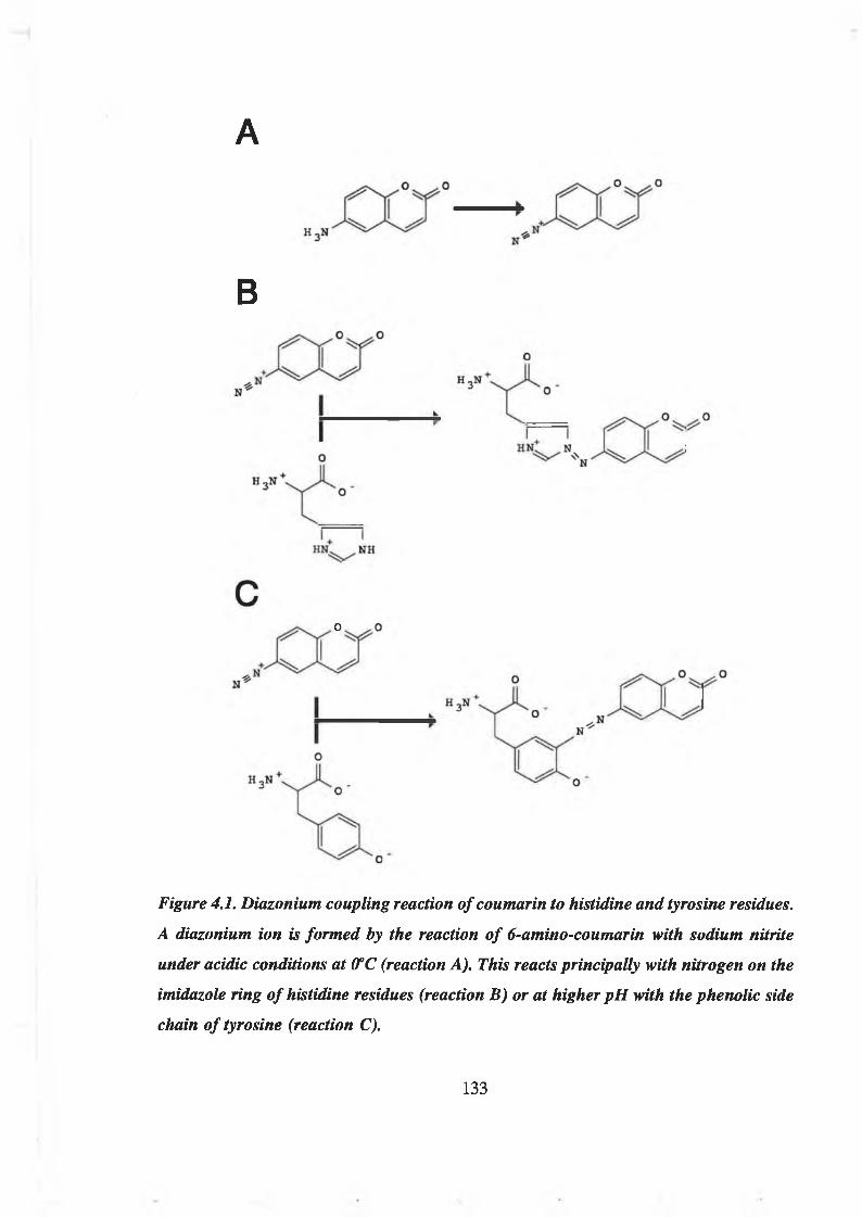
A
B
c>
Figure 4.1. Diazonium coupling reaction o f coumarin to histidine and tyrosine residues.
A diazonium ion is formed by the reaction o f 6-amino-coumarin with sodium nitrite
under acidic conditions at 0°C (reaction A). This reacts principally with nitrogen on the
imidazole ring o f histidine residues (reaction B) or at higher pH with the phenolic side
chain o f tyrosine (reaction C).
133

6-NH2-coumarin could thus be directly covalently linked to a solid surface. This strategy
may oblate problems associated with the interaction of phage antibodies with the protein
moiety of the hapten-carrier conjugate and avoid the need to produce and optimise the
production of such conjugates.
4.1.3 Affinity selection of antibodies from libraries using BIAcore™
Selection and screening procedures using solid supports encourage the isolation of high
affinity interactions over lower affinity ones (Kretzschmar et a l, 1995). In studies of
antibody kinetics, it has been shown that the equilibrium affinity constant is mostly
determined by the dissociation rate constant, kd (Tijssen, 1985). Selection in this way
encourages the isolation of antibodies with low dissociation rate constants. Such antibodies
are optimal for subsequent use in ELISA and other immunoassay formats. However,
antibodies are being increasingly used in immunosensor systems (Turner, 1997). These
employ the detection of an antibody-antigen interaction by transduction to an electrical
signal using a physical property such as surface plasmon resonance (Stenberg et a l, 1991)
or electrochemistry (Ghindilis et a l, 1998). As well as having the potential for rapid,
‘real-time’ analysis, these systems also offer the possibility of the repeated use of the
sensor surface. To accomplish this, the surface must be regenerable. That is, the antibody-
antigen interaction that took place as a result of a previous analysis must be fully
dissociated, leaving behind a re-usable sensor surface of either antigen or antibody,
depending on the methodology employed. The ease of dissociation of the antibody-antigen
interaction and, thus, the ease of regeneration of the sensor surface is primarily determined
by the dissociation rate of the antibody used. Since most antibodies isolated via ELISA
have low dissociation rates, they require more severe procedures to bring about full
dissociation, making regeneration less efficient and more damaging to the sensor surface,
reducing the number of regenerations possible.
As an alternative to enzyme immunoassay selection and screening procedures, some
sensor devices, such as BIAcore™, facilitate using the sensor device to screen for clones
with different dissociation rates, which would be of greater use subsequently in the same
sensor and other devices (Malmborg et a l, 1996).
134

4.1.4 Induction of protein expression from phagemid pHENl
As is also the case with pComb3, production of scFv antibody protein, and pill protein
from the pHENl phagemid is controlled by the LacT, promoter/operator. LacL is a low
level constitutive promoter that is repressed by glucose and greatly up-regulated by certain
inducers such as lactose (Old and Primrose, 1980). Production of protein from pHENl is
controlled by the incorporation of repressors and inducers in the growth medium. E. coli
TGI (pHENl) and E. coli HB2151 (pHENl) are normally cultured in 1% (w/v) glucose
to repress constitutive expression when not required. High level induction is brought about
by the removal of glucose from the medium, and replacement with a non-metabolisable
inducer analogue of lactose, isopropyl-|5-D-thiogalactopyranoside (IPTG). For scFv
expression on the surface of phage, constitutive protein translation occurs in association
with pill. For soluble scFv protein production, large yields of protein are preferable, and
induction with IPTG is used following the initial growth of the cultures to mid-log phase.
Inducer concentration can vary but a concentration of 1 mM is typical (Buchner and
Rudolph, 1991).
Growth conditions normally employed for high level scFv induction require a culture of
mid-log phase, grown at 37°C, cultured in 1% (w/v) glucose to be transferred to a medium
containing 1 mM IPTG and growth at the lower temperature of 25-30°C. This has also
been found to be optimal for protein yield (Skerra et al., 1991). Conditions of induction
can affect the production and secretion characteristics of the proteins synthesised.
Temperature, inducer concentration and growth phase may all affect the yield of protein.
The age and condition of the cells may affect the release of antibody from the periplasm
to the culture supernatant. Proteolytic degradation may also be a factor. Large scale
protein production may also contribute to the overloading of protein production machinery
and in the accumulation of protein as insoluble inclusion bodies.
4.1.5 Screening of antibody clones from phage display libraries
Screening of phage following affinity selection is performed using phage ELISA. This can
be performed on polyclonal phage, showing the general level of enrichment occurring in
135

each round of selection, or on individual clones to identify antigen-binding recognition.
Positive clones can be selected from the population and used for the production of soluble
scFv antibody. In the Nissim library, soluble scFv production is achieved by the transfer
of the pHENl scFv-containing phagemid from the amber suppressor E. coli strain TGI
to the non-suppressor E. coli HB2151. In the absence of helper phage and pin, soluble
scFv is produced (Hoogenboom et al., 1991).
Screening of positive clones has also been performed on blotting membranes (Pharmacia
Biotech, 1996). Individual E. coli clones expressing scFv (either as phage or soluble
antibody) were blotted onto nitrocellulose containing antigen. Following blocking of the
membrane, binding was detected with secondary antibodies. Positive clones could then be
selected from the original culture plate.
It has been noted that the translation of exogenous protein in E. coli does not always
result in the formation of correctly folded, soluble protein. Several factors are involved
in the efficient folding and secretion of proteins from intracellular matrices. One factor
is the presence of a leader or signal peptide at the C terminus of the protein (Cheung et
al., 1992). These characteristic sequences guide proteins across membranes, before being
processed by enzymatic cleavage or resulting in the anchoring of the protein in a
particular membrane. To facilitate secretion of scFv from the intracellular compartment
of E. coli, upstream of the scFv is a leader sequence derived from the E. coli pectate lyase
protein, pelB. Presence of the correct leader sequence is, however, no guarantee of
transport. The ability of a protein to form a correctly folded, soluble molecule is
sequence-dependant, and so difficult to predict. In eukaryotic cells, many chaperon
proteins are present which guide the folding of proteins (Takagi et al., 1988; Pelham,
1989). These mechanisms are not present in E. coli and so correct folding is solely
dependant on the particular protein structure translated. It is thus likely that, while some
clones may secrete into the periplasm and culture supernatant, others will not be capable
of doing so. These may form misfolded, insoluble inclusion bodies within the E. coli
cytoplasm (Buchner and Rudolph, 1991).
Analysis of positive scFv clones can be performed by ELISA. Here, culture supernatant
136

from scFv clones is analysed for the presence of scFv binding to antigen. In the Nissim
library, recognition is via a c-myc peptide tag of approx. 1.5 kDa, that is recognised by
mycl-9E10 anti-c-myc antibody. Direct detection of scFv can also be facilitated using
blotting membranes such as nitrocellulose and PVDF (Towbin and Gordon, 1984; Stott,
1989). These do not indicate antigen binding activity, but merely the presence of scFv
protein.
The presence of scFv material in the E. coli periplasm can also be detected by the gentle
breaking of the outer membrane using freezing and thawing (Hughes et al., 1969).
Soluble, active scFv otherwise incapable of passage across the outer membrane can thus
be detected.
4.1.6 Isolation and refolding of scFv antibody protein from insoluble inclusion bodies
As has been stated, many scFv clones fail to secrete antibody, either due to misfolding
of protein upon translation, or due to the overloading of the cellular machinery. It may
not always be possible to isolate clones that secrete antibody. Protocols have been
developed for the isolation of soluble protein from intracellular compartments (Hughes et
al. 1969). These require the lysis of the inner membrane, the denaturation and
solubilisation of the protein inclusion bodies and the controlled refolding of the denatured
protein to the active form. Lysis requires more severe treatment of the cells than outer
membrane rupture, such as physical disruption techniques, like sonication, explosive
decompression (X-press) or shearing against glass beads.
Denaturation is brought about by the use of high concentrations of guanidine
hydrochloride or urea. This results in linearised peptide chains, removing inter- and intra
molecular interactions, with resulting protein solubility.
Refolding is performed by the gradual removal of denaturant from the protein
environment, either by dialysis, or by the direct addition of a dilution buffer. Refolding
begins from a point of low protein concentration. This dilute solution discourages
intermolecular interactions during refolding. Gradual removal of denaturant allows
137
intramolecular interactions to come about. Hydrophobic residues will associate and
predominate on internal surfaces, whereas hydrophilic interactions will occur with the
aqueous matrix. Some protein will refold correctly. Yields of up to 40% (w/w) correctly
folded material has been produced (Buchner and Rudolph, 1991). Yield can be further
enhanced by the purification of scFv prior to refolding (section 4.1.7).
The level of refolded antibody has been improved by the incorporation of arginine at 0.4
M. This aids in the stabilisation of the protein during refolding. Refolding is a gradual
process, taking up to 5 days to reach completion.
4.1.7 Affinity-purification of antibodies
For the production of high titre scFv material, without the presence of contaminants
capable of the degradation of the sample, or causing problems associated with non-specific
background interactions when used in assays, an efficient means of purification is
required. Purification has been achieved using a protein A-scFv fusion protein system
(Djojonegoro et al., 1994). Many protein purification systems now used employ the highly
efficient technique of metal chelate affinity chromatography (Skerra et al., 1991; Vaughan
et al., 1996). This exploits the highly specific interaction that occurs between nickel ions
and sequences of histidine residues. This can be used to purify scFv of any specificity,
whether correctly folded or not. This can be particularly useful for the isolation of
antibody from intracellular lysates. The Nissim library, however, does not incorporate the
(His)6 sequence tag necessary to do this. In the past, scFv from this library have been
purified using anti-c-myc antibody (Kipriyanov and Little, 1997). This, however, was
considered relatively inefficient. The traditional alternative is the use of antigen
immobilised on a solid support in an affinity column (Hermanson et al., 1992). In this
way, scFv may be introduced to the column under conditions which promote specific
interaction while allowing the removal of other non-specific proteins in the sample.
Conditions are then found which result in the dissociation of antibody from antigen,
without permanent effect on the antibody species.
138

4.1.8 Composition of affinity matrices
The range of affinity matrices available is broadly similar to the range of affinity ligands
available for affinity selection. Carrier-hapten conjugates can be coupled to activated
supports such as sepharose activated with CNBr (Pharmacia). Alternatively, haptens can
be coupled directly via functional groups such as covalent linkage of 7-hydroxycoumarin-
4-acetic acid to EAH-sepharose (Heiko Reinartz, Dublin City University, Personal
Communication).
Traditionally, purification of polyclonal antibodies and many monoclonal antibodies using
affinity columns containing antigen begins from a point of high ionic strength (0.5 M
NaCl) to reduce non-specific background binding (Catty, 1989). Elution of antibody
following sufficient washing is facilitated by the use of either high (11.5) or low (2.2) pH
buffers that disrupt the antibody-antigen interaction, followed by immediate neutralisation
of the purified species (Heiko Reinartz, Dublin City University, Personal Communication).
Monoclonal antibodies, however, exhibit specific individual characteristics that may differ
significantly in a number of ways from the ’average’ characteristics of a polyclonal
antibody population (Catty, 1989). As a consequence, more individual antibody
purification strategies may need to be developed to take into account this nature. This
principally involves optimising the environmental conditions used for purification such as
temperature, pH, ionic strength, use of ionic and non-ionic detergents, the presence of
non-polar organic solvents, and the use of chaotropic agents. Any of these factors may
affect the efficiency of the affinity purification as well as the quality of the antibody
material subsequently derived.
4.1.9 Concentration of purified antibodies
Protein isolated from affinity purification is normally in dilute solution and requires
concentration if it is to be used as an effective reagent. Several methods such as reverse
osmosis, ultrafiltration and membrane filtration can be applied.
139

4.1.10 Immunoassay strategies for the measurement of drug-hapten concentrations
The quantitative analysis of drug haptens and other small molecules using antibodies is
best facilitated by the heterogeneous competition immunoassay (Tijssen, 1985). Two
competition strategies can be envisaged (Fig 4.2). In one scenario, antibody recognising
the analyte is immobilised on a solid surface, such as the wells of an immunoassay plate.
Enzyme-labelled analyte competes with free analyte present in standards and unknown test
samples for binding to the antibody. The amount of enzyme label present is inversely
proportional to the level of free analyte in the sample. This proportionality is log-linear
over a range of analyte concentration which is usually determined by the antibody’s
affinity. Higher affinity antibodies yield more sensitive assays (Asignal/Aconcentration)
with lower limits of detection. This strategy requires the production of an enzyme-labelled
conjugate of the analyte.
The second strategy involves competition of free antibody for bound analyte-protein
conjugate and free analyte. The quantity of antibody binding to the surface is also
inversely proportional to the level of free analyte over a specific concentration range.
Detection this time is either via enzyme label directly attached to antibody, or the
incorporation of intermediate antibody species to amplify the signal. Both strategies are
equally acceptable. The latter, however, utilises hapten-protein conjugates that are
normally synthesised as part of the antibody production strategy, and so requires no
additional conjugates to be prepared.
The nature of competition immunoassays requires the optimisation of several parameters
before it can perform effectively (Crowther, 1995). Since free analyte is competing with
surface-bound analyte, the maximum concentration of free drug that can be measured is
dependant on the amount of surface bound analyte. If only small concentrations of bound
analyte are employed, antibody will saturate at a lower concentration, reducing the amount
of free drug that can be bound. However, oversaturation of the surface will only result in
wasted hapten-protein conjugate as excess amounts will not be adsorbed to the surface.
140

To compete effectively between bound and free antigen, antibody must be present in
limiting concentrations. Otherwise, it may be capable of saturating both bound and free
antigen with the result that no decrease in signal is obtained when free antigen is
introduced. Experimentally, the dilution of antibody which yields 70% maximal binding
to surface-bound antigen in the absence of free antigen has been used as an effective
working dilution in such immunoassays (Crowther, 1995).
141

Antigen
Antibody
Figure 4.2. Competition ELISA strategies for the analysis o f haptens. A. Antibody
competes for free and bound antigen. B. Enzyme-labelled and unlabelled antigen
compete for binding to surface-bound antibody. The enzyme is detected with a suitable
substrate or amplified with secondary antibodies.
142

4.1.11 Antibody kinetics and equilibria
Antibody kinetic equilibria data may also be estimated using competition immunoassay
strategies (Tijssen, 1985; Friguet et al., 1985). This can be deduced from equilibrium
equations of antibody-antigen binding. The equilibrium is defined by the equation:
KAb + Ag ** A b . Ag ( 1 )
k a
Where Ab is antibody and Ag is antigen and Ab.Ag is the antibody-antigen complex.
The on rate is defined by:
ka[Ab].[Ag] in M'1 .s'1
The off rate is defined by:
kd[Ab.Ag] in s’1
The association equilibrium constant, KA, is defined by:
Ka = — = — ‘ — AT1 (2)A k d [Ab] . [Ag\
The dissociation equilibrium constant, KD, is defined by:
K = - i - = = [A b] ■ [A g ] ^ ^ )D k . {Ab.Ag] M (3)
The association equilibrium constant can be found experimentally. For binding to
monovalent hapten, the point at which 50% binding occurs:
[Aby nbound] I B ound]
143

Since [Ab] = [AbAg], then:
k a = 4s = T T T ( 4 )A k d [A g ]
This is performed experimentally according to Friguet et al. (1985). Antibody and a range
of free antigen concentrations are allowed to reach equilibrium. At some antigen
concentration, half of the antibody is bound at equilibrium. Samples from equilibration
are then assayed against bound antigen to measure the quantity of unbound antibody. 50%
binding can be calculated and the association equilibrium constant determined.
A non-equilibrated approximation of affinity measurement was used by Van Heyningen
et al. (1983) and modified by Roben (1993). In this instance, [Ab] = [Ab.Ag] is estimated
by competing antibody for free and surface-bound antigen. This assumes that at any given
instant the rate constants for binding to free and surface-bound antigen are the same and
that the amount of antibody not bound to either free or surface-bound antigen is
negligible:
[A b .A g Fiee\ + [A b .A g sui£ace] - lA b Tot3l\ * 0 ( 5 )
In this instance, when:
[Ab.AgFrJ = [Ab.AgSurface]
This approximates to:
[Ab] = [AbAg]
thus yielding an estimate of the association equilibrium constant.
144

4.1.12 Analysis of antibody kinetics using BIAcore™
BIAcore is an analytical device based on the optical property of surface plasmon
resonance (SPR). BIAcore acts as a mass balance. A wedge of light is focused to a point
on the surface of a gold film. The intensity of the reflected wedge of light is analysed at
a photodiode array detector. At an angle of incidence to the gold film, known as the
surface plasmon resonance angle, a component of the incident light is propagated through
the gold film as an evanescent wave. This excites delocalised surface electrons in the gold
film known as plasmons, which absorb energy from the incident light. This leads to a
decrease in the intensity of the reflected light at this angle. The angle at which surface
plasmon resonance occurs is dependent on the characteristics of the gold film, including
the refractive index of the opposite face of the gold film. Changes in the bulk refractive
index at this side of the gold film will change the resonance angle. This change in bulk
refractive index can be related to the mass bound to the gold film. The resonance angle
is converted to an electrical signal which is expressed as response units (RU). Attachment
of affinity ligands to the gold surface results in detectable mass changes. Coupling is
normally performed using a dextran matrix attached to the gold film. This can be used to
couple most species, which it maintains in solution. Rate constants for antibody-antigen
interactions can be calculated using kinetic results arrising from BIAcore analysis
(O’Shannessy and Winzor, 1996), based on the reaction scheme described in Equation 1
above.
145

RESULTS
4.2 PRODUCTION OF ANTI-COUMARIN ANTIBODIES FROM THE NISSIM
LIBRARY
4.2.1 Phage ELISA of anti-coumarin phage antibodies
The phage ELISA for the anti-coumarin clones taken from the second and third rounds
of affinity selection is shown in Fig. 4.3. Five clones out of 42 were positive after two
rounds of affinity selection. This had risen to 12 out of 42 after three rounds. No positive
clones to coumarin were detected prior to affinity selection (Fig. 4.4). These were
designated a-C l to a-C17 accordingly.
4.2.2 Phage ELISA of anti-coumarin phage antibodies raised against 6-amino-
coumarin coupled via N-oxysuccinimide ester
The phage ELISA of clones screened following affinity selection against 6-NH2-coumarin
directly coupled to an N-oxysuccinimide (NOS) 96 well microtitre plate, and screened
using coumarin-BSA is shown in Fig. 4.5. Four positive clones out of 94 could be
identified. These were designated a-C(NOS)l to a-C(NOS)4 accordingly.
4.2.3 Screening of anti-coumarin positive clones for soluble scFv antibody production
The results of dot blotting of individual anti-coumarin clones following affinity selection
to either coumarin-BSA or 6-NH2-coumarin are shown in Fig. 4.6. Out of the 21 positive
anti-coumarin phage antibodies, only three showed significant scFv present in periplasmic
lysates (Fig. 4.6a), and two of these showed antibody present in the culture supernatant
(Fig. 4.6b). 27 clones were selected at random from the anti-coumarin library following
three rounds of affinity selection. Several showed good secretion into the periplasm, with
some extracellular expression also. The three anti-coumarin secretors were all from the
anti-coumarin library selected using coumarin-BSA. None of the four clones selected on
6-NH2-coumarin showed any secretion. The blot was repeated (Fig. 4.7).
146

21 2 3 4 5 6 p37 8 9 10 11 12
A. . > *ir "**'b <r / m . . -• *>' rC <W\<>. '■D CVV; V V )E X ■' • ' © ' ■ ' ' ,'V|F CXX'- ■■ ■'
r n, m, k ■» Jh» , |
G ■' ■ v; .1h
/ /'•VnVkAFigure 4.3. Phage EUSA analysis of anti-coumarin library following 2 (P2) and 3 (P3)
rounds of affinity selection against coumarin-BSA. Clones from the second round of
affinity selection showed 5 positives out of 42, and clones following three rounds of
selection showed 12 good positives (with some weaker positives) from the same number
of clones. Negative control: H6. Positive control: H7. The positive control failed to
work, but positive wells could be clearly denoted without it.
1 2 3 4 5 6 7 8 9 10 11 12A 0.093 0.090 0.080 0.075 0.077 0.091 0.076 0.091 0.085 0.084 0.084 0.092B 0.106 0.082 0.075 0.086 0.069 0.081 0.071 0.078 0.084 0.070 0.082 0.087C 0.100 0.081 0.078 0.086 0.075 0.321 0.081 0.076 0.076 0.087 0.323 0.090D 0.106 0.091 0.081 0.079 0.080 0.077 0.076 0.074 0.079 0.081 0.099 0.088E 0.086 0.082 0.077 0.071 0.081 0.084 0.080 0.081 0.094 0.081 0.086 0.091F 0.083 0.082 0.081 0.079 0.086 0.078 0.078 0.079 0.079 0.083 0.092 0.121G 0.155 0.075 0.079 0.080 0.072 0.085 0.075 0.093 0.084 0.073 0.088 0.093H 0.087 0.086 0.096 0.080 0.083 0.080 M i l 0.083 0.082 0.083 0.085 0.094
Figure 4.4. Phage ELISA data of 94 randomly selected clones from the original,
unpanned Nissim library screened against coumarin-BSA. No clones were found to be
positive in the 94 clones selected. Negative control (H6): a -NIP clone against coumarin-
BSA. Positive control (H7): Or NIP clone against NIP-BSA.
147

1 2 3 4 5 6 7 8 9 10 11 12
A B C D E F G H
Figure 4.5. Phage ELISA of anti-coumarin clones selected using 6-NH2-coumarin
coupled via N-oxysuccinimide-activated microtitre plates. Four clones out of 94 were
found to be positive to the coumarin-BSA conjugate used for screening. The four clones
selected were BIO (a-C(NOS)l), C4 (u-C(NOS)2), D9 (a-C(NOS)3) and H2 (a-
C(NOS)4). Negative control: H6. Positive control: H7.Periplasm Supernatant
1 2 3 4 5 6 7 8 9 10 11 12
Bcn
Figure 4.6. Dot blot analysis of periplasmic lysates (columns 1-6) and culture
supernatants (columns 7-12) of 21 anti-coumarin clones selected on coumarin-BSA
(Fig. 4.3) and 6-NH2-coumarin (Fig. 4.5). The 21 anti-coumarin clones (A1-D3 and A7-
D12)Three clones from selection on coumarin-BSA proved positive for periplasm
containing scFv. Two of the clones that were positive for periplasmic secretion were
also found to be positive for secretion into culture supernatant. None of the four clones
selected with 6-NH2-coumarin showed solubility in either periplasm or supernatant.
148

Supernatant Periplasm
1 2 3 4 5 6 7 8 9 10 11 12
AW ■
B
C
D
E 9 e> # • |
F •
G
H • •
Figure 4.7. Repeat analysis of dot blots of 21 anti-coumarin clones from phage EUSA.
Clones were numbered sequentially a -Cl to (X-C21 in the order A l, Bl, Cl... for
supernatants and A7, B7, C7... for periplasmic lysates, respectively. This confirmed the
previous result of three clones secreting scFv, either into the periplasm or culture
supernatant, or both. These were E1,E7 (a-C5), E2,E8 (OrC13) and F2,F8 (OrC14).
Negative controls: G5 and G il. Positive controls H5 and H ll.

This again showed the same three positive clones, designated a-C5, a-C13 and a-C14.
All three showed extracellular secretion of scFv, particularly a-C14, but only a-C5 and
(X-C13 showed good levels of periplasmic scFv.
4.2.4 Optimisation of affinity purification of anti-coumarin scFv clones
4.2.4.1 Effect of high pH on OrC14 elution from coumarin-BSA
A 10 ml sample of culture supernatant from a-C14 was affinity purified on a coumarin-
BSA column on a pH gradient from pH 7 to 12 (Fig. 4.8a). Dot blots of fractions showed
six fractions to contain scFv antibody at pH between 8 and 10. This was repeated and
positive fractions were obtained between pH 10.5 and 11.5 (Fig. 4.8b). A third gradient
(Fig. 4.8c) showed a single positive fraction at pH 11.5. However, the signals achieved
by dot blotting were extremely poor compared to controls, indicating small quantities of
antibody binding to the coumarin-BSA column.
4.2.4.2 Batch purification of a -C5 and a-C13
5 ml volumes of periplasmic lysates of a-C5 and a-C13 were batch purified in 12.5 ml
coumarin-BSA coupled to sepharose 4B which had been equilibrated with PBS. Washing
was with 50 mM Tris/0.5 M NaCl, pH 8.5, and elution was with 50 mM Tris/0.5 M NaCl,
pH 12, and concentration of the antibody by reverse osmosis. In both a-C5 and a-C13,
purification of a 31 kDa scFv fragment did occur following elution and concentration (Fig.
4.9). However, these purified fractions were no longer detected when analysed by western
blotting using the anti-c-myc antibody, mycl-9E10 (Fig. 4.10). Also present in unpurified
fractions was a lower molecular weight fragment of approximately 20 kDa which
recognised anti-c-myc antibody, but was subsequently lost upon purification, and was not
present in purified fractions in SDS-PAGE analysis. This loss of recognition was also
associated with loss of activity on ELISA (Figs. 4.11a and 4.11b). These clearly showed
excellent responses from periplasmic lysates to coumarin-BSA, but not to BSA. However,
following purification, nearly all activity had been lost.
150

Fraction number Fraction number Fraction number
Figure 4.8A, B, and C. Purification of a-C14 using pH gradient elution. Analysis of fractions from affinity purification of cirC14 on a pH
gradient from 7 to 12 showed most antibody eluting between pH 10.5 and 11.5. Successive improvement in the control of pH showed elution
to occur between 10.5 and 11.5. Positive fractions containing antibody are shown in a shaded circle. 4.8A shows antibody in several fractions
across a range of pH values. Why this was so is not clear. In 4.8B only a single fraction at pH 11.4 is positive. 4.8C. Again, only a single
fraction proves positive at pH 10.5.
151

a - C 5 a-C131 2 3 4 5 6 7 8 9 10
Figure 4.9. SDS-PAGE analysis of batch-purification of a-C5 and a-C13. Lanes: 1,6:
Molecular weight markers; 2,7: Supernatant; 3,8: Periplasmic lysate; 4,9: Unbound
fraction; 5,10: Affinity-purified fraction. In both cases, purification of a 31 kDa protein
fragment can be seen following elution of scFv at pH 12. The concentrations of non
specific protein in supernatants and periplasmic lysates was very high and led to
overloading of the gel.a-C5 a-C13
1 2 3 4 5 6 7 8
Figure 4.10. Western blot analysis of batch-purification samples of OrC5 and a-C13.
Recognition was with a-c-myc antibody. Lanes: 1,5: Supernatant; 2,6: Periplasmic
lysate; 3,7: Unbound fraction; 4,8: Affinity-purified fraction. In both cases, recognition
of the 31 kDa scFv antibody is seen in periplasmic lysates, unbound fractions and
column washings. However, only small responses are detected from the purified 31 kDa
fraction.
152

1 10 100 1000
1/Antibody dilution
1 10 100 10001 /Antibody dilution
Figure 4.11 A and B. ELISA analysis of periplasmic lysate and affinity-purified samples
from the batch purification of a rC5 (A) and a-C13 (B). Periplasmic lysates of both a-
C5 and a-C13 exhibited titres in excess of 1 in 1,000 to coumarin-BSA (C-BSA), with
no response to BSA alone. Only marginal responses were produced using samples of
a -C5 and a -C13 which had been affinity-purified samples.
153

4.2A.3 Optimisation of OrC13 affinity purification
A 5 ml sample of (X-C13 periplasmic lysate was dialysed with 10 mM Tris, pH 7.4,
overnight at 4°C and affinity purified on a 12.5 ml coumarin-BSA column using three
gradients:
1. Tris buffer gradient, pH 7.4, from 10 mM to 50 mM.
2. NaCl gradient in 50 mM Tris, pH 7.4, from 0 M NaCl to 0.5 M NaCl.
3. pH gradient in 50 mM Tris/0.5 M NaCl from pH 7.4 to 12.
10 x 10 ml fractions were collected on each of the three 100 ml gradients, and analysed
by dot blotting (Fig. 4.12).
No antibody eluted as a result of increasing the ionic strength of the Tris buffer from 10
mM to 50 mM. However, significant amounts of antibody eluted as a result of increasing
the NaCl concentration fro 0 to 0.5 M. Only negligible amounts of antibody then
continued to elute in response to an increase in pH. Another significant difference from
previous affinity purifications using high pH elution was that very little antibody was lost
in the ’unbound’ fraction of material when added to the column. The individual fractions
from salt elution were analysed by western blotting (Figs. 4.13). Single purified 31 kDa
bands were detected in samples 3 to 5. Analysis of these same fractions by dot blotting
showed them to be detected by the anti-c-myc antibody (Fig. 4.12).
4.2 A A Comparison of phosphate-buffered saline (PBS) and Tris as buffers in ELISA
of a-C5 scFv
An ELISA of a-C5 was carried out according to section 2.3.1.12.1, using either 50 MM
Tris, pH 7.4, or PBS as wash and diluent reagents throughout the assay. The assay using
Tris gave titres greatly in excess of 1 in 1,000, whereas use of PBS reduced this to below
1 in 1,000 (Fig. 4.14).
154
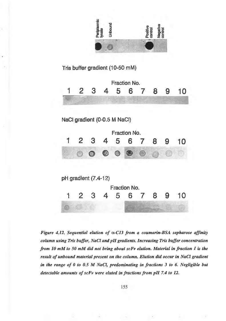
Tris buffer gradient (10-50 mM)
Fraction No.
1 2 3 4 5 6 7 8 9 10
N a d gradient (0-0.5 M NaCI)
Fraction No.
1 2 3 4 5 6 7 8 9 10e ® • ® © o
pH gradient (7.4-12)
Fraction No,
1 2 3 4 5 6 7 8 9 10
Figure 4.12. Sequential elution of OrC13 from a coumarin-BSA sepharose affinity
column using Tris buffer, NaCI and pH gradients. Increasing Tris buffer concentration
from 10 mM to 50 mM did not bring about scFv elution. Material in fraction 1 is the
result of unbound material present on the column. Elution did occur in NaCI gradient
in the range of 0 to 0.5 M NaCI, predominating in fractions 3 to 6. Negligible but
detectable amounts of scFv were eluted in fractions from pH 7.4 to 12.

Fraction No. 8 7 6 5 4 3 2 1 mw
— ^ 31 kDa
Figure 4.13. Western blot analysis of fractions from NaCl gradient elution of U-C13.
Recognition of scFv was with a-c-myc antibody. This shows the presence of a 31 kDa
protein fragment present in fractions 3 to 5.
156

1/Antibody dilution
Figure 4.14. The titration of a-C5 antibody against coumarin-BSA (C-BSA) using either
Tris buffer or PBS as wash buffers and diluent. Antibody diluted and washed with PBS,
pH 7.2, greatly reduced scFv titre, as compared with the use of 50 mM Tris, pH 7.4,
which yielded titres greatly in excess of 1 in 1,000. The absence of NaCl in the Tris
buffer did not lead to an increase in the non-specific binding of scFv to BSA.
157

4.2.4.5 Purification of a-C5 and OrC13 using NaCl gradient
10 ml samples of periplasmic lysate of a-C5 and a-C13 in 50 mM Tris, pH 7.4 were
purified on a 12.5 ml coumarin-BSA column using a 50 mM Tris, pH 7.4, 0 to 0.5 M
NaCl gradient, collecting 10 x 10 ml fractions. Dot blots of purification fractions for oc-C5
and a-C13 are shown in Figs. 4.15 and 4.16, respectively. Both showed elution of
significant levels of antibody in fractions 3 to 6. Later fractions contained lower yields
of scFv also. The fractions containing the greatest yields of antibody were pooled,
concentrated and analysed by SDS-PAGE and western blotting. SDS-PAGE showed
purification of two bands, one at approximately 31 kDa, and the other at approx. 29.5 kDa
(Figs. 4.17 and 4.18). The lower molecular weight band was present in large amounts in
both samples following concentration. In the a-C13 sample, the higher molecular weight
component was entirely lost following concentration of the affinity-purified sample. TTie
31 kDa molecular weight fragment isolated from affinity purification and detected by
SDS-PAGE was also detected on western blots which recognised the c-myc peptide tag
(Fig. 4.18). However, loss of the 31 kDa fragment was associated with loss of recognition
on western blots, the 29.5 kDa band showing no recognition by anti-c-myc. The loss of
recognition on western blots was also accompanied by loss of activity in ELISA (Figs.
4.19 and 4.20). Periplasmic lysates yielded titres in excess of 1 in 1,000. However, nearly
all activity was lost following affinity purification, a-C5 being marginally better than a-
C13. The 20 kDa fragment recognising anti-c-myc antibody was again present in
unpurified samples.
4.2.4.6 Purification of a-C14 using NaCl gradient
Culture supernatant from cultures of a-C14 were filtered through sterile 0.45 (am filters
and 0.15% (w/v) streptomycin sulphate added. Supernatant was diluted 1 in 4 with 50 mM
Tris, pH 7.4, and purified on a 12.5 ml coumarin-BSA column. Antibody was washed and
eluted as in section 4.2.4.5 and samples analysed by dot blotting (Fig. 4.21). Although
overall yields were poorer than for previous purifications of a-C5 and a-C13, antibody
did elute into fractions 4 to 6 on the salt gradient. Again, however, when analysed by
158

4.22), significant activity was lost when the purified fractions were concentrated. This was
again associated with loss of the western blot-active 31 kDa component and the
appearance of the non-active 29.5 kDa component (Figs. 4.23 and 4.24).
4.2.4.7 Optimised purification of anti-coumarin scFv antibodies from culture
supernatants and periplasmic lysates
Affinity purification of anti-coumarin clones was as previously described, with the
additional incorporation of 1 mM PMSF into all buffers used in affinity purification and
final samples. Culture supernatant from a-C14 production was concentrated 10-fold by
ultrafiltration (Amicon) and dialysed into 50 mM Tris, pH 7.4. Purified antibody-
containing fractions were pooled, dialysed into 50 mM Tris, pH 7.4 and concentrated on
Centricon 10 units (Amicon). In all three cases, elution profiles were as previously
described, with elution taking place in fractions 2 to 6 for a-C5 and a-C13, corresponding
to 0.05 M to 0.15 M NaCl, and fractions 4 to 6 for a-C14, corresponding to 0.15 M NaCl
(Fig. 4.25). All three samples showed elevated levels of the 31 kDa component over the
29.5 kDa fragment (Fig. 4.26). This also gave rise to titres in excess of 1 in 10,000 by
ELISA (Figs. 4.27a, 4.27b and 4.27c), even after two weeks storage at 4°C. Typical yields
of purified scFv were in the order of 1 mg scFv per litre of original culture.
4.2.4.8 Modification of scFv ELISA using anti-human F(ab’)2 antibody
The results of ELISA using a HRP-labelled anti-human F(ab’)2 antibody to detect scFv
a-C5 and a-C13 are shown in Figs. 4.28a and 4.28b. Both periplasmic lysates showed
decreased responses in association with decreases in antibody concentration. However,
overall signals were typically very poor with absorbances no higher than 0.3 at 492 nm.
159

ctooE T3
<dIZ s
C3
13
a SEc
CO o_ ^ D
ID.5 o© J= CM -S ■— ¿3**= ¡2 1. “2 t/j c2 « l a o om3 a-3 o_ o
<1 • • O @
Fraction No.3 4 5 6 7 8 9
Figure 4.15. Dot blot analysis of fractions of a-C5 purified in an NaCl gradient.
Culture supernatant, periplasmic lysate, unbound material from the column and column
wash were all analysed as were fractions eluted in NaCl gradient from 0 to 0.5 NaCl
(Fractions 1 to 10). This was followed by a wash in glycine buffer, pH 2.2. Highest
levels of scFv were found infractions 3 to 6. Lower concentrations of scFv was present
in the supernatant, with a considerable amount in the periplasmic lysate. The unbound
fraction was the material that passed through the column without binding. This
contained appreciable amounts of scFv. Little antibody was present in the buffer wash
prior to elution and also little eluted in glycine buffer, pH 2.2 which followed NaCl
elution. Positive control: Or NIP.
c oas —E -a¡3 c <oi 9 « 3 CM •> '5r " S f f l 3 *- . S oIL # 1 & %%
3 ® O T c 5 TO X « O nOT a. >* z> m 3 0.3 £ 8
O • • ©
Fraction No.1 2 3 4 5 6 7 8 9 10
© • • * « 0 9
Figure 4.16. Dot blot analysis of fractions of a-C13 purified in an NaCl gradient.
Highest levels of scFv were found in fractions 3 to 6. Quantities of scFv in supernatant,
periplasm, unbound fraction, buffer and low pH washes was similar to that for a-C5
(Fig. 4.16). Positive control: a-NIP.
160

a-C5 a-Cl31 2 3 4 5 6 7 8 910 1112
Figure 4.17. SDS-PAGE analysis of samples from the purification of a-C5 and a-C13
by NaCl elution from a coumarin-BSA sepharose affinity column. Lanes: 1,6,7,12:
Molecular weight markers; 2,8: Supernatant; 3,9: Periplasmic lysate; 4,10: Affinity-
purified fraction; 5,11: Affinity-purified and concentrated. Affinity purification brought
about the enrichment of a 31 kDa protein fragment. This had degraded to a 29.5 kDa
protein band following concentration. This was more significant in the a -C13 sample
than in a -C5. Although enrichment of the scFv was significant, several other molecular
weight species were still present following purification.
161

f
a-C51 2 3 4
a-C13 5 6 7 8
31 kDa
20 kDa -►
Figure 4.18. Western blot analysis of samples from the purification ofarC5 and a -C13
by NaCl elution from a coumarin-BSA sepharose affinity column. Lanes: 1,5:
Supernatant; 2,6: Periplasmic lysate; 3,7: Affinity-purified; 4,8: Affinity-purified and
concentrated. Recognition of 31 kDa components by the anti-c-myc antibody are clearly
present in the periplasmic lysates and affinity-purified samples of both OrC5 and QL-C13.
Following concentration, the recognition of a-C13 is completely lost, associated with
the complete loss of the 31 kDa band. However, the presence of significant levels of the
a-C5 31 kDa component in the affinity-purified fraction has led to its recognition by
anti-c-myc. High levels of protein in samples such as a-C13 periplasm led to non
specific binding and smearing.
162

1/Antibody Dilution
Figure 4.19. Analysis of samples from the NaCl gradient purification of a-C5 by
ELISA. Titres in excess of 1 in 1,000 were achieved for periplasmic lysate to coumarin-
BSA (C-BSA). This had dropped to less than 1 in 100 in the affinity-purified sample.
Only a negligible signal was obtained from culture supernatants.
1/A ntibody Dilution
Figure 4.20. Analysis of samples from the NaCl gradient purification of c/rC13 by
ELISA. Titres in excess of 1 in 1,000 were achieved for periplasmic lysate to coumarin-
BSA (C-BSA). Only negligible activity remained in the sample following affinity
purification. Culture supernatant yielded a signal only marginally higher than
background values.
163

Fraction No.1 2 3 4 5 6 7 8 9 10
© • e
Figure 4.21. Dot blot analysis of OrC14 fractions purified by NaCl gradient elution on
a coumarin-BSA sepharose affinity column. Antibody eluted primarily into fractions 4
to 6. Recognition ofscFv was with a -c-myc antibody. Large concentrations ofscFv were
present in the culture supernatant, and the diluted culture supernatant used for
application of sample to the affinity column. Fractions of sample after passage through
the column showed little unbound material upon initial application of sample, but
significantly more at the end of sample application. Some ScFv also eluted in pH 2.2
buffer. Positive control: u-NIP. Negative control: HB2151.
164

1 /Antibody dilution
Figure 4.22. ELISA analysis of samples from a-C14 purification by NaCl gradient
elution. Culture supernatant gave a titre in excess of 1 in 10,000 to coumarin-BSA (C-
BSA). This had decreased to 1 in 1,000following affinity purification. Signals were only
marginally above baseline levels following concentration.
165

1 2 3 4 5 6i
Figure 4.23. SDS-PAGE analysis of samples from the purification of a-C14 by NaCl
gradient elution on a coumarin-BSA sepharose affinity column. Lanes: 1,6: Molecular
weight markers; 2: Supernatant; 3: Unbound fraction; 4: Affinity-purified fraction; 5:
Affinity-purified and concentrated. The presence of a 31 kDa and a 29.5 kDa fragment
are both clearly visible in the affinity-purified sample, but only the 29.5 kDa component
is visible following concentration of the scFv.
166

Figure 4.24. Western blot analysis of samples from the purification of a-C 14 by NaCl
gradient elution. ScFv was detected with a-c-myc antibody. Lanes: Supernatant; 2:
Unbound fraction; 3: Affinity-purified fraction; 4: Affinity-purified and concentrated.
Recognition of the 31 kDa scFv is apparent in supernatant, unbound material and
affinity-purified samples, but has again been lost in the concentrated affinity-purified
sample. This blot corresponds to the SDS-PAGE gel in Fig. 4.23.
167

NaCI (M)0 .05 .1 .15 .2 .25 .3 .35 .4 .45 .5
a-C5 0 o • • © o o' €.*
x-C13 © o o m * i.
x-C14 • ©c BYE
Figure 4.25. Dot blot analysis of Fractions from the affinity purification of ctrC5, a-C13
and a -C14 by NaCI gradient elution on a coumarin-BSA sepharose affinity column,
with the incorporation ofPMSF. Recognition ofscFv was with a -c-myc antibody. o C 5
and a -C13 show maximum scFv elution in fractions from 0.05 to 0.25 M NaCI and a -
C14 shows maximum elution in fractions from 0.15 to 0.3 M NaCI.
168

1 2 3 4 5
Figure 4.26. SDS-PAGE analysis of arC5, a-C13 and OrC14 affinity-purified and
concentrated using NaCl gradient in the presence of PMSF. Lanes: 1,5: Molecular
weight markers; 2: oC 5; 3: a-C13; 4: a-C M All three scFv preparations show the
greatly improved yield of 31 kDa fragment over the 29.5 KDa component.
169
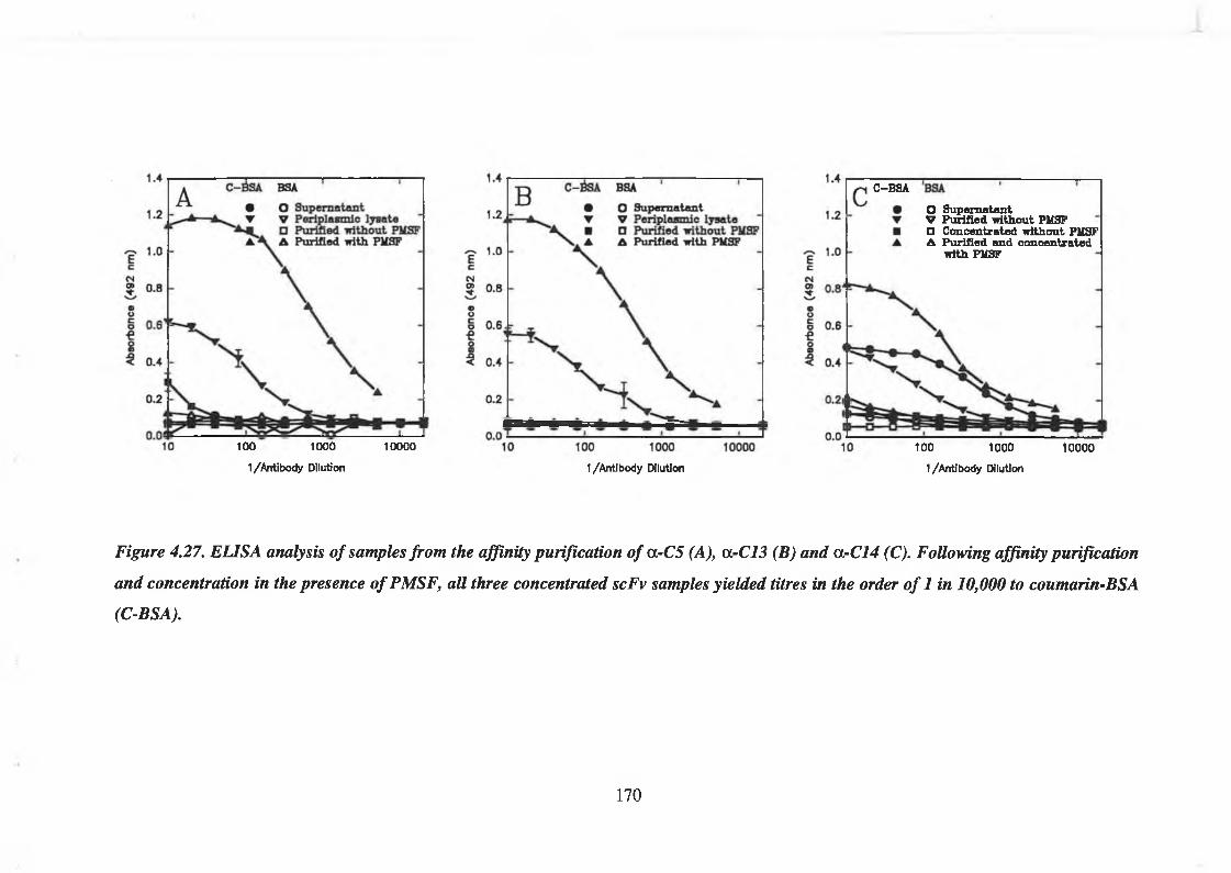
100 1000
1/Antibody Dilution
10000
C-BSA
O Supernatant V Purified without PMSF □ Concentrated without PMSF A Purified and concentrated
■with PMSF
1/Antibody Dilution
100 1000
1/Antibody Dilution
10000
Figure 4.27. ELISA analysis of samples from the affinity purification of a-C5 (A), a-C13 (B) and a-C14 (C). Following affinity purification
and concentration in the presence of PMSF, all three concentrated scFv samples yielded titres in the order of 1 in 10,000 to coumarin-BSA
(C-BSA).
170

Eceg£oucQ■e8S
Q 1 /Antibody dilution 1/Antlbody dilution
1/Antibody dilution 1/Antibody dilution
Fig. 4.28A and B. ELISA analysis ofarC5 (A) and a-C13 (B) scFv using HRP-labelled
anti-human F(ab')2 antibody. Response to decreases in antibody concentration were only
achieved at high dilutions of conjugated antibody. However, signals were extremely low,
the highest signals being less than 0.3 AU at 492 nm.
171
4.2.5 Characterisation of anti-coumarin scFv antibodies
4.2.5.1 Optimisation of competition ELISA for the detection of free coumarin
4.2.5.1a Selection of optimal antibody concentration
Optimal antibody concentration was calculated according to (Crowther, 1995) from the
titres yielded in Figs. 4.27a, b and c. This was selected as 70% maximum binding, and
corresponded to 1 in 400, 1 in 200 and 1 in 100 dilutions of a-C5, a-C13 and a-C14
antibodies, respectively.
4.2.5.1b Selection of optimal coumarin-BSA coating concentrations
The effect of coating concentration on signal for all three anti-coumarin scFvs is shown
in Fig. 4.29. Levels above 150 (jg/ml did not increase signals, so 150 (ig/ml was selected
as optimal coating concentration of coumarin-BSA.
4.2.5.1c Competition EUSA analysis of free coumarin using anti-coumarin scFv
The ability of the three anti-coumarin clones to compete between bound coumarin-BSA
and free coumarin was assessed (Fig. 4.30). All three clones showed no recognition of
free coumarin in the presence of 0.05% methanol, at coumarin concentrations ranging
from 0.05 to 50 |ag/ml. Competition ELISAs were repeated over a range of antibody
dilutions (Figs. 4.31 A, 4.32B and 4.32C), but these assays confirmed the original result
that showed no recognition between the anti-coumarin scFv antibodies and free drug.
4.2.6 Assessment of the effect of methanol and NaCl concentration on competition
ELISA with a-C5, a-C13 and a-C14
The effect of methanol concentrations up to 25%, and NaCl concentrations up to 0.5 M
on the binding of a-C5, a-C13 and a-C14 to free coumarin were assessed at the 1 (ig/ml
free coumarin level (Figs. 4.32, 4.33 and 4.34). NaCl significantly reduced the binding of
172

all three scFv antibodies to surface coumarin-BSA. The presence of methanol appeared
to enhance the interaction of antibody with coumarin-BSA significantly in the case of a-
C14 (Fig. 4.34).
Data for different methanol concentrations were grouped, and student independent and
paired t-tests performed, comparing the means of populations in the presence and absence
of 1 |ig/ml coumarin. In the cases of a-C5 and a-C13, a significant difference in binding
to coumarin-BSA in the presence and absence of free coumarin was detected at the 0 and
Table. 4.2. Mean (X) and standard deviation (s) of populations from ELISA results
investigating the effect of NaCl concentration on the binding of anti-coumarin
scFvs in the presence and absence of free coumarin (n=5).
NaCl concentration (M)
0 0.001 0.01 0.1 0.25 0.5
a-C5
1 ng/ml X 0.631 0.630 0.622 0.356 0.110 0.105
coumarin s 0.012 0.007 0.045 0.039 0.010 0.005
No free X 0.656 0.644 0.641 0.390 0.117 0.099
coumarin s 0.012 0.007 0.006 0.026 0.007 0.006
a-C13
1 ng/ml X 0.703 0.702 0.705 0.610 0.250 0.222
coumarin s 0.005 0.006 0.003 0.028 0.008 0.028
No free X 0.715 0.719 0.713 0.628 0.261 0.218coumarin s 0.009 0.007 0.008 0.025 0.014 0.015
cc-C14
1 (ig/ml X 0.604 0.596 0.597 0.216 0.091 0.087coumarin s 0.031 0.037 0.026 0.040 0.004 0.007
No free X 0.607 0.602 0.604 0.221 0.092 0.081coumarin s 0.024 0.026 0.029 0.058 0.005 0.002
173

0.001 M NaCl concentration levels (n=5). These were significant at the 95% confidence
level (Table 4.2) and Figs. 4.35A, B and C. In most instances, the signal in the presence
of 1 ng/ml free coumarin was lower than that with no free coumarin, but this was only
statistically significant at the 0 and 0,001 M NaCl concentrations for a-C5 and a-C13.
4.2.7 The effect of NaCl concentration on anti-coumarin scFv binding to coumarin-
BSA
The effect of NaCl concentration from 0 to 0.2 M on the binding of the three anti-
coumarin-BSA clones is shown in Fig. 4.36. Maximum binding occurred at 0 M NaCl and
this decreased rapidly to baseline levels when the NaCl concentration reached 0.2 M.
4.2.8 Competition of anti-coumarin-BSA scFv antibodies against coumarin-BSA
When normalised to a percentage of maximum binding, all three anti-coumarin-BSA scFv
clones showed competition binding profiles to coumarin-BSA that were very similar to
one another (Fig. 4.37). Relative affinities of the scFvs to coumarin-BSA were calculated
as the molarity of free antigen which yielded 50% maximum absorbance, according to
Roben (1993) and Tijssen (1985). This yielded relative affinities for the three clones of
3 x 10⁷ M'1, 4.5 x 10⁷ M'1 and 2.5 x 10⁷ M"1 for a-C5, a-C13 and a-C14, respectively.
Since the coupling ratio of coumarin to BSA was 11.5:1, this would give relative affinities
to the hapten of 3.45 x 10⁸ M'1, 5.18 x 10⁸ M'1 and 2.88 x 10s M'1 for a-C5, a-C13 and
oc-C14 respectively. This resulted in an affinity ranking of a-C13>a-C5>a-C14.
4.2.9 Competition of a-C5 scFv with different coumarin-protein conjugates
The interaction of the a-C5 scFv clone with different coumarin-protein conjugates was
assessed. Four conjugates were selected: three coumarin-BSA conjugates with different
coupling ratios of coumarin to protein (11.5, 5.7 and 2.77 moles coumarin/mole BSA),
and a coumarin-ovalbumin conjugate (7 moles coumarin/mole ovalbumin). These were
used as competition reagents against bound coumarin-BSA (11.5 moles/mole) (Fig. 4.38).
174

All conjugates showed that they had been recognised by a-C5 scFv. In the case of the
three coumarin-BSA conjugates, the level of recognition was proportional to the level of
conjugation. a-C5 also recognised coumarin when attached to ovalbumin, giving a
response similar to that of the coumarin-BSA (11.5 moles/mole) conjugate. However,
ovalbumin is a smaller protein than BSA, being only approximately 44 kDa, this would
equate to a coupling ratio of 10.5 moles coumarin/mole BSA, very similar to the 11.5
moles/mole conjugate.
4.2.10 Assessment of the multi-specificity of the anti-coumarin scFv clones to
different coumarin derivative-BSA conjugates
Fig. 4.39 shows that the three anti-coumarin scFv antibodies bound specifically to
coumarin-BSA, but not to 7-hydroxycoumarin-BSA or 7-hydroxycoumarin-glucuronide-
BSA (see section 5). The level of binding to 7-hydroxycoumarin-BSA and 7-
hydroxycoumarin-glucuronide-BSA was only fractionally higher than that for the control
BSA.
4.2.11 Use of anti-coumarin-BSA scFv antibodies as western blot reagents
All three antibodies were used as western blot reagents on samples of coumarin-BSA,
BSA, coumarin-OVA and OVA. However, due to their intolerance of NaCl, salt-
containing buffers were substituted with 50 mM Tris, pH 7.4. The level of non-specific
interaction between antibody and proteins on the surface of a membrane can be controlled
by the incorporation of NaCl at different concentrations as well as detergents and the
number of washing steps involved. Normally, a level is selected which reduces non
specific binding to a negligible level, while still giving good interaction with the specific
protein. Low stringency buffers containing no NaCl resulted in high levels of non-specific
background staining (results not shown).
175

Log C-BSA conc. (m g/m l)
Figure 4.29. The effect of coumarin-BSA coating concentration on signal for the
optimisation of competition ELISA in the analysis of coumarin. Increasing coating
concentration above 150 pg/ml did not result insignificant increases in the amount of
scFv bound. Optimal coumarin-BSA coating concentration for competition ELISA was
estimated to be 150 fig/ml.
Free coumarin (ug/m l)
Figure 4.30. Competition ELISA of free coumarin (in 0.05 % methanol) against
coumarin-BSA using a-C5, a-C73 and OrC14. No competition with free coumarin was
apparent at concentrations of free drug from 0.05 to 50 pg/ml.
176

0.1 1 10
Frw ooumorin (uM)
a -C 1 3 dilution
O 1/10 • 1/20 V 1 /4 0 r i/bo □ l/ieo ■ 1/880 A 1 /840 A 1/1J280
EcC*i3.
a-C14 dilution 0 1/10 • i/soV 1 /4 0 ▼ 1 /8 0 □ 1 /1 8 0 ■ 1 /380 A 1 /840 A 1 /1 ,860
Fn* coumortn (uM)
Figure 4.31 A, B and C. Repeat analysis of competition EUSAs using cl -C5, a-C13 and a-C14 for the detection of free coumarin using a
range of scFv concentrations. No observable interaction between scFv and free coumarin was apparent at any of the drug concentrations
or antibody dilutions analysed.
0.1 1 1 1 1----- 1—0.001 0.01 0.1 1 10 100
Fr»« coumorfn (uM)
177

a. 1 /Lig/ml free coumarin b. No free coumarin0.7
0.6
CM2 0.4€>o0 0.3■e
1 0.2
0.1
0.0 0.
Figure 4.32. The effect of NaCl and methanol concentration on the binding of o.-C5 in the presence of 0 and 1 fig/ml coumarin. Increasing
NaCl concentration drastically reduced the binding interaction between a-C5 and coumarin-BSA. Methanol concentration had no significant
effect between 0.01 and 25 % (v/v) (as found by paired and independent student t-tests). Significant reduction in the binding to coumarin-BSA
occurred in the presence of 1 fig/ml coumarin at the 0 and 0.001 M NaCl levels. (Values for 0 M NaCl not shown due to logarithmic scale
used. See Table 4.2).
178

a. 1 /xg/m l free coumarin b. No free coumarin
NaCI (M) NaCI (M)
Figure 4.33. The effect of NaCI and methanol concentration on the binding of a-C/3 in the presence of 0 and 1 fig/ml coumarin. Increasing
NaCI concentration drastically reduced the binding interaction between a-C13 and coumarin-BSA. Methanol concentration had no significant
effect between 0.01 and 25 %(v/v). Significant reduction in the binding to coumarin-BSA occurred in the presence of 1 fig/ml coumarin at
the 0 and 0.001 NaCI levels.
179

a. 1 /¿g/m l free coumarin b. No free coumarin
0.001
NaCI (M)
0.01 0.1
NaCI (M)
Figure 4.34. The effect of NaCI and methanol concentration on the binding of a-C14 in the presence of 0 and 1 fig/ml coumarin. Increasing
NaCI concentration drastically reduced the binding interaction between a-C14 and coumarin-BSA. Methanol concentration had a significant
effect, increasing the level of interaction between a -C14 and coumarin-BSA between 0.01 and 25 %(v/v), most significantly on samples
containing 0.01 M and 0.1 M NaCI (see insert). No significant reduction in binding to coumarin-BSA was observed in the presence of 1/ig/ml
coumarin.
180

0.7
0.6
~ 0.5 c
CM
? 0.4
s§ 0.3■e8€ 0.2
0.1
0.0
Isa 1 w i/m icou marin 0 /ifl/mlcou marin
0.001 0.01 0.1 0.25 0.5NaCI (M)
0.8
0.7
~ 0.6
c CM? 0.5
8o 0.4■eon5 0.3
0.2
0.1
B
P S i 1 /*g/m lt ____ coumarin
Ih i n o ¿ig/mi coumarin
0.001 0.01 0.1 NaCI (M)
0.25 0.5
0.7
0.6
~ 0.5 c <M? 0.4DO0 0.31 «5 0.2
0.1 -
0.0
K M 1 /xg/ml coumarin 0 jig /m l coumarin
0.001 0.01 0.1 0.25 0.5NaCI (M)
Figure 4.35A, B and C. Competition immunoassay of OrC5 (A), a -C13 (B) and a-C14 (C) against 1 fig/ml free coumarin at different NaCI
concentrations. All three scFv clones show slightly lower absorbances in the presence of 1 fig/ml coumarin compared to 0 fig/ml coumarin
at the 0, 0.001, 0.01 and 0.1 M NaCI concentrations. This was significant for arC5 and a-C l3 at the 0 and 0.001 M NaCI concentrations
(as determined by independent and paired t-tests).
181

NaCI (M)
Figure 4.36. The effect of NaCI concentration on the binding of anti-coumarin-BSA
scFvs to coumarin-BSA. All three scFvs show NaCI sensitivity from as low as 0.05 M.
Baseline signals occur when NaCI concentration reaches 0.2 M.
Log C—BSA conc. (M)
Figure 4.37. Relative affinities of a-C5, a -C13 and a-C14. All three scFvs show 50%
binding with free coumarin-BSA at approx. 5 x 10⁷ M'1 coumarin-BSA, which
corresponds to a molarity of coumarin of approximately 5.75 x 10s M'1.
182

1----------- 1----------- 1----------- r-O C-BSA (2.77 m oles/m ole)
0.5 |- • C-BSA (6.7 m oles/m ole)V C-BSA (11.5 m oles/m ole) ▼ C-OVA (7 m oles/m ole)
0.4 -CM?W©§ 0-3 h-Ok_
g $
0.2 h
0.1 0.001 0.01 0.1 1 10 100
Conjugate conc. (ug/ml)
1000
Figure. 4.38. Competition of OrC5 scFv for different coumarin-protein conjugates.
Competition for coumarin-BSA (C-BSA) conjugates appears to be proportional to the
level of conjugation of coumarin on the BSA carrier protein, oo-C5 also recognised
coumarin-ovalbumin (C-OVA) to the same degree as coumarin-BSA (11.5 moles
coumarin/mole BSA). Due to the lower molecular weight of ovalbumin, this would be
equivalent to a 10.5:1 coupling ratio on BSA.
183

BSA C-BSA 70HC-BSA 70HCG-BSA
Figure 4.39. ELISA of anti-coumarin scFv antibodies a -C5, a -C13 and a -C14 to
coumarin and its derivatives 7-hydroxycoumarin (70HC) and 7-hydroxycoumarin-
glucuronide (70HCG) conjugated to bovine serum albumin (BSA). Antibody bound only
to the coumarin-BSA conjugate and showed only background binding to 70HC-BSA
and 70HCG-BSA as compared with the BSA control
184

The BIAcore sensorgrams for the three anti-coumarin scFv clones are shown in Fig. 4.40.
Antibodies were analysed at 1 mM concentrations on a surface of coumarin-BSA (2.77
moles/mole) of 1,600 response units (RU). Kinetic data was calculated for the three
clones. Analysis showed that the three clones did not adhere perfectly to first order
kinetics. However, all three clones gave affinity constants based on the observed on and
off rates in the 109 M 1 range and were affinity ranked in the order a-C13>a-C5>a-C14.
This affinity ranking corresponded with that found by competition immunoassay (Fig.
4.37). Controls using BSA coupled to the sensor chip surface (6,000 RU) did show some
background binding of the scFv antibody to the BSA-dextran matrix (Fig. 4.41) of 31.6
RU, compared to 167.2 RU for the a-C5 clone.
3000
2500
3 * 2000Q£38 1500€>09c o
n 1000 «a:
500
00 200 400 600 800 1000 120C
Time (s)
Figure 4.40. BIAcore sensorgrams of anti-coumarin clones a-C5, a -C l3 and a-C14 to
a coumarin-BSA surface coupled to dextran and yielding 1,600 response units (RU). All
three clones show association and dissociation phases of the three clones. Kinetic data
based on these sensorgrams yielded affinity constants in the range of l ( f M'1.
4.2.12 Characterisation and kinetic analysis of anti-coumarin-BSA scFv antibodies
using BIAcore™
185

3000 ---------- 1— r 1 i i
r
2500 - -
3 2000 -
------Coumarin-BSA surface----- BSA control surface
£3 1500 —
maog 1000 - -
O'
500 -
n -------- 1_____ I_____ 1_____ I_____ I_____0 200 400 600 800 1000 1200
Time (e)
Figure 4.41. BIAcore sensorgrams of a-C5 scFv (1 mM) passed over a surface of
coumarin-BSA (1,600 RU) linked to dextran and over bovine serum albumin (BSA)
linked to dextran (6,000 RU). This showed some interaction of the scFv with the BSA-
dextran surface (31. 6 RU) as compared to 167.2 RU over the coumarin-BSA surface.
186

4.3 PRODUCTION OF ANTI-7-HYDROXYCOUMARIN ANTIBODIES FROM THE
NISSIM LIBRARY
4.3.1 Phage ELISA screening of first anti-7-hydroxycoumarin library
93 clones from the first anti-7-hydroxycoumarin library, following three rounds of affinity
selection on 70HC-BSA with BSA as blocking reagent, were screened on 70HC-BSA and
BSA (Fig. 4.42). This showed that all the clones which were positive to 70HC-BSA were
positive on the BSA control also. Positives were selected as those having values in excess
of the positive control well, H7. This creates some discrepancies between the two plates.
However, all clones which show values well in excess of the negative control on one
plate, also did so on the other, indicating that these clones were also positive, if not
reaching the positive control signal.
4.3.2 Phage ELISA analysis of second anti-7-hydroxycoumarin library
A second anti-7-hydroxycoumarin library was prepared with affinity selection using
alternating 70HC-protein conjugates and different blocking proteins (see section 2.3.3.4
and 2.3.3.6). Screening of 42 clones against 70HC-0VA and OVA showed no response
to the OVA control, while showing good enrichment of positive clones to 70HC-0VA
(Fig. 4.43).
A second phage ELISA of 96 clones was performed (Fig. 4.44). From this, 43 positive
clones were selected and denoted a-70H Cl to a-70HC43 and used for further analysis.
4.3.3 Analysis of supernatant and periplasmic lysates of anti-7-hydroxycoumarin
clones for the presence of scFv
Dot blot analysis of the periplasmic lysates and culture supernatants of the 43 positive
anti-7-hydroxycoumarin clones is shown in Fig. 4.45. All 43 clones showed no secretion
of antibody into periplasm or culture supernatant. Clones picked at random from the
library showed secretion in 2 out of 5 clones. Since no individual clone exhibited better
187

secretion characteristics than another, the first twelve clones, (X-70HC1 to a-70HC12
were selected for further studies on antibody refolding. a-70H Cl to (X-70HC11 were
analysed a second time and compared to controls and again showed poor scFv production.
Clones (X-70HC6 to a-70H C ll appeared slightly better, with (X-70HC10 and a-70H C ll
looking moderately better than the rest of the population.
4.3.4 Use of BIAcore for the affinity selection of anti-7-hydroxycoumarin clones from
the Nissim library
To enrich for antibodies to 7-hydroxycoumarin-BSA, the phage antibodies of the original
Nissim library were passed across a BIAcore chip surface immobilised with 7-
hydroxycoumarin-BSA according to section 2.3.3.5. The phage counts eluting from the
chip surface are shown in Fig. 4.46. A total of approximately 2.74 x 10⁸ phage antibodies
(tfu) from the original Nissim library were passed across the BIAcore chip surface. Phage
antibody eluting in wash buffer fell to a level of approximately 5 x 10² tfu/ml. This level
of phage elution was quite constant. Upon the addition of glycine buffer, however,
specific elution of bound phage did occur. A total output of eluted phage of 1.26 x 10⁴
tfu was produced. This represented an enrichment in excess of 1 in 20,000. No signal was
produced by this binding on BIAcore sensorgrams (data not shown).
188

7-hydroxycoumarin-bovine serum albumin
Slá&Ml SàèlyÉ IfeilÉl S i l
Bovine serum albumin
« l i
Figure 4.42. Phage ELISA analysis of anti-7-hydroxycoumarin library clones on 7-
hydroxycoumarin-BSA and BSA surfaces following three rounds of affinity selection.
Both ELISAs show similar patterns of positives (shaded) and negatives (unshaded).
Discrepancies only occur for wells that fall slightly below the positive control value, but
are still positive readings. Positive clones are shaded. Positive control: H7. Negative
control: H6.
189

7-hy droxyc oumarin- ovalbumin Ovalbumin
1 Ü Ü § i 3 4 5 H I 7 8 9 10 11 12A 0.348 l l i i ! 0.416 0.294 0.277 l l i l i i 0.083 0.122 0.108 0.077 0.078 0.136B 0.431 ¿III!0.407 0.265 0.456 0.375 0.099 0.112 0.106 0.076 0.107 0.103C 0.314 0.377 0.356 0.293 0.292 0.365 0.076 0.096 0.089 0.081 0.077 0.097D 0.437 0.315 0.436 0.477 0.409 0.100 0.082 0.146 0.116 0.150 0.129h i lilll 0.475 0.485 0.466 0.402 0.173 0.126 0.128 0.129 0.133 0.124F 0.487 ' MMi0.463 0.461 0.459 0.417 0.136 0.132 0.121 0.114 0.115 0.115G 0.382 0.277 0.323 0.354 0.273 0.390 0.091 0.084 0.093 0.107 0.073 0.124H 0.382 $1111 0.330 0.365 0.474 0.494 11111110.158 0.077 0.090 0.119 0.069
Figure 4.43. Phage ELISA of anti-7-hydroxy coumarin library following three rounds
of affinity selection on alternating protein conjugates of 7-hydroxy coumarin with
different blocking proteins with screening on 7-hydroxycoumarin-ovalbumin (70HC-
OVA) and ovalbumin (OVA). All clones when screened on 70HC-0VA showed elevated
signals above background when compared to the same clones on OVA. None of the
clones screened on OVA gave signals above the negative control (H6). Positive control:
H 7 ' 1 2 3 4 5 6 7 8 9 10 11 12
A B C D E F G H
Figure 4.44. Phage EUSA analysis of anti-7-hydroxy coumarin library. 43 clones
showing the highest responses were selected as positive from the phage ELISA. Positive
control: H7. Negative control: H6.
190

Periplasm Supernatant
1 2 3 4 5 6 7 8 9 10 11 12
A B C D E F G H
Figure 4.45. Dot blot analysis of supernatant and periplasmic lysates of anti-7-
hydroxycoumarin positive clones from phage ELISA. The 43 positive clones (rows A to
G and HI and H7) were compared to five clones taken at random from the library (H2
to H6 and H8 to H12). All 43 clones failed to demonstrate any secretion into the
periplasm or culture supernatant. The control clones tended to show higher levels of
scFv production, especially H3 and H4.
191

105
E\33=£ 104 -M
0)OfO-CQ_
103
0 5 10 15 20 25 30 35 40
Time (min)
Figure 4.46. Use of BIAcore for the enrichment of anti-7-hydroxycoumarin positive
clones from the Nissim library. Approximately 2.75 x l ( f phage antibodies were injected
onto the sensor ship immobilised with 7-hydroxycoumarin-BSA. The phage titres eluting
from the sensor are shown. During washing with buffer, titres fell to 5 x 10² tfu/ml.
When glycine elution buffer was applied to the sensor (pH 2.2), this titre increased to
7 x 10* tfu/ml. Over the 20 minute elution period, this equated to 1.26 x 10⁴ phage.
192

4.3.5 Comparison of lysis methods in the isolation of scFv from intracellular
compartments
The effect of periplasmic and intracellular lysis of clones a-70HC10 and a-70H C ll is
shown in Fig. 4.47. This showed that freeze-thawing again released no scFv into the
periplasm, but some antibody present in the intracellular compartment following
sonication. Dot blot signals were also improved when antibody was denatured in 7.5 M
guanidine hydrochloride.
Clones a-70HC6 to a-70HC8 were also sonicated and denatured in 8 M urea, which was
also found to improve signals on dot blots, but still showed very low production of anti-7-
hydroxycoumarin scFv antibody (Fig. 4.48).
4.3.6 Analysis of denatured and refolded anti-7-hydroxycoumarin clones
Clones a-70HC6 to a-70H C ll were denatured and refolded in arginine buffer for two
days. Refolding greatly improved dot blot signals (Fig. 4.49). This was particularly
evident in clones a-70HC10 and a-70H C ll.
4.3.7 Induction and affinity purification of (X-70HC10
Analysis of the fractions from the affinity purification of a refolded intracellular lysate of
a-70HC10 is shown in Fig. 4.50. Antibody eluted from the 7-hydroxycoumarin column
(7-hydroxycoumarin-4-acetic acid coupled to EAH-sepharose 4B) in glycine buffer, pH
2.2 in fractions 3 to 5. Following pooling of these fractions, they were found to have
enriched for a 31 kDa protein fragment as seen by SDS-PAGE analysis (Fig. 4.51), as
well as a lower molecular weight fragment, the molecular weight of which could not be
accurately determined from the gel. Only the 31 kDa fragment was recognised by anti-c-
myc antibody on western blots (Fig. 4.52). Analysis under reducing and non-reducing
conditions indicated that a-70HC10 was a single peptide. Recognition of the 31 kDa
fragment only occurred on western blots following purification. Antibody may not have
been present in sufficient quantities to yield a signal on western blots prior to purification.
193

Figure 4.47. Dot blot analysis of lysis methods in the release of scFv from a-70HC10
and 0-70HC11. Positive clone a-NIP showed good release of scFv following freeze-
thawing and sonication, as well as into the culture supernatant. TGI (pHENl) also
showed secretion of c-myc peptide. No antibody was present in the periplasmic lysate
following freeze-thawing, but was present following sonication and dénaturation.
Dénaturation of (X-70HC10 and (X-70HC11 significantly improved signals.
Figure 4.48. Dot blot analysis of the sonication and dénaturation of U-70HC6, a-
70HC7 and a-70HC8 clones. Low yields of scFv were produced following sonication.
Signals were again enhanced following dénaturation in 8 M urea. The negative control,
HB2151 showed no scFv production in all samples as expected. Positive assay controls:
a -NIP and TGI (pHENl).
c . i . I SON.W \tN 1 mu
ICI lantl-Niri
ICI tyllMl)
194

Sonicated
Denatured
Refolded
Figure 4.49. Dot blot analysis of anti-7-hydroxy coumarin scFv clones a-70HC6 to a-
70HC11 (a-70HC9 omitted). All five clones tested showed poor signals of scFv after
sonication. This improved upon dénaturation and was significantly better following
refolding in arginine. a-70HC10 was significantly better than the others and a-
70HC11 was moderately better. Positive clones TGI (pHENl) and TGI (o NIP) showed
good levels of c-myc peptide and soluble scFv, respectively. Negative control HB2151
showed no scFv production as expected.
St SON
•St DEN
tci tci70HM 70*0-1« 70HC-11 IpKWH HM151
• t
• tf t 1 1
195

- s w
f f j < t£ / Fractions
2
to ? < € * » KM u|M| A «WCTt*»/S1 X. * j ’\ jf
4 5 6 7 8 9H £ * r
10 11 12 13 14 1510 '1 /t jj '5
16 17 18 19 20 21n i? if ** ¿ 4 11
22 23 24 25 -ve +ve1 I Z X I < r -vc.
•Figure 4.50. Dot blot analysis of fractions from the elution of a-70HC10. ScFv eluted
at pH 2.2 from the EAH-sepharose 7-hydroxycoumarin column primarily in fractions
3 to 5 with some material in fractions 2 and 6 also. Positive control: OrNIP. Negative
control: HB2151.
196

31 kDa "4
Figure 4.51. SDS-PAGE analysis of a-70HC10 refolded scFv antibody before and after
affinity purification. Lanes: 1-4: Reducing conditions; 5-8: Non-reducing conditions;
1,5: a-NIP (positive control); 2,6: HB2151 (negative control); 3,8: Purified U-70HC10;
4,7: Unpurified Or70HC10. Purification resulted in the enrichment of a 31 kDa
fragment and a lower molecular weight band that was not recognised by western
blotting (Fig. 4.52). Purification reduced the amount of non-specific protein in the
sample. Analysis under reducing and non-reducing conditions showed that the 31 kDa
protein was a single peptide fragment.
197

1 2 3 4 5 6 7 8
31 kDa
■ L
Figure 4.52. Western blot analysis of Or70IIC10 refolded scFv antibody before and
after purification. Detection of scFv was with a-c-myc antibody. Lanes: 1-4: Non
reducing conditions; 5-8: Reducing conditions; 1,5: a-NIP; 2,6: HB2151; 3,8:
Unpurified a-70HC10; 4,7: Purified Or70HC10. Purification led to the recognition of
a 31 kDa fragment under both reducing and non-reducing conditions. No antibody
could be detected in the unpurified samples.
198

4.3.8 ELISA of purified (X-70HC10
The ELISA of the purified (X-70HC10 scFv against 70HC-BSA and BSA is shown in Fig.
4.53. The antibody showed no response to BSA, but did respond to 70HC-BSA with a
very poor signal which was proportional to the amount of 70HC-BSA on the surface,
with signals on the 6.25 ng/ml 70HC-BSA wells still higher than controls at 1 in 20
dilution of scFv.
4.3.9 Affinity purification of a-70HC9
Refolded a-70HC9 was affinity purified on 70HC-EAH-sepharose and analysed by dot
blotting (Fig. 4.54). This showed a-70HC9 to be present in fractions 1 to 4. A sizeable
amount did not bind to the column and passed through in the unbound fraction. Dot blot
analysis also showed that antibody did not elute at pH 3.5 (Fig. 4.55). SDS-PAGE also
showed that although enrichment of a 31 kDa fragment occurred, poor levels of
purification were achieved (Fig. 4.56). Western blotting showed the presence of the 31
kDa component in the refolded lysate material, the unbound fraction and the affinity-
purified sample, detected by a-c-myc antibody. This was still present following
concentration of a-70HC9 (Fig. 4.57), which was also shown by dot blotting (Fig. 4.58).
199

Ec
CM0!>
«0 c D■e1 £
0.18
0.16
0.14
0.12
0.10
70HC-BSA Cone (ug/m l)
10 100 1000
1/Antibody dilution
10000
Figure 4.53. ELISA analysis of purified a-70HC10 scFv. Antibody recognised 7-
hydroxycoumarin-BSA (70HC-BSA) but not BSA alone. Signals were very low, even
at low dilutions (0.17 AU at 1 in 10), but gave titres in excess of 1 in 100.
200

Off Unbound Hash l 2 3
4 5
ffC9
Figure 4.54. Dot blot analysis of fractions from the purification of a-70HC9. Detection
of scFv was with Orc-myc antibody. Positive control (+ve): Or NIP. Negative control (-
ve): HB2151. A large proportion of the antibody did not bind to the column and passed
through in the unbound fraction. Antibody eluted in fractions 1 to 4.
T««C> H 2.2
#h f
0Ufikotlftd Hm I
'I l i l t t
1i
Huh
PR . y |
Unbound H u h E l u i t f
Figure 4.55. Dot blot analysis showing elution of a-70HC9 in pH 2.2 and pH 3.5
buffers. Detection of scFv was with Orc-myc antibody. Positive control (+ve): a-NIP.
Negative control (-ve): HB2151.ScFv eluted at pH 2.2, but not at pH 3.5.
201

1 2 3 4 5 6 7 8
31 kDa - t
Figure 4.56. SDS-PAGE analysis of affinity-purified Or70HC9. Lanes: 1: Molecular
weight markers; 2: HB2151; 3: a-NIP; 4: Refolded Or70HC9; 5: Unbound fraction;
6: Affinity-purified a-70HC9; 7: Concentrated column washings; 8: Concentrated
affinity-purified fraction. Enrichment of a 31 kDa protein fragment can be seen
following concentration of the affinity-purified sample.
Figure 4.57. Western blot analysis of affinity-purified Or70HC9. Detection of a 31 kDa
fragment was seen in the refolded sample, the unbound fraction and in the concentrated
affinity-purified sample, although the latter is extremely faint. No scFv was detected in
column washings. Lanes: 1: Molecular weight markers; 2: a-NIP; 3: HB2151; 4:
Refolded a-70HC9; 5: Unbound fraction; 6: Affinity-purified O.-70HC9; 7:
Concentrated column washings; 8: Concentrated affinity-purified a-70HC9.
1 2 3 4 5 6 7 8
31 kDa
202

70HC9
RtfoUtdCone.
elu*t«
Unbound70HC9conc.
MTinitypur* f i
1-4
-v*
Figure 4.58. Dot blot analysis of affinity-purified O-70HC9. ScFv material was detected
in the unbound fraction and in the pooled affinity-purified fractions 1 to 4. The signal
was significantly enhanced following concentration of the affinity purified fraction
(Or70HC9 conc.). No antibody was detected in concentrated column washings (conc.
eluate). Negative control (-ve): HB2151. Positive control (+ve): Or NIP.
203
4.3.10 Induction optimisation of (X-70HC10
a-70HC10 culture supernatant, periplasmic lysates and intracellular lysates were analysed
by dot blotting for the presence of scFv following induction with IPTG (Fig. 4.59). During
induction, several parameters were monitored. These were growth period prior to induction
(4, 8 and 16 h), IPTG concentration (0.01, 0.1 and 1 mM) and growth period after
induction. No scFv was present in culture supernatant or periplasmic lysate under any of
the conditions tested. ScFv was present in most intracellular lysates, but no great
differences in scFv yield were evident due to the changes in growth conditions. In general,
8 h growth pre-induction showed the overall highest levels of antibody, with 0.1 mM
IPTG and 16 h post-induction growth being marginally better. Also, the 1 mM IPTG with
16 h pre-induction growth was very good at 0 and 4 h post-induction. These two scenarios
were analysed a second time, this time also looking at the effect of growth temperature
post-induction. This now showed 8 h growth pre-induction with 0.1 M IPTG and 2 h post
induction growth at 25°C to be optimal for scFv antibody yield (Fig. 4.60). However,
further scale ups using these induction conditions failed to significantly increase scFv
yield (data not shown).
4.3.11 Precipitation of purified and concentrated scFv preparations
Following denaturation and refolding of a-70HC9 and a-70HC10, these were
characterised by the precipitation of protein material following affinity purification and
concentration. When analysed, no viable titres were obtained for a-70HC9 (data not
shown).
204

Culture supernatantl i
1 bN 8.1 *K ,81 «K
« h
1 nH 8.1 xK .81 nK
4 h
I hK nfl1 "l 11 — . ........... 1 ’48 h
24 h|
16 h
4h
Oh
Periplasm
t i h t I n • »tI « A
1; 1 «X 9,1 •« .»1 it 1«K 1,1 »* .#1 #'
1 a i . i H i46 h
24 h
16 h
4 h
Oh
Intracellular lysate
| 1 nH
I t h 7 1 '■
9.1 nH ,01 nH 1 UK
i %
8.1 oh >1 »« 1 »M
4 »
#.1 *K
twr•.»1 a*
48 h
24 h
16 h
4h : #
Oh fcl? . , i
205

Figure 4.59 (previous page). Analysis of culture supernatants, periplasmic lysates and
intracellular lysates of (X-70HC10 for the presence of scFv following induction under
different conditions. All show pre-induction growth periods of 4, 8 and 16 h, IPTG
inducer concentrations of 0.01, 0.1 and 1 mM IPTG and post-induction growth periods
of 0 to 48 h at 3(fC. No scFv was found in periplasm or supernatant, but was present
in intracellular lysates, generally most significantly following 8 h pre-induction culture
growth, but samples of 8 h pre-induction growth, 0.1 mM IPTG, 16 h post-induction
growth and 16 h pre-induction growth, 1 mM IPTG, and 0 h post-induction growth
showing slightly elevated levels above the others.
25°C
• h 1 h 2 h 3 h 4 h I t h 20 h 24 hli It prtinJuction;1 IPTC - Vi
30°C *37°C
■25'C
1 b rrtittati»*; l .l nN IP1C
m '
30“C
37°C
Figure 4.60. Dot blot analysis of the induction of a-70HC10. Induction was performed,
either with 16 h preincubation growth period and 1 mM IPTG, or 8 h preincubation
growth and 0.1 mM IPTG, both with post-induction growth up to 24 h at 25, 30 and
37°C. Maximum yields of scFv from the 16 h pre-induction sample was at 4 h and 25°C.
However, most scFv resulted from 8 h pre-incubation after 2 h, also at 25°C. Negative
control: HB2151. Positive control: a-NIP.
206
4.3.12 Characterisation of anti-7-hydroxycoumarin phage antibodies
Six anti-7-hydroxycoumarin clones positive from affinity selection were prepared for the
production of phage-expressed antibody (section 2.3.3.3). These phage antibody
preparations were characterised by ELISA (Fig. 4.61). All six clones showed very similar
profiles of recognition of 70HC-BSA, but with high responses to BSA also. These were
designated a-70H C 0 to denote them as phage antibody clones, rather than soluble scFv
antibodies.
4.3.13 Reselection, screening and isolation of a-7-hydroxycoumarin scFv clones
A further library of anti-7-hydroxycoumarin clones was prepared. Phage ELISA was
performed using 7-hydroxycoumarin-OVA and OVA as control (Fig. 4.62). This showed
that most clones bound to 70HC-0VA, but not to ovalbumin alone. These clones were
also tested against a 70HC-BSA conjugate synthesised using EDC/NHS coupling
chemistry (Keating, 1998) (Fig. 4.63). No response was seen to this conjugate. The clones
were also tested for the presence of periplasmic or extracellular scFv production. Seven
clones showed good levels of periplasmic scFv production (Fig. 4.64). These were in
positions B2, B4, G4, A5, B5, D5 and H5, with B5 and D5 being the poorer secretors of
the group. Nearly all the clones showed some level of extracellular expression, with C4,
D5 and G4 being better than average (Fig. 4.64). Large scale periplasmic lysates of two
clones, A5, and B4, were prepared and competition immunoassays performed using the
periplasmic lysate. (Figs. 4.65 and 4.66). Both clones only showed poor responses on
ELISA, but in both cases, competition with free drug could not be detected. In both
instances, the presence of larger concentrations of 7-hydroxycoumarin increased the signal.
207

OJAoco■e
<
a —70HC$B11.0
a —70HC$D!1.0
a —70HC$D2
10 100 1000 10000
1/Phage dilution
10 100 1000 10000
1 /Phage dilution
10 100 1000 10000
1/Phage dilution
0.0 1-------- 1--------10 100 1000 10000
1/Phage dilution
a —70HC&E11 .0 1----------- 1----------
' 10 100 1000 10000
1/Phage dilutton
a —70HCSH1
Ecjjj 0.6
EcNCT>'*■
0.6
10 100 1000 10000
1/Phage dilutton
a —70HC$F11 n
es03
9Oc
IOCD-Q<
Figure 4.61. ELISA analysis of anti-7-hydroxycoumarin phage antibodies. Phage
antibody clones were designated a-70HC<£> to indicate attachment of the antibody to the
phage particle. AU six clones showed near identical titration profiles, with good
responses to 70HC-BSA, but were also characterised by unacceptably high background
responses to BSA alone. Open circles: 70HC-BSA; Filled circles: BSA.
208

7-hydroxycoumarin-ovalbumin Ovalbumin
1 2 3 4 5 6 7 8 9 10 11 12A 0.259 0.319 0.186 0.177 0.166 0.258 0.070 0.173 0.116 0.096 0.096 0.161B 0.289 0.230 0.319 0.357 0.209 0.250 0.139 0.134 0.201 0.205 0.124 0.146C 0.216 0.279 0.247 0.279 0.250 0.251 0.141 0.169 0.144 0.155 0.143 0.137D 0.172 0.305 0.322 0.211 0.344 0.230 0.123 0.203 0.185 0.122 0.222 0.136E 0.183 0.233 0.277 0.285 0.250 0.204 0.145 0.134 0.151 0.179 0.152 0.140F 0.232 0.283 0.227 0.288 0.275 0.299 0.142 0.174 0.152 0.179 0.152 0.140G 0.307 0.163 0.272 0.305 0.144 0.251 0.176 0.114 0.149 0.173 0.144 0.151H 0.312 0.210 0.228 0.298 0.261 0.104 0.195 0.147 0.146 0.164 0.128 0.424
Figure 4.62. Phage ELISA analysis of anti-7-hydroxycoumarin library. 42 clones were
analysed against 7-hydroxycoumarin-ovalbumin and ovalbumin. Clones did not bind to
ovalbumin, but most bound to 7-hydroxycoumarin-ovalbumin. Positive control: H12.
Negative control: H6.
7-hydroxycoumarin-BSA (EDC/NHS) BSA
1 2 3 4 5 6 7 8 9 10 11 12A 0.091 0.100 0.084 0.080 0.077 0.097 0.070 0.076 0.086 0.072 0.080 0.086B 0.081 0.089 0.111 0.142 0.088 0.091 0.069 0.079 0.088 0.086 0.079 0.080C 0.076 0.098 0.079 0.105 0.094 0.092 0.065 0.076 0.071 0.092 0.075 0.069D 0.078 0.108 0.134 0.105 0.142 0.086 0.073 0.089 0.080 0.106 0.117 0.072E 0.079 0.085 0.094 0.104 0.136 0.083 0.082 0.071 0.101 0.080 0.101 0.070F 0.082 0.106 0.090 0.107 0.103 0.106 0.072 0.084 0.073 0.076 0.075 0.066G 0.111 0.096 0.099 0.113 0.094 0.088 0.083 0.066 0.085 0.080 0.080 0.064H 0.131 0.096 0.082 0.125 0.102 0.140 0.100 0.081 0.072 0.087 0.080 0.452
Figure 4.63. Phage ELISA analysis of anti-7-hydroxy coumarin library. Clones that were
positive on 7-hydroxycoumarin-ovalbumin (Fig. 4.62) were not positive for the 7-
hydroxycoumarin-bovine serum albumin (BSA) conjugate prepared by EDC/NHS
coupling chemistry. Positive control: H12. Negative control: H6.
209

iG
)"
nm
oo
aj
>Supernatant
1 2 3 4 5 6Periplasm
1 2 3 4 5 6
Figure 4.64. Dot blot analysis of supernatants and periplasmic lysates of 42 clones from
the anti-7-hydroxycoumarin scFv library. Recognition of scFv was with a-c-myc
antibody. All the culture supernatants showed some level of scFv secretion, with clones
at positions D5 and G4 showing particularly good levels of secretion. In periplasmic
lysates, several clones showed secretion of scFv, wit clones at positions B2, B4, G4, A5,
B5, D5 and H5 with B5 and D5 being the poorest secretors.
210

Free 70HC (ug/ml)
Figure 4.65. Competition enzyme immunoassay of Or70HCA5periplasmic lysate against
free 7-hydroxycoumarin (70HC) and 7-hydroxycoumarin-ovalbumin. No competition
occurred. At high free 7-hydroxycoumarin concentrations, the signal was elevated,
indicating enhanced binding to the bound conjugate.
Free 70HC (ug/ml)
Figure 4.66. Competition enzyme immunoassay of Or70HCB4periplasmic lysate against
free 7-hydroxy coumarin (70HC) and 7-hydroxy coumarin-ovalbumin. As in the case of
Or70HCA5, no competition was noted, and signals were heightened at elevated 7-
hydroxycoumarin concentrations.
211

DISCUSSION
4.4 PRODUCTION OF ANTI-COUMARIN scFv ANTIBODIES USING THE
NISSIM LIBRARY
4.4.1 Selection of scFv antibodies to coumarin
Phage ELISA analysis of rounds of affinity selection clearly showed the library enriching
for antibodies recognising the coumarin-BSA conjugate. No anti-coumarin clones were
detected in the library prior to affinity selection. 12% of clones were positive after two
rounds of selection and 28.5% after three rounds. Selection using 6-NH2-coumarin coupled
to plates was less efficient. However, since no detailed work was performed on optimising
the coupling chemistry involved, enrichment may not have been as efficient as might be
possible using direct coupling chemistries.
4.4.2 Screening of scFv antibodies to coumarin
Screening of positive scFv-producing clones can be performed using ELISA. This would
simultaneously show the recognition of scFv for antigen, while also demonstrating
extracellular expression if culture supernatant were used. However, many scFv clones
positive by phage ELISA, prove negative when culture supernatant is analysed. Many
clones may not be efficiently secreted due to particular protein structures that result in
misfolding and intracellular aggregation. Others may secrete efficiently into the periplasm,
but do not reach the extracellular matrix. Nissim et al. (1993) suggested that failure of
detection using ELISA was due to a reduced sensitivity of the ELISA assay system as
compared with phage ELISA. This is probably not the reason for their failure to detect
scFv positive clones. Considering the problems associated with ELISA screening of
clones, dot blotting was employed as a more reliable and sensitive technique for detecting
scFv. Although dot blotting does not demonstrate interaction of the scFv with its antigen,
recognition of antigen had already been shown to occur by phage ELISA.
Screening of clones was also extended to include periplasmic lysates of scFv-carrying E.
212

coli clones. Such lysates can yield concentrated scFv material (Roben, 1993) and do not
have the liquid handling problems associated with large volumes of culture supernatant.
They are thus, an important source of antibody material. Also, many clones that do not
efficiently secrete into supernatants may secrete efficiently into the periplasmic space.
The inability of many scFv clones to secrete efficiently was borne out in section 4.1.3
where only three out of 21 clones showed efficient secretion, two of these into periplasm
and supernatant (a-C5 and a-C13) and a-C14 solely into culture supernatant. Clones
selected at random from the coumarin library after three rounds of affinity selection
showed a variety of good and poor secretors with higher levels of expression than clones
already selected as positive to coumarin. This may suggest that clones recognising
coumarin excrete less efficiently from E. coli than clones not recognising the conjugate.
The four clones screened as positive from selection using 6-NH2-coumarin all failed to
secrete. However, the fact that positive clones were detected using phage ELISA may be
very important for the future production of anti-hapten antibodies without the requirement
for drug-protein conjugates, as will be discussed in more detail.
Having selected three clones and elucidated their secretion characteristics, it was decided
to treat a-C5 and a-C13 as periplasmic lysates and a-C14 as a culture supernatant for the
purposes of antibody production.
4.4.3 Purification of scFv antibodies to coumarin
Initial attempts to affinity purify the anti-coumarin scFv a-C14 were carried out in a
fashion typical of that used for polyclonal antibody purification and many monoclonal
antibodies (Catty, 1989, Heiko Reinartz, Dublin City University, Personal
Communication), employing high pH elution of antibody from a coumarin-BSA affinity
column. Initially, extremely small quantities of scFv bound to the affinity column and
following high pH elution, only small quantities of antibody were recovered. This did not
improve following careful optimisation of pH conditions used for washing and elution, a-
C5 and a-C13 were also tested under similar conditions, except this time using batch
purification with periplasmic lysate being allowed to mix for a period of time with the
213

affinity matrix. This again failed to improve the yield of scFv. It also served to identify
another characteristic of the affinity purification, namely that although a 31 kDa scFv
component was purified which was detected by western blots, subsequent concentration
resulted in its breakdown to a 29.5 kDa fragment that was no longer recognised by dot
blotting, or detected by ELISA, suggesting the disappearance of the short c-myc peptide
tag of approximately 1.5 kDa from the scFv molecule. A 20 kDa molecular weight
fragment recognising anti-c-myc antibody, but incapable of binding to antigen was also
formed. There appears to be two sites of significant proteolytic cleavage on the three scFv
antibody clones (Fig. 4.67), which leads to the formation of the characteristic protein
fragments as seen on western blots and SDS-PAGE gels. The 29.5 kDa fragment is
purified by affinity chromatography, but is not detected by western blotting, whereas the
20 kDa fragment is detected by blotting, but because it lacks the major part of the VH
chain, is not affinity purified.
o'®#®
G'®tV|_| chain y Linker VL chain y
W T F H 1
#-®
I■;____ __ 1 ______ ___ _
CDR3 VÀ3 c-myc31 kDa
20 kDa29.5 kDa 1.5 kDa
Figure 4.67. Major protein fragments produced by the proteolytic cleavage of the anti-
coumarin antibodies (X-C5, U-C13 and a-C14. Cleavage at two sites produces a 29.5
kDa fragment not detected by western blotting, but recognising antigen and a 20 kDa
fragment detected by anti-c-myc antibody, but incapable of recognising antigen.
214

The ionic conditions of affinity purification were more thoroughly investigated due to
early difficulties in the establishment of an effective affinity purification strategy.
Periplasmic lysate of a-C13 was dialysed into 10 mM Tris, pH 7.4, prior to interaction
with the affinity column, this time, with 10 mM Tris buffers used for equilibration instead
of PBS. The effects of three gradients were investigated sequentially. Firstly, an increase
in Tris buffer concentration from 10 to 50 mM showed no affect on the antibody-antigen
interaction. Antibody did elute on the second gradient, however, when an increase in NaCl
concentration from 0 to 0.5 M was employed. It was clear that large amounts of antibody
had bound in the absence of, and eluted in the presence of approx. 0.15 M NaCl, a similar
ionic strength as is found in PBS which contains a mixture of KC1 and NaCl. Culture
supernatants of 2xTY media contain approx. 0.85 M NaCl. These antibodies in the
presence of PBS or 2xTY would be significantly inhibited from binding to the affinity
matrix. The levels obtained following high pH elution were the result of small quantities
of scFv remaining attached to the affinity column. This was further confirmed when a-C5
was assayed by ELISA using both PBS and Tris as buffers, resulting in greatly differing
titres for the two systems. Subsequent purification and analysis of these scFvs was
modified to allow for this salt intolerance and 50 mM Tris, pH 7.4, was used instead of
PBS.
Subsequent purifications of anti-coumarin scFv were performed with an NaCl gradient
between 0 and 0.5 M NaCl. This resulted in the efficient purification of the 31 kDa scFv
fragment. However, degradation of the scFv, associated with loss of c-myc activity
continued to be a problem. This was more significant when antibody was concentrated
after affinity purification. It was suspected that degradation might be enzymatic due to the
presence of protein contaminants from periplasm or culture supernatant. Such proteolysis
should be inhibited by the presence of PMSF in lysis buffers. During affinity purification,
however, protective PMSF may be removed during washing and elution. This may allow
the reactivation of serine protease contaminants in the sample which coelute with the
scFv. When concentrated, this activity may be greatly increased, bringing about an
acceleration in the degradation of the antibody. If this was the case, then the inclusion of
PMSF in wash buffers, elution buffers and purified fractions should reduce the level of
degradation.
215

All three scFvs were repurified taking into account all the characteristics of these
antibodies. This resulted in the more efficient enrichment of the active 31 kDa scFv
component that was detected by western blotting and gave ELISA titres of 1 in 10,000
to coumarin-BSA with no detection of BSA alone. Antibody activity was also found to
be stable upon storage at 4°C over several weeks at least.
The anti-c-myc antibody (mycl-9E10) used for recognition of scFv in ELISA and western
blots would only be capable of detecting the 31 kDa fragment. An antibody recognising
scFv, however, might recognise both 31 kDa and 29.5 kDa species, and so loss of the c-
myc peptide tag during purification would not affect use of the antibody in assays.
However, no anti-scFv antibodies were commercially available. The closest antibody
recognising human scFv that was available was an anti-human F(ab’ )2 antibody. This was
used in conjunction with scFv in ELISA. Although a result was obtained, it was extremely
poor. This is likely to be because only a fraction of the anti-human F(ab’)2 antibody is
being used in scFv recognition, as there is only a single heavy and light chain variable
region sequence. The synthetic nature of the CDRH3 region and the presence of a
synthetic linker may all contribute to reduce the amount of anti-human F(ab’)2 antibody
specific for the scFv to a very small proportion of the total polyclonal mixture, thus
making it insensitive and non-viable in ELISA.
4.4.4 Characterisation of scFv antibodies to coumarin
To serve its purpose, an antibody must be applicable in the assay system for which it was
developed. The three anti-coumarin clones isolated here were produced for the detection
of the drug coumarin in a competition immunoassay, in which antibody would compete
for the free analyte coumarin and the surface bound coumarin-BSA conjugate. For a
competition assay to function effectively, several parameters must be optimised (Tijssen,
1985). Firstly, the scFv antibody concentration used in the assay must be optimised so as
to make it limiting. If antibody is capable of binding to free and bound antigen in excess,
this will not result in a measurable signal. Optimal antibody concentration was determined
from titres of the three scFvs and taken at 70 % of maximum binding. At this level, they
216

will be limiting, but still provide enough antibody to bind antigen at a range of
concentrations. Surface coumarin-BSA coating concentration was also maximised in this
instance so as to effectively compete with free analyte and provide a high blank signal.
Only as much antigen is used when increasing concentration does not result in an increase
in signal. In the case of all three scFvs, this was found to be 150 ^g/ml.
oc-C5, a-C13 and a-C14 were tested in competition ELISA assays with free coumarin
concentrations from 50 to 0.05 (ig/ml. All three antibodies showed no detectable
interaction with free drug at any of these concentrations. Coumarin was present in 0.05%
methanol for efficient dissolution. Repetition of the assays varying antibody concentration
again failed to show interaction with free drug.
The three anti-coumarin scFvs had displayed particular physicochemical characteristics.
Significantly, they were capable of binding to the conjugate, coumarin-BSA. They did not,
however, show any interaction with BSA alone. This suggested that interaction of
antibody and antigen was absolutely dependent on the presence of the coumarin moiety.
However, the lack of significant recognition of free drug indicated that interaction with
the coumarin moiety alone was not capable of forming a high affinity antibody-antigen
complex.
The NaCl sensitivity of the three scFv antibodies suggested that an ionic or electrostatic
interaction between antigen and antibody was significantly important for the stabilisation
of the antibody-antigen interaction. However, the extent of this type of interaction in the
overall stabilisation of the complex must be limited due to this sensitivity. Coumarin is
extremely hydrophobic and possesses no structures that would bring about electrostatic
interactions with the scFv. Therefore, it must be concluded that this essential stabilising,
but weak electrostatic force must be as a result of an interaction with the antibody and
some component of the BSA carrier protein, probably a component of the amino acid by
which coumarin is coupled, i.e. histidine or tyrosine. Complete stabilisation of the
antibody-antigen interaction requires both hydrophobic interactions between scFv and
electrostatic interactions between scFv and amino acid present on the parent protein. The
three scFv antibodies thus probably principally recognise a combined hapten-protein
217

epitope of the coumarin-BSA conjugate.
The fact that coumarin was required to stabilise the antibody-antigen interaction suggested
that there must be some affinity between the scFvs and free coumarin. Since the principle
interactions between coumarin-BSA or coumarin and scFv appeared to be either
electrostatic and/or hydrophobic in nature, then it might be suggested that adjustment of
the ionic and polar environment in which the competition between coumarin-BSA and
coumarin takes place, may enhance the interaction between free coumarin and scFv over
coumarin-BSA and scFv. This was attempted by looking at the combined effects of NaCl
and methanol concentration present during the antibody-antigen competition. However,
increasing NaCl concentration only succeeded in destabilising coumarin-BSA-scFv
interaction, without enhancing interaction with coumarin alone. Increasing methanol
concentration also succeeded in only having a positive effect on the interaction of
coumarin-BSA with scFv. This effect was only significant in the case of a-C14, but not
a-C5 or a-C13. The effect of NaCl concentration on the interaction between scFv and
coumarin-BSA was examined more closely and seemed to confirm earlier observations
that antibody-antigen interaction was all but prevented at an NaCl concentration of 0.2 M.
In the cases of a-C5 and a-C13, as the effect of methanol concentration was not
significant, these data were used as replicates (n=5) from which to calculate means and
standard deviations of absorbance for each NaCl concentration used. When populations
in the presence and absence of coumarin were compared, significant differences were
detected at the 0 and 0.001 M NaCl levels for a-C5 and a-C13, showing that binding to
surface coumarin-BSA was reduced in the presence of 1 fig/ml coumarin in the case of
<x-C5 and a-C13. This was not so for a-C14. However, since there was greater standard
deviation in this population due to the significant effect of methanol, the presence of an
effect showing binding to free coumarin may have been lost.
Since the scFvs could not be effectively characterised using free coumarin to measure
affinity, relative affinity measurements were made using coumarin-BSA conjugate. The
method used was an approximation of the affinity equilibrium constant according to Van
Heyningen et al. (1983) and Roben (1993). All three scFvs yielded very similar results,
218

3 x 10⁷ M'1, 4.5 x 10⁷ M'1 and 2.5 x 10⁷ M'1 for a-C5, a-C13 and a-C14, respectively.
This figure represents moderate affinity, but was calculated based on a single mole of
coumarin-BSA. With a multivalent hapten, the formula for KA becomes:
K* = T d = n [ A g \ M 1
where n is the valence of the antigen. Up to 11.5 scFv molecules may bind to a single
coumarin-BSA molecule. If this were the case, relative affinities would be 3.5 x 10⁸ M \
5.18 x 10⁸ M'1 and 2.88 x 10⁸ M'1 for a-C5, a-C13 and a-C14, respectively, under the
conditions used.
Further confirmation of the characteristics of the three scFv antibodies resulted from their
interaction with other coumarin-protein conjugates. The level of interaction between scFv
and coumarin-BSA conjugates was directly related to the extent of drug conjugation on
the BSA protein, again indicating the requirement for a shared coumarin-BSA epitope.
Interaction with coumarin-OVA to the same extent as coumarin-BSA again suggested that
the epitope did not depend on topographically proximal amino acid structures, but on the
amino acid to which the coumarin molecule was linked, namely tyrosine and tryptophan.
This could be further confirmed by the use of other coumarin-protein conjugates
(coumarin-THG), enzymatic digests of coumarin-BSA and conjugates of coumarin-
histidine and coumarin-tyrosine.
a-C5, a-C13 and a-C14 could not be effectively used for the analysis of free coumarin,
but were capable of the characterisation of coumarin-protein conjugates by ELISA. Their
use as reagents on western blots was, however, limited by their NaCl sensitivity. Blotting
incorporates stringent washing steps using PBS and other salt buffers to prevent
background staining. When these buffers were substituted with buffers suitable for the
scFv interaction with coumarin-protein conjugates, the lack of stringency resulted in high
background. Their use as reagents in blots is limited by this sensitivity, but may be
optimised by careful control of NaCl concentration and the use of other agents such as
non-polar detergents like Tween 20.
219

The multi-specificity of the three clones was also assessed using conjugates of coumarin,
7-hydroxycoumarin and 7-hydroxycoumarin-glucuronide coupled to BSA. This showed
that the three antibodies were specific for the coumarin hapten and showed no significant
specificity for the other haptens. This is not surprising as coumarin and 7-
hydroxycoumarin were coupled in opposing orientations to the carrier protein, coumarin
at the 6-position and 7-hydroxycoumarin at the 3-position (Fig. 4.68). When these are
presented to the scFv, they are bound to present significantly different surfaces. This may
have been less significant if the haptens had been free. 7-hydroxycoumarin-glucuronide
possesses the intervening glucuronic acid molecule, and a stable interaction between the
coumarin moiety and the amino acid to which it was linked was unlikely to take place.
BSACoumarin-BSA
0H
7-hydroxycoumarin-Q lucuronide-BSA
Figure 4.68. Orientation of coumarin, 7-hydroxycoumarin and 7-hydroxycoumarin-
glucuronide on the carrier protein. Coumarin was coupled at the 6-position, 7-
hydroxycoumarin at the 3-position and 7-hydroxycoumarin-glucuronide via the carboxyl
group of the glucuronide moiety.
The three anti-coumarin scFv clones were also analysed by BIAcore. Kinetic analysis was
performed on the three clones and the relative affinities for the three clones calculated.
220

Kinetic analysis showed that the three clones deviated from pseudo-first-order kinetics.
This could occur for two main reasons (Nieba et al., 1996; O’Shannnessy and Winzor,
1996). One is that binding is taking place with the matrix rather than the haptens present
on the protein. This was demonstrated as occurring on the control surface. Since no
binding to the BSA control occurred in ELIS As, it is probable that most of this interaction
was a non-specific one between the antibody and the charged dextran matrix. This,
coupled to the ionic sensitivity of these clones would suggest that some electrostatic
interaction may occur between the dextran matrix and the scFv. Also, first order kinetics
can be distorted by the presence of high concentrations of surface ligand. In this instance,
rebinding may take place. Efforts were made to greatly reduce the amount of bound ligand
(1,600 RU), but a lower concentration may have been more optimal.
Using the data from the observed on and off rates, all the clones had affinity
measurements in the nanomolar range, with an affinity ranking of a-C13>oc-C5>a-C14.
These affinity measurement were strongly dependent on the matrix used for the
measurement, ie., HEPES buffer without NaCl was used. The use of the standard
phosphate-buffered saline buffer would have significantly reduced the affinity
measurements. Indeed, 0.15 M NaCl was incorporated into the HEPES buffer to serve as
a regeneration buffer. Such an easily regenerable sensor surface could have applications
in sensor technology where an easily regenerable surface is an ideal characteristic of the
assay system.
The calculated affinities were an order of magnitude greater than that found by
competition enzyme immunoassay, which were calculated using equilibrium data. This
may suggest a significant error in the kinetic data as generated by BIAcore analysis and
demonstrates some of the possible inadequacies of the kinetic models used for such
analyses. However, both studies yielded affinity rankings for the three clones in the order
of a-C13>a-C5>a-C14.
221

4.4.5 Summary
The three anti-coumarin scFv antibodies a-C5, a-C13 and a-C14 all exhibited
characteristics very similar to one another. All three possessed NaCl sensitivity and bound
to coumarin-BSA with high affinity in the absence of NaCl, but to free coumarin with
poor affinity. None of the antibodies bound to BSA alone. All three showed relative
affinity measurements in the subnanomolar and nanomolar range by ELISA and
BIAcore™ respectively. Major differences were observed in their secretion characteristics,
a-C5 and a-C13 secreting into periplasm and supernatant in roughly equal quantities,
whereas (X-C14 secreted more efficiently into culture supernatant, bypassing the periplasm.
This could lead to the suggestion that the structure of the antigen binding sites may be
very similar, if not identical, then at least exhibiting certain critical polar and non-polar
regions. a-C5 and a-C13 appear so alike that they may be derived from the same clone.
a-C14 may possess sequence variations that do not affect antigen binding but aid in more
efficient release of protein from the periplasm across the outer membrane. Nucleic acid
sequence analysis of the three clones would confirm or refute this.
Screening of anti-coumarin antibodies using the Nissim library resulted in the isolation
of three scFv antibodies that were capable of recognising coumarin with low affinity and
a combined coumarin-BSA epitope with relatively high affinity. These also had a
characteristic sensitivity to greater than 0.05 M NaCl which made them unsuitable as
western blot reagents, but made them easily regenerable on the BIAcore sensor chip
surface. This electrostatic interaction also resulted in some interaction with the BIAcore
sensor chip dextran matrix, which contributed to non-linearity when kinetic parameters for
the scFvs were measured.
222

4.5 PRODUCTION OF ANTI-7-HYDROXYCOUMARIN scFv ANTIBODIES USING
THE N1SSIM LIBRARY
4.5.1 Selection and screening of scFv antibodies to 7-hydroxycoumarin
Production of the anti-7-hydroxycoumarin scFv library began with affinity selection
against a 7-hydroxycoumarin-BSA (70HC-BSA) conjugate using BSA as blocking reagent
and free in solution to remove non-specific BSA binders. In principle, this methodology
should have been capable of the removal of anti-BSA clones from the phage population,
while enriching for clones recognising 7-hydroxycoumarin only. Initially, however, this
did not occur, and the library enriched for anti-BSA clones instead. It is clear that the use
of soluble protein to remove reactivity with surface BSA, either as blocking reagent or as
carrier protein is not sufficient. This may be as a result of the very large numbers of
phage particles in the phage population used for affinity selection. If enough phage is
present, binding of clones to free BSA and surface BSA may reach an equilibrium. Even
following washing, many anti-BSA phage may be still adhering to the surface. Since the
number of epitopes available to anti-BSA binders is greater than those to anti-7-
hydroxycoumarin binders, and since the number of sites available to bind to BSA is far
greater than to 7-hydroxycoumarin (the 70HC-BSA conjugate used possessed 2.7:1 moles
7-hydroxycoumarin to BSA), then the numbers of anti-BSA clones enriched could greatly
exceed the number of anti-7-hydroxycoumarin clones, even following a single round of
affinity selection. With subsequent rounds of enrichment, this bias may only be
exacerbated, until a point is reached where the population consists almost entirely of anti-
BSA clones, making it impossible to screen anti-7-hydroxycoumarin clones.
The enrichment methodology was modified so as to avoid this enrichment to the carrier
protein or blocking reagent. Changing the carrier protein and the blocking protein used
on each round of affinity selection would ensure that enrichment of phage antibody to
carrier protein or blocking protein on a previous round of selection would be removed in
the following round. This was very successful, and all clones subsequently screened were
not positive to the carrier protein or blocking reagent.
223

This phenomenon did not arise in the case of enrichment of anti-coumarin clones (section
4.2). What may have been crucial here was the level of conjugation of hapten to carrier
protein. In the coumarin-BSA conjugate, the coupling ratio was 11.5:1 moles coumarin
to BSA, whereas for 70HC-BSA, this was only 2.7:1. Such a reduction in the number of
hapten epitopes may bias affinity selection too much in favour of the carrier protein.
A high hapten:protein coupling ratio would be ideal for such affinity selection strategies.
With peptides, approximately 10:1 molar ratio will effectively cover the surface of the
protein, so revealing very few carrier protein epitopes for antibody binding (Andrew
Williams, Cambridge Antibody Technology, personal communication). However, haptens
such as coumarin and 7-hydroxycoumarin, having much lower molecular masses, would
require much higher coupling ratios to achieve this near total occlusion of the carrier
protein. Such an attempt would result in problems associated with the physical
characteristics of such a conjugate. The hydrophobicity of these molecules would lead to
an extremely insoluble conjugate that could not function as a useful affinity ligand
(Hermanson, 1997). For effective selection of antibodies to 7-hydroxycoumarin,
modification of the enrichment procedure in the way performed seems appropriate.
Alternatively direct coupling of haptens to solid supports may also be another viable
strategy. This was tested briefly for anti-coumarin antibodies which did appear to result
in levels of enrichment. No optimisation of these methodologies was performed and
significantly better levels of enrichment may be achievable.
As has been stated, not all clones positive by phage ELISA result in the secretion of scFv.
Indeed, in most cases, this seems to be a relatively small proportion of the overall
population. In the case of the 7-hydroxycoumarin screening, however, all 43 clones
positive to 70HC-BSA were found to be incapable of secretion. Clones taken at random
from the population, i.e., not necessarily anti-7-hydroxycoumarin clones, showed greater
levels of secretion. This phenomenon was also true of the coumarin library. It was initially
suggested that such a result might be a consequence of the production of scFv molecules
to hydrophobic haptens. The presence of hydrophobic residues in the scFv to bind to the
hydrophobic hapten may result in difficulties associated with refolding where large areas
of hydrophobicity are not normally exposed on the external surfaces of such globular
224

proteins. However, later rescreening found there to be populations of soluble anti-7-
hydroxycoumarin clones, and that the earlier difficulties with insolubility might have been
artefacts of the original affinity selection.
4.5.2 Isolation of scFv antibodies to 7-hydroxycoumarin from intracellular lysates
Prior to reselection and rescreening, however, the possibility of isolating scFv from
intracellular lysates was investigated. In the conversion of whole monoclonal antibodies
into antibody fragments for prokaryotic expression () it has been found that many of the
resulting molecules are insoluble due to incorrect folding, so preventing secretion and
resulting in intracellular inclusion bodies. The development of protocols to solubilise,
denature and then refold these molecules into their correct conformation allowed the
possible use of these molecules. Lysis of the inner membrane was normally performed by
sonication or physical disruption techniques such as the use of a Beadbeater apparatus
(Hughes et al., 1969). Neither was significantly better than the other in terms of the yield
of scFv released. In practical usage, however, sonication was the simpler of the
techniques.
Following cell lysis, denaturation was performed in either 7.5 M guanidine hydrochloride
or 8 M urea. Again, these performed equally well, and both resulted in elevated signals
of scFv on blots. Denaturation may have resulted in the greater availability for recognition
of the c-myc peptide by anti-c-myc antibody.
Efficient refolding is a lengthy procedure and can take up to 5 days (Buchner and
Rudolph, 1991). Refolding also requires large concentrations of arginine which has been
found to assist in the refolding of the antibody protein.
When applied to the anti-7-hydroxycoumarin clones a-70HC9 and (X-70HC10, the
refolding methodology did result in scFv that was capable of binding to 7-
hydroxycoumarin coupled to EAH sepharose, indicating recognition of 7-
hydroxycoumarin. Much scFv material did not attach, however. Actual yield could not be
measured due to the presence of non-specific protein, but yields of up to 40% correctly
225

folded antibody have been achieved with these protocols (Buchner and Rudolph, 1991).
Surprisingly, however, for affinity purification, much non-specific protein material was
still present following extensive washing of the column prior to elution. Intracellular
lysates contain very high concentrations of protein in comparison to periplasmic lysates
and more extensive optimisation of washing and elution procedures would be required to
improve the level of enrichment.
The main obstacle in the efficient use of scFv refolded from intracellular lysates was the
initial yield of scFv from the cells. Yields of exogenous protein can be dramatically
effected by the growth conditions of the culture such as growth temperature (Cabilly,
1989; Takagi et al, 1988) and inducer concentration (Takagi et a l, 1988). For this reason,
optimisation of the yield of scFv from a-70HC10 was attempted by varying several
factors: growth period prior to induction; inducer concentration; growth period post
induction and growth temperature post-induction. All these have been shown in the past
to be factors important in determining antibody yield. In this instance, however, although
small variations in yield could sometimes be noted when growth periods and inducer
concentrations were varied in small scale tests, for full scale antibody production, yields
were not seen to be visibly improved upon previous analyses. This poor yield may relate
to the build up of these antibodies as inclusion bodies within the cell. Such deposits may
inhibit the correct functioning of the cell, maybe even causing cell death before large
yields of protein have built up.
Purified antibodies were also found to be prone to precipitation following concentration.
This predisposition to precipitate may be connected with their inability to refold correctly
during translation. This, in combination with an initial low yield of scFv, resulted in very
low yields of correctly folded, soluble antibody. a-70HC10 managed to show selective
recognition of 70HC-BSA over BSA on ELISA, but with extremely poor signals. This
combination of characteristics meant that the production of scFv anti-7-hydroxycoumarin
antibodies from intracellular lysates was not a viable option for the isolation of antibody
material useful in immunoassays or other applications.
226
4.5.3 Production of phage antibodies to 7-hydroxycoumarin
If antibody could not be efficiently secreted, then antibody on the surface of phage - so-
called ’phage antibodies’- might be used as an alternative source of antibody material.
Secretion of scFv attached to pill and assembled on the surface of the virion is assured,
and large yields of phage antibody can be isolated. Six such clones were isolated and
analysed by ELISA. Although supernatants from clones were shown to have no reactivity
to the carrier protein, when used as concentrated phage stocks, a sizeable response to BSA
was detected in all six clones, below the signal for 70HC-BSA. The presence of large
amounts of virion protein in these concentrated phage stocks may have resulted in
significant degrees of non-specific interaction between virion and BSA, contributing to a
high background and a very poor signal:noise ratio. In this form, such phage antibodies
would have little use as affinity reagents. Application of these molecules may be found
in a pVIII-type system (Kang et al., 1991) for scFvs. Here, the copy number of scFv on
each virion would be considerably increased. This would also in turn decrease non
specific interaction between phage and surface coatings, improving the signalrnoise ratio
of the assay and allowing the use of higher dilutions.
4.5.4. Affinity selection of scFv antibodies to 7-hydroxycoumarin using BIAcore
The potential of using BIAcore to select phage antibody clones to 7-hydroxycoumarin
from the Nissim library was explored. Malmborg et al. (1996) had demonstrated that
BIAcore could be used to select positive clones from a mixture of positive and negative
phage clones. Malmborg et al. (1996) crucially demonstrated that below a specific phage
titre of 3 x 10⁸ tfu/ml, BIAcore was not capable of detecting a binding signal. Thus, it
would be unlikely that any signal would result from a population of unselected phage such
as the Nissim library used here. However, absence of a sensorgram signal did not mean
that selection was not taking place. By monitoring phage counts, it was clear that elution
of bound phage could be brought about after attachment to the BIAcore sensor chip
surface. The level of enrichment was significant, yielding in excess of 1 in 20,000
enrichment of specific phage. This was well in excess of the 5-fold enrichment
experienced by Malmborg and Borrebaeck (1995). However, since they began with a
227

mixture of 1:10 specific antibody and non-specific phage stock, the mixture was already
significantly enriched and unlikely to result in large enrichment factors. Malmborg et al.
(1996) also found that antibodies isolated in the early part of the elution phase had higher
dissociation rate constants than those isolated later. It was demonstrated that specific
phage could be selected from a large, diverse, unpanned antibody library in much the
same way as has been achieved on solid supports. Such a selection procedure also has the
potential to be used to select antibodies with high dissociation rates with the potential for
use in regenerable immunosensor systems.
4.5.5 Optimised affinity selection and screening of scFv antibodies to 7-
hydroxycoumarin
The affinity selection of antibodies to 7-hydroxycoumarin using the conjugates available
was eventually achieved. This was done using rounds of affinity selection with different
carrier proteins and different blocking reagents. In this way, two problems with the
enrichment were eradicated. Firstly, the enrichment of antibodies to the blocking protein
were prevented as a different one was used each time. Secondly, responses to the protein
carrier were minimised also. This strategy did indeed result in clones that did not respond
to the carrier protein alone, or the blocking solution. However, when tested against a
conjugate of 7-hydroxycoumarin in which the drug was attached to the protein via a
different linker at the 4-position rather than the 3-position, no response from the antibody
occurred. Although some clones were now found to be secretors of soluble scFv, when
tested against free 7-hydroxycoumarin, no competition was seen. A similar effect was
noted as had occurred with anti-coumarin clones. That is, at higher free drug
concentrations, the response to the bound conjugate was enhanced.
4.5.6 Characterisation of scFv antibodies to 7-hydroxycoumarin
Although further analysis of these clones was not performed, and they were only used in
an unpurified form, they seemed to possess certain characteristics in common with the
previously screened anti-coumarin scFv clones. Firstly, they did bind to the drug-protein
conjugate to which they were raised, but not to the free drug or carrier protein alone.
228

They also showed that the increasing hydrophobicity of the test matrix due to the presence
of high concentrations of 7-hydroxycoumarin, enhanced the interaction of the antibody
with the drug-protein conjugate. This may indicate that an electrostatic interaction between
the antibody and the antigen is a major stabilising influence. Since there are no polar or
ionic components present on 7-hydroxycoumarin, it could also be concluded that a
component of the protein provides this interaction. It might be concluded then, that the
production of antibodies to coumarin and 7-hydroxycoumarin resulted in the selection of
clones which did not bind to the free drug with high affinity , but selected antibodies to
a drug-amino acid conjugate.
4.6 THEORIES AS TO WHY scFv ANTIBODY PRODUCTION STRATEGIES DID
NOT RESULT IN ANTIBODIES WHICH SIGNIFICANTLY RECOGNISE FREE
DRUG
Keating (1998) used hybridoma technology for the production of antibodies to 7-
hydroxycoumarin. Similar results were encountered in which clones selected using 7-
hydroxycoumarin conjugates coupled to proteins differing from that used for immunisation
did not result in the selection of clones that were capable of binding free drug.
Danilova (1994) also obtained similar results in the isolation of hybridomas to small
haptens such as digoxin and thyroxine. These exhibited all the characteristics of the anti-
coumarin and anti-7-hydroxycoumarin clones described. That is, they bound thyroxine-
BSA, but not BSA or free thyroxine with high affinity, but were capable of showing
competition with the free thyroxine-BSA conjugate. Danilova concluded that these clones
also bound to a combined epitope of the hapten-carrier and that this was not dependent
on the carrier protein as antibody bound equally well to other protein-hapten conjugates,
as did the anti-coumarin clones.
A mechanism can be proposed as to why, in the case of selection and screening using
antibody scFv libraries, clones were selected that were not capable of binding to the free
haptens, coumarin and 7-hydroxycoumarin, but bound to a combined epitope of the drug-
hapten conjugate. This mechanism can also be extended to explain why this phenomenon
229

also occurs in the case of hybridoma screening.
In the selection of scFv to these haptens, selection was performed using, in one instance,
the same coumarin-BSA conjugate throughout selection and screening. In the other case,
selection used different conjugates, differing by their protein, but not their mode of
attachment. In the case of coumarin, the coupling ratio was high, and this resulted in
enrichment of clones to the drug-carrier conjugate, rather than to the protein alone. At
lower coupling ratios, as was initially the case with 7-hydroxycoumarin-BSA, however,
enrichment to the carrier came to predominate, and so coupling ratio is crucial in
removing these effects. However, the alternative strategy of alternating the carrier protein
and the blocking solution also circumvented this.
Affinity selection is performed using immobilised antigen on immunoassay plates. Such
methodologies have the potential to select for clones with the highest affinities to the
antigen. Such high affinities are based on the sum of attractive and repulsive forces, based
on hydrophobic interactions, Van der Waals forces, ionic bonds and hydrogen bonding
taking place between the antigen and the antibody. Since coumarin and 7-
hydroxycoumarin are small, hydrophobic molecules, they would have a tendency to
interact by a small number of hydrophobic forces alone. Such interaction may be of only
low affinity. This molecule would occupy only a small pocket of the antigen binding site.
Other interactions between the antibody and the antigen may provide higher affinity
interactions. Such might involve interactions, not with the drug alone, but with parts of
the protein, namely that part of the protein that is proximal to the hapten: the amino acid
to which it is linked. Indeed, even a single electrostatic interaction can contribute
significantly to the overall affinity of such an interaction (Tijssen, 1985). Enrichment of
these clones over those that interact with hapten alone thus result in difficulties during
screening anti-hapten antibodies from the enriched population. Again, during the screening
process, which is an ELISA-based technique, these lower affinity binders are passed over
for the higher affinity clones which yield the highest signals.
In the case of hybridoma technology, similar events may occur both in vivo and in vitro
to bring about the same effects. In the antibody response following immunisation, affinity
230

maturation takes place, where high affinity clones are selected from lower affinity clones,
which are apoptosed (Kuby, 1997). Indeed, it was shown by Reinartz (Dublin City
University, Personal Communication) that only a small percentage of a polyclonal
antibody response was found to be to the hapten, and the rest to components of the carrier
protein. Already, the population of B cells may already be heavily biased in favour of
high affinity clones not recognising the free hapten, but a hapten-carrier combination. Use
of a hapten-protein conjugate during screening that just differs by the protein used is then
not sufficient to prevent the selection of clones that recognise the hapten-amino acid
epitope, as this is the same in both instances. The use of different protein carriers was
again shown to be of no significance in the case of anti-7-hydroxycoumarin antibodies,
as the resulting clones still recognised both conjugates.
It is clear, then, that the use of conjugates differing by the protein, but using the same
coupling methodology are not sufficient for the selection of antibodies to these small
haptens. Danilova’s assertion - that the orientation of the hapten should be changed, along
with the coupling methodology and the carrier, between, in the case of hybridomas,
immunisation and screening, and in the case of scFv antibodies, during affinity selection
and screening - is a valid one. Changing the orientation of the hapten and changing the
coupling methodology would reduce the bias towards selecting and screening clones that
relied on a certain interaction with the carrier, or, indeed, the linker. Also, antibodies
would be selected that simultaneously bound different orientations of the same hapten,
perhaps in different parts of the antigen binding site, which might significantly affect the
resulting affinity.
Other viable alternatives for selection and screening are the use of direct covalent
attachment of the haptens to solid phases. This would remove problems associated with
the carrier protein, but may not remove problems associated with the linker, or the
orientation of the hapten. Steric hindrance may also be a concern in this instance.
Selection and screening using BIAcore may also be a viable alternative. Its ability to
select low affinity binders may show that many of these are capable of interacting with
the hapten and not the hapten-carrier combination. Another alternative would be the
development of a screening method based on a competitive immunoassay in which
231

binding to free drug could be detected. Such work was attempted (data not shown), but
was found not to be feasible. Several parameters of a competition assay require
optimisation prior to the assay itself, such as optimum antibody, antigen and free analyte
concentrations (Catty, 1989). Such analyses cannot be performed simultaneously on large
numbers of clones as occurs with phage ELISA. This would further diminish the chances
of detecting clones that were capable of binding to free antigen.
4.7 CONCLUSION
Antibody scFv fragments were screened from a naive library against coumarin and 7-
hydroxycoumarin. The resulting clones were incapable of binding with high affinity to the
free drug due mainly to the nature of the conjugates used to select and screen them. For
the effective selection and screening of monoclonal antibodies to these haptens, either by
genetic or hybridoma technology, the development of more sophisticated conjugates is
desired. These conjugates should differ, not only by their carrier proteins, but also by their
linker methodology and thus, the orientation of the hapten. It is felt that this will remove
the problems encountered in these antibody production strategies.
232

5. 7-HYDROXYCOUMARIN-GLUCURONIDE: PRODUCTION,
PURIFICATION, ANALYSIS AND CONJUGATION FOR THE PRODUCTION
OF ANTIBODIES

5.1 INTRODUCTION
In vitro metabolic studies are useful for several reasons. They can be used to assess the
relative contributions of various tissues and organs in the metabolism of a particular
pharmaceutical component. The data gathered in this way can be compared to what is
known of its metabolism in the in vivo situation. In the case of coumarin (Pelkonen et al.,
1997), there exists large interspecies differences in its metabolism. Such in vitro models
can establish which organs are responsible for these variations in metabolism. Such
variations may have significance for the toxicity or therapeutic effects of coumarin, 7-
hydroxycoumarin and their metabolites.
In vitro studies of the human metabolism of coumarin have also been performed using
tissue biopsies (Deasey et al., 1995). This can yield important data as to the relative
contribution of human organs in the metabolism of coumarin in man. Inter-individual
differences in man may also have relevance for such factors as toxicity and therapy.
Coumarin is also a useful model for the metabolism of a simple organic substrate.
The phase I metabolism of coumarin has been very well investigated (Pelkonen et al.
1997). However, for the reasons described in section 1.14, its phase II metabolism has
been unjustifiably ignored. Although less important in terms of toxicity as phase I
metabolism, phase II metabolism may play a significant role in the effectiveness of
coumarin therapy. The development of in vitro metabolic methodologies and allied
analytical techniques for 7-hydroxycoumarin-glucuronide are essential for such studies.
Also, the development of suitable derivatives for the purpose of antibody production lead
to the possibility of developing immunoanalytical tools to complement the solid state
methods used. The production of 7-hydroxycoumarin-glucuronide also has the potential
to be used in further pharmacological studies.
In the case of Tegtmeier (1988) and Brunner and Tegtmeier (1994), the purpose of their
metabolic studies was the detoxification of chemical agents in an in vitro metabolic
5.1.1 In vitro metabolic studies of 7-hydroxycoumarin
234

system incorporating hollow fibre membrane technology. Glucuronidation is often
correctly described as a major detoxification pathway for many xenobiotic substances.
This has applications for environmantal clean up as well as the pharmacological
applications described.
The metabolism of 7-hydroxycoumarin to its glucuronide has been described (section
1.14.1). It was assumed that the techniques used by Tegtmeier (1988) for the in vitro
metabolism of lignins would be applicable to 7-hydroxycoumarin also. Here, the enzyme
UDP-glucuronyl transferase was isolated from porcine liver. This was incubated with the
necessary substrates: the aglycone and UDP-glucuronic acid. Other reaction components
were also required. The presence of magnesium ions were necessary as an enzyme
cofactor, Saccharolactone is an inhibitor of P-glucuronidase activity. This stops the reverse
reaction of the removal of glucuronic acid from the glycone derivative, in our case, 7-
hydroxycoumarin-glucuronide.
As well as the production of the glucuronide by in vitro means, analytical methods have
to be applied to the study. Two methodologies were used: HPLC and CE.
5.1.2 The use of HPLC for the analysis of coumarin metabolism
In HPLC, the separation of components in a matrix is dependent on their partition in a
mobile and a stationary phase (Fallon et al., 1987). In the case of HPLC, the stationary
phase is a narrow bore column solid phase, and the mobile phase is liquid. In the type of
chromatography used here, called reverse phase chromatography, the solid phase is a
hydrophobic material produced by the fusing of silica with long chain aliphatic
hydrocarbon chains of 18 carbons in length (C l8). Separation of relatively hydrophobic
components is then brought about by adjusting, amongst other conditions, the composition
of the mobile phase. A very hydrophobic mobile phase will encourage hydrophobic
components to travel with it, whereas a less hydrophobic phase would cause these to be
retarded on the hydrophobic stationary phase. In general, more hydrophobic phase will
migrate more slowly than less hydrophobic components. Egan and O’Kennedy (1994)
deveoloped HPLC conditions for the analysis of coumarin and 7-hydroxycoumarin. This
235

employed an isocratic mobile phase of methanol:water and acetic acid, with UV detection
at 320 nm
Detection of the analytical species is normally performed by a single wavelength
ultraviolet detector. For coumarin analysis, this is typically 320 nm. However, photodiode
array analysis can also be applied to the separation of such components for the more
detailed analysis that this technique yields.
5.1.3 Reverse phase chromatography
Reverse phase chromatographic separation can be applied to large scale chromatography
in semi- and fully preparative separations. The principal is the same as that described
above, except that only low pressures are used to perform the separation, and large
column volumes are used to achieve larger yields.
5.1.4 Capillary electrophoresis (CE)
CE is a relatively modem analytical technique. It is a development of other electrophoretic
systems such as gel electrophoresis (Beckman, 1991). Analytical mixtures are separated
by the application of a high electrical potential to a sample in an ionic buffer in a narrow
bore capillary. The fused silica capillary is negatively charged and attracts positively
charged ions from the buffer. Cations in the buffer migrate towards the cathode, carrying
water with them. This results in a net flow of buffer towards the cathode. Most solutes
will migrate towards the cathode, regardless of their charge due to this electroosmotic
flow. Dectection is via a detector window close to the cathodic terminal of the capillary.
UV absorption is again the detection method most commonly employed. CE results in
near instantaneous, high speed separations. This alters the requirements for sample clean
up in certain analytical procedures, often simplifying the methodology. Also, unlike
HPLC, it is capable of analysis in aqueous environments, so extractions with organic
solvents is not required. Bogan et al. (1996) developed a CE separation strategy for the
analysis of urine samples for the presence of 7-hydroxycoumarin-glucuronide.
236

5.1.5 Liquid/liquid extraction
Liquid extractions are commonly used for the isolation of organic hydrophobic
components from aqueous mixtures. The imiscibility of organic solvents with water makes
this possible. The extracted species partitions between the two phases, normally with
much higher partition in the organic phase. The extraction of more water miscible solutes
is more difficult, however, with such techniques and is normally far less efficient. Phases
of various polarity can be constructed to attempt to partition the solute more efficiently
into the organic phase. In this way, moderately hydrophobic components may be extracted
into organic phases.
5.1.6 Conjugation strategies
The diazonium coupling methodology of haptens containing an aromatic amine have been
described in section 4.1.2. The resulting conjugates are chemically robust. They do,
however, require the chemical synthesis of the aromatic amine if one is not naturally
available. This is performed under relatively severe chemical conditions. Such conditions
may not be compatible with a compound such as 7-hydroxycoumarin-glucuronide.
Alternatively, it might be conceived that an amine be introduced to 7-hydroxycoumarin
prior to in vitro conjugation with glucuronic acid. In this way, production of an amino
conjugate of 7-hydroxycoumarin-glucuronide might be possible.
Alternatively, coupling chemistries such as 1-ethyl-3-(3-dimethylamino-
propyl)carbodiimide / N-hydroxysuccinimide (EDC/NHS) could be applied to 7-
hydroxycoumarin-glucuronide (Hermanson, 1997). This requires the presence of a
carboxyl group, one of which is present on the glucuronic acid moiety of 7-
hydroxycoumarin-glucuronide. This strategy tends to result in less stable conjugates, but
is simple to perform. The carbodiimide, EDC can react with carboxyl groups, forming a
very short-lived o-acylisourea intermediate. This is then capable of the interaction with
primary amines, forming an amide bond. The reaction can be improved by the
introduction of NHS. This results in the formation of a much more stable NHS ester
237

intermediate of the carboxyl-containing compound, which can then react with the primary
amine (Fig. 5.1).
238

Figure 5.1. EDC/NHS reaction chemistry with 7-hydroxy coumarin-glucuronide. The carboxyl group of the glucuronic acid moiety of 7-
hydroxycoumarin-glucuronide reacts with EDC to form the ubstable and short-lived o-acylisourea reactive intermediate. This can react
directly with primary amines on proteins, but these are normally at a relatively low concentration in solution. Addition of NHS results
in the production of higher concentration of more stable reactive intermediates which can react with primary amines over several hours.
239

5.2.1 Initial analysis of the in vitro production of 7-hydroxycoumarin glucuronide by
HPLC
All the individual components of the reaction mixture were first analysed by HPLC under
the conditions described (section 2.3.4.2). These components were shown not to have
absorbances or retention times that would interfere with the analysis of 7-
hydroxycoumarin-glucuronide (data not shown).
Figs 5.2 A to 5.2 D show chromatograms of all 4 reaction mixtures taken at 0 and 23 h.
All chromatograms (except the samples lacking 7-hydroxycoumarin) show peaks at
approximately 9 to 9.5 min, corresponding to 7-hydroxycoumarin, and a smaller peak at
11 min, corresponding to coumarin which is a contaminant of 7-hydroxycoumarin. Smaller
peaks between 1.52 min and 2.5 min correspond to the other reaction components.
In sample A which contained all the reaction components, a peak between 2.98 and 3.26
min, corresponding to 7-hydroxycoumarin-glucuronide was apparent. This was not present
in the other three reaction mixtures. The area of this peak increased over time (Fig. 5.2
A). Reaction mixture A was also spiked with 7-hydroxycoumarin-glucuronide, which led
to a further increase in the peak at 3.26 min in the sample after 23 h incubation (data not
shown).
RESULTS
5.2 The in vitro production of 7-hydroxycoumarin-glucuronide
240

Timô (min)
Time (min)
Fig. 5.2 A and B. HPLC chromatograms of enzymatic reaction mixtures from the initial
analysis of in vitro 7-hydroxycoumarin production at 0 and 23 h. A contained all
reaction components and B lacked the substrate UDP-glucuronide acid. All
chromatograms show peaks A,B, and C, components of the reaction mixture, Tris,
saccharolactone and magnesium chloride, respectively, and peaks E and F, the 7-
hydroxycoumarin and coumarin contaminant, respectively. Both chromatograms showed
shifts in the retention times of the components over time. In the case o f A, the retention
times increased marginally, and in B, decreased. Only in sample A was there seen the
appearance of peak D, 7-hydroxy coumarin-glucuronide, which increased over time.
241

0.06 -------1-------1-------1-------1-------1-------r
c^ 0.04 -
0 2 4 6 8 10 12 14 16
Time (min)
Time (min)
Fig. 5.2 C and D. HPLC chromatograms of enzymatic reaction mixtures from the initial
analysis of in vitro 7-hydroxy coumarin production at 0 and 23 h. C lacked the substrate
7-hydroxycoumarin, and as a consequence, lacked peak E. Also, the coumarin
contaminant (peak F) was not present. No production of 7-hydroxycoumarin-
glucuronide was observed in either sample. In sample D, which lacked UDP-glucuronyl
transferase enzyme, no shift in retention times was observed for the 7-hydroxycoumarin
(peak E) and coumarin (peak F) peaks.
242

The rate of formation of 7-hydroxycoumarin-glucuronide in sample A is shown in Fig.
5.3. Production of 7-hydroxycoumarin-glucuronide had an initial rate of 7.93 x 10'2
jiM/min, and a specific activity of 3.3 x 10'3 (iM/min/mg protein. Approximately 14.4 foM
7-hydroxycoumarin-glucuronide had been formed after 23 h. This corresponded to a yield
of 7-hydroxycoumarin-glucuronide of 0.29 nmoles or 0.097 ig. The level of 7-
hydroxycoumarin-glucuronide production was calculated from a standard curve of 7-
hydroxycoumarin-glücuronide concentration as shown in Fig. 5.4.
243

Figure 5.3. In vitro production of 7-hydroxycoumarin-glucuronide from the enzymatic
conjugation of 7-hydroxycoumarin to glucuronic acid via porcine liver UDP glucuronyl
transferase as analysed by HPLC. The initial rate of product formation was
approximately 7.93 x Iff2 fiM/min (r*=0.99) (dashed line).
Figure 5.4. Standard curve of 7-hydroxy coumarin concentration prepared in enzymatic
reaction mixture according to section vs. peak area as analysed by HPLC (n=2)
(r*=0.99).
244

5.2.2 Repeat analysis of the in vitro production of 7-hydroxy coumarin-glucuronide by
HPLC
The in vitro production of 7-hydroxycoumarin-glucuronide was repeated using bovine
UDP glucuronyl transferase, isolated according to section 2.3.4.1. The formulation of the
reaction mixture was adjusted according to section 2.3.4.3, and HPLC analysis conditions
were adjusted according to section.
Table 5.1 shows the mean peak absorbance ratio for the 7-hydroxycoumarin-glucuronide
standards and their percentage relative standard deviation (n=4).
Table 5.1. Concentration of 7-hydroxycoumarin-glucuronide vs. the peak absorbance ratio for 7-hydroxycoumarin-glucuronide and 4-hydroxycoumarin.
7-hydroxy coumarin- glucuronide (jiM)
Peak absorbance ratio 7- hydroxycoumarin-
glucuronide/4- hydroxycoumarin +/-
standard deviation (n=4)
Standard Error (%)
0 0 0
1.47 0.006+/- 0.0002 1.3
2.96 0.01+/- 0.0002 3.0
5.92 0.02+/- 0.0015 8.3
14.79 0.02+/- 0.0015 3.2
29.58 0.09+/- 0.003 3.2
59.15 0.19+/- 0.0069 3.7
147.89 0.46+/- 0.0099 2.2
236.62 0.73+/- 0.0407 5.6
295.77 0.88+/- 0.059 6.7
The reaction mixture was monitored over 150 min. Fig. 5.5 shows chromatograms for the
reaction mixture at 0, 20 and 70 min. It was clear that an increase in the 70HCG peak
at 13.2 min (peak A) occurred over time. A decrease in the 7-hydroxycoumarin peak at
16.2 min (peak B) could also be observed over time.
245
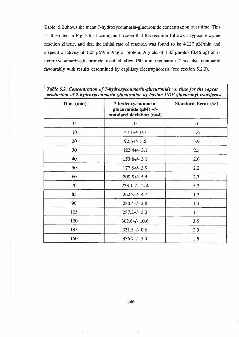
Table 5.2 shows the mean 7-hydroxycoumarin-glucuronide concentration over time. This
is illustrated in Fig. 5.6. It can again be seen that the reaction follows a typical enzyme
reaction kinetic, and that the initial rate of reaction was found to be 4.127 (iM/min and
a specific activity of 1.03 ^M/min/mg of protein. A yield of 1.35 |xmoles (0.46 ^g) of 7-
hydroxycoumarin-glucuronide resulted after 150 min incubation. This also compared
favourably with results determined by capillary electrophoresis (see section 5.2.3).
Table 5.2. Concentration of 7-hydroxycoumarin-glucuronide vs. time for the repeat production of 7-hydroxycoumarin-glucuronide by bovine UDP glucuronyl transferase.
Time (min) 7-hydroxycoumarin- glucuronide (fiM) +/-
standard deviation (n=4)
Standard Error (%)
0 0 0
10 47.1+/- 0.7 1.6
20 92.6+/- 5.5 5.9
30 122.4+/- 3.1 2.5
40 153.8+/- 3.1 2.0
50 177.8+/- 3.9 2.2
60 200.5+/- 5.5 2.7
70 233.1+/- 12.4 5.3
85 262.3+/- 4.7 1.7
90 280.4+/- 3.8 1.4
105 287.2+/- 3.0 1.1
120 302.6+/- 10.6 3.5
135 331.3+/- 0.6 2.0
150 336.7+/- 5.0 1.5
246

Time (min)Figure 5.5. HPLC chromatograms of the repeat production of 7-hydroxycoumarin-
glucuronide. Shown are offset chromatograms at 0, 20 and 70 min. This shows an
increase in the 7-hydroxycoumarin-glucuronide peak at 13.2 min (peak A) over time.
A decrease in the 7-hydroxy coumarin peak at 16.2 min (peak B) could also be observed
over time.
Time (min)
Figure 5.6. In vitro production of 7-hydroxycoumarin-glucuronide from the enzymatic
conjugation of 7-hydroxy coumarin to glucuronic acid via bovine liver UDP glucuronyl
transferase as analysed by HPLC. The initial rate of product formation was linear
between 0 and 40 min and was estimated to be approximately 4.13 pM/min (r2=0.995).
247
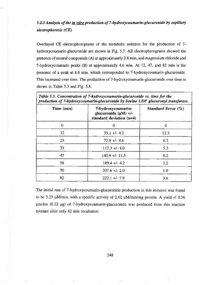
5.2.3 Analysis of the in vitro production of 7-hydroxy coumarin-glucuronide by capillary
electrophoresis (CE)
Overlayed CE electropherograms of the metabolic solution for the production of 7-
hydroxycoumarin-glucuronide are shown in Fig. 5.7. All electropherograms showed the
presence of neutral compounds (A) at approximately 3.8 min, and magnesium chloride and
7-hydroxycoumarin peaks (B) at approximately 4.6 min. At 12, 47, and 82 min is the
presence of a peak at 8.6 min, which corresponded to 7-hydroxycoumarin-glucuronide.
This increased over time. The production of 7-hydroxycoumarin-glucuronide over time is
shown in Table 5.3 and Fig. 5.8.
Table 5.3. Concentration of 7-hydroxycoumarin-glucuronide vs. time for the production of 7-hydroxycoumarin-glucuronide by bovine UDP glucuronyl transferase.
Time (min) 7-hydroxycoumarin- glucuronide (|iM) +/-
standard deviation (n=4)
Standard Error (%)
0 0 0
12 35.1 +/- 4.3 12.3
23 72.8 +/- 4.6 6.3
35 112.3 +/- 6.0 5.3
47 140.9 +/- 11.5 8.2
58 189.4 +/- 4.2 2.2
70 207.6 +/- 2.0 1.0
82 222.1 +/- 7.9 3.6
The initial rate of 7-hydroxycoumarin-glucuronide production in this instance was found
to be 3.23 (iM/min, with a specific activity of 2.02 (iM/min/mg protein. A yield of 0.36
(amoles (0.12 jag) of 7-hydroxycoumarin-glucuronide was produced from this reaction
mixture after only 82 min incubation.
248

Time (min)
Fig. 5.7. CE electropherograms from the in vitro elucuronidation of 7-hydroxycoumarin
to 7-hydroxy coumarin-glucuronide by bovine UDP-glucuronyl transferase.
Electropherograms at 12, 47 and 82 min have been offset to show the increase in 7-
hydroxycoumarin-glucuronide peak (C) at 6.5 min. All electropherograms showed
typical peaks of neutral reaction components (peak A) and a magnesium chloride peak
(B).
249

Time (mln)
Figure 5.8. In vitro production of 7-hydroxycoumarin-glucuronide from the enzymatic
conjugation of 7-hydroxycoumarin to glucuronic acid via bovine liver UDP glucuronyl
transferase as analysed by CE. The initial rate of product formation was linear between
0 and 35 min and was estimated to be approximately 3.23 fiM/min (r2=0.995).
250

5.3 Purification of 7-hydroxycoumarin-glucuronide
5.3.1 Separation of 7-hydroxycoumarin and 7-hydroxycoumarin-glucuronide by reverse
phase column chromatography
Initially, pure samples of both 7-hydroxycoumarin (100 (il of 10 mg/ml) and 7-
hydroxycoumarin-glucuronide (100 (il of 1 mg/ml) were analysed using a methanol mobile
phase with flow rates of approximately 0.3 ml/min with fractions collected every 2.5 min
(Figs. 5.9 A and B). These showed the elution of 7-hydroxycoumarin at between 4 and
8 ml and 7-hydroxycoumarin-glucuronide at about 3 to 6 ml, showing considerable peak
overlap. A mobile phase of 700:300:2 water:methanol:acetic acid (v/v/v) resulted in 7-
hydroxycoumarin eluting immediately from the column (fractions 1 to 5, and the 7-
hydroxycoumarin at fractions 20 to 25 (Fig. 5.10). Using a simple combination of
methanol and water and varying the ratio of the two, increasing quality of separation of
both components could be achieved, with increasing separation occurring by increasing
the ratio of water to methanol, so making the mobile phase more polar (Fig. 5.11 A, B
and C.). Good separation was achieved when the ratio of water to methanol was in excess
of 40:60 (v/v).
251

0.06
4 6
Volume (mis)
EcoaIOeoco£ss
0 o \nf72krnà*£0 2 4 6
Volume (mls)
Figure 5.9 A and B. Elution of 100 /il of 10 mg/ml 7-hydroxycoumarin (A) and 100 fil 1 mg/ml 7-hydroxycoumarin-glucuronide (B)from
a Lichroprep RP-18 reverse phase silica column in methanol mobile phase at approx. 0.3 ml/min (flow rate shown in dashed line).
Fractions were collected over 2.5 min intervals and analysed at 320 nm (hatched box and solid line). 7-hydroxy coumarin eluted primarily
in 4 to 8 mis and 7-hydroxy coumarin-glucuronide between 3 and 6 ml.
252
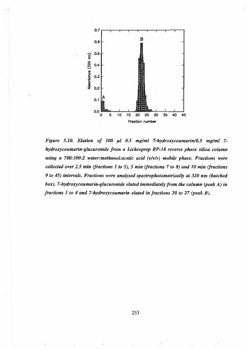
Fraction number
Figure 5.10. Elution of 100 fil 0.5 mg/ml 7-hydroxycoumarin/0.5 mg/ml 7-
hydroxycoumarin-glucuronide from a lichroprep RP-18 reverse phase silica column
using a 700:300:2 water:methanol:acetic acid (v/v/v) mobile phase. Fractions were
collected over 2.5 min (fractions 1 to 5), 5 min (fractions 7 to 8) and 10 min (fractions
9 to 45) intervals. Fractions were analysed spectrophotometrically at 320 nm (hatched
box). 7- hydroxy coumarin-glucuronide eluted immediately from the column (peak A) in
fractions 1 to 4 and 7-hydroxycoumarin eluted infractions 20 to 27 (peak B).
253

oCNIO
O■e
10 15 20 25 30Fraction number Fraction number Fraction Number
Figure 5.11 A, B and C. Separation of 100 fil 7-hydroxycoumarin/7-hydroxycoumarin-glucuronide (0.5 mglml/0.5 mg/ml) on a Lichroprep
RP-18 reverse phase silica column in a mobile phase of 80:20 (A), 60:40 (B) and 50:50 (C) methanol:water (v/v). Flow rate was
approximately 0.2 mUmin and fractions were collected over 2.5 min intervals and analysed spectrophotometrically at 320 nm. Separation
of peaks of 7-hydroxy coumarin-glucuronide (peak A) and 7-hydroxy coumarin (peak B) increased with the increase in the ratio of water
to methanol, with good separation being achieved at 50:50 methanol:water (v/v).
254

5.3.2 Purification of 7-hydroxycoumarin-glucuronide from crude enzymatic reaction
mixtures by reverse phase column chromatography
Enzymatic reaction mixtures from the in vitro production of 7-hydroxycoumarin-
glucuronide were applied to the reverse phase column according to the conditions
described in section 2.3.5.2. Increasing volumes of reaction mixture were applied to the
column from 100 to 400 pi (Figs. 5.12 A, B, C and D). All showed complete separation
of the 7-hydroxycoumarin-glucuronide peaks from the 7-hydroxycoumarin peaks, up to
a sample volume of 400 pi, although this did result in the peaks becoming less well
resolved from one another.
255

0 5 10 15 20 25Volume (mis)
1.0
0.8
E cO 0.6G«o| 0.4o n
-O <0.2
0.00 5 10 15 20 25 30
Fraction number
Figure 5.12 A and B. Purification of 7-hydroxycoumarin-glucuronide from 100 p i (A),
200 pi (B) of enzymatic reaction mixture on a Lichroprep RP-18 reverse phase silica
column in a 50:50 methanol: water (v/v) mobile phase, with a flow rate o f approximately
0.2 ml/min and fractions collected over 5 min intervals and spectrophotometric analysis
at 320 nm. Separation of 7-hydroxycoumarin-glucuronide (peak A) and 7-
hydroxycoumarin (peak B) was complete at 100 and 200 p i of sample.
256

0.8 -
E c
8 0.6 Ot>o
0.4
0.2
0.0 _i_ -L .
5 10 15 20 25 30Fraction number
Fraction number
Figure 5.12 C and D. Purification of 7-hydroxy coumarin-glucuronide from 300 fit (C),
400 fil (D) of enzymatic reaction mixture on a Lichroprep RP-18 reverse phase silica
column in a 50:50 methanol:water (v/v) mobile phase, with a flow rate of approximately
0.2 ml/min and fractions collected over 5 min intervals and spectrophotometric analysis
at 320 nm. Separation of 7-hydroxy coumarin-glucuronide (peak A) and 7-
hydroxycoumarin (peak B) was still complete at 300 and 400 fil o f sample.
257

5.3.3 Purification of 7-hydroxycoumarin from, coumarin treated patient urine using
reverse phase column chromatography
100 pi samples of control urine, control urine spiked with 100 ng/ml 7-hydroxycoumarin-
glucuronide and coumarin-treated volunteer urine were applied to the reverse phase
column as described in section 2.3.5.2. (Fig. 5.13 A, B and C). All three samples showed
the presence of glucuronides, as indicated by the large absorbance at 254 nm. This was
present in all samples in elution volumes between 6 and 12 mis. The spiked sample
showed an elevation in the 320 nm signal over the control urine. This 320 nm absorbance
was much greater in the sample of coumarin-treated urine and is indicative of the presence
of 7-hydroxycoumarin-glucuronide. No other significant peaks were noted in the urine
samples tested.
5.3.4 Extraction of 7-hydroxycoumarin-glucuronide from urine in dichloromethane
(DCM): methanol
The absorbance of the extracted samples of treated urine at 320 nm are shown in Fig.
5.14. This showed a clear increase in the 320 nm absorbance at a DCMrmethanol ratio
of 70:30 (v/v), indicating the highest extraction efficiency for 7-hydroxycoumarin-
glucuronide from urine.
258

10 20 30Volume (ml)
B
KNXS254 nm 320 nm
10 20
Volume (mi)
30
1.0
0.9
0.80.7^.c0.6 v
E0.5 ~
0.4 ^I
0.3 c
0.2
0.1
400.0
Volume (mls)
Figure 5.13 A, B and C. Purification of 7-hydroxycoumarin-glucuronide by reverse phase column chromatography from control (A),
spiked (B) and treated (C) urine. Flow rate: (dotted line). A shows the presence of quantities of glucuronides in the urine, as observed
by the absorbance at 254 nm (hatched bars). When spiked with 7-hydroxy coumarin-glucuronide (B), an increase in the 320 nm
absorbance was observed at the same place in which the glucuronides were eluting (solid line). C clearly shows a further elevation in
the ratio of 320/254 nm absorbance from the treated urine sample, indicating the presence of 7-hydroxycoumarin-glucuronide in
association with other glucuronides in the urine.
259

DCM:MeOH
Figure 5.14. Extraction of 7-hydroxycoumarin-glucuronide into different proportions
of DCM:methanol. Samples were analysed by HPLC. This shows that the greatest 320
nm absorbance was found in samples extracted with an approximate ratio of
DCM'.methanol of 70:30 (v/v).
260

5.3.5 Purification of 7-hydroxycoumarin-glucuronide from coumarin treated patient
urine and enzymatic reaction mixtures by reverse phase chromatography following
extraction in dichloromethane (DCM):methanol (70:30 v/v)
The chromatograms of the extracted enzymatic reaction mixtures are shown in Figs. 5.15
A and B. In both chromatograms, where 100 pi (5.15 A) and 400 pi (5.15 B) of extracted
enzymatic sample were used, it would appear that significant concentration of 7-
hydroxycoumarin taken place. However, the ratio of 7-hydroxycoumarin-glucuronide to
7-hydroxycoumarin had clearly decreased, indicating inefficient extraction of the
conjugated 7-hydroxycoumarin. This methodology showed a decreased efficiency over the
direct application of enzymatic reaction mixture to the column.
Chromatography of the extracted treated urine sample also showed a decrease in
efficiency as compared with unextracted samples applied directly to the reverse phase
column, indicating significant losses of 7-hydroxycoumarin-glucuronide during the
extraction procedure (Fig. 5.16).
261
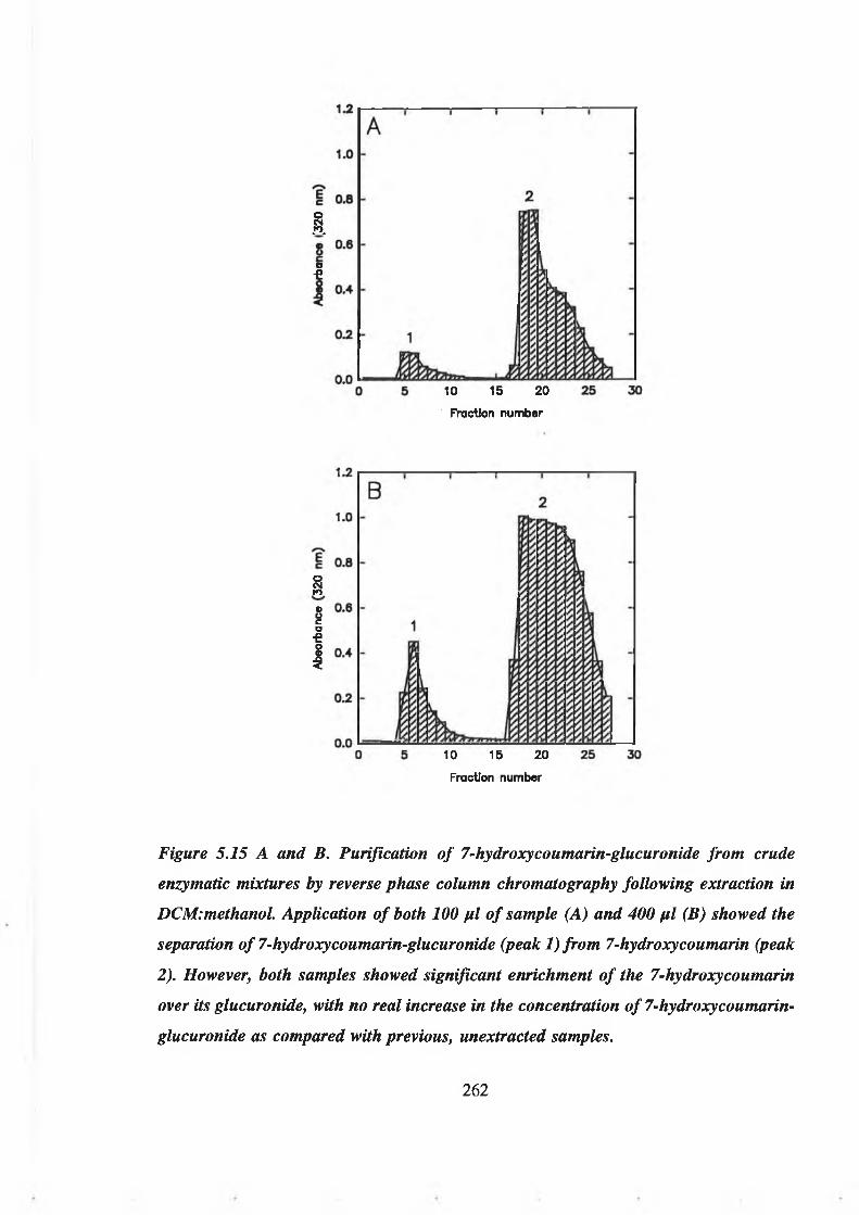
AD€
10 15 20Fraction number
10 15 20Fraction number
§©Oc0 €1
Figure 5.15 A and B. Purification of 7-hydroxycoumarin-glucuronide from crude
enzymatic mixtures by reverse phase column chromatography following extraction in
DCM'.methanol. Application of both 100 fil of sample (A) and 400 fil (B) showed the
separation of 7-hydroxy coumarin-glucuronide (peak 1) from 7-hydroxy coumarin (peak
2). However, both samples showed significant enrichment of the 7-hydroxy coumarin
over its glucuronide, with no real increase in the concentration of 7-hydroxycoumarin-
glucuronide as compared with previous, unextracted samples.
262

0 5 10 15 20 25 30Fraction number
Figure 5.16. Chromatographic separation of extracted treated urine sample. This shows
that 7-hydroxycoumarin-glucuronide predominated in the sample, but some 7-
hydroxycoumarin had also become extracted and concentrated in the sample (peaking
infraction 13). The overall yield was also relatively low with maximum absorbances at
320 nm less than 0.3.
263

5.4.1 Thermal stability of 7-hydroxycoumarin-glucuronide at 60°C
The peak area for samples of 7-hydroxycoumarin-glucuronide over time when incubated
at 60°C is shown in Fig. 5.17. No apparent decrease in peak area was noted after 80 min
incubation indicating good thermal stability of the compound at 60°C.
5.4 Characterisation of 7-hydroxycoumarin-glucuronide
Time (min)
Figure 5.17. Analysis of 7-hydroxycoumarin-glucuronide by HPLC during incubation
at 6(fC over time. No reduction in 7-hydroxycoumarin-glucuronide peak size were
observed over the 160 min incubation period.
264

5.4.2 Stability of 7-hydroxycoumarin-glucuronide with respect to pH
The peak height for samples of 7-hydroxycoumarin-glucuronide in aqueous solvent at
different pH is shown in Fig. 5.18. Samples showed good stability at pH 7 and below, but
began to decrease above pH 7 indicating instability in basic environments. However,
degradation of the sample only became significant above pH 10.
pH
Figure 5.18. Analysis of 7-hydroxycoumarin-glucuronide by HPLC following
preparation in different pH buffers. From pH 2 to 7, no reduction in peak is observed.
However, above pH 7, significant decreases in the peak size of 7-hydroxycoumarin-
glucuronide were observed. This became substantial at pH in excess of 10.
265

Production of a 3-amino derivative of 7-hydroxycoumarin-glucuronide was investigated
for its potential use in the conjugation of 7-hydroxycoumarin-glucuronide to proteins. This
first required the production of 3-amino-7-hydroxycoumarin from the precursor 3-
acetylamino-7-acetocoumarin, followed by the enzymatic glucuronidation of the 3-amino-
7-hydroxycoumarin.
5.5.1 Production of 3-amino-7-hydroxycoumarin
The infrared spectrophotometric scan of 3-amino-7-hydroxycoumarin is shown in Fig.
5.19. This compares exactly with equivalent IR scans by Bogan (1996), indicating the
production of 3-amino-7-hydroxycoumarin from the 3-acetylamino-7-acetocoumarin
derivative. HPLC analysis shows the synthesised compound to be mostly 3-amino-7-
hydroxycoumarin, with a small amount of unreacted 3-acetylamino-7-acetocoumarin (Fig.
5.20).
5.5.2 In vitro enzymatic production of 3-amino-7-hydroxycoumarin-glucuronide
Components in the production of 3-amino-7-hydroxycoumarin-glucuronide were analysed
by HPLC according to section 2.3.4.2 using dual channel spectrophotometric monitoring
at 320 and 254 nm as well as photodiode array analysis between 200 and 400 nm.
Fig. 5.21 shows the chromatogram and the photodiode array scan for 7-hydroxycoumarin-
glucuronide. Worthy of note is the retention time of 3.33 min and the absorbance maxima
at 320 nm, with a peak rising to 200 nm. When analysed by HPLC and PDA analysis, 3-
acetylamino-7-acetocoumarin (Rt= 5.45 min) and 3-amino-7-hydroxycoumarin (Rt=4.32
min) both contained the coumarin absorbance maxima at 320 nm (Fig. 5.22). However,
3-acetylamino-7-acetocoumarin possessed a UV absorbance peak rising to 200 nm,
whereas 3-amino-7-hydroxycoumarin showed a dip at 200 nm. When the enzymatic
reaction mixture was analysed by HPLC, 5 distinct components could be identified. Two
of these at 4.3 and 5.42 min could be clearly identified as the original components of the
5.5 In vitro enzymatic production of 3-amino-7-hydroxycoumarin-giucuronide
266

mixture, 3-amino-hydroxycoumarin and 3-acetylamino-7-acetocoumarin (Fig. 5.23). Three
new components, all with greater polarity than the parent compounds were also present.
Two of them closely resembled one of the parental compounds with respect to their
photodiode array scan, with the peak at 2.7 min resembling 3-amino-7-hydroxycoumarin
and the peak at 2.9 min resembling 3-acetylamino-7-acetocoumarin. A third peak at 3.6
min also possessed the characteristic absorbance peak at 320 nm typical of coumarins. Its
UV absorbance ridge, however, did not resemble the other components in the mixture. No
further identification of these components was performed.
267

Tran
smitt
ance
(%
)
Figure 5.19. Infrared spectra of 3-amino-7-hydroxy coumarin. This shows a spectra
typical of that expected for 3-amino-7-hydroxy coumarin, according to Bogan (1994).
268

ffi5I
I .« ; 1
s g
Figure 5.20. HPLC and photodiode array analysis of 3-amino-7-hydroxycoumarin.
Absorbance at 254 nm (solid line). Absorbance at 320 nm (dotted line). Two peaks were
present in the sample. The reaction product 3-amino-7-hydroxycoumarin at 4.32 min
predominated in the sample with the presence of a smaller quantity of unreacted 3-
acetylamino-7-acetocoumarin (5.45 min) also present. These showed typical PDA scans,
both with absorbances at the 320 nm region, with one showing a rise in absorbance to
200 nm, the other showing a decrease in the same region.
269

Figure 521. HPIC and photodiode array analysis of 7-hydroxycoumarin-glucuronide.
Absorbance at 254 nm (solid line). Absorbance at 320 nm (dotted line). A single peak
of 7-hydroxycoumarin-glucuronide was obtained at 3.33 min. This had the typical PDA
scan as shown, with a peak at 320 nm, a ridge at 280 nm, and a rise up to 200 nm.
270

I
Figure 522. HPLC and photodiode array analysis of the in vitro metabolic production
of 3-amino-7-hydroxycoumarin-glucuronide. Absorbance 254 nm (solid line).
Absorbance at 320 nm (dashed line). Five components were present in the reaction
mixture. Two at 4.20 and 5.42 min were the original reaction components, 3-amino-7-
hydroxycoumarin and 3-acetylamino-7-acetocoumarin, respectively. Three new
components, all showing decreased hydrophobicity were produced at 2.69,2.99 and 3.59
min. The PDA scan of peak 2.69 min resembled 3-amino-7-hydroxycoumarin, whilst
peak at 2.9 min resembled that of 3-acetyl-7-acetocoumarin. The peak at 3.59 min did
not particularly resemble either of the starting components.
271

5.6 Production and analysis of a 7-hydroxycoumarin-glucuronide-bovine serum
albumin conjugate prepared by EDC/NHS coupling chemistry
The 7-hydroxycoumarin-glucuronide-bovine serum albumin conjugate was analysed by
SDS-PAGE (Fig. 5.23). This indicated the presence of a protein component of
approximately 66 kDa, corresponding to bovine serum albumin. When analysed by size
exclusion chromatography and photodiode array analysis (Fig. 5.24), this showed a
predominant protein peak at 21.9 min. Concomitant with the protein absorbance maxima
at approximately 280 nm was a second absorbance maxima at 320 nm indicating the
presence of a coumarin moiety attached to the protein component. When compared to the
spectrophotometric scan of bovine serum albumin alone, no such absorbance maximum
could be seen at 320 nm.
272

Figure 5.23. Analysis of bovine serum albumin and 7-hydroxycoumarin-bovine serum
albumin conjugate by SDS-PAGE. Lanes: 1: bovine serum albumin; 2: 7-
hydroxycoumarin-bovine serum albumin. The gel shows that both samples contained
protein with a molecular weight of approximately 66 kDa, corresponding to the
molecular weight of bovine serum albumin.
273

Wavelength (nm)
Figure 5.25. Photodiode array scans of 7-hydroxycoumarin-glucuronide-bovine serum
albumin conjugate (solid line) and bovine serum albumin control (dot-dash line). Both
samples exhibited the typical 260 nm absorbance peak indicating the presence of a
protein (peak A). However, the 7-hydroxycoumarin-glucuronide-bovine serum albumin
conjugate exhibited a second peak at 320 nm (peak B), indicating the presence of a
coumarin-type molecule attached to the protein. This was not present in the bovine
serum albumin control sample.
274

DISCUSSION
The purpose of studying the production, analysis, purification and conjugation of 7-
hydroxycoumarin-glucuronide were outlined in section 1.14.
The study of the in vitro metabolism of 7-hydroxycoumarin-glucuronide served several
purposes. The need for information on the pharmacokinetics of 7-hydroxycoumarin are
important in its use as a therapy in several conditions such as high protein lymphoedema,
brucellosis and experimental cancer therapies. Variations in phase II metabolism of the
drug may relate to differences in the efficiency of the treatment regime. Inadequacies in
phase II metabolism may also relate to sporadic cases of hepatotoxicity during such
therapies. Studying individual differences in their ability to metabolise 7-hydroxycoumarin
in liver samples such as liver biopsies may further confirm in vivo analyses, pointing to
deficiencies in phase II metabolism in the liver. Such procedures have been used in the
phase I metabolism of coumarin (Deasy, et al., 1995). Interspecies studies have also
served to elucidate the metabolic pathways of different organisms, leading to a fuller
understanding of the development of these pathways. Studies of in vitro metabolism in
different organs has also yielded information about the sites that are important in the
drug’s metabolism, and consequently its distribution in the organism. In vitro metabolism
is also a source of the glycone for purification and use in other methodologies, such as
in standards, and its application in the development of a conjugate for the production of
antibodies to the glucuronide. Such antibodies would themselves be applied in various
studies of phase II metabolism.
5.8 In vitro production of 7-hydroxycoumarin-glucuronide and analysis by HPLC
For the in vitro production and analysis of 7-hydroxycoumarin-glucuronide, an effective
enzymatic reaction had to be developed as well as a method or methods of analysis of the
reaction product formed. Tegtmeier et al. (1988) and Brunner and Tegtmeier (1984) had
developed methods of in vitro glucuronidation using hollow fibre membrane systems for
the biochemical detoxification of lypophilic compounds in which suitable reaction
conditions for porcine UDP-glucuronyltransferases had been elucidated. Egan and
275

O’Kennedy (1994) had also developed HPLC conditions capable of the direct detection
and resolution of 7-hydroxycouraarin and coumarin. These two methodologies were used
as starting points for the analysis of the in vitro metabolism of 7-hydroxycoumarin by
porcine UDP-glucuronyltransferase enzymes isolated from liver. HPLC analysis showed
that the 7-hydroxycoumarin substrate and 7-hydroxycoumarin-glucuronide could be
separated using this methodology. It also illustrated very clearly that all the reaction
components (substrates and enzyme) were necessary to detect formation of the
glucuronide product. Analysis of initial rates of product production (v0) showed the rate
to be very slow at 3.3 x 10'3 pM/min/mg under the initial reaction conditions used.
In subsequent experimentation, changes were made to both the enzymatic reaction mixture
and the analytical methodology employed. With respect to the reaction mixture, UDP-
glucuronyltransferase enzyme was isolated from bovine liver. The other notable alteration
was the reduction of the 7-hydroxycoumarin substrate concentration. The analytical
methodology also underwent some notable changes. The separation methodology used a
gradient system. This was done primarily to increase the retention time of the glucuronide
species, bringing it further from the void volume, while still giving good resolution of all
components. Calculation of 7-hydroxycoumarin-glucuronide concentration was determined
by ratio analysis against an internal standard, 4-hydroxycoumarin, which was also
effectively resolved by the methodology applied. As a result v0 of the bovine enzyme was
found to be 4.13 pM/min (1.03 pM/min/mg protein). This was an increase in over 300-
fold specific reaction rate over the initial reaction. It is not clear whether this is due
primarily to the change in the source of liver enzyme, or due to substrate inhibition in the
original reaction mixture due to the very high concentration of 7-hydroxycoumarin
employed.
With respect to the HPLC methodologies used, no direct comparison can be made
between the two methodologies with regard to precision, since the initial experiment was
not performed with replicates. However, internal standardisation has been shown to be
superior in terms of precision over direct measurements (Christian, 1986), so it might be
assumed that this was also the case here. The standard errors of the standards used for
analysis were all below 8% and typically below 5%, indicating acceptable precision. The
276

analytical methodology was, however, slower than the original chromatographic
separation, requiring 25 min, as opposed to approximately 16 min for the original isocratic
separation. However, due to sample stability and automation of sampling, this was not an
inconvenience.
In terms of the overall yields of these methodologies for the production of 7-
hydroxycoumrin-glucuronide, the initial methodology produced just less than 0.1 pg of
conjugate in the 23 h reaction period, compared to nearly 0.5 |ig for the second reaction
within 2.5 h. Thus, the enzymatic methodologies employed were shown to be capable of
yielding microgram quantities of 7-hydroxycoumarin-glucuronide.
5.9 In vitro production of 7-hydroxycoumarin-glucuronide and analysis by CE
The technique of capillary electrophoresis was applied to the analysis of the enzymatic
reaction mixture. As has been discussed, CE offers rapid separations of reaction
components based on their electrophoretic mobility in certain ionic solvents in high
electrical potentials. A method had been developed by Bogan et al. (1996) for the analysis
of 7-hydroxycoumarin-glucuronide in urine samples. This methodology was applied to the
analysis of 7-hydroxycoumarin-glucuronide in crude enzymatic reaction mixtures.
Crucially, treatment of the reaction mixtures for CE analysis with trichloroacetic acid was
not appropriate and was not required. Changes in pH brought about by such treatments
would significantly alter the separation characteristics of the assay. However,
electrophoresis brings about instantaneous separation of the reaction components, at which
point, the reaction ceases. To perform this analysis then, samples must be taken and
analysed immediately. This is not a difficulty, since separation takes just 7.5 min, a
periodicity quite convenient for regular sampling of the enzymatic reaction mixture. Use
of this methodology for the analysis of enzymatic reaction mixtures demonstrated the
resolution of 7-hydroxycoumarin-glucuronide from other reaction components.
The initial rate of reaction in the CE study was comparable to the HPLC study using
bovine liver UDP-glucuronyltransferase, with a v0 of 3.23 fxM/min, but with a specific
activity of 2.02 (jM/min/mg protein, yielding only 0.12 |ig of 7-hydroxycoumarin-
277

glucuronide in the 82 min incubation, with the rate of reaction dropping more rapidly than
in the study which used HPLC. The reasons for these discrepancies could be several fold.
Mostly, they might relate to the quality of the liver material used, its treatment and
preparation and the yield of UDP-glucuronyltransferase enzyme protein in the sample.
This does illustrate a very important consideration when extrapolating data from such
experiments to the actual in vivo enzyme reaction rates of intact whole organs. It is
probable that specific activities of in vitro prepared enzyme mixtures are lower than that
in whole, in tact tissues. However, no significant studies comparing in vivo and in vitro
metabolic rates have been performed to illustrate this.
The establishment of an in vitro metabolic reaction mixture for the conversion of 7-
hydroxycoumarin to its glucuronide, accompanied by two reliable analytical methodologies
by which to analyse the reaction products yielded a system that could be used to probe
the phase II metabolic characteristics of different organs and species with respect to 7-
hydroxycoumarin. These methodologies have been successfully exploited by Bogan et al.
(1997) and Duffy et al. (1997) for the analysis of phase II metabolism of 7-
hydroxycoumarin in rabbit tissues. It also resulted in a third source of 7-hydroxycoumarin-
glucuronide, in addition to chemical synthesis and in vivo isolation from treated urine.
5.10 Purification of 7-hydroxycoumarin- glucuronide
Two metabolic sources of 7-hydroxycoumarin-glucuronide were available. Firstly, the
enzymatic synthesis methodologies outlined in section 5.2, and secondly, 7-
hydroxycoumarin-glucuronide from treated volunteer urine samples.
In the case of the enzymatic reaction mixtures, 7-hydroxycoumarin-glucuronide was in the
presence of quantities of 7-hydroxycoumarin, coumarin and protein material. If 7-
hydroxycoumarin-glucuronide from such a source was to have any application as a
standard, or for the production of conjugates, it must first be separated from these other
components.
278

Since 7-hydroxycoumarin-glucuronide was successfully separated from 7-hydroxycoumarin
and coumarin using reverse phase chromatography by HPLC, it was assumed that such
a reverse phase system might be applicable to the column chromatographic preparative-
scale separation of these components also.
A reverse phase column with C-18 packing was prepared (section 2.3.5.1). Initially, pure
samples of both 7-hydroxycoumarin and 7-hydroxycoumarin-glucuronide were applied to
the column in a methanol mobile phase. These experiments helped to establish the
separation methodologies applied later, such as pump speeds, flow rates, collection
intervals etc. Initially, both samples were shown to elute within the first 10 ml of mobile
phase, so rapid purification times could be established. However, in the mobile phase
used, inadequate differences in retentions of the two components were found.
The conditions of the mobile phase in the initial isocratic HPLC separation were applied
to the preparative column. In this case, 7-hydroxycoumarin-glucuronide and 7-
hydroxycouamarin were seen to separate very well. However, 7-hydroxycoumarin-
glucuronide eluted in the void volume and could not be successfully fractionated from the
column. Under these conditions, the mobile phase was too polar to bring about adequate
retardation of the water soluble glucuronide on the column. The coumarin contaminant of
7-hydroxycoumarin was not important in these separations because either its concentration
was too small to be noted in chromatograms, or it coeluted with 7-hydroxycoumarin under
the conditions applied.
A more simple mobile phase of methanol:water was applied to the separation. It was
found that with higher methanol concentrations, 7-hydroxycoumarin-glucuronide was
significantly retarded on the column. Decreases in methanol concentration further retarded
7-hydroxycoumarin on the column. At 50:50 methanol:water (v/v), good separation and
retention of both components could be established. Good separation also occurred at 60:40
methanol:water (v/v).
Having established separations for pure mixtures of 7-hydroxycoumarin and 7-
hydroxycoumarin-glucuronide, the methodology was applied to the separation of crude
279

reaction mixtures. A range of volumes of the reaction mixture were applied to the column
up to 400 pi, which brought about concomitant increases in the peaks of both 7-
hydroxycoumarin and 7-hydroxycoumarin-glucuronide. The two compounds were still
resolvable with 400 pi sample volumes.
The second readily available source of 7-hydroxycoumarin-glucuronide was the
administration of coumarin to a healthy human volunteer and harvesting of the urine
samples subsequent to this. Studies of the pharmacokinetics of coumarin metabolism in
humans has shown its rapid excretion as 7-hydroxycoumarin-glucuronide in the urine,
which takes place within 24 h, with maximum excretion approximately 2 h post
administration (Pelkonen et al., 1997). Thus, urine from control and treated healthy
volunteers was taken, the latter 2 h after administration of 1 g p.o. coumarin in 10 x 100
mg tablets. These were separated by the same analytical technique as the enzymatic
mixtures. In this case, no 7-hydroxycoumarin peak was noted. In the control urine, a
significant 254 nm absorbance peak was noted in the same position as would be found
7-hydroxycoumarin-glucuronide, indicating the presence of glucuronides, but with a low
320 nm absorbance, suggesting non-coumarin-derived glucuronides. When the control
urine was spiked with 7-hydroxycoumarin-glucuronide, the 320 nm signal showed an
increase at the same position as the 254 nm glucuronide peak. This suggested that natural
glucuronides present in the urine were coeluting with 7-hydroxycoumarin-glucuronide.
When treated urine was separated, a larger 254/320 nm peak also eluted at the same
position as in the control urine sample. The higher 320/254 nm ratio also indicated that
this was, indeed, the position at which 7-hydroxycoumarin-glucuronide was eluting.
As has been stated, many natural metabolic waste products are excreted as glucuronides,
notably bilirubin and testosterone, and other hormones (Reid and Leppard, 1983). It is
clear from this that the separation technique employed was not applicable for the direct
separation of 7-hydroxycoumarin-glucuronide from treated urine samples because it does
not separate glycone derivatives of different compounds.
Although simple to analyse directly, water-miscible glucuronide compounds are difficult
to purify using liquid/liquid separation methodologies as they are not easily extracted with
280

organic solvents. The presence of the 7-hydroxycoumarin moiety would, however, endow
them with some lipophilicity. An attempt was made to exploit this by the development
of an extraction procedure for 7-hydroxycoumarin-glucuronide. This was based on an
organic phase composed of dichloromethane (DCM) and methanol. These were chosen
because the polar methanol would dissolve in the water-immiscible DCM, but could still
dissolve quantities of 7-hydroxycoumarin-glucuronide. Various ratios of DCM:methanol
were tested against pure samples of 7-hydroxycoumarin-glucuronide using equal volumes
of the organic and inorganic mixtures. The presence of the glucuronide was analysed by
the 320 nm absorbance of the dried, rehydrated samples. Maximum 320 nm readings were
at a DCM:methanol ratio of 70:30 (v/v).
The extraction procedure was applied to the enzymatic reaction mixtures and urine
samples containing 7-hydroxycoumarin glucuronide. As compared with unextracted
enzyme mixtures, the yield of 7-hydroxycoumarin-glucuronide decreased significantly,
whereas the level of 7-hydroxycoumarin in these samples was significantly increased. In
urine, the yield of glucuronide in the sample had also decreased substantially, although
the glucuronide that was present did show a high 320 nm absorbance. This may indicate
that the extraction methodology was more efficient at extracting 7-hydroxycoumarin-
glucuronide than other glucuronides present in the urine.
Overall, employment of an extraction methodology resulted in inefficient extraction of the
7-hydroxycoumarin-glucuronide, both from enzyme reaction mixtures and from samples.
The availability of large volumes of urine, however, may allow for an improvement in the
extraction methodology. Using large volumes of urine and small volumes of organic
mixture may increase the partitioning of significant quantities of 7-hydroxycoumarin-
glucuronide into the organic phase. This may also remove significant quantities of other
glucuronides from the sample that do not possess lipophilic functional groups.
When extracted, separated samples from treated urine were analysed by HPLC, 7-
hydroxycoumarin-glucuronide was shown to be present in significant amounts with only
minimal contaminants present and was considered acceptable for use in further
experimental procedures.
281

5.11 Physical analysis of 7-hydroxycoumarin-glucuronide
Some physical characteristics of the 7-hydroxycoumarin-glucuronide were analysed by
HPLC. The molecule exhibited good thermal stability at 60°C. Of note was the sensitivity
of the glucuronide to alkaline pH. Even mildly alkaline conditions brought about
degradation of the conjugate, but this was significant above pH 10. This finding is
substantially in accordance with other glucuronides which undergo rapid alkaline
hydrolysis of the (3-D ether linkage (Wilson et al., 1983). Indeed, this lability is of
importance when analysing such metabolites in urine. The composition and pH of the
urine can vary substantially depending on dietary habits and the administration of other
drugs such as antacids which make urine basic and may lead to deconjugation. Also, urine
loses carbon dioxide upon standing and becomes alkaline. This results in the precipitation
of salts such as inorganic phosphates and may also bring about deconjugation of 7-
hydroxycoumarin-glucuronide and precipitation of insoluble 7-hydroxycoumarin leading
to inaccurate results. This is important when data derived from such metabolic studies is
related to the phase II inter-individual variability in humans and possible associated
hepatotoxic effects (Bogan, 1996). Such analyses may benefit by being acidified upon
collection.
5.12 Conjugate production
For the production of antibodies to small haptens - as has already been illustrated several
times throughout this thesis - it is necessary to conjugate them to large immunogens such
as proteins. As has also been demonstrated, coumarin and 7-hydroxycoumarin lack any
functional groups that can be readily applied to a conjugation reaction. Production of
amino derivatives of coumarin and 7-hydroxycoumarin have been used to produce
conjugates for the purpose of antibody production (Egan, 1993; Bogan, 1996).
Of concern is the orientation of the hapten as displayed to the antigen-binding site. It
might be thought that orientation of coumarin and 7-hydroxycoumarin in directions that
leave the pyrone ring proximal to the antigen-binding site may bring about associated
cross-reactivities with the resulting antibodies. For this reason, conjugation of coumarin
282

was at the 6 position and 7-hydroxycoumarin at the 3 position, resulting in opposing
orientations (Egan, 1993; Bogan, 1996). This did yield antibodies that showed no cross
reactivity to either species.
The same concerns were initially applied to the conjugation of 7-hydroxycoumarin-
glucuronide to proteins such as bovine serum albumin. The only functional group
available for attachment with this molecule is the carboxyl group present on the
glucuronic acid moiety. This coupling methodology of drug glucuronides has been
successfully used (Abbott Laboratories, 1984). However, conjugation at the glucuronic
acid moiety may orient the hapten with the 7-hydroxycoumarin pyrone ring proximal to
the antigen-binding site, again leading to concerns of cross-reactivity. To circumvent this,
glucuronide conjugates of 7-hydroxycoumarin with functional groups would be desirable
such as 3-amino-7-hydroxycoumarin-glucuronide. In this way, the hapten could be
oriented with its glucuronic acid component greatly exposed to the antibody, theoretically
minimising cross reactivity.
Conjugate synthesis required the production of 3-amino-7-hydroxycoumarin from 3-
acetylamino-7-acetocoumarin, previously synthesised by Bogan (1996). Infrared analysis
of the product was in accordance with Bogan (1996). HPLC analysis, however, showed
that the reaction was not complete and contaminants of 3-acetylamino-7-acetocoumarin
remained. This two component mixture was applied to in vitro glucuronidation.
Identification of the components by HPLC, coupled with photodiode array detection
yielded 5 components in the reaction mixture. Two of these were the original components
of the sample. Three new derivatives of significance were found in the mixture. These all
had lower retention times, so greater polarity than the parent molecules. Two of these
possessed photodiode array spectra similar to the two parental species and a third, which
also appeared to be a coumarin derivative due to its absorbance at 320 nm, but otherwise,
not significantly recognising the parental components.
It was presumed that glucuronidation of the parental components did take place in this
mixture and that this resulted in the hydrophillic components produced. However, because
no definitive identification of the components could be made without significant further
283

production, purification and anlysis by a methodology such as nuclear magnetic resonance,
no further characterisation of these species was attempted. However, the potential clearly
exists for the in vitro metabolic production of a glycone derivative of 3-amino-7-
hydroxycoumarin.
Since pursuance of the production of a 3-amino- derivative of 7-hydroxycoumarin-
glucuronide was found to be prohibitively complex, production of a conjugate of 7-
hydroxycoumarin-glucuronide, coupled via the glucuronic acid moiety was pursued, with
due regard given to the potential of such a methodology to result in antibodies that would
be cross-reactive with either 7-hydroxycoumarin or coumarin.
The well established chemistry of l-ethyl-3(3-dimethylaminopropyl)carbodiimide
(EDC)/N-hydroxysuccinimide (NHS) was shown to be applicable to the linkage of
glucuronide conjugates to proteins (Abbott Laboratories, 1984). The method adopted for
7-hydroxycoumarin-glucuronide conjugation initially used a high pH of 9.6. Although
awareness was given to the instability of 7-hydroxycoumarin-glucuronide at elevated pH,
this was most pronounced above pH 10, and a high pH would ensure full dissociation of
the carboxyl group of the glucuronic acid for full reactivity. When analysed by photodiode
array and compared to controls, the approximately 66 kDa species was shown to possess
a 320 nm absorbance peak, indicating attachment of a coumarin moiety to the protein.
Since linkage could only be via the glucuronic acid, this suggested attachment of an in
tact 7-hydroxycoumarin-glucuronide molecule to bovine serum albumin. This conjugate
could be used in the production of antibodies to 7-hydroxycoumarin-glucuronide.
CONCLUSIONS
A method for the in vitro glucuronidation of 7-hydroxycoumarin using liver-isolated UDP-
glucuronyltransferase was established. This also led to the development of two analytical
techniques for the measurement of the enzymatic process based on HPLC and CE.
Chromatographic techniques were also developed for the purification of 7-
hydroxycoumarin from enzymatic reaction mixtures and urine.
284

Attempts were made to produce in vitro glucuronidated conjugates of 3-amino-7-
hydroxycoumarin, but the complex mixtures produced meant that this was not a viable
option. Instead, conjugation was performed via the carboxyl group of the glucuronide
using EDC/NHS coupling chemistry. This resulted in stable 7-hydroxycoumarin-
glucuronide-BSA conjugates with the potential to be used in the production of antibodies,
either as polyclonal sera, or the selection and screening of clones from antibody phage
display libraries.
285

6. OVERALL CONCLUSIONS

The purpose of this research was the development of genetic techniques for the production
of antibodies to the experimental chemotherapeutic agent, coumarin, and its major human
phase I metabolite, 7-hydroxycoumarin. Also, this research studied the phase II
metabolism of 7-hydroxycoumarin to 7-hydroxycoumarin-glucuronide.
The first system for the production of antibody fragments to coumarin and 7-
hydroxycoumarin to be evaluated was the pComb3 system for the production of Fab
antibody fragments. The system required the production of light and heavy chain gene
libraries from the splenocytes of immunised mice. These heavy and light chain gene
libraries were successfully created in the cloning vector pGEM-T. However, all attempts
to transfer these libraries to the phagemid vector pComb3 failed. Initially, it was thought
that problems lay with restriction-digestion of the light and heavy chain genes. However,
this was eventually shown not to be the case as these could be transferred to and from the
pGEM-T vector. From my own results and the experiences of others (Burioni et al.,
1997), it became clear that difficulties in cloning were due to the characteristics of the
vector, pComb3. Modified versions of this plasmid have been created, but time did not
allow for their use in this instance.
As an alternative to the pComb3 vector system, work was developed using an alternative,
naive human library, commonly referred to as the Nissim library (Nissim et al., 1994).
Here, a complete human antibody repertoire in the form of single chain scFv antibodies
was present, along with increased diversity brought about by the presence of a random
CDR sequence on the antibody heavy chain.
The library was evaluated at various stages for its ability to produce antibodies to
coumarin and 7-hydroxycoumarin. It became evident that the conditions used for both
affinity selection and screening would have a crucial impact on the quantity and type of
antibody clones that would be selected from the library. This was particularly so for the
small hydrophobic haptens that were being investigated, i.e., coumarin and 7-
hydroxycoumarin. It became clear that the design of the hapten-protein conjugates
required for selection and screening was of great significance. At low hapten coupling
ratios, antibodies to the hapten were not sufficiently enriched over clones recognising the
287

carrier protein. At higher hapten coupling ratios, however, clones were successfully
isolated that bound to the hapten-protein conjugate, but not to the carrier protein alone.
Also, alternating the carrier protein used in affinity selection resulted in antibody clones
with the same characteristics. Other methods of selection were also briefly explored, such
as the use of haptens coupled directly to solid phases and use of BIAcore as an affinity
selection system.
Detailed analysis of the clones derived from the affinity selection strategies, however,
showed that these antibodies possessed only poor affinities to the hapten and bound
predominantly to a combined protein-hapten epitope via a weak electrostatic interaction.
Further characterisation suggested that this was most probably an interaction with the
amino acid by which the hapten was attached to the parent protein. The structural
simplicity and hydrophobic nature of these haptens militated against producing antibodies
recognising free drug alone. To avoid this, conjugates utilising different conjugate
chemistries would be desirable. In this way, selection would not be made predominantly
against the type of linkage between hapten and carrier protein, but to the hapten alone.
The other strategy of direct attachment of haptens may also be useful in this regard.
In vitro methods for the production of 7-hydroxycoumarin-glucuronide were developed
using liver UDP-glucuronyl transferase from both bovine and porcine sources. These
methods were accompanied by the development of methods for the analysis of 7-
hydroxycoumarin-glucuronide using HPLC and CE. Methods were also developed for the
purification of 7-hydroxycoumarin-glucuronide, from in vitro enzymatic synthesis, and
from the urine of patients treated with coumarin. Finally, different means of producing
protein conjugates of the glucuronide were investigated. An in vitro enzymatic synthesis
route was found not to be viable due to the complexity of the reaction product. Linkage
of the 7-hydroxycoumarin-glucuronide via the carboxyl group of the glucuronide residue
using EDC/NHS chemistry was simpler and resulted in a higher yield of pure product,
which could then be used for antibody production in either the ways previously discussed,
or through the more traditional means of polyclonal and hybridoma production.
288

In summary, this work has contributed to the understanding of the mechanisms at work
during affinity selection strategies to small, hydrophobic haptens, and has illustrated the
special considerations required when working with such molecules. A significant
contribution has also been made to the study of phase II metabolism of coumarin, by the
development of in vitro metabolic methodologies and the analytical tools to accompany
them. Also, the production of conjugates with which to produce antibodies to the phase
II metabolite leaves possible further immunoanalytical research in this area.
289

7. REFERENCES

Abbott Laboratories (1984). Alkaline phosphatase lebeled steroid hormone glucuronides.
European patent No. 0114615.
Abou-Issa, H., Curley, R.W. Jr., Panigot, M.J., Tanagho, S.N., Sidhu, B.S. and Alshafie,
G.A. (1997). Chemotherapeutic evaluation of N-(4-hydroxyphenyl) retinamide-O-
glucuronide in the rat mammary tumor model. Anticancer Res. 17:3,335-3,339.
Adair, J.R. (1992). Engineering antibodies for therapy. Immunol. Rev. 130:5-40.
Adair, J., and Bright, S. (1993). Engineering antibodies. The Biochemist 15:6-10.
Archer, A.W. (1988). Determination of cinnamaldehyde, coumarin, and cinnamyl alcohol
in cinnamon and cassia by high-performance liquid chromatography. J. Chromatogr.
447:272-276.
Baca, M., Presta, L.G., O’Connor, S.J. and Wells, J.A. (1997). Antibody Humanization
using monovalent phage display. J. Biol. Chem. 272:10678-10684.
Barbas HI, C.F., Kang, A.S., Lemer, R.A., and Benkovic, SJ. (1991). Assembly of
combinatorial antibody libraries on phage surfaces: the gene in site. Proc. Natl. Acad. Sci.
(USA) 88:7978-7982.
Barbas HI, C.F., Björling, E„ Chiodi, F., Dunlop, N., Cababa, D., Jones, T.M., Zebedee,
S.L., Persson, M.A.A., Nara, P.L., Norrby, E., and Burton, D.R. (1992a). Recombinant
human Fab fragments neutralize type 1 immunodeficiency virus in vitro. Proc. Natl. Acad.
Sci. (USA) 89:9339-9343.
Barbas IE, C.F., Crowe, Jr., J.E., Cababa, D., Jones, T.M., Zebedee, S.L., Murphy, B.R.,
Chanock, R.M., and Burton, D.R. (1992b). Human monoclonal Fab fragments derived
from a combinatorial library bind to respiratory syncytial virus F glycoprotein and
neutralize infectivity. Proc. Natl. Acad. Sci. (USA) 89:10164-10168.
291

Barbas ID, C.F., Rosenblum, J.S., and Lemer, R.A. (1993). Direct selection of antibodies
that coordinate metals from semisynthetic combinatorial libraries. Proc. Natl. Acad. Sci.
(USA) 90:6385-6389.
Barbas HI, C.F. and Burton, D.R. (1996). Selection and evolution of high-affinity human
anti-viral antibodies. Trends Biotechnol. 14:230-234.
Bass, S., Greene, R., and Wells, J.A. (1990). Hormone phage: an enrichment method for
variant proteins with altered binding properties. Proteins: Struct. Funct. Genet. 8:309-314.
Bautista, D, and Shulman, M.J. (1993). A hit-and-run system for introducing mutations
into the Ig H chain locus of hybridoma cells by homologous recombination. J. Immunol.
151:1950-1958.
Beaty, M.W., Fetsch, P., Wilder, A.M., Marineóla, F. and Abatti, A. (1997). Effusion
cytology of malignant melanoma. A morphologic and immunocytochemical analysis
including application of the MART-1 antibody. Cancer 81:57-63.
Beckman (1991). Introduction to capillary electrophoresis. Beckman Instruments Inc.
USA.
Berlingozzi, S. and Fabbrini, L. (1954). Paper chromatography separation of coumarin and
its derivatives. Sperimentale, Sez. Chim. Biol. 5:1-5.
Better, M., Chang, C.P., Robinson, R.R., and Horwitz, A.H. (1988). Escherichia coli
secretion of an active chimeric antibody fragment. Science 240:1041-1043.
Biocca, S., Neuberger, M.S., and Cattaneo, A. (1990). Expression and targeting of
intracellular antibodies in mammalian cells. EMBO J. 9:101-108.
Blackburn, G.M. and Deng, S.-X. (1993). Approaches to catalytic antibodies for amide
hydrolysis. The Biochemical Society, Meeting No. 647, University of Sheffield F6.
292

Bodey, B., Siegel, S.E. and Kaiser, H.E. (1996). Human cancer detection and
immunotherapy with conjugated and non-conjugated monoclonal antibodies. Anticancer
Res. 16:661-674.
Bogan, D.P. (1996). Development of novel methods for the detection of coumarin and its
metaboiltes and their applications. PhD thesis, Dublin City University, Ireland.
Bogan, D.P. and O’Kennedy, R. (1996). The simultaneous determination of coumarin, 7-
hydroxycoumarin and 7-hydroxycoumarin-glucuronide in human plasma and serum by
high-performance liquid chromatography. J. Chromatogr. A 686:267-273.
Bogan, D.P., Thornes, R.D., Tegtmeier, M., Scafer, E.A. and O’Kennedy, R. (1996). The
direct determination of 7-hydroxycoumarin and 7-hydroxycoumarin, in urine, by capillary
electrophoresis. Analyst 121:243-247.
Bogan, D.P., Keating, G.J., Reinartz, H., Duffy, C.F., Smyth, M.R. and O’Kennedy, R.,
Analysis of coumarins, In "Coumarins: Biology, Applications and Mode of Action",
O’Kennedy, R. and Thornes, R.D. (Eds.),John Wiley and Sons, New York, 1997a, pp.
267-302.
Bogan, D.P., Duffy, C.F. and O’Kennedy, R. (1997b). In vitro glucuronidation of 7-
hydroxycoumarin and determination of 7-hydroxycoumarin and 7-hydroxycoumarin
glucuronide by capillary electrophoresis. J. Chromatogr. A 772:321-326.
Brinkmann, U., Gallo, M., Brinkmann, E., Kunwar, S., and Pastan, I. (1993). A
recombinant immunotoxin that is active on prostate cancer cells and that is composed of
the Fv region of monoclonal antibody PR1 and a truncated form of Pseudomonas
exotoxin. Proc. Natl. Acad. Sci. (USA) 90:547-551.
Brinkmann, U., Reiter, Y., Jung, S-H., Lee, B., and Pastan, I. (1993). A recombinant
immunotoxin containing a disulfide-stabilized Fv fragment. Proc. Natl. Acad. Sci. (USA)
90:7538-7542.
293

Britton, K.E. (1996). Radiolabeled monoclonal antibodies in diagnosis and therapy of
cancer. Summary and Perspectives. Acta Oncol. 35:385-390.
Brodeur, P.H. and Riblet, R. (1984). The immunoglobulin heavy chain variable region
(Igh-V) locus in the mouse. I. One hundred Igh-V genes comprise seven families of
homologous genes. Eur. J. Immunol. 14:922-930.
Briiggemann, M., Caskey, H.M., Teale, C., Waldmann, H., Williams, G.T., Surani, M.A.,
and Neuberger, M.S. (1989). A repertoire of monoclonal antibodies with human heavy
chains from transgenic mice. Proc. Natl. Acad. Sci. (USA) 86:6709-6713.
Briiggemann, M. and Neuberger, M.S. (1996). Strategies for expressing human antibody
repertoires in transgenic mice. Immunol. Today 17:391-397.
Brunner, G. and Tegtmeier, F. (1984). Enzymatic detoxification using lipophilic hollow-
fiber membranes: I. Glucuronidation reactions. Artificial Organs 8:161-166.
Buchner, J. and Rudolph, R. (1991). Renaturation, purification and characterization of
recombinant Fab-fragments produced in Escherichia coli. Biotechnology 9:157-162.
Burioni, R., Plaisant, P., Delli Carri, V., Vannini, A., Spanu, T., Clementi, M., Fadda, G.
and Varaldo, P.E. (1997). An improved phage display vector for antibody repertoire
cloning by construction of combinatorial libraries. Res. Virol. 148:161-164.
Burton, D.R., Barbas HI, C.F., Persson, M.A.A., Koenig, S., Chanock, R.M., and Lemer,
R.M. (1991). A large array of human monoclonal antibodies to type 1 human
immunodeficiency virus from combinatorial libraries of asymptomatic seropositive
individuals. Proc. Natl. Acad. Sci. (USA) 88:10134-10137.
Burton, D.R. and Barbas III, C.F. (1992). Antibodies from libraries. Nature 359:782-783.
294

Cabilly, S., Riggs, A.D., Pande, H., Shively, J.E., Holmes, W.E., Rey, M., Perry, L.J.,
Wetzel, R. and Heyneker, H.L. (1984). Generation of antibody activity from
immunoglobulin polypeptide chains produced in Escherichia coli. Proc. Natl. Acad. Sci.
(USA) 81:3,273-3,277.
Campbell, A.M. (1986). Monoclonal antibody technology. Burdon, R.H. and van
Knippenberg, P.H. (Eds.), Laboratory techniques in biochemistry and molecular biology.
Vol. Xni, Elsevier, Oxford.
Casadei, J., Powell, M.J., and Kenten, J.H. (1990). Expression and secretion of aequorin
as a chimeric antibody by means of a mammalian expression vector .Proc. Natl. Acad. Sci.
(USA) 87:2047-2051.
Casley-Smith, J.R. and Casley-Smith, J.R. High-protein oedemas and the benzo-pyrones,
J.B. Lippincott, Sydney, 1986.
Casley-Smith, J.R. and Casley-Smith, J.R., Coumarin in the treatment of lympoedema and
other high-protein oedemas, In "Coumarins: Biology, Applications and Mode of Action",
O’Kennedy, R. and Thornes, R.D. (Eds.),John Wiley and Sons, New York, 1997, pp. 143-
184.
Catty, D. Antibodies: a practical approach. IRL Press, Oxford, England, 1989.
Chaudhary, V.K., Queen, C., Janghans, R.P., Waldmann, T.A., FitzGerald, D.J., and
Pastan, I. (1989). A recombinant immunotoxin consisting of two antibody variable
domains fused to Pseudomonas exotoxin. Nature (Lond.) 339:394-397.
Chen, S.Y. and Marasco, W.A.(1996). Novel genetic immunotoxins and intracellular
antibodies for cancer therapy. Semin. Oncol. 23:148-153.
Chen, Y.-CJ., Danon, T., Sastry, L., Mubarak, M., Jandra, K.D., and Lemer, R.A. (1993).
Catalytic antibodies from combinatorial libraries. J. Am. Chem. Soc. 115:357-358.
295

Chesnut, J.D., Baytan, A.R., Russell, M., Chang, M.P., Bernard, A., Maxwell, I.H. and
Hoeffler, J.P. (1996). Selective isolation of transiently transfected cells from a mammalian
cell population with vectors expressing a membrane anchored single-chain antibody. J.
Immunol. Meth. 193:17-27.
Cheung, S.C., Dietzschold, B., Koprowski, H., Notkins, A.L., and Rando, R.F. (1992). A
recombinant human Fab expressed in Escherichia coli neutralizes rabies virus. J. Virol.
66:6714-6720.
Cholerton, S.C., Idle, M.E., Vas, A., Gonzalez, F.J. and Idle, F.R. (1992). Comparison of
a novel thin-layer chromatographic-fluorescence detection method with a
spectrofluorimetric method for the determination of 7-hydroxycoumaiin in human urine.
J. Chromatogr. 575:325-330.
Chomezynski, P. and Saachi, N. (1987). Single-step method of RNA isolation by acid
guanidium thiocyanate-phenol-chloroform extraction. Anal. Biochem. 162:156.
Christian, G.D. Analytical chemistry 4 ed., John Wiley and Sons, New York, 1986.
Clark, J.I., Alpaugh, RK., von Mehren, M., Schultz, J., Gralow, J.R., Cheever, M.A.,
Ring, D.B. and Weiner, L.M. (1997). Induction of multiple anti-c-erb-2 specificities
accompanies a classical idiotypic cascade following 2B1 bispecific monoclonal antibody
treatment. Cancer Immunol. Immunother. 44:265-272.
Clarke, D.J. and Burchell, B., The uridine diphosphate glucuronosyltransferase multigene
family: function and regulation In "Conjugation-deconjugation reactions in drug
metabolism and toxicity", Kauffman, F.C. (Ed.), Springer-Verlag, New York, 1994, pp.
3-44.
Co, M.S., and Queen, C. (1991). Humanized antibodies for therapy. Nature (Lond.)
351:501-502.
296

Cooke, D, Fitzpatrick, B, O’Kennedy, R., McCormack, T. and Egan D., Coumarins -
multifaceted molecules with many analytical and other applications, In "Coumarins:
Biology, Applications and Mode of Action", O’Kennedy, R. and Thornes, R.D.
(Eds.),John Wiley and Sons, New York, 1997, pp. 303-332.
Cox, D. O’Kennedy, R. and Thornes, R.D. (1989). The rarity of liver toxicity in patients
treated with coumarin (1,2 benzo-pyrone). Human Toxicol. 8:501-506.
Crowther, J.R. ELISA: Theory and Practice. Walker, J.M. (Ed.), Methods in Molecular
Biology, Humana Press, Totowa, New Jersey, Vol. LXII, 1995.
Curtis, P.J. (1992). Bifunctional antibodies and method of preparing same. Patent No. WO
92/10209.
Cwirla, S.E., Peters, E.A., Barrett, R.W., and Dower, W.J. (1990). Peptides on phage: a
vast library of peptides for identifying ligands. Proc. Natl. Acad. Sci. (USA) 87:6378-6382.
Danishefsky, S. (1993). Catalytic antibodies and disfavoured reactions. Science 259:469-
470.
Danilova, N.P. (1994). ELISA screening of monoclonal antibodies to haptens: influence
of the chemical structure of hapten-protein conjugates. J. Immunol. Meth. 173:111-117.
Deasey, B., Dempsey, E., Smyth, M.R., Egan, D. and O’Kennedy, R. (1994).
Development of an antibody-based biosensor for determination of 7-hydroxycoumarin
(umbelliferone) using horseradish peroxidase-labelled anti-7-hydroxycoumarin antibody.
Anal. Chim Acta. 294:291-297.
Deasey, B., Bogan, D.P., Smyth, M.R., O’Kennedy, R. and Fuhr, U. (1995). Study of
coumarin metabolism by human liver microsomes using capillary electrophoresis. Anal.
Chim. Acta 310:101-107.
297

Deonarain, M.P., Rowlinson-Busza, G., George, A.J. and Epenetos, A.A. (1997).
Redesigned anti-human placental alkaline phosphatase single-chain Fv: soluble expression,
characterization and in vivo tumour targeting. Protein Eng. 10:89-98.
Devlin, J.J., Panganiban, L.C., and Devlin, P.E. (1990). Random peptide libraries: a source
of specific protein binding molecules. Science 249:404-406.
Dildrop, R. (1984). A new classification of mouse VH sequences. Immunology Today 5:85-
86, and centre pages.
Djojonegoro, B.M., Benedik, MJ. and Wilson, R.C. (1994). Bacteriophage surface display
of an immunoglobulin-binding domain of Staphylococcus aureus Protein A. Biotechnology
12:169-172.
Duffy, C.F., Bogan, D.P. and O’Kennedy, R. (1997). Determination by HPLC of the
UDP-glucuronyl transferase activity for 7-hydroxycoumarin in rabbit tissue samples. J.
Liq. Chroma. & Rel. Technol. 20:217-226.
van Duk, J., Tsuruo, T., Segal, D.M., Bolhuis, R.L.H., Colognola, R., van de Griend, R.J.,
Fleuren, G.J. and Wamaar, S.O. (1989). Bispecific antibodies reactive with the multidrug-
resistance-related glycoprotein and CD3 induce lysis of multidrug-resistant tumor cells.
Int. J. Cancer 44:738-743.
Ebbinghaus, S.W., Mohler, J.L. and Marshall, M.E., Renal cell carcinoma: The
background, rationale and current development of coumarin (1,2-benzopyrone) as a
potential therapeutic agent, In "Coumarins: Biology, Applications and Mode of Action",
O’Kennedy, R. and Thornes, R.D. (Eds.),John Wiley and Sons, New York, 1997, pp. 209-
240.
Edelmann, G.M. and Poulik, M.D. (1961). Studies on structural units of y-globulins. J.
Exp. Med. 113:861.
298

Egan, D, O’Kennedy, R., Moran, E., Cox, D., Prosser, E. and Thornes, R.D. (1990). The
pharmacology, metabolism, analysis, and applications of coumarin and coumarin-related
compounds, Drug Metabol. Rev. 22:503-529.
Egan, D.A. and O’Kennedy, R. (1992). Rapid and sensitive determination of coumarin and
7-hydroxycoumarin and its glucuronide conjugate in urine and plasma by high-
performance liquid chromatography. J. Chromatogr. 582:137-143.
Egan, D.A. (1993). Analytical, immunological and toxicological studies of coumarin and
coumarin-related compounds. PhD thesis, Dublin City University, Ireland.
Egan, D.A. and O’Kennedy, R. (1993a). Spectrofluorimetric method for the quantification
of 7-hydroxycoumarin in urine and plasma using both extracted and unextracted samples.
Analyst 118:201-203.
Egan, D.A. and O’Kennedy, R. (1993b). The production and characterisation of anti-7-
hydroxycoumarin antibodies and their use in the development of an enzyme-linked
immunosorbent assay. J. Ir. Coll. Phys. Surg. 22:72.
Fallon, A., Booth, R.F.G. and Bell, L.D. (1987). Applications of HPLC in biochemistry.
Burdon, R.H. and van Knippenberg, P.H. (Eds.), Laboratory techniques in biochemistry
and molecular biology. Vol. XVII, Elsevier, Oxford.
Fentem, J.H. and Fry, J.R. (1993). Species differences in the metabolism and
hepatotoxicity of coumarin. Comp. Biochem. Physiol. KMC: 1-8.
Fiorentini, S., Matczak, E., Gallo, R.C., Reitz, M.S., Keydar, I. and Watkins, B.A. (1997).
Humanization of an antibody recognizing a breast cancer-specific epitope by CDR-
grafting. Immunotechnology 3:45-59.
299

Fowler, R.G., Schaeper, R.M., and Glickman, B.W. (1986). Characterization of mutational
specificity within the lacl gene for a mutD5 mutator strain of Escherichia coli defective
in 3’—>5’ exonuclease (proofreading) activity. J. Bacteriol. 167:130-137.
Friguet, B., Chaffotte, A.F., Djavadi-Ohaniance, L. and Goldberg, M.E. (1985).
Measurements of the true affinity constant in solution of antigen-antibody complexes by
enzyme-linked immunosorbent assay. J. Immunol. Meth. 77:305-319.
Gherardi, E., and Milstein, C. (1992). Original and artificial antibodies. Nature (Lond.)
357:201-202.
Ghindilis, A.L., Atanasov, P., Wilkins, M. and Wilkins, E. (1998). Immunosensors:
electrochemical sensing and other engineering approaches. Biosensors Bioelectronics
13:113-131.
Gibbs, R.A., Taylor, S., and Benkovic, SJ. (1992). Antibody-catalyzed rearrangement of
the peptide bond. Science 258:803-805.
Glockshuber, R., Malia, M., Pfitzinger, I., and Pliickthun, A. (1990). A comparison of
strategies to stabilize immunoglobulin Fv-fragments. Biochem. 29:1362-1367.
Gram, H., Marconi, L.-A., Barbas IH, C.F., Collet, T.A., Lemer, R.A., and Kang, A.S.
(1992). In vitro selection and affinity maturation of antibodies from a naive combinatorial
library. Proc. Natl. Acad. Sei. (USA) 89:3576-3580.
Hawthorne, S.B., Krieger, M.S. and Miller, D J. (1988). Analysis of flavour and fragrance
compounds using super-critical fluid extraction coupled with gas chromatography. Anal.
Chem. 60:427-477.
Hermanson, G.T. Bioconjugate Techniques. Academic Press, London, 1997.
300

Hermanson, G.T., Mallia, A.K. and Smith, P.K. Immobilized affinity ligand techniques.
Academic Press, San Diego, 1992.
Hilvert, D., Hill, K.W., Nared, K.D., and Auditor, M.-T.M. (1989). Antibody catalysis of
a Diels-Alder reaction. J. Am. Chem. Soc. 111:9261-9262.
Holliger, P., Prospero, T., and Winter, G. (1993). "Diabodies": small bivalent and
bispecific antibody fragments. Proc. Natl. Acad. Sci. (USA) 90:6444-6448.
Holliger, P. and Winter, G. (1993). Engineering bispecific antibodies. Curr. Op. Biotech.
4:446-449.
Holvoet, P, Laroche, Y., Lijnen, H.R., Van Hoef, B., Brouwers, E., de Cock, F.,
Lauwreys, M., Gansemans, Y., and Collen, D. (1992). Biochemical characterization of
single chain chimeric plasminogen activators consisting of a single-chain Fv fragment of
a fibrin-specific antibody and single-chain urokinase. Eur. J. Biochem. 210:945-952.
Hoogenboom, H.R., Griffiths, A.D., Johnson, K.S., Chiswell, D.J., Hudson, P. and Winter,
G. (1991). Multi-subunit proteins on the surface of filamentous phage: methodologies for
displaying antibody (Fab) heavy and light chains. Nucleic Acids Res. 19:4133-4137.
Hoogenboom, H.R. and Winter, G. (1992). By-passing immunisation: Human antibodies
from synthetic repertoires of germline VH gene segments rearranged in vitro. J. Mol. Biol.
227:381-388.
Hsieh-Ma, S.T., Eaton, A.M., Shi, T., and Ring, D.B. (1992). In vitro cytotoxic targeting
by human mononuclear cells and bispecific antibody 2B1, recognizing c-erbB-2
protooncogene product and Fey receptor III. Cancer Res. 52:6832-6839
Hu, S., Shively, L., Raubitschek, A., Sherman, M., Williams, L.E., Wong, J.Y., Shively,
J.E. and Wu, A.M. (1996). Minibody: A novel engineered anti-carcinoembryonic antigen
301

antibody fragment (single-chain Fv-CH3) which exhibits rapid, high level targeting of
xenografts. Cancer Res. 36:3055-3061.
Hughes, D.E., Wimpenny, J.W.T. and Lloyd, D. The disintegration of micro-organisms
In "Methods in Microbiology", Norris, J.R. and Ribbons, D.W. (Eds.), Vol. VB, Academic
ress, London, 1969, pp 1-53.
Huse, W.D., Sastry, L„ Iverson, S.A., Kang, A.S., Alting-Mees, M., Burton, D.R.,
Benkovic, S.J., and Lemer, R.A. (1989). Generation of a large combinatorial library of
the immunoglobulin repertoire in phage lambda. Science 246:1275-1281.
Huston, J.S., Levinson, D., Mudgett-Hunter, M., Tai, W.S., Novotny, J., Margolies, M.N.,
Ridge, R.J., Bruccoleri, R.E., Haber, E., Crea, R., and Oppermann, H. (1988). Protein
engineering of antibody binding sites: Recovery of specific activity in an anti-digoxin
single-chain Fv analogue produced in Escherichia coli. Proc. Natl. Acad. Sei. (USA)
85:5879-5883.
Innis, M.A. and Gelfand, D.H. Optimization of PCRs In Innis, M.A., Gelfand, D.H.,
Sninsky, J.J. and White, TJ. (Eds.), PCR protocols: a guide to methods and applications.
Academic Press, London, 1990.
Iverson, B.L., and Lemer, R.A. (1989). Sequence-specific peptide cleavage catalyzed by
an antibody. Science 243:1184-1188.
Jandra, K.D., Weinhouse, M.I., Danon, T., Pacelli, K.A., and Scloeder, D.M. (1991).
Antibody Bait and Switch catalysis: a survey of antigens capable of inducing abzymes
with acyl-transfer properties. J. Am. Chem Soc. 113:5427-5434.
Jones, P.T., Dear, P.H., Foote, J., Neuberger, M.S., and Winter, G. (1986). Replacing the
complementarity-determining regions in a human antibody with those from a mouse.
Nature (Lond.) 321:522-525.
302

Kabat, E.A., Wu, T.T., Reid-Miller, M., Perry, H.M. and Gottesman, K.S. Sequences of
proteins of immunological interest 4 ed. US Department of Health and Human Services,
1987.
Kang, A.S., Barbas, C.F., Jandra, K.D., Benkovic, S.J., and Lemer, R.A. (1991a). Linkage
of recognition and replication functions by assembling combinatorial antibody Fab
libraries along phage surfaces. Proc. Natl. Acad. Sci. (USA) 88:4363-4366.
Kang, A.S., Jones, T.M., and Burton, D.R. (1991b). Antibody redesign by chain shuffling
from random immunoglobulin libraries. Proc. Natl. Acad. Sci. (USA) 88:11120-11123.
Kao, C.-Y.Y., and Sharon, J. (1993). Chimeric antibodies with anti-dextran-derived
complementarity-determining regions and anti-/?-azophenylarsonate-derived framework
regions. J. Immunol. 151:1968-1979,
Karius, D. and Marriott, M.A. (1997). Immunologic advances in monoclonal antibody
therapy: implications for oncology nursing. Oncol. Nurs. Forum 24:483-494.
Keating, G. (1998). Biosensor-based studies of coumarin compounds. PhD thesis, Dublin
City University, Ireland.
Keating, G. and O’Kennedy, R., The chemistry and occurrence of coumarins In
"Coumarins: Biology, Applications and Mode of Action", O’Kennedy, R. and Thornes,
R.D. (Eds.), John Wiley and Sons, New York, 1997, pp. 23-66.
Kikuchi, K. and Hilvert, D. (1996). Antibody catalysis via strategic use of haptenic
charge. Acta Chem. Scand. 50:333-336.
Kipriyanov, S.M. and Little, M. (1997). Affinity purification of tagged recombinant
proteins using immobilized single chain Fv fragments. Anal. Biochem. 244:189-191.
303

Kohen, F„ Kim, J.B., Lindner, H.R., Eshhar, Z„ and Green, B. (1980). Monoclonal
immunoglobulin G augments hydrolysis of an ester of the homologous hapten: an
esterase-like activity of the antibody-containing site? FEBS Lett. 111:427-431.
Kostelny, S.A., Cole, M.S. and Tso, J.Y. (1992). Formation of a bispecific antibody by
the use of leucine zippers. J. Immunol. 148:1,547-1,553.
Kouki, T., Inui, T., Hachiya, T., Okabe, H., Ochi, Y. and Kajita, Y. (1997). Demonstration
of thyroid stimulating activity within H chain fragments of TSAb-IgG by protease
digestion and reduction. Clin. Endocrinol. 46:183-188.
Kretzschmar, T., Zimmerman, C. and Geiser, M. (1995). Selection procedures for
nonmatured phage antibodies: a quantitative comparison and optimization strategies. Anal.
Biochem. 224:413-419.
de Kruif, J. and Logtenberg, T. (1996). Leucine zipper dimerized bivalent and bispecific
scFv antibodies from a semi-synthetic antibody phage display library. J. Biol. Chem.
271:7630-7634.
Kuby, J. Immunology, 2 ed., Academic Press, New York, 1997.
Landry, D.W., Zhao, K., Yang, G.X-Q., Glickman, M., and Georgiadis, T.M. (1993).
Antibody-catalyzed degradation of cocaine. Science 259:1899-1901.
Lebecque, S., Dolecek, C., Laffer, S., Visco, V., Denepoux, S., Pin, J.J., Guret, C.,
BoltzNitulescu, G., Weyer, A. and Valenta, R. (1997). Immunologic characterization of
monoclonal antibodies that modulate human IgE binding to the major birch pollen allergen
Bet v 1. J. Allergy Clin. Immunol. 99:374-384.
Levi, M., Sallberg, M., Ruden, U., Herlyn, D., Maruyama, H., Wigzell, H., Marks, J., and
Wahren, B. (1993). A complementarity-determining region synthetic peptide acts as a
304

miniantibody and neutralizes human immunodeficiency virus type 1 in vitro. Proc. Natl.
Acad. Sei. (USA) 90:4374-4378.
Lemer, R.A., Benkovic, S.J., Schultz, P.G. (1991). At the crossroads of chemistry and
immunology: catalytic antibodies. Science 252:659-667.
Lindhofer, H., Menzel, H., Gunther, W., Hultner, L. and Thierfelder, S. (1996). Bispecific
antibodies target operationally tumor-specific antigens in two leukemia relapse models.
Blood 88:4651-4658.
Little, M., Fuchs, P., Breitling, F., and Dübel, S. (1993). Bacterial surface presentation of
proteins and peptides: an alternative to phage technology? Trends in Biotech. 11:3-5.
LoBuglio, A.F., and Saleh, M.N. (1992). Advances in monoclonal antibody therapy of
cancer. Am. J. Med. Sei. 304:214-224.
Mallender, W.D. and Voss, E.W. Jr. (1994). Construction, expression and activity of a
bivalent bispecific single-chain antibody. J. Biol. Chem. 269:199-206.
Malmborg, A.-C. and Borrebaeck, C.A.K. (1996). BIAcore as a tool in antibody
engineering. J. Immunol. Meth. 183:7-13.
Malmborg, A.-C., Duenas, M., Ohlin, M., Söderlind, E. and Borrebaeck, C.A.K. (1996).
Selection of binders from phage displayed antibody libraries using the BIAcore™
biosensor. J. Immunol. Meth. 198:51-57.
Marasco, W.A., Haseltine, W.A., and Chen, S.Y. (1993). Design, intracellular expression,
and antibody activity of a human anti-human immunodeficiency virus type 1 gpl20
single-chain antibody. Proc. Natl. Acad. Sei. (USA) 90:7889-7893.
305

Mateo, C., Moreno, E., Amour, K., Lombardero, J., Harris, W. and Pérez, R. (1997).
Humanization of a mouse monoclonal antibody that blocks the epidermal growth factor
receptor: recovery of antagonistic activity. Immunotechnology 3:71-81.
McCafferty, J., Griffiths, A.D., Winter, G., and Chiswell, DJ. (1990). Phage antibodies:
filamentous phage displaying antibody variable domains. Nature (Lond.) 348:552-554.
McGregor, D.P., Molloy, P.E., Cunningham, C. and Harris, W.J. (1994). Spontaneous
assembly of bivalent single chain antibody fragments in Escherichia coli. Mol. Immunol.
31:219-226.
McMahon, M. and O’Kennedy, R. Natural selection of therapeutic antibodies from
combinatorial libraries. Discovery and Development of Novel Therapeutic Agents for the
21st Century, Tamarron, Colorado, March 16-21, 1997, p.9.
Meekel, A.A., Resmini, M. and Pandit, U.K. (1997). Regioselectivity and
enantioselectivity in an antibody catalyzed hetero Diels-Alder reaction. Bioorg. Med.
Chem 4:1051-1057.
von Mehren, M. and Weiner, L.M. (1996). Monoclonal antibody-based therapy. Curr.
Opin. Oncol. 8:493-498.
Mohler, J.L., Williams, B.T., Freeman, J.A. and Marshall, M.E., The potential role of
coumarins in the therapy of prostate cancer, In "Coumarins: Biology, Applications and
Mode of Action", O’Kennedy, R. and Thornes, R.D. (Eds.),John Wiley and Sons, New
York, 1997, pp. 241-254.
Motmans, K., Thirion, S., Heyligen, H., Janssens, J., Raus, J. and Vandeyver, C. (1996).
Immunotherapy for cancer: construction, expression and functional characterization of
chimeric antibodies. Eur. J. Cancer Prev. 5:512-519.
306

Mueller, B.M., Reisfeld, R.A., and Gillies, S.D. (1990). Serum half-life and tumour
localization of a chimeric antibody deleted of the CH2 domain and directed against the
disialoganglioside GD2. Proc. Natl. Acad. Sci. (USA) 87:5702-5705.
Nieba, L., Krebber, A. and Pluckthun, A. (1996). Competition BIAcore for measuring true
affinities: large differences from values determined by binding kinetics. Anal. Biochem.
234:155-165.
Niemeijer, N.R., de Boer, J.H., Gerding, T.K. and de Zeeuw, R.A. (1989). Optimization
of the in vitro glucuronidation of ibuprofen using factorial design. Pharm. Weekbl. Sci.
Ed. 11:232-235.
Nissim, A., Hoogenboom, H.R., Tomlinson, I.M., Flynn, G., Midgley, C., Lane, D. and
Winter, G. (1994). Antibody fragments from a ’single pot’ phage display library as
immunochemical reagents. EMBO J. 13:692-698.
Nolan, O. and O’Kenndy, R. (1990). Bifunctional antibodies: concept, production and
applications. Biochem. et Biophys. Acta 1040:1-11.
Nolan, O. and O’Kennedy, R. (1992). Bifunctional antibodies and their potential clinical
applications. Int. J. Clin. Lab. Res. 22:21-27.
Ochi, A., Hawley, R.G., Shulman, M.J., and Hozumi, N. (1983). Transfer of cloned
immunoglobulin light-chain gene to mutant hybridoma cells restores specific antibody
production. Nature (Lond.) 302:340-342.
Ohlin, M. and Borrebaeck, C.A. (1996). Characteristics of human antibody repertoires
following active immune response in vivo. Mol. Immunol. 33:583-592.
Ohta, T., Nakano, T., Egashira, Y. and Sanada, H. (1997). Antioxidant activity of ferulic
acid beta-glucuronide in the LDL oxidation system. Biosci. Biotechnol. Biochem.
61:1,942-1,943.
307

Oi, V.T., Morrison, S.L., Herzenberg, L.A., and Berg, P. (1983). Immunoglobulin gene
expression in transformed lymphoid cells. Proc. Natl. Acad. Sci. (USA) 80:825-829.
Old, R.W. and Primrose, S.B. Principles of gene manipulation. 4 ed., Blackwell Scientific
Publications, Oxford, 1989.
Orlandi, R., Giissow, D.H., Jones, P.T., and Winter, G. (1989). Cloning immunoglobulin
variable domains for expression by the polymerase chain reaction. Proc. Natl. Acad. Sci.
(USA) 86:3833-3837.
O’Shannessy, D. and Winzor, D.J. (1996). Interpretation of deviations from pseudo-first-
order kinetic behaviour in the characterization of ligand binding by biosensor technology.
Anal. Biochem. 236:275-283.
Pack, P., Knappik, A., Kreber, C. and Pliickthun, A. (1992). Mono- and bivalent antibody
fragments produced in E. coli: binding properties and folding in vivo. Harnessing
Biotechnology for the 21st century, American Chemical Society, Washington D.C., pp.
10-13.
Pack, P. and Pliickthun, A. (1992). Miniantibodies: use of amphipathic helices to produce
functional, flexibly linked dimeric Fv fragments with high avidity in Escherichia coli.
Biochem. 31:1,579-1,584.
Pack, P., Muller, K, Zahn, R. and Pliickthun, A. (1995). Tetravalent miniantibodies with
high avidity assembling in Escherichia coli. J. Mol. Biol. 246:28-34.
Parmley, S.F., and Smith, G.P. (1988). Antibody-selectable filamentous fd phage vectors:
affinity purification of target genes. Gene 73:305-318.
Paul, S. (1996). Natural catalytic antibodies. Mol. Biotechnol. 5:197-207.
308

Pelham, H.R.B. (1989). Control of protein exit from the endoplasmic reticulum. Annu.
Rev. Cell Biol. 5:1-23.
Pelkonen, O., Raunio, H., Rautio, A., Pasanen, M. and Lang, M.A., The metabolism of
coumarin, In "Coumarins: Biology, Applications and Mode of Action", O’Kennedy, R. and
Thornes, R.D. (Eds.),John Wiley and Sons, New York, 1997, pp. 67-92.
Penelope, A. (1989). Will dAbs challenge mAbs? Nature (Lond.) 341:484-485.
Pereira, S., Van Belle, P., Elder, D., Maruyama, H., Jacob, L., Sivanandham, M., Wallack,
M., Siegel, D. and Herlyn, D. (1997). Combinatorial antibodies against human malignant
melanoma. Hybridoma 16:11-16.
Persic, L., Roberts, A., Wilton, J., Cattaneo, A., Bradbury, A. and Hoogenboom, H.R.
(1997). An integrated vector system for the eukaryotic expression of antibodies or their
fragments after selection from phage display libraries. Gene 187:9-18.
Persson, M.A.A., Caothien, R.A., and Burton, D.R. (1991). Generation of diverse high
affinity human monoclonal antibodies by repertoire cloning. Proc. Natl. Acad. Sci. (USA)
88:2432-2436.
Pharmacia Biotech. (1996). Simultaneous screening of large numbers of recombinant
antibody clones using a colony lift assay. Science Tools 1:10-11.
Piller, N.B., Mode of action of coumarin in the treatment of thermal injuries,
In "Coumarins: Biology, Applications and Mode of Action", O’Kennedy, R. and Thornes,
R.D. (Eds.),John Wiley and Sons, New York, 1997, pp. 185-208.
Ping, T., Tometta, M.A., Ames, R.S., Bankosky, B.C., Griego, S., Silverman, C., Porter,
T., Moore, G. and Sweet, R.W. (1996). Isolation of a neutralizing human RSV antibody
from a dominant, non-neutralizing immune repertoire by epitope-blocked panning. J.
Immunol. 157:772-780.
309

Pliickthun, A. and Pack, P. (1997). New protein engineering approaches to multivalent and
bispecific antibody fragments. Immunotechnol. 3:83-105.
Porter, R.R. and Weir, R.C., (1966). Subunits of immunoglobulins and their relationship
to antibody specificity. J. Cell. Physiol. 67:51-64.
Reading, C.L., Bator, J., and O’Kennedy, R. (1989). Monoclonal antibodies in tumor
therapy In Moulds, J.M., and Masouredis, S.P. (eds.) Monoclonal antibodies pp. 145-179.
American Assoc, of Blood Banks.
Reid, E. and Leppard, J.P. Drug metabolite isolation and determination. Plenum Press,
New York, 1983.
Reinartz, H.W., Quinn, J.G., Zänker, K. and O’Kennedy, R. (1996). A bispecific
multivalent antibody studied by realtime interaction analysis for the development of an
antigen-inhibition ELISA. Analyst 121:757-771.
Rice, D., and Baltimore, D. (1982). Regulated expression of an immunoglobulin K gene
introduced into a mouse lymphoid cell line. Proc. Natl. Acad. Sei. (USA) 79:7862-7865.
Riechmann, L., Clark, M., Waldmann, H., and Winter, G. (1988). Reshaping human
antibodies for therapy. Nature (Lond.) 332:323-327.
Ritschel, W.A., Brady, M.E., Tan, H.S.I., Hoffman, K.A., Yiu, I.M. and Grummich, K.W.
(1977). Pharmacokinetics of coumarin and its 7-hydroxymetabolites upon intravenous and
peroral administration of coumarin in man. Eur. J. Clin. Pharmacol. 12:457-461.
Roben (1993). The antibody response as it relates to Human Immunodeficiency Virus
Type 1. PhD thesis, Dublin City University, Ireland.
310

Robert, A., Samuelson, P., Andr6oni, C., Bächi, T., Uhlen, M., Binz, H., Nguyen, T.N.
and Stahl, S. (1996). Surface display on staphylococci: a comparative study. FEBS Lett.
390:327-333.
Roguska, M.A., Pedersen, J.T., Henry, A.H., Searle, S.M., Roja, C.M., Avery, B., Hoffee,
M., Cool, S., Lambert, J.M., Blätter, W.A., Rees, A.R. and Guild, B.C. (1996). A
comparison of two murine monoclonal antibodies humanized by CDR-grafting and
variable domain resurfacing. Protein Eng. 9:895-904.
Roitt, I, Brostoff, J. and Male (Eds.). Immunology, 5 ed., Gower Medical Publishing,
London, 1998.
Russell, S.J., Hawkins, R.E., and Winter, G. (1993). Retroviral vectors displaying
functional antibody fragments. Nucleic Acids Res. 21:1081-1085.
Ryabova, L.A., Desplancq, D., Spirin, A.S. and Pliickthun, A. (1997). Functional antibody
production using cell-free translation: Effects of protein disulfide isomerase and
chaperones. Nature Biotech. 15:79-84.
Saiki, RK., Scharf, S., Faloona, F., Mullis, K.B., Horn, G.T., Erlich, H.A., and Amheim,
N. (1985). Enzymatic amplification of ß-globin genomic sequences and restriction site
analysis for diagnosis of sickle cell anaemia. Science 230:1350-1354.
Salem, A. and Hope, W. (1997). Role of morphine glucuronide metabolites in morphine
dependence in the rat. Pharmacol. Biochem. Behavior 57:801-807.
Sambrook, J., Fritsch, E.F. and Maniatis, T. Molecular cloning: a laboratory manual, 2 ed.,
Cold Spring Harbour Laboratory Press, 1989.
Sastry, L., Alting-Mees, M., Huse, W.D., Short, J.M., Sorge, J.A., Hay, B.N., Jandra,
K.D., Benkovic, S.J., and Lemer, R.A. (1989). Cloning of the immunological repertoire
in Escherichia coli for generation of monoclonal catalytic antibodies: construction of a
311

heavy chain variable region-specific cDNA library. Proc. Natl. Acad. Sci. (USA) 86:5728-
5732.
Savage, P., Rowlinson-Busza, G., Verhoeyen, M., Spooner, R.A., So, A., Windust, J.,
Davis, P.J., and Epentos, A.A. (1993). Construction, characterization and kinetics of a
single chain antibody recognizing the tumour-associated antigen placental alkaline
phosphatase. Br. J. Cancer 68:738-742.
Sawyer, J.R., Tucker, P.W., and Blattner, F.R. (1992). Metal-binding chimeric antibodies
expressed in Escherichia coli. Proc. Natl. Acad. Sci. (USA) 89:9754-9758.
Scanlan, T.S., Prudent, J.R., and Schultz, P.G. (1991). Antibody-catalyzed hydrolysis of
phosphate monoesters. J. Am. Chem. Soc. 113:9397-9398.
Schibler, U., Marcu, K.B. and Perry, R.P. (1978). The synthesis and processing of the
messenger RNAs specifying heavy and light chain immunoglobulins in MPC-11 cells. Cell
15:1495-1509.
Scott, J.K., and Smith, G.P. (1990). Searching for peptide ligands with an epitope library.
Science 249:386-390.
Seliger, B., The effects of coumarin and its metabolites on cell growth and development,
In "Coumarins: Biology, Applications and Mode of Action", O’Kennedy, R. and Thornes,
R.D. (Eds.),John Wiley and Sons, New York, 1997, pp. 93-102.
Shalaby, M.R., Shepard, H.M., Presta, L., Rodrigues, M.L., Beverley, P.C.L., Feldmann,
M. and Carter, P. (1992). Development of humanized bispecific antibodies reactive with
cytotoxic lymphocytes and tumor cells overexpressing the HER2 protooncogene. J. Exp.
Med. 175:217-225.
312

Sharon, J. (1990). Structural correlates of antibody affinity: three engineered amino acid
substitutions can increase the affinity of an anti-/?-azophenylarsonate antibody 200-fold.
Proc. Natl. Acad. Sci. (USA) 87: 4814-4817.
Sims, M.J., Hassal, D.G., Brett, S., Rowan, W., Lockyer, M.J., Angel, M., Lewis, A.P.,
Hale, G., Waldmann, H., and Crowe, J.S. (1993). A humanized CD18 antibody can block
function without cell destruction. J. Immunol. 151:2296-2308.
Skerra, A., and Pliickthun, A. (1988). Assembly of a functional immunoglobulin Fv
fragment in Escherichia coli. Science 240:1038-1041.
Skerra, A., Pfitzinger, I. and Pliickthun, A. (1991). The functional expression of antibody
Fv fragments in Escherichia coli: Improved vectors and a generally applicable purification
technique. Biotechnology 9:273-278.
Smith, G.P. (1985). Filamentous fusion phage: novel expression vectors that display
cloned antigens on the virion surface. Science 228:1315-1317.
Smith, G.P. (1991). Surface presentation of protein epitopes using bacteriophage
expression systems. Curr. Opin. Biotech. 2:668-673.
Soderlind, E„ Lagerkvist, A.C.S., Duenas, M., Malmborg, A.-C., Agala, M., Danielsson,
L., and Borrebaeck, C.A.K. (1993). Chaperonin assisted phage display of antibody
fragments on filamentous bacteriophages. Biotech. 11:503-507.
Sotaniemi, E.A., Rautio, A., Backstrom, M., Arvela, P. and Pelkonen, O. (1995). Hepatic
cytochrome P450 isozyme (CYP2A6 and CYP3A4) activities and fibrotic process in liver.
Br. J. Clin. Pharmacol. 39:71-76.
Stenberg, E., Persson, B., Roos, H. and Urbaniczky, C. (1991). Quantitative determination
of surface concentration of protein with surface plasmon resonance using radiolabeled
proteins. J. Colloid & Interface Science 143:513-526.
313

Stott, D.I. (1989). Immunoblotting and dot blotting. J. Immunol. Meth. 119:153-187.
Sun, M., Li, L., Gao, Q.S. and Paul, S. (1984). Antigen recognition by an antibody light
chain. J. Biol. Chem. 269:734-738.
Surfus, J.E., Hank, J.A., Oosterwijk, E., Welt, S., Lindstrom, M.J., Albertini, M.R.,
Schiller, J.H. and Sondel, P.M. (1996). Anti-renal-cell carcinoma chimeric antibody G250
facilitates antibody-dependent cellular cytotoxicity with in vitro and in vivo interleukin-2-
activated effectors. J. Immunother. Emphasis Tumor Immunol. 19:184-191.
Swain, T. (1953). Identification of coumarins and related compounds by filter paper
chromatography, Biochem. J. 53:200-208.
Takagi, H„ Yasushi, M„ Makoto, T., Haruo, I. and Masayori, I. (1988). Control of folding
of proteins secreted by a high expression secretion vector, pIN-III-ompA: 16-fold increase
in production of active subtilisin E in Escherichia coli. Biotechnology 6:948-950.
Tang, Y., Jiang, N., Parakh, C. and Hilvert, D. (1996). Selection of linkers for a catalytic
single-chain antibody using phage display technology. J. Biol. Chem. 271:15682-15686.
Tavladoraki, P., Benvenuto, E., Trinca, S., De Martinis, D., Cattaneo, A., and Galeffi, P.
(1993), Transgenic plants expressing a functional single-chain Fv antibody are specifically
protected from virus attack. Nature (Lond.) 366:469-472.
Tawfik, D.S., Lindner, A.B., Chap, R., Eshhar, Z. and Green, B.S. (1997). Efficient and
selective /?-nitrophenyl-ester-hydrolyzing antibodies elicited by a p-nitrobenzyl
phosphonate hapten. Eur. J. Biochem. 244:619-626.
Tegtmeier, F., Belsner, K. and Brunner, G. (1988). Enzymatic synthesis of glucuronides
using lipophilic hollow fiber membranes. Bioprocess Eng. 3:43-47.
314

Tijssen, P. (1985). Practice and theory of enzyme immunoassay. Burdon, R.H. and van
Knippenberg, P.H. (Eds.), Laboratory techniques in biochemistry and molecular biology,
Elsevier, Oxford, Vol. XV.
Tizard, I. R. Immunology: An introduction, 4 ed., Saunders College Publishing, London,
1995.
Towbin, H. and Gordon, J. (1984). Immunoblotting and dot blotting - current status and
outlook. J. Immunol. Meth. 72:313-340.
Tso, J.Y., Kostelny, S.A. and Cole, M.S. (1993). Bispecific antibody heterodimers. Patent
No. WO 93/11162.
Turner, A.P.F. (1997). Immunosensors: the next generation. Nature Biotech. 15:421.
Tutt, A., Stevenson, G.T., and Glennie, M J. (1991). Trispecific F(ab’)3 derivatives that
use cooperative signalling via the TcR/CD3 complex and CD2 to activate and redirect
resting cytotoxic T cells. J. Immunol. 147:60-69.
Ueda, Y., Tsumoto, K., Watanabe, K., and Kumagai, I. (1993). Synthesis and expression
of a DNA encoding the Fv domain of anti-lysozyme monoclonal antibody, HyHELlO in
Streptomyces lividans. Gene 129:129-134.
Utsumi, S, and Karush, F. (1964). The subunits of purified rabbit antibody. Biochem.
3:1329-1388.
Van Heyningen, V., Brock, D.J.H. and Van Heyningen, S. (1983). A simple method for
ranking the affinities of monoclonal antibodies. J. Immunol. Meth. 62:142.
Van Iersel, M.L.P.S., Henderson, C.J., Walters, D.G., Price, R.J., Wolf, C.R. and Lake,
B.G. (1994). Metabolism of (3-14C) coumarin by human liver microsomes. Xenobiotica
24:795-803.
315

Vaughan, T.J., Williams, A.J., Pritchard, K., Osbourn, J.K., Pope, A.R., Eamshaw, J.C.,
McCafferty, J., Hodits, R.A., Wilton, J. and Johnson, K.S. (1996). Human antibodies with
sub-nanomolar affinities isolated from a large non-immunized phage display library.
Nature Biotech. 14:309-314.
Von Mehren, M. and Weiner, L.M. (1996). Monoclonal antibody-based therapy. Curr.
Opin. Oncol. 8:493-498.
Wade, H. and Scanlan, T.S. (1997). The structural and functional basis of antibody
catalysis. Annu. Rev. Biophys. Biomol. Struct. 26:461-493.
Wagner, J., Lemer, R.A. and Barbas, C.F. IE. (1996). Synthesis of five enantiomerically
pure haptens designed for in vitro evolution of antibodies with peptidase activity. Bioorg.
Med. Chem. 4:901-916.
Walker, M.R., Bevan, L.J., Daniels, J., Rottier, M.M.A., Rapley, R., and Roberts, A.M.
(1992). Isolation and amplification of human IgE Fd encoding mRNA from human
peripheral blood lymphocytes. J. Immunol. Meth. 149:77-85.
Ward, E.S., Giissow, D., Griffiths, A.D., Jones, P.T., and Winter, G. (1989). Binding
activities of a repertoire of single immunoglobulin variable domains secreted from
Escherichia coli. Nature (Lond.) 341:544-546.
Webb, K.S., Ware, J.L., Parks, S.F., Waither, P.J. and Paulson, D.F. (1985). Evidence for
a novel hybrid immunotoxin recognizing ricin a-chain by one antigen-combining site and
a prostate-restricted antigen by the remaining antigen-combining site: potential for
immunotherapy. Cancer Treat. Rep. 69:663-672.
Wedrychowski, A., Kim, Y.-W., and Chang, T.W. (1993). Immune enhancers composed
of polyvalent binding sites of anti-CD3 antibodies. Biotech. 11:486-489.
316

Weiner, G.T., Kostelny, S.A., Hillstrom, J.R., Cole, M.S., Link, B.K., Wang, S.L. and
Tso, J.Y. (1994). The role of T cell activation in anti-CD3 x antitumour bispecific
antibody therapy. J. Immunol. 152:2385-2392.
Weinmann, I., History of the development and applications of coumarin and coumarin-
related compounds, In "Coumarins: Biology, Applications and Mode of Action",
O’Kennedy, R. and Thornes, R.D. (Eds.),John Wiley and Sons, New York, 1997, pp. 1-22.
Wentworth, P. Datta, A., Blakey, D., Boyle, T., Partridge, L.J. and Blackburn, G.M.
(1996). Toward antibody-directed "abzyme" prodrug therapy, ADAPT: carbamate prodrug
activation by catalytic antibody and its in vitro application to human tumor cell killing.
Proc. Natl. Acad. Sci. (USA) 93:799-803.
Wheeler, A.P. and Bernard, G.R. (1996). Applications of molecular biology and
biotechnology: antibody therapy of sepsis. J. Crit. Care 11:77-94.
Williamson, R.A., Burioni, R., Sanna, P.P., Partridge, L.J., Barbas IE, C.F., and Burton,
D.R. (1993). Human monoclonal antibodies against a plethora of viral pathogens from
single combinatorial libraries. Proc. Natl. Acad. Sci. (USA) 90:4141-4145.
Wilson, I.D., Bhatti, A., Illing, H.P.A., Bryce, T.A. and Chamberlain, J. Chromatography,
hydrolysis and rearrangement of the glucuronide conjugate of isoxepac, as affecting its
assay in urine, In Drug metabolite isolation and determination, Reid, E. and Leppard, J.P.
(Eds.), Plenum Press, New York, 1983, 181.
Wright, A. and Morrison, S.L. (1997). Effect of glycosylation on antibody function:
implications for genetic engineering. Trends Biotechnol. 15:26-32.
Wright, M., Grim., J., Deshane, J., Kim, M., Strong, T.V., Siegal, G.P. and Curiel, D.T.
(1997). An intracellular anti-ebB-2 single-chain antibody is specifically cytotoxic to
human breast carcinoma cells overexpressing erbB-2. Gene Ther. 4:317-322.
317

Zebedee, S.L., Barbas HI, C.F., Horn, Y.-L., Caothien, R.H., Graff, R., DeGraw, J., Pyati,
J., LaPolla, R., Burton, D.R., Lemer, R.A., and Thornton, G.B. (1992). Human
combinatorial antibody libraries to hepatitis B surface antigen. Proc, Natl. Acad. Set
(USA) 89:3175-3179.
Zlabinger, G.J., Coumarin as an immunomodulator, In "Coumarins: Biology, Applications
and Mode of Action", O’Kennedy, R. and Thornes, R.D. (Eds.),John Wiley and Sons,
New York, 1997, pp. 103-124.
318

APPENDIX
Prize winning essays in Biochemistry

The Antibody Army: Friend or Foe?
Royal Irish Academy, National Committee for Biochemistry, Scince Writing Competition Winner, 1996.
A n tib o d ie s ! Even my five year old nephew has heard of them: some alien superhero producing antibodies against a mutant virus created by an evil scientist to destroy the world...and all that on a children’s cartoon. But what are they? They are protein molecules found in your bloodstream. Their function is to seek out foreign bodies such as viruses and bacteria and mark them for destruction. Hence the name ’anti-bodies’. The antibody army is a large one with an estimated one hundred million million million antibody molecules circulating in the body.
The shape of antibody molecules (also called immunoglobulins) is essential to the way they seek out alien invaders in the bloodstream. Their basic structure is often thought of in terms of the shape of the letter Y. The two arms of the Y are identical to one another. Each is able to attach to the foreign invader which is now called an antigen. The stem part of the Y interacts with the rest of the body’s immune system, which results in the destruction of the foreign body.
Although all antibodies have the same basic structure, they can combine to form larger molecules that each perform slightly different jobs (Figure 1). The mainstay of the antibody army is the single Y molecule, called IgG (meaning immunoglobulin G). Another single Y molecule is called IgE. IgE have a special function in helping to combat parasitic infections. These have gained much attention because of their link with allergic responses, something we will return to later. IgA molecules are made up of two Ys linked tail to tail. These are important in protecting the external linings of the body, most importantly, the lungs and the alimentary canal. The biggest Ig molecule is IgM. Here, five Ys join in a ring structure. These are produced early on in the body’s response to infection and so are weaker than the IgG molecules that are produced later. The presence of 10 places at which to attach the enemy invader helps to compensate for this weakness.
So how do these molecules seek out enemy invaders and go about having them destroyed, and how can they tell whether they are enemy invaders or not? Although the basic structure of each antibody is the same, the areas at the tips of the Y arms are different from one antibody to the next.
Figure 1. Antibody subclasses. IgG is the main antibody present in the body, accounting for 70- 75%. IgA accounts for between 15 and 20%, and IgM makes up about 10%. IgE is only present in trace quantities.
Each antibody is produced by a different antibody- producing cell called a B cell. It is reckoned that the body can generate more than one hundred million different antibody molecules in this way. These will all attach themselves to slightly different chemical structures including those present in the body. Early in human development, before birth, the immune system is in a learning mode. Protected by the mother’s immune system, and so being free of any foreign bodies, the only molecules present are that of the growing infant. These are called ’self antigens. Many of the antibodies produced will attach to these self antigens. These antibodies are then removed from the immune system, or rather the B cells that produce them are removed. All that should remain are B cells that produce antibodies to molecules not already present in the body. This still leaves many millions of combinations that can seek out and bind to antigens present on viruses, bacteria and parasites. The immune system is now ready to take on all comers. However, it still has a lot to learn.
If the body becomes infected with, for example, a bacteria it has never encountered before, then somewhere in the immune system there is an antibody or even several antibodies that can bind to it. These could be spread out through the blood stream and lymphatic system. Lymph nodes are like busy market places for cells of the immune system. Here, many B cells can come into close contact with other immune cells, along with the invading bacteria. In this way, the bacteria has more chance
320

of coming into contact with B cells that produce antibodies which can attach to it. When this happens, the B cells producing these antibodies are triggered to multiply very rapidly and produce more antibody against the bacteria. So, often the first sign of infection can be the swelling of lymph nodes due to this extra activity. Most people are familiar with the swelling of their tonsils in response to a bacterial infection.
The antibodies produced at this early stage do not bind to the foreign body that strongly, so IgM molecules are used which can bind at 10 sites per molecule. In the meantime, the B cells undergo further changes that ’fine tune’ the structure of the antibody so as to bind more and more strongly to the foreign antigen. This strength of antibody binding is called affinity. Eventually, the low affinity IgM molecules are replaced by high affinity IgG molecules. To become active, these IgG molecules must attach at both arms. This then allows the stem of the antibody to attach to other cells and molecules of the immune system. One important set of such molecules is called ’complement’. Binding of antibody to the bacteria causes a series of changes in the complement molecules, one result of which is the Membrane Attack Complex, where a hole is formed in the membrane of the bacteria, allowing its contents to spill out and be destroyed (Figure 2). Many other cells also attack foreign bodies such as cytotoxic T cells (Tc) which recognize antigen and natural killer (NK) cells which recognize the stem part of the antibody that is bound to antigen. These produce cocktails of enzymes that can also attack and break down bacteria and parasites. If the immune system wins the batde, all the foreign invaders are destroyed. Many of the B cells that produced the antibodies now go on to form memory cells. These are long-lived B cells that stay dormant, but are activated when the same invader tries to attack again. This is thankfully why, with most diseases, we only suffer them once.
Understanding the workings of the immune system allows us to go one step further and prevent the body from ever having to suffer a disease. This is the principle of vaccination. Here, the body is allowed to encounter the enemy on safer territory. In the case of viral immunizations, such as the measles, mumps and rubella viruses (MMR), the body is supplied either with virus that has been killed or virus that has been severely disabled, so making it incapable of causing disease. Antibodies are still produced against it and this results in
Figure 2. Antibody-mediated destruction o f a bacterial cell. Antibodies attract proteins in the blood called complement. These form a ring structure in the membrane surrounding the bacterial cell causing its contents to leak out. It also attracts other cells to the bacteria which release enzymes that break down the bacteria. The B cells producing these antibodies can go on to form long-lived B cells.
subsequent resistance to disease.
So the antibody army keeps us alive from moment to moment. Its surveillance of our body for foreign invaders is constant and unceasing and goes on mosdy without us noticing. The antibodies of our immune system are something we take for granted. The immune system is, however, extremely complex, and much of it remains undiscovered and poorly understood. Such complexity also leads to fallibility, and the immune system can fail to function in many ways. Many of these relate to how the antibody carries out its functions. In these instances, the antibody is guilty of treachery, turning on the body and inflicting damage.
Disease brought about by the failure of antibodies to function correctly can be broadly categorized into two groups between which there is considerable overlap. These are hypersensitivity and autoimmunity. Hypersensitivity refers to the misguided or exaggerated immune response, whereas autoimmunity refers to the attack by the immune system upon its own body.
The most common form of hypersensitivity is that of the allergic reaction. Most allergies are against substances in the environment that are of little or no harm, such as pollen, dust, penicillin and nut oils. The immune system, for reasons that are poorly understood, designates these as dangerous foreign invaders and mounts an immunological attack against them. In these cases the allergic response is
321

led by IgE antibodies. These are particularly powerful activators of immune cells called mast cells. These cells are stimulated to release extremely powerful and fast acting chemicals such as histamines, heparin, and prostaglandins. These bring about an immediate and extreme inflammatory response. In hay fever, the symptoms are runny nose, streaming eyes, itching, etc. This response is quite localized and not life threatening. Many people are also allergic to the dust mite, or rather its faeces. When these enter the lungs in airborne dust, an asthmatic response may result which inflames the airways, restricting breathing. This can, in the most extreme of circumstances, close the airways completely. The Asthma Society of Ireland estimate that a quarter of a million Irish people currently suffer with asthma. The incidence is much higher among children where 1 in 7 are thought to be affected, compared with 1 in 20 adult sufferers. Other more severe allergic responses are against antigens such as bee venom, certain nut oils and penicillin. Challenge with these antigens results in a systemic effect known as anaphylaxis. Here, mast cells are stimulated by IgE throughout the body. The most severe result of this is the loss of fluid from the blood stream into the surrounding tissues. This can result in the collapse of the circulation which may cause heart failure and death. An injection of adrenalin reverses most of these effects, along with a drip to restore blood volume.
The production of IgE antibodies is believed to be important in the battle against parasitic infections such as worms. Modern Western man is less prone to parasitic disease, and so this element of our immune system is less used. It is not yet clear why the body decides to make IgE antibodies to substances such as pollen, penicillin and nut oils, and not to other substances in the environment, or more importantly, why it makes them at all. Many believe that modem day environmental factors such as pollution and diet may be contributing to the allergic response. Whatever their cause, allergies are increasing in our modern world.
Another disease brought about by the misguided production of antibodies is haemolytic disease of the newborn. This can occur when a mother has a different blood group to her unborn infant. This occurs predominantly with an antigen found on red blood cells called Rhesus D. if the unborn infant has the Rhesus D antigen (Rh+) and the mother does not (Rh‘), the mother will produce antibodies to the infant’s Rhesus antigen. At the birth of a first child, some of the infant’s red blood cells can be
passed back into the mother’s circulation. This is the time at which the mother’s immune system produces anti-Rhesus antibodies. If the mother has subsequent Rh+ babies, these anti-Rhesus IgG antibodies will cross the placenta and attack the growing infant’s red blood cells, causing widespread symptoms, damaging the spleen and liver which becomes enlarged and results in jaundice. These symptoms can be prevented by the administration of an anti-D antibody to the mother following the birth of her first Rh+ child. These antibodies clear up any of the infant’s blood cells that may have entered her body and so a response is not mounted by the mother’s immune system. This has greatly reduced the incidence of, and resulting death from this disease since the introduction of anti-D in the 1970s.
Autoimmunity can result in many diseases that attack various parts of the person’s own body. Three such diseases are myasthenia gravis, rheumatoid arthritis and multiple sclerosis.
Figure 3. Myasthenia gravis. In a normal nerve impulse, the signal reaches the motor neuron end plate and causes the release of acetylcholine (ACh) which then crosses the synapse and binds to ACh receptors on the muscle. Autoantibodies associated with myasthenia gravis bind to the ACh receptors and prevent the binding o f ACh. The impulse is weakened and this leads to decreased muscular movement.
Myasthenia gravis is a very rare disease which results in extreme muscular weakness. In this condition, ’self’ antibodies (also called autoantibodies) are produced that cause the blocking of signals that are transmitted by nerve cells (neurons) from the nervous system to the muscles (Figure 3). To allow a muscle to move, an electrical impulse must pass from the nervous system (initially from the brain) to a muscle. To do this, it must travel from one neuron to the next. These
322

neurons have gaps between them called synapses. For the signal to pass from the end of one neuron to the start of the next, several types of transmitter chemical can be released from the neuron carrying the signal which travels across the synapse and stimulates the next neuron to trigger an impulse. One of these chemicals is called acetylcholine (ACh). In myasthenia gravis, antibodies are produced that bind to the receptors of ACh on the last nerve that connects to the muscle. Not as much ACh is allowed to reach the muscle, and this results in a weakened signal which impairs muscle movement.
Rheumatoid arthritis is a familiar disease to many. It results in the inflammation of the joints, often those in the hands, wrists and elbows. Other joints are also affected. This all results in limited joint movement, pain and joint deformation which can often be permanent. According to the Arthritis Foundation of Ireland, 1% of the Irish population are affected by rheumatoid arthritis, with women being three times more susceptible than men.
It is believed that rheumatoid arthritis is caused by an autoantibody of the IgM class. This antibody is, itself directed against an altered IgG antibody called rheumatoid factor. One theory suggests that an IgG antibody is initially produced to combat damage due to injury or infection at the joint. This IgG may itself become damaged as a result of the inflammatory response. This damaged IgG may then be seen by the immune system as foreign, whereupon, it produces an IgM antibody against it.
This would lead to further damage due to inflammation and further modification of antibody (Figure 4). This cyclical pattern of inflammation leads to the long-term, progressively worsening symptoms that are characteristic of rheumatoid arthritis.
Multiple sclerosis (MS) is another disease that affects cells of the nervous system. This time, the cells affected are those of the central nervous system, namely the brain and the spinal cord. These areas are also made up of neurons. These neurons in particular have a protective coating called myelin. This acts like an insulator and helps the passage of electrical impulses along neurons. MS is caused by the production of autoantibodies to this
myelin layer. This results in its destruction and the impaired functioning of the nerves themselves when damaged myelin is replaced by scar tissue (Figure 5). Symptoms resulting from this are lack of muscular control, or loss of movement. The disease is slow but progressive. It is characterized by unpredictable periods of remission and exacerbation and has also been known to spontaneously clear up completely. MS is a very relevant Irish problem. Its prevalence is particularly high here, with approximately 1 in 1,000 suffers.
A/rt-mytfln AjYttxxfiM
Figure 4. Rheumatoid arthritis. Initially, IgG present in the joints becomes damaged in some way. This rheumatoid factor (RF) causes the production o f an IgM antibody against it. This IgM recruits immune chemicals to the region which damage surrounding tissue such as cartilage and the synovial membrane. They also further damage IgG producing more rheumatoid factor, and so producing more anti-RF IgM antibody.
Figure 5. Multiple sclerosis. Autoantibodies are produced to the myelin sheath surrounding nerves of the central nervous system. These result in damage which is repaired by scar tissue. This damaged tissue impairs the quality o f the myelin sheath and reduces the quality o f the nerve impulses.
The cause of MS is still very unclear. Most now believe it is a result of a combination of factors. These may be a viral infection and an environmental factor. Genetic predisposition may also be involved. Many candidates for both the viral and environmental cause have been put forward. The most popular viral culprit at present is the
323

measles virus. In some instances, measles infection may bring about damage of myelin. This damaged myelin may then be seen by the immune system as foreign which then produces antibodies against it. These antibodies may cause the myelin further damage. Some believe that parts of the measles virus look like myelin, and so in producing antibodies against the virus, the same antibodies also cause damage to the myelin. The relationship between measles virus and MS is made hard to study because disease symptoms do not occur until many years after measles infection. Attempts to unequivocally link the two have not yet proved possible.
Antibody soldiers are very powerful and have at their disposal the considerable resources of the immune system. This gives them the strength to
defend us from very many bacteria, viruses and parasites. They do, however, carry a double-edged sword, which, when turned upon the body can inflict considerable damage, and even kill. Research in immunology is some of the most exciting and fast moving in modern science. The untold complexity of the immune system ensures this will be a long task. As more becomes known, however, ways of directing the immune system to even greater good will be discovered. One such exciting area is the immunotherapy of many cancers. Further knowledge of the immune system will no doubt help us in the fight against many diseases. This knowledge will also show why the immune system sometimes turns on itself, causing what seems to be needless self destruction. In understanding this, even better cures for autoimmune diseases such as rheumatoid arthritis, myasthenia gravis and multiple sclerosis will inevitably follow. We wait.
324

It’s Life Jim, but not as we Know it!
Royal Irish Academy, National Committee for Biochemistry, Science Writing Competition, Runner up, 1997.
With the discovery of viruses just over 100 years ago, and with the more recent discoveries of viroids (naked nucleic acids) and prions (the cause of BSE or mad cow disease), these biologically simple creations are challenging the traditional definitions of that elusive entity...LIFE!
MERRING! No its not a Serbo-Croatian swear word, but an acronym used to teach the characteristics possessed by all living things: Movement, Excretion, Respiration, Reproduction, Irritability, Nutrition and Growth. All living things can move in some way, whether towards a stimulus such as food or light in plants, or by locomotion as in animals and humans. Excretion involves the removal of harmful waste products from an organism. Everyone is familiar with the waste product produced by yeast fermentation, alcohol. To produce energy with which to carry out the processes of life, all living things must perform respiration. This involves the release of energy from energy-rich molecules such as glucose. Although carbon-containing (organic) sources of energy are the most common, many microorganisms can use non-carbon (inorganic) sources such as sulphur bacteria in hot volcanic springs. To perpetuate themselves, all organisms must also possess the most important quality of life. Its ability to reproduce. This can be in the simple form of asexual reproduction from a single parent, or sexual reproduction with one offspring resulting from two parents. Both result in the perpetuation of the species and also introduce genetic diversity, resulting in its evolution. Irritability involves the sensing of, and response to the environment. In higher mammals such as humans, this is extremely complex and is controlled through our nervous system, but it is also present in extremely simple forms of life, for example when an amoeba responds to the presence of a harmful substance in its surroundings and move away from it. Nutrition is the intake of all the chemical substances required for the life processes of an organism which it is incapable of producing for itself. Nutritional complexity tends to increase with the complexity of the organism. Humans must take in large numbers of nutrients including carbon-containing compounds for energy and biosynthesis, the building blocks of life. Plants, however, require only carbon dioxide, oxygen, water, energy from the sun and several other inorganic components from which they synthesise all the materials with which they need to survive. Finally, a result of these combined life processes is an increase in the size and complexity of all living things, simply referred to as growth. Growth periods vary enormously from as little as 20 minutes for some bacteria to hundreds of years for some of our oldest trees.
However diverse, all forms of life on our planet have these seven traditionally defined characteristics. They have also all been found to carry out these life processes using the same fundamental building blocks. Cells. Alone, or in combination with other cells, all these functions can be performed.
The Cell - The Protein Factory
lust as diverse forms of life perform these same basic functions, so too all cell types, no matter how different, also cany out the same fundamental processes, the most important one of which is the production of proteins. Nearly all the functions of the cell are carried out using some protein or other. They are either structural or enzymes which direct and control all the reactions that take place within the cell. So important are they that the cell directs large amounts of resources to their production. In fact a quarter of the cell’s dry mass is made up of the protein-making machinery, ribosomes. These are like individual self-contained assembly lines for proteins in the giant protein factory which is the cell (Figure 1). All the genetic information for the production of all these proteins is contained in the nucleus of the cell in the form of DNA (deoxyribonucleic acid). The DNA is like a permanent record of what the cell does and how it does it, and is extremely precious as it must be replicated faithfully to each generation. It is too precious to be used again and again to promote protein production directly. Much like an important piece of computer software, the DNA is used as the master copy for the production of working copies of the information. In a cell, these working copies for protein production are called RNA (ribonucleic acids), and though different from DNA in many ways, still encode the same information for protein production. Another reason for the presence of RNA is that DNA contains not only the information necessary for that cell, but also the information for every cell in the body. Obviously not all this information is required for our single cell, and only the information necessary is converted into RNA. The RNA molecules then travel
325

to the ribosome protein assembly line and the RNA is read into the ribosome like a punch tape. The information is read off the RNA in sequence, three nucleic acids at a time, each triplet encoding for a different amino acid to be joined to the growing chain. Eventually, the whole protein is synthesized, correctly folded and transported off to the part of the cell where it performs its function. All these proteins contribute to the performance of all our ’life’ tasks: movement, excretion...etc. In this way a basic mechanism for the existence of life can be seen in this three step process of DNA to RNA to protein. However, nature has found ways of producing much simpler forms of reproductive structure that are clearly ’life’, but do not conform to the basic cellular definition.
Figure 1. The Cell -The Protein Factory
DNA in the nucleus is transcribed to RNA which is read by ribosomes and converted into protein sequences. These proteins become folded and undergo further modifications and are transported to the region o f the cell where they perform their function.
Viruses
Before the invention of the microscope, the cause of microbial disease remained unknown. All that could be shown was that certain tissues from infected individuals remained ’infectious’ when brought into contact with healthy individuals, but when passed through fine clays, many such infectious tissues were rendered innocuous. Then with the invention of the light microscope, the cause of so many of these diseases (bacteria and other microorganisms) could now be seen directly.
It was a Russian scientist, Iwanowsky, who first observed that some infections still remained upon filtration. These smaller infectious agents he called viruses. And although most of the characteristics of viruses were defined indirectly, they were only visualized directly for the first time in 1942 with the invention of the electron microscope, the only device capable of visualizing these extremely small entities.
If viruses are not cells, what are they? They are basically composed of a nucleic acid (DNA or RNA), surrounded by a protein coat called a capsid. The capsid is composed mostly of repeating protein subunits which
TRANSPORTATION TO LOCATION
PROTEIN ■ l ■DNA
MASTER COPYF O L D IN G /PACKAGING
NUCLEUS
326

tend to give viruses the most fascinating geometric shapes (Figure 2), but which protects the nucleic acid from environmental hazards such as ultraviolet radiation. Some viruses also surround themselves with a membrane called an envelope which they ’steal’ from other cells. In fact everything a virus has, it has stolen from a cell.
Viruses carry out only one of our seven life functions; reproduction. When a virus comes into contact with some suitable uninfected host cells, (for example, influenza viruses in the air when someone sneezes for instance). These get carried to the cells of the lungs. Viruses have then developed clever ways of getting the cell to bring it inside. When this occurs, the cell is now doomed to become a slave to the viruses voracious reproductive appetite. The viral nucleic acid has been specially designed to hijack all the assembly line machinery of the cell to producing its own proteins and replicating its own nucleic acids. These then assemble into mature virus particles called virions. Eventually the cell becomes full to bursting with virus particles. The cell bursts (and dies), leaving the virions to go off and infect neighbouring cells or to be passed to other individuals.
Viruses have ’discovered’, if you like, that they do not need all the machinery for reproducing themselves They can just borrow it (steal it?) from cells that do. This brings us closer to a definition of what constitutes life: that which can perpetuate itself. Many may argue with such a definition for various reasons, but all those things that we clearly agree are life exhibit this characteristic at least.
IFigure 2. The Adenovirus
The adenovirus is composed o f an icosahedral capsid encapsidating a DNA nucleic acid.
Viroids
Several plant diseases had been studied for which no infectious agent (fungal, bacterial or viral) could be identified. The only component that could be found in diseased extracts was RNA. After much study, it became clear that these plants were being infected by another new agent never seen before; a naked nucleic acid. This they called a viroid.
327

Viroid RNA is fascinating in that it alone is able to redirect the plant cell for its own reproduction. It is extremely resistant to the environment such as ultraviolet radiation and is at a lesser risk that animal viruses because of the absence of a plant immune system. The viroid RNAs are also inclined to bend and fold producing much secondary structure, which may allow them to behave like a protein and actually ’do things’, rather than just get replicated. In fact, the RNA in cells also possess much secondary structure which is all involved in the control of protein production. The RNA from plant viroids is extremely similar to some of the plant’s own nucleic acid. It is likely that the viroid ’evolved’ to become separated from the plant RNA and developed the ability to be replicated by the plant’s cells in much the same way as viruses are.
Now we see two very important elements to our new definition of life: reproduction and evolution. What is occurring is that as these viroids and viruses (and indeed cells) replicate, they do so quite faithfully, but not so faithfully as mistakes cannot happen. This leads to minor changes that may or may not be an improvement on the previous generation. Those changes that are an improvement - that is they help survival and further reproduction - may result in those individuals doing better than others in the population and so replicate more successfully. Evolution is just a result of the demand put on a faulty replication mechanism to improve.
Many have speculated that these RNA molecules could have been the first forms of life on our planet. Only they have been shown to possess the ability to carry information in the form of the genetic code, but also like proteins to ’do things’, because of their secondary structure.
Viruses and viroids both fit our new definition. But is there anything else out there that doesn’t conform to these rules? A group of diseases called the spongiform encephalopathies were thought for a time to be either the result of viruses or viroids, but no genetic material could ever be found associated with the disease. Their cause has only been seriously established in the last decade or so.
Prions
An isolated tribe were discovered in Papua New Guinea in 1957 who still practised cannibalism. It was also a male dominated society, and at meal times, the men would get all the good quality muscle tissue, but the women and children would be left with the offal and brains to eat. Accordingly, there was a high incidence of a disease in the women and children called Kura. This is a neurological disorder, very similar to diseases in the Western world such as Creutzfeldt-Jacob’s disease (CJD) and Gerstmann-Straussler syndrome (GSS). These neurodegenerative disorders are ultimately fatal. A similar disease in sheep, scrapie, has also resulted in the infection of cattle via dietary supplements which resulted in the cattle epidemic bovine spongiform encephalopathy (BSE) or mad cow disease. Spread of the disease to humans from meat containing infected offal has also been identified. The disease reached epidemic proportions in the United Kingdom, but due to strict control measures in Ireland, only isolated cases have occurred with no transmission to man having occurred here.
Out of any infectious disease this group of diseases has probably seen the most argument and debate as to their cause. Doctors who had studied patients with these diseases knew it to have a genetic component (nucleic acid), because diseases such as fatal familial insomnia (as the name suggests), were passed on in families. (Diseases such as CJD are familial and have also been contracted from infected growth hormone extract injected to treat dwarfism). Other scientists, however, could also demonstrate that these diseases were infectious and could be passed from one individual to the next, but that the infectious material did not contain any nucleic acids at all.
Following much intensive research, it has been found that the infectious component of this group of diseases is a protein, and for this reason they were termed prions (protein infectious agents). Prior to this, only agents carrying genetic information were shown to be capable of causing infectious disease. How was a protein able to bring about its own propagation, and what is the involvement of the genetic component that has been shown to exist?
I am sure much detail has still to be worked out, but the basic, underlying mechanism has now been elucidated sufficiently (Figure 3). The prion protein is a normal protein produced by cells in the brain. Its exact function has not been concluded yet. Genetic mutations in the DNA that codes for this protein have been shown to be linked with many of the cases of the disease that are passed down through families. These mutations bring about
328

an incorrect, misfolded version of the protein. Normally, such misshaped proteins would be destroyed, but in this instance, the changes in the protein’s shape lead to several interesting characteristics. The protein becomes insoluble, which also renders it resistant to biochemical breakdown by protein-digesting enzymes called proteases. This insoluble, indigestible protein material builds up in areas of the brain forming crystalline structures which gradually damage brain tissue in small, discrete patches, giving the brain a sponge-like appearance, hence spongiform encephalopathies. This, however, is only half the story. If a healthy, uninfected individual ingests (or injects) some of this material into their body, the harmful protein resists any attempts at digestion and enters the body. When it reaches the brain, the ’bad’ prion protein comes into contact with ’good’ prion protein. The two proteins join together to form a complex in some way, which results in the bad prion protein changing the shape of the good protein. In a sense, bad prion protein has brought about its own replication. These two molecules of bad prion protein now come into contact with others, also resulting in their conversion. This process moves relentlessly on until enough damage accrues, causing death. No cure is yet available for these diseases, although drugs are available which slow their progression.
INFECTIOUS ___BAD'
Scrapie PRIONBSE CJD Kuru
GENFTIC
CJD (familial) GSS FFI
UNINFECTEDMATERIAL
Figure 3.
Mechanism of Prion Replication
Misfolded ’bad’ prion protein from either infectious or genetic sources comes into contact with correctly folded prion protein. Following complex formation, the correctly folded prion protein becomes misfolded. Now both proteins can go on to form complexes with correctly folded protein.
Does the prion fit our definition of life? Well it can reproduce itself using the components of a cell, just as viruses do. It does not, however, control its own evolution. This is selected for by genetic changes in a protein gene in the original ’host’ individual, in whom such a mutation is of no evolutionary benefit. Once having left its original ’host’, however, it does not appear to require an evolutionary mechanism by which to be sustained. But lacking the ability to mutate and change with the times, measures to control infection and genetic screening to reduce familial propagation of the disease may bring about their demise.
'GOOD'PRION
PRION COMPLEX 'BAD'PRIONS
329

Thus, viruses and viroids do get safely into our definition of what is life. The prion is on shakier ground, however, but maybe if discovered on another planet, in another galaxy, we would hear that now familiar trekky phrase "It’s life Jim, but not as we know it."
330