the pro-proliferative effects of nicotine and its ... · the pro-proliferative effects of nicotine...
TRANSCRIPT

The Pro-Proliferative Effects of Nicotine and ItsUnderlying Mechanism on Rat Airway Smooth MuscleCellsFang He1, Bing Li2*, Zhuxiang Zhao1, Yumin Zhou1, Guoping Hu1, Weifeng Zou1, Wei Hong1, Yimin Zou1,
Changbin Jiang1, Dongxing Zhao1, Pixin Ran1*
1 Guangzhou Institute of Respiratory Diseases, The First Affiliated Hospital, Guangzhou Medical University, Guangzhou, Guangdong, China, 2 The Research Center of
Experiment Medicine, Guangzhou Medical University, Guangzhou, Guangdong, China
Abstract
Recent studies have shown that nicotine, a major component of cigarette smoke, can stimulate the proliferation of non-neuronal cells. Cigarette smoking can promote a variety of pulmonary and cardiovascular diseases, such as chronicobstructive pulmonary disease (COPD), atherosclerosis, and cancer. A predominant feature of COPD is airway remodeling,which includes increased airway smooth muscle (ASM) mass. The mechanisms underlying ASM remodeling in COPD havenot yet been fully elucidated. Here, we show that nicotine induces a profound and time-dependent increase in DNAsynthesis in rat airway smooth muscle cells (RASMCs) in vitro. Nicotine also significantly increased the number of RASMCs,which was associated with the increased expression of Cyclin D1, phosphorylation of the retinoblastoma protein (RB) andwas dependent on the activation of Akt. The activation of Akt by nicotine occurred within minutes and depended upon thenicotinic acetylcholine receptors (nAchRs). Activated Akt increased the phosphorylation of downstream substrates such asGSK3b. Our data suggest that the binding of nicotine to the nAchRs on RASMCs can regulate cellular proliferation byactivating the Akt pathway.
Citation: He F, Li B, Zhao Z, Zhou Y, Hu G, et al. (2014) The Pro-Proliferative Effects of Nicotine and Its Underlying Mechanism on Rat Airway Smooth MuscleCells. PLoS ONE 9(4): e93508. doi:10.1371/journal.pone.0093508
Editor: Qingzhong Xiao, William Harvey Research Institute, Barts and The London School of Medicine and Dentistry, Queen Mary University of London, UnitedKingdom
Received October 30, 2013; Accepted March 6, 2014; Published April 1, 2014
Copyright: � 2014 He et al. This is an open-access article distributed under the terms of the Creative Commons Attribution License, which permits unrestricteduse, distribution, and reproduction in any medium, provided the original author and source are credited.
Funding: This work was supported by National Natural Science Foundation of China (81170043), National Natural Science Foundation of Guangdong(S2011020002789), Doctoral Fund of Ministry of Education of China (20104423110001). The funders had no role in study design, data collection and analysis,decision to publish, or preparation of the manuscript.
Competing Interests: The authors have declared that no competing interests exist.
* E-mail: [email protected] (BL); [email protected] (PR)
Introduction
Chronic Obstructive Pulmonary Disease (COPD) is currently
the fourth leading cause of death worldwide and is expected to rise
to the third leading cause. COPD is and will be a global health
challenge in the next decades [1]. COPD is an inflammatory lung
disease that is characterized by a progressive and largely
irreversible airflow obstruction, which involves structural changes
of the lung, including emphysema and small airway remodeling
[2].
Cigarette smoking is one of the main risk factors for the
development of COPD [3]. Cigarette smoke (CS) has previously
been shown to induce features of airway remodeling in animal
models, including airway wall thickening, increased ASM mass,
goblet cell hyperplasia and collagen deposition [4,5,6,7,8].
Cigarette smoke contains many toxic constituents, including
nicotine—the major addictive component in cigarette smoke that
might play a more significant role than previously realized [9].
Recent studies have shown that nicotine can stimulate the
proliferation of non-neuronal cells [10]. Nicotine stimulates
proliferation in aortic smooth muscle cells, bronchial epithelial
cells, and lung cancer cells via the nicotinic acetylcholine receptor
[11,12].
The activation of Akt by nicotine was detected in cultured
normal airway cells and lung tumors [13]. Nicotine and its
derivatives can regulate cellular proliferation and apoptosis by
activating the Akt pathway [12,13,14]. The nicotine-induced
proliferation of rat pulmonary artery smooth muscle cells is at least
partially attributed to an up regulation of Cyclin D1 [15]. Akt, also
known as protein kinase B (PKB), is a central node in a complex
cascade of signaling pathways regulating cell proliferation,
apoptosis, transcription and cell migration [16]. Akt triggers a
network that positively regulates cell cycle progression through
G1/S by inactivating GSK3b, which results in increased Cyclin
D1.
Based on these observations, we hypothesized that nicotine
might play an important role in the pathogenesis of COPD by
activating the Akt pathway via the nAchRs on RASMCs. In this
study, we demonstrate that nicotine could induce proliferative and
early biochemical effects in RASMCs, such as the activation of the
PI3K/Akt pathway. To assess the activation of Akt in an in vitro
model system, we established primary RASMCs derived from
large airways. The nicotine treatment activated Akt in the
RASMCs at nanomolar doses within minutes. Multiple a and bsubunits of the nAchRs, which bind to nicotine, were expressed in
the RASMCs. Using pharmacologic inhibitors, we showed that the
PLOS ONE | www.plosone.org 1 April 2014 | Volume 9 | Issue 4 | e93508

nicotinic activation of Akt depends upon PI3K and specific
nAchRs. Once activated by nicotine, Akt increased the phosphor-
ylation of downstream substrates, including GSK3b, and up
regulated Cyclin D1 in vitro. Our results suggest that the
subsequent steps resemble growth factor-induced cell proliferation,
including the phosphorylation and inactivation of RB (retinoblas-
toma protein) and the enhanced recruitment of E2F to prolifer-
ative promoters. These events can be expected to contribute to the
growth and progression of RASMCs exposed to nicotine through
cigarette smoke.
Materials and Methods
Isolation of Rat Airway Smooth Muscle CellsMale SD rats (4-8-weeks-old) were anesthetized with pentobar-
bital sodium (130 mg/kg i.p.). Entire isolated tracheas were
rapidly removed and placed into cold HBSS. After dissecting the
smooth muscle layer and removing the mucosal and connective
tissues, the tracheal smooth muscle was chopped using a McIlwain
tissue chopper at a setting of 500 mm. Primary rat airway smooth
muscle cells were isolated by enzymatic digestion. The enzymatic
digestion was performed using Ham’s F12 medium containing
0.5% fetal bovine serum (FBS) supplemented with collagenase IV
(2 mg/ml, Sigma Chemical, St. Louis, MO, USA), papain (1 mg/
ml, Sigma Chemical, St. Louis, MO, USA). The cells were
passaged by trypsinization using 0.25% trypsin, and only cells
from passages 3–5 were used in subsequent experiments. The
morphology of the isolated cells was assessed by immunofluores-
cence staining using a smooth muscle a-actin antibody. The purity
of the cells was verified by laser-scanning confocal immuno-
fluoresence microscope [14]. The cells were maintained in 25-cm2
flasks using Ham’s F12 medium containing 10% fetal bovine
serum (FBS), 100 U/ml penicillin and 100 mg/ml streptomycin,
in a humidified atmosphere of 95% air and 5% CO2 at 37uC. The
care and use of the animals was in compliance with regulations
designated by the Chinese Association for Laboratory Animal
Science Policy. All of the experimental protocols were approved by
the Institutional Animal Care and Use Committee of Guangzhou
Medical University.
RASMCs Nicotine ExposureFor the dose-dependent induction of Akt phosphorylation,
100 mM, 10 mM, 1 mM, and 0.1 mM nicotine (Merck, Germany)
was used to stimulate the RASMCs for 30 min. For the time-
dependent induction of Akt phosphorylation, 10 mM nicotine was
used. To assess the phosphorylation of downstream substrates,
10 mM nicotine was added for 30 min. For the in vitro kinase
assays, 10 mM LY294002 (Sigma-Aldrich) or 1 mM SB216763
(Sigma-Aldrich) was added to the reactions 30 min prior to the
addition of the 10 mM nicotine for 30 min. To assess the role of
different nAchRs in mediating Akt activity, 10 mM nicotine was
added for 30 min with or without a 30-min pretreatment with
100 mM MCA (Sigma-Aldrich) or 1 mM MG624 (Sigma-Aldrich).
To examine of the effects of nicotine on cell proliferation, the
RASMCs were pretreated for 30 min with 10 mM LY294002,
1 mM SB216763, 100 mM MCA, or 1 mM MG624, in addition to
the treatment for 24 hours with 10 mM nicotine. The cells were
harvested for cell counts, flow cytometry analysis, and EDU
incorporation assays to evaluate the effects of nicotine on the cells
undergoing DNA replication.
Cell CountingCells were seeded in triplicate into 6-well plates at a density of
16105 cells per well and incubated overnight. The cells were then
washed twice with PBS, and the media were changed to 0.5%
FBS-containing medium for 24 h. The RASMCs were exposed to
nicotine at various concentrations (100 mM, 10 mM, 1 mM, and
0.1 mM) for different treatment periods (0, 24, and 48 h). At the
end-point, the cells were harvested by trypsinization and counted
with a Zeiss Coulter Counter (Beckman Coulter, Miami, FL).
Flow Cytometry AnalysisCells were seeded in triplicate into 100-mm Petri dishes at a
density of 16105 cells per plate and incubated overnight. The cells
were then washed twice with PBS, and the media were changed to
0.5%-FBS-containing medium for 24 h. The RASMCs were
exposed to nicotine at various concentrations (100 mM, 10 mM,
1 mM, and 0.1 mM) for different treatment periods (0, 24 h). The
cells (16106) were harvested, washed twice with PBS, resuspended
in 0.6 ml PBS, and fixed by the addition of 1.4 ml 100% ethanol
at 4uC overnight. The fixed cells were rinsed twice with PBS,
resuspended in propidium iodide (PI) solution containing 50 mg/
ml PI and 50 mg/ml RNaseA (Sigma) in PBS, and incubated at
37uC for 30 min in the dark. The stained cells were analyzed using
a FACScan Flow cytometer and Cell Quest analysis software
(Becton Dickinson, San Jose, CA, USA). The flow cytometric
analysis was repeated 3 times [17].
RT-PCRTotal RNA was extracted from the RASMCs in each group
using Trizol (Invitrogen). Efficient extraction was verified by
electrophoresis on a 1.5% agarose gel and an absorbance (A260 /
280) value of 1.8–2.0. The complementary DNA was generated
using the PrimeScriptTMRT reagent KIT (Takara Biotechnology,
Dalian, China). PCR was performed using an iCycler (Bio-Rad)
with a touch-down program with annealing temperatures from
55uC to 53uC.Subunit-specific primers (Takara Biotechnology,
Dalian, China) for the nAchRs were synthesized using the
sequences below (Table 1).
EDU Incorporation AssayEDU incorporation assays were performed using the Cell-
LightTM EDU imaging detecting kit, according to the manufac-
turer’s protocols (RiboBio, Guangzhou, China). The RASMCs
were seeded in triplicate into 96-well plates at a density of 16104
cells per well and incubated in Ham’s F12 medium containing
10% FBS overnight. The cells were then washed twice with PBS,
and the media were changed to 0.1% FBS-containing medium for
24 h. The cells were next re-stimulated with 10 mM nicotine for 0,
12, 15, 18, 21, or 24 h. For the indicated experiments, 10 mM
nicotine was added for 30 min with or without a 30-min
pretreatment with 100 mM MCA, 1 mM MG624, 10 mM
LY294002, or 1 mM SB216763. All of the EDU incorporation
experiments were performed according to the kit’s manufacturer’s
protocol [18,19]. The EDU-positive cells were quantified by
fluorescence microscopy. The assay was performed in triplicate
and repeated three times. The proliferation rate of the RASMCs
was calculated as the percentage of EDU-positive nuclei to total
nuclei in five high-power fields per well [20].
Immunocytochemistry and ImmunofluorescenceThe cells were seeded onto sterile round coverslips that had
been placed inside 12-well plates. At 70% confluence, the medium
was changed to 0.1% FBS-containing medium for 24 h. The cells
were next re-stimulated with 10 mM nicotine for 0, or 12 h. All of
the wells were washed twice with cold PBS, and a subset of the
wells was fixed with 4% paraformaldehyde at room temperature
The Proliferative Effects of Nicotine on RASMCs
PLOS ONE | www.plosone.org 2 April 2014 | Volume 9 | Issue 4 | e93508

for 20 min. The cells were then treated with 0.2% TritonX-100
(Sigma-Aldrich) at room temperature for 10 min. The cells on the
coverslips were blocked in 2% donkey serum for 1 h at room
temperature and then incubated with a-SMA mouse pAb (1:200,
Cell Signaling Technology, USA), Cyclin D1 mouse pAb (1:100,
Abcam, UK) over night at 4uC. The antibody binding was
detected with a peroxidase-conjugated anti-mouse antibody
according to the manufacturer’s instructions; the slides were
washed three times with cold PBS, and treated with the secondary
antibody, donkey anti-mouse IgG (1:250, Santa Cruz Biotechnol-
ogy) for 1 h in the dark. The slides were again rinsed with PBS
three times, mounted with 50% glycerol and stored in the dark.
The immunofluorescence was examined using a Zeiss Axio Imager
2 Microscope (Carl Zeiss, Germany). The assay was performed in
triplicate and repeated three times.
Whole-Cell and Nuclear Protein Extraction and WesternBlot
After the indicated pharmacological treatments described
above, the whole-cell extracts were prepared by washing the
treated cells with ice-cold PBS. The cells were lysed and incubated
on ice in the presence of phosphatase and protease inhibitor
cocktails (Keygenbio, Nanjing). To prepare nuclear extracts, first,
the cells were collected in ice-cold PBS in the presence of
Phosphatase Inhibitors to limit further protein modifications, then,
the cells were resuspended in Hypotonic Buffer to swell the cell
membrane and made it fragile. Addition of the Detergent caused
leakage of the cytoplasmic proteins into the supernatant. After
collection of the cytoplasmic fraction, the nuclei were lysed and the
nuclear proteins were solubilized in the Lysis Buffer in the
presence of the Protease Inhibitor Cocktail (Active Motif, USA).
The protein concentrations were determined using a BCA Protein
Assay Kit (Keygenbio, Nanjing). Forty micrograms of the protein
samples was loaded and resolved on 10% SDS-PAGE gels and
transferred to polyvinylidene fluoride membranes (Millipore, MA).
The membranes were blocked in 5% nonfat milk/PBST (16PBS,
0.10% Tween-20) for 1 h at room temperature and then
incubated with primary antibodies (16 PBS, 5% nonfat milk,
and 0.10% Tween-20; 1:1,000 or 1:500 antibody) overnight at
4uC. The membranes were subsequently washed with PBST three
times and incubated with a peroxidase-conjugated secondary
antibody diluted in 5% nonfat milk/PBST at room temperature
for 1 h. The membranes were then washed in PBST. The
immunoreactive bands were detected using an enhanced chemi-
luminescence system and Western blotting detection reagents
(ECL) (Santa Cruz Biotechnology, USA). Densitometry of the
immunoblotted bands was performed using the Quantity One
image analysis software (Bio-Rad). The primary antibodies were
Table 1. RT-PCR Primers.
Gene Primer sequence (Sense/antisense) Product size
a1 nAchR 59-GACTACAGCAGTGTGGTCCG-39 (sense) 490 bp
59-CCCACTCTCCGCTCTCCATG -39 (antisense)
a2 nAchR 59- TCTGATGTGGTCATCGTGCG -39 (sense) 610 bp
59-CAGTGAGGCAGGAGATGAGC-39 (antisense)
a3 nAchR 59-TGAGGTGTCCATGTCTCAGC-39 (sense) 725 bp
59-GGCATGGTGTGTGTGGTTGG-39 (antisense)
a4 nAchR 59-CTGGGACCCTGGTGACTAC-39 (sense) 380 bp
59-AGCACTCGTACTTCCTGGTG-39 (antisense)
a5 nAchR 59-CTCTTCCTCCACACACAACGC-39 (sense) 790 bp
59- CATGGTCCCAGCTACTCAGG-39 (antisense)
a6 nAchR 59- CAACGGAGTACGATGGCATCG-39 (sense) 250 bp
59- CAGCCTTGTCGTAAGTCCAGG-39 (antisense)
a7 nAchR 59-GGTCCTGGTCCTATGGAGG-39 (sense) 524 bp
59-GCAGAAACCATGCACACCAG-39 (antisense)
a9 nAchR 59-CTCATCACCTGGGACTCACC-39 (sense) 454 bp
59- GAGGCTGGCATGATCTCTGC-39 (antisense)
a10 nAchR 59-CCTCACCTATGGCTGCTGCTC-39 (sense) 514 bp
59- GCTTCCTGGTGGCATAGACAC-39 (antisense)
b1 nAchR 59-AGGTCTGCCTCAGGAGCTAC-39 (sense) 680 bp
59-GCTTACCAGACCTGCCATTCC-39 (antisense)
b2 nAchR 59-ACTCACGGTGTTCCTGCTGC-39 (sense) 550 bp
59-GGTCGATCACCATGGCAACG-39 (antisense)
b3 nAchR 59-CCGTTTTGGTCTCTTTGACGG-39 (sense) 582 bp
59-CAGAGAGTGGCTCCTAGTGG-39 (antisense)
b4 nAchR 59-CACTGTCCCAGCTCATCAGTG-39 (sense) 650 bp
59-GAGCAGCAGGAAGAACGTGAG-39 (antisense)
Unique primers for each nAchR subunit were used for the relevant PCR reactions. All of the results were sequenced and compared with the known subunit sequences toconfirm that the correct subunit was being amplified.doi:10.1371/journal.pone.0093508.t001
The Proliferative Effects of Nicotine on RASMCs
PLOS ONE | www.plosone.org 3 April 2014 | Volume 9 | Issue 4 | e93508

Akt rabbit mAb (1:1,000, Cell Signaling Technology, USA),
phospho-Akt (Ser473)(p-Akt) rabbit mAb (1:1,000, Cell Signaling
Technology, USA),GSK3b rabbit mAb (1:1,000, Cell Signaling
Technology, USA), phospho-GSK3b (Ser9)(p-GSK3b) rabbit
mAb (1:1,000, Cell Signaling Technology, USA),Cyclin D1 rabbit
mAb (1:1,000, Cell Signaling Technology, USA),RB rabbit pAb
(1:500, Bioworld Technology, USA), phospho-RB (Ser807)(p-RB)
rabbit pAb (1:500, Bioworld Technology, USA). GAPDH mouse
pAb (1:1,000, JetWay Biotechnology, China), b-Tubulin mouse
mAb (1:500, Santa Cruz Biotechnology) and Lamin B mouse pAb
(1:2000, Biovision, USA) were used as loading standards. The
secondary antibodies used were anti-rabbit IgG (1:2,000; Santa
Cruz Biotechnology) and anti-mouse IgG (1:2,000, Santa Cruz
Biotechnology). The immunoblot experiments were performed at
least three times [21,22].
Statistical AnalysisAll of the data are presented as the mean values 6 the standard
deviation (S.D.) from at least three independent experiments. The
differences between two groups were analyzed by Student’s t-test
and between multiple groups by an ANOVA. The statistical
Figure 1. Nicotine promotes the proliferation of airway smooth muscle cells (RASMCs). (A) The cell numbers in the nicotine-treatedgroups were significantly increased, and the nicotine-mediated cell proliferation was time-dependent. (B) The percentage of the nicotine-treated cellsin S-phase was significantly increased compared with the control group. (C) Nicotine promoted DNA replication; the DNA replication was increasedby the nicotine treatment at every time point examined. The role of nicotine was particularly significant at the 24 h time point. *P,0.05, comparedwith the control group, N = 3; #P,0.001, compared with the control group, N = 3.doi:10.1371/journal.pone.0093508.g001
The Proliferative Effects of Nicotine on RASMCs
PLOS ONE | www.plosone.org 4 April 2014 | Volume 9 | Issue 4 | e93508

The Proliferative Effects of Nicotine on RASMCs
PLOS ONE | www.plosone.org 5 April 2014 | Volume 9 | Issue 4 | e93508

analyses were performed using SPSS 13.0 software (Chicago, IL).
P,0.05 was considered statistically significant.
Results
Nicotine Induces Proliferation-related Changes inRASMCs
To investigate the effects of nicotine on the proliferation of
RASMCs in vitro, the RASMCs were stimulated with increasing
concentrations of nicotine (0.1 mM–100 mM) for different treat-
ment periods. The concentrations applied were equivalent to the
doses experienced by smokers, which range from 0.2 mM to
100 mM [23,24,25]. We found that nicotine treatment significantly
increased the RASMC cell numbers. There were statistically
significant differences between the treated and control groups, and
the nicotine-mediated cell proliferation was time-dependent
(Figure 1A). Nicotine also affected the cell cycle—an increase in
the percentage of cells in S-phase was maximally observed
following treatment with 10 mM nicotine. Nicotine increased the
percentage of S-phase cells significantly compared with the control
group (Figure 1B). After the exposure to 10 mM nicotine for
different treatment periods (0, 12, 15, 18, 21 or 24 h), an EDU
incorporation assay was performed to detect the effects of the
nicotine on the cells undergoing DNA replication. The nicotine
increased the population of cells undergoing active DNA
replication. Compared with the control group, active DNA
replication was increased dramatically by nicotine at the 12- and
24-h time points (Figure 1C). These data suggest that nicotine
induces proliferation-related changes in RASMCs.
Nicotine Induces Cyclin D1 Activity and RBPhosphorylation
The activities of Cyclin D1 serve to integrate the extracellular
signaling during the G1 phase with the cell cycle engine that
regulates DNA replication and mitosis. The retinoblastoma
protein (RB) plays a central role in regulating cell cycle progression
and is inactivated in a wide variety of cancers [26,27]. The
inactivation of RB by kinases associated with Cyclin D1 and E
facilitates the activation of E2F-regulated proliferative gene
promoters, which promotes S-phase entry [27]. The effect of the
nicotine stimulation on the Cyclin/CDK activity, as well as on RB
function, was examined. To this end, we stimulated RASMCs with
10 mM nicotine for different treatment periods (0, 12, 16, 20, and
24 h). At the 12 h and 24 h time points, the exposure of the cells
to nicotine significantly increased the Cyclin D1 and RB
phosphorylation in a time-dependent manner by Western blot
analysis (Figure 2A). In vitro kinase assays revealed that the kinase
activity associated with Cyclin D1 was greatly enhanced upon
nicotine stimulation, and the nicotine stimulation led to the
accumulation and nuclear localization of Cyclin D1 (Figure 2B,
2C). Furthermore, the nicotine treatment resulted in the dissoci-
ation of E2F1 from RB that was correlated with the induction of
RB phosphorylation. Thus, nicotine stimulation appeared to affect
various components of the cell cycle machinery, similar to growth
factor stimulation.
Nicotine Induces the Proliferation of RASMCs via nAchRsThe nAchRs belong to the superfamily of ligand-gated ion
channels, which are predominantly expressed in neural tissues.
However, the nAchRs have recently been reported to be expressed
in other tissues [28,29]. Upon nicotine binding, the nAchRs
mediate the biologic effects of tobacco components. We examined
the expression of the nAchR subunits in RASMCs. We performed
nAchR subunit-specific RT-PCR analyses for the a1, a4, a7, a9,
a10 and b1, b2, and b4 subunits in the RASMCs. To determine
which nAchRs might facilitate the nicotine-induced proliferation
of RASMCs, we treated the RASMCs with pharmacological
inhibitors directed against the specific a subunit-containing
nAchRs (the non-specific nAchR antagonist MCA and the a7
nAchR antagonist MG624) and measured proliferation-related
changes in the RASMCs after nicotine treatment. Our results
showed that the cell numbers were increased significantly in the
10 mM nicotine-treated group compared with the control-treated
group. When the cells were treated in the presence of MCA or
MG624, the cell numbers were decreased by 76% or 71%,
respectively, compared with the 10 mM nicotine-treated group
(Figure 3A, 3B). The EDU incorporation assays showed that the
level of EDU incorporation in the control group was
15.4262.23%, whereas a 10 mM nicotine treatment increased
the population of S-phase cells to 21.5560.83%. When the cells
were treated in the presence of MCA or MG624, the percentages
of S-phase cells decreased to 15.9961.81% or 15.9661.87%,
respectively (Figure 3C). Similar results were obtained in our
Western blot analysis. The exposure of the RASMCs to nicotine
for 12 or 24 h resulted in a significant increase in Cyclin D1
expression and RB phosphorylation. In the presence of MCA or
MG624, the expression of Cyclin D1 and the phosphorylation of
RB were sharply decreased (Figure 3D, 3E). Our results indicated
that the proliferative effects of nicotine require nAchR function.
The nicotine-induced proliferation of the RASMCs was signifi-
cantly inhibited by the non-specific nAchR antagonist MCA and
the a7 nAchR antagonist MG624.
Nicotine-induced The Activation of The Akt PathwayTo investigate whether nicotine can activate Akt, we treated the
RASMCs in vitro with various concentrations of nicotine (0.1 mM
-100 mM) for different treatment periods (0, 5, 15, 30, or 60 min).
Nicotine increased the phosphorylation of Akt at S473 (p-Akt) in a
time-dependent manner (Figure 4A, 4C). The nicotine-stimulated
phosphorylation of Akt in the RASMCs was evident within 15
minutes of the treatment and peaked at 60 minutes. In
experiments designed to test the dose-dependent response to
nicotine, we found that nicotine increased the phosphorylation of
Akt at doses as low as 0.1 mM, and the maximum phosphorylation
was observed at 10 mM (Figure 4C). To confirm that the increased
Akt phosphorylation at S473 was indicative of increased kinase
activity, we measured the phosphorylation of GSK3b, the
downstream substrate of Akt. We found that the nicotine
treatment increased the phosphorylation of GSK3b at S9 (p-
GSK3b) in a time-dependent manner (Figure 4B, 4D), beginning
as early as 30 min after the treatment. This dose-response
Figure 2. Nicotine Induces Cyclin D1 Activity and RB Phosphorylation. (A) Nicotine increased the levels of Cyclin D1 and phosphorylated RB,as indicated by Western blot. The expression of Cyclin D1 increased significantly in response to the nicotine treatment at the 12 and 20 h time points.Furthermore, nicotine induced the phosphorylation of RB and RB, which was maximal at 24 h. (B) The immunofluorescence analysis demonstrates thesignificant accumulation and nuclear localization of Cyclin D1 upon a 12-h nicotine treatment. (C) The western blot analysis shows that nicotineinduces an significant increase in Cyclin D1 production at the 12 h time point examined in the whole-cell and nuclear extracts. b-tubulin was used asa loading control for the whole-cell fraction and Lamin B was used as a loading control for the nuclear fraction. *P,0.05, compared with the controlgroup, N = 3; #P,0.05, compared with the nicotine-treated group 10 mM, N = 3.doi:10.1371/journal.pone.0093508.g002
The Proliferative Effects of Nicotine on RASMCs
PLOS ONE | www.plosone.org 6 April 2014 | Volume 9 | Issue 4 | e93508

The Proliferative Effects of Nicotine on RASMCs
PLOS ONE | www.plosone.org 7 April 2014 | Volume 9 | Issue 4 | e93508

relationship was similar to that of the nicotine-induced Akt
phosphorylation in the RASMCs. Nicotine treatment increased
the GSK3b phosphorylation at concentrations as low as 0.1 mM
nicotine, but the maximum phosphorylation was observed with
10 mM nicotine (Figure 4D). The PI3K inhibitor LY294002
completely abrogated the nicotine-induced Akt phosphorylation
(Figure 4E), indicating that PI3K mediates the nicotine-induced
Akt phosphorylation in RASMCs. Our results indicated that
nicotine induced the activation of Akt in a PI3K-dependent
manner, which increased the phosphorylation of downstream
substrates including GSK3b.
Nicotine Induces The Activation of The Akt Pathway vianAchRs
To determine whether the nAchRs might mediate the nicotine-
induced activation of Akt, we treated RASMCs with pharmaco-
logical inhibitors directed against specific a subunit-containing
nAchRs (the nonspecific nAchR antagonist MCA and the a7
antagonist MG624) and measured the Akt activation after
treatment with nicotine. Our results indicated that the levels of
Akt and GSK3b phosphorylation in the MCA group and MG624
group were not significantly different when untreated with nicotine
compared with the control group. However, the nAchRs
antagonists MCA and MG624 inhibited the nicotine-induced
phosphorylation of Akt and GSK3b. The levels of Akt and GSK3bphosphorylation of the MCA +10 mM nicotine-treated and the
MG624 +10 mM nicotine-treated groups decreased significantly
compared with the 10 mM nicotine-treated group. The inhibition
of the a7 antagonist MG624 was particularly significant. These
results suggest that nicotine activates the Akt pathway through the
nAchRs (Figure 5A, 5B).
Nicotine Promotes The Proliferation of RASMCs via AktPathway
Activation of the PI3K/Akt pathway promotes cellular prolif-
eration in multiple cell types [13,30]. Recent studies have shown
that nicotine can regulate cellular proliferation and apoptosis by
activating the Akt pathway [12]. We tested whether the nicotine-
induced activation of Akt is necessary for the proliferation of
RASMCs. We treated RASMCs with the PI3K inhibitor
LY294002 and the GSK3b inhibitor SB216763 and measured
the proliferation-related changes in the RASMCs after treatment
with nicotine. Our results showed that the treatment with 10 mM
nicotine significantly increased the cell numbers compared with
the control-treated group. When the cells were treated with
nicotine in the presence of LY294002, the cell number was
reduced from (2.4260.04) 6105 to (1.7060.13) 6105 compared
with the 10 mM nicotine-treated group (Figure 6A). The EDU
incorporation assays showed that the level of EDU incorporation
in the control group was only 12.2861.55% and increased to
19.0561.33% upon 10 mM nicotine treatment. When the cells
were treated with nicotine in the presence of LY294002, the
percentage of S-phase cells was reduced to 14.9460.58%
(Figure 6B). Similar results were obtained in the Western blotting
analysis. At 12 or 24 h after the treatment of the RASMCs with
nicotine, the levels of Cyclin D1 expression and RB phosphory-
lation were significantly increased; when the cells were treated
with nicotine in the presence of LY294002, the levels of Cyclin D1
expression and RB phosphorylation were dramatically reduced
(Figure 6C, 6D). However, the effects of SB216763 were different
from those of LY294002. When the cells were treated in the
presence of SB216763, the cell numbers increased from
(2.4260.04) 6105 to (2.6460.04) 6105 compared with the
10 mM nicotine treatment group (Figure 6A). The EDU incorpo-
ration assays showed that the SB216763 +10 mM nicotine
treatments increased the percentage of S-phase cells from
19.0561.33% to 21.6060.83% compared with the 10 mM
nicotine treatments (Figure 6B). Similar results were obtained in
our Western blot analysis, which showed that the level of Cyclin
D1 expression and RB phosphorylation was significantly increased
by the SB216763 +10 mM nicotine treatment compared with the
10 mM nicotine treatment (Figure 6C, 6D). Our results showed
that the nicotine-induced proliferation of the RASMCs was
significantly inhibited by the PI3K inhibitor LY294002. The
proliferative effects of nicotine required the activation of the Akt
pathway.
Discussion
COPD is a condition that is characterized by the airflow
limitation in the peripheral airways that is not fully reversible and
that generally becomes progressively worse over time [31]. A
predominant feature of COPD is airway remodeling, which
includes an increased airway smooth muscle (ASM) mass. The
inhalation of noxious particles or gases, particularly cigarette
smoke, represents a major risk factor.
Although the mechanisms involved in the development and
progression of airway remodeling in COPD are largely unknown,
chronic inflammation of the airways associated with the release of
profibrotic cytokines and growth factors is presumably of major
importance; the inflammatory changes are coupled to a repair or
remodeling process that thickens the walls of these airways [32].
Recent studies have indicated that increased airway smooth
muscle mass may contribute to the airway remodeling observed in
COPD [2–5]. Airway remodeling could also result from the direct
effects of cigarette smoke on ASMCs proliferation, independent of
inflammation [33].
Cigarette smoke is a complex mixture of over 4,000 compounds;
nicotine is one of the more important components of cigarette
smoke, which was originally believed to only be responsible for
tobacco addiction. It is also widely marketed as an aid to smoking
cessation in the form of nicotine-replacement products. The
nicotine supplements now used for smoking cessation include
patches, nasal sprays, chewing gum, and the transdermal patches,
Figure 3. The effect of nAchRs on the nicotine-induced proliferation of RASMCs. (A, B) The RASMCs were treated with 10 mM nicotine for24 h in the presence or absence of the nAchR subunit inhibitors MCA and MG624. The cells were harvested by trypsinization and then counted. Thecell numbers in the nicotine- treated groups were significantly increased, whereas in the MCA + 10 mM nicotine-treated and MG624 +10 mM nicotine-treated groups, the cell numbers decreased significantly compared with the 10 mM nicotine-treated group. (C) Nicotine increased the levels of EDUincorporation at the 24 h time point. When the RASMCs were treated with nicotine in the presence of MCA or MG624, the percentage of S-phase cellswas sharply reduced. (D) Nicotine increased the levels of Cyclin D1 at the 12 h time point, as detected by Western blot. When the RASMCs weretreated with nicotine in the presence of MCA or MG624, the levels of Cyclin D1 decreased significantly compared with the nicotine-treated group. (E)Nicotine increased the level of RB phosphorylation at the 24 h time point as detected by Western blot. When the RASMCs were treated with nicotinein the presence of MCA or MG624, the levels of RB phosphorylation were significantly decreased compared with the nicotine-treated group. The roleof MG624 was particularly significant. *P,0.05, compared with the control group, N = 3; #P,0.05, compared with the nicotine-treated group (10 mM),N = 3.doi:10.1371/journal.pone.0093508.g003
The Proliferative Effects of Nicotine on RASMCs
PLOS ONE | www.plosone.org 8 April 2014 | Volume 9 | Issue 4 | e93508

The Proliferative Effects of Nicotine on RASMCs
PLOS ONE | www.plosone.org 9 April 2014 | Volume 9 | Issue 4 | e93508

these non-smoking nicotine delivery modalities can result in serum
concentrations of nicotine that are equivalent to those observed in
active smokers [34]. The impact and risks of long-term nicotine
supplementation are unknown [35].
In human smokers, nicotine is not only found in blood plasma
but also in saliva and induced sputum. Once inhaled, nicotine is
quickly taken up by the blood stream and distributed throughout
the body. The nicotine plasma concentration in active and passive
smokers ranges between 1 mM and 0.01 mM. The nicotine
concentrations found in saliva can be up to 10 mM during
‘‘smoking days’’ [36]. Five minutes after smoking a cigarette, the
induced sputum contains a surprising 34 mM of nicotine [37].
Therefore, the lungs and bronchial surfaces of smokers might be
exposed to significantly increased nicotine concentrations com-
pared with that measured in the bloodstream [38]. Recent studies
have shown that nicotine can stimulate the proliferation of non-
neuronal cells. Nicotine stimulates proliferation in bronchial
epithelial cells, lung cancer cells, and most notably in aortic
smooth muscle cells [39,40,30,22]. Further investigation into the
biological activities of nicotine on ASMCs is warranted.
In this study, our in vitro data provide evidence that nicotine
might contribute to airway remodeling through direct proliferative
effects on RASMCs that might be involved in the increased ASM
mass in COPD. We used nicotine concentrations of 100 mM-
0.1 mM as representative concentrations for significant effects. The
nicotine concentration that was demonstrated to induce significant
proliferation of RASMCs was 10 mM. This conclusion is further
supported by the discovery of up regulated Cyclin D1 expression
and RB phosphorylation. In addition, the percentage of S-phase
cells detected by flow cytometry and the level of replicating cells
detected by EDU incorporation assay revealed an increase in S-
phase cells, suggesting an enhanced G1/S phase transition of the
RASMCs (Figure 1 and 2). Cyclin D1 is a critical regulator of cell
cycle progression and plays a key role in controlling the G1/S
Figure 4. The nicotine-induced phosphorylation of Akt and GSK3b in RASMCs. (A, B) RASMCs were exposed to 10 mM nicotine for differenttreatment periods (0, 5, 15, 30, and 60 min). Nicotine treatment increased the Akt and GSK3b phosphorylation in a time-dependent manner in theRASMCs, as assessed by Western blot analysis. (C, D) RASMCs were exposed to the indicated concentrations of nicotine (0.1 mM–100 mM) for 30 min.The phosphorylation of Akt and GSK3b was increased by the nicotine treatment at every concentration examined. (E) RASMCs were treated with10 mM nicotine for 30 min in the presence or absence of the PI3K inhibitor LY294002, or the GSK3b inhibitor SB216763, and the Akt phosphorylationwas assessed by Western blot. Treatment with LY294002 +10 mM nicotine dramatically decreased the levels of phosphorylated Akt compared withtreatment with 10 mM nicotine alone. In contrast, the GSK3b inhibitor SB216763 elicited no effect. When LY294002 was added to the culture mediumwithout nicotine stimulation, the level of phosphorylated Akt was reduced compared with that of the control group. *P,0.05, compared with thecontrol group, N = 3; #P,0.05, compared with the nicotine group (10 mM), N = 3.doi:10.1371/journal.pone.0093508.g004
Figure 5. The effects of nicotinic antagonists on the activation of Akt in RASMCs. (A) RASMCs were treated with 10 mM nicotine for 30 minin the presence or absence of the nonspecific nAchR subunit inhibitor MCA and the a7-antagonist MG624. The levels of Akt phosphorylation in MCA+10 mM nicotine-treated and MG624 +10 mM nicotine-treated groups were decreased significantly compared with the nicotine-treated group. (B)RASMCs were treated with 10 mM nicotine for 30 min in the presence of MCA or MG624. The levels of GSK3b phosphorylation were also decreased;the role of MG624 was particularly significant. *P,0.05, compared with the control group, N = 3; #P,0.05, compared with the nicotine-treated group(10 mM), N = 3.doi:10.1371/journal.pone.0093508.g005
The Proliferative Effects of Nicotine on RASMCs
PLOS ONE | www.plosone.org 10 April 2014 | Volume 9 | Issue 4 | e93508

Figure 6. The effects of the Akt pathway on the nicotine-induced proliferation of RASMCs. (A) RASMCs were treated with 10 mM nicotinefor 24 hours in the presence or absence of the PI3K inhibitor LY294002 or the GSK3b inhibitor SB216763. The cells were harvested by trypsinizationand counted. In the LY294002 +10 mM nicotine-treated group, the cell numbers were significantly decreased, whereas the cell numbers in theSB216763 +10 mM nicotine-treated group increased significantly compared with the 10 mM nicotine-treated group. (B) RASMCs were treated withnicotine for 24 hours in the presence of LY294002. The EDU incorporation assays revealed that the percentage of S-phase cells dramaticallydecreased, whereas the percentage of S-phase cells in the SB216763 +10 mM nicotine-treated group increased compared with the nicotine-treatedgroup. (C, D) RASMCs were treated with nicotine in the presence or absence of LY294002 or SB216763. The levels of Cyclin D1 expression at the 12 htime point and of RB phosphorylation at the 24 h time point were detected by Western blot analysis. The levels of Cyclin D1 expression and RBphosphorylation in the LY294002 +10 mM nicotine-treated group were significantly decreased, whereas they were significantly increased in the
The Proliferative Effects of Nicotine on RASMCs
PLOS ONE | www.plosone.org 11 April 2014 | Volume 9 | Issue 4 | e93508
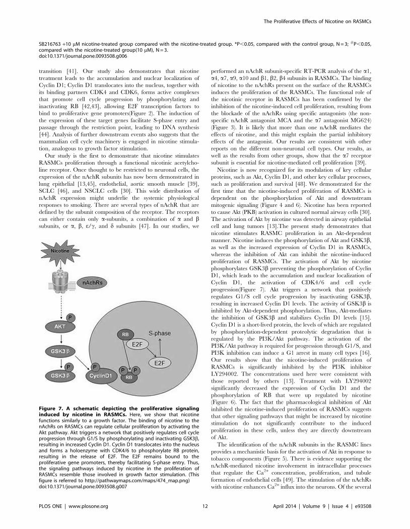
transition [41]. Our study also demonstrates that nicotine
treatment leads to the accumulation and nuclear localization of
Cyclin D1; Cyclin D1 translocates into the nucleus, together with
its binding partners CDK4 and CDK6, forms active complexes
that promote cell cycle progression by phosphorylating and
inactivating RB [42,43], allowing E2F transcription factors to
bind to proliferative gene promoters(Figure 2). The induction of
the expression of these target genes facilitate S-phase entry and
passage through the restriction point, leading to DNA synthesis
[44]. Analysis of further downstream events also suggests that the
mammalian cell cycle machinery is engaged in nicotine stimula-
tion, analogous to growth factor stimulation.
Our study is the first to demonstrate that nicotine stimulates
RASMCs proliferation through a functional nicotinic acetylcho-
line receptor. Once thought to be restricted to neuronal cells, the
expression of the nAchR subunits has now been demonstrated in
lung epithelial [13,45], endothelial, aortic smooth muscle [39],
SCLC [46], and NSCLC cells [30]. This wide distribution of
nAchR expression might underlie the systemic physiological
responses to smoking. There are several types of nAchR that are
defined by the subunit composition of the receptor. The receptors
can either contain only a-subunits, a combination of a and bsubunits, or a, b, e/c, and d subunits [47]. In our studies, we
performed an nAchR subunit-specific RT-PCR analysis of the a1,
a4, a7, a9, a10 and b1, b2, b4 subunits in RASMCs. The binding
of nicotine to the nAchRs present on the surface of the RASMCs
induces the proliferation of the RASMCs. The functional role of
the nicotinic receptor in RASMCs has been confirmed by the
inhibition of the nicotine-induced cell proliferation, resulting from
the blockade of the nAchRs using specific antagonists (the non-
specific nAchR antagonist MCA and the a7 antagonist MG624)
(Figure 3). It is likely that more than one nAchR mediates the
effects of nicotine, and this might explain the partial inhibitory
effects of the antagonist. Our results are consistent with other
reports on the different non-neuronal cell types. Our results, as
well as the results from other groups, show that the a7 receptor
subunit is essential for nicotine-mediated cell proliferation [39].
Nicotine is now recognized for its modulation of key cellular
proteins, such as Akt, Cyclin D1, and other key cellular processes,
such as proliferation and survival [48]. We demonstrated for the
first time that the nicotine-induced proliferation of RASMCs is
dependent on the phosphorylation of Akt and downstream
mitogenic signaling (Figure 4 and 6). Nicotine has been reported
to cause Akt (PKB) activation in cultured normal airway cells [30].
The activation of Akt by nicotine was detected in airway epithelial
cell and lung tumors [13].The present study demonstrates that
nicotine stimulates RASMC proliferation in an Akt-dependent
manner. Nicotine induces the phosphorylation of Akt and GSK3b,
as well as the increased expression of Cyclin D1 in RASMCs,
whereas the inhibition of Akt can inhibit the nicotine-induced
proliferation of RASMCs. The activation of Akt by nicotine
phosphorylates GSK3b preventing the phosphorylation of Cyclin
D1, which leads to the accumulation and nuclear localization of
Cyclin D1, the activation of CDK4/6 and cell cycle
progression(Figure 7). Akt triggers a network that positively
regulates G1/S cell cycle progression by inactivating GSK3b,
resulting in increased Cyclin D1 levels. The activity of GSK3b is
inhibited by Akt-dependent phosphorylation. Thus, Akt-mediates
the inhibition of GSK3b and stabilizes Cyclin D1 levels [15].
Cyclin D1 is a short-lived protein, the levels of which are regulated
by phosphorylation-dependent proteolytic degradation that is
regulated by the PI3K/Akt pathway. The activation of the
PI3K/Akt pathway is required for progression through G1/S, and
PI3K inhibition can induce a G1 arrest in many cell types [16].
Our results show that the nicotine-induced proliferation of
RASMCs is significantly inhibited by the PI3K inhibitor
LY294002. The concentrations used here were consistent with
those reported by others [13]. Treatment with LY294002
significantly decreased the expression of Cyclin D1 and the
phosphorylation of RB that were up regulated by nicotine
(Figure 6). The fact that the pharmacological inhibition of Akt
inhibited the nicotine-induced proliferation of RASMCs suggests
that other signaling pathways that might be increased by nicotine
stimulation do not significantly contribute to the induced
proliferation in these cells, unless they are directly downstream
of Akt.
The identification of the nAchR subunits in the RASMC lines
provides a mechanistic basis for the activation of Akt in response to
tobacco components (Figure 5). There is evidence supporting the
nAchR-mediated nicotine involvement in intracellular processes
that regulate the Ca2+ concentration, proliferation, and tubule
formation of endothelial cells [49]. The stimulation of the nAchRs
with nicotine enhances Ca2+ influx into the neurons. Of the several
SB216763 +10 mM nicotine-treated group compared with the nicotine-treated group. *P,0.05, compared with the control group, N = 3; #P,0.05,compared with the nicotine-treated group(10 mM), N = 3.doi:10.1371/journal.pone.0093508.g006
Figure 7. A schematic depicting the proliferative signalinginduced by nicotine in RASMCs. Here, we show that nicotinefunctions similarly to a growth factor. The binding of nicotine to thenAchRs on RASMCs can regulate cellular proliferation by activating theAkt pathway. Akt triggers a network that positively regulates cell cycleprogression through G1/S by phosphorylating and inactivating GSK3b,resulting in increased Cyclin D1. Cyclin D1 translocates into the nucleusand forms a holoenzyme with CDK4/6 to phosphorylate RB protein,resulting in the release of E2F. The E2F remains bound to theproliferative gene promoters, thereby facilitating S-phase entry. Thus,the signaling pathways induced by nicotine in the proliferation ofRASMCs resemble those involved in growth factor stimulation. (Thisfigure is referred to http://pathwaymaps.com/maps/474_map.png)doi:10.1371/journal.pone.0093508.g007
The Proliferative Effects of Nicotine on RASMCs
PLOS ONE | www.plosone.org 12 April 2014 | Volume 9 | Issue 4 | e93508

types of neuronal nAchRs, the a7 subunit-containing nAchRs
exhibit high calcium permeability. PI3K and its downstream
target Akt have been reported to be important for calcium-
mediated survival in a wide variety of cells [50]. It will be
interesting to study the involvement of Ca2+ in the context of
nicotine-induced proliferation of RASMCs. The flux of Ca2+
through the nAchRs might represent the intrinsic link between
nicotine, the nAchRs and the Akt pathway. Clearly, further studies
are needed to clarify the role of nicotine in the proliferation of
SMCs in airway remodeling. For example, the calcium-activated
signaling and in vivo models might yield important information in
this respect.
Taken together, our observations demonstrate that nicotine
binding to the nAchRs on RASMCs can regulate cellular
proliferation by activating the Akt pathway (Figure 7). This study
elucidates a novel aspect of the pathogenesis of COPD and might
open new avenues for the therapeutic targeting in COPD.
Author Contributions
Conceived and designed the experiments: PR BL FH. Performed the
experiments: FH YZ WZ. Analyzed the data: FH ZZ GH CJ. Contributed
reagents/materials/analysis tools: WH YZ DZ. Wrote the paper: FH PR
BL.
References
1. WHO.int.World Health Statistics. [updated 2008; cited 2012 April 25].
Available from: http://www.who.int/whosis/whostat/EN_WHS08_Full.pdf
2. Hogg JC, Timens W (2009) The pathology of chronic obstructive pulmonary
disease. Annu Rev Pathol 4:435–459.
3. Macnee W (2007) Pathogenesis of chronic obstructive pulmonary disease. Clin
Chest Med 28(3):479–513.
4. Brass DM, Savov JD, Gavett SH, Haykal-Coates N, Schwartz DA (2003)
Subchronic endotoxin inhalation causes persistent airway disease. Am J Physiol
Lung Cell Mol Physiol 285:L755–L761.
5. Churg A, Wang R, Wang X, Onnervik PO, Thim K, et al. (2007) Effect of an
MMP-9/MMP-12 inhibitor on smoke-induced emphysema and airway
remodeling in guinea pigs. Thorax 62:706–713.
6. Wright JL, Postma DS, Kerstjens HA, Timens W, Whittaker P, et al. (2007)
Airway remodeling in the smoke exposed guinea pig model. Inhal Toxicol
19:915–923.
7. Vernooy JH, Dentener MA, Van Suylen RJ, Buurman WA, Wouters EF (2002)
Long-term intratracheal lipopolysaccharide exposure in mice results in chronic
lung inflammation and persistent pathology. Am J Respir Cell Mol Biol 26:152–
159.
8. Toward TJ, Broadley KJ (2002) Goblet cell hyperplasia, airway function, and
leukocyte infiltration after chronic lipopolysaccharide exposure in conscious
Guinea pigs: effects of rolipram and dexamethasone. J Pharmacol Exp Ther
302:814–821.
9. Wongtrakool C, Wang N, Hyde DM, Roman J, Spindel ER (2012) Prenatal
nicotine exposure alters lung function and airway geometry through a7 nicotinic
receptors. Am J Respir Cell Mol Biol 46(5):695–702.
10. Zou W, Zou Y, Zhao Z, Li B, Ran P (2013) Nicotine Induced Epithelial-
mesenchymal Transition via Wnt/b-catenin Signaling in Human Airway
Epithelial Cells. American Journal of Physiology Lung Cellular and Molecular
Physiology 304(4):L199–209.
11. Cucina A, Fuso A, Coluccia P, Cavallaro A (2008) Nicotine inhibits apoptosis
and stimulates proliferation in aortic smooth muscle cells through a functional
nicotinic acetylcholine receptor. J Surg Res. 150(2):227–35.
12. Lam DC, Girard L, Ramirez R, Chau WS, Suen WS, et al. (2007) Expression of
nicotinic acetylcholine receptor subunit genes in non-small-cell lung cancer
reveals differences between smokers and nonsmokers. Cancer Res 67(10):4638–
47.
13. West KA, Brognard J, Clark AS, Linnoila I R, Yang X, et al. (2003) Rapid Akt
activation by Nicotine and a tobacco carcinogen modulates the phenotype of
normal Human airway epithelial cells. J. Clin. Invest 111,81–90.13.
14. Minna JD (2003) Nicotine exposure and bronchial epithelial cell nicotinic
acetylcholine receptor expression in the pathogenesis of lung cancer. J. Clin.
Invest 111:31–33.
15. Wang R, Xu YJ, Liu XS, Zeng DX, Xiang M (2012) CCN2 promotes cigarette
smoke-induced proliferation of rat pulmonary artery smooth muscle cells
through up regulating cyclin D1 expression. Journal of Cellular Biochemistry
113:349–359
16. Liang J, Slingerland JM (2003) Multiple roles of the PI3K/PKB (Akt) pathway in
cell cycle progression. Cell Cycle 2(4):339–45
17. Zhang W, Kong G, Zhang J, Wang T, Ye L, et al. (2012)MicroRNA-520b
inhibits growth of hepatoma cells by targeting MEKK2 and cyclin D1. PLoS
One 7(2):e31450.
18. Salic A, Mitchison TJ (2008) A chemical method for fast and sensitive detection
of DNA synthesis in vivo. Proc Nat l Acad Sci U S A 105:2415–2420.
19. Limsirichaikul S, Niimi A, Fawcett H, Lehmann A, Yamashita S, et al. (2009)
Arapidnon-radio active technique for measurement of repair synthesis in
Primary human fibroblasts by incorporation of ethynyldeoxyuridine (EDU).
Nucleic Acids Res 37:e 31.
20. Wang F, Zhao XQ, Liu JN, Wang ZH, Wang XL, et al. (2012) Antagonist of
microRNA-21 improves balloon injury-induced rat iliac artery remodeling by
regulating proliferation and apoptosis of adventitial fibroblasts and myofibro-
blasts. J Cell Biochem 113(9):2989–3001.
21. Brognard J, Clark AS, Ni Y, Dennis PA (2001) Akt/Protein Kinase B Is
Constitutively Active in Non-Small Cell Lung Cancer Cells and Promotes
Cellular Survival and Resistance to Chemotherapy and Radiation. Cancer Res
61(10):3986–97.
22. Dasgupta P, Rastogi S, Pillai S, Ordonez-Ercan D, Morris M, et al. (2006)
Nicotine induces cell proliferation by beta-arrestin-mediated activation of Srcand Rb-Raf-1 pathways. J Clin Invest 116(8):2208–2217.
23. Russell MA, Jarvis M, Iyer R, Feyerabend C (1980) Relation of nicotine yield ofcigarettes to blood nicotine concentrations in smokers. Br. Med. J 280:972–976.
24. Armitage AK, Dollery CT, George CF, Houseman TH, Lewis PJ, et al. (1975)Absorption and metabolism of nicotine from cigarettes. Br. Med. J 4:313–316.
25. Fu XW, Wood K, Spindel ER (2011) Prenatal nicotine explosure increasesGABA signaling and mucin expression in airway epithelium. Am J Respir Cell
Mol Biol 2011,44(2):222–229
26. Nevins JR (2001) The Rb/E2F pathway and cancer. Hum. Mol. Genet 10:699–703.
27. Stevaux O, Dyson NJ (2002) A revised picture of E2f Transcriptional regulationand Rb function. Curr. Opin. Cell Biol 14:684–691.
28. Lindstrom J (1997) Nicotinic acetylcholine receptors in health and disease. Mol.Neurobiol 15:193–222.
29. Grando SA, Horton RM, Mauro TM, Kist DA, Lee TX, et al. (1996) Activationof keratinocyte nicotinic cholinergic receptors stimulates calcium influx and
enhances cell differentiation. J. Invest. Dermatol 107:412–418.
30. Carlisle DL, Liu X, Hopkins TM, Swick MC, Dhir R, et al. (2007) Nicotine
activates cell-signaling pathways through muscle-type and neuronal nicotinicacetylcholine receptors in non-small cell lung cancer cells. Pulm Pharmacol Ther
20(6):629–41.
31. Ye L, Wang X, Jin M (2009) Role of Airway Epithelial Cells in Development of
Chronic Obstructive Pulmonary Disease. Journal of Epithelial Biology and
Pharmacology 2,44–50.
32. Hogg JC, Chu F, Utokaparch S, Woods R, Elliott WM, et al. (2004) The nature
of small-airway obstruction in chronic obstructive pulmonary disease.N Engl J Med 350(26):2645–53.
33. Pera T, Gosens R, Lesterhuis AH, Sami R, van der Toorn M, et al. (2010)Cigarette smoke and lipopolysaccharide induce a proliferative airway smooth
muscle phenotype. Respir Res 11:48. doi: 10.1186/1465-9921-11-48.
34. Zevin S, Gourlay SG, Benowitz NL (1998) Clinical pharmacology of nicotine.
Clin. Dermatol 16:557–564.
35. Heeschen C, Jang JJ, Weis M, Pathak A, Kaji S, et al. (2001) Nicotine stimulates
angiogenesis and promotes tumor growth and atherosclerosis. Nat. Med 7:833–839.
36. Lindell G, Farnebo LO, Chen D, Nexo E, Rask Madsen J, et al. (1993) Acuteeffects of smoking during modified sham feeding in Duodenal ulcer patient-
s.Ananalysis of nicotine, acid secretion, gastrin, catecholamines, epidermal
growth factor, prostaglandin E2, and bile acids. Scand J Gastroenterol28(6):487–494.
37. Clunes LA, Bridges A, Alexis N, Tarran R (2008) In vivo versus in vitro airwaySurface liquid nicotine levels following cigarette smoke exposure. J Anal Toxicol
32(3):201–207.
38. Xu Y, Zhang Y, Cardell L-O (2010) Nicotine enhances murine airway
contractile responses to kinin receptor agonists via activation of JNK- andPDE4-related intracellular pathways. Respir Res 11(1): 13.
39. Dasgupta P, Chellappan SP (2006) Nicotine-mediated cell proliferation and
angiogenesis: new twists to an old story. Cell Cycle 5(20):2324–8.
40. Yu J, Huang NF, Wilson KD, Velotta JB, Huang M, et al. (2009) nAChRs
mediate human embryonic stem cell-derived endothelial cells: proliferation,apoptosis, and angiogenesis. PLoS One. 4(9):e7040
41. Sherr CJ (1994) G1 phase progression.Cyclingoncue. Cell 79:551–555.
42. Morikawa-Futamatsu K, Adachi S, Maejima Y, Tamamori-Adachi M, Suzuki J,
et al. (2006) HMG-CoA reductase inhibitor fluvastatin prevents angiotensin II-induced cardiachypertrophy via Rhokinase and Inhibition of cyclinD1. Life Sci
79:1380–1390.
43. Ouyang W, Li J, Zhang D, Jiang BH, Huang DC (2007) PI-3K/Akt signal
pathway plays a crucial role in arsenite-induced cell proliferation of Humankeratinocytes through induction of cyclinD1. J Cell Biochem 101:969–978.
44. Takahashi-Yanaga F, Sasaguri T (2008) GSK-3beta regulates cyclin D1expression: a new target for chemotherapy. Cell Signal 20(4):581–9.
The Proliferative Effects of Nicotine on RASMCs
PLOS ONE | www.plosone.org 13 April 2014 | Volume 9 | Issue 4 | e93508

45. Wang,Y, Pereira EF, Maus AD, Ostlie NS, Navaneetham D, et al. (2001)
Human bronchial epithelial And endothelial cells express alpha7 nicotinicacetylcholine receptors. Mol. Pharmacol 60, 1201–1209.
46. Tarroni P, Rubboli F, Chini B, Zwart R, Oortgiesen M, et al. (1992) Neuronal-
type nicotinic receptors in human neuro-Blastoma and small-cell lung carcinomacell lines. FEBS Lett 312,66–70.
47. Conti_Tronconi BM, McLane KE, Raftery MA, Grando SA, Protti MP (1994)The nicotinic acetylcholine receptor: structure and autoimmune pathology.
Critical Reviews in Biochemistry and Molecular Biology 69–123.48,49,50,51,52.
48. Tsurutani J, Castillo SS, Brognard J, Granville CA, Zhang C, et al. (2005)
Tobacco components stimulate Akt-dependent proliferation and NFkappaB-
dependent survival in lung cancer cells. Carcinogenesis 26(7):1182–95.
49. Li XW, Wang H (2006) Nonneuronal nicotinic a-7 receptor, a new endothelial
target for revascularization. Life Sci 78;p. 1863.
50. Nakayama H, Numakawa T, Ikeuchi T (2002) Nicotine-induced phosphoryla-
tion of Akt through epidermal growth factor receptor and Src in PC12h cells.
J Neurochem 83(6):1372–9.
The Proliferative Effects of Nicotine on RASMCs
PLOS ONE | www.plosone.org 14 April 2014 | Volume 9 | Issue 4 | e93508