the primo vascular system: human biological system ... · • the pvs system may have interesting...
TRANSCRIPT

1
The Primo Vascular System: A Unique Human Biological System Shifting Medical Paradigm
Prospective Osteopathic Applications for Cranial, Visceral, Lymphatic, and Fascial Techniques
_____________________________ Bruno Chikly, MD, DO a & d, Paul Roberts, DOMP b, Jörgen Quaghebeur, MSc, DO, PhD Med Sci. c
a. Chikly Health Institute, Chikly Health Institute (CHI), 8924 E. Pinnacle Peak Rd, Ste G5, #248, Scottsdale, AZ 85255 USA
b. Sosa Osteopathy Centre, 21 Yiu Wa Street #801, Causeway Bay, Hong Kong.
c. Department of Research, Flanders International College of Osteopathy (FICO), Santvoortbeeklaan 23, B 2100 Antwerp, Belgium
Corresponding Author: Bruno Chikly, MD, DO
Highlights
• The primovascular system (PVS) is a rediscovered morphodynamic system initially described in 1961.
• Using state of the art technology, a scientific team reinitiated the PVS research in 2002 confirming its existence in many structures of the central and peripheral nervous systems, on the surface of most viscera, in lymph and blood vessels, as well as in adipose tissue.
• The PVS carries a high concentration of nucleic acids and adult small embryoniclike stem cells as well as hormones in secretory granules.
• The PVS has circulatory properties, endocrine functions, supports antiinflammatory processes and may be a new player in the physiopathology and treatment of cancer.
• The PVS system may have interesting implications for osteopathic manual medicine. The presence of “primovessels” in several vessels and nerves, viscera and fascia, and in the brain and spinal cord could potentially open novel possibilities of integration for cranial, lymphatic, visceral and fascial approaches in osteopathic manipulative treatment.
Keywords:
Acupuncture BongHan Ducts Circulation
2
Lymph Lymph Drainage Therapy Lymphatic System Manual Therapy Manual Lymphatic Therapy Osteopathy Osteopathic Lymphatic Technique Osteopathic Manipulation Osteopathic Manipulative Medicine Palpation Primovascular System
Abbreviations/Acronyms:
BHD: BongHan Duct BHK: BongHan Kim BHS: BongHan System LDT: Lymph Drainage Therapy MLDT: Manual Lymph Drainage Therapy MT: Manual Therapy OLT: Osteopathic Lymphatic Technique OMM: Osteopathic Manual Medicine PF: Primovascular fluid or Primofluid PN: Primovascular nodes or Primonodes PV: Primovascular vessels or Primovessels PVS: PrimoVascular System

3
Abstract
This article describes the historical background and pathophysiologic aspects of the PrimoVascular System (PVS). The PVS is depicted as being the most recent body system to be discovered, with a specific anatomical and immunohistochemical signature that sets it apart from the arteriovenous and lymphatic systems. It is comprised of immune functions, endocrine functions, and is found to be deeply involved in several biological processes, including tissue regeneration, inflammation and cancer.
Though ongoing research has been conducted since 2002, the original discovery was actually made in the 1960's by BongHan Kim, a North Korean scientist, which went virtually unnoticed for a span of nearly 40 years.
The presence of “primovessels” in and around several vessels and nerves, viscera and fascia, and in the brain and spinal cord, reveals a common link that could potentially open novel possibilities of integration for cranial, lymphatic, visceral and fascial approaches in osteopathic manual medicine.
Introduction
Even in an age where a profuse amount of scientific data is revealed on a daily basis, it is rare to suggest the existence of another human body system. A novel circulatory system was first described by BongHan Kim as the "Substance of Kyungrak" in the 1960’s. Advanced evaluation methods later confirmed this system as the Primo Vascular System (PVS) and its existence in multiple anatomical structures in animals and the human body.
A relatively recent body system like the lymphatic system was officially discovered as far back as 1627 1 . The PVS being the latest candidate to the list of human body system, thought formally described in 19611962, was not scientifically authenticated until 2002.
This literature review article presents the past and the present research on the PrimoVascular system, and describes its historical background and physiologic aspects as well as the current state of understanding on PVS.
The presence of the PVS, if fully confirmed, suggests possible implications that may fundamentally reshape our vision of anatomy, physiology and medicine, consequently impacting osteopathic manipulative medicine and manual therapy.
Discovery of the PVS There are two distinct eras of discovery and research of the PVS occurring nearly 40 years apart.
A. Initial discovery: from 1961 to 1965
BongHan Kim (1916?), professor at the Pyongyang Medical School in North Korea, embarked on scientific research to discover the substratum for the acupuncture meridians. While head of the department of physiology in Pyongyang, BongHan Kim made his initial announcement about the anatomical substratum for the acupuncture meridian system (“Kyungrak” in Korean), called "Substance of Kyungrak", on August 18,
4
1961, which he then published in 1962 2. In 1962, he wrote in his manuscript that the acupuncture meridian system “consists of bundles of tubular structures and it is clearly distinguishable from nervous, blood vessels and lymph systems in histological and experimentalbiological characters.” 2 and ”the diameter of the tubular structures range between 20 and 50 microns.” 2. BongHan Kim subsequently published five detailed articles about his research from 1962 to 1965 38. He used histological techniques (Hematoxylin and Eosin, Mason’s trichrome, Verhoff, silver staining, Feulgen reaction, Acridine orange), radioactive tracers, and electrophysiological methods. He identified the PVS using a specific blue dye, unfortunately his reports lack full scientific descriptions of his materials, methods, and protocols, which proved highly challenging for researchers of the time wanting to confirm his findings.
One of BongHan’s reports were translated into English and made readily available for research teams 7, which led numerous groups in China, Japan, and Russia to try to confirm his findings9. Fujiwara and Yu, in Japan, were able to partially reproduce Kim’s findings inside blood vessels, and on the surfaces of viscera10 , and some Chinese teams may have also reproduced some of BongHan Kim’s results in rabbits 11 , however, one of the main reasons that scientific teams could not properly reproduce his results was that the specific blue dye that BongHan used to make the system visible and conduct his research, was never fully described. The scientific community of the time eventually abandoned the idea of a definite BongHan system of ducts and nodes. Around 1965, the institute in North Korea, where BongHan Kim was working, unexpectedly closed. The BongHan theory has been largely forgotten for about four decades, and to this day BongHan Kim’s fate is unknown 12. Table 1: Reported discoveries of BongHan Kim.
Reported Discoveries of BongHan Kim
BongHan Kim (BHK) claimed he discovered a new circulatory system inside plants, invertebrate, and mammals, especially rabbits. He said this was the “Kyungrak System”, meaning the system of “acupuncture meridians and collaterals” in Korean. The so called Kyungrak system (or BongHan/BH system) consisted of a network of BongHan ducts (or BH ducts), BongHan corpuscles (or BH corpuscles), and BongHan liquor (or BH liquor), circulating inside of them, a liquid high in chromatin. BongHan Kim, using radiotracer, described the BH ducts as being comprised of many smaller ductules composed of endothelial cells with characteristic rodshaped nuclei. He applied biochemical and histochemical analyses of the BH system and found that the fluid inside the ducts is transparent, and contains more nucleic acids, especially DNA, than any other tissue. He described the components of the BH liquor as: Total nitrogen content: 3.12–3.40% Non protein nitrogen content: 0.10–0.17% Lipids: 0.57–1.00% Reduced sugar: 0.10–0.12% Total hyaluronic acid: 170.4mg% More than 19 free amino acids More than 16 free mononucleotides Very abundant DNA and RNA nucleotides
5
For BongHan Kim, the BH ducts also contain “sanals“, meaning “live egg” in Korean which seems to have a function equivalent to that of stem cells. BongHan Kim described these “sanals”, modernly renamed Primomicrocell or Pmicrocell, as having hematopoietic functions, and being able to regenerate injured tissues, and heal wounds. BHK described 5 subsystems (BHS = BongHan System):
1. The intravascular BHS: located inside the blood vessels, the heart and the lymphatic vessels. 2. The organ surface BHS: freely floating on the surfaces of viscera. 3. Theextravascular BHS: running along the blood and lymphatic vessels, as well as the nerves. It is located just outside of these structures. 4. Thenervous BHS: located inside the central and peripheral nervous systems, floating in the cerebrospinal fluid. 5. The intraorgan BHS: located inside visceral parenchyma.
Electrical conductivity of a BongHan vessel recorded by BongHan Kim: BongHan Kim identified three periodic potentials of 1530 seconds, 710 seconds, and 2025 seconds. The method used to measure these signals was not specifically described. According to the research of BongHan Kim, damage to BongHan vessels can “modify the frequency and amplitude of the heart and change the peristaltic motion of intestines.” It can also reduce nerve excitability and decrease muscular contractions.
B. Scientific Confirmation: from 2002 to the Present Time
Around 2000, KwangSup Soh, professor in the Department of Physics at the Seoul National University (1976–2011) also formed a Biomedical Physics Laboratory to examine the scientific reality of the acupuncture meridians. He chose Dr. BC Lee as the main researcher to reexamine the findings of BongHan Kim using the latest technology available. After a series of trials and errors they finally observed BongHan ducts floating on the surfaces of rabbit viscera, such as the liver, the stomach, the intestines and the bladder 13. In 2002 KwangSup Soh proposed a change to the terminology specifically changing the name “BongHan system” to “Primovascular system” (see Table 2). This change was later endorsed by the 2010 International Symposium on the Primo Vascular System 14. Table 2: Primo Vascular System Terminology.
Primo Vascular System Terminology Terminology changes of KwangSup Soh (2002/2010) BongHan system (initially called the “Kyungrak” system) was changed to Primovascular system (or PVS) BongHan duct was changed to Primovessel or Primovascular vessels (or PV) BongHan corpuscle was changed to Primonode (or Pnode) BongHan liquid was changed to Primofluid (or Pfluid) BongHan “Sanal” was changed to Primomicrocell (or Pcell)
In 2008, Dr. BC Lee of the Soh group and his team discovered a dye that can be sued in a specific manner to identify the PVS: Trypan blue 15.

6
From there, the Seoul National University (SNU) could observe the PVS in the bovine heart, the brain ventricles, and in the central canal of spinal cords, as well as, for the first time, in the abdominal adipose tissues. In 2011, Prof. K.S. Soh’s team of the Seoul National University Department of Physics formed the Nano Primo Research Center of SNU’s Advanced Institute of Convergence Technology, where he would head the research. Identification of the PVS To visualize the PVS, a great deal of advanced technology has been used 16, including confocal laser scanning microscopy 17, scanning electron microscopy (SEM), cryoSEM, focusedionbeam SEM, high voltage transmission electron microscopy (TEM) 18, electron microscopy (for the surfaces of mammalian organs) 18, 19 ; atomic force microscopy20; fluorescent nanoparticle21; immunohistochemistry22,23; proteomic analysis 24; and the ELISA technique for the primofluid content 25, 26. Before the discovery of Trypan blue 27, Janus green 28 and Alcian blue dyeing 29 were used with moderate success to identify the PVS. Trypan blue is not a specific marker of PVS. Trypan blue has been widely used in the analysis of cell cultures to stain dead or dying cells in culture and as early as the 1930's this dye was known to stain damaged or inflamed tissue (e.g. Menkin V. 1931) 30. Trypan blue also has been used to stain lymphatic vessels (e.g., Leak LV et al. 1978) 31, and Li H and Li J. (2003) 32. To identify the PVS, Soh’s team used subjects in a non pathological state and the method for detecting PVS requires specific procedures that were developed in the Nano Primo Research Center. If this procedure is not followed the PVS sample is not detectable (Prof. Soh KS, personal communication). Image # 1 Stereomicroscopic in situ image of a branched and rejoined primo vascular system (blue stain) in the thoracic duct of a subject rat. Two regions of branching (arrows) and rejoining (open arrows) are observed. In Kim S, Jung SJ, Cho SY, et al. A Method for the Observation of the Primo Vascular System in the Thoracic Duct of a Rat. Evid Based Complement Alternat Med 2013; 2013: 5. http://dx.doi.org/10.1155/2013/536560. Reprinted with the kind permission of Professor Kwang-Sup Soh. Anatomical Overview of the PVS The primovascular vessels have been identified in rabbits as thin semitransparent structures of an average

7
diameter of approximately 20 to 30 μm 19, 33, 34. Each vessel contains several, (up to 20), smaller ductules, 3–10 μm in diameter that are lined by a single layer of endothelial cells and surrounded by extracellular matrix (ECM) 34. Each ductule is filled with a liquid called primofluid 18, 19, 34. They can be connected to primovascular nodes (PN) 35. Nowadays, primovessels are fairly easily differentiated from blood vessels, and they present many differences with lymph vessels. Primovessels are not lymphatic, nor blood vessels, as they don’t express LYVE1 (specific lymphatic endothelial marker, a CD44 homolog), nor CD31 (blood vessel specific marker). The rodshaped nuclei of the PVS endothelium cells are aligned parallel to the wall of the primovessels, and can be specifically stained by Acridine orange, fluorescent phalloidin (Factin), or DAPI 9, 27, 33, 3638 . Rodshaped nuclei are hall marks of the PVS, regardless of which animals or organs the PVS was taken from. One problem is that the rodshaped nuclei can easily be seen and identified in longitudinal views, but are very difficult to see or identify in cross section images. Image # 2 Illustration of one isolated subvessel (top) and a bundle of subvessels of the primo vessel. In Stefanov M, Potroz M, Kim J, et al. The Primo Vascular System as a New Anatomical System. J Acupunct Meridian Stud 2013; 6(6): 3318. http://www.ncbi.nlm.nih.gov/pubmed/24290797. Reprinted with the kind permission of Professor Kwang-Sup Soh. Composition of the Primofluid
8
The composition of the primofluid of rats has been found to be rich in granulocytes and secretory granules: mast cells (20%), histiocytes (53%), eosinophils (16%), neutrophils (5%), round immature stemlike cells (3%), and relatively poor in lymphocytes (1%) 39. In the PVS, researchers measured a high concentration of adult small embryoniclike stem cells (very small embryoniclike stem cells) expressing the stem cell biomarkers oct4, nanog, and CD133 34, 39, 40. Location of the PrimoVascular System
i. Heart Inside the bovine heart 41, on top of bovine endocardium 42, Lee et al. visualized a complex arrangement of endocardial vessels (20 μm in thickness).
ii. Blood Vessels Primovessels has been identified inside large blood vessels 4347. In particular, primovessels have been identified floating inside the abdominal artery and the caudal vena cava of rabbits 48, rats 49, and mice 46. PVS has also been discovered in the superior sagittal sinus of a rabbit brain 50.
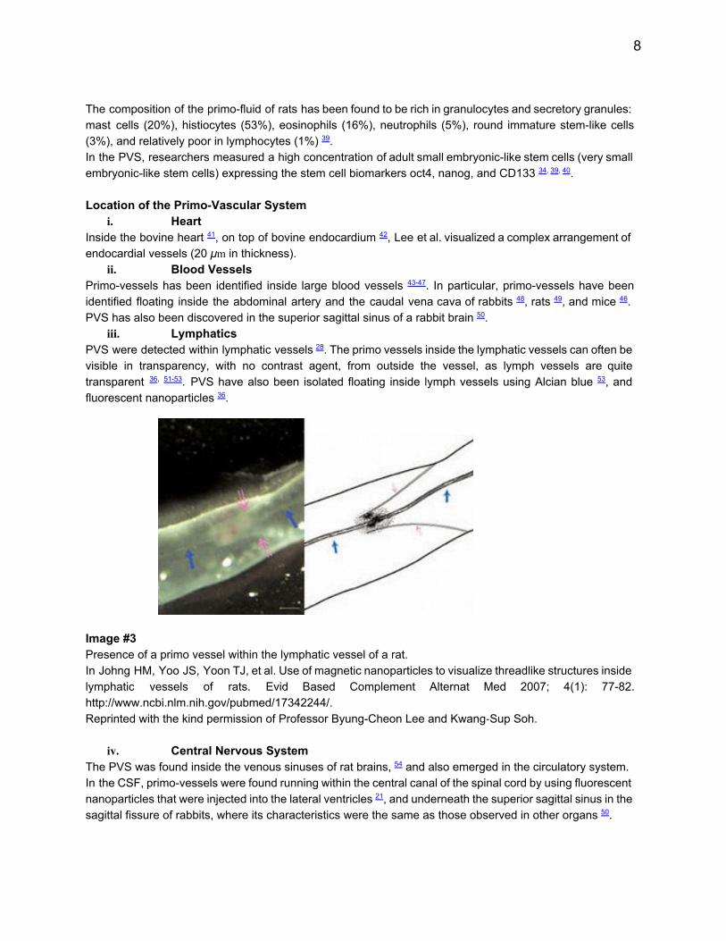
iii. Lymphatics PVS were detected within lymphatic vessels28. The primo vessels inside the lymphatic vessels can often be visible in transparency, with no contrast agent, from outside the vessel, as lymph vessels are quite transparent 36, 5153. PVS have also been isolated floating inside lymph vessels using Alcian blue 53, and fluorescent nanoparticles 36. Image #3 Presence of a primo vessel within the lymphatic vessel of a rat. In Johng HM, Yoo JS, Yoon TJ, et al. Use of magnetic nanoparticles to visualize threadlike structures inside lymphatic vessels of rats. Evid Based Complement Alternat Med 2007; 4(1): 7782. http://www.ncbi.nlm.nih.gov/pubmed/17342244/. Reprinted with the kind permission of Professor ByungCheon Lee and Kwang-Sup Soh.
iv. Central Nervous System The PVS was found inside the venous sinuses of rat brains, 54 and also emerged in the circulatory system. In the CSF, primovessels were found running within the central canal of the spinal cord by using fluorescent nanoparticles that were injected into the lateral ventricles21, and underneath the superior sagittal sinus in the sagittal fissure of rabbits, where its characteristics were the same as those observed in other organs 50.
9
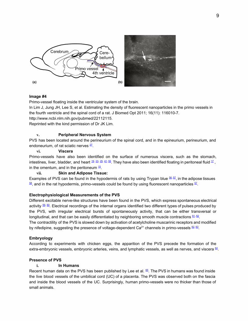
Image #4 Primovessel floating inside the ventricular system of the brain. In Lim J, Jung JH, Lee S, et al. Estimating the density of fluorescent nanoparticles in the primo vessels in the fourth ventricle and the spinal cord of a rat. J Biomed Opt 2011; 16(11): 1160107. http://www.ncbi.nlm.nih.gov/pubmed/22112115. Reprinted with the kind permission of Dr JK Lim.
v. Peripheral Nervous System PVS has been located around the perineurium of the spinal cord, and in the epineurium, perineurium, and endoneurium, of rat sciatic nerves 27.
vi. Viscera Primovessels have also been identified on the surface of numerous viscera, such as the stomach, intestines, liver, bladder, and heart 19, 23, 25, 41,55. They have also been identified floating in peritoneal fluid17 , in the omentum, and in the peritoneum 15.
vii. Skin and Adipose Tissue: Examples of PVS can be found in the hypodermis of rats by using Trypan blue 56,57, in the adipose tissues 58, and in the rat hypodermis, primovessels could be found by using fluorescent nanoparticles 57. Electrophysiological Measurements of the PVS Different excitable nervelike structures have been found in the PVS, which express spontaneous electrical activity 59, 60. Electrical recordings of the internal organs identified two different types of pulses produced by the PVS, with irregular electrical bursts of spontaneously activity, that can be either transversal or longitudinal, and that can be easily differentiated by neighboring smooth muscle contractions 61, 62. The contractility of the PVS is slowed down by activation of acetylcholine muscarinic receptors and modified by nifedipine, suggesting the presence of voltagedependent Ca2+ channels in primovessels 62, 63.
Embryology According to experiments with chicken eggs, the apparition of the PVS precede the formation of the extraembryonic vessels, embryonic arteries, veins, and lymphatic vessels, as well as nerves, and viscera 64. Presence of PVS
i. In Humans Recent human data on the PVS has been published by Lee et al. 65. The PVS in humans was found inside the live blood vessels of the umbilical cord (UC) of a placenta. The PVS was observed both on the fascia and inside the blood vessels of the UC. Surprisingly, human primovessels were no thicker than those of small animals.

10
ii. In Animals BongHan Kim primarily used rabbits for his experiments on the PVS, but affirmed that he had identified the PVS in hydra, fish, amphibians, birds, and numerous mammals, including humans 4. Since 2002 the PVS has been located in mice, rats, rabbits, dogs, pigs, and cows.
Functions of the PVS i. Circulatory Functions/Transport
The PVS circulates in a network of vessels and nodes with multiple independent paths that can stay connected. The entire PVS network has yet to be fully mapped in humans. The speed of the primoflow was measured at approximately 0.3 ± 0.1 mm/second in rabbits, using Alcian blue 19, and at around 100800 m/second with direct measurement using fluorescent nanoparticles 19, 66, 67. Within the primofluid, the circulatory nature of the PVS can help transport chemical substances, factors of inflammation, possibly medication, as well as cancer cells (metastases), in pathological processes. The PVS has been foreseen as a potentially novel drug delivery route, particularly for cancer treatment 68.
i. Immune and Regenerative Functions Primofluid contains a high concentration of very small embryoniclike stem cells, approximately 14 m in diameter, whose exact function still needs to be determined 40, 69. A research team led by BS Kwon, at the National Cancer Center of Korea, 39 confirmed that the PVS was abundant with other immune cells, such as macrophages, eosinophils, and mast cells 18.
ii. Endocrine Functions: Neurotransmitter Pathway The PVS has also been described as an endocrine organ which transports different hormones 25, 26, 70. Catecholamines (adrenalin and noradrenalin) have been identified in the primofluid of the organ surface of rabbits and rats with the ELISA technique 25, 39.
iii. Bioluminescence (Biophotons) Popp et al. described different types of biophoton emissions within living systems 71, 73. The PVS contains a high concentration of nucleic acids, and is surrounded by collagen. Consequently, Lee and Soh suggested that the PVS can be a good medium to transport, or communicate, tissue bioluminescence (biophoton) 53, 68. Role of PVS in Pathology
i. Inflammatory Process Wang et al. in China showed that the PVS of infected rats carries pathological products, which may be involved in inflammatory processes 73.
ii. Cancer In mammals, the PVS has been identified on the fascia surrounding tumor tissue, or connected to tumors9,
74, 75. The PVS may be a newly recognized player in cancer growth control, and may potentially bea novel path for cancer metastasis, as primovessels are more concentrated around tumor sites, and migration of tumor cells is more efficient inside the PVS than in the lymphatic system 7477. Applications for Osteopathic Manual Medicine: Potential Integration of Cranial, Lymphatic, Visceral and Fascia Approaches G.W. Sutherland (18731954), that may have continued the legacy of Swedenborg(16881772)78., described the “potency” of the cerebrospinal fluid, calling it the “fluid within the fluid”, and again toward the end of his life he described it as a “liquid light” 79. It would be interesting to debate, whether or not, Sutherland’s descriptions of a “fluid within the fluid” and of a “liquid light”, might potentially be actual early descriptions of some aspects of the yet unknown primovascular system.
11
Primo vessels are ubiquitous channels for transporting fluid with immune and endocrine functions. Initially, primovessels could, for example, be accessed manually through the lymphatic vessels using specialized osteopathic lymphatic techniques. Furthermore, like for most structures of the body, primovessels specific motilities could be looked for (this remains to be confirmed by further research). The PVS could reasonably link tissue functions across "systems". The PVS plays intriguing roles in tissue where osteopathic manipulation has distinct efficacy, including fascia, membrane, lymph, viscera, nerve and central nervous system. Since osteopathic manipulation can benefit vasculature, lymph, and capillary flow, it is reasonable to suggest that this additional fluid system can also be affected which would both positively influence, and further expand, the scope of osteopathic practice. Discussion The work of the Soh group is undoubtedly intriguing and would need to be confirmed by a number of other scientific teams. Circulatory aspects of the PVS have been described, Kim BH showed three periodic potentials of 1530 seconds, 710 seconds, and 2025 seconds, for the PVS but he did not describe a pulsatory type measurement. It remains a challenge to measure such rhythmic aspects in the human body. Further research evaluating the physiological importance and circulatory effects of the PVS is required and eagerly awaited. Future endeavors may hopefully reveal more convincing evidence and substantiate a rhythmic aspect of this novel circulatory system, and its potential palpation by trained osteopaths, as the PVS is on the surface of many organs and in the subcutaneous layer. If the anatomy and pathophysiology of the PVS, in particular its applications to inflammation, endocrinology, and oncology are confirmed, it will expand our understanding of the human body systems and will create a general paradigm change in medicine. Conclusion It is difficult to fathom that the prolific PrimoVascular System was not identified in medicine until recently. Both BongHan Kim and KwangSup Soh initially targeted their research at discovering the meridian system of acupuncture, but found something more surprising and far reaching. The discovery of the PVS in intravascular and extravascular spaces, in the central and peripheral nervous systems, on the surface of and within viscera, in cutaneous layers, and in most body systems, may signify a novel and complete morphodynamic system, with potential to reshape paradigms in medicine and osteopathic manipulative treatment. Acknowledgements The authors are grateful for the help and advice provided to them by Professor KwangSup Soh, Director of the Nano Primo Research Center Advanced Institute of Convergence Technology in Seoul National University. Author’s Contribution The authors contributed equally to this work. Disclosure Statement The three authors have no direct or indirect financial interests. References 1. Aselli G. De Lactibus Seu Lactis Venis, Quarto Vasorum Mesaraicorum Genere Novo Inventa [Dissertatio Mediolan]; 1627. 2. Kim BH. Great Discovery in Biology and Medicine: Substance of Kyungrak. Pyongyang: Foreign
12
Languages Publishing House; 1962. 3. Kim BH. Study on the reality of acupuncture meridians. J Jo Sun Med 1962; 9: 513. 4. Kim BH. The Kyungrak system. J Jo Sun Med 1965; 108: 138. 5. Kim BH. The sanal theory. J Acad Med Sci DPR Korea 1965; 108: 3962. 6. Kim BH. Sanals and hematopoiesis. J Jo Sun Med 1965: 16. 7. Kim BH. On the Kyungrak system. J Acad Med Sci DPR Korea 1963; 90: 141. http://primosystem.wikispaces.com/Kim+Bong+Han+Paper+in+1963. Accessed: 27/12/2014. 8. Kim BH. On the Kyungrak System. Pyongyan: D. P. R. K.; 1964. 9. Yoo JS, Ayati MH, Kim HB, Zhang WB, Soh KS. Characterization of the primovascular system in the abdominal cavity of lung cancer mouse model and its differences from the lymphatic system. PLoS ONE 2010; 5(4): e9940. http://www.ncbi.nlm.nih.gov/pubmed/20376343/. Accessed: 27/12/2014. 10. Fujiwara S, Yu SB. 'Bonghan theory’ morphological studies. Igaku no Ayumi 1967; 60: 56777. Accessed: [Japanese]. 11. Liu JL, Jing XH, Shi H, et al. Historical Review about Research on "Bonghan System" in China. Evid Based Complement Alternat Med 2013; 2013: 636081. http://www.ncbi.nlm.nih.gov/pubmed/23861708/. Accessed: 20/02/2015. 12. Soh KS, Kang KA, Ryu YH. 50 years of BongHan theory and 10 years of primo vascular system. Evid Based Complement Alternat Med 2013; 2013: 587827. http://www.ncbi.nlm.nih.gov/pubmed/23983793. Accessed: 27/12/2014. 13. Soh KS. Bonghan circulatory system as an extension of acupuncture meridians. J Acupunct Meridian Stud 2009; 2(2): 93106. http://www.ncbi.nlm.nih.gov/pubmed/20633480. Accessed: 27/12/2014. 14. Soh KS, Kang KA, Harrison DK. The Primo Vascular System. Its Role in Cancer and Regeneration. New York: Springer; 2012. 15. Lee BC, Kim KW, Soh KS. Visualizing the network of Bonghan ducts in the omentum and peritoneum by using Trypan blue. J Acupunct Meridian Stud 2009; 2(1): 6670. http://www.ncbi.nlm.nih.gov/pubmed/20633476. Accessed: 27/12/2014. 16. Stefanov M, Kim J. Primo vascular system as a new morphofunctional integrated system. J Acupunct Meridian Stud 2012; 5(5): 193200. http://www.ncbi.nlm.nih.gov/pubmed/23040098. Accessed: 27/12/2014. 17. Shin HS, Johng HM, Lee BC, et al. Feulgen reaction study of novel threadlike structures (Bonghan ducts) on the surfaces of mammalian organs. Anat Rec B New Anat 2005; 284B(1): 3540. http://onlinelibrary.wiley.com/doi/10.1002/ar.b.20061/. Accessed: 27/12/2014. 18. Lee BC, Yoo JS, Ogay V, et al. Electron microscopic study of novel threadlike structures on the surfaces of mammalian organs. Microsc Res Tech 2007; 70(1): 3443. http://www.ncbi.nlm.nih.gov/pubmed/17019695. Accessed: 27/12/2014. 19. Sung B, Kim MS, Lee BC, et al. Measurement of flow speed in the channels of novel threadlike structures on the surfaces of mammalian organs. Naturwissenschaften 2008; 95(2): 11724. http://www.ncbi.nlm.nih.gov/pubmed/17713750. Accessed: 27/12/2014. 20. Kwon J, Baik KY, Lee BC, et al. Scanning probe microscopy study of microcells from the organ surface Bonghan corpuscle. Applied Physics Letters 2007; 90(17): 13. http://scitation.aip.org/content/aip/journal/apl/90/17/10.1063/1.2732183. Accessed: 27/12/2014. 21. Lim J, Jung JH, Lee S, et al. Estimating the density of fluorescent nanoparticles in the primo vessels in the fourth ventricle and the spinal cord of a rat. J Biomed Opt 2011; 16(11): 1160101160107. http://www.ncbi.nlm.nih.gov/pubmed/22112115. Accessed: 25/05/2015. 22. Soh KS, Hong S, Hong JY, Lee BC, Yoo JS. Immunohistochemical characterization of intravascular Bonghan duct. Microcirculation 2006; 13: 166. http://ispvs.org/L.O.P(Soh)%202011.10.28.pdf. Accessed: 15/01/2015.

13
23. Kim MS, Hong JY, Hong S, et al. BongHan corpuscles as possible stem cell niches on the organ surfaces. J Kor Pharmacopunct Inst 2008; 11: 512. http://www.journal.ac/sub/view2/300. Accessed: 27/12/2014. 24. Lee SJ, Lee BC, Nam CH, et al. Proteomic analysis for tissues and liquid from bonghan ducts on rabbit intestinal surfaces. J Acupunct Meridian Stud 2008; 1(2): 97109. http://www.ncbi.nlm.nih.gov/pubmed/20633461. Accessed: 27/12/2014. 25. Kim J, Ogay V, Lee BC, et al. CatecholamineProducing Novel Endocrine Organ: Bonghan System. Medical Acupuncture 2008; 20(2): 97102. http://online.liebertpub.com/doi/abs/10.1089/acu.2008.0600. Accessed: 27/12/2014. 26. Ogay V, Kim MS, Seok HJ, Choi CJ, Soh KS. Catecholaminestoring cells at acupuncture points of rabbits. J Acupunct Meridian Stud 2008; 1(2): 8390. http://www.ncbi.nlm.nih.gov/pubmed/20633459. Accessed: 27/12/2014. 27. Lee BC, Eom KH, Soh KS. Primovessels and primonodes in rat brain, spine and sciatic nerve. J Acupunct Meridian Stud 2010; 3(2): 1115. http://www.ncbi.nlm.nih.gov/pubmed/20633524. Accessed: 27/12/2014. 28. Lee BC, Yoo JS, Baik KY, Kim KW, Soh KS. Novel threadlike structures (Bonghan ducts) inside lymphatic vessels of rabbits visualized with a Janus Green B staining method. Anat Rec B New Anat 2005; 286(1): 17. http://www.ncbi.nlm.nih.gov/pubmed/16177995. Accessed: 27/12/2014. 29. Jung SJ, Cho SY, Bae KH, et al. Protocol for the observation of the primo vascular system in the lymph vessels of rabbits. J Acupunct Meridian Stud 2012; 5(5): 23440. http://www.ncbi.nlm.nih.gov/pubmed/23040104. Accessed: 27/12/2014. 30. Menkin V. Studies on inflammation: VI. Fixation of Trypan blue in inflamed areas of frogs. J Exp Med 1931; 53(2): 17983. http://www.ncbi.nlm.nih.gov/pubmed/19869834/. Accessed: 22/05/2015. 31. Leak LV, Schannahan A, Scully H, Daggett WM. Lymphatic vessels of the mammalian heart. Anat Rec 1978; 191(2): 183201. http://www.ncbi.nlm.nih.gov/pubmed/666017. Accessed: 25/05/2015. 32. Li H, Li J. Development of the peritoneal lymphatic stomata and lymphatic vessels of the diaphragm in mice. Annals of anatomy = Anatomischer Anzeiger : official organ of the Anatomische Gesellschaft 2003; 185(5): 4118. http://www.ncbi.nlm.nih.gov/pubmed/14575267. Accessed: 25/05/2015. 33. Noh YI, Rho M, Yoo YM, Jung SJ, Lee SS. Isolation and morphological features of primo vessels in rabbit lymph vessels. J Acupunct Meridian Stud 2012; 5(5): 2015. http://www.ncbi.nlm.nih.gov/pubmed/23040099. Accessed: 27/12/2014. 34. Ogay V, Bae KH, Kim KW, Soh KS. Comparison of the characteristic features of Bonghan ducts, blood and lymphatic capillaries. J Acupunct Meridian Stud 2009; 2(2): 10717. http://www.ncbi.nlm.nih.gov/pubmed/20633481. Accessed: 27/12/2014. 35. Lee BC, Jhang SU, Choi JH, et al. DiI staining of fine branches of Bonghan ducts on surface of rat abdominal organs. J Acupunct Meridian Stud 2009; 2(4): 3015. http://www.ncbi.nlm.nih.gov/pubmed/20633506. Accessed: 27/12/2014. 36. Johng HM, Yoo JS, Yoon TJ, et al. Use of magnetic nanoparticles to visualize threadlike structures inside lymphatic vessels of rats. Evid Based Complement Alternat Med 2007; 4(1): 7782. http://www.ncbi.nlm.nih.gov/pubmed/17342244/. Accessed: 27/12/2014. 37. Jia ZF, Soh KS, Zhou Q, Dong B, Yu WH. Study of novel threadlike structures on the intestinal fascia of dogs. J Acupunct Meridian Stud 2011; 4(2): 98101. http://www.ncbi.nlm.nih.gov/pubmed/21704951. Accessed: 27/12/2014. 38. An P, Dai J, Su Z, et al. Putative primovascular system in mesentery of rats. J Acupunct Meridian Stud 2010; 3(4): 23240. http://www.ncbi.nlm.nih.gov/pubmed/21185537. Accessed: 27/12/2014. 39. Kwon BS, Ha CM, Yu S, et al. Microscopic nodes and ducts inside lymphatics and on the surface of internal organs are rich in granulocytes and secretory granules. Cytokine 2012; 60(2): 58792.
14
http://www.ncbi.nlm.nih.gov/pubmed/22884518. Accessed: 27/12/2014. 40. Ogay V, Soh KS. Identification and Characterization of Small StemLike Cells in the Primo Vascular System of Adult Animals. In: Soh KS, Kang KA, Harrison DK, eds. The Primo Vascular System. New York: Springer; 2012: 14955. 41. Lee BC, Kim HB, Sung B, et al. Structure of the Sinus in the Primo Vessel Inside the Bovine Cardiac Chambers. In: Soh KS, Kang KA, Harrison DK, eds. The Primo Vascular System. New York: Springer; 2012: 5762. 42. Lee BC, Kim HB, Sung B, et al. Network of endocardial vessels. Cardiology 2011; 118(1): 17. http://www.ncbi.nlm.nih.gov/pubmed/21372571. Accessed: 27/12/2014. 43. Jiang XW, Lee BC, Choi C, et al. Method for Observing Intravascular BongHan Duct. J Kor Orient Prevent Med Soc 2002; 6: 1626. http://arxiv.org/pdf/physics/0211086.pdf. Accessed: 27/12/2014. 44. Shin HS, Soh KS. Electrical method to detect a Bonghan duct inside blood vessels. New Physics 2002; 45(6): 3768. http://www.jntong.co.kr/jn/request.html?cno=300. Accessed: 21/01/2015 [Korean]. 45. Baik KY, Lee BC, Johng HM, et al. Long threadlike structure inside the blood vessels of rats. Newest Med J 2004; 47: 1822. Accessed: 21/01/2015. 46. Yoo JS, Kim MS, Ogay V, Soh KS. In vivo visualization of bonghan ducts inside blood vessels of mice by using an Alcian blue staining method. Indian J Exp Biol 2008; 46(5): 3369. http://www.ncbi.nlm.nih.gov/pubmed/18697616. Accessed: 27/12/2014. 47. Lee BC, Baik KY, Johng HM, et al. Fluorescent method for observing intravascular Bonghan duct. Journal of the Korean Institute of Herbal Acupuncture 2005; 8: 59. http://www.researchgate.net/publication/263362213. Accessed: 27/12/2014. 48. Kim J, Jung J, Potroz M. Summary of BongHan Kim’s Publications. In: Soh KS, Kang KA, Harrison DK, eds. The Primo Vascular System. New York: Springer; 2012: 717. 49. Lee BC, Baik KY, Johng HM, et al. Acridine orange staining method to reveal the characteristic features of an intravascular threadlike structure. Anat Rec B New Anat 2004; 278(1): 2730. http://www.ncbi.nlm.nih.gov/pubmed/15170690. Accessed: 20/02/2015. 50. Nam MH, Lim J, Choi SH, Kim S, Soh KS. A primo vascular system underneath the superior sagittal sinus in the brain of a rabbit. J Acupunct Meridian Stud 2012; 5(5): 2107. http://www.ncbi.nlm.nih.gov/pubmed/23040101. Accessed: 27/12/2014. 51. Jung SJ, Lee SH, Bae KH, et al. Visualization of the Primo Vascular System Afloat in a Lymph Duct. J Acupunct Meridian Stud 2014; 7(6): 33745. http://www.jamskpi.com/article/S20052901(14)001812/abstract. Accessed: 27/12/2014. 52. Lee BC, Soh KS. Contrastenhancing optical method to observe a Bonghan duct floating inside a lymph vessel of a rabbit. Lymphology 2008; 41(4): 17885. http://www.ncbi.nlm.nih.gov/pubmed/19306664. Accessed: 27/12/2014. 53. Lee C, Seol SK, Lee BC, et al. Alcian blue staining method to visualize bonghan threads inside large caliber lymphatic vessels and xray microtomography to reveal their microchannels. Lymphat Res Biol 2006; 4(4): 18190. http://www.ncbi.nlm.nih.gov/pubmed/17394401. Accessed: 27/12/2014. 54. Lee HS, Park WH, Je AR, Kweon HS, Lee BC. Evidence for novel structures (primo vessels and primo nodes) floating in the venous sinuses of rat brains. Neurosci Lett 2012; 522(2): 98102. http://www.ncbi.nlm.nih.gov/pubmed/22728058. Accessed: 27/12/2014. 55. Johng HM, Shin HS, Yoo JS, Lee BC. Bonghan ducts on the surface of rat liver. J Intl Soc Life Info Sci 2014; 22(2): 469. http://www.qigonginstitute.org/shopping/preview_abstract.php?id=3566. Accessed: 27/12/2014. 56. Lee BC, Soh KS. Visualization of acupuncture meridians in the hypodermis of rat using Trypan blue. J Acupunct Meridian Stud 2010; 3(1): 4952. http://www.ncbi.nlm.nih.gov/pubmed/20633516. Accessed: 20/02/2015.

15
57. Lee BC, Su Z, Sung B, et al. Network of the Primo Vascular System in the Rat Hypodermis. In: Soh KS, Kang KA, Harrison DK, eds. The Primo Vascular System. New York: Springer; 2012: 13946. 58. Lee BC, Bae KH, Jhon GJ, Soh KS. Bonghan system as mesenchymal stem cell niches and pathways of macrophages in adipose tissues. J Acupunct Meridian Stud 2009; 2(1): 7982. http://www.ncbi.nlm.nih.gov/pubmed/20633479. Accessed: 27/12/2014. 59. Islam MA, Thomas SD, Sedoris KJ, et al. Tumorassociated primo vascular system is derived from xenograft, not host. Exp Mol Pathol 2013; 94(1): 8490. http://www.ncbi.nlm.nih.gov/pubmed/23000426. Accessed: 27/12/2014. 60. Choi JH, Han TH, Lim CJ, Lee SY, Ryu PD. Basic Electrophysiological Properties of Cells in the Organ Surface Primo Vascular Tissues of Rats. In: Soh KS, Kang KA, Harrison DK, eds. The Primo Vascular System. New York: Springer; 2012: 2439. 61. Cho SJ, Lee SH, Zhang W, et al. Mathematical Distinction in Action Potential between PrimoVessels and Smooth Muscle. Evid Based Complement Alternat Med 2012; 2012: 269397. http://www.ncbi.nlm.nih.gov/pubmed/22319544/. Accessed: 27/12/2014. 62. Park SH, Lee BC, Choi CJ, et al. Bioelectrical Study of Bonghan Corpuscles on Organ Surfaces in Rats. J Korean Phys Soc 2009; 55(6): 288693. http://www.kps.or.kr/jkps/abstract_view.asp?articleuid=DAEB8C9DEF3D4B7E8EF2AA770502C18E. Accessed: 27/12/2014. 63. Choi JH, Lim CJ, Han TH, et al. TEAsensitive currents contribute to membrane potential of organ surface primonode cells in rats. J Membr Biol 2011; 239(3): 16775. http://www.ncbi.nlm.nih.gov/pubmed/21153632. Accessed: 27/12/2014. 64. Lee SY, Lee BC, Soh KS, Jhon GJ. Development of the Putative Primo Vascular System Before the Formation of Vitelline Vessels in Chick Embryos. In: Soh KS, Kang KA, Harrison DK, eds. The Primo Vascular System. New York: Springer; 2012: 7782. 65. Lee BS, Lee BC, Park JE, et al. Primo vascular system in human umbilical cord and placenta. J Acupunct Meridian Stud 2014; 7(6): 2917. http://www.ncbi.nlm.nih.gov/pubmed/25499562. Accessed: 27/12/2014. 66. Darras JC, Albarède P, de Vernejoul P. Nuclear medicine investigation of transmission of acupuncture information. Acupuncture in Medicine 1993; 11(1): 228. http://aim.bmj.com/content/11/1/22.abstract. Accessed: 27/12/2014. 67. Zhang WB, Tian YY, Li H, et al. A discovery of low hydraulic resistance channel along meridians. J Acupunct Meridian Stud 2008; 1(1): 208. http://www.ncbi.nlm.nih.gov/pubmed/20633451. Accessed: 27/12/2014. 68. Soh KS. Bonghan Duct and Acupuncture Meridian as Optical Channel of Biophoton. J Korean Phys Soc 2004; 45(5): 11968. http://www.google.be/url?sa=t&rct=j&q=&esrc=s&source=web&cd=1&ved=0CCQQFjAA&url=http%3A%2F%2Fwww.kps.or.kr%2Fjkps%2FdownloadPdf.asp%3Farticleuid%3D%257B5ED5A03EF6EA4A69BE4B948EEFD651B9%257D&ei=ZkSfVIvJKYPmaIHAgPAO&usg=AFQjCNGEmRsHlprEr73YpwUY4Hfkah6TlQ&bvm=bv.82001339,d.d2s. Accessed: 27/12/2014. 69. Ratajczak MZ, Kucia M, Ratajczak J, ZubaSurma EK. A multiinstrumental approach to identify and purify very small embryonic like stem cells (VSELs) from adult tissues. Micron 2009; 40(3): 38693. http://www.ncbi.nlm.nih.gov/pubmed/19028104. Accessed: 27/12/2014. 70. Yoo JS, Choi K, Baik KY, Soo CD, Soh KS. Liquidphase microextraction method in capillary electrophoresis to detect adrenaline in Bonghan lipid. J Int Soc Life Inf Sci 2005; 23(2): 2925. http://www.islis.airi.org/en/journalE/abst232E.htm. Accessed: 19/01/2015. 71. Popp FA. Properties of biophotons and their theoretical implications. Indian J Exp Biol 2003; 41(5): 391402. http://www.ncbi.nlm.nih.gov/pubmed/15244259. Accessed: 27/12/2014.
16
72. Popp FA, Li KH, Gu Q. Recent Advances in Biophotons Research and Its Applications. Singapore: World Scientific; 1992. 73. Wang X, Shi H, Cui J, et al. Preliminary research of relationship between acute peritonitis and celiac primo vessels. Evid Based Complement Alternat Med 2013; 2013: 569161. http://www.ncbi.nlm.nih.gov/pubmed/24069050/. Accessed: 27/12/2014. 74. Yoo JS, Kim HB, Ogay V, et al. Bonghan ducts as possible pathways for cancer metastasis. J Acupunct Meridian Stud 2009; 2(2): 11823. http://www.ncbi.nlm.nih.gov/pubmed/20633482. Accessed: 27/12/2014. 75. Yoo JS, Kim HB, Won N, et al. Evidence for an additional metastatic route: in vivo imaging of cancer cells in the primovascular system around tumors and organs. Mol Imaging Biol 2011; 13(3): 47180. http://www.ncbi.nlm.nih.gov/pubmed/20567924. Accessed: 27/12/2014. 76. Soh KS, Yoo JS. A transformative approach to cancer metastasis: Primo vascular system as a novel microenvironment for cancer stem cells. Cancer Cell & Microenvironment 2014; 1: 142. http://www.smartscitech.com/index.php/CCM/article/view/142. Accessed: 27/12/2014. 77. Akers W, Liu Y, Sudlow G, et al. Identification of Primo Vascular System in Murine Tumors and Viscera. In: Soh KS, Kang KA, Harrison DK, eds. The Primo Vascular System. New York: Springer; 2012: 17983. 78. Fuller DB. Osteopathy and Swedenborg: The Influence of Emanuel Swedenborg on the Genesis and Development of Osteopathy, Specifically on Andrew Taylor Still and William Garner Sutherland. Bryn Athyn, PA: Swedenborg Scientific Association Press; 2012. 79. Sutherland WG. Primary Respiratory Mechanism. In: Wales AL, editor. Teachings in the Science of Osteopathy. Portland, OR: Rudra Press Publication; 1950. p. 199.