the pre participation physical exam: more than just ... winter... · the pre participation physical...
TRANSCRIPT

The Pre Participation Physical Exam:
More Than Just Checking a Box
January 28, 2017
Steven M. Erickson, MD
Medical Director, Sports Medicine and
Concussion Specialists
Co-Chief, Sports Medicine Center
Program Director, Primary Care Sports Medicine
Fellowship

Purpose of Pre Participation Exams
• The primary goal of CV screening of athletes is to
identify underlying cardiac disorders predisposing to
SCA/D with the intent to reduce morbidity and
mortality by mitigating risk through individualized,
patient-centered, and disease specific medical
management. (AMSSM Position Statement)
• Evaluate for medical and orthopedic
contraindications for participation
• Address / maximize management of chronic medical
problems


When to do them?
• 2015 AHA Bethesda Conference, AMSSM,
AAP
• High school and below - 2 years (AIA=yearly)
• College - yearly
• 4-6 weeks before the season starts to allow
sufficient time for further evaluation if
necessary

Where to do them?
• American Academy of Pediatrics position
statement recommends that all pre-
participation physical examinations occur in a
physician’s office, ideally with the athlete’s
primary care physician or sports medicine
specialist

AHA/ACC guidelines
• Revisited in March 2007 in response to 25 year
study in Italy and in 2016
• No changes from 2004 Bethesda conference
• Standardized medical forms recommended
• Parents should fill out for minors
• 14 element screening strategy

AHA/ACCF guidelines
1. Exertional chest pain/discomfort
2. Unexplained syncope or near-syncope (deemed not to be vasovagal)
3. Exertional dyspnea/fatigue
4. Recognition of heart murmur
5. Elevated systemic blood pressure (3 Readings)
6. Death from heart disease in relative <50 years old

AHA/ACCF guidelines
7. Disability of family member from heart disease at age <50
8. Family history of Marfan’s, long QT syndrome, ion channelopathies (Brugada), clinically important arrhythmias, hypertrophic or dilated cardiomyopathies
9. Auscultation for detection of heart murmurs (LVOT obstructions)

AHA/ACCF guidelines
10. Signs of Marfan’s
11. Femoral / radial pulse examination to rule out
coarctation of the aorta
12. Seated, bilateral brachial BP’s

Standard Questions • Do you ever get chest pain, palpitations, light
headed, dizziness, pass out or almost pass out during or after exercise?
• Do you have any family history of heart disease or death at a young age (<50 years old) from a heart problem?
• Do you have a history of a heart murmur or have you ever been told you could not participate in sports due to a heart problem?

Auscultation
• Any diastolic or grade II systolic murmur
greater needs further evaluation
• Provocative maneuvers
• Murmur of HOCM increases with Valsalva or decreases
with squatting and increases with standing
• Reverse for aortic stenosis
• EKG and Echo for any abnormalities on
screening history and physical examination

Blood Pressure
• 3 separate elevated blood pressure readings to
diagnose HTN
• Varies by age
• Mild to moderate w/o end organ damage - no restriction to
activity
• Severe to very severe - restrict from high static sports until
BP controlled
• All athletes with diagnosis of HTN receive cardiac
work-up

Sudden Cardiac Death (SCD)
Definition:
• Unexpected death that occurs within 1 hour
after the onset of symptoms or change in
clinical status.
• Unexpected death occurring within 24 hours,
if un-witnessed, in a previously
asymptomatic person.

Sudden Cardiac Death (SCD)
• 1:200,000 US high school athletes*
• <300 deaths/year in all levels of competition (? 25 million sports participants in the US)
• 9:1 male : female – Exercise related death rate for females would be
1:769,000
*VanCamp et al. MSSE;1995.

Sudden Cardiac Death
• Pete Maravich - Anomalous LCA
• Flo Jo – HCM
• Hank Gathers – HCM
• Krissy Taylor – ARVD
• Darryl Kyle - CAD

Epidemiology
• In the general adult (>35yrs) population:
• SCD accounts for 50% of all cardiac deaths
• ~ 300,000 events / year
• Incidence of 1 – 2 per 1,000 patient years
• Usually the result of occult coronary artery disease.
Maron B. JACC 32:1881-4, 1998
Epidemiology

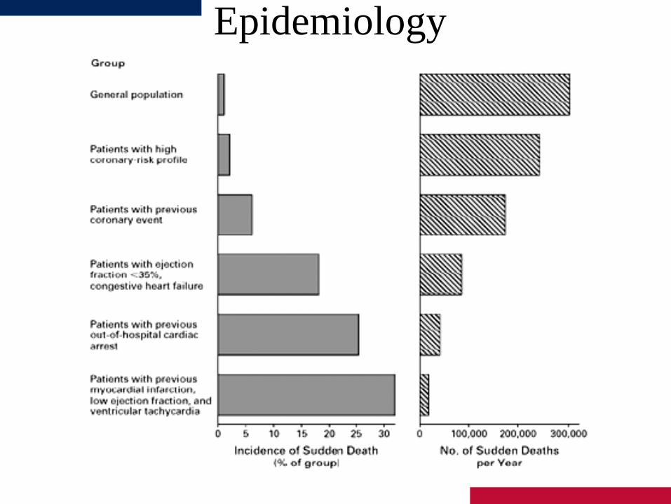
Epidemiology In the high school athlete population:
The risks are orders of magnitude lower!
• Risk of SCD: – 1 : 217,400 participants per year
– 0.46 / 100,000 person years annually
– 1 : 72,500 for one athlete over 4 year HS career
“It’s a very large haystack to look through…”
Maron B. JACC 32:1881-4, 1998.

Epidemiology
• Most SCD’s occurred in the most popular
sports. Maron B. JAMA 1996 Jul 17;276(3):199-204
SCD: Epidemiology
• Most SCD’s occur during practice or
competitions. Maron B. JAMA 1996 Jul 17;276(3):199-204

Epidemiology
• Cardiomyopathies and coronary
abnormalities account for most of the
cases of SCD in young athletes.
Maron B. JAMA 1996 Jul 17;276(3):199-204


Causes of Sudden Death in Athletes
1. Hypertrophic Cardiomyopathy
2. Congenital Coronary Artery Syndrome
3. Marfan’s Syndrome
4. Coronary Artery Disease
5. Sickle Cell Trait
6. Asthma
7. Others

Hypertrophic Obstructive
Cardiomyopathy
• Most common cause of SCD in athletes in the USA

Hypertrophic Obstructive Cardiomyopathy
• Exam
– High frequency SEM @ LUSB
– Increased with Valsalva, decreased with squatting
and increases with standing
– S4 common
Hypertrophic Obstructive Cardiomyopathy
• Asymmetric septal hypertrophy.
• Myocardial fiber disarray and
interstitial fibrosis.

Hypertrophic Obstructive Cardiomyopathy
• Myocyte hypertrophy in the absence of known
• Stimuli for ventricular hypertrophy (AS, CoA, HTN).
• Due to mutations of sarcomeric proteins.
• Autosomal dominant inheritance, variable and
age
• Dependent penetrance.
• Sporadic cases common (new mutations).
• Prevalence: 1-2 per 1,000 persons


Hypertrophic Obstructive Cardiomyopathy
• Clinical Signs & Symptoms • Dyspnea on exertion
• Chest pain
• Syncope or pre-syncope
• Palpitations
• Sudden death
• Mid-systolic murmur that increases in intensity with standing (dynamic auscultation)
• None of the symptoms are specific for HCM ! Mid-systolic murmur fairly specific but insensitive (hypertrophic non-obstructive cardiomyopathy)

ECG in Hypertrophic Obstructive
Cardiomyopathy
• >90% EKG’s abnormal
• Q III, Q aVF, Q V6, S V1, R V6

Hypertrophic Obstructive
Cardiomyopathy
• Conventional ECG criteria for LVH in children lack sufficient sensitivity and specificity for diagnosis of HCM.
• Dipchand A. Am J C 83(4):628, 1999.

Hypertrophic Obstructive Cardiomyopathy
• Newer ECG protocols developed with better
accuracy in differentiating between HCOM
and athletic heart

Hypertrophic Obstructive
Cardiomyopathy
• Echocardiography is the gold standard for the diagnosis of HCM.

Hypertrophic Obstructive Cardiomyopathy
Echocardiogram
• Left ventricular end diastolic septum thickness ≥
15mm for adults, <12mm is normal
• > 2 standard deviations from the mean for relative
to body surface area in children

Hypertrophic Obstructive Cardiomyopathy
• What if ventricular septum is 13-15mm?
– Athletic heart or HOCM?

Athletic Heart Syndrome (AHS) vs.
HOCM
• AHS
–Reduced LV mass with short deconditioning
periods
• MRI best for assessment
–LV end-diastolic dimension >55mm favors
AHS over HOCM

Athletic Heart Syndrome (AHS) vs.
HOCM
• HOCM
– Abnormal Doppler derived LV diastolic filling or
relaxation indices
– Family member with HOCM
– LV cavity <45 mm in diastole
– MRI again may help measure hypertrophy or lack
of

Hypertrophic Obstructive
Cardiomyopathy
• B-MHC, MyBP-C, & cTn-T account for
most HCM cases. There are many mutations
at each locus making “rule out” genetic
testing problematic.
Roberts R. Circ 104:2113, 2001.
Spirito P. NEJM 336(11):775, 1997

Hypertrophic Obstructive
Cardiomyopathy
• Most patients with HCM are non-
obstructive and asymptomatic.
How do you find these and what do you do
with them?
Spirito P. NEJM 336(11):775, 1997.

AHA/ACC Recommendations
• Participation in competitive athletics for
asymptomatic, genotype-positive HCM
patients without evidence of LV hypertrophy
by 2-dimensional echocardiography and CMR
is reasonable, particularly in the absence of a
family history of HCM-related sudden death
(Class IIa; Level of Evidence C).

Hypertrophic Obstructive
Cardiomyopathy
• Death from VT / Vfib
• OR…Nothing…
• OR…
• Symptoms
• palpitations
• syncope
• chest pain
• DOE

Hypertrophic Obstructive Cardiomyopathy
• Yearly echo in those with family history of
HOCM
• Disqualify from exertional sports?
• May participate in 1A sports such as golf,
bowling


Congenital Coronary Artery Anomalies
• Origin of LCA from right sinus of Valsalva-
most common
• Single coronary artery
• Origin of coronary artery from PA
• Coronary artery hypoplasia

Congenital Coronary Artery Anomalies
• Aberrant coronary arteries compromise the second
leading cause of SCD in young athletes.
• The highest risk pattern is LCA from the right Sinus of
Valsalva followed by RCA from the left Sinus of Valsalva.
• The SCD event is always occurs with strenuous exercise
or shortly thereafter.
• The presumed mechanism of SCD is dynamic
compression of the coronary arteries during exercise
leading to ischemia and lethal ventricular arrhythmia.

Congenital Coronary Artery Anomalies
• Proximal coronary
artery origins as seen
from the parasternal
short axis view.
• Can be seen easily in
most pediatric
patients.
Snider R. Echo in Ped Heart Dis. Mosby 1997.

Congenital Coronary Artery Anomalies
• Highest risk
pattern:
LMCA from right
Sinus of Valsalva
Davis J. JACC 37:593, 2001.

Congenital Coronary Artery Anomalies
• Second highest risk: RCA from left Sinus of
Valsalva.
Davis J. JACC 37:593, 2001.

Congenital Coronary Artery Anomalies
• Epidemiology of Aberrant Coronaries One large prospective pediatric echo study 2,388 children evaluated 1997-1999 inclusive. Referral for “murmur” or “ventricular function.” 4 cases of aberrant coronaries (0.17%) identified. 1 case “missed” by echo. SCD with aberrant coronary at autopsy. Prevalence higher than in other reported screening studies. Referral bias?
Davis J. JACC 37:593, 2001.

Congenital Coronary Artery Anomalies
• The Pyramid problem: Aberrant coronaries are 2nd leading cause of SCD. The prevalence of aberrant coronaries in the general asymptomatic “healthy” population is unknown.
• The math doesn't work out: Estimated prevalence 0.1-0.2% of general population? 4,000,000 live US births annually. 4,000-8,000 affected children / yr. Observed risk of SCD (all causes) in sports 1 per 217,000 / yr. Either the prevalence of aberrant coronaries is much lower than currently reported OR the incidence of SCD in aberrant coronary arteries is quite low.

Congenital Coronary Artery Anomalies
• Symptoms
– Death is most common
– Rarely
• exertional chest pain
• syncope
• infarction

Congenital Coronary Artery Anomalies
• Diagnosis
– Arteriography
– CT angiogram
• Corrective surgery if stable

Primary Electrical Disorders
• Wolff-Parkinson-White
• Long QT
• ARVD
• Brugada
• Rare disorders associated with development of malignant ventricular arrhythmias and associated sudden cardiac death.

Wolff-Parkinson-White
• Ventricular pre-excitation by an accessory bypass tract.
• Delta wave, short PR, +/- unusual T-waves on ECG Bypass tract allows for conventional SVT.
• Bypass tract also allows for rapid conduction to ventricle with atrial fibrillation (pre-excited a fib).

Wolff-Parkinson-White • Risk of SCD in WPW is very low (0.5% per decade).
• Risk of SVT and pre-excited a fib eliminated by
successful radiofrequency ablation.
• Most patients with rapid bypass tract conduction have
SVT symptoms but SCD can be the first presentation
of WPW.
• Ablation of completely asymptomatic people with
WPW is controversial.
• Ablation for asymptomatic WPW may be warranted in
high-risk occupations (pilot, military, etc).

Long QT Syndrome
• Genetic disease involving cardiac ion channels (Na+, K+).
• Results in delayed repolarization seen on surface ECG as long QT interval.
• Risk of polymorphic VT / VF “Torsades” & SCD. Diagnosed by QTc > 460 msec on
• ECG along with other electrical and clinical parameters.
• Avoidance of QT prolonging drugs, exercise restriction, and Beta blockade are cornerstones of therapy.
Ackerman M. Mayo Clin Proc 73:250, 1998.

Long QT Syndrome

Arrhythmogenic right ventricular
cardiomyopathy (ARVC)
• Familial condition
• Myocyte death, replacement with fibrous or adipose tissue in right ventricle
– Causes ventricular or supraventricular arrhythmias which can be fatal
• Most common cause of SCD in Veneto, Italy
– <5% in USA

ARVD Diagnostic Criteria Major Criteria
Right ventricular dysfunction Severe dilatation and reduction of RV EF with little or no LV impairment Localized RV aneurysms Severe segmental dilatation of the RV Tissue characterization Fibrofatty replacement of myocardium on endomyocardial biopsy Conduction abnormalities Epsilon waves in V1 - V3. Localized prolongation (>110 ms) of QRS in V1 - V3 Family history Familial disease confirmed on autopsy or surgery
Minor Criteria Right ventricular dysfunction Mild global RV dilatation and/or reduced ejection fraction with normal LV. Mild segmental dilatation of the RV Regional RV hypokinesis Tissue characterization Conduction abnormalities Inverted T waves in V2 and V3 in an individual over 12 years old, in the absence of a RBBB. Late potentials on signal averaged EKG. Ventricular tachycardia with a LBBB morphology Frequent PVCs (> 1000 PVCs / 24 hours) Family history Family history of sudden cardiac death before age 35 Family history of ARVD
Corrado D, Heart. 2000 May;83(5):588-95.

Brugada Syndrome
• Rare genetic (AD) electrical myopathy
Spike & dome ST segment in V1-V3
Prevalence of Brugada ECG pattern 1-2 per 1,000
Treatment: ICD
Priori S. Circ 105:1342, 2002.


Marfan’s Syndrome
• Diagnosis (2 of 4 features):
– Family history
– Cardiovascular abnormality (aortic aneurysm,
MVP, CHF symptoms)
– Musculoskeletal abnormality (arm span>height,
kyphoscoliosis, pectus cavus)
– Ocular abnormality (ectopic lens, myopia)
• Usually die from aortic dissection and rupture


Coronary Artery Disease
• Consider exercise stress testing for:
– male >45; female>55
– diabetics, smoker, fam Hx CAD, Chol>250,
HDL<30
– anyone with exertional chest pain, syncope, or
palpitations

Other causes
• Myocarditis
– Viral illness followed by CHF symptoms
– 50% Coxsackie B
– Afebrile 24 hours prior to play
• Aortic stenosis - SD with or without exercise

Commotio Cordis
• Induction of VF by mechanical stimulus during “vulnerable
phase” of repolarization. “R-on-T phenomenon”
No genetic or electrical susceptibility needed.
Requires critically timed impact directly over cardiac
silhouette.
Seen in small ball and high impact sports: baseball,
hockey, lacrosse, karate.
Preventable through use of RIF or “safety” balls and
possibly rigid chest protectors.
Rescue therapy: Early defibrillation (AEDs)

Commotio Cordis
• Induction of VF with low energy (30 mph)
baseball impact 15-30 msec before t-wave
peak in swine.
Link M. NEJM 338:1805, 1998.

Screening Guidelines 1996
• Uniform Screening for all High School & College
athletes
• Detailed PE & Fam Hx upon entry to sports and then
q 2 years
• No testing required (ECG, echo, labs)
Circ 94:850-856, 1996.
Screening Guidelines 2007
• Update to 1996 screening guidelines
• Addresses differences in recommendations between European Society of Cardiology and International Olympic committee guidelines.
• Emphasis on AHA 14 element screen Screening Guidelines 2007 12 element AHA screen Personal history Family History Physical exam
• EKG and ECHO not routinely required!!!!

Does Screening Work ? • Study of 42,368 athletes in Italy.
3 eras (1979-2004) Pre screening (< 82) Early screening ( 83-93) Late screening (93-04)
• 55 SCD in 42,368 athletes 1.9 /100,000 person-years 89% reduction in SCD with screening 3,035 (7%) initially disqualified but ultimately cleared for sports 879 (2%) of screened athletes disqualified
• Majority of cases due to ARVC rather than HOCM
JAMA 296:1593, 2006

Does Screening Work?
• 89% reduction in SCD after athlete screening
started in 1982. No change in
unscreened nonathletes.
JAMA 296:1593, 2006

Does Screening Work ?
• European Society
of Cardiology H&P
screening elements
Similar to US - AHA
guidelines
• JAMA 296:1593, 2006

Does Screening Work?
• ESC EKG screening criteria. LVH criteria different
(30mm).
JAMA 296:1593, 2006

Does Screening Work ?
• 5, 615 high school athletes screened by: General H & P and BP measurement Dynamic cardiac auscultation
Resting ECG
• Three year follow up period:
582 / 5615 (10%) had 1 or >1 screening abnormalities 115 (2%) Hx 175 (3.2%) PE 20 (0.3%) BP 146 (2.6%) ECG ECHO and exercise stress test performed if screen abnormal
Fuller, C. Med & Sci in Sports Exercise 29:1131, 1997.

Does Screening Work ? • No cases of HCM identified despite high prevalence in
general population and high prevalence in SCD.
• ? Self selection bias ?
• 22 athletes disqualified after abnormal screen and follow up echo and/or exercise stress test
• 1 severe AI, 5 severe hypertension (ok)
• 1 SVT treated with ablation => “requalified”
• 15 “other arrhythmias” (6 WPW, 5 PVC’s, 4 RBBB)
• One sudden death during track practice
• Dx: aberrant right coronary artery (normal screen)
Fuller, C. Med & Sci in Sports Exercise 29:1131, 1997.

Pros of EKG screening • Clearly shown to dramatically reduce SCD in the Italian population
below that of the non athletic population (from 3.6-0.4/100,000 over 24 yrs)
• Remarkably sensitive in the Italian population for HCM as 51/53=96% who subsequently died of HCM had positive ECGs.
• *Nevada data clearly indicate that ECGs are much better than PPE for detecting underlying cardiac disorders associated with SCD in the US
• Current US strategy has no data to support that it reduces SCD in athletes.
• ECG clearly increases detection of disorders like LQTS and ARVD that cause sudden death in the non athletic population as well.
• Once more identified, genetic/family clusters can be identified
• More money is spent on health initiatives that save fewer lives (ex.Meningococcal vaccine)

Cons to screening EKG
• Italian data not proven and potentially not applicable in US with marked greater heterogeneity of the population
• Estimated 250 to 2000/100,000 athletes would be held from competition for each life saved (estimates depend on SCD incidence figures) with no data on long term health or social effects on the disqualified athletes (DM, obesity, BP, depression, CAD)
• No gender or racial norms exist for ECG in the US at this point
• Assuming Italian data apply to US, 7% of those initially disqualified would return to play after echo, stress test, etc (which amounts to 500,000 of 7,300,000 athletes in 2006) demonstrate a normal workup
• Cost of >$2 B per year and no infrastructure like Italian exists in US
• Applying Italian data, cost per life/year saved is between $44,000 and $330,000

AHA/ACC Recommendation
• Mandatory and universal mass screening with 12-lead
ECGs in large general populations of young healthy
people 12 to 25 years of age (including on a national
basis in the United States) to identify genetic/
congenital and other cardiovascular abnormalities is
not recommended for athletes and nonathletes alike.
(Class III, no evidence of benefit; Level of Evidence
C).

Sickle Cell Trait
• Usually benign – sudden death may
occur in high heat or
altitude

Sickle Cell Trait
• Sports participation should not be restricted
but preventative measures should be taken
• avoid dehydration
• increase training intensity gradually
• wear cool clothing
• caution in high temp, humidity, or altitude
• stop activity if cramps occur

Sickle Cell Trait • Symptoms
• legs weak and cramping
• short of breath
• collapse
• LUQ abdominal pain from splenic infarct
• Exam
• obtunded
• hyperventilation
• hypotensive

Conclusion • Sudden cardiac death, although tragic, is very rare!
• HCM and aberrant coronary arteries are the cause of SCD in over 2/3 of cases.
• Electrical disorders, other cardiomyopathies, and aortic rupture (Marfan’s) compromise most of the remaining 1/3 of cases.
• There are well documented guidelines for pre participation screening of prospective athletes.
• Current guidelines emphasize PE & Fam Hx and do not require an ECG or ECHO evaluation.

Conclusion • Addition of ECG screening would improve detection of
HCM and allow detection of most electrical disorders but at increased cost.
• False positive ECG’s requiring echo confirmation
• False negative ECG’s in HCM (~10%)
• Benign nonspecific ECG changes requiring further evaluation / follow up (NL variants, “athlete’s heart”)
• No screening methodology other than detailed echocardiography (or CT / MRI) would allow for detection of coronary artery abnormalities.

Conclusion
• Pre Participation physical exam not just for
cardiovascular screening
• Screening for sickle cell trait with appropriate
education can prevent morbidity and mortality due to
sickle crisis with strenuous exercise
• Screening and appropriate management of asthma can
improve athletic performance and prevent morbidity
from reactive airway disease

Thank You!!!!!
Banner University Sports
Medicine Center
602-839-7285