the plant cell wall–decomposing machinery underlies … · lcms/ms analysis of s ... magnetic...
TRANSCRIPT

www.sciencemag.org/cgi/content/full/science.1205411/DC1
Supporting Online Material for
The Plant Cell Wall–Decomposing Machinery Underlies the Functional Diversity of Forest Fungi
Daniel C. Eastwood,* Dimitrios Floudas, Manfred Binder, Andrzej Majcherczyk, Patrick Schneider, Andrea Aerts, Fred O. Asiegbu, Scott E. Baker, Kerrie Barry, Mika
Bendiksby, Melanie Blumentritt, Pedro M. Coutinho, Dan Cullen, Ronald P. de Vries, Allen Gathman, Barry Goodell, Bernard Henrissat, Katarina Ihrmark, Hävard Kauserud,
Annegret Kohler, Kurt LaButti, Alla Lapidus, José L. Lavin, Yong-Hwan Lee, Erika Lindquist, Walt Lilly, Susan Lucas, Emmanuelle Morin, Claude Murat, José A. Oguiza, Jongsun Park, Antonio G. Pisabarro, Robert Riley, Anna Rosling, Asaf Salamov, Olaf
Schmidt, Jeremy Schmutz, Inger Skrede, Jan Stenlid, Ad Wiebenga, Xinfeng Xie, Ursula Kües, David S. Hibbett, Dirk Hoffmeister, Nils Högberg, Francis Martin, Igor V.
Grigoriev, Sarah C. Watkinson
*To whom correspondence should be addressed. E-mail: [email protected]
Published 14 July 2011 on Science Express
DOI: 10.1126/science.1205411
This PDF file includes:
Materials and Methods
SOM Text
Figs. S1 to S6
Tables S1 to S15
References (30–89)

2
SUPPORTING ONLINE MATERIAL
TABLE OF CONTENTS
1 MATERIALS AND METHODS....................................................................................3
1.1 Nucleic acid extraction for genomic study...............................................................3
1.2 Sequencing and assembly..........................................................................................3
1.3 EST clustering and assembly....................................................................................4
1.4 Genome annotation....................................................................................................5
1.5 Molecular clock analysis............................................................................................6
1.6 Comparative study of the evolution of carbohydrate active and oxidoreductase
enzyme gene repertoires............................................................................................6
1.7 Comparative transcriptomic analysis of Serpula lacrymans growth on defined
glucose media and on Pinus sylvestris sapwood......................................................7
1.8 Proteomic analysis of Serpula lacymans grown on wood........................................9
1.9 Genome analysis of natural product genes and secondary metabolite analysis
under nutrient asymmetry......................................................................................10
1.10 S. lacrymans interaction with Picea sylvestris seedlings......................................11
2 ADDITIONAL RESULTS AND DISCUSSION..........................................................11
2.1 Genome assembly, annotation and analysis...........................................................11
2.2 Molecular clock analysis...........................................................................................12
2.3 Comparative study of the evolution of carbohydrate active and oxidoreductase
enzyme gene repertoires...........................................................................................12
2.4 Comparison between transcriptomic and proteomic data....................................15
2.5 Natural product genes in the Serpula lacrymans genome and description of the
atromentin locus involved in variegatic acid production……………………......17
2.6 S. lacrymans interaction with Picea sylvestris seedlings.........................................17
3 FURTHER FUNDING SOURCES.................................................................................18
4 SUPPLEMENTARY FIGURES....................................................................................19
Fig S1. LCMS/MS analysis of S. lacrymans S7.9 split plate extract and purified
Fractions.............................................................................................................19
Fig S2. CAFE anaylis of the total number of protein families in each species node.20
Fig S3. Reconciliation analysis of lignocellulose-active enzymes from sequenced
fungal genomes...................................................................................................21
Fig S4. Functional characterisation of S. lacrymans transcripts with significant
differentiation when grown on wood..............................................................39
Fig S5. S. lacrymans S7.9 protein models with similar iron reductase domain........40
Fig S6. Interaction between S. lacrymans and roots of Picea sylvestris......................41
5 SUPPLEMENTARY TABLES.......................................................................................42
Table S1. Genomic libraries included in the Serpula lacrymans genome assembly...42
Table S2. Summary statistics of the output of the S. lacrymans S7.9 whole genome
shot gun assembly............................................................................................42
Table S3. S. lacrymans final assembly statistics............................................................42
Table S4. Predicted gene models and supporting lines of evidence............................43
Table S5. Characteristics of predicted gene models.....................................................43
Table S6. Functional annotation of proteins.................................................................43
Table S7. Comparison of the putative CAZy enzymes from genome sequenced fungi
with differing nutritional modes...................................................................44
Table S8. Comparison of the putative oxidoreductase enzymes from genome
sequenced fungi with differing nutritional modes......................................46
Table S9. Gene families and organisms used for reconciliation analysis....................48

3
Table S10. Number of orthologs between Serpula strains and with other
Agaricomycetes.............................................................................................49
Table S11. Posterior probability distribution for divergence times for major lineages
in the Agaricomycetes..................................................................................49
Table S12. S. lacrymans transcriptomic comparison from cultures grown on either
glucose or wood wafers................................................................................50
Table S13. Extracellular proteins from S. lacrymans S7 grown in solid state wood
Culture..........................................................................................................55
Table S14. Comparison of proteins identified as expressed on solid state wood
culture with the transcriptomic study........................................................56
Table S15. Comparison of secondary metabolic genes in sequenced basidiomycetes
........................................................................................................................57
6 REFERENCES.................................................................................................................58
1. MATERIALS AND METHODS
1.1 Nucleic acid extraction for genomic study
DNA extraction. Serpula lacrymans monokaryons S7.9 and S7.3, prepared
from S. lacrymans S7 dikaryon by Dr O. Schmit, University of Hamburg, were
inoculated in Czapek dox (30 gL-1
sucrose, 1 gL-1
monosodium glutamate, 1 gL-1
KH2PO4, 0.5 gL-1
MgSO4 x7H2O, 0.01 gL-1
FeSO4 x7H2O) liquid medium at 20C for
21 days. Cultures were harvested and filtered between muslin before freeze drying.
Harvested mycelia were ground under liquid nitrogen and DNA extracted following
the CTAB protocol described on the JGI website
(http://my.jgi.doe.gov/general/index.html). DNA quantity and quality was verified by
agarose gel electrophoresis and Nanodrop spectrophotometry (Thermo Scientific,
Hertfordshire, UK).
RNA extraction. S. lacrymans S7.9 was cultured under a range of conditions
to maximise the number of genes expressed. The fungus was cultured on 1) Czapek
dox medium (described above), 2) Czapek dox where either sucrose and monosodium
glutamate was reduced to 3 gL-1
or 0.1 gL-1
respectively, 3) Czapek dox where the
sucrose was replaced by carboxymethyl cellulose, 4) complex medium consisting of
20 gl-1
malt extract (BD Difco, Oxford, Uk) and 5 gL-1 yeast extract, 6) aged culture,
grown in Czapek dox for 10 weeks, and 7) heat and cold shock, cultures grown for 21
days in Czapek dox at 20 C were placed at either 25 C or 4 C for four hours prior
to harvesting. Mycelium was harvested and freeze dried as above. Total RNA was
extracted using the phenol:chloroform protocol described previously (30). RNA
quantity and quality was verified by formamide agarose gel electrophoresis and
Nanodrop spectrophotometry (Thermo Scientific).
1.2 Sequencing and assembly
Serpula lacrymans S7.9 was sequenced using Sanger sequencing on ABI
3730XL capillary machines (Life Technologies, California, USA). Three different
sized libraries were used as templates for the plasmid subclone sequencing process
and both ends were sequenced. 234,528 reads from the 2.5 kb sized library, 291,744
reads from the 6.4 kb sized library, and 29,952 reads from a 39.8 kb fosmid library
were sequenced (table S1). A total of 564,672 reads were assembled using a modified
version of Arachne (31) v.20071016 with parameters maxcliq1=100,
correct1_passes=0 and BINGE_AND_PURGE=True (see table S2 for scaffold and
4
contigs totals). This produced 68 scaffold sequences, with L50 of 2.9 Mb, 22 scaffolds
larger than 100 kb, and total scaffold size of 43.0 Mb. Each scaffold was screened
against bacterial proteins, organelle sequences and GenBank using megablast against
Genbank NR and blastp against a set of known microbial proteins. No scaffolds were
identified as contamination. We classified additional scaffolds as unanchored rDNA
(18 scaffolds), mitochondrion (1 scaffold), and small repetitive (3 scaffolds).
Additional scaffolds were removed if they consisted of greater than 95% 24mers that
occurred 4 other times in the scaffolds larger than 50kb or if the scaffold contained
only unanchored rDNA sequences. The final assembly contains 46 scaffolds that
cover 42.4 Mb of the genome with a contig L50 of 228.0 kb and a scaffold L50 of 2.9
Mb (table S3)
Serpula lacrymans S7.3 genome was sequenced using 454 (Roche,
Connecticut, USA) pyrosequencing platforms, which included 6 half runs of unpaired
454 Titanium data (table S1). All general aspects of library construction and
sequencing can be found at the JGI website (http://www.jgi.doe.gov/). After filtering
for low quality reads and contaminants, the resulting 454 reads were assembled with
the Newbler assembler version 2.3-PreRelease-9/14/2009 to the final estimated
assembled coverage of 24X (table S1) and resulted in 6088 contigs with an N/L50 of
247/45.5Kb. To improve the assembly it was scaffolded against Serpula lacrymans
S7.9, using Nucmer show-tilings (32). The final assembly included 2128 scaffolds
with an N/L50 of 7/2.9Mb (table S3).
1.3 EST clustering and assembly
Serpula lacrymans S7.9 total RNA was used to extract PolyA RNA using
oligo (dT) magnetic beads (Absolutely mRNA™ Purification kit, Stragene, Agilent
Technologies, California, USA). PolyA RNA was reversed transcribed using
Superscript III (Invitrogen) using a dT15VN2 primer. Second strand cDNA was
synthesized by nick translation with E. coli DNA ligase, E. coli DNA polymerase I,
and RNase H and blunt end repaired using T4 polymerase (Invitrogen, Life
Technologies, California, USA). The dscDNA was fragmented and 300-800 base pair
fragments were gel purified using a 2% agarose gel. The purified fragments were then
used to create the 454 single stranded cDNA library as described below (454 library
preparation kit, Roche).
The fragment ends were polished using T4 ligase and T4 polynucleotide
kinase (Roche). Adaptors containing primer sequences and a biotin tag were ligated to
the fragment ends (Roche). The fragments with properly ligated adapters were
immobilized onto magnetic streptavidin coated beads (Roche). Nicks or gaps
between the adapters and the dscDNA fragments were repaired using the fill-in
polymerase (Roche). The non-biotinylated strands of the immoblized dscDNA
fragments were melted off to generate the single stranded cDNA library for 454
sequencing.
The ESTs were evaluated for the presence of polyA tails (which if present
were removed) then evaluated for length, removing ESTs with fewer than 50 bases
remaining. Additionally, ESTs consisting of more than 50% low complexity
sequence were removed from the final set of ESTs. The resulting set of ~751k ESTs
were used for clustering.
For clustering, ESTs were evaluated with malign, a kmer based alignment tool
(Chapman, unpublished), which clusters ESTs based on sequence overlap (kmer = 16,
seed length requirement = 32 alignment ID >= 98%). EST clusters were then each
5
assembled using CAP3 (33) to form consensus sequences. Clustering and assembly
of all ESTs resulted in 107,971 consensus sequences and 69,437 singlets.
1.4 Genome annotation
Both genomes were annotated using the JGI annotation pipeline, which takes
multiple inputs (scaffolds, ESTs, and known genes) and runs several analytical tools
for gene prediction and annotation, and deposits the results in the JGI Genome Portal
(http://www.jgi.doe.gov/Serpula) for further analysis and manual curation.
Genomic assembly scaffolds were masked using RepeatMasker (34) and the
RepBase library of 234 fungal repeats (35). tRNAs were predicted using tRNAscan-
SE (36). Using the repeat-masked assembly, several gene prediction programs falling
into three general categories were used: 1) ab initio - FGENESH (37); GeneMark (38)
trained on full-length Serpula lacrymans genes, 2) homology-based - FGENESH+;
Genewise (39) seeded by BLASTx alignments against GenBank‟s database of non-
redundant proteins (NR: http://www.ncbi.nlm.nih.gov/BLAST/), and 3) EST-based -
EST_map (http://www.softberry.com/) seeded by Serpula lacrymans EST contigs.
Genewise models were extended where possible using scaffold data to find start and
stop codons. EST BLAT alignments (40) were used to extend, verify, and complete
the predicted gene models. The resulting set of models was then filtered for the best
models, based on EST and homology support, to produce a non-redundant
representative set. This representative set (12,917 and 14,495 genes in S7.9 and S7.3,
respectively – tables S4-S5) was subject to further analysis and manual curation.
Measures of model quality include proportions of the models complete with start and
stop codons (>82% of models), consistent with ESTs (>63% of models covered over
75% of exon length), supported by similarity with proteins from the NCBI NR
database (>68% of models). Quality metrics for gene models are summarized in tables
S4-S5.
All predicted gene models were functionally annotated using SignalP (41),
TMHMM (42), InterProScan (43), BLASTp (44) against nr, and hardware-accelerated
double-affine Smith-Waterman alignments (deCypherSW;
http://www.timelogic.com/decypher_sw.html) against SwissProt
(http://www.expasy.org/sprot/), KEGG (45), and KOG (46). KEGG hits were used to
assign EC numbers (http://www.expasy.org/enzyme/), and Interpro and SwissProt hits
were used to map GO terms (http://www.geneontology.org/). Functional annotations
are summarized in Table S6. Manual curation of the automated annotations was
performed by using the web-based interactive editing tools of the JGI Genome Portal
to assess predicted gene structures, assign gene functions, and report supporting
evidence.
Multigene families were assembled from 158,123 predicted proteins found in S.
lacrymans, representatives from Agaricomycotina, Pucciniomycotina and
Ustilagomycotina phyla using the MCL algorithm (47) with inflation parameter set to
3.0. Multigene families were then analyzed for evolutionary changes in protein family
size using the CAFE program (48). The latter program uses a random birth and death
process to model gene gain and loss across a user specified tree structure. The
distribution of family sizes generated under the random model provides a basis for
assessing the significance of the observed family size differences among taxa (p-value
0.001). CAFE estimates for each branch in the tree whether a protein family has not
changed, has expanded or contracted. A phylogenetic tree was constructed using 203
highly conserved, core gene representatives of S. lacrymans and other basidiomycetes
(see the FunyBASE at http://genome.jouy.inra.fr/funybase/).

6
1.5 Molecular clock analysis
A subset of the 191 taxa multigene dataset from Binder et al. (2010) (17) was
supplemented with sequences of Postia placenta, Phanerochaete chrysosporium,
Heterobasidion irregulare (previously H. annosum), and Fomitiporia mediterranea.
Alignments for three nuclear ribosomal genes (nuc-ssu = 1783 bp, nuc-lsu = 1399 bp,
5.8S = 159 bp) and three protein coding genes (rpb1 = 427 bp, rpb2 = 2087 bp, tef1 =
1016 bp) were made separately using MacClade 4.08 (49). The concatenated
alignment including 32 species and a total of 6871 sites was built and edited using
TextWrangler (Bare Bones Software, Inc.).
Twelve tMRCA‟s (time to most recent common ancestors) were specified
using BEAUTi v.1.4.8 (50) to define the monophyletic groups resolved in previous
analyses (17, 51, 52). Fossil calibrations were applied to the marasmioid clade in the
Agaricales and to the Suillineae in the Boletales, reflecting the minimum age
estimates for both groups. Archaeomarasmius legetti from mid-Cretaceous amber was
found in a well-characterized layer of clay in New Jersey that dates to 94-90 Mya (53)
and we calibrated the marasmioid clade (fig 1A) including Armillaria mellea,
Chondrostereum purpureum, Crinipellis perniciosa, and Hemimycena gracilis with
this estimate. The permineralized suilloid ectomycorrhiza fossil associated with pine
roots in the middle Eocene Princeton chert of British Columbia (54) was used to
calibrate the Suillineae (represented by Gomphidius roseus and Suillus pictus) with 50
myr as a minimum age estimate. We estimated the absolute timing for the split
between the saprotrophic Serpula lineage and ectomycorrhizal Austropaxillus lineage
under the GTR model using an uncorrelated relaxed clock with normal rate
distribution, invoking the Yule birth-death process of speciation as the tree prior.
Three Bayesian relaxed clock analyses were run for 10 million generations using
BEAST v1.4.8 (50), saving trees every 1000th
generation. The resulting log file was
inspected with Tracer v1.4 (55) to confirm that the estimated sample sizes for
statistics represent appropriate values of posterior distributions. The runs converged to
stable likelihood values after one million generations and 9000 ultrametric trees from
each run were analyzed in TreeAnnotator v1.4.8 (50) to estimate the 95% credible
node intervals referred to as highest posterior densities (HPD), which mark the lower
and upper boundaries of the time estimates.
1.6 Comparative study of the evolution of lignocellulolytic carbohydrate active and
oxidoreducases enzyme gene repertoires Genomes and focal gene families. Carbohydrate active enzyme (CAZy) and
oxidoreductase families involved in lignocellulose decomposition in S. lacrymans
were compared with other genome sequenced fungi exhibiting a range of nutritional
modes (tables S7-8). A subset of these fungi and enzymes were selected for further
analysis to investigate lignocelluloytic gene repertoires (table S9). The analyses
included data from 7 Basidiomycota and 3 Ascomycota genomes, basidiomycetes
Serpula lacrymans S7.9 v1.0 Phanerochaete chrysosporium v2.0, Postia placenta
MAD-698, Heterobasidion irregulare v1.0 (previously identified as H. annosum),
Schizophyllum commune v1.0, Laccaria bicolor, Coprinopsis cinerea okayama
7#130, and the ascomycetes Trichoderma reesei v2.0 (anamorph of Hypochrea
jecorina), Magnaporthe grisea 70-15 and Stagonospora nodorum. The data for the
first six basidiomycetes and T. reesei were retrieved using the databases of Joint
Genome Institute (www.jgi.doe.gov), while the data for C. cinerea, M. grisea and S.

7
nodorum were retrieved using the databases of the Broad Institute
(www.broadinstitute.org).
Nineteen gene families with various roles in wood degradation were targeted
(table S9), separated into two broad functional classes of gene families, including
genes encoding Carbohydrate-Active enzymes (CAZY‟s, e.g., cellobiohydrolases,
endoglucanases, esterases) and various oxidoreductases implicated in lignocellulolysis
(e.g. Class II peroxidases, multicopper oxidases). In families containing enzymes with
diverse roles, e.g. glycoside hydrolases (GH) 5, 28 and 61 and carbohydrate esterases
(CE) 1 and 16, only enzymes with direct lignocelluloytic activity were considered in
the analysis, e.g. endoglucanases and mannanases in GH5 and cinnamoylesterases in
CE16. In addition highly truncated sequences were also omitted from the analysis.
This was done to ensure duplications and losses of enzymes related to wood
decomposition would be identified and not masked by fluctuations in functional
groups not involved in decomposition. The query sequences used in blast searches
were obtained from CAZY base (www.cazy.org), UniProt (www.uniprot.org), or
relevant studies.
Blast searches, alignment, and phylogenetic analysis. Blastp searches were
conducted with the e-value set to -5, using the „best models‟ database for each
genome. The recovered protein models were checked for intron-exon structure,
functional domains and putative function as displayed on the genome browser or by
using Pfam (http://pfam.sanger.ac.uk/) and InterProScan
(http://www.ebi.ac.uk/Tools/InterProScan/). Alignments were performed using
MAFFT version 6 (http://mafft.cbrc.jp/alignment/software, (56)) and adjusted
manually in MacClade 4.08 OS X (49), with alternative models being used in some
cases, including reprediction of truncated models using FGENESH
(http://linux1.softberry.com/berry.phtml). The latter models carry the suffix „mod‟.
Poorly aligned areas were excluded. The appropriate protein evolution model was
selected for each dataset using ProtTest1.4.mac (57) and an unrooted maximun
likelihood analysis was performed in RAxML Blackbox with 1000 rapid bootstrap
replicates with the final ML search under the GAMMA+P-Invar model
(http://phylobench.vital-it.ch/raxml-bb/, (58)).
Gene tree/species tree reconciliation analysis. Gene family phylogenies
were reconciled with an organismal phylogeny reflecting current understanding of
relationships among the ten genomes (59) using Notung (60, 61). The default settings
for costs of duplications and losses were used (1.5 and 1.0 respectively), and two
analyses were performed for each gene family, using edge weight thresholds (EWT)
set to 90 or 75 (bootstrap frequencies). Gene trees were rooted to minimize the cost of
duplications and losses, and topological rearrangements were performed according to
the EWT setting. The implied duplications and losses for each gene family under both
EWT settings were mapped onto the species tree, and the total duplications and losses
across all CAZY gene families and all oxidoreductase gene families were compiled
and mapped separately on the species tree.
1.7 Comparative transcriptomic analysis of Serpula lacrymans growth on defined
glucose media and on Pinus sylvestris sapwood
Sample preparation. S. lacrymans S7 was grown on shavings of Pinus
sylvestris (L.) sapwood placed on moist soil separated by a nylon net for a period of
ten days at 20oC and 80% relative humidity. The nylon net covered with S. lacrymans
mycelium was removed and and RNA was extracted following the CTAB protocol
outlined below. Control cultures were prepared using MMN agar medium (5 gL-1

8
glucose, 0.5 gL-1
NH4NO3, 0.5 gL-1
KH2PO4, 0.5 gL-1
MgSO4.7H2O, 0.167 gL-1
thamine).
RNA extraction. The RNA was extracted from the culture isolates using a 2%
cetyltrimethyl ammonium bromide (CTAB) protocol. Briefly, mycelia were ground
under liquid nitrogen and extraction buffer added (CTAB, polyvinyl pyrrolidone and
-mercaptoethanol) with acid phenol. Chloroform:isoamyl alcohol extraction and
lithium chloride precipitation was carried out according to established protocols (30).
All samples were treated with RQ1 DNASE (Promega, Stockholm, Sweden)
according to maufacturer‟s instructions. The quantity of the RNA was analyzed with
NanoDrop and Qubit™ fluorometer (Invitrogen). The quality of the extracted RNA
was determined using 2100 Bioanalyzer (Agilent Technologies, Edinburgh, UK). To
determine the integrity of the extracted RNA, the samples were divided into two,
where one sample was incubated in 37°C for 2 hours and reanalyzed on the Agilent
2100 Bioanalyzer to establish whether the RNA had been degraded.
cDNA synthesis and amplification. Total RNA (50-100 ng) was used to
synthesize first strand cDNA. First and second stand cDNA synthesis and
amplification was performed using the SMARTer™ pico PCR cDNA synthesis kit
and Advantage®
2 PCR kit (Clontech, Saint-Germain, France), according to the
protocol from the manufacturer. The quantity of the cDNA was analyzed with
NanoDrop and Qubit™ fluorometer (Invitrogen). The quality was determined using
the 2100 Bioanalyzer.
Array design and analysis. The Serpula lacrymans S7.9 custom-exon
expression array (4 x 72K) manufactured by Roche NimbleGen Systems Limited
(Madison, WI) (http://www.nimblegen.com/products/exp/index.html) contained five
independent, nonidentical, 60-mer probes per gene model coding sequence. For
12,797 of the 12,917 annotated protein-coding gene models probes could be designed.
For 8 gene models no probes could be generated and 112 gene models shared all five
probes with other gene models. Included in the array were 2130 random 60-mer
control probes and labelling controls. For 1200 randomly chosen gene models,
technical duplicates were included on the array.
Single dye labeling of samples, hybridization procedures, data acquisition,
background correction and normalization were performed at the NimbleGen facilities
(NimbleGen Systems, Reykjavik, Iceland) following their standard protocol.
Microarray probe intensities were quantile normalized across all chips. Average
expression levels were calculated for each gene from the independent probes on the
array and were used for further analysis. Raw array data were filtered for non-specific
probes (a probe was considered as non-specific if it shared more than 90% homology
with a gene model other than the gene model it was made for) and renormalized using
the ARRAYSTAR software (DNASTAR, Inc. Madison, WI, USA). For 994 gene
models no reliable probe was left. A transcript was deemed expressed when its signal
intensity was three-fold higher than the mean signal-to-noise threshold (cut-off value)
of the random oligonucleotide probes present on the array (50 to 100 arbitrary units).
Gene models with an expression value higher than three-fold the cut-off level were
considered as transcribed. The maximum signal intensity values were ~65000
arbitrary units. A Student t-test with false discovery rate (FDR) (62) multiple testing
correction was applied to the data using the ARRAYSTAR software (DNASTAR).
Transcripts with a significant p-value (<0.05) were considered as differentially
expressed. The complete expression dataset is available as series (accession number
GSE27839) at the Gene Expression Omnibus at NCBI
(http://www.ncbi.nlm.nih.gov/geo/).

9
1.8 Proteomic analysis of Serpula lacrymans grown on wood
Fungal cultures. Serpula lacrymans S7 (dikaryon) culture was maintained on
malt agar medium (2% malt extract and 1.5% agar) at 20°C in the dark. Millet culture
was prepared from 100 g millet (soaked in water overnight and autoclaved) inoculated
with three 10 mm diameter pieces of 7 days old agar culture and incubated for 3
weeks until substrate was completely overgrown by the fungus. Picea abies wood was
chopped to particles of approximately 1-5x0.5x1 mm and dried for three days at 80°C
(moisture content of 4.4% and water content of 4.2%). Wood cultures of S. lacrymans
with calcium silicate were prepared by autoclaving 100 g wood mixed with 2 g meta
calcium silicate (CaSiO3, reagent grade, Alfa Aesar, Karlsruhe, Germany) and 240 ml
water in 1 L preserving jar and inoculated with about 2 g of the millet culture.
Samples without calcium were prepared in the same way omitting calcium silicate.
The initial moisture content and water content of the wood cultures was 280% and
74%, respectively. Cultures in several replications were incubated for 30 days at 20°C
in dark.
Protein extraction. Extracellular proteins were extracted from each culture
using 500 ml of 500 mM Tris buffer pH 7.0 containing 1% (v/v) Tween 80 and 1 mM
PMSF (phenylmethanesulfonylfluoride). Samples were evacuated to remove air from
the material, sonicated twice for 5 min and incubated for about 40 min. Separated
liquids from parallel samples were combined and stored at -20°C.
After thawing, samples were centrifuged at 25.000 g for 60 min (4°C) and
collected supernatants kept in an ice-bath. Sodium chloride and sodium deoxycholate
were added to the samples to final concentrations of 1 M and 0.05%, respectively
(63). Proteins were precipitated by addition of TCA from 100% stock solution to 10%
final concentration; 100% trichloroacetic acid (TCA) stock solution contained 100 g
TCA in 45.4 ml water. After mixing, samples were allowed to stand on ice for 30 min.
Thereafter, phosphotungstic acid was added from 10% stock solution to the final
concentration of 0.5% (64). Samples were mixed well and incubated on ice overnight.
Precipitated proteins were collected by centrifugation at 25.000 g for 30 min and
further processed as previously described (65). Total protein in this step was
determined by the Bradford Reagent (Pierce, Germany). Protein aliquots for 2D-
electrophoresis were freeze-dried and samples for shot gun analysis suspended in 100
mM ammonium bicarbonate.
Shotgun protein identification. Protein digestion and peptide fractionation
was performed as previously described (66, 67). Briefly, aliquots of approximately
500 µg total protein from each experiment (three replicates) suspended in 100 mM
ammonium bicarbonate were digested with sequencing grade trypsin (Promega,
Germany) in an enzyme:substrate ratio of 1:40 (w/w) at 37°C for 16 h. Thereafter,
samples were reduced with dithiothreitol (DTT) and alkylated with iodoacetic acid.
After addition of a new amount of trypsin samples were digested again for 30 min at
58°C (68). The digested peptides were de-salted with a C18 Sep-Pak (Waters, Milford
MA) and dried in vacuum centrifuge. Samples were dissolved in 8 M urea and total
peptide amount determined by BCA Protein Reagent (Pierce, Germany) calibrated
with a tryptic bovine serum albumen (BSA) digest. Samples containing 300 µg total
peptides were brought up with 8 M urea and IPG buffer 3-10 (Amersham) to 350 µl
and applied to a 18 cm IPG dry strip (Amersham, Munich, Germany). Using an
IPGphor II (Amersham), the following focusing protocol was applied: active
rehydration for 6 h at 20 V, current limit of 50 μA per strip at 20 °C, 1 h 300 V, 1 h
gradient to 1000 V, 3 h gradient to 4000 V, 3 h gradient to 8000 V, 8000 V upto 50

10
kVh. Each gel strip was cut into 26 sections and peptides extracted with three
sequential solutions containing 0%, 50%, and 100% acetonitrile in water with 0.1%
trifluoroacetic acid. Supernatants were combined, dried and redissolved in 5% formic
acid. Samples were cleaned of salts and residual oil using STAGE tips (69), dried and
stored at -20 °C before analysis.
LC-MS/MS analysis. Peptide analysis was performed using 1100 LC
(Agilent, Böblingen, Germany) interfaced to an Esquire3000 ion trap mass
spectrometer (Bruker-Daltonic, Germany) via an electro spray ionization (ESI) unit.
Each peptide fraction was dissolved in 5 µl of 5% formic acid and 4 µl samples were
loaded on a 180 µm i.d. capillary column packed with 3 µm Reprosil-Pur C18-AQ
(Dr. Maisch GmbH, Ammerbuch, Germany), conditioned in 98% of solvent A (0.1%
formic acid in water) and 2% of solvent B (0.1% formic acid in 90% acetonitrile).
After 20 min isocratic elution at 2 µl/min peptides were eluted by gradients of solvent
B: 15% in 5 min, 40% in 90 min, 50% in 5 min, and 90% in 5 min. Mass spectrometer
was setup to take four averages of MS-spectra (200 to 1500 mu) and four averages of
MS/MS-spectra (200 to 3000 mu) of two most abundant precursor ions. The Dynamic
Exclusion was set to non-single charged precursor ions and an exclusion time of 5
min. The MS/MS spectra were extracted by DataAnalysis (V. 3.0, Bruker Daltonic)
and peptides identified using Mascot (V. 2.2, Matrix Science, Uk). Target database
was constructed from annotated genomes of S. lacrymans S7.9 and S7.3 and the
SwissProt database. Searches against a decoy database, created by randomizing the
target database, indicated the false discovery rate (FDR) of 0.25%. All searches were
run as a tryptic digest with one missing cleavage allowed, fixed
carbamidomethylation of cysteine and variable oxidation of methionine. Mass
tolerances were set to 1.4 Da and 0.4 Da for the MS and MS/MS spectra, respectively.
Mascot results were extracted from raw DAT-files using a VB-script and transferred
to an SQL-database (Microsoft SQL Server 2005). SQL queries were used to extract
proteins with at least two peptides with scores higher than the corresponding identity
score. Analogous data processing was performed on false/positive searches of the
decoy database and additional proteins were identified using an average peptide
scoring (APS) method (70, 71) with restriction to proteins matched by at least two
confidently identified peptides.
1.9 Genome analysis of natural product genes and secondary metabolite analysis
under nutrient asymmetry
Genome analysis. The S. lacrymans genome was analysed for natural product
genes putatively involved in secondary metabolite production.
Culture preparation. S. lacrymans S7 was grown on solid standard „Serpula
Czapek Dox‟ medium (per liter: 30g sucrose, 1g monosodium glutamate, 1g KH2PO4,
0.5g MgSO4 .7H2O, 0.01g FeSO4 x7H2O, 20g agar). After 3-4 weeks, mycelia
including the solid medium of six plates (10 cm in diameter) were shredded and
exhaustively extracted with ethyl acetate. After solvent evaporation under reduced
pressure the crude extract was solved in methanol.
Pigment identification. The crude extract was analyzed on an Agilent 1200
HPLC instrument equipped with a Zorbax Eclipse XDB C-18 column (150 x 4.6 mm,
3.5 µm particle size) and a guard column (Agilent Technologies). The following
gradient was applied (solvent A: water, solvent B: acetonitrile): initial hold for 2 min
at 5% B, then linear increase to 95% B within 20 min, at a flow rate of 0.5 ml/min.
Chromatograms were recorded at λ = 254 nm. Variegatic acid and its precursors,
atromentic acid and xerocomic acid, were identified by their masses during

11
electrospray mass spectrometry (positive and negative mode) and by their UV spectra,
which were compatible to those published (72).
Sample preparation. The crude extract was adsorbed into a 3 ml Bakerbond
silica gel solid phase extraction cartridge, followed by sequential elution with
cyclohexane, ethyl acetate, and methanol. Variegatic acid and derivatives desorbed
into two ethyl acetate fractions, one of which (4 mg) was composed of 60% variegatic
acid and 15% xerocomic acid and was tested in the ferrozine assay, along with crude
extract. HPLC analysis of the crude and two ethyl acetate fractions were recorded,
data showed that the sample of fraction 3 was the most purified, while the raw extract
the least (fig S1).
Ferrozine iron reduction assay. Three Serpula extract samples were
analyzed for their iron reducing capabilities using a ferrozine assay. Ferrozine [3-(2-
pyridyl)-5,6-bis(4-phenylsulfonic acid)-1,2,4-triazine] reacts with divalent iron and
forms a stable magenta colored complex that can easily be determined and measured
photometrical at a fixed wave length of 562nm (73). Three S. lacrymans extracts
prepared as outlined above (raw extract and 2 ethyl acetate purified fractions) before
being assessed for iron reducing capability. The Serpula extract samples were diluted
in 5.0 ml 50% (v/v) ethanol solution and care was taken to ensure they were well
mixed into solution and stored tightly covered at 4 C. Samples (0.5 M) were
assayed in a reaction consisting of 50 M FeCl3 and 10 mM ferrozine in 1 M acetic
acid and 1M sodium acetate buffer (pH 4.5), in 10 ml total volume. Control samples
containing 50 M 2,3-dihydroxybenzoic acid (DHBA) as iron reducing agent were
included as a reference. After 5, 20, 60, and 120 minutes incubation, the ferrozine was
added and measurements were recorded after a further 2 minutes incubation at 562 nm
using a spectrophotometer. Exposure to air was minimized and sample vials were
purged with nitrogen to prevent oxidation.
1.10 S. lacrymans interaction with Picea sylvestris seedlings
Experimental microcosm systems were constructed using the established
mycorrhizal synthesis system (29). Two-week-old seedlingswere aseptically
inoculated with agar plugs from of S. lacrymans S7 in Petri dishes with growth
substrate of sterilized fine sphagnum peat : vermiculite : 1/10 strength liquid MMN
mixture in the ratio 1 : 4 : 2. Five replicate microcosms were constructed for each tree
species/fungus combination (two tree species × three fungi × five replicates, or 30
microcosms in total). Inoculated microcosms were incubated in a growth chamber at
20 ° C, with a 16 h light : 8 h dark photoperiod.
2. ADDITIONAL RESULTS AND DISCUSSION
2.1 Assembly, annotation and analysis
The 42.4 MB genome of Serpula lacrymans S7.9 was sequenced to 8x read
depth coverage using Sanger platform and assembled into 46 scaffolds using Arachne
(31). The second strain S7.3 was sequenced with 454 (Roche) pyrosequencing and
assembled using assembled S7.9 as a template for scaffolding (table S3). 12,917 and
14,495 genes were predicted in S7.9 and S7.3, respectively, using a combination of
gene predictors and validated with ESTs (tables S4-S5). On average orthologs
between two strains show 98.5% amino acid identity and arranged into large scaffold-
long syntenic blocks while only a half of this number (5,000-5600) are orthologous to
other Agaricomycetes with 57-61% identity (table S10) and still significant syntenic
blocks. Interestingly, S7.3 shows more genes than S7.9, that may affect differences in
functional profiles (tables S6, 10). In S. lacrymans, 581 gene families showed a

12
significant expansion, 3,571 showed no change and 906 families had undergone
contraction by comparison to a putative common ancestor Basidiomycota having
5,058 gene families (fig S2).
2.2 Molecular clock analysis
All Bayesian searches converged independently on a tree topology that is
consistent with previously published phylogenetic inferences (17, 52). The relaxed
molecular clock analyses with normal fossil calibration priors produced broadly
spaced estimates for the HDP of the non-calibrated nodes (table S11, fig 1A). This
result is not unexpected since the approach taken was aimed at estimating the
minimum node ages conservatively. Our main objective was to provide a minimum
age estimate for the split between Serpula lacrymans and Austropaxillus spp. within a
comprehensive phylogenetic framework. The posterior time estimate for the
Agaricomycetidae (Agaricales, Amylocorticiales, Atheliales, Boletales, and Jaapiales)
dates the most recent common ancestor at 166.1 Mya (95% HPD 189.8 – 126.5) and
this estimate largely overlaps with the fossil record of Pinaceae as potential hosts
dating back to the lower Jurassic (74-76). The minimum timing for the origin of the
Boletales is estimated at 113.4 mya (HPD 140.5 – 87.3), which is consistent with the
findings of Hibbett and Matheny (2009) (77) suggesting that the Boletales are young
enough to have been associated with Pinaceae or rosids (Eurosids I).
Collectively, previous studies (12, 19, 78) suggest that S. lacrymans has an
Asian origin and it became cosmopolitan by gradually expanding its natural range
over the northern hemisphere in association with pinaceous hosts. Contemporary
Austropaxillus spp. on the other hand are bound to Nothofagus and to some extent to
Eucalyptus and have a southern hemisphere distribution extending over Australia,
Tasmania, New Zealand, Papua New Guinea, and parts of South America. In addition,
the ancestral nutritional mode of the most exclusive clade containing Serpula and
Austropaxillus was estimated as brown-rot saprotrophic with a single transitional
event to ECM leading to Austropaxillus (12). The split between Serpula and
Austropaxillus estimated at 34.9 mya (HPD 53.1 – 15.0) coincides with the separation
of South America and Australia from Antarctica about 31 mya ago (79, 80). This
finding is consistent with a vicariant distribution of Austropaxillus and its main host,
Nothofagus, although dispersal may have played a major role in the current
biogeography of the tree hosts (81). Thus, it appears plausible that the transition from
a saprotrophic ecology to ECM in Austropaxillus occurred after a host switch from
Pinaceae to Nothofagaceae before the complete break-up of Pangaea. Alternatively,
extinct or unsampled Serpula-like fungi have acquired the ability to function as ECM
partners and morphological elaboration from resupinate to stipitate-pileate forms
became manifested with the occurrence of Austropaxillus. A comparative genomic
study between Serpula and Austropaxillus would ultimately help to clarify whether
Serpula is genetically predisposed to enter ectomycorrhizal associations.
2.3 Comparative study of the evolution of lignocellulolytic carbohydrate active and
oxidoreducases enzyme gene repertoires CAFE analysis identified gene families that had undergone significant
expansion and contraction (fig S2). A more detailed analysis of genes involved in
lignocellulose decomposition revealed a link between brown rot evolution and gene
repertories. 783 protein models were retrieved and used in the phylogenetic and
reconciliation analyses. 767 protein models were included in the final mapping of

13
gene losses and duplications, including 521 from basidiomycete genomes and 246
from ascomycete genomes.
Lignocellulolytic Carbohydrate Active enzymes (CAZY’s). The lowest
number of CAZY genes families selected for study in basidiomycetes was found in L.
bicolor (14 genes), while in ascomycetes. T. reesei (32 genes) had the fewest CAZY
genes. The highest number of selected CAZY genes families in basidiomycetes and
ascomycetes were identified in Coprinopsis cinerea (75 genes) and St. nodorum (87
genes) respectively (fig 1B).
The distribution of gene copies was not homogeneous among the genomes and
among the gene families. C. cinerea, Sc. commune and Ph. chrysosporium had
representative copies from all the CAZY families included in the study, while L.
bicolor lacked copies in half of the families, S. lacrymans and P. placenta lacked
copies for some of the families and H. irregulare (previously classified as H.
annosum) only for the Glycoside Hydrolases family 11 (tables S7 and 9). The
Glycoside Hydrolases family 74 included the lowest number of gene copies (8 copies)
while the Glycoside Hydrolases families 61 and 3 had the most prominent
representation in the dataset with 139 and 104 copies respectively (table S9). The
lineages leading to C. cinerea, Sc. commune, Ph. chrysosporium and St. nodorum
have undergone net expansions in CAZY gene families, while the lineages leading to
L. bicolor, S. lacrymans, P. placenta, H. irregulare and T. reesei have undergone net
contractions (fig 1B).
The common ancestor of the basidiomycetes was estimated to have 66 to 83
total gene copies for the selected CAZY gene families (fig 1B), depending on the edge
weight threshold (EWT). The genomes of C. cinerea, Sc. commune and Ph.
chrysosporium had similar gene numbers to the common basidiomycete ancestor,
although the similarity did not coincide in gene diversity, as several gene families had
undergone expansions and contractions. The rest of the basidiomycete genomes had a
decreased number of genes compared to the common ancestor (fig 1B).
A more detailed examination of the results for S. lacrymans, P. placenta and
L. bicolor, which have undergone parallel reductions in CAZY gene families,
suggested that: 1) a number of gene families are dispensable and lost in all or two out
of the three genomes (table S9), 2) the three species bear a low number of lineage
specific duplications (figs 1B & S3A-M) much lower than the lineage specific
duplications for the other species, 3) despite the shared similarities among the three
genomes, L. bicolor bears a lower number of copies than the two brown rot species
for most of the gene families that all three species include gene copies (table S9).
H. irregulare also had an increased number of lineage specific gene losses (fig
1B) but the gene distribution pattern observed was different from the three species
referred above. Thus in spite of the increased losses, H. irregulare had at least one
gene copy in all but one gene family (table S9).
Oxidoreductases. The highest number of copies for oxidoreductase gene
families was present in Ph. chrysosporium (43 copies) followed by H. irregulare (36
copies), while the lowest number of copies were identifed in S. lacrymans and P.
placenta (19 copies each) and T. reesei (fig 1C & table S8). The differences among
the genomes in total gene copies were less prominent in the dataset compared to the
CAZY dataset, with the exception of Ph. chrysosporium, H. irregulare and T. reesei .
As with the CAZYs, the distribution of oxidoreductase gene copies was not
equally dispersed among the gene families and genomes. The genomes of Ph.
chrysosporium and H. irregulare include at least one copy for each one of the gene
families studied, while the rest of the genomes lack copies for one or more families

14
(table S8). The cellobiose dehydrogenases had the lowest number of gene copies (12
copies), while the highest number of gene copies was in the multicopper oxidases (92
copies).
The summarized reconciliation results suggest that the lineages leading to C.
cinerea, L. bicolor, Sc. commune, H. irregulare and Ph. chrysosporium have
undergone net expansion in oxidoreductases while the lineage leading to S. lacrymans
has undergone net gene contraction, and the lineage leading to P. placenta is almost
constant regarding gene copies numbers (fig 1C).
Gene families encoding Class II peroxidases, which are implicated in
ligninolysis, had expanded independently in the lineages of Ph. chrysosporium and H.
irregulare, while the rest of basidiomycetes either have had no expansions or bore a
low number of losses (fig S3N). Genes for this family were absent from the genomes
of S. lacrymans and Sc. commune. Furthermore, the multicopper oxidases have
undergone extensive gene duplication in the genomes of C. cinerea, H. irregulare and
L. bicolor, and thus gene expansions are associated not only with white rot species
(fig S3O). Regarding the four gene families involved in oxidative degradation of
cellulose in relation to the Fenton reaction (cellobiose dehydrogenase, oxalate
oxidases/decarboxylases, iron reducing glycopeptides and quinone reductases), all the
wood degrading species, with the exception of H. irregulare, had an increased number
of gene copies as compared to C. cinerea and L. bicolor. Serpula lacrymans and P.
placenta had lineage specific duplications for some of these families (fig S3P-S), and
the former was the only species among the basidiomycetes that has duplications in
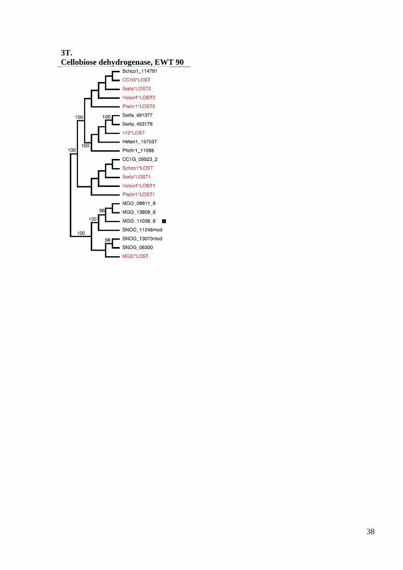
cellobiose dehydrogenases (fig S3T).
Discussion. Reconciliation analyses suggest that the 12 CAZY gene families
in S. lacrymans, P. placenta, L. bicolor and T. reesei have undergone net contraction.
A reduction in CAZY gene content was noted previously for the brown rot species
Postia placenta (Polyporales), including losses of genes encoding
exocellobiohydrolases and cellulolytic enzymes with cellulose binding modules
(CBM1) (13). A similar pattern of reduction in CAZYs is evident in S. lacrymans
(Boletales), which represents an independent origin of brown rot, as well as in the
ectomycorrhizal L. bicolor (Agaricales) (15). Surprisingly, T. reesei, which is a very
efficient cellulolytic fungal species, also has reduced gene content for several CAZY
families (table S9). The reconciliation results support those findings especially in this
case where T. reesei was compared with the aggressive plant pathogens M. grisea and
St. nodorum. In contrast, the white rot basidiomycete Sc. commune contains a great
variety of CAZY enzymes (82), which reconciliation analysis shows to have arisen
through multiple recent duplications in the lineage leading to Sc. commune.
Brown rot has evolved independently in multiple lineages of basidiomycetes
(4). Gene content and reconcilation analyses suggest that these transitions have been
characterized by extensive gene losses in the CAZY families, and that the common
ancestor of Agaricomycetes (Basidiomycota) was able to utilize cellulose and
hemicellulose in a similar fashion as contemporary white rot species. The brown rot
mechanisms of S. lacrymans and P. placenta demonstrate remarkable convergence in
their cellulose degradation biochemistry, but they are distinct as S. lacrymans has a
cellobiohydrolase of the Glycoside Hydrolase family 6 as well as genes that carry a
CBM1 domain for the families 6 and 5. Another brown rot species of the Boletales,
Coniophora puteana, also produces cellobiohydrolases (83) and genes coding for
cellobiohydrolases of both the Glycoside Hydrolase families 6 and 7 have been cloned
from this species (84). The differences in gene content between S. lacrymans and P.
placenta reflect the independent origin of the two brown rot lineages.

15
Evolution of a mycorrhizal lifestyle in L. bicolor also appears to involve
reductions in CAZY gene families. Similar patterns of gene loss have been suggested
in the evolution of other mycorrhizal taxa, including the basidiomycetes Amanita
bisporigera (85) and the ascomycete Tuber melanosporum (14).
The convergent evolutionary events in gene losses and duplications among
gene families related to wood degradation for L. bicolor, S. lacrymans and P. placenta
as they were highlighted above especially for the CAZY gene families, indicate a
possible transition from a brown rot lifestyle towards a mycorrhizal one. This is
further supported by a study (17) that placed several brown rot genera as basal in the
Boletales. The most prominent finding in that study was the placement of
Austropaxillus, a south hemisphere mycorrhizal genus, as sister group of S. lacrymans
and Serpula himantioides.
2.4 Comparison between S. lacrymans transcriptomic and proteomic data
S. lacrymans gene transcripts on wood and glucose medium were analysed
using NimbleGen microarrays containing 11,804 probes produced from the S.
lacrymans S7.9 genome database. A total of 517 gene features were significantly
(P<0.01) regulated 4-fold or greater between treatments, 300 on wood substrate and
217 on glucose. Secreted proteins were extracted from wood-based cultures, separated
by 2D-gel electrophoresis and peptides identified by LC-MS/MS. Twenty-eight
putatively secreted proteins were identified, 22 (78%) of which were in 10% of the
most highly expressed transcripts and 17 (68%) were differentially regulated on
wood.
A comparison of transcriptomes of white rot Ph. chrysposporium and the
closely related brown rot P. placenta showed that Ph. chrysosporium expressed a
conventional system of synergistically acting endo- and exo- hydrolase enzymes and
relatively few oxidoreductases (26). P. placenta produced a larger proportion of
hemicellulose degrading and oxidoreductase enzymes, fewer cellulases, and appeared
to lack exocellobiohydrolase activity (26). There was evidence that a hydroquinone-
driven Fenton‟s system for non-enzymic hydroxyl radical cleavage of cellulose,
typical of brown rot decay fungi takes the place of the enzymic hydrolysis of α-
cellulose in P. placenta (13). The S. lacrymans wood-induced transcriptome showed
similarities to both P. placenta and Ph. chrysosporium, with glycoside hydrolase
(GH) and oxidoreductase enzymes accounting for 35.7% of genes upregulated on
wood, while lipase and sugar transporter transcripts were also increased (table S12).
GH enzymes accounted for 50% proteins identified (table S13) and a large proportion
of transcripts were regulated more than 20 fold on wood (33.9%, fig S4), with GH
families 5, 61, 3 and 28 represented more than once. Two endoglucanases (Prot id:
433209 and 453342) were expressed more than 100 fold more on wood, and
hemicellulose-degrading enzymes (1,3-β-galactosidase, mannanases, endoxylanases,
galactouronases, glucuronidases) were prominent. The S. lacrymans GH6
cellobiosehydrolase gene (absent in P. placenta) was not highly expressed, suggesting
that, as in P. placenta, the depolymerisation of cellulose by S. lacrymans occurs in the
absence of known exo-acting cellobiohydrolases.
Also highly represented amongst the S.lacrymans transcripts from wood
cultures were cytochrome P450 (18 genes), aldo-keto reductases (11 genes) and short
chain dehydrogenases (8 genes), while FAD-dependent pyridine nucleotide disulphide
oxidoreductase (451883) and glucose-methanol-choline oxidoreductase (439506)
were upregulated 109 and 38 fold respectively. Aromatic ring hydroxylases, alcohol
dehydrogenases, multicopper oxidase and aldehyde dehydrogenases were also
16
identified. No evidence of upregulated hydroquinone synthesis was discovered; 1,4-
benzoquinone reductase and hydroquinol-1,2-dioxygenases described in P. placenta
were not differentially regulated or highly expressed in S. lacrymans, although a ferric
reductase-like protein (478753) possibly involved in iron redox cycling was expressed
(fig S5). Unexpectedly, neither S. lacrymans cellobiose dehydrogenase CDH gene
(453176 and 453175) was highly or differentially expressed.
Transcriptomic data was obtained from S. lacrymans S7 (dikaryon) cultures
grown on wood shavings on soil for 10 and 30 days and compared with mycelium
grown on a minimal medium with glucose as the main carbon source. For ease of
presentation 10 day wood cultures were compared with glucose medium. Genes were
identified as significantly differentially regulated (up or down) if transcript levels 4
fold or more between treatments. A total of 517 microarray features were identified as
being differentially regulated, 300 had greater expression in the wood samples
compared with glucose medium and 217 lower levels (fig S4; table S12 for gene list
and annotation of 50 most differentially transcribed on wood).
Proteomic data was obtained from S. lacrymans S7 cultures growing on wood
particles (Picea abies about 1-5x0.5x1 mm) for 30 days either in the presence or
absence of calcium ions (meta calcium silicate, 2g/100g wood). Low quantities of
extracellular proteins were obtained, leading to the identification of 39 proteins, 29 of
which having an extracellular signal peptide motif. Twenty four proteins were
characterised from the S. lacrymans S7.9 monokaryon and 15 from the S. lacrymans
S7.3 monokayon (table S13), proteins identified were compared with transcriptomic-
derived data (table S14). The microarray was designed using gene models of the
monokaryon S7.9, therefore, proteins identified from S. lacrymans S7.3 were
compared against the S7.9 database by BLAST and the ortholog identified was
compared with the transcriptomic data. In some instances (e.g. closely related genes
familes such as glycoside hydrolase 3) more than one S7.9 gene model gave the same
Blast similarity score, in which case the transcript level of each S7.9 ortholog was
examined.
The effect of calcium on protein expression was examined for the proteomic
study, 11 proteins appeared to be associated with cultures in the presence of calcium
ions only; 6 proteins were detected on wood without calcium and 22 were associated
with the fungus growing on wood both with and without calcium. As calcium was not
used in the transcriptomic study, the comparison between the two was conducted
using the 28 proteins identified as being present in wood when calcium was not
added.
Analysis revealed that 3 proteins were not represented by a probe on the
microarray (protein number 1, 6 and 29), presumably because a suitable probe could
not be designed. Therefore comparison is based on 25 proteins rather than 28.
Seventeen (68%) proteins identified in the proteomic study were highlighted as
differentially regulated in the transcriptomic study. Of the remaining 8 proteins, 6 had
transcript levels in the top 10% signal intensity in wood cultures, but their expression
on glucose was also high and therefore these genes were not identified as strongly up-
regulated in the transcriptomic study. Of the proteins identified in the proteomic study
22 (88%) were in the top 10% of genes with greatest transcript levels from the
transcriptomic study and 9 (36%) were in the top 1%. Gycoside hydrolase and
carboxylesterase enzymes were well represented in both studies.

17
2.5 Natural product genes in the Serpula lacrymans genome and description of the
atromentin locus involved in variegatic acid production
Serpula species are known as producers of small molecule natural products,
and three distinct classes of metabolites were reported in the literature: i) members of
the pulvinic acid family of pigments (86) which are common among members of the
Boletales, ii) the himanimides (87), and iii) polyine acids (88). Genes for five
polyketide synthases (PKSs), 15 nonribosomal peptide synthases (NRPSs) or NRPS-
like enzymes, and two PKS/NRPS-hybrids, were identified. For example, none of the
known secondary S. lacrymans metabolites requires PKS activity during biosynthesis.
Some products of NRPS genes may participate in the fungus´ primary metabolism, as
they resemble α-aminoadipate reductases (NPS5, NPS9, NPS10, NPS11) or
siderophore-synthesizing enzymes (NPS4). Two genes (nps6 and nps8) whose
products show an identical domain organization encode putative PKS/NRPS hybrid
enzymes. Also, putative genes for 10 terpene cyclases were found, some of which
may catalyze secondary product formation.
Putative atromentin locus and pigment formation. Atromentin is the central
intermediate en route to pulvinic acid-derived pigments, such as xerocomic,
variegatic, and atromentic acid. A cluster of genes resembling the atromentin
biosynthesis locus in Tapinella panuoides (24) was found on scaffold 9, thus making
these very likely candidates to govern pigmentation of S. lacrymans fruiting bodies
and undifferentiated mycelia. We expect the central enzymes are encoded by i) the
gene nps3 (74% identical amino acids to the T. panuoides quinone synthetase AtrA,
an NRPS-like enzyme) and amt1 (59% identical amino acids to the T. panuoides L-
tyrosine:2-oxoglutarate aminotransferase AtrD). As in T. panuoides, a third reading
frame in opposite transcriptional direction putatively encoding an alcohol
dehydrogenase was identified between the aminotransferase and quinone synthetase
genes.
Tailoring enzymes. Numerous reading frames which may encode tailoring
enzymes were identified. Most of these are located in the vicinity of NRPS and PKS
genes thus suggesting the biosynthesis gene cluster paradigm holds up for the
Boletales the same way as it does for aspergilli and numerous other filamentous fungi
(89).
Prenyl transferase activity is required during himanimide assembly. Consistent
with this, genes for two putative metal independent prenyl transferases - ppt1 on
scaffold 12 (clustered with nps1) and ppt2 on scaffold 17 - were detected.
Halogenated natural products have not yet been described from S. lacrymans.
However, two genes (hal1 on scaffold 18, clustered with a PKS gene, and hal2 on
scaffold 5) which very likely code for flavin-dependent halogenases indicate the
capacity to synthesize as yet unknown halogenated secondary products. Although
some pathways may be tightly regulated we expect, based on the genomic data (table
S15), the S. lacrymans secondary metabolome to be much more diverse than evident
from current chemical data.
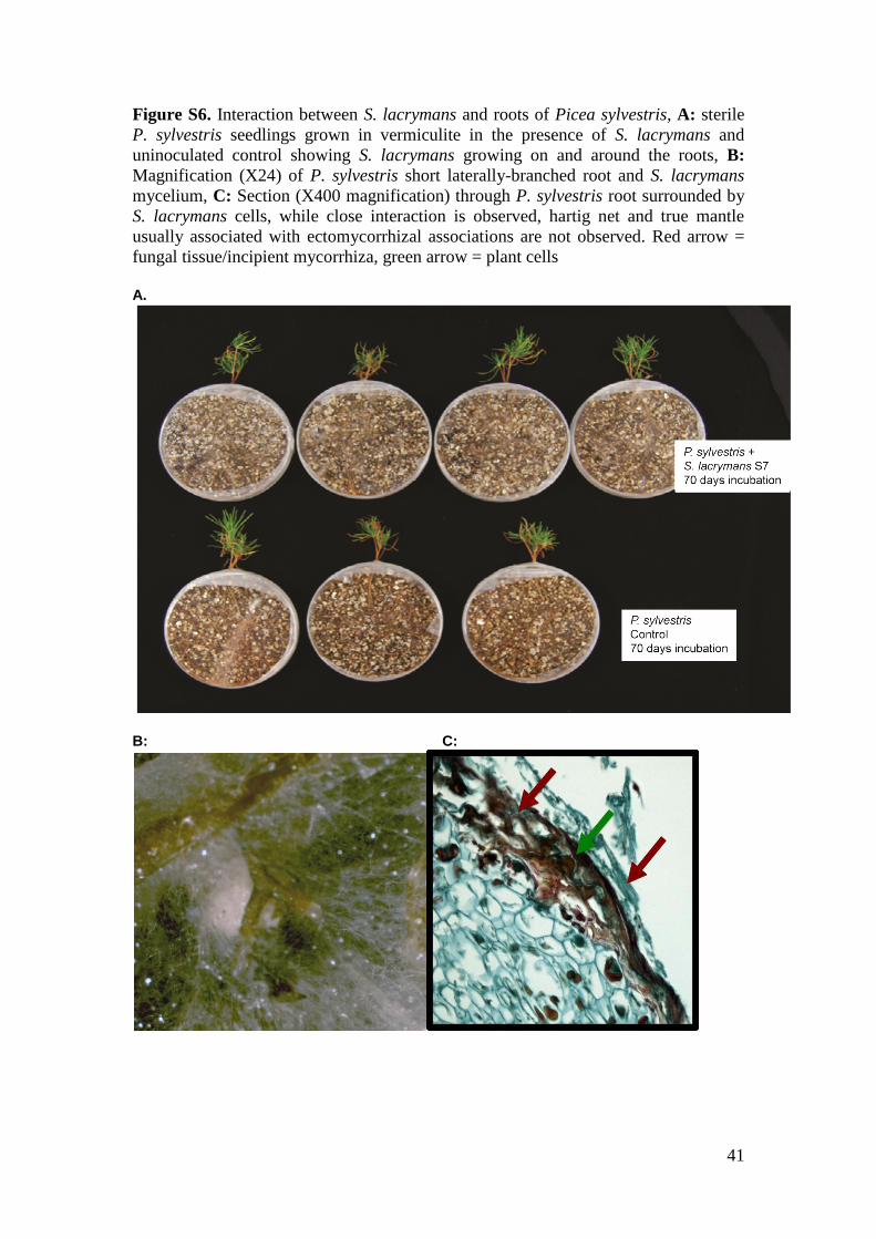
2.6 S. lacrymans interaction with Picea sylvestris seedlings
In co-culture with Picea sylvestris seedlings, S. lacyrmans S7 was observed to
grow towards and around the roots of the plant (fig S6). In addition, the roots were
observed to form short lateral branches commonly associated with micorrhizal
associations (fig S6B). However, while microscopic investigation revealed a close
association of the fungus and root cells, no evidence of hartig net or true mantle
necessary for mycorrhizal formation were observed (fig S6C). The observations
18
suggest a transient interaction between fungus and plant in which P. sylvestris
seedling in attempting to form symbiotic association interact with a fungus foraging
for nutrients. The interaction does not proceed to form structures expected for
mycorrhizal associations and it is not clear from this experiment to what extent this is
due to the plant not recognising a compatable fungal partner or S. lacrymans lacking
the necessary machinery to develop the association further. The data do suggest a
close relationship which can form between tree roots and fungi and point to a scenario
in which saprotrophic fungi could evolve into partners in a mycorrhizal mutualistic
association.
3. Further funding sources
Funding is acknowledged from the "Wealth Out of Waste" programme funded by a
EPSRC grant to the Warwick Innovative Manufacturing Centre, INRA, the Region
Lorraine Council, the European Commission within the Project ENERGYPOPLAR
(FP7-211917), the Network of Excellence EVOLTREE (FP6-016322), and the US
Department of Energy – Oak Ridge National Laboratory Scientific Focus Area for
Genomics Foundational Sciences (Project Plant–Microbe Interfaces), FORMAS, Carl
Tryggers Foundation, KSLA, Ångpanneföreningen, Ministry of Science and Culture
of Lower Saxony (VW-Vorab 11-76251-99-9/04 ZN 2043/ZN 2128) and the
Deutsche Bundesstiftung Umwelt, ANR (grant ANR-07-BIOE-006), the Natural
Environment Research Council,UK (grant UK NER/A/S/2002/882)and Helsinki
University Research Fund and Academy of Finland. The CAZy database is funded in
part by GIS-IBiSA.

19
4. Supplementary Figures Figure S1. LCMS/MS analysis of S. lacrymans S7.9 split plate extract and purified fractions used to test for iron reducing activity in the Ferrozine assay

20
Figure S2. CAFE anaylis of the total number of protein families in each species node. The figure
represents the total number of protein families in each species or node. We take into
consideration families with at least 10 members and 2 species. The numerals on branches
show numbers of expanded (left, blue), unchanged (middle, black) or contracted (right, red)
protein families along lineages by comparison to the putative pan-proteome.

21
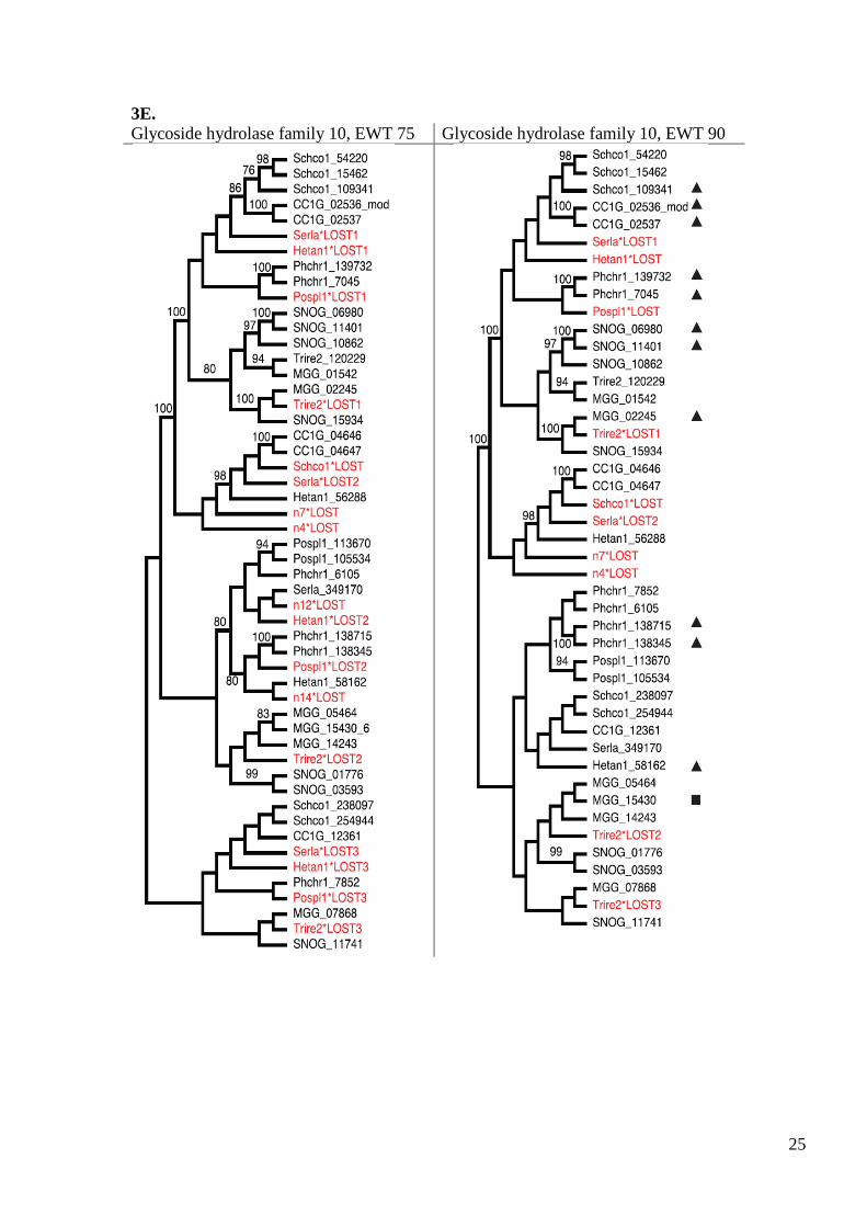
Figure S3. Reconciliation analysis of lignocellulose-active enzymes from sequenced fungal genomes
using edge weight thresholds (EWT) set to 90 or 75 (bootstrap frequencies). CC1G - Coprinopsis
cinerea, Hetan1 – Heterobasidion irregulae (previously identified as H. annosum), Lacbi - Laccaria
bicolor, MGG -Magnaporthe grisea, Pospl – Postia placenta, Phchr1 - Phanerochaete chrysosporium,
Serla- Serpula lacrymans; Schco – Schizophyllum commune, SNOG – Stagonospora nodorum, Trite -
Trichoderma reesei. Red text indicates gene losses. - N-terminal cellulose binding module, - C-
terminal cellulose binding module
3A:
Glycoside hydrolase family 3 EWT 75 Glycoside hydrolase family 3 EWT 90

22
3B. Glycoside hydrolase family 5, EWT 75 Glycoside hydrolase family 5, EWT 90

23
3C.
Glycoside hydrolase family 6, EWT 75 Glycoside hydrolase family 6, EWT 90

24
3D.
Glycoside hydrolase family 7, EWT 75 Glycoside hydrolase family 7, EWT 90
25
3E.
Glycoside hydrolase family 10, EWT 75 Glycoside hydrolase family 10, EWT 90

26
3F.
Glycoside hydrolase family 11, EWT 75 & 90

27
3G.
Glycoside hydrolase family 12, EWT 75 Glycoside hydrolase family 12, EWT 90

28
3H.
Glycoside hydrolase family 28, EWT 75 Glycoside hydrolase family 28, EWT 90

29
3I.
Glycoside hydrolase family 61, EWT 75

30
3J.
Glycoside hydrolase family 61, EWT 90

31
3K.
Glycoside hydrolase family 74, EWT 75 Glycoside hydrolase family 74, EWT 90
3L.
Carbohydrate esterase family 1, EWT 75 & 90

32
3M.
Carbohydrate esterase family 16, EWT 75 Carbohydrate esterase family 16, EWT 90

33
3N.
Class II peroxidases, EWT 75 Class II peroxidases, EWT 90
A - Basidiomycete class II peroxidises; B – Cytochrome C peroxidises (not included in
duplications-losses counting)

34
3O.
Multicopper oxidases, EWT 75 Multicopper oxidases, EWT 90

35
3P.
Glyoxal oxidases, EWT 75 Glyoxal oxidases, EWT 90

36
3Q.
Quinone reductase, EWT 75 Quinone reductase, EWT 75, EWT 90
3R.
Iron-binding glycoproteins/reductases,
EWT 75
Iron-binding glycoproteins/reductases,
EWT 90

37
3S.
Oxalate oxidases/decarboxylases, EWT 75 Oxalate oxidases/decarboxylases, EWT 90
38
3T.
Cellobiose dehydrogenase, EWT 90

39
Figure S4. Functional characterisation of S. lacrymans transcripts with significant increased regulation (4 fold or greater, ANOVA P<0.01)
when grown on wood compared with glucose-based medium (MMN) identified by microarray analysis (n=300 genes). Gene list and heat map of
relative expression level is provided for the 30 S. lacrymans genes with greatest fold increase in transcript levels on solid wood substrate.
Carbon metabolism
Oxidoreducatse/monooxygenase activity
Transport
Lipid metabolism
Other functions
Predicted proteins with orthologs in other fungi
Predicted proteins, new for S. lacrymans

40
Figure S5. S. lacrymans S7.9 protein models with similar iron reductase domain
(cd00241), including putatively annotated cellobiose dehydrogenase genes 453175
and 453176.
Iron reductase domain = cd00241; CBM – cellulose binding domain; CDH –
cellobiose dehydrogenase oxidoreductase domain; SIGN – signal peptide cleavage
motif; LNK – linking sequence without specific function.
453175
453176
452187
417465
Serpula lacrymans var.lacrymans S7.9
Serpula lacrymans var.lacrymans S7.9
Serpula lacrymans var.lacrymans S7.9
Serpula lacrymans var.lacrymans S7.9
JUNK SIGN cd00241 LINK CDH
SIGN cd00241 LINK CDH
SIGN cd00241
SIGN cd00241 LINK CBM
Accession
Number Genome Database Domain Structure
41
Figure S6. Interaction between S. lacrymans and roots of Picea sylvestris, A: sterile
P. sylvestris seedlings grown in vermiculite in the presence of S. lacrymans and
uninoculated control showing S. lacrymans growing on and around the roots, B:
Magnification (X24) of P. sylvestris short laterally-branched root and S. lacrymans
mycelium, C: Section (X400 magnification) through P. sylvestris root surrounded by
S. lacrymans cells, while close interaction is observed, hartig net and true mantle
usually associated with ectomycorrhizal associations are not observed. Red arrow =
fungal tissue/incipient mycorrhiza, green arrow = plant cells A.
B: C:
42
5. Supplementary Tables
Table S1. Genomic libraries included in the Serpula lacrymans genome assembly and their
respective assembled sequence coverage levels in the final release.
Library Type Average Insert
Size
Number of
Reads
Assembled Sequence
Coverage (X)
S7.9:
Sanger, 3kb 2,552 234,528 3.59
Sanger, 8kb 6,426 291,744 3.98
Sanger, fosmid 39,854 29,952 0.44
Total 556,224 8.01
S7.3:
454, std N/A 2,489,569 24.06
Total 2,489,569 24.06
Table S2. Summary statistics of the output of the S. lacrymans whole genome shotgun assembly S7.9
before screening and removal of organelles and contaminating scaffolds. The table shows total
contigs and total assembled basepairs for each set of scaffolds greater than the given size.
Size Number Contigs Scaffold Size Basepairs % Non-gap
Basepairs
5,000,000 1 51 5,733,305 5,701,910 99.45%
2,500,000 8 245 27,090,049 26,889,349 99.26%
1,000,000 15 350 38,261,814 37,960,259 99.21%
500,000 20 388 42,140,175 41,797,878 99.19%
250,000 20 388 42,140,175 41,797,878 99.19%
100,000 22 399 42,458,091 42,101,004 99.16%
50,000 25 405 42,692,124 42,298,518 99.08%
25,000 25 405 42,692,124 42,298,518 99.08%
10,000 34 418 42,825,041 42,429,394 99.08%
5,000 43 432 42,886,961 42,488,184 99.07%
2,500 67 468 42,968,800 42,566,449 99.06%
1,000 68 469 42,970,512 42,568,161 99.06%
0 68 469 42,970,512 42,568,161 99.06%
Table S3. S. lacrymans final assembly statistics.
S7.9 S7.3
Assembly method Arachne (Sanger) Newbler (454/Roche)
Main genome scaffold total 46 2133
Main genome contig total 434 3303
Main genome scaffold sequence total 42.8 Mb 47.0 Mb
Main genome contig sequence total 42.4 Mb (0.9% gap) 41.2 Mb (12.4% gap)
Main genome scaffold N/L50 6/2.9 MB 7/2.7 Mb
Main genome contig N/L50 62/228.0 KB 143/86.6 kb
Number of scaffolds > 50 KB 24 49
% main genome in scaffolds > 50 KB 99.60% 95.10%

43
Table S4. Predicted gene models and supporting lines of evidence
S7.9 S7.3
# gene models 12917 14495
% complete (with start and stop codons) 87% 82%
% genes with homology support 69% 68%
% genes with Pfam domains 42% 40%
% with 100% EST support 33.00% 26.00%
% with > 50% EST support 74.00% 70.00%
Table S5. Characteristics of predicted gene models.
S7.9 S7.3
Avg.gene length, bp 1600 1501
Avg. protein length, aa 339 322
Avg. exon frequency 5.6 exons/gene 5.3 exons/gene
Avg. exon length, bp 222 226
Avg. intron length, bp 77 75
Table S6. Functional annotation of proteins.
S7.9 S7.3
Proteins assigned to a KOG 5475 (42%) 5908 (41%)
KOG categories genome-wide 3011 3112
Proteins assigned a GO term 5148 (40%) 5423 (37%)
GO terms genome-wide 2114 2154
Proteins assigned an EC number 1843 (14%) 2027 (14%)
EC numbers genome-wide 619 647
Proteins assigned a Pfam domain 5421 (42%) 5781 (40%)
Pfam domains genome wide 2152 2250
44
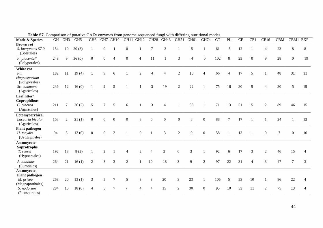
Table S7. Comparison of putative CAZy enzymes from genome sequenced fungi with differing nutritional modes Mode & Species GH GH3 GH5 GH6 GH7 GH10 GH11 GH12 GH28 GH43 GH51 GH61 GH74 GT PL CE CE1 CE16 CBM CBM1 EXP
Brown rot
S. lacrymans S7.9
(Boletales)
154
10
20 (3)
1
0
1
0
1
7
2
1
5
1
61
5
12
1
4
23
8
8
P. placenta*
(Polyporales) 248
9 36 (0) 0 0 4 0 4 11 1 3 4 0 102 8 25 0 9 28 0 19
White rot
Ph.
chrysosporium
(Polyporales)
182
11
19 (4)
1
9
6
1
2
4
4
2
15
4
66
4
17
5
1
48
31
11
Sc. commune
(Agaricales)
236 12 16 (0) 1 2 5 1 1 3 19 2 22 1 75 16 30 9 4 30 5 19
Leaf litter/
Coprophilous
C. cinerea
(Agaricales)
211
7
26 (2)
5
7
5
6
1
3
4
1
33
1
71
13
51
5
2
89
46
15
Ectomycorrhizal
Laccaria bicolor
(Agaricales)
163
2
21 (1)
0
0
0
0
3
6
0
0
8
0
88
7
17
1
1
24
1
12
Plant pathogen
U. maydis
(Ustilaginales)
94
3
12 (0)
0
0
2
1
0
1
3
2
0
0
58
1
13
1
0
7
0
10
Ascomycete
Saprotrophs
T. reesei
(Hypocreales)
192
13
8 (2)
1
2
1
4
2
4
2
0
3
1
92
6
17
3
2
46
15
4
A. nidulans
(Eurotiales)
264 21 16 (1) 2 3 3 2 1 10 18 3 9 2 97 22 31 4 3 47 7 3
Ascomycete
Plant pathogen
M. grisea
(Magnaporthales)
268
20
13 (1)
3
5
7
5
3
3
20
3
23
1
105
5
53
10
1
86
22
4
S. nodorum
(Pleosporales)
284 16 18 (0) 4 5 7 7 4 4 15 2 30 0 95 10 53 11 2 75 13 4

45
*A full list of gene identification numbers is provided in additional file 1, additional table 1
†P. placenta figures are for the sequenced dikaryon; Figures in parentheses glycoside hydrolase family 5 with putative carbohydrate-binding
module 1 (cellulose). GH, glycoside hydrolases; GT, glycosyl transferases; PL, polysaccharide lyases; CE, carbohydrate esterases; CMB,
carbohydrate-binding module; CBM1, cellulose-binding module; EXP, plant expansin-like.

46
Table S8. Comparison of putative oxidoreductase enzymes from genome sequenced fungi with differing nutritional modes* Mode & Species POX MCO CDH AAOX GlyOX PryOX GluOX QR AOX OXO IGP
Brown rot
S. lacrymans S7.9
(Boletales)
0
6
2
6
3
0
0
2
1
3
3
P. placenta†
(Polyporales)
? 8 0 8 5 0 0 2 2 10 8
White rot
Ph. chrysosporium
(Polyporales)
16
5
1
3
7
1
1
4
1
7
3
Sc. commune
(Agaricales)
0 5 1 1 2 1 4 4 1 5 7
Leaf litter/
Coprophilous
C. cinerea
(Agaricales)
1
17
1
14
6
0
1
3
2
1
0
Ectomycorrhizal
Laccaria bicolor
(Agaricales)
1
11
0
4
10
0
2
2
2
1
1
Plant pathogen
U. maydis
(Ustilaginales)
0
0
0
0
1
0
1
1
0
0
0
Saprotrophic
Ascomycete
T. reesii
(Hypocreales)
0
7
0
1
1
0
1
1
2
3
0
A. nidulans
(Eurotiales)
0 11 1 5 0 1 1 0 2 2 0
Ascomycete
Plant pathogen
M. grisea
(Magnaporthales)
2
12
3
1
1
0
2
1
2
2
0
S. nodorum
(Pleosporales)
5 8 3 2 1 0 2 1 3 3 0
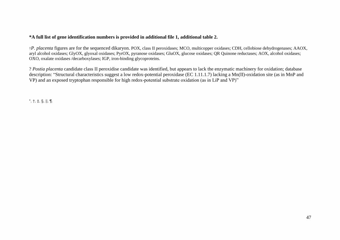
47
*A full list of gene identification numbers is provided in additional file 1, additional table 2.
†P. placenta figures are for the sequenced dikaryon. POX, class II peroxidases; MCO, multicopper oxidases; CDH, cellobiose dehydrogenases; AAOX,
aryl alcohol oxidases; GlyOX, glyoxal oxidases; PyrOX, pyranose oxidases; GluOX, glucose oxidases; QR Quinone reductases; AOX, alcohol oxidases;
OXO, oxalate oxidases /decarboxylases; IGP, iron-binding glycoproteins.
? Postia placenta candidate class II peroxidise candidate was identified, but appears to lack the enzymatic machinery for oxidation; database
description: “Structural characteristics suggest a low redox-potential peroxidase (EC 1.11.1.7) lacking a Mn(II)-oxidation site (as in MnP and
VP) and an exposed tryptophan responsible for high redox-potential substrate oxidation (as in LiP and VP)”
*, †, ‡, §, ||, ¶.

48
Table S9. Gene copy distribution for the gene families and organisms used in reconciliation analysis*. Red = Gene families absent in genome, blue – highest
number of gene copies per family for Basidiomycete genomes only. GH – glycoside hydroalse, CE – carbohydrate esterase, † - GH family 5 endoglucanases
(EC3.2.1.4) & mannanases (EC3.2.1.78) only, ‡ - CE1 excluding S-formylglutathione hydrolases, § - CE16 cinnamoylesterases only, no lysophospholypases. Gene Family Coprinopsis
cinerea
Laccaria
bicolor
Schizophyllum
commune
Serpula
lacrymans
Heterobasidion
irregulare
Postia
placenta
Phanerochaete
chrysosporium
Trichoderma
reesei
Magnaporthe
grisea
Stagonospora
nodorum
CAZYs
GH Family 3 7 2 12 10 12 5 9 13 18 16
GH Family 5† 6 3 3 8 6 5 5 3 4 5
GH Family 6 5 0 1 1 1 0 1 1 3 4
GH Family 7 4 0 2 0 1 0 7 2 5 5
GH Family 10 5 0 5 1 2 2 6 1 6 7
GH Family 11 6 0 1 0 0 0 1 2 5 7
GH Family 12 1 2 1 1 4 2 2 1 3 4
GH Family 28 3 3 3 7 8 7 4 3 2 4
GH Family 61 30 3 22 5 10 2 16 3 21 27
GH Family 74 1 0 1 1 1 0 2 1 1 0
CE Family 1‡ 3 0 9 0 1 0 3 0 3 6
CE Family 16§ 4 1 4 2 4 5 2 2 2 2
Oxidoreductases
Class II peroxidases 1 1 0 0 8 ? 16 0 2 5
Multicopper oxidases 17 11 5 6 17 4 5 7 12 8
Cellobiose
dehydrogenases
1 0 1 2 1 0 1 0 3 3
Glyoxal oxidases 6 10 2 3 5 3 7 1 1 1
Quinone reductases 3 2 4 2 1 1 4 1 1 1
Oxalate oxidases/
decarboxylases
1 1 5 3 3 5 7 3 2 3
Iron binding
glycopeptides
0 1 7 3 1 5 3 0 0 0
CAZY/oxidoreductase
s /total
75/29/104 14/26/40 64/24/88 36/19/55 50/36/86 28/19/47 58/43/101 32/12/44 73/21/94 87/21/108

49
*A full list of gene identification numbers is provided in additional file 1, additional table 3.
? Postia placenta candidate class II peroxidise candidate was identified, but appears to lack the enzymatic machinery for oxidation; database
description: “Structural characteristics suggest a low redox-potential peroxidase (EC 1.11.1.7) lacking a Mn(II)-oxidation site (as in MnP and
VP) and an exposed tryptophan responsible for high redox-potential substrate oxidation (as in LiP and VP)”.
Table S10. Number of of orthologs (% amino acid identity) between Serpula strains and with other Agaricomycetes
S7.9 S7.3
Serpula lacrymans (non-self) 10378(98.5%) 10378(98.5%)
Agaricus bisporus 5354(58%) 5616(58%)
Coprinopsis cinerea 5585(57%) 5859(57%)
Laccaria bicolor 5578(60%) 5840(60%)
Phanerochaete chrysosporium 5138(60%) 5341(60%)
Postia placenta 5097(61%) 5330(61%)
Table S11. Posterior probability distribution for divergence times (in millions of years before present) for major lineages in the Agaricomycetes and the
Serpula-Austropaxillus split using relaxed molecular clock analyses with normal distribution.
Node tMRCA Posterior means 95% HPD
1 Ingroup (-Fomitiporia) 180.7 219.4 – 140.6
2 Russulales 114.4 178.1 – 66.9
3 Polyporales 98.4 150.9 – 57.6
4 Agaricomycetidae 166.1 189.8 – 126.5
5 core Agaricomycetidae (-Jaapia) 143.9 174.2 – 119.3
6 Agaricales 124.0 155.2 – 109.4
7 Boletales (inc. S. lacrymans) 113.4 140.5 – 87.3
8 Amylocorticiales 93.7 127.9 – 43.4
9 Atheliales 77.7 118.5 – 33.2
10 Marasmioid clade 92.0 93.9 – 90.0
11 Suillineae 52.0 54.0 – 50.0
12 Serpula-Austropaxillus 34.9 53.1 – 15.0

50
Table S12. S. lacrymans transcriptomic comparison from cultures grown on either glucose or wood wafers. 50 genes with the greatest
differential expression when grown on wood compared with glucose cultures are shown.
Sequence
ID
Average
glucose
10 days
Average
wood 10
days
Ratio wood
10d/glucose
10D
Signal
peptide Interpro definition
335267 4 5935 1355,044 * Glycoside hydrolase, family 61
433209 150 33665 224,987 *
Cellulose-binding region, fungal ; Glycoside hydrolase, family 5 ;
Cellulose-binding region, fungal ; Cellulose-binding region, fungal ;
Cellulose-binding region, fungal ; Cellulose-binding region, fungal ;
Glycoside hydrolase, family 5 ; Glycoside hydrolase, catalytic core ;
Cellulose-binding region, fungal
453342 29 4307 148,172 * Glycoside hydrolase, family GH74 with BNR repeat
452187 260 31801 122,372 *
Cellulose-binding region, fungal ; Cellulose-binding region, fungal ;
Cellulose-binding region, fungal ; Cellulose-binding region, fungal ;
Carbohydrate-binding family 9/cellobiose dehydrogenase, cytochrome ;
Cellulose-binding region, fungal
445922 234 25504 109,013 * Glycoside hydrolase, family GH43 ; -galactosidase
451883 72 7797 108,642
Flavin-containing monooxygenase FMO ; FAD-dependent pyridine
nucleotide-disulphide oxidoreductase
447766 121 8898 73,354 Amidase signature enzyme ; Amidase signature enzyme
349170 493 35476 71,977 *
Glycoside hydrolase, family 10 ; Glycoside hydrolase, family 10 ;
Glycoside hydrolase, family 10 ; Glycoside hydrolase, family 10 ;
Glycoside hydrolase, catalytic core
372243 52 3510 67,380
Aminoacyl-tRNA synthetase, class I, conserved site ; Glycoside hydrolase,
catalytic core
433208 409 24635 60,182
Glycoside hydrolase, family 5 ; Glycoside hydrolase, family 5 ; Glycoside
hydrolase, catalytic core
453971 228 13133 57,688 *
Glycoside hydrolase, family 28 ; Glycoside hydrolase, family 28 ; Pectin
lyase fold/virulence factor
371352 127 6979 55,084
Oligopeptide transporter OPT superfamily ; Tetrapeptide transporter,
OPT1/isp4 ; Oligopeptide transporter OPT superfamily

51
480589 250 13356 53,330 *
Glycoside hydrolase, family 5 ; Serine/threonine protein kinase, active site ;
DNA topoisomerase, type IIA, subunit B or N-terminal ; Glycoside
hydrolase, catalytic core
434626 150 7823 52,128 * Bacterial alpha-L-rhamnosidase ; Six-hairpin glycosidase-like
417964 199 10315 51,742 *
475099 316 16233 51,434
362527 22 1088 49,779
Cytochrome P450 ; Cytochrome P450, E-class, group I ; Cytochrome P450
; Cytochrome P450 ; Cytochrome P450
432855 325 15793 48,544 *
Sugar transporter ; General substrate transporter ; Sugar transporter ; Major
facilitator superfamily ; Sugar transporter, conserved site ; MFS general
substrate transporter
454077 464 22294 48,035
Glycoside hydrolase, family 3, N-terminal ; Glycoside hydrolase, family 3,
C-terminal ; Glycoside hydrolase, catalytic core ; Glycoside hydrolase,
family 3, C-terminal
363565 194 9197 47,348 *
477654 321 14645 45,655 * Lipase, GDSL ; Esterase, SGNH hydrolase-type
360767 399 18001 45,166
Major facilitator superfamily MFS-1 ; Major facilitator superfamily ; MFS
general substrate transporter
357854 253 10882 43,047 * Lipase, GDSL Esterase, SGNH hydrolase-type
408489 173 7248 41,901 NAD-dependent epimerase/dehydratase ; NAD(P)-binding
453784 218 9017 41,425 * Glycoside hydrolase, family 61
361086 541 21583 39,897 * Glycoside hydrolase, family 5 ; Glycoside hydrolase, catalytic core
465649 742 28838 38,854 * Glycoside hydrolase, family 61
469302 186 7172 38,560
Major facilitator superfamily MFS-1 ; Major facilitator superfamily ; MFS
general substrate transporter
439506 249 9491 38,100
Glucose-methanol-choline oxidoreductase, N-terminal ; Glucose-methanol-
choline oxidoreductase, C-terminal ; Glucose-methanol-choline
oxidoreductase ; Glucose-methanol-choline oxidoreductase, N-terminal ;
Glucose-methanol-choline oxidoreductase, N-terminal
355924 530 19410 36,588 General substrate transporter ; Sugar transporter ; Major facilitator

52
superfamily ; MFS general substrate transporter
471097 80 2910 36,452
440027 192 6884 35,800 * Aromatic-ring hydroxylase ; Monooxygenase, FAD-binding
433131 366 12605 34,395 * Carboxylesterase, type B ; Carboxylesterase, type B
458151 863 29183 33,796 * Chitin-binding, domain 3
442919 833 27457 32,945 * Carboxylesterase, type B ; Carboxylesterase, type B
478367 148 4738 32,100 *
Glycoside hydrolase, family 12 ; Glycoside hydrolase, family 12 ;
Concanavalin A-like lectin/glucanase
371376 177 5554 31,458 *
Glycoside hydrolase, family 28 ; Parallel beta-helix repeat ; Glycoside
hydrolase, family 28 ; Pectin lyase fold/virulence factor
434546 829 25656 30,931 *
Glycoside hydrolase, family 3, N-terminal ; Glycoside hydrolase, family 3,
N-terminal ; Glycoside hydrolase, family 3, C-terminal ; Glycoside
hydrolase, catalytic core ; Glycoside hydrolase, family 3, C-terminal
439032 204 5949 29,134 * Pectinesterase, catalytic ; Pectin lyase fold/virulence factor
451920 425 12053 28,332 * Lipase, GDSL ; Esterase, SGNH hydrolase-type
469301 258 7265 28,193 MFS general substrate transporter
355683 783 21495 27,442 *
Cellulose-binding region, fungal ; Glycoside hydrolase, family 5 ;
Cellulose-binding region, fungal ; Cellulose-binding region, fungal ;
Cellulose-binding region, fungal ; Cellulose-binding region, fungal ;
Glycoside hydrolase, family 5 ; Glycoside hydrolase, catalytic core ;
Cellulose-binding region, fungal
408717 754 20156 26,742 *
Carboxylesterase, type B ; Carboxylesterase, type B ; Carboxylesterase,
type B
362272 507 13172 25,956 Glycoside hydrolase, family 5 ; Glycoside hydrolase, catalytic core
391341 174 4291 24,628
450352 1232 30308 24,597
443184 232 5572 24,024
Cytochrome P450, E-class, group I ; Cytochrome P450 ; Cytochrome P450
; Cytochrome P450
445039 882 21161 23,998
Glycoside hydrolase, family 1 ; Glycoside hydrolase, family 1 ; Glycoside
hydrolase, family 1 ; Glycoside hydrolase, catalytic core

53
441797 454 10901 23,989 *
Histidine acid phosphatase ; Histidine acid phosphatase, eukaryotic ;
Histidine acid phosphatase ; Histidine acid phosphatase
453756 212 5089 23,987

54
Table S13. Extracellular proteins from S. lacrymans (dikaryon) grown in solid state wood culture. Protein Nr refers to protein number in
annotated genomes of S. lacrymans monokayrons S7.9 and S7.3; A – all models, F – filtered models
wit
h C
a
wit
ho
ut
Ca
bo
th
1 beta-glucosidase, GH1 79-A-433206 Q Q25BW4 PF00232 Glycosyl hydrolase family 1 cl01046 Glycosyl hydrolase family 1
2 beta-mannosidase GH2, 732-F-107546 S Q2UN00 PF00703 Glyco_hydro_2 COG3250 Beta-galactosidase/beta-glucuronidase
(beta-galactosidase/beta-glucuronidase)
3 beta-glucosidase, GH3 732-F-103008 S Q4WGT3 PF00933 Glycosyl hydrolase family 3 N terminal domain cl03393 Glyco_hydro_3_C super family
PF01915 Glycosyl hydrolase family 3 C terminal domain cl07971 Glycosyl hydrolase family 3 N terminal domain
4 beta-glucosidase, GH3 732-F-117918 Q A1CA51 PF00933 Glycosyl hydrolase family 3 N terminal domain cl03393 Glyco_hydro_3_C super family
PF01915 Glycosyl hydrolase family 3 C terminal domain cl07971 Glycosyl hydrolase family 3 N terminal domain
PF07691 PA14 domain
5 exo-1,4-beta-xylosidase, GH3 732-A-78770 S B8NYD8 PF00933 Glycosyl hydrolase family 3 N terminal domain cl03393 Glyco_hydro_3_C super family
PF01915 Glycosyl hydrolase family 3 C terminal domain cl07971 Glycosyl hydrolase family 3 N terminal domain
6 beta-glucosidase, GH3 79-A-406110 S Q4WGT3 PF00933 Glycosyl hydrolase family 3 N terminal domain cl03393 Glyco_hydro_3_C super family
PF01915 Glycosyl hydrolase family 3 C terminal domain cl07971 Glycosyl hydrolase family 3 N terminal domain
7 endo-1.4-beta-glucanase, (cellulase) GH5 732-A-106926 Q XP_001838381 PF00150 Cellulase (glycosyl hydrolase family 5) cl12144 Cellulase super family
8 endo-1.4-beta-xylanase, (xylanase) GH10 79-F-349170 S O60206 PF00331 Glycosyl hydrolase family 10 cl01495 Glyco_hydro_10 super family
9 alpha-glucosidase, GH31 79-F-447930 S Q9P999 PF01055 Glycosyl hydrolases family 31 cl11402 GH31 super family
PRK10658 alpha-xylosidase YicI
10 alpha-glucosidase, (maltase) GH31 79-A-411290 S P29064 PF01055 Glycosyl hydrolases family 31 cd06602 GH31_MGAM_SI_GAA
COG1501 Alpha-glucosidases, GH family 31
11 beta-galactosidase, (lactase) GH35 79-F-453704 Q P29853 PF01301 Glycosyl hydrolases family 35 cl03154 Glyco_hydro_42
PF10435 Beta-galactosidase. domain 2 cl11086 Beta-galactosidase, domain 2
12 beta-galactosidase, (lactase) GH35 732-F-167797 S Q700S9 PF01301 Glycosyl hydrolases family 35 cl03154 Glyco_hydro_42
PF10435 Beta-galactosidase. domain 2 cl11086 Beta-galactosidase, domain 2
COG1874 LacA
13 alpha-1.2-mannosidase, GH47 732-F-102103 S Q12563 PF01532 Glycosyl hydrolase family 47 cl08327 Glyco_hydro_47 super family
14 alpha-N-arabinofuranosidase, GH51 79-F-412847 S Q8NK90 PF06964 Alpha-L-arabinofuranosidase C-terminus cl01412 Alpha-L-AF_C super family
15 alpha-1.2-mannosidase, GH92 732-F-158402 Q EFI98309 PF07971 Glycosyl hydrolase family 92 cl14007 aman2_put super family
16 beta-glucuronidase, GH79 79-F-468146 S B0DA80 no Pfam no Pfam no CDD no CDD
17 polyphenol oxidase (tyrosinase) 732-F-71478 Q Q00024 PF00264 Common central domain of tyrosinase cl02830 Tyrosinase super family
18 multicopper oxidase (laccase) 79-F-362730 S Q12719 PF00394 Multicopper oxidase cl11412 Cu-oxidase super family
PF07731 Multicopper oxidase TIGR03388 ascorbase
PF07732 Multicopper oxidase
19 MOX/AOX, alcohol oxidase 79-F-471949 Q A8DPS4 PF00732 GMC oxidoreductase cl02950 GMC_oxred_N super family
PF05199 GMC oxidoreductase cl08434 GMC_oxred_C super family
COG2303 BetA
20 secreted FAD-oxidoreductase 79-F-459686 S EFI92548 PF01565 FAD binding domain cl10516 FAD_binding_4 super family
PF08031 Berberine and berberine like cl10516 FAD_binding_4 super family
Pfam Pfam domain CDD CDD domain
Glycosyl hydrolases
Oxidoreductases
Protein
#ProteinName Protein Nr
Protein in
Signal
peptideNCBInr
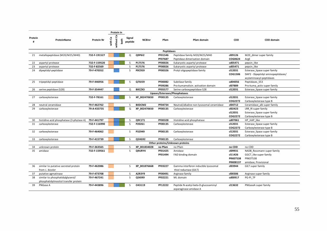
55
wit
h C
a
wit
ho
ut
Ca
bo
th
21 metallopeptidase (M20/M25/M40) 732-F-191527 Q Q9P6I2 PF01546 Peptidase family M20/M25/M40 cl09126 M20_dimer super family
PF07687 Peptidase dimerisation domain COG0624 ArgE
22 aspartyl protease 732-F-159520 S P17576 PF00026 Eukaryotic aspartyl protease cd05471 pepsin_like
23 aspartyl protease 732-F-82569 S P17576 PF00026 Eukaryotic aspartyl protease cd05471 pepsin_like
24 dipeptidyl-peptidase 79-F-470352 S P0C959 PF00326 Prolyl oligopeptidase family cl12031 Esterase_lipase super family
COG1506 DAP2 - Dipeptidyl aminopeptidases/
acylaminoacyl-peptidases
25 tripeptidyl-peptidase 79-F-446956 S Q70J59 PF00082 Subtilase family cd04056 Peptidases_S53
PF09286 Pro-kumamolisin. activation domain cl07889 Pro-kuma_activ super family
26 serine peptidase (S28) 79-F-354447 Q B0CZX3 PF05577 Serine carboxypeptidase S28 cl12031 Esterase_lipase super family
27 carboxylesterase 732-F-78161 S XP_002470650 PF00135 Carboxylesterase cl12031 Esterase_lipase super family
COG2272 Carboxylesterase type B
28 neutral ceramidase 79-F-462762 S B0DCM2 PF04734 Neutral/alkaline non-lysosomal ceramidase cl04712 Ceramidase_alk super family
29 carboxylesterase 79-A-433735 Q XP_002470650 PF00135 Carboxylesterase cl02423 LRR_RI super family
cl12031 Esterase_lipase super family
COG2272 Carboxylesterase type B
30 histidine acid phosphatase (3-phytase A) 79-F-441797 S Q9C1T1 PF00328 Histidine acid phosphatase cd07061 HP_HAP_like
31 carboxylesterase 732-F-116990 S P20261 PF00135 Carboxylesterase cl12031 Esterase_lipase super family
COG2272 Carboxylesterase type B
32 carboxylesterase 79-F-464062 S P32949 PF00135 Carboxylesterase cl12031 Esterase_lipase super family
COG2272 Carboxylesterase type B
33 carboxylesterase 79-F-415739 S Q5XG92 PF00135 Carboxylesterase
34 unknown protein 79-F-363565 S XP_001834028 no Pfam no Pfam no CDD no CDD
35 amidase 732-F-159561 S Q9URY4 PF01425 Amidase cl09931 NADB_Rossmann super family
PF01494 FAD binding domain cl11426 GGCT_like super family
PRK07538 PRK07538
PRK08137 amidase, Provisional
36 similar to putative secreted protein 79-F-462086 S XP_001876668 PF03227 Gamma interferon inducible lysosomal cl03944 GILT super family
from L. bicolor thiol reductase (GILT)
37 putative agmatinase 79-F-473708 S A2R3Y9 PF00491 Arginase family cl00306 Arginase super family
38 similar to phosphatidylglycerol/ 79-F-467241 S Q5KIR9 PF02221 ML domain cd00917 PG-PI_TP
phosphatidylinositol transfer protein
39 PNGase A 79-F-443896 S O43119 PF12222 Peptide N-acetyl-beta-D-glucosaminyl cl13632 PNGaseA super family
asparaginase amidase A
Other proteins/Unknown proteins
Pfam Pfam domain CDD CDD domain
Peptidases
Lipases/Esterases/Phosphatases
Protein
#ProteinName Protein Nr
Protein in
Signal
peptideNCBInr

56
Table S14. Comparison of proteins identified as expressed on solid state wood culture
in the proteomic study with transcript expression in the transcriptomic study.
Proteomic
Protein #
S7.9
Protein
number
Protein Name # peptides
identified
Transcript
fold change
Relative
transcript
level
Ranking
based on
expression†
1 433206 Glycoside
Hydrolase 1
13 No Probe
2 361341 Glycoside
Hydrolase 2
9 6.9 19573 225 (1.9)
3 434546
354470
359881
452594
Glycoside
Hydrolase 3
13 30.9
8.3
0.79
1.3
25656
19873
3964
3684
59 (0.5)
212 (1.8)
3832 (28.7)
4058 (34.4)
4 362919
451868
446869
Glycoside
Hydrolase 3
13 12.9
6.8
2.0
15220
26195
2851
505 (4.2)
53 (0.45)
4846 (41.1)
5 454077 Glycoside
Hydrolase 3
7 48.0 22294 116 (0.98)
6 406110 Glycoside
Hydrolase 3
13 No Probe
7 465929
362272
Glycoside
Hydrolase 5
7 8.4
26.0
23026
13172
102 (0.9)
689 (5.8)
8 349170 Glycoside
Hydrolase 10
3 72.0 35476 8 (0.07)
9 447930 Glycoside
Hydrolase 31
10 22.6 15957 438 (3.7)
10 411290 Glycoside
Hydrolase 31
4 No Probe
11 453704 Glycoside
Hydrolase 35
7 2.6 9763 1190 (10.1)
12 415055 Glycoside
Hydrolase 35
7 9.0 12872 725 (6.1)
13 445448 Glycoside
Hydrolase 47
5 1.6 25242 68 (0.6)
14 412847 Glycoside
Hydrolase 51
2 8.8 7527 1820 (15.4)
15 413209 Glycoside
Hydrolase 92
6 2.0 11074 953 (8.1)
16 468146 Glycoside
Hydrolase 79
7 8.1 22796 106 (0.9)
17 435855
462697
Polyphenol
Oxidase
15 0.98
0.92
20491
1042
180 (1.5)
7340 (62.2)
18 362730 Multicopper
Oxidase
4 1.5 8202 1596 (13.5)
19 471949 MOX/AOX
Alcohol Oxidase
4 7.6 26661 50 (0.4)
20 459686 FAD-
Oxidoreductase
3 No Probe
21 463883 Metallopeptidase 6 1.3 15994 433 (3.7)

57
22 470882
*
Aspartyl Peptidase 3 0.95 13689 636 (5.4)
23 470882
*
Aspartyl Peptidase 3 NA
24 470352 Dipeptidyl
Peptidase
2 1.2 13460 660 (5.6)
25 446956 Tripeptidyl
Peptidase
3 6.6 15243 502 (4.3)
26 354447 Serine Peptidase 5 0.43 4831 3161 (26.8)
27 443197 Carboxyesterase 9 15.9 17016 359 (3.0)
28 462762 Neutral
Ceramidase
2 1.1 6932 2059 (17.4)
29 433735 Carboxylesterase 6 No Probe
30 441797 Histidine Acid
Phosphatase
4 24.0 27457 41 (0.35)
31 442919 Carboxylesterase 6 33.0 10901 982 (8.3)
32 464062 Carboxylesterase 5 7.5 37155 5 (0.04)
33 415739 Carboxylesterase 3 16.0 20823 161 (1.4)
34 363565 Unknown 5 47.4 9197 1319 (11.2)
35 447766
447765
Amidase 6 73.4
14.1
8898
5367
14.02 (11.9)
2862 (24.3)
36 462086 Thiol Reductase 5 0.70 4436 3456 (29.3)
37 473708 Agmatinase 3 2.8 6909 2123 (18.0)
38 467241 Phosphatidyl
Transfer Protein
4 0.86 15310 494 (4.2)
39 443896 PNGase A 3 9.1 16970 362 (3.1)
* no homolog identified in S7.9, e-value similarity to S7.9 470882 for proteins 21 and
22 were 1xe-103 and 1xe-99 respectively. †Ranking of gene based on relative
transcript levels in 10 day wood cultures, based on 11804 probes. Figure in brackets
indicate the position as a percentage, i.e. 5.0 = transcript is in the top 5% highly
expressed genes. Proteins 10, 20, 23, 28, 32, 34, 35, 36, 37 and 38 were associated
with the presence of Calcium. Only proteins 37 and 38 were shown to be differentially
regulated in the transcriptomic study.
Table S15. Comparison of secondary metabolic genes in sequenced basidiomycetes
Species Polyketide
synthases
Terpene cyclases Prenyl transferases
S. lacrymans 5 10 2
C. cinerea 2 3 1
L. bicolor 4 3 2
Sc. commune 1 2 1
Ph. chrysosporium 1 3 0
P. placenta 4 5 1
C. neoformans 1 1 0
U. maydis 1 1 0

References and Notes 1. F. Martin et al., Sequencing the fungal tree of life. New Phytol. 190, 818 (2011).
doi:10.1111/j.1469-8137.2011.03688.x Medline
2. F. Martin, in Biology of the Fungal Cell, R. J. Howard, N. A. R. Gow, Eds. (Springer Berlin Heidelberg, 2007), vol. 8, pp. 291-308.
3. R. L. Gilbertson, Wood rotting fungi of North America. Mycologia 72, 1 (1980). doi:10.2307/3759417
4. D. S. Hibbett, M. J. Donoghue, Analysis of character correlations among wood decay mechanisms, mating systems, and substrate ranges in homobasidiomycetes. Syst. Biol. 50, 215 (2001). doi:10.1080/10635150151125879 Medline
5. K.-E. Eriksson, R. A. Blanchette, P. Ander, Microbial and enzymatic degradation of wood and wood components. (Springer-Verlag, Berlin; New York, 1990).
6. B. D. Lindahl et al., Spatial separation of litter decomposition and mycorrhizal nitrogen uptake in a boreal forest. New Phytol. 173, 611 (2007). doi:10.1111/j.1469-8137.2006.01936.x Medline
7. R. R. Northup, Z. Yu, R. A. Dahlgren, K. A. Vogt, Polyphenol control of nitrogen release from pine litter. Nature 377, 227 (1995). doi:10.1038/377227a0
8. B. Goodell et al., Low molecular weight chelators and phenolic compounds isolated from wood decay fungi and their role in the fungal biodegradation of wood. J. Biotechnol. 53, 133 (1997). doi:10.1016/S0168-1656(97)01681-7
9. T. Shimokawa, M. Nakamura, N. Hayashi, M. Ishihara, Production of 2,5-dimethoxyhydroquinone by the brown-rot fungus Serpula lacrymans to drive extracellular Fenton reaction. Holzforschung 58, 305 (2005). doi:10.1515/HF.2004.047
10. V. Arantes, A. M. Milagres, T. R. Filley, B. Goodell, Lignocellulosic polysaccharides and lignin degradation by wood decay fungi: the relevance of nonenzymatic Fenton-based reactions. J. Ind. Microbiol. Biotechnol. 38, 541 (2011). doi:10.1007/s10295-010-0798-2 Medline
11. S. F. Curling, C. A. Clausen, J. E. Winandy, Experimental method to quantify progressive stages of decay of wood by basidiomycete fungi. Int. Biodeterior. Biodegradation 49, 13 (2002). doi:10.1016/S0964-8305(01)00101-9
12. M. Binder, D. S. Hibbett, Molecular systematics and biological diversification of Boletales. Mycologia 98, 971 (2006). doi:10.3852/mycologia.98.6.971 Medline
13. D. Martinez et al., Genome, transcriptome, and secretome analysis of wood decay fungus Postia placenta supports unique mechanisms of lignocellulose conversion. Proc. Natl. Acad. Sci. U.S.A. 106, 1954 (2009). doi:10.1073/pnas.0809575106 Medline
14. F. Martin et al., Périgord black truffle genome uncovers evolutionary origins and mechanisms of symbiosis. Nature 464, 1033 (2010). doi:10.1038/nature08867 Medline
15. F. Martin et al., The genome of Laccaria bicolor provides insights into mycorrhizal symbiosis. Nature 452, 88 (2008). doi:10.1038/nature06556 Medline

16. D. Martinez et al., Genome sequence of the lignocellulose degrading fungus Phanerochaete chrysosporium strain RP78. Nat. Biotechnol. 22, 695 (2004). doi:10.1038/nbt967 Medline
17. M. Binder, K. H. Larsson, P. B. Matheny, D. S. Hibbett, Amylocorticiales ord. nov. and Jaapiales ord. nov.: early diverging clades of agaricomycetidae dominated by corticioid forms. Mycologia 102, 865 (2010). doi:10.3852/09-288 Medline
18. A. J. Eckert, B. D. Hall, Phylogeny, historical biogeography, and patterns of diversification for Pinus (Pinaceae): phylogenetic tests of fossil-based hypotheses. Mol. Phylogenet. Evol. 40, 166 (2006). doi:10.1016/j.ympev.2006.03.009 Medline
19. H. Kauserud et al., Asian origin and rapid global spread of the destructive dry rot fungus Serpula lacrymans. Mol. Ecol. 16, 3350 (2007). doi:10.1111/j.1365-294X.2007.03387.x Medline
20. O. Schmidt, Molecular methods for the characterization and identification of the dry rot fungus Serpula lacrymans. Holzforschung 54, 221 (2000). doi:10.1515/HF.2000.038
21. Materials and methods are available as supporting material on Science Online.
22. B. L. Cantarel et al., The Carbohydrate-Active EnZymes database (CAZy): an expert resource for Glycogenomics. Nucleic Acids Res. 37, (Database issue), D233 (2009). doi:10.1093/nar/gkn663 Medline
23. G. Vaaje-Kolstad et al., An oxidative enzyme boosting the enzymatic conversion of recalcitrant polysaccharides. Science 330, 219 (2010). doi:10.1126/science.1192231 Medline
24. P. Schneider, S. Bouhired, D. Hoffmeister, Characterization of the atromentin biosynthesis genes and enzymes in the homobasidiomycete Tapinella panuoides. Fungal Genet. Biol. 45, 1487 (2008). doi:10.1016/j.fgb.2008.08.009 Medline
25. M. Tlalka, M. Fricker, S. Watkinson, Imaging of long-distance α-aminoisobutyric acid translocation dynamics during resource capture by Serpula lacrymans. Appl. Environ. Microbiol. 74, 2700 (2008). doi:10.1128/AEM.02765-07 Medline
26. A. Vanden Wymelenberg et al., Comparative transcriptome and secretome analysis of wood decay fungi Postia placenta and Phanerochaete chrysosporium. Appl. Environ. Microbiol. 76, 3599 (2010). doi:10.1128/AEM.00058-10 Medline
27. T. Nilsson, J. Ginns, Cellulolytic activity and the taxonomic position of selected brown-rot fungi. Mycologia 71, 170 (1979). doi:10.2307/3759230
28. B. O. Lindahl, A. F. S. Taylor, R. D. Finlay, Defining nutritional constraints on carbon cycling in boreal forests - Towards a less 'phytocentric' perspective. Plant Soil 242, 123 (2002). doi:10.1023/A:1019650226585
29. R. Vasiliauskas, A. Menkis, R. D. Finlay, J. Stenlid, Wood-decay fungi in fine living roots of conifer seedlings. New Phytol. 174, 441 (2007). doi:10.1111/j.1469-8137.2007.02014.x Medline
30. J. Sambrook, D. W. Russell, 3rd Edn (Cold Spring Harbor Laboratory Press, NY, 2001).

31. D. B. Jaffe et al., Whole-genome sequence assembly for mammalian genomes: Arachne 2. Genome Res. 13, 91 (2003). doi:10.1101/gr.828403 Medline
32. S. Kurtz et al., Versatile and open software for comparing large genomes. Genome Biol. 5, R12 (2004). doi:10.1186/gb-2004-5-2-r12 Medline
33. X. Huang, A. Madan, CAP3: A DNA sequence assembly program. Genome Res. 9, 868 (1999). doi:10.1101/gr.9.9.868 Medline
34. A. F. A. Smit, R. Hubley, P. Green, RepeatMasker Open-3.0. 1996-2010.
35. J. Jurka et al., Genome Res. 110, 462 (2005).
36. T. M. Lowe, S. R. Eddy, tRNAscan-SE: a program for improved detection of transfer RNA genes in genomic sequence. Nucleic Acids Res. 25, 955 (1997). doi:10.1093/nar/25.5.955 Medline
37. A. A. Salamov, V. V. Solovyev, Ab initio gene finding in Drosophila genomic DNA. Genome Res. 10, 516 (2000). doi:10.1101/gr.10.4.516 Medline
38. K. Isono, J. D. McIninch, M. Borodovsky, Characteristic features of the nucleotide sequences of yeast mitochondrial ribosomal protein genes as analyzed by computer program GeneMark. DNA Res. 1, 263 (1994). doi:10.1093/dnares/1.6.263 Medline
39. E. Birne, R. Durbin, Genome Res. 10, 547 (2000). Medline doi:10.1101/gr.10.4.547
40. W. J. Kent, BLAT—the BLAST-like alignment tool. Genome Res. 12, 656 (2002). Medline
41. H. Nielsen, J. Engelbrecht, S. Brunak, G. von Heijne, Identification of prokaryotic and eukaryotic signal peptides and prediction of their cleavage sites. Protein Eng. 10, 1 (1997). doi:10.1093/protein/10.1.1 Medline
42. K. Melén, A. Krogh, G. von Heijne, Reliability measures for membrane protein topology prediction algorithms. J. Mol. Biol. 327, 735 (2003). doi:10.1016/S0022-2836(03)00182-7 Medline
43. E. M. Zdobnov, R. Apweiler, InterProScan—an integration platform for the signature-recognition methods in InterPro. Bioinformatics 17, 847 (2001). doi:10.1093/bioinformatics/17.9.847 Medline
44. S. F. Altschul, W. Gish, W. Miller, E. W. Myers, D. J. Lipman, Basic local alignment search tool. J. Mol. Biol. 215, 403 (1990). Medline
45. M. Kanehisa et al., KEGG for linking genomes to life and the environment. Nucleic Acids Res. 36, (Database issue), D480 (2008). doi:10.1093/nar/gkm882 Medline
46. E. V. Koonin et al., A comprehensive evolutionary classification of proteins encoded in complete eukaryotic genomes. Genome Biol. 5, R7 (2004). doi:10.1186/gb-2004-5-2-r7 Medline
47. A. J. Enright, S. Van Dongen, C. A. Ouzounis, An efficient algorithm for large-scale detection of protein families. Nucleic Acids Res. 30, 1575 (2002). doi:10.1093/nar/30.7.1575 Medline

48. T. De Bie, N. Cristianini, J. P. Demuth, M. W. Hahn, CAFE: a computational tool for the study of gene family evolution. Bioinformatics 22, 1269 (2006). doi:10.1093/bioinformatics/btl097 Medline
49. D. R. Maddison, W. P. Maddison, MacClade 4 Sunderland, Massachusetts: Sinauer Associates (2005).
50. A. J. Drummond, A. Rambaut, BEAST: Bayesian evolutionary analysis by sampling trees. BMC Evol. Biol. 7, 214 (2007). doi:10.1186/1471-2148-7-214 Medline
51. M. Binder et al., The phylogenetic distribution of resupinate forms across the major clades of mushroom-forming fungi (Homobasidiomycetes). Syst. Biodivers. 3, 113 (2005). doi:10.1017/S1477200005001623
52. P. B. Matheny et al., Major clades of Agaricales: a multilocus phylogenetic overview. Mycologia 98, 982 (2006). doi:10.3852/mycologia.98.6.982 Medline
53. D. S. Hibbett, D. Grimaldi, M. J. Donoghue, Fossil mushrooms from Miocene and Cretaceous ambers and the evolution of Homobasidiomycetes. Am. J. Bot. 84, 981 (1997). doi:10.2307/2446289 Medline
54. B. A. LePage, R. S. Currah, R. A. Stockey, G. W. Rothwell, Fossil Ectomycorrhizae from the Middle Eocene. Am. J. Bot. 84, 470 (1997). doi:10.2307/2446014
55. A. Rambaut, A. J. Drummond, Tracer v1.4 http://beast.bio.ed.ac.uk/Tracer (2007).
56. K. Katoh, K. Kuma, H. Toh, T. Miyata, MAFFT version 5: improvement in accuracy of multiple sequence alignment. Nucleic Acids Res. 33, 511 (2005). doi:10.1093/nar/gki198 Medline
57. F. Abascal, R. Zardoya, D. Posada, ProtTest: selection of best-fit models of protein evolution. Bioinformatics 21, 2104 (2005). doi:10.1093/bioinformatics/bti263 Medline
58. A. Stamatakis, P. Hoover, J. Rougemont, A Rapid Bootstrap Algorithm for the RAxML Web Servers. Syst. Biol. 75, 758 (2008). doi:10.1080/10635150802429642
59. T. Y. James et al., Reconstructing the early evolution of Fungi using a six-gene phylogeny. Nature 443, 818 (2006). doi:10.1038/nature05110 Medline
60. D. Durand, B. V. Halldórsson, B. Vernot, A hybrid micro-macroevolutionary approach to gene tree reconstruction. J. Comput. Biol. 13, 320 (2006). doi:10.1089/cmb.2006.13.320 Medline
61. B. Vernot, M. Stolzer, A. Goldman, D. Durand, Reconciliation with non-binary species trees. J. Comput. Biol. 15, 981 (2008). doi:10.1089/cmb.2008.0092 Medline
62. Y. Benjamini, Y. Hochberg, J. R. Statist. Soc. B 57, 289 (1995).
63. A. Bensadoun, D. Weinstein, Assay of proteins in the presence of interfering materials. Anal. Biochem. 70, 241 (1976). doi:10.1016/S0003-2697(76)80064-4 Medline
64. H. Y. Yeang, F. Yusof, L. Abdullah, Protein purification for the Lowry assay: acid precipitation of proteins in the presence of sodium dodecyl sulfate and other biological detergents. Anal. Biochem. 265, 381 (1998). doi:10.1006/abio.1998.2893 Medline

65. D. Fragner, M. Zomorrodi, U. Kües, A. Majcherczyk, Optimized protocol for the 2-DE of extracellular proteins from higher basidiomycetes inhabiting lignocellulose. Electrophoresis 30, 2431 (2009). doi:10.1002/elps.200800770 Medline
66. B. J. Cargile, J. L. Bundy, T. W. Freeman, J. L. Stephenson, Jr., Gel based isoelectric focusing of peptides and the utility of isoelectric point in protein identification. J. Proteome Res. 3, 112 (2004). doi:10.1021/pr0340431 Medline
67. J. Krijgsveld, S. Gauci, W. Dormeyer, A. J. Heck, In-gel isoelectric focusing of peptides as a tool for improved protein identification. J. Proteome Res. 5, 1721 (2006). doi:10.1021/pr0601180 Medline
68. J. Havlis, H. Thomas, M. Sebela, A. Shevchenko, Fast-response proteomics by accelerated in-gel digestion of proteins. Anal. Chem. 75, 1300 (2003). doi:10.1021/ac026136s Medline
69. J. Rappsilber, Y. Ishihama, M. Mann, Stop and go extraction tips for matrix-assisted laser desorption/ionization, nanoelectrospray, and LC/MS sample pretreatment in proteomics. Anal. Chem. 75, 663 (2003). doi:10.1021/ac026117i Medline
70. C. L. Chepanoske, B. E. Richardson, M. von Rechenberg, J. M. Peltier, Average peptide score: a useful parameter for identification of proteins derived from database searches of liquid chromatography/tandem mass spectrometry data. Rapid Commun. Mass Spectrom. 19, 9 (2005). doi:10.1002/rcm.1741 Medline
71. I. Shadforth, T. Dunkley, K. Lilley, D. Crowther, C. Bessant, Confident protein identification using the average peptide score method coupled with search-specific, ab initio thresholds. Rapid Commun. Mass Spectrom. 19, 3363 (2005). doi:10.1002/rcm.2203 Medline
72. M. Gill, W. Steglich, Prog. Chem. Org. Nat. Prod. 51, 1 (1987).
73. L. L. Stookey, Ferrozine---a new spectrophotometric reagent for iron. Anal. Chem. 42, 779 (1979). doi:10.1021/ac60289a016
74. D. W. Malloch, K. A. Pirozynski, P. H. Raven, Ecological and evolutionary significance of mycorrhizal symbioses in vascular plants (A Review). Proc. Natl. Acad. Sci. U.S.A. 77, 2113 (1980). doi:10.1073/pnas.77.4.2113 Medline
75. J. W. G. Cairney, Evolution of mycorrhiza systems. Naturwissenschaften 87, 467 (2000). doi:10.1007/s001140050762 Medline
76. X.-Q. Wang, D. C. Tank, T. Sang, Phylogeny and divergence times in Pinaceae: evidence from three genomes. Mol. Biol. Evol. 17, 773 (2000). Medline
77. D. S. Hibbett, P. B. Matheny, The relative ages of ectomycorrhizal mushrooms and their plant hosts estimated using Bayesian relaxed molecular clock analyses. BMC Biol. 7, 13 (2009). doi:10.1186/1741-7007-7-13 Medline
78. A. M. Bresinsky, M. Jarosch, M. Fischer, I. Schönberger, B. Wittmann-Bresinsky, Phylogenetic Relationships within Paxillus s. I. (Basidiomycetes, Boletales): Separation of a Southern Hemisphere Genus. Plant Biol. 1, 327 (1999). doi:10.1111/j.1438-8677.1999.tb00260.x

79. F. Florindo, A. K. Cooper, P. E. O'Brien, Introduction to ‘Antarctic Cenozoic palaeoenvironments: geologic record and models’. Palaeogeogr. Palaeoclimatol. Palaeoecol. 198, 1 (2003). doi:10.1016/S0031-0182(03)00405-X
80. L. A. Lawyer, L. M. Gahagan, Palaeogeogr. Palaeoclimatol. Palaeoecol. 198, 11 (2003). doi:10.1016/S0031-0182(03)00392-4
81. L. G. Cook, M. D. Crisp, Not so ancient: the extant crown group of Nothofagus represents a post-Gondwanan radiation. Proc. Biol. Sci. 272, 2535 (2005). doi:10.1098/rspb.2005.3219 Medline
82. R. A. Ohm et al., Genome sequence of the model mushroom Schizophyllum commune. Nat. Biotechnol. 28, 957 (2010). doi:10.1038/nbt.1643 Medline
83. D. R. Schmidhalter, G. Canevascini, Purification and characterization of two exo-cellobiohydrolases from the brown-rot fungus Coniophora puteana (Schum ex Fr) Karst. Arch. Biochem. Biophys. 300, 551 (1993). doi:10.1006/abbi.1993.1076 Medline
84. T. Kajisa, K. Igarashi, M. Samejima, The genes encoding glycoside hydrolase family 6 and 7 cellulases from the brown-rot fungus Coniophora puteana. J. Wood Sci. 55, 376 (2009). doi:10.1007/s10086-009-1042-4
85. S. Nagendran, H. E. Hallen-Adams, J. M. Paper, N. Aslam, J. D. Walton, Reduced genomic potential for secreted plant cell-wall-degrading enzymes in the ectomycorrhizal fungus Amanita bisporigera, based on the secretome of Trichoderma reesei. Fungal Genet. Biol. 46, 427 (2009). doi:10.1016/j.fgb.2009.02.001 Medline
86. A. Bresinsky, Z. Naturforsch. 28c, 627 (1973).
87. P. Aqueveque, T. Anke, O. Sterner, Z. Naturforsch. 57, 257 (2002).
88. I. W. Farrell, J. W. Keeping, M. G. Pellatt, V. Thaller, Natural acetylenes. Part XLI. Polyacetylenes from fungal fruiting bodies. J. Chem. Soc. Perkin Trans. 22, 2642 (1973). doi:10.1039/p19730002642
89. D. Hoffmeister, N. P. Keller, Natural products of filamentous fungi: enzymes, genes, and their regulation. Nat. Prod. Rep. 24, 393 (2007). doi:10.1039/b603084j Medline
Acknowledgements: Drs J. Schilling, U Minnesota and Dr D. Barbara, U Warwick critically reviewed the manuscript, Tony Marks designed graphics, B Wackler and M. Zomorrodi gave technical assistance. Assembly and annotations of S.lacrymans genomes are available at http://www.jgi.doe.gov/Serpula and DDBJ/EMBL/GenBank, accessions AECQB00000000 and AEq. C00000000. Complete microarray expression dataset is available at the Gene Expression Omnibus (http://www.ncbi.nml.nhi.gov/geo/) accession GSE27839. The work conducted by the U.S. Department of Energy Joint Genome Institute and supported by the Office of Science of the U.S. Department of Energy under Contract No. DE-AC02-05CH11231. Further financial support is acknowledged as supporting material on Science online.