the physiology of the cardiac - ju med: class of...
TRANSCRIPT

Sheet #22 Dr. Faisal Mohammad 11/5/2014
P a g e | 1 Done by Osama Abu Shawar
بسم هللا العزيز الحكيم
The Physiology of the cardiac
Introduction:
We have three types of muscles in our body:
1. Skeletal muscle.
2. Smooth muscle.
3. Cardiac muscle.
The cardiovascular system consists of: heart (pump) and cardiac vessels.
The Heart:
The heart is the center and the pump of the cardiovascular system by the action of
its muscle layer, it pumps blood to the circulations. We have two circulations: The
greater circulation that distributes the blood to all over the body and is also called
systemic because it distributes the blood to all systems except the lungs, they receive
blood through the lesser (or pulmonary) circulation.

Sheet #22 Dr. Faisal Mohammad 11/5/2014
P a g e | 2 Done by Osama Abu Shawar
The wall of heart has three layers: (from inside to outside)
1. Endocardium (innermost layer, epithelium)
2. Myocardium (major layer, Muscle)
3. Pericardium (outermost layer, two layers: Visceral (close to the heart), and Parietal
with the pericardial space in between which contains a fluid for protection).
A comparison between Skeletal and Cardiac Muscles:
- Skeletal muscles are spindle in shape and start form the origin to the insertion
so they are long, while the cardiac muscle cells are rectangular in shape and
smaller.
- Both are striated due to the presence of sarcomeres that contain contractile
muscle fibers (proteins). There are four types of contractile proteins: Myosin
forming the thick filaments, and the thin filaments are three types:
Tropomyosin forming the doubly helical line to witch actin “beads” are
connected. And Troponin.
- Skeletal muscle cells are not connected to each other, and motor nerves
supply a number of fibers to cause their contraction (a motor unit). While
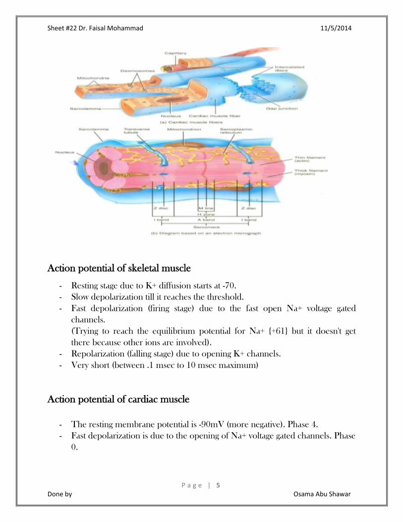
cardiac muscle cells are connected with each other by intercalated disks with
gab junctions between them. The gab junctions conduct electricity from one
cell to another; so they form low resistance areas, and they open or close as
Sheet #22 Dr. Faisal Mohammad 11/5/2014
P a g e | 3 Done by Osama Abu Shawar
response to a change in voltage (voltage-gated channels). When they open the
ions move from one cell to another fast, and thus cardiac muscles are
electrically coupled. Any change (action potential) in one cell spreads into all
cells at the same time through gap junction. This electrical change (action
potential) is followed by a mechanical change (the contraction), and since the
cells receive the action potential at the same time they contract at the same
time as one unit and the heart will work as a pump.
Pathologically, if each cardiac muscle cell contracts by itself (Atrial or
Ventricular fibrillation) the heart will not function and ventricular fibrillation
will cause death.
Gab junctions are hexagonal proteins with an open and a close conformation.
- Skeletal muscles are voluntary, it cannot contract unless it is innervated. While
cardiac muscles are involuntary, although it is supplied by the autonomic
nervous system (sympathetic and parasympathetic). This innervation of the
cardiac muscle is not important for the initiation of cardiac muscle contraction
(during cardiac transplants the autonomic supply is cut but the heart is still
running). The contraction of cardiac muscle follows (None or All law).
In the muscle we call:
The plasma membrane sarcolemma SL.
The endoplasmic reticulum sarcoplasmic reticulum SR.
The cytoplasm sarcoplasm.
- The sarcolemma of cardiac and skeletal muscles have deep invaginations
called the T-tubules or transverse tubules. The T-tubules of skeletal muscles is
slender and longer and occur in the I band so each sarcomere has two T-
tubules, while in the cardiac muscle they are wider and shorter and occur at
the Z line (disk) so each sarcomere has one T-tubule.
The sarcomere is the distance between two Z lines.
- The sarcoplasmic reticulum (which stores Ca++) in the skeletal muscle is well-
developed, while it is less developed in the cardiac muscle so the cell doesn’t
store enough Ca++ for its contraction and needs an extra source of Ca+ from
outside (extracellular fluid around cells – interstitial fluid). That’s why during
heart transplant, the heart is put into a solution with Calcium.
- Cardiac muscle is contracted all the time which requires more energy, that’s
why it has lots of mitochondria to supply ATP compared to skeletal muscles
that, on the other hand, have much more nuclei.
*Ca+ ions bind to troponin to initiate contraction while relaxation occurs when Ca+
unbinds from troponin.

Sheet #22 Dr. Faisal Mohammad 11/5/2014
P a g e | 4 Done by Osama Abu Shawar
*after contraction Ca+ gets back to the SR or leaves the cell through Na\Ca
exchanger and Ca pump.
( المعلومه لتوضيح فقط أدناه الفقرتان )
In both cardiac and skeletal muscles, muscular force production is controlled
primarily by changes in the intracellular Ca+ concentration. In general, when calcium
raises, the muscles contract and, when calcium falls, the muscles relax.
Troponin is a component of thin filaments (along with actin and tropomyosin), and
is the protein complex to which calcium binds to trigger the production of muscular
force.
T tubule sarcomere mitochondria shape
nuclei
Skeletal
muscle
Longer
and
cylinder
two
poor (less)
spindle
more
Cardiac
muscle
Shorter
and wider
one
rich (more)
rectangular
less
Sheet #22 Dr. Faisal Mohammad 11/5/2014
P a g e | 5 Done by Osama Abu Shawar
Action potential of skeletal muscle
- Resting stage due to K+ diffusion starts at -70.
- Slow depolarization till it reaches the threshold.
- Fast depolarization (firing stage) due to the fast open Na+ voltage gated
channels.
(Trying to reach the equilibrium potential for Na+ {+61} but it doesn't get
there because other ions are involved).
- Repolarization (falling stage) due to opening K+ channels.
- Very short (between .1 msec to 10 msec maximum)
Action potential of cardiac muscle
- The resting membrane potential is -90mV (more negative). Phase 4.
- Fast depolarization is due to the opening of Na+ voltage gated channels. Phase
0.

Sheet #22 Dr. Faisal Mohammad 11/5/2014
P a g e | 6 Done by Osama Abu Shawar
(There's an increment in the permeability of Na+ and decrease in the
permeability of K+).
- Partial repolarization is due to the opening of transient K+ and Cl- specialized
channels. Phase 1.
- Plateau (maintaining depolarization) is due to slow opening of Ca+ channels.
Phase 2.
(This induces releasing Ca+ from SR and this process plays the main role in
contraction).
- Repolarization is due to the opening of K+ voltage gated channels. Phase 3.
- Return to resting stage by the Na-K pump for the rearrangement of ions.
Phase 4.
- Longer, occurs in about 200 – 400 (normally 300 msec) thanks to the long
refractory period (an absolute refractory period from the beginning to half of
the repolarization stage where the muscle cannot contract, and a relative
refractory after it where the muscle may contract to a stronger stimulus)

Sheet #22 Dr. Faisal Mohammad 11/5/2014
P a g e | 7 Done by Osama Abu Shawar
The Contraction and Tetanization:
In the skeletal action potential:
Because of the short action potential, if there's another stimuli, new action potential
and another contraction will start in the relative refractory period of first action
potential and this is called tetanus or spasm (summation of contraction
no relaxation).
(Relative refractory period: the last half of the repolarization phase till the action
potential has ended).
In the cardiac action potential:
The muscle contracts and relaxes before a new action potential starts (no tetanus).
Why?.
Due to the very long action potential (long absolute refractory period) in the heart
(plateau).

Sheet #22 Dr. Faisal Mohammad 11/5/2014
P a g e | 8 Done by Osama Abu Shawar
)ما تبقى من الصفحه هو مقدمة للمحاضرة القادمه(
** Na+ channels have M gate (activated) and H gate (inactivated).
M gate H gate
Location Extracellular intracellular
During resting stage Closed opened
During depolarization (less
negative)
Opened closed
Time constant (that's need to
open)
Fast slow
During depolarization:
1. M gate opens.
2. Na enters.
3. H gate closes (due to the threshold is reached).
Summary:
Resting
membrane
potential
Number
of
phases
Partial
repolarization
plateau
Ca+
channel
tetanus
time

Sheet #22 Dr. Faisal Mohammad 11/5/2014
P a g e | 9 Done by Osama Abu Shawar
Skeletal
action
potential
-70
two
___
___
___
present
10
msec
Cardiac
action
potential
-90
five
present
present
present
___
300
msec
Phase 0 Phase 1 Phase 2 Phase 3 Phase 4
Depolarization
(Na+ influx)
Partial
repolarization
(K+ efflux and
Cl- influx).
Plateau
(slow Ca+
influx)
Fast
repolarization
(K+ efflux)
Resting
membrane
potential
*cardiac muscle cells connected with each other through gap junction.
*the skeletal muscle cell has to be stimulated by impulses to contract, but cardiac
muscle is able to contract involuntarily.
*the cardiac muscles are syncytium structure that's why they receive the action
potential as a one unit.
*the electrical change is followed by mechanical change in cardiac muscle.
*the decrease in the permeability of K+ at phase 0 & 1 contributes to the
maintenance of depolarization in phase 2 (plateau).
......ستة أعوام
حتما سنواجه فيهن.....
مادة صعبه!......
دكتور ما بشرح!........
امتحانات كالنار!......
مادة ال تنتهي!.......
تعب وسهر!.....
هموم وأرق!....
والحل؟؟!!!
{قال تعالى: فليستجيبوا لي وليؤمنوا بي وإذا سألك عبادي عني فإني قريب أجيب دعوة الداع إذا دعان
........}لعلهم يرشدون
Sheet #22 Dr. Faisal Mohammad 11/5/2014
P a g e | 1 0 Done by Osama Abu Shawar
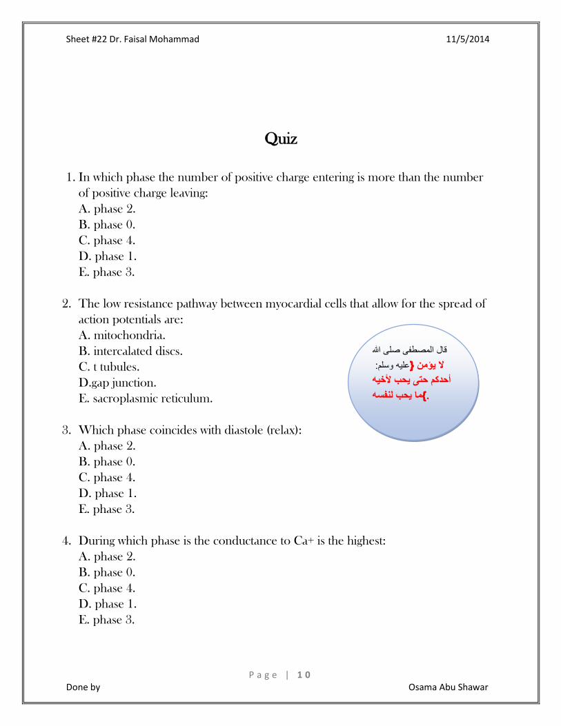
Quiz
1. In which phase the number of positive charge entering is more than the number
of positive charge leaving:
A. phase 2.
B. phase 0.
C. phase 4.
D. phase 1.
E. phase 3.
2. The low resistance pathway between myocardial cells that allow for the spread of
action potentials are:
A. mitochondria.
B. intercalated discs.
C. t tubules.
D.gap junction.
E. sacroplasmic reticulum.
3. Which phase coincides with diastole (relax):
A. phase 2.
B. phase 0.
C. phase 4.
D. phase 1.
E. phase 3.
4. During which phase is the conductance to Ca+ is the highest:
A. phase 2.
B. phase 0.
C. phase 4.
D. phase 1.
E. phase 3.
قال المصطفى صلى هللا
ال يؤمن {عليه وسلم:
أحدكم حتى يحب ألخيه
.}ما يحب لنفسه

Sheet #22 Dr. Faisal Mohammad 11/5/2014
P a g e | 1 1 Done by Osama Abu Shawar
5. During which phase is the membrane potential is closest to the K+ equilibrium
potential:
A. phase 2.
B. phase 0.
C. phase 4.
D. phase 1.
E. phase 3.
إعداد: أسامة أبو شاور...
All of these questions are (past paper). 1 2 3 4 5
B D E A C