the pesky patellofemoral joint: an ongoing orthopaedic enigma pesky... · 2 pfp diagnosis csm 2019...
TRANSCRIPT

1
PFP Diagnosis CSM 2019 Lisa Hoglund, PT, PhD
The Pesky Patellofemoral Joint: An Ongoing Orthopaedic Enigma
Differential Diagnosis of Patellofemoral Pain
Lisa Hoglund, PT, PhD
Associate Professor, Department of Physical Therapy
Thomas Jefferson University
Philadelphia, Pennsylvania, USA
The presenter has no financial relationships or product endorsements to disclose
Presented at 2019 APTA Combined Sections Meeting, Washington D.C. Jan. 24th, 8-10am.
Learning Objectives
• By the end of this session, the learner will be able to:
• Choose clinically-relevant tests and measures to enhance clinical decision-making when
treating individuals with PFP
Diagnosis of Patellofemoral Pain
• Challenging
• No gold standard
• Diagnostic tests – poor accuracy
• Clusters of tests – no improvement to accuracy
• Cook 2012, Nunes 2013
• Diagnosis of exclusion
• First – rule out all other possible causes of anterior knee pain
• Cook 2012, Witvrouw 2014, Crossley 2016
Differential Diagnosis: Anterior Knee Pain
• Medical screening: Appropriate for physical therapy?
• Tumors, fractures, septic arthritis, slipped capital femoral epiphysis, etc.
• Red flags – Review of Systems
• OSPRO-ROS
• Fractures – Ottawa Knee Rule or Pittsburgh Knee Decision Rule
• Risk factors
• Key red flags
• Non-mechanical pain, insidious onset, no improvement in 4 weeks
• Childs 2008, George 2015, Konan 2013
Differential Diagnosis
• Other musculoskeletal conditions
• Lumbar spine
• Hip
• Knee – meniscus, ligaments, cartilage
• Patellar or tibial apophysitis
• Patellar tendinopathy
2
PFP Diagnosis CSM 2019 Lisa Hoglund, PT, PhD
• Patellar subluxation or dislocation
• Psychosocial issues – yellow flags
• Noehren 2016
Symptoms
• Retropatellar or peripatellar pain
• Papadopoulos 2015, Crossley 2016
• Functional tasks loading PFJ with flexed knee
• Squatting
• Stair ascent or descent
• Running
• Sitting with flexed knees
• Collins 2016
Provocative Diagnostic Tests
• Squatting – most accurate
• LR+ = 1.8, LR- = 0.20
• Stair Climbing
• LR+ = 1.3-1.7, LR- = 0.1-0.6
• Eccentric Step-down Test
• LR+ = 2.3, LR- = 0.7
• Cook 2012, Nunes 2013, Papadopoulos 2015
Non-provocative Diagnostic Tests
• Patellar Tilt Test
• LR+ = 5.4, LR- = 0.6
• Passive Gliding Patella
• Superior/inferior
• LR+ = 1.4, LR- = 0.7
• Medial/lateral
• LR+ = 1.8, LR- = 0.7
• Lateral Pull (Active Instability Test)
• LR+ = 249 (?), LR- = 0.8
• Cook 2012, Nunes 2013, Papadopoulos 2015
Summary: Diagnosis of Patellofemoral Pain
• Retropatellar or peripatellar pain
• Rule out other possible causes of anterior knee pain
• Reproduction of anterior knee pain with activities loading the PF joint in a flexed-knee
position
• Squatting, stair ascent/descent, prolonged sitting with flexed knees, etc.
• Cook 2012, Nunes 2013, Papadopoulos 2015, Crossley 2016, Collins 2016
3
PFP Diagnosis CSM 2019 Lisa Hoglund, PT, PhD
References
• Cook D, Mabry L, Reiman MP, et al. Best tests/clinical findings for screening and diagnosis
of patellofemoral pain syndrome: a systematic review. Physiother. 2012;98:93-100.
• Nunes GS, Stapait EL, Kirsten MH, et al. Clinical test for diagnosis of patellofemoral pain
syndrome: systematic review with meta-analysis. Phys Ther Sport. 2013;14:54-59.
• Witvrouw EK, Crossley K, Davis I, et al. The 3rd International Patellofemoral Research
Retreat: an international expert consensus meeting to improve the scientific understanding and
clinical management of patellofemoral pain. Br J Sports Med. 2014;48(6):408.
• Crossley KM, Callaghan MJ, van Linschoten R. Patellofemoral pain. Br J Sports Med.
2016;50(4):247-250.
• Childs JD, Cleland JA, Elliott JM, et al. Neck pain: clinical practice guideline linked to the
International Classification of Functioning, Disability, and Health from the Orthopaedic
Section of the American Physical Therapy Association. J Orthop Sports Phys Ther.
2008;38(9):A1-A34.
• George SZ, Beneciuk JM, Bialosky JE, et al. Development of a review-of-systems screening
tool for orthopaedic physical therapists: results from the optimal screening for prediction of
referral and outcome (OSPRO) cohort. J Orthop Sports Phys Ther. 2015;45;(7):512-526.
• Konan S, Zang TT, Tamimi N, et al. Can the Ottawa and Pittsburgh rules reduce requests for
radiography in patients referred to acute knee clinics? Ann R Coll Surg Engl. 2013;95(3):188-
191.
• Noehren B, Shuping L, Jones A, et al. Somatosensory and biomechanical abnormalities in
females with patellofemoral pain. Clin J Pain. 2016;32(10):915-919.
• Papadopoulos K, Stasinopoulos D, Ganchev D. A systematic review of reviews in
patellofemoral pain syndrome. Exploring the risk factors, diagnostic tests, outcome
measurements and exercise treatment. The Open Sports Medicine Journal. 2015;9:7-17.
• Crossley KM, Stefanik JJ, Selfe J, et al. 2016 patellofemoral pain consensus statement from
the 4th International Patellofemoral Pain Research Retreat, Manchester. Part 1: terminology,
definitions, clinical examination, natural history, patellofemoral osteoarthritis and patient-
reported outcome measures. Br J Sports Med. 2016;50(14):839-843.
• Collins NJ, Vicenzino B, van der Heijden RA, et al. Pain during prolonged sitting is a
common problem in persons with patellofemoral pain. J Orthop Sports Phys Ther.
2016;46(8):658-663.

Patellofemoral Pain Classification Scheme CSM 2019 Lori Bolgla, PT, PhD, MAcc, ATC
The Pesky Patellofemoral Joint: An ongoing orthopaedic enigma
Lori A Bolgla, PT, PhD, MAcc, ATC Professor of Physical Therapy Kellett Chair in Allied Health Sciences Department of Physical Therapy Augusta University Augusta, GA The presenter has no financial relationships or product endorsements to disclose
Presented at 2019 APTA Combined Sections Meeting, Washington D.C. Jan. 24th, 8-10am.
Proposed Classification System for the Treatment of Individuals with Patellofemoral Pain
A. Overuse/Overload without other Impairment • Loss of tissue homeostasis (Post & Dye, 2017) • Moderate relationship between self-reported pain and increased patellofemoral joint
metabolic activity (Draper et al, 2012) • Significant relationship between pain intensity and physical activity level (Briani et al,
2017) • Foot kinematics and hip strength are not predictors of injury for individuals who develop
patellofemoral pain following a “start-to-run” program (Thijs et al, 2008; Thijs et al, 2011)
• Summary Points − Onset from rapid increase in patellofemoral joint loading without adequate time for
tissue recovery − Subgroups may not necessarily exhibit other impairments like impaired muscle
performance, muscle coordination or mobility − Supports the importance of patient education for activity modification (Barton et al,
2015)
B. Muscle Performance Deficits • Subgroup of individuals with patellofemoral pain exhibit hip and knee weakness that
respond favorably to strengthening exercises (Bolgla et al, 2016) • Measurement considerations when assessing hip and knee muscle strength
− Hand-held dynamometry with stabilization straps

Patellofemoral Pain Classification Scheme CSM 2019 Lori Bolgla, PT, PhD, MAcc, ATC
− Use of the “make” test − 3 trial with coefficient of variation less than 10% − Data expressed as a percent of body mass
• Guidance for identifying muscle weakness (expressed as percent body mass) based on responders from a large-scale randomized controlled trial comparing outcomes for a hip/core- or quadriceps-based rehabilitation program (Ferber et al, 2015; Bolgla et al, 2016)
− Hip abductors: 38.8% (males) and 32.2% (females) − Hip extensors: 27.4% (males) and 22.2% (females) − Hip external rotators: 13.0% (males) and 12.0% (females) − Knee extensors: 44.9% (males) and 37.4% (females)
• Summary Points − Subgroup of individuals with patellofemoral pain with hip and/or knee weakness − Hip weakness most likely a result, not a cause, of patellofemoral pain onset (Rathleff
et al, 2014; Herbst et al, 2015) − Need for ongoing works to identify threshold values to identify hip and knee
weakness − Identifying hip and knee weakness values will assist in the development and
implementation of individually-tailored interventions
C. Muscle Coordination Deficits • Subgroup of individuals without evident hip and/or knee weakness • Based on concept of an increased dynamic Q-angle during dynamic tasks (Powers, 2003)
− Contralateral pelvic drop − Hip adduction − Hip internal rotation − Knee abduction
• Limitations of measuring the frontal plane projection angle during a single-leg squat to identify faulty mechanics (Räisänen et al, 2018)
• Support for the use of the dynamic valgus index that incorporates both the hip and knee frontal plane projection angles (Scholtes & Salsich, 2017)
• Use of mobile devices and apps to quantify the dynamic valgus index (unpublished data from Bolgla et al, 2018)
• Summary Points − Commercially-available video cameras can be used to identify patients who exhibit
altered hip and/or knee mechanics during a dynamic task (Scholtes & Salsich, 2017; Gwynne & Curran, 2018)
Patellofemoral Pain Classification Scheme CSM 2019 Lori Bolgla, PT, PhD, MAcc, ATC
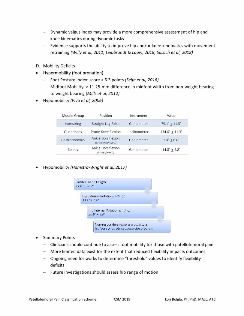
− Dynamic valgus index may provide a more comprehensive assessment of hip and knee kinematics during dynamic tasks
− Evidence supports the ability to improve hip and/or knee kinematics with movement retraining (Willy et al, 2011; Leibbrandt & Louw, 2018; Salsich et al, 2018)
D. Mobility Deficits • Hypermobility (foot pronation)
− Foot Posture Index: score > 6.3 points (Selfe et al, 2016) − Midfoot Mobility: > 11.25-mm difference in midfoot width from non-weight bearing
to weight bearing (Mills et al, 2012) • Hypomobility (Piva et al, 2006)
• Hypomobility (Hamstra-Wright et al, 2017)
• Summary Points
− Clinicians should continue to assess foot mobility for those with patellofemoral pain − More limited data exist for the extent that reduced flexibility impacts outcomes − Ongoing need for works to determine “threshold” values to identify flexibility
deficits − Future investigations should assess hip range of motion
Patellofemoral Pain Classification Scheme CSM 2019 Lori Bolgla, PT, PhD, MAcc, ATC
E. References
1. Barton CJ, Lack S, Hemmings S, Tufail S, Morrissey D. The 'Best Practice Guide to Conservative Management of Patellofemoral Pain': incorporating level 1 evidence with expert clinical reasoning. Br J Sports Med. 2015;49(14):923-934.
2. Bolgla LA, Earl-Boehm J, Emery C, Hamstra-Wright K, Ferber R. Pain, function, and strength outcomes for males and females with patellofemoral pain who participate in either a hip/core- or knee-based rehabilitation program. Int J Sports Phys Ther. 2016;11(6):926-935.
3. Briani RV, Pazzinatto MF, De Oliveira Silva D, Azevedo FM. Different pain responses to distinct levels of physical activity in women with patellofemoral pain. Braz J Phys Ther. 2017;21(2):138-143.
4. de Oliveira Silva D, Barton CJ, Briani RV, et al. Kinesiophobia, but not strength is associated with altered movement in women with patellofemoral pain. Gait Posture. 2018;68:1-5.
5. Draper CE, Fredericson M, Gold GE, et al. Patients with patellofemoral pain exhibit elevated bone metabolic activity at the patellofemoral joint. J Orthop Res. 2012;30(2):209-213.
6. Ferber R, Bolgla L, Earl-Boehm JE, Emery C, Hamstra-Wright K. Strengthening of the hip and core versus knee muscles for the treatment of patellofemoral pain: a multicenter randomized controlled trial. J Athl Train. 2015;50(4):366-377.
7. Ferreira AS, de Oliveira Silva D, Briani RV, et al. Which is the best predictor of excessive hip internal rotation in women with patellofemoral pain: rearfoot eversion or hip muscle strength? Exploring subgroups. Gait Posture. 2018;62:366-371.
8. Gwynne CR, Curran SA. Two-dimensional frontal plane projection angle can identify subgroups of patellofemoral pain patients who demonstrate dynamic knee valgus. Clin Biomech. 2018;58:44-48.
9. Herbst KA, Barber Foss KD, Fader L, et al. Hip strength is greater in athletes who subsequently develop patellofemoral pain. Am J Sports Med. 2015;43(11):2747-2752.
10. Leibbrandt DC, Louw QA. Targeted functional movement retraining to improve pain, function, and biomechanics in subjects with anterior knee pain: a case series. J Sport Rehabil. 2018;27(3):218-223.
11. Mills K, Blanch P, Dev P, Martin M, Vicenzino B. A randomised control trial of short term efficacy of in-shoe foot orthoses compared with a wait and see policy for anterior knee pain and the role of foot mobility. Br J Sports Med. 2012;46(4):247-252.
12. Piva SR, Goodnite EA, Childs JD. Strength around the hip and flexibility of soft tissues in individuals with and without patellofemoral pain syndrome. J Orthop Sports Phys Ther. 2005;35(12):793-801.
13. Post WR, Dye SF. Patellofemoral pain: an enigma explained by homeostasis and common sense. Am J Orthop. 2017;46(2):92-100.
Patellofemoral Pain Classification Scheme CSM 2019 Lori Bolgla, PT, PhD, MAcc, ATC
14. Powers CM. The influence of altered lower-extremity kinematics on patellofemoral joint dysfunction: a theoretical perspective. J Orthop Sports Phys Ther. 2003;33(11):639-646.
15. Powers CM. The influence of abnormal hip mechanics on knee injury: a biomechanical perspective. J Orthop Sports Phys Ther. 2010;40(2):42-51.
16. Raisanen AM, Pasanen K, Krosshaug T, et al. Association between frontal plane knee control and lower extremity injuries: a prospective study on young team sport athletes. BMJ Open Sport Exerc Med. 2018;4(1):e000311.
17. Rathleff MS, Rathleff CR, Crossley KM, Barton CJ. Is hip strength a risk factor for patellofemoral pain? A systematic review and meta-analysis. Br J Sports Med. 2014;48(14):1088.
18. Scholtes SA, Salsich GB. A dynamic valgus index that combines hip and knee angles: Assessment of utility in females with patellofemoral pain. Int J Sports Phys Ther. 2017;12(3):333-340.
19. Selfe J, Janssen J, Callaghan M, et al. Are there three main subgroups within the patellofemoral pain population? A detailed characterisation study of 127 patients to help develop targeted intervention (TIPPs). Br J Sports Med. 2016;50(14):873-880.
20. Thijs Y, De Clercq D, Roosen P, Witvrouw E. Gait-related intrinsic risk factors for patellofemoral pain in novice recreational runners. Br J Sports Med. 2008;42(6):466-471.
21. Thijs Y, Van Tiggelen D, Roosen P, De Clercq D, Witvrouw E. A prospective study on gait-related intrinsic risk factors for patellofemoral pain. Clin J Sport Med. 2007;17(6):437-445.
22. Willy RW, Scholz JP, Davis IS. Mirror gait retraining for the treatment of patellofemoral pain in female runners. Clin Biomech. 2012;27(10):1045-1051.

The Pesky Patellofemoral Joint: An ongoing orthopaedic enigma
Richard Willy, PT, PhD Assistant Professor of Physical Therapy School of Physical Therapy and Movement Sciences Missoula, MT, USA The presenter has no financial relationships or product endorsements to disclose
Presented at 2019 APTA Combined Sections Meeting, Washington D.C. Jan. 24th, 8-10am.
I. Slide 1: Cover slide: Best Practice Treatment Strategies for individuals with Patellofemoral Pain (PFP)
II. Slide 2: Disclosures III. Slide 3: Outline for talk IV. Slide 4: Case study Description V. Slide 5: Case study: objective finding
VI. Slide 6: gait analysis VII. Slide 7: Viewing injuries in the context of the Tissue homeostasis model and envelope of
function, as per Dye 2005, can be helpful to guide the clinician and the runner.(1, 2) VIII. Slide 8: Exercise therapy is the standard of care for the treatment of the individual with
PFP(3). Absolute rest is contra-indicated. IX. Slide 9: Quadriceps strength training has the greatest volume of research supporting its
use with individuals with PFP.(4) X. Slide 10: PFJ contact forces and stresses are higher in endrange knee extension and knee
flexion for non-weight bearing and weight bearing knee extension exercises, respectively. However, constant resistance knee extension machines, which are far more common, have more moderate amounts of PFJ stress throughout the range of motion, except for endrange non-weight bearing knee extensions where PFJ stress is consistently high.(5)
XI. Slide 11: EMG biofeedback to augment quadriceps exercises is not supported(6) nor is NMES(7)
XII. Slide 12: Hip plus knee exercise therapies are associated with greater treatment effects compared with knee exercise therapy alone.(8)
XIII. Slide 14: Yet, PFJ taping is helpful but mainly in the early stages of rehabilitation.(9) Mechanism for pain reduction is likely not biomechanical as MRI evidence indicates tape does not alter PFJ alignment or contact area.(10)
XIV. Slide 13: Problems with exercise therapy literature in PFP(11):

a. very few actually describe exercise interventions in sufficient detail to replicate b. Most interventions lack evidence-based parameters that ensure that optimal
strengthening is achieved e.g., rest intervals, %1RM, number of sets XV. Slide 14: Hip strengthening, however, does not appear to alter mechanics.(12).
XVI. Slide 15: PFJ loads of common functional tasks, including walking and running.(13-15) This large difference between walking PFJ loads and running/jumping PFJ loads illustrates why athletes have such a hard time returning to activity.
XVII. Slide 15: How much pain should we allow during exercise? What guidelines can we give our patients? Is hyperalgesia present in our patients?(16)
XVIII. Slide 16-18: Treatment plan for case study XIX. Slide 19, 20: Gait retraining to address frontal plane mechanics in runners(17, 18) and to
be used during a return to running program(1) XX. Slide 21: Review future directions and limitations
XXI. Slide 22: Take home message XXII. Slide 23: Acknowledgements
1. Willy RW, Meira EP. Current Concepts in Biomechanical Interventions for Patellofemoral Pain. Int J Sports Phys Ther. 2016;11(6):877-90. 2. Dye SF. The pathophysiology of patellofemoral pain: a tissue homeostasis perspective. Clin Orthop Relat Res. 2005(436):100-10. 3. van der Heijden RA, Lankhorst NE, van Linschoten R, Bierma-Zeinstra SM, van Middelkoop M. Exercise for treating patellofemoral pain syndrome. The Cochrane database of systematic reviews. 2015;1:CD010387. 4. Van Der Heijden RA, Lankhorst NE, Van Linschoten R, Bierma-Zeinstra SM, Van Middelkoop M. Exercise for treating patellofemoral pain syndrome: an abridged version of Cochrane systematic review. European journal of physical and rehabilitation medicine. 2016;52(1):110-33. 5. Powers CM, Ho KY, Chen YJ, Souza RB, Farrokhi S. Patellofemoral joint stress during weight-bearing and non-weight-bearing quadriceps exercises. The Journal of orthopaedic and sports physical therapy. 2014;44(5):320-7. 6. Collins NJ, Bisset LM, Crossley KM, Vicenzino B. Efficacy of nonsurgical interventions for anterior knee pain: Systematic review and meta-analysis of randomized trials. Sports Medicine. 2012;42(1):31-49. 7. Lake DA, Wofford NH. Effect of therapeutic modalities on patients with patellofemoral pain syndrome: A systematic review. Sports Health. 2011;3(2):182-9. 8. Lack S, Barton C, Sohan O, Crossley K, Morrissey D. Proximal muscle rehabilitation is effective for patellofemoral pain: A systematic review with metaanalysis. British Journal of Sports Medicine. 2015;49(21):1365-76. 9. Crossley KM, Middelkoop MV, Callaghan MJ, Collins NJ, Rathleff MS, Barton CJ. 2016 Patellofemoral pain consensus statement from the 4th International Patellofemoral Pain Research Retreat, Manchester. Part 2: Recommended physical interventions (exercise, taping, bracing, foot orthoses and combined interventions). British Journal of Sports Medicine. 2016;50(14):844-52.

10. Ho KY, Epstein R, Garcia R, Riley N, Lee SP. Effects of Patellofemoral Taping on Patellofemoral Joint Alignment and Contact Area During Weight Bearing. The Journal of orthopaedic and sports physical therapy. 2017;47(2):115-23. 11. Holden S, Rathleff MS, Jensen MB, Barton CJ. How can we implement exercise therapy for patellofemoral pain if we don't know what was prescribed? A systematic review. British journal of sports medicine. 2018;52(6):385. 12. Willy RW, Davis IS. The effect of a hip-strengthening program on mechanics during running and during a single-leg squat. The Journal of orthopaedic and sports physical therapy. 2011;41(9):625-32. 13. Willy RW, Halsey L, Hayek A, Johnson H, Willson JD. Patellofemoral Joint and Achilles Tendon Loads During Overground and Treadmill Running. J Orthop Sports Phys Ther. 2016;46(8):664-72. 14. Teng HL, Powers CM. Sagittal plane trunk posture influences patellofemoral joint stress during running. The Journal of orthopaedic and sports physical therapy. 2014;44(10):785-92. 15. Trepczynski A, Kutzner I, Kornaropoulos E, Taylor WR, Duda GN, Bergmann G, et al. Patellofemoral joint contact forces during activities with high knee flexion. Journal of Orthopaedic Research. 2012;30(3):408-15. 16. Rathleff MS, Roos EM, Olesen JL, Rasmussen S, Arendt-Nielsen L. Lower mechanical pressure pain thresholds in female adolescents with patellofemoral pain syndrome. Journal of Orthopaedic and Sports Physical Therapy. 2013;43(6):414-21. 17. Willy RW, Scholz JP, Davis IS. Mirror gait retraining for the treatment of patellofemoral pain in female runners. Clin Biomech 2012;27(10):1045-51. 18. Noehren B, Scholz J, Davis I. The effect of real-time gait retraining on hip kinematics, pain and function in subjects with patellofemoral pain syndrome. British Journal of Sports Medicine. 2010;45(9):691-6.

1
PFOA Management CSM 2019 Lisa Hoglund, PT, PhD
The Pesky Patellofemoral Joint: An Ongoing Orthopaedic Enigma
Patellofemoral Pain and Patellofemoral Osteoarthritis Onset: Management Across the Life Span
Lisa Hoglund, PT, PhD
Associate Professor, Department of Physical Therapy
Thomas Jefferson University
Philadelphia, Pennsylvania, USA
The presenter has no financial relationships or product endorsements to disclose
Presented at 2019 APTA Combined Sections Meeting, Washington D.C. Jan. 24th, 8-10am.
Learning Objectives
• By the end of this session, the learner will be able to:
• Explain how factors associated with PFP may contribute to PF OA and describe specific
intervention strategies when treating individuals with PF OA
Prevalence of Patellofemoral Pain
• Varies by population
• Annual prevalence general population: 22.7%
• Smith 2018
• Not just a problem for young adults & adolescents
• PearlDiver Record Database - diagnosis rates
• PFP: 1.5%-7.3% all patients seeking medical care in USA
• Diagnosis rates increased with age – to 50-59 years
• Glaviano 2015
Patellofemoral Osteoarthritis: Imaging
• Osteoarthritis – multifactorial disease
• Cartilage, subchondral bone, synovial tissue, joint capsule, muscle
• Knee: medial TF, lateral TF, PF
• Radiographic findings
• Joint space narrowing, osteophytes
• MRI
• Abnormal cartilage morphology, bone marrow lesions
• Hart 2017
Patellofemoral Osteoarthritis
• Possible long-term result of PFP
• PFP and PF OA – possible continuum
• Lack of longitudinal studies
• One retrospective review – persons undergoing PFJ arthroplasty
• Crossley 2014, Thomas 2010, Utting 2005
• PFP – frequent persistent symptoms – up to 20 years
• Nimon 1998

2
PFOA Management CSM 2019 Lisa Hoglund, PT, PhD
• Similar symptoms, impairments, functional limitations
• Crossley 2016
Patellofemoral Osteoarthritis (cont.)
• Highly prevalent in adults
• 38% (population-based)
• 43% (symptom-based)
• Hart 2017
• Anterior knee pain – stair climbing
• Min-no pain – level ambulation
• van Middelkoop 2018
• Significant cause of disability
• Stair ascent & descent, sit-to-stand, car & bathtub transfers
• Hoglund 2015, van Middelkoop 2018
Muscle and Static Alignment Factors Associated with PF OA
• Proximal muscle weakness:
• Quadriceps
• Hip abd, hip ER, hip ext
• Hoglund 2014, Stefanik 2011, van Middelkoop 2018
• LE static malalignment
• Elahi 2000, Cahue 2004
Biomechanics Associated with PF OA
• Faulty dynamic mechanics: inconsistent
• Sit-to-stand: dynamic genu valgus
• Hoglund 2014
• Gait: conflicting
• No differences
• Crossley 2012, Pohl 2013
• Inc. anterior pelvic tilt
• Late stance – inc. contralateral pelvic drop, dec. hip extension & inc. hip adduction
• Crossley 2018
• Stair descent: inc. anterior pelvic tilt
• Fok 2013
How should we treat the patient with PF OA?
• Limited evidence
• Patellar taping or bracing
• Multimodal approach
• Two trials
• Hip abductor strengthening, VMO retraining, jt mobilization, patellar taping
• van Middelkoop 2018
• Foot orthoses – prefab orthosis (6°varus wedge) and flat insole – dec. knee pain in stepdown
test
• Collins 2017
3
PFOA Management CSM 2019 Lisa Hoglund, PT, PhD
Pilot Study: Exercise Intervention
• Principles for treating patients with PFP: hip focus hip + knee
• Core/trunk strengthening + neuromuscular reeducation
• Hoglund 2018
PF OA: Exercise Focus (Hoglund 2018)
• 6 weeks, 2x/week + home program
• Hip focus + abdominal strengthening/stabilization – lying
• Decreased PFJ stress
• Progressed to standing hip, knee, pelvic/trunk stabilization
• Neuromuscular reeducation
• Functional ex: sit-to-stand
PF OA Exercise Intervention Results -1
PF OA Exercise Intervention Results -2
Patient-Reported Outcome Measure for PF OA
• Knee Injury and Osteoarthritis Outcome Score (KOOS)
• Valid, reliable, and responsive for knee OA, focal cartilage lesions, meniscal tear, ACL
tear, postsurgical
• MDC: older pts 20 pts per subscale, younger pts 14.3 – 19.6 pts
• Collins 2016
• KOOS-PF
• Smallest detectable change: 16 pts
• Minimal important change: 14.2 pts
• Crossley 2018
PF OA Physical Performance Measures
• Recommended core set of PPM for pts with knee and hip OA (OARSI):
• 30” Chair Stand Test
• 40 meter Fast Paced Walk
• Stair-climb Test
• Additional recommended PPM
• TUG
• PF OA participants – longer time vs controls
• Hoglund 2015
• 6-minute Walk Test
• Dobson 2017
References
• Smith BE, Selfe J, Thacker D, et al. Incidence and prevalence of patellofemoral pain: a
systematic review and meta-analysis. PLoS ONE. 2018;13(1):e0190892.
https://doi.org/10.1371/journal.pone.0190892
• Glaviano NR, Kew M, Hart JM, et al. Demographic and epidemiological trends in
4
PFOA Management CSM 2019 Lisa Hoglund, PT, PhD
patellofemoral pain. Int J Sports Phys Ther. 2015;10(3):281-290.
• Hart HF, Stefanik JJ, Wyndow N, et al. The prevalence of radiographic and MRI-defined
patellofemoral osteoarthritis and structural pathology: a systematic review and meta-analysis.
Br J Sports Med. 2017;51:1195-1208.
• Crossley KM. Is patellofemoral osteoarthritis a common sequela of patellofemoral pain? Br
J Sports Med. 2014;48(6):409-410.
• Thomas MJ, Wood L, Self J, et al. Anterior knee pain in younger adults as a precursor to
subsequent patellofemoral osteoarthritis: a systematic review. BMC Musculoskelet Disord.
2010; 11:201-2474-11-201.
• Utting MR, Davies G, Newman JH. Is anterior knee pain a predisposing factor to
patellofemoral osteoarthritis? Knee. 2005;12(5):362-365.
• Nimon G, Murray D, Sandow M, et al. Natural history of anterior knee pain: a 14- to 20-year
follow-up of nonoperative management . J Pediatr Orthop. 1998;18:118-122.
• Crossley KM, Stefanik JJ, Selfe J, et al. 2016 Patellofemoral pain consensus statement from
the 4th International Patellofemoral Pain Research Retreat, Manchester. Part 1: Terminology,
definitions, clinical examination, natural history, patellofemoral osteoarthritis and patient-
reported outcome measures. Br J Sports Med. 2016;50:839–843.
• Van Middelkoop M, Bennell KL, Callaghan MJ, et al. International patellofemoral
osteoarthritis consortium: Consensus statement on the diagnosis, burden, outcome
measures, prognosis, risk factors and treatment. Semin Arthritis Rheum. 2018;47(5):666-675.
• Hoglund LT, Lockard MA, Barbe MF, et al. Physical performance measurement in persons
with patellofemoral osteoarthritis: A pilot study. J Back Musculoskel Rehabil. 2015;28:335-
342.
• Hoglund LT, Hillstrom HJ, Barr-Gillespie AE, et al. Frontal plane knee and hip kinematics
during sit-to-stand and proximal lower extremity strength in persons with patellofemoral
osteoarthritis: a pilot study. J Appl Biomech. 2014;30:82-94.
• Stefanik JJ, Guermazi A, Zhu Y, et al. Quadriceps weakness, patella alta, and structural
features of patellofemoral osteoarthritis. Arthritis Care Res. 2011;63(10):1391-1397.
• Elahi S, Cahue S, Felson DT, et al. The association between varus-valgus alignment and
patellofemoral osteoarthritis. Arthritis Rheum. 2000;43(8):1874-1880.
• Cahue S, Dunlop D, Hayes K, et al. Varus-valgus alignment in the progression of
patellofemoral osteoarthritis. Arthritis Rheum. 2004;50(7):2184-2190.
• Crossley KM, Dorn TW, Ozturk H, et al. Altered hip muscle forces during gait in people with
patellofemoral osteoarthritis. Osteoarthritis Cartilage. 2012;20(11):1243-1249.
• Pohl MB, Patel C, Wiley JP, et al. Gait biomechanics and hip muscular strength in patients
with patellofemoral osteoarthritis. Gait Posture. 2013;37:440-444.
• Crossley KM, Schache AG, Ozturk H, et al. Pelvic and hip kinematics during walking in
people with patellofemoral joint osteoarthritis compared to healthy age-matched controls.
Arthritis Care Res. 2018;70(2):309-314.
• Fok LA, Schache AG, Crossley KM, et al. Patellofemoral joint loading during stair
ambulation in people with patellofemoral osteoarthritis. Arthritis Rheum. 2013;65(8):2059-
2069.
• Collins NJ, Hinman RS, Menz HB, et al. Immediate effects of foot orthoses on pain during
functional tasks in people with patellofemoral osteoarthritis: a cross-over, proof-of-concept
study. Knee. 2017;24:76-81.
• Hoglund LT, Pontiggia L, Kelly JD. A 6-week hip muscle strengthening and lumbopelvic-hip
5
PFOA Management CSM 2019 Lisa Hoglund, PT, PhD
core stabilization program to improve pain, function, and quality of life in persons with
patellofemoral osteoarthritis: a feasibility pilot study. Pilot Feasibility Stud. 2018 Apr 6;4:70.
• Collins NJ, Prinsen CAC, Christensen R, et al. Knee Injury and Osteoarthritis Outcome Score
(KOOS): systematic review and meta-analysis of measurement properties. Osteoarthritis
Cartilage. 2016;24:1317-1329.
• Crossley KM, Macri EM, Cowan SM, et al. The patellofemoral pain and osteoarthritis
subscale of the KOOS (KOOS-PF): development and validation using the COSMIN checklist.
Br J Sports Med. 2018;52:1130–1136.
• Dobson F, Hinman RS, Hall M, et al. Reliability and measurement error of the Osteoarthritis
Research Society International (OARSI) recommended performance-based tests of physical
function in people with hip and knee osteoarthritis. Osteoarthritis Cartilage. 2017; 25:1792-
1796.