the permeability of rat transitional …jcb.rupress.org/content/28/1/21.full.pdfin other tissues,...
TRANSCRIPT
T H E P E R M E A B I L I T Y OF R A T
T R A N S I T I O N A L E P I T H E L I U M
Keratinizat ion and the Barrier to Water
R. M. H I C K S
From the Bland-Sutton Institute of Pathology, the Middlesex Hospital Medical School, London, England
A B S T R A C T
Permeability barriers must exist in transitional epithelium to prevent the free flow of water from underlying blood capillaries through the epithelium into the hypertonic urine, and such a barrier has now been demonstrated in isolated bladders. This barrier is passive in function and can be destroyed by damaging the luminal surface of the transitional epi- thelium with sodium hydroxide and 8 M urea solutions, by digesting it with trypsin, leci- thinase C, and lecithinase D, or by treating it with lipid solvents such as Triton N 100 and saponin. From this it is concluded that the barrier depends on the integrity of lipo- protein cell membranes. The barrier function is also destroyed by sodium thioglycollate solutions, and electron microscope investigations show that sodium thioglycollate damages the thick asymmetric membrane which limits the luminal face of the superficial squamous cell. Cytochemical staining shows the epithelium to contain disulfide and thiol groups and to have a concentration of these groups at the luminal margin of the superficial cells. It thus appears that the permeability barrier also depends on the presence of disulfide bridges in the epithelium, and it is presumed that these links are located in keratin. Because of the effect of thioglycollates, both on the barrier function and on the morphology of the mem- brane, it is suggested that keratin may be incorporated in the thick barrier membrane. It is proposed that the cells lining the urinary bladder and ureters should be regarded as a keratinizing epitheluim.
The transitional epithelium in the mammal presents a barrier to the passage of salts and water between blood and urine (1). No dilution of the strongly hypertonic urine occurs by the passage of water across the epithelium from the intra- and extracellular fluids, and there is only a very slow movement of ions across the cells in the reverse direction, from the urine into the blood. It was evident from a study of the subcellular morphology of this epithelium that the barrier function is subdivided into an extracellular and an intra- cellular component (2). The extracellular barrier has been identified, by analogy with the epithelial linings of other cavitary organs, as the junctional
complex formed by the lateral cell membranes of adjacent superficial squamous cells at their luminal borders, and reasons have been given for thinking that the intracellular barrier must also be at the luminal edge of the epithelium. This intracellular barrier was tentatively identified as the unusual, thick, asymmetric cell membrane which limits the free surface of the squamous cell adjacent to the urine. It was proposed that the barrier function might, in part, be due to keratin incorporated in the thick surface membrane (2). Keratin is apparently also present in the ceils of this epithelium in the form of tonofilaments, which,
21
on June 17, 2018jcb.rupress.org Downloaded from http://doi.org/10.1083/jcb.28.1.21Published Online: 1 January, 1966 | Supp Info:
in o the r tissues, are general ly bel ieved to be kera t in fibers (3, 4).
I n this paper , fu r the r observat ions are m a d e w h i c h con f i rm the effect iveness of the pe rmeab i l i t y ba r r i e r in t rans i t ional ep i the l ium. Ev idence is p resen ted tha t the bar r ie r func t ion is des t royed by pro teoly t ic agents , l ipid solvents, and sod ium thioglycol la te solutions. T h e thioglycol la te is p r e s u m e d to ac t by b reak ing disulfide br idges in kera t in , a n d it is shown tha t it also physical ly d i s rup ts the thick cell m e m b r a n e . T h e exis tence of disulf ide links, b o t h t h r o u g h o u t the cy top lasm
a n d in a c o n c e n t r a t e d b a n d a t the lumina l edge of the ep i the l ium, is shown cytochemical ly . These results suppor t the suggest ion tha t this is a kera- t inizing ep i the l ium.
M A T E R I A L S AND M E T H O D S
ANIMALS: Adult male and female rats of the Wistar strain were used. They were killed by dis- location of the neck.
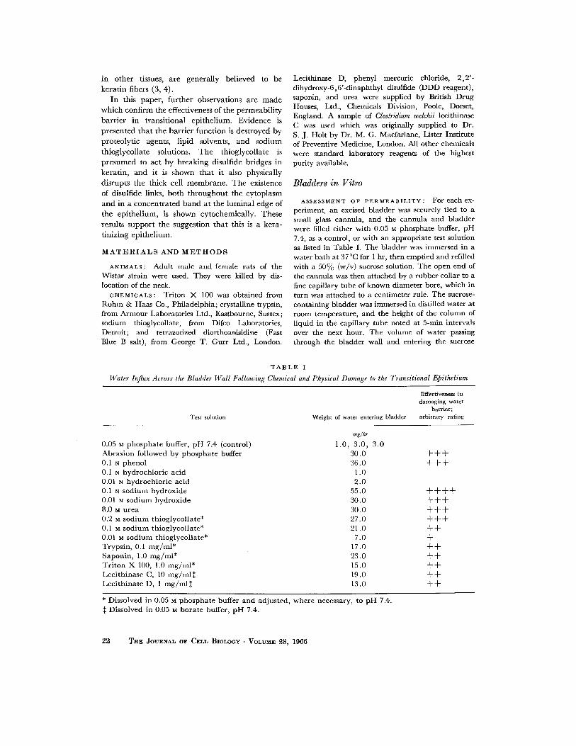
CHEMICALS: Tri ton X 100 was obtained from R o h m & Haas Co., Philadelphia; crystalline trypsin, f rom Armour Laboratories Ltd., Eastbourne, Sussex; sodium thioglycollate, f rom Difco Laboratories, Detroit ; and tetrazotized diorthoanisidine (Fast Blue B salt), from George T. Gurr Ltd., London.
Lecithinase D, phenyl mercuric chloride, 2 ,2 ' - d ihydroxy-6,6 ' -dinaphthyl disulfide (DDD reagent) , saponin, and urea were supplied by British Drug Houses, Ltd., Chemicals Division, Poole, Dorset, England. A sample of Clostridium welchii lecithinase C was used which was originally supplied to Dr. S. J . Holt by Dr. M. G. Macfarlane, Lister Insti tute of Preventive Medicine, London. All other chemicals were s tandard laboratory reagents of the highest purity available.
Bladders in Vi tro
ASSESSMENT OF PERMEABILITY: F o r e a c h e x - periment, an excised bladder was securely tied to a small glass cannula, and the cannula and bladder were filled either with 0.05 M phosphate buffer, p H 7.4, as a control, or with an appropriate test solution as listed in Table I. The bladder was immersed in a water bath at 37°C for 1 hr, then empt ied and refilled with a 50% (w/v) sucrose solution. The open end of the cannula was then at tached by a rubber collar to a fine capillary tube of known diameter bore, which in turn was a t tached to a cent imeter rule. The sucrose- containing bladder was immersed in distilled water at room temperature , and the height of the column of l iquid in the capillary tube noted at 5-nlin intervals over the next hour. The volume of water passing through the bladder wall and entering the sucrose
T A B L E I
Water Influx Across the Bladder Wall Following Chemical and Physical Damage to the Transitional Epithelium
Test solution Weight of water entering bladder
Effectiveness in damaging water
barrier; arbitrary rating
0.05 M p h o s p h a t e buffer, p H 7.4 (control) Abras ion fol lowed by p h o s p h a t e buffer 0.1 N phenol 0.1 N hydroch lo r i c acid 0.01 N hydroch lo r ic acid 0.1 N sod ium hydrox ide 0.01 N sod ium hydrox ide 8.0 M urea 0.2 M sodium thioglycol la te* 0.1 M sodium thioglycol la te* 0.01 M sod ium thioglycol la te* Tryps in , 0.1 m g / ml * Saponin , 1.0 m g / m l * T r i t o n X 100, 1.0 m g / ml * Lec i th inase C, 10 m g / m l ~ Lec i th inase D, 1 m g / m l ~
mg/hr
1.0, 3.0, 30.0 36.0
1.0 2.0
55.0 30.0 30.0 27.0 21.0
7.0 17.0 23.0 15.0 19.0 13.0
3.0 + + + + + +
+ + + + + + + + + + + + + + + + + + + + + + + + + +
* Dissolved in 0.05 ~ p h o s p h a t e buffer and adjus ted, where necessary, to pH 7.4. Dissolved in 0.05 M bora t e buffer, p H 7.4.
22 THE JOURNAL OF CELb BIOLOGY • VOLUME ~8, 1966

solution was approximate ly equal to v:r2h, where r is the radius of the capillary tube and h the he igh t of the c o l u m n of liquid.
ENZYME DIGESTIONS OF THE EPITHELIUM" Solutions of lecithinases C and D (4 to l0 un i t s /ml ) and trypsin (0.25 m g / m l ) were prepared according to Holt (5). These solutions were in t roduced into isolated bladders, and the bladders then incubated, as described, at 37°C for 1 hr. T h e enzyme solutions were then replaced by 50~o sucrose and the b ladder permeabi l i ty de termined.
Bladders and Ureters in Vivo
T H I O G L Y C O L L A T E I N J E C T I O N S : Animals w e r e
anesthet ized with ether, and the left ureter and b ladder were exposed. T h e bladder , if not a l ready collapsed, was empt ied by gentle pressure. A 0.1 M or 0.01 M solution of sod ium thioglycollate in 0.05 M phospha te buffer, p H 7.4, was injected into the uppe r end of the ureter unti l the b ladder was slightly distended. A c l amp was placed over the lower end of the ureter before the needle of the syringe was with- drawn, thus t r app ing some of the thioglycollate solution in the ureter. After an interval of 5, 10, or 15 rain, the ureter and bladder were injected in situ with o s m i u m tetroxide, and removed for fur ther fixation and inspection in the electron microscope.
In o ther animals , the c o m m o n bile duc t was exposed. A 0.01 M sod ium thioglycollate solution in 0.05 ~ phospha te buffer was injected into the upper end of the bile duc t and t rapped in the duc t by c l amping the lower end. After 5 rain, the duc t was injected with o s m i u m tetroxide, excised, and pre- pa red for inspection in the electron microscope. Bile ducts were taken f rom other animals to act as con- trois.
E L E C T R O N M I C R O S C O P Y : Bladders and u r e t e r s
were fixed by injecting cold 4 % (w/v) o s m i u m tetroxide, buffered with phospha te (6) to p H 7.3, directly into the lumen. T h e organ was then removed and sectioned, unde r cold o s m i u m tetroxide, into approximate ly 0.5 to 1.0 m m 3 cubes or cylinders. Fixat ion was cont inued for 1 hr, and the tissue was then dehydra ted in e thanol and e m b e d d e d in Epikote 812 (Shell Chemica l Co., Ltd. , London) essentially by the me thod of Luf t (7).
Sections showing silver-to-gold interference colors were cu t wi th glass knives on a Por ter -Blum micro- tome, m o u n t e d on copper electromesh grids, double- s ta ined (2) with u rany l acetate (8) and a lead salt (9), and examined in a Siemens EImiskop 1.
L I G H T M I C R O S C O P Y : F o r l ight microscopy, whole ureters and bladders were fixed in 4 % glutara l - dehyde buffered to p H 7.4 with sod i um cacodylate (10) for 24 hr, t hen washed for 24 h r in 0.25 M sucrose buffered with 0.1 M sod ium cacodylate to p H
7.4. The tissues were embedded in paraffin wax, and 5 /z sections were cu t and m o u n t e d on clean glass slides. After dewaxing, the sections were s ta ined to show - - S H groups by the 2 ,2 ' - d ihyd roxy -6 ,6 ' - d inaph thy l disulfide (DDD) me thod of Barrnet t and Se l igman (11) according to the schedule g iven by Pearse (12). Control sections were incuba ted in sa tura ted phenyl mercur ic chloride for 2 days before staining. To illustrate - - S - - S - - as well as - - S H groups, the sections were reduced wi th a thioglycollate solution (12) before s ta ining with the D D D reagent .
Experimental Procedure
In a first set of experiments , the permeabi l i ty to water of excised bladders was assessed by measur ing the ra te at which water would cross the b ladder wall to enter a 50~o sucrose solution. The role of the t ransi t ional ep i the l ium in control l ing this was invest igated by d a m a g i n g the l ining of the bladder , ei ther chemical ly or physically, by scra tching it wi th broken glass.
Since one agent caus ing an increase in b ladder per- meabi l i ty was found to be sod ium thioglycollate, which is known to act as a kera t in solvent (3), fur ther exper iments were unde r t aken in which the fine s t ructure of the b ladder ep i the l ium was examined , with the electron microscope, for d a m a g e after t r ea tmen t with thioglycollate solutions. T h e b l a d d e r s used for the in vitro permeabi l i ty exper iments were not entirely satisfactory for electron microscope investigations. Control bladders, in which the barr ier funct ion r ema ined intact , showed wide distension of the extracel lular spaces between epithelial cells after immers ion in distilled water (Fig. 1). After experi- menta l d a m a g e to the barrier function, gross desquama t ion of the ep i the l ium as well as subcel lular d a m a g e was seen. U n d e r these conditions, it was not possible to detect the primary site of act ion of sod ium thioglycollate. In vivo exper iments were therefore performed, in which the blood supply to the b ladder was ma in t a ined so tha t the ep i the l ium was subjected nei ther to osmotic shock nor to anoxia. The lumina l side of the ep i the l ium was t hen exposed to sod ium thioglycollate solutions of progressively lower con- centrat ions unti l a min ima l a m o u n t of cell d a m a g e was detectable with the electron microscope. T h e d a m a g e seen could thus be a t t r ibuted, wi th some confidence, to the thioglycollate solutions and not to the exper imenta l conditions.
I n parallel in vivo experiments , bile duc t epithe- l i um was inspected as a control before and after exposure to sod ium thioglycollate.
I n a final set of experiments , the ep i the l ium was s ta ined cytochemical ly to reveal the location of disulfide and sulphydryl groups and therefore, by implicat ion, the location of keratin.
R. M. HICK~ Keratinization of Transitional Epithelium 23
R E S U L T S
Bladder Permeability and Epithelial Damage
The permeabi l i ty of whole excised bladders was measured by calculat ing the ra te at which water, from distilled water in which the b ladder was suspended, entered a 5 0 % sucrose solution con- tained within the bladder . F rom the internal d iameters of the bladders below their point of a t t a c h m e n t to the cannulae , it appeared tha t the surface area of the ep i the l ium in contac t wi th the sucrose solutions was approximate ly 0.75 to 1.0 cm 2, bu t this est imate is undoub ted ly low because of the folding of the epithelial surface. Moreover, those bladders in which the epi the l ium was damag ed distended far more dur ing the course of the experiments t han did u n d a m a g e d control bladders. For these reasons, no a t t empt has been made to relate the ra te of water passage across the b ladder to a un i t area, and the ra te figures given in Tab le I are not strictly comparable . They arc included here to show the basis on which the a rb i t ra ry ra t ing was made and to give an idea of the order of b ladder permeabil i ty. These ex- per iments were performed at room tempera ture , wi th no supply of oxygen or nut r ients to the b ladder wall, 2 to 3 h r after removing the organ from the animal.
Wa te r entered control bladders a t a relatively
low rate, approximate ly 1 to 3 m g / h r . Damag ing the epithel ium, mechanica l ly by scratching it l ightly with broken glass, or by the nonspecific corrosive action of dilute phenol, damaged the barr ier function of the b ladder and caused a ten- fold increase in the ra te at which water entered the sucrose solution.
Incuba t ion of bladders conta in ing dilute hydro- chloric acid for 1 hr at 37°C did not increase thei r permeabi l i ty above t ha t of control bladders in- cuba ted wi th phosphate buffer. Sodium hydroxide, on the other hand , increased the rate at which water entered the sucrose solution even more ef- fectively than phenol or abrasion, and 8 M urea solutions also increased the water flow. Incuba t ion of b ladders conta in ing trypsin, or lecithinases C or D, increased the permeabi l i ty of the b ladder to water, as did Tr i ton X 100 and saponin. The ef- fect on the b ladder of three concentra t ions of sodium thioglycollate in phosphate buffer was investigated, and at each concent ra t ion the per- meabi l i ty was raised above normal .
The Effect of Thioglycollate Solutions on the
Thick Cell Membrane
The m e m b r a n e on the lumina l surface of the squamous cells is unusual ly thick, measur ing abou t l l 5 A across (Fig. 2). The th inner dense leaflet of the uni t s t ructure adjacent to the cytoplasm is
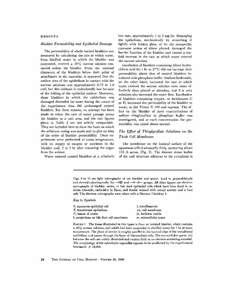
Figs. 8 to 11 are light micrographs of rat bladder and ureter, fixed in glutaraldehyde and stained cytochemically for - -SH and --~S--S--- groups. All other figures are electron micrographs of bladder, ureter, or bile duct epithelial cells which have been fixed in os- mium tetroxide, embedded in Epou, and double stained with uranyl acetate and a lead salt. The electron micrographs were taken with a Siemens Elmiskop 1.
Key to Symbols
S, squamous epithelial cell T, transitional epithelium U, lumen of ureter b, projections on bile duct cell membrane
t, tonofilaments cm, cell membrane fv, fusiform vesicle ec, extraeellular space
FIGURE 1 The tissue illustrated in this figure is from an isolated bladder, which contains a 50% sucrose solution, and which had been suspended in distilled water for 1 hr at room temperature. The plane of section is roughly parallel to the luminal edge of the transitional epithelium and passes through the layer of intermediate cells. The extracellular spaces (ec) between the cells are widely distended and contain little or no electron-scattering material. The morphology of the subcellular organeUes appears to be unaffected by the experimental treatment. X le,000.
24 THE JOURNAL OF CELL BIOLOGY • VOLUME ~8, 1966

R. M. ttlCXS Keratlni~ation of Tramitional Epithelium 25

abou t 25 A, the central l ight b a n d abou t 30 A, and the outer dense leaflet abou t 60 A thick.
The effect on the fine s tructure of the transi- t ional epi the l ium of in vivo injections of 0.1 to 0.01 M sodium thioglycollate solutions into the lumen of the b ladder or ureter was investigated with the electron microscope after 5, 10, or 15 rain exposure times. T he epi thel ium was grossly damaged by 0.1 M thioglycollate solutions and by 15 rain exposure to a 0.01 M solution. M a n y of the superficial and in termedia te cells were rup tured and others shed into the lumen. Those remain ing a t tached to the basement m e m b r a n e and sub- epithelial tissues showed a var iable degree of cytoplasmic damage, frequently wi th rup tured cell membranes and large " e m p t y " areas in the cytoplasm. After only 5 min exposure to 0.01 M thioglycollate, early signs of damage to the epi- the l ium could be seen. The luminal surface of the epithelial cells tended to be more f lat tened in appearance wi th fewer crests t han normal , and the cytoplasm immedia te ly below the m e m b r a n e became thick and dense owing to condensat ion of tonofi laments (Fig. 3). At intervals, breaks in the m e m b r a n e were seen (Fig. 4) and the tonofila- ments appeared to fray out f rom the cell surface into the lumen (Figs. 4 and 5). At this stage, the cytoplasmic contents only a few microns below the cell surface appeared to be substantial ly nor- mal (Fig. 5), and no damage to the membranes of cytoplasmic vesicles or mi tochondr ia , or to the cytoplasmic tonofi laments was observed, a l though a few spaces be tween the tonofi laments appeared.
The Effect of Sodium Thioglycollate on the
Cell Membrane of the Common Bile Duct
The m e m b r a n e on the lumina l surface of the c o m m o n bile duc t in the ra t was also found to be asymmetr ic (Fig. 6). The short, s tubby microvill i are covered by an approximate ly 1 l0 A thick membrane , wi th a wider than average cent ra l l ight band. At the tips of the microvilli, the dense leaflet on the lumina l surface of the un i t mem- b rane is f imbria ted and is projected into, or covered with, small dense blobs approximate ly 150 to 400 A in d iameter (Fig. 6). I t is not clear whether these blobs are a pa r t of the m e m b r a n e un i t s t ructure or a very closely applied form of extraneous coat.
This m e m b r a n e was inspected after in vivo injections of sodium thioglycollate into the bile duc t lumen, and no damage to the m e m b r a n e or under ly ing tissues was observed after 5 rain ex- posure to 0.01 M sodium thioglycollate (Fig. 7).
Cytochemical Staining for - - S H and
- - S - - S - - groups
Sections of glutaraldehyde-f ixed bladders were stained by the D D D method (1 l, 12) for sulphy- dryl groups. Weak positive staining of the cyto- plasm th roughou t the epi thel ium was obta ined, and a more intense band of stain appeared to be located at the lumina l marg in of the superficial cells (Fig. 8). W h e n sections were reduced wi th sodium thioglycollate, before being stained, to
FIGURE ~ This figure shows part of the triple-layered cell membrane of a squamous cell (S) adjacent to the ureteric lumen (?.7). The asymmetric unit structure of this 115 A thick membrane can be clearly seen. The dense leaflet (dl) adjacent to the ureteric lumen is ap- proximately 60 A thick, while the inner leaflet (d2) adjacent to the cytoplasm is about ~5 A thick. X 800,000.
FIGURES 3 and 4 These figures are part of ureteric squamous cells and show the effect on the thick cell membrane of 5 min in vivo exposure to 0.01 M sodium thioglycollate. The unit structure of the membrane has disappeared in Fig. 8, and there is a condensation of tonofilaments (t) at the cell surface. In Fig. 4, remnants of the membrane can be seen (arrows), and tonofilaments appear to fray out into the ureteric lumen (U). X 60,000.
FIGURE 5 This field shows part of the luminal edge of a bladder epithelial cell (S), after in vivo exposure to 0.01 M sodilma thioglycollate. Remnants of the thick surface membrane (arrows) can be seen, but elsewhere the membrane has gone, and the cell surface is ragged in appearance. The thick membranes of the cytoplasmic vesicles (fv), ~ to 5 tt below the surface of the cell, appear to be intact. X 60,000.
26 THE JOURNAL OF CELL BIOLOGY • VOLUME ~8, 1966

R. M. I~CKS Keratinization of Transitional Epithelium 27
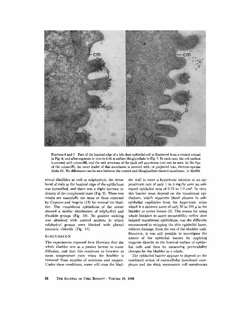
FIGLrRES 6 and 7 Part of the luminal edge of a bile duct epithelial cell is illustrated from a control animal in Fig. 6, and after exposure in vivo to 0.01 M sodium thioglycollate in Fig. 7. In each case, the cell surface is covered with microviili, and the unit structure of the thick cell membrane (cm) can be seen. At the tips of the microvilli, the outer leaflet of this membrane is covered with, or projected into, electron-opaque blobs (b). No differences can be seen between the control and thioglycollate-treated membrane. X 80,000.
reveal disulfides as well as sulphydryls, the dense band of stain at the luminal edge of the epithelium was intensified, and there was a slight increase in density of the cytoplasmic stain (Fig. 9). These two results are essentially the same as those reported by Capurro and Angrist (13) for normal rat blad- der. The transitional epithelium of the ureter showed a similar distribution of sulphydryl and disulfide groups (Fig. 10). No positive staining was obtained with control sections in which sulphydryl groups were blocked with phenyl mercuric chloride (Fig. 11).
D I S C U S S I O N
The cxperimcnts reported here illustratc that thc whole bladder acts as a passive barrier to water diffusion, and that this continucs to function at room temperature even when the bladder is removed from supplies of nutrients and oxygen. Under these conditions, water will cross thc blad-
der wall to enter a hypertonic solution at an ap- proximate rate of only 1 to 3 m g / b r over an esti- mated epithelial area of 0.75 to 1.0 cm ~. In vivo, this barrier must depend on the transitonal epi- thelium, which separates blood plasma in sub- epithelial capillaries from the hypertonic urine which is a distance away of only 50 to 100/z in the bladder or ureter lumen (2). The reason for using whole bladders to assess permeability rather than isolated transitional epithelium, was the difficulty encountered in stripping the thin epithelial layer, without damage, from the rest of the bladder wall. However, it was still possible to investigate the nature of the epithelial barrier by applying reagents directly to the luminal surface of epithe- lial cells and then by measuring permeabili ty changes for the bladder as a whole.
The epithelial barrier appears to depend on the combined action of extracellular junctional com- plexes and the thick asymmetric cell membranes
28 T~v. JOURNAL OF CELL BIOLOGY • VOLUME 28, 1966

FiGrffaES 8 to 11 These light micrographs show portions of rat bladder (Figs. 8, 9, and 11) and ureter (Fig. 10) after cytochemical staining with the DDD reagent (11, 1~) for - -SH groups.
Fig. 8 shows a distribution o f - -SH groups throughout the transitional epithelium (T), and a thin line of intense stain (s) at the luminal margin of the epithelium.
In Figs. 9 and 10, the tissue was reduced with sodium thioglycollate before staining, to show the dis- tribution of both --S---S-- and - -SH groups. In ureter and bladder, the line of intense stain (s) at the luminal margin of the epithelium is more pronounced than it was before reduction of the tissue (cf. Fig. 8), and there is a slight increase in density of the cytoplasmic stain.
The tissue in Fig. 11 was incubated in phenyl mercuric chloride to block the--SH groups before stain- ing with the DDD reagent. Neither the cytoplasm nor the luminal edges of the epithelial cells stain after this treatment. Figs. 8 to 11, X 1400.
found only on the luminal face of the superficial cells (2). If the epithelium is damaged, by scratch- ing it lightly with broken glass or by the nonspeci- fic corrosive action of phenol, the barrier func- tion is drastically reduced, and water enters the bladder (Table I). The barrier appears to be fairly acid-resistant, but is badly damaged by sodium hydroxide and 8 M urea, both of which are powerful protein solvents. I t is also damaged by digestion with the proteolytic enzyme trypsin. Incubation with lecithinases, which disrupt the phospholipid lecithin, and with saponin or Tri ton X 100, which react with cholesterol (I 4, 15), also render the bladder permeable to water.
Most interesting, however, is the damage to barrier function obtained by incubation with thioglycollate solutions, which are known to break disulfide bonds by reduction to sulphydryls. These results indicate that the barrier in transitional epithelium is dependent in part upon a phos- pholipoprotein structure, i.e. the cell membrane, and in part upon the presence of disulfide groups. This supports the previous suggestions that the asymmetric membrane on the luminal surface of the squamous cell is the intracellular barrier and that keratin may be included in its s t ru~ure (2), for keratin and keratohyalin are the only commonly found animal structural proteins rich
R. M. HICKS Keratinization of Transitional Epithelium 29
in sulfur, and the water-proofing properties of keratin are well known (3).
The over-all thickness of this barrier membrane in the rat is about 115 A, which agrees well with the figure of 100 to 110 A reported for the same membrane in mouse transitional epithelium (16, 17). I t is damaged by sodium thioglycollate, and structural changes can be detected after only 5 min exposure to a 0.01 M solution. At this early stage of damage, tonofilaments, mitochondria, and the thick membranes of cytoplasmic vesicles below the cell surface still appear to be normal, which sug- gests that the thick barrier membrane is the first structure to be attacked by the thioglycollate solu- tions. To investigate whether this effect of thio- glycollates is peculiar to the transitional epithe- l ium or is common to all cell membranes, thio- glycollate solutions were injected into the common bile duct. The asymmetric membrane of bile duct epithelial cells, unlike that of the transitional epithelium, is not damaged by sodium thioglycol- late, and it may therefore be assumed that disulfide links are not an important part of its structure. These epithelial cells, in the bile duct of the rat, may be expected to function like those of the gall bladder in other species and to be active in water and cation transport (18) ; an impermeable keratin-containing barrier would be of no ad- vantage in this situation.
I t can be concluded from these results that the barrier function of transitional epithelium may well depend on the inclusion of some form of keratin in the thick surface membrane, unless some other sulfur-containing fibrous protein can subsequently be isolated from this tissue. The tran- sitional epithelium undoubtedly contains some keratin or other cystine-rich protein, as the cyto- chemical staining results obtained with the D D D method (11, 12) for combined - - S - - S - - a n d - - S H groups illustrate. I t is not possible to infer from these light micrographs (Figs. 8 to 11) which morphological substructures contain the reactive
sulphydryl radicles. The weak cytoplasmic stain is probably due to the presence of tonofilaments throughout the cells, if tonofilaments are, in fact, a form of keratin, as is generally stated for other tissues (3, 4). The concentration of stain at the
R E F E R E N C E S
1. ENGLUND, S. E., Acta Radiol., Suppl., 1956, 135, 1. 2. HICKS, R. M., J. Cell Biol., 1965, 26, 25. 3. MERCER, E. H., in International Series of Mono-
graphs on Pure and Applied Biology, No.
luminal edge of the squamous cells which had previously been observed by Capurro and Angrist (13) may be interpreted as due to keratin in the thick membrane of the cell surface and cytoplasmic vesicles. Alternatively, it could be due to keratin in tonofilaments, for although these fibrils are present throughout the epithelium, they are particularly plentiful toward the luminal edge of the superficial squamous cells.
The presence of tonofilaments and disulfide groups, together with the known ability of the bladder epithelium to produce keratinized plaques in response to stress (19, 20), indicates that transi- tional epithelium may properly be regarded as a keratinizing epithelium. This is using Kligman's definition of keratinization as " the synthesis of a peculiar fibrous protein" and "no t the synonym for horny later formation" (21). This epithelium is mesodermal in derivation and is therefore an exception to the statement that "keratin-forming cells are all of ectodermal origin" (22). Transi- tional epithelium is thus comparable to vaginal and oral mucosae, both of which are noncornified, keratinizing epithelia. These epithelia, however, unlike transitional epithelium or epidermis, are relatively permeable to salts and water, and this has been attributed to their lack of a horny layer (23), although it is apparent that the thickness of the horny layer in epidermis is no guide to the effectiveness of the barrier (21, 23). I t is equally possible that differences in permeability between oral mucosa and transitional epithelium are due to differences in the structure of their superficial cell membranes, which in oral mucosa are thick- ened and coated by an amorphous extracellular material, which may be polysaccharide in nature, and which originates in cytoplasmic membrane- coating granules (24). This evidently represents a form of membrane specialization which differs from that seen in transitional epithelium and which is related to a difference in function.
This work was supported by the British Empire Cancer Campaign. I am most grateful to Dr. M. A. Epstein and Dr. S. J. Holt for their advice and encouragement, and to Mr. G. Ball and Mr. T. Heather for their excellent technical assistance.
Received for publication 8 July 1965.
12, in Modern Trends in Physiological Sciences Division, (P. Alexander and Z. M. Bacq, editors), New York, Pergamon Press, Inc., 1961, 43.
30 TnE JOURNAL OF CELL BIOLOGY . VOLUME 28, 1966
4. ODLAND, G. F., in The Epidermis, (W. Montagna and W. C. Lobitz, editors), New York, Academic Press, Inc., 1964, 237.
5. HOLT, S. J. , Exp. Cell Research., Suppl., 1959, 7, 1.
6. MILLONIG, G., J. Appl. Physics, 1961, 32, 1637. 7. LUFT, J. H., or. Biophysic. and Biochem. Cytol.,
1961, 9,409. 8. WATSON, M. L., J. Biophysic. and Biochem. Cytol.,
1958, 4,475. 9. MILLOmO, G., J. Biophysic. and Biochem. Cytol.,
1961, 11, 736. 10. SABATINI, D. D., BENSCH, K. G., and BARRNETT,
R. J., J. Histochem. and Cytochem., 1962, 10, 652.
11. BARRNETT, R. J. , and SELIGMAN, A. M., J. Nat. Cancer Inst., 1952, 13, 215.
12. PEARSE, A. G. E., in Histochemistry, Theoretical and Applied, London, J. & A. Churchill, Ltd., 2nd edition, 1960, 807.
13. CAI'URRO, P., and ANORXST, A., Arch. Path., 1962, 73, 325.
14. BANOHAM, A. D., and HORNE, R. W., Nature, 1962, 196,952.
15. GLAUERT, A.M., DINGLE, J .T. , and Lucy, J.A., Nature, 1962, 196, 953.
16. PORTER, K.R., KENYON, K.R., and BADENHAU- SEN, S., Anat. Rec., 1965, 151, 401.
17. PORTER, K. R., and BONNEVILLE, M. A., An Introduction to the Fine Structure of Cells and Tissues, London, Lea & Febiger, 1963.
18. KALE, G. I., LANE, N., WHEELER, H. O., and WHITLOCK, R. T., Anat. Rec., 1965, 151, 369.
19. CAPURRO, P., ANORIST, A., BLACK, J. , and MouMolS, B., Cancer Research, 1960, 20, 563.
20. ROE, F. J. C., Brit. J. Urol., 1964, 36, 238. 21. KLIGMAN, A. M., in The Epidermis, (W.
Montagna and W. C. Lobitz, editors), New York, Academic Press, Inc., 1964, 387.
22. MATOLTSY, A. G., in Comparative Biochemistry, (M. Florkin and H. S. Mason, editors), New York, Academic Press, Inc., 1962, 4, 343.
23. MALKINSON, F. D., in The Epidermis, (W. Montagna and W. C. Lobitz, editors), New York, Academic Press, Inc., 1964, 435.
24. MATOLTSY, A. G., and PARAKKAL, P. F., J. Cell Biol., 1965, 24, 297.
R. M. HICKS Keratinization of Transitional Epithelium 31