the pelvic kidney of male ambystoma maculatum (amphibia
TRANSCRIPT

The Pelvic Kidney of Male Ambystoma maculatum(Amphibia, Urodela, Ambystomatidae) with SpecialReference to the Sexual Collecting Ducts
Dustin S. Siegel,1* David M. Sever,2 and Robert D. Aldridge1
1Department of Biology, Saint Louis University, St. Louis, Missouri 631032Department of Biological Sciences, Southeastern Louisiana University, Hammond, Louisiana 70402
ABSTRACT This study details the gross and micro-scopic anatomy of the pelvic kidney in male Ambystomamaculatum. The nephron of male Ambystoma macula-tum is divided into six distinct regions leading sequen-tially away from a renal corpuscle: (1) neck segment,which communicates with the coelomic cavity via a ven-trally positioned pleuroperitoneal funnel, (2) proximaltubule, (3) intermediate segment, (4) distal tubule, (5)collecting tubule, and (6) collecting duct. The proximaltubule is divided into a vacuolated proximal region anda distal lysosomic region. The basal plasma membraneis modified into intertwining microvillus lamellae. Theepithelium of the distal tubule varies little along itslength and is demarcated by columns of mitochondriawith their long axes oriented perpendicular to the basallamina. The distal tubule possesses highly interdigitat-ing microvillus lamellae from the lateral membranesand pronounced foot processes of the basal membranethat are not intertwined, but perpendicular to the basallamina. The collecting tubule is lined by an epitheliumwith dark and light cells. Light cells are similar to thoseobserved in the distal tuble except with less mitochon-dria and microvillus lamellae of the lateral and basalplasma membrane. Dark cells possess dark euchromaticnuclei and are filled with numerous small mitochondria.The epithelium of the neck segment, pleuroperitonealfunnel, and intermediate segment is composed entirelyof ciliated cells with cilia protruding from only the cen-tral portion of the apical plasma membrane. The collect-ing duct is lined by a highly secretory epithelium thatproduces numerous membrane bound granules thatstain positively for neutral carbohydrates and proteins.Apically positioned ciliated cells are intercalatedbetween secretory cells. The collecting ducts anastomosecaudally and unite with the Wolffian duct via a commoncollecting duct. The Wolffian duct is secretory, but not tothe extent of the collecting duct, synthesizes neutral car-bohydrates and proteins, and is also lined by apical cili-ated cells intercalated between secretory cells. Althoughfunctional aspects associated with the morphologicalvariation along the length of the proximal portions ofthe nephron have been investigated, the role of a highlysecretory collecting duct has not. Historical data thatimplicated secretory activity concordant with mating ac-tivity, and similarity of structure and chemistry to sex-ual segments of the kidneys in other vertebrates, leadus to believe that the collecting duct functions as asecondary sexual organ in Ambystoma maculatum.J. Morphol. 271:1422–1439, 2010. � 2010 Wiley-Liss, Inc.
KEY WORDS: urogenital; nephron; anatomy; histology;histochemistry; ultrastructure
INTRODUCTION
Salamanders possess paired kidneys that cangenerally be divided into cranial and caudal por-tions. The cranial portion of the male kidney hasbeen termed the sexual (Francis, 1934; Rodgersand Risley, 1938) or genital kidney (Adams, 1940;Williams et al., 1984), in which the nephrons aremodified for the transport of sperm from the vasaefferentia of the testes to the Wolffian ducts. Inplethodontid salamanders, this cranial portion ofthe kidney is lost (Strickland, 1966), and only thenephrons forming the epididymal complex remain.Williams et al. (1984) and Aranzabal (2003, 2009)reviewed variation in cranial kidney structure.
The caudal portion of the male adult kidney,termed the definitive kidney (Baker and Taylor,1964) or simply the pelvic kidney (Spengel, 1876),is involved in urine transport from the nephronsto the cloaca and the production of a sexual secre-tion in some taxa (Aron, 1924; (Norris, unpub-lished data; but see Norris, 1987)). In the Protei-dae (Chase, 1923; Rosenquist and Baker, 1967)and Sirenidae (Willett, 1965), the proximal regionsof the nephron communicate with the Wolffianduct via collecting ducts that exit the pelvic kidneylaterally. These collecting ducts fuse with the Wolf-fian duct individually along its caudal length. Thismorphology is typical of the larval and femalestructure in salamanders (Francis, 1934; Rodgers
Contract grant sponsor: National Science Foundation; Contractgrant number: DEB-0809831.
*Correspondence to: Dustin S. Siegel, Department of Biology,Saint Louis University, 3507 Laclede Avenue, St. Louis, MO 63103.E-mail: [email protected]
Received 21 June 2010; Revised 19 July 2010;Accepted 26 July 2010
Published online 24 September 2010 inWiley Online Library (wileyonlinelibrary.com)DOI: 10.1002/jmor.10883
JOURNAL OF MORPHOLOGY 271:1422–1439 (2010)
� 2010 WILEY-LISS, INC.

and Risley, 1938), and we term this the ‘‘simplecondition.’’
In Ambystomatidae (Baker and Taylor, 1964),Amphiumidae (Baker, 1945), Cryptobranchidae(Ratcliff, 1965), Dicamptodontidae (de Marco,1952), Hynobiidae (Yamagiwa, 1924), Plethodonti-dae (Strickland, 1966), and Salamandridae (Aron,1924; Francis, 1934; Baker, 1965), the collectingducts exit the kidney laterally and communicatewith the cloaca. Each collecting duct communi-cates with the cloaca in Cryptobranchidae andHynobiidae, whereas in the other taxa, the collect-ing ducts anastomose caudally and form a commoncollecting tube. This tube either communicatesdirectly with the cloaca (Plethodontidae and someSalamandridae) or with the cloaca through theWolffian duct (Ambystomatidae and some Sala-mandridae). These conditions arise in malesbecause of a caudal migration of the collectingtubules during development (Rodgers and Risley,1938). We term this the ‘‘complex condition.’’
In males, the epithelium lining the collectingduct tubules of the complex condition varies sea-sonally with reproductive activity in the Salaman-dridae (Aron, 1924; Adams, 1940; Miller and Rob-bins, 1954) and, perhaps, the Ambystomatidae(Norris, unpublished data, see Norris, 1987). Dur-ing reproductive activity, secretory granules fill theapical regions of the collecting duct epithelium andthe ducts nearly double in diameter (Aron, 1924;Adams, 1940; Miller and Robins, 1954). No functionhas been attributed to this increase in secretionsynthesis (Norris, 1987), although the secretionhas been previously referred to as ‘‘albuminoi-diques’’ (Aron, 1924). However, considering theirincreased activity during the reproductive season,it appears as if the collecting ducts in salamanders(at least in the complex condition) have evolved afunction as secondary sexual organs.
Besides the three investigations discussed above(Aron, 1924; Adams, 1940; Miller and Robins,1954), no data exist on the potentially ‘‘sexual’’collecting ducts in salamanders. Although thedescription is limited, the study by Sakai andKawahara (1983) supplies an electron micrographof the collecting duct epithelium. Here, we describethe histology, histochemistry, and ultrastructure ofthe nephron of Ambystoma maculatum in an effortto elucidate functional and morphological aspectsof the collecting ducts in salamanders. We alsoreview the literature on structures associated withthe pelvic kidney and create synonymies for thehistorical terminology.
MATERIALS AND METHODS
Four male Ambystoma maculatum were utilized from the SaintLouis University Museum collection. Two were captured in apond on March 18, 2010 in Crawford County, MO. Snout-ventlengths were 78 and 80 mm. These salamanders were injectedwith McDowell’s-Trump fixative (Electron Microscopy Sciences,
Hatfield, PA) and were submerged in the same fixative for 4 h.After initial fixation, the urogenital tracts were removed and sub-merged in a second solution of McDowell’s-Trump fixative for 48h. The left urogenital tract was then rinsed and dehydrated viaincreasing concentrations of ethanol (35, 70, 95, and 100%), dif-ferentiated with toluene, and then submerged in melted para-plast under vacuum for 12 h before embedding in paraffin.Transverse sections were sliced at 7 lm, affixed to slides with al-bumin, and stained with hematoxylin and eosin for general struc-tural analysis, brilliant blue (BB) for protein concentration, orperiodic acid-Schiff ’s (PAS) for neutral carbohydrates followingthe protocols of Kiernan (1990). Slides were viewed with a LeicaDM4500 microscope (Leica Microsystems, Wetzlar, Germany) anddigital micrographs were obtained via a Quicam 12-Bit MonoFast 1394 Cooled digital camera (Qimaging Corporation, BritishColumbia, Canada). Images were subsequently uploaded intoAdobe Creative Suite (Adobe Systems, San Jose, CA) for labeling.The other two salamanders were also collected in CrawfordCounty, MO, in May; however, the year of collection was notreported. These salamanders were fixed in formalin, and theirentire urogenital tracts were prepared as described above.
The right urogenital tracts of salamanders fixed in McDo-well’s-Trump fixative were rinsed in phosphate buffered saline(PBS; pH 7.4) and subsequently postfixed in 2% osmium tetrox-ide in PBS (pH 7.4). Tissues were then rinsed with PBS (pH7.4), dehydrated via a graded series of ethanol (70, 85, 95, and100%), submerged in a 1:1 mixture of ethanol and propylene ox-ide, followed by pure propylene oxide, and subsequently embed-ded in Epon (EmBed 812, Electron Microscopy Science, Hat-field, PA) for ultrathin sectioning with a Leica EM UC6 ultra-microtome (Leica Microsystems, Wetzlar, Germany). Sections of75 nm were taken, placed on copper grids, and stained withuranyl acetate and lead citrate. Grids were then viewed with aJEOL JEM 100S TEM (JEOL USA, Peabody, MA) and photo-graphed with a L3C CCD digital camera (Scientific Instrumentsand Applications, Duluth, GA). Images were subsequentlyuploaded into Adobe Creative Suite for labeling.
RESULTSGross Morphology and Nomenclature
Major structures of the adult male salamanderkidney have been termed differently in previousinvestigations (Table 1). The pelvic kidneys restdorsal to the pelvic bones, hence the terminologyutilized here. On gross dissection of the venter, thekidneys can be observed; however, observation ofthe entire pelvic kidney apparatus is not possibleas the collecting ducts travel laterally and thenbend medially covering the remaining kidneystructures (Fig. 1A,B). We utilize the terminologyof collecting ducts due to the fact that no apparentcollecting ducts are observed more proximallywithin the kidney. Any terminology that previouslyutilized ‘‘ureters’’ for these structures is inappro-priate due to the lack of homology with theamniote ureters. Medial to the collecting ducts, thepaired lightly pigmented Wolffian ducts travel ven-tral to the kidney. All of these structures lie retro-peritoneal within the same pleuroperitoneum.
By pulling the collecting ducts laterally, themore proximal ducts of the kidney are exposed(Fig. 1B). The proximal kidney ducts are tightlycoiled within a narrow medial portion of the kid-ney (Fig. 1B). These proximal ducts open into �40,large, less-coiled, collecting ducts (Fig. 1B). Thecollecting ducts anastomose caudally and empty
THE PELVIC KIDNEY OF Ambystoma maculatum 1423
Journal of Morphology

into the Wolffian ducts via common collectingducts. These common collecting ducts have beenpreviously termed ureters; however, this is inap-propriate for the same reason as the misnomencla-ture of the collecting ducts.
Structural Organization of the Kidney
The nephron of male Ambystoma maculatumcan be divided into six distinct histological regions
leading sequentially from a renal corpuscle: (1)neck segment, (2) proximal tubule, (3) intermedi-ate segment, (4) distal tubule, (5) collecting tubule,and (6) collecting ducts. The neck segment commu-nicates with the coelomic cavity via a pleuroperito-neal funnel through the ventral kidney wall. Therenal corpuscles are arranged in a longitudinalrow and occupy an area between the midsagittalplane and medial aspect of the kidney. The neckregion tubule branches from the lateral aspect of
TABLE 1. Ontology of the pelvic kidney structures from gross examination in salamanders
Proposedterminology
Historicalterminology Source(s) Taxon(a)* Definition
Pelvic kidney Pars renalis Blum, 1985 Textbook Caudal portion of thekidney. The functionalunits of this portion ofthe kidney are thenephrons. The nephronsobtain filtrate from theblood stream throughrenal corpuscle and thecoloemic cavity throughthe perotineal funnels.
Lumbar kidney Witchi, 1937; Rodgersand Risley, 1938;Adams, 1940
Ambystomatidae:Salamandridae
Definitive kidney Francis, 1934; Baker andTaylor, 1964; Baker,1965; Ratcliff, 1965;Willett, 1965;Strickland, 1966;Rosenquist and Baker,1967
Ambystomatidae;Cryptobranchidae;Plethondontidae;Salamandridae;Sirenidae; Proteidae
Uriniferous kidney Kent and Carr, 2001;Kardong, 2008
Textbook
Excretory kidney Willett, 1965; Rosenquistand Baker, 1967
Sirenidae; Proteidae
Pelvic kidney Spengel, 1876(Beckenniere); Chase,1923; Yamagiwa, 1924(Beckenniere); Sakaiand Kawahara, 1983
Hynobiidae; Proteidae;Salamandridae
Collecting ducts Collecting ducts McCurdy, 1937; Witchi,1937; Rogers andRisley, 1938; Adams,1940; Strickland, 1966
Ambystomatidae;Salamandridae;Plethodontidae
Lateral tubules of thekidney in which thenephrons empty urine.In Proteidae andSirenidae, thesetubules empty into theWolffian ducts multipletimes along theirlengths. In othersalamander taxa, thesetubules bend caudallyand interact with thecloaca individually(e.g., Cryptobranchidaeand Hynobiidae),anastomose and emptyinto the cloaca withthe Wolffian duct (e.g.,Ambystomatidae; someSalamandridae), oranastomose and emptyinto the cloaca (e.g.,Plethodontidae; someSalamandridae). In atleast Ambystomatidae,Plethodontidae, andSalamandridae, thesetubules are secondarysexual organs.
Renal-collecting ducts Miller and Robbins, 1954 SalamandridaeSecondary urinary
ductsBlum, 1985 Textbook
Urinary tubules Baker and Taylor, 1964;Baker, 1965; Ratcliff,1965; Willett, 1965;Strickland, 1966;Rosenquist and Baker,1967
Ambystomatidae;Cryptobranchidae;Plethodontidae;Salamandridae;Sirenidae; Proteidae
Collecting tubules Chase, 1923; Yamagiwa,1924 (sammelrohrchender beckenniere);Baker, 1945; Kent andCarr, 2001
Amphiumidae;Hynobiidae; Proteidae;Textbook
Ureters Francis, 1934; Adams,1940; Rosenquist andBaker, 1967; Sakai andKawahara, 1983
Proteidae;Salamandridae
Urinary collectingtubules
Rosenquist and Baker,1967
Proteidae
Urinary collectingducts
Mintz, 1947 Ambystomatidae
Accessory urinaryducts
Kardong, 2008 Textbook
Common collectingduct
Ureter McCurdy, 1936; Witchi,1936; Baker andTaylor, 1964;Strickland, 1966
Ambystomatidae;Salamandridae;Plethodontidae
The common ductresulting from theanastomosis of thecollecting tubules.
*No terminology was given for structures associated with the pelvic kidney in Dicamptodontidae (DeMarco, 1952). There are cur-rently no descriptions of the urogenital structures associated with the pelvic kidney in Rhyacotritonidae.
1424 D.S. SIEGEL ET AL.
Journal of Morphology

the renal corpuscle and transitions into the proxi-mal tubule at a more lateral aspect of the kidney.The proximal tubule possesses numerous convolu-tions in the lateral kidney before stretching medi-ally and transitioning to the intermediate segment.The intermediate segment transitions to the distaltubule in the medial portion of the kidney. The dis-tal tubule is less convoluted than the proximal andstretches laterally before transition to the collectingtubule and, subsequently, the collecting duct. Thecollecting ducts exit the kidney mass laterally butremain enclosed in a common serosa with the kid-ney. The collecting ducts have the greatest diameterand epithelial height (�220.3 and �40.5 lm, respec-tively) followed by the proximal tubules (�84.6 and�23.2 lm), distal tubules (�50.7 and �15.3 lm),and the ciliated regions (�41.5 and �12.3 lm).
Histology and UltrastructureRenal Corpuscle. The renal corpuscle is com-
posed of the Bowman’s capsule and a fine network
of glomerular capillaries. The Bowman’s capsulehas a parietal epithelium that is continuous with avisceral epithelium, which is in close associationwith the capillaries of the glomerulus (Fig. 2A).Parietal epithelial cells are squamous, have nar-row intercellular canaliculi, and are adhered bydesmosomes along the entire canaliculi length(Fig. 2B). The nuclei of the parietal epithelium arehighly heterochromatic, and small round mito-chondria and electron lucent vesicles of varyingsizes are dispersed throughout the cytoplasm (Fig.2B). Parietal cells rest against a basal lamina andshare a lamina propria with the distal convolutedtubules (Fig. 2B). The epithelial layers of the renalcorpuscle stain basophilic with no reaction to BBor PAS.
The visceral epithelium of the Bowman’s capsuleis squamous, modified into podocytes, and abutsthe basal lamina of the glomerular capillaries (Fig.2C). Adjacent podocytes are adhered by desmo-somes, and desmosomes also join nonadjacentpodocytes that are brought into close association
Fig. 1. Gross anatomy of the caudal urogenital tract of male Ambystoma maculatum. A: Ventral overview of the pelvic kidneyand cloaca highlighting the orientation of the intact kidney and ducts. B: High magnification of the pelvic kidney with the collect-ing ducts distended lateral/dorsal. Cd, collecting ducts; Cl, cloaca; Pk, pelvic kidney; Pn, proximal regions of the nephron; Wd, Wolf-fian duct.
THE PELVIC KIDNEY OF Ambystoma maculatum 1425
Journal of Morphology

Fig. 2. Fine structure of the renal corpuscle of male Ambystoma maculatum. A: Low magnification of a renal corpuscle (tolui-dine blue). B: High magnification of the parietal epithelium of a renal corpuscle (uranyl acetate and lead citrate). C: High magnifi-cation of the visceral epithelium of a renal corpuscle (uranyl acetate and lead citrate). D: High magnification of the cytoplasmiccontents of a podocyte (white arrows indicate filtration slits; uranyl acetate and lead citrate). Bl, basal lamina; Cp, capillary; Cs,capsular space; Dt, distal tubule; Ds, desmosomes; Go, Golgi complex; Mt, mitochondria; Np, nuclei of parietal epithelium; Nu,nuclei; Pd, podocyte; Pn, podocyte nuclei; Rbc, red blood cell; Rer, rough endoplasmic reticulum; Vs, vesicles.
1426 D.S. SIEGEL ET AL.
Journal of Morphology

by the convolutions of the glomerular capillaries(Fig. 2C). Nuclei are heterochromatic (Fig. 2D).Small and elongated mitochondria fill the perinu-clear space of the podocytes and profiles of roughendoplasmic reticulum, and lucent vesicles of vary-ing sizes are also common throughout the cyto-plasm (Fig. 2D). A mildly electron-dense materialfills the lumina of larger vesicles (Fig. 2D). The ba-sal portion of the podocytes is modified into foot-like projection and creates fenestrations along thewalls of the glomerular capillaries (Fig. 2D).
Neck Region, Intermediate Segment, andPleuroperitoneal Funnel. The parietal epithe-lium of the renal corpuscle is continuous with theepithelial lining of the nephron (Fig. 3A). Thelumen of the nephron is continuous with the cap-sular space (Fig. 3A). The first region of the neph-ron distal to the renal corpuscle is the ciliatedneck region. The epithelium of this region is sim-ple cuboidal with centrally located nuclei (Fig. 3A).
All of the cells of the neck region are ciliated andonly two other regions of the nephron possess anepithelium completely lined by cilia: (1) the pleuro-peritoneal funnel (Fig. 3B) and (2) the intermedi-ate segment (Fig. 3C) between the proximal anddistal tubules. The epithelium of all of theseregions is basophilic with no reaction to PAS andonly an apical reaction of the ciliated cells withBB. The cytology of all of these regions is identical;however, from 1 lm thick sections it appears thatthe nuclei of the pleuroperitoneal funnel epithelialcells stain slightly darker with toluidine blue (Fig.3B), and the cilia of the intermediate segment arenoticeably longer than the other respective regions(Fig. 3C). Thus, the cytological description belowencompasses all of the completely ciliated nephronregions.
Ciliated cells of the aforementioned regions arefilled apically with small, slightly elongated mito-chondria, and lucent vacuoles are often inter-
Fig. 3. Structure of the neck region (A), pleuroperitoneal funnel (B), and intermediate segment (C), of male Ambystoma macula-tum (toluidine blue). D: High magnification of a ciliated cell from the intermediate segment (uranyl acetate and lead citrate). Bb,basal bodies; Bl, basal lamina; Ci, cilia; Cn, ciliated cell nuclei; Co, coelom; Dt, distal tubule; Lu, lumen; Mt, mitochondria; Pt, prox-imal tubule.
THE PELVIC KIDNEY OF Ambystoma maculatum 1427
Journal of Morphology

spersed with the mitochondria (Fig. 3D). Elon-gated cilia anchor to basal bodies in the centralportion of the apical cell surface (Fig. 3D). The pe-ripheral region of the apical cell surface is smooth(Fig. 3D). Intercellular canaliculi are narrow andnontortuous. Apical tight junctions and distal des-
mosomes adhere to adjacent ciliated cells. The ba-sal plasma membrane is unmodified and contoursto the basal lamina (Fig. 3D).
Proximal Tubule. The proximal tubule can bedivided cytologically into two distint regions: (1) aproximal vacuolated region (Fig. 4A) and (2) a dis-
Fig. 4. Fine structure of the early proximal tubule of male Ambystoma maculatum. A: Low magnification of the epithelium ofthe early proximal tubule (toluidine blue). B: High magnification of a principle cell in the early proximal tubule (uranyl acetateand lead citrate). C: High magnification of the apical cytoplasm from a cell in the early proximal tubule (uranyl acetate and lead ci-trate). Ac, apical canaliculi; Bl, basal lamina; Cn, ciliated cell nuclei; Ds, desmosome; Ev, endocytic vesicles; Ft, filtrate; Gj, gapjunction; Lu, lumen; Ly, lysosome; Mt, mitochondria; Mv, microvilli; Nu, nuclei; Pn, principle cell nuclei; Va, vacuoles.
1428 D.S. SIEGEL ET AL.
Journal of Morphology
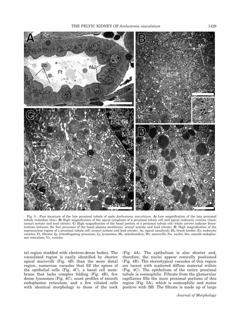
tal region studded with electron-dense bodies. Thevacuolated region is easily identified by shorterapical microvilli (Fig. 4B) than the more distalregion, numerous vacuoles that fill the apices ofthe epithelial cells (Fig. 4C), a basal cell mem-brane that lacks complex folding (Fig. 4B), fewdense lysosomes (Fig. 4C), scant profiles of smoothendoplasmic reticulum, and a few ciliated cellswith identical morphology to those of the neck
(Fig. 4A). The epithelium is also shorter and,therefore, the nuclei appear centrally positioned(Fig. 4B). The stereotypical vacuoles of this regionare lucent with scattered diffuse material within(Fig. 4C). The epithelium of the entire proximaltubule is eosinophilic. Filtrate from the glomerularcapillaries fills the more proximal portions of thisregion (Fig. 5A), which is eosinophilic and stainspositive with BB. The filtrate is made up of large
Fig. 5. Fine structure of the late proximal tubule of male Ambystoma maculatum. A: Low magnification of the late proximaltubule (toluidine blue). B: High magnification of the apical cytoplasm of a proximal tubule cell and apical endocytic vesicles (inset;uranyl acetate and lead citrate). C: High magnification of the basal portion of a proximal tubule cell (white arrows indicate fenes-trations between the foot processes of the basal plasma membrane; uranyl acetate and lead citrate). D. High magnification of thesupranuclear region of a proximal tubule cell (uranyl acetate and lead citrate). Ac, apical canaliculi; Bb, brush border; Ev, endocyticvesicles; Ft, filtrate; Ip, interditigating processes; Ly, lysosomes; Mt, mitochondria; Mv, microvilli; Nu, nuclei; Ser, smooth endoplas-mic reticulum; Vs, vesicles.
THE PELVIC KIDNEY OF Ambystoma maculatum 1429
Journal of Morphology

globular substances of varying electron densi-ties and smaller granular substances (Figs. 4Aand 5A).
The distal region of the proximal tubule pos-sesses a simple columnar epithelium with basalheterochromatic nuclei (Fig. 5A). The central as-pect of the apices of the epithelial cells is domed,and long microvilli cover the apices of every epi-thelial cell (Fig. 5A,B). Between the apical micro-villi, intracellular canaliculi invaginate into theepithelial cells, and endocytic vesicles are observedbelow the surface of the apical plasma membrane(Fig. 5B and inset). Smooth endoplasmic reticulumis abundant throughout the epithelial cells (Fig.5B–D), whereas large circular mitochondria aggre-gate beneath the apical and basal plasma mem-brane (Fig. 5B,C). Electron-dense lysosomes are re-stricted to the supranuclear space of the proximaltubular epithelial cells (Fig. 5D), and these inclu-sions stain positive with PAS. The lateral plasmamembrane and intercellular canaliculi are distinctwith little digitation. Desmosomes adhere to theadjacent epithelial cells along the entire length ofthe intercellular canaliculi, whereas tight junc-tions are present at its apical extremity with a gapjunction located more basally. Numerous invagina-tions form small foot-like projections at the basalregion of the plasma membrane creating a fenes-trated membrane region atop the basal lamina(Fig. 5C). The basal foot processes are highly inter-digitating. Small lucent vesicles are often presentat the distal tips of the foot-like projections (Fig.5C).
Distal tubule and collecting Tubule. Thelumen of the distal tubule is filled with filtrate;however, the filtrate is more homogeneous thanthat observed in the more proximal tubules (Fig.6A). The epithelium of the distal tubule is simplecuboidal with centrally positioned nuclei (Fig. 6A).Epithelial cell cytoplasm is basophilic with a weakreaction to BB. Nuclei are either heterochromaticor mostly euchromatic with dense aggregations ofchromatin dispersed around the nuclear periphery(Fig. 6A,B). Mitochondria fill the cytoplasm ofthese cells and partially occlude the observation ofother cytoplasmic contents (Fig. 6B). Mitochondriaare oriented perpendicular to the apical plasmamembrane and are highly elongated. Invaginationsof the basal plasma membrane form very deep in-tracellular canaliculi basally (Fig. 6C). This causesthe basal foot processes of the epithelial cells totake on an almost filamentous appearance, withthe distal ends of the filaments abutting the basallamina (Fig. 6C). A highly dense material occupiesthe cytoplasm of the distal extremity of the footprocesses (Fig. 6C). The basal foot processes do notinterdigitate to the extent of the proximal tubule.
Microvilli are common on the apical surface ofthe cells but not to the extent of the proximaltubules. Adjacent epithelial cells of the distal
tubule are adhered by an apical tight junctionwith one slightly more basal desmosome (Fig. 6D).The intercellular canaliculi basal to the junctionalcomplexes are highly convoluted from lateralmicrovillus projections with interdigitating lamel-lae from every epithelial cell (Fig. 6D). Dispersedbetween mitochondria, profiles of smooth andrough endoplasmic reticulum are present. Smalllipid droplets are often observed basal to thenuclei. Little cellular variation exists along thelength of the distal tubules. Mitochondria in themore proximal regions of the distal tubule arelarger, less numerous, and are less organized per-pendicular to the basal lamina. The foot processesare also larger resulting in a decrease in the basalmembrane surface area.
The collecting tubule has often been describedas a portion of the distal tubule (see Discussion);however, many differences are observed. In termsof cytoplasmic contents, collecting tubule cells areidentical to those of the distal tubule. Nuclei tendto be less round and more irregularly shaped (Fig.6E), and less-pronounced labyrinths exist betweenthe basal plasma membrane and basal lamina andlaterally between adjacent epithelial cells. Mito-chondria are also less numerous. Odd cells thatstain deep purple with toluidine blue are alsofound intercalated between the majority light cells(Fig. 6E). The only prominent feature of thesedark cells is numerous small mitochondria and adark euchromatic nucleus (Fig. 6E).
Collecting Ducts. The collecting ducts arelined by tall columnar principle cells with basalheterochromatic nuclei (Fig. 7A). Ciliated cells arescattered in between the apices of adjacent colum-nar principle cells (Fig. 7A). The lumen of the col-lecting duct is difficult to observe in 1-lm sectionsstained with toluidine blue due to its dark stainingcontents that are identical in staining intensity tothe numerous secretory granules found in the col-lecting duct epithelium (Fig. 7A).
Rough endoplasmic reticulum is abundant in theperinuclear region, and its cisternae are filled witha diffuse material in the principle cells of the col-lecting ducts (Fig. 7B,C). A large supranuclearGolgi complex is stereotypical (Fig. 7B). Invagina-tions of the basal plasma membrane are scarce;however, interdigitating microvillus projectionsline the lateral aspects of the principle cells (Fig.7D). The principle cells rest on the basal lamina ofmultiple layers of fibroblasts and collagen fibers(Fig. 7C).
The large prominent electron-dense granules ofthe principle collecting duct cells are formed fromfusion of condensing vacuoles with transportvesicles from the Golgi complex (Fig. 7D). Maturegranules dominate the apices of the collecting ductepithelial cells, and this granulated region makesup about 4/5 of every cell (Fig. 7A). Mature gran-ules are homogeneously electron dense (Fig. 8A,B),
1430 D.S. SIEGEL ET AL.
Journal of Morphology

Fig. 6. Fine structure of the distal tubule and collecting tubule of male Ambystoma maculatum. A: Low magnification of the dis-tal tubule (toluidine blue). B: High magnification of the loan cell type in the distal tubule (uranyl acetate and lead citrate). C: Highmagnification of the basal portion of a distal tubule cell (uranyl acetate and lead citrate). D: High magnification of the supranu-clear region of a distal tubule cell (uranyl acetate and lead citrate). E: Low magnification of the collecting tubule (toluidine blue).Bl, basal lamina; Bp, basal processes; Dn, dark-cell nuclei; Ds, desmosome; Ft, filtrate; Ic, intercellular canaliculi; Ln, light-cellnuclei; Lu, lumen; Mt, mitochondria; Nu, nuclei; Tj, tight junction.
THE PELVIC KIDNEY OF Ambystoma maculatum 1431
Journal of Morphology

are eosinophilic, are stain positive with PAS, andhave a weak reaction to BB. In some principlecells, small to large cellular inclusions fill spacesbetween secretory granules (Fig. 8A,B). Theseinclusions are mildly electron dense, nonuniform,and typically have scattered regions of highlyelectron-dense material internally (Fig. 8A,B). Inextreme cases, entire principle cells are filled withan inclusion. The cellular inclusions and maturesecretory granules are released into the lumen viamerocrine secretion (Fig. 8B); however, immatureand mature secretory granules are also found inthe collecting duct lumen (Fig. 8B) indicating the
possibility of cellular rupture or an apocrine modeof secretion.
The cytology of the apical ciliated cells is identi-cal to the ciliated cells of the previously discussedciliary regions (Fig. 8C) with few exceptions.Unlike the ciliated nephron regions, the ciliatedcells in the collecting ducts do not abut a basallamina but instead rest on convex corners of adja-cent principle cells. Microvillus projections fromthe basal portions of the ciliated cells interdigitatewith the lateral membranes of the principle cellsand are adhered apically with the principle cellsby an apical tight junction and one slightly more
Fig. 7. Fine structure of the sexual collecting ducts of male Ambystoma maculatum. A: Low magnification of a sexual collectingduct (toluidine blue). B: High magnification of the perinuclear region of a principle cell of the sexual collecting duct epithelium(uranyl acetate and lead citrate). C: High magnification of the basal portion of a sexual collecting duct principle cell (uranyl acetateand lead citrate). D: High magnification of the lateral aspects of two adjacent principle cells of the sexual collecting duct epithelium(uranyl acetate and lead citrate). Bl, basal lamina; Cn, ciliated cell nuclei; Cv, condensing vacuoles; Fb, fibroblast; Ic, intercellularcanaliculi; Mt, mitochondria; Nu, nuclei; Pn, principle cell nuclei; Rer, rough endoplasmic reticulum; Sg, secretory granule.
1432 D.S. SIEGEL ET AL.
Journal of Morphology

basal desmosome (Fig. 8D). Cilia also cover theentire surface of cells of the collecting ducts (Fig.8C).
Wolffian Duct. Like the collecting ducts, theWolffian ducts are lined by a layer of principlecells that are simple tall columnar (Fig. 9A). The
Fig. 8. Fine structure of the sexual collecting ducts of male Ambystoma maculatum. A: High magnification of the supranuclearregion of a sexual collecting duct principle cell (uranyl acetate and lead citrate). B: High magnification of the apical portion of asexual collecting duct principle cell (black arrow indicates a merocrine type secretion in process; uranyl acetate and lead citrate).C: High magnification of the apical region of a ciliated cell in the sexual collecting duct (uranyl acetate and lead citrate). D: Highmagnification of the lateral aspects of adjacent principle and ciliated cells of the sexual collecting duct (uranyl acetate and lead ci-trate). Ci, cilia; Cn, ciliated cell nuclei; In, cellular inclusions; Mf, microfilaments; Nu, nuclei; Rer, rough endoplasmic reticulum;Ds, desmosome; Sg, secretory granule; Sm, secretory material; Tj, tight junction.
THE PELVIC KIDNEY OF Ambystoma maculatum 1433
Journal of Morphology

epithelial cells have a narrow width and ciliatedcells rest in between the apices of adjacent princi-ple cells. Nuclei of both cell types are located in abasal position and are heterochromatic with thoseof the ciliated cells being slightly darker thanthose of the principle cells in 1 lm thick toluidineblue stained tissues (Fig. 9A). The cytoplasm ofthe Wolffian duct epithelium is basophilic.
The principle cells possess abundant profiles ofrough endoplasmic reticulum that are most densesupranuclear (Fig. 9B). Electron-dense transport
vesicles from supranuclear rough endoplasmicreticulum are abundant in the cytoplasm basal tothe trans region of a medially oriented Golgi com-plex (Fig. 9C). Lysosomes are also common in theprinciple cell cytoplasm and aggregate along theintercellular canaliculi (Fig. 9B,C). Intercellularcanaliculi are narrow, and few interdigitatingmicrovillus projections are present between adja-cent cells (Figs. 9B and 10A). Profiles of smoothendoplasmic reticulum are observed infranuclearand are often in association with lipid droplets of
Fig. 9. Fine structure of the Wolffian duct of male Ambystoma maculatum. A: Low magnification of a Wolffian duct (toluidineblue). B: High magnification of the supranuclear region of principle cells from the Wolffian duct (uranyl acetate and lead citrate).C: Higher magnification of the supranuclear region of a principle cell of a Wolffian duct (uranyl acetate and lead citrate). D: Highmagnification of the basal portion of a Wolffian duct principle cell (uranyl acetate and lead citrate). Bl, basal lamina; Cn, ciliatedcell nuclei; Go, Golgi complex; Ic, intercellular canaliculi; Ld, lipid droplets; Lu, lumen; Ly, lysosomes; Mt, mitochondria; Nu, nuclei;Pn, principle cell nuclei; Rer, rough endoplasmic reticulum; Ser, smooth endoplasmic reticulum; Tv, transport vesicles.
1434 D.S. SIEGEL ET AL.
Journal of Morphology

varying diameters (Fig. 9D). The principle cellsrest against a basal lamina with no villus modifi-cations of the basal cell membrane (Fig. 9D).
Like the principle cells of the collecting ducts,the principle cells of the Wolffian ducts are filledwith electron-dense secretory granules that stainpositive with PAS but weakly with BB; however,these granules are much smaller in diameter (Fig.10A). Dark elongated mitochondria are also abun-dant in the apical region of the principle cells (Fig.10A). The secretory process of the Wolffian ductprinciple cells is odd and can most accurately betermed atypical apocrine. The apical cell mem-brane appears to bulge and rupture, and the apicalgranules degranulate and become vesicular (Fig.10B). Ciliated cells of the Wolffian duct are identi-cal in structure to those found in the collectingducts, although basal lipid droplets are often foundin the Wolffian duct ciliated cells.
DISCUSSIONNephron
The nephron of male Ambystoma maculatumwas similar in structure to that of other amphib-ians previously described (for review, see Møbjerget al. 2004). Thus, we restrict our discussion to sal-
amander nephron morphology, as we provide littledata that add to the functional or morphologicalevolution of the vertebrate nephron. However, werefer readers to Møbjerg et al. (2004) and Maun-sbach and Boulpaep (1984), who provide compara-tive qualitative and quantitative analyses withnonurodelan taxa. Most studies on amphibiannephron morphology have described a renal cor-puscle, neck region, proximal tubule, intermediatesegment, distal tubule, collecting tubules, and col-lecting ducts (Møbjerg et al., 1998; Møbjerg et al.,2004).
In comparison with the proximal tubule ofAmbystoma tigrinum, Amphiuma means (termedproximal segment), Necturus maculosus, and Tri-turus pyrrhogaster, the distal portion of the proxi-mal tubule of Ambystoma maculatum is practicallyidentical. The apical border is covered entirely bymicrovilli, and lysosomes (reportedly with acidphosphatase activity) fill the cytoplasm in all taxain at least one section of the proximal tubule(Clothier et al., 1978; Sakai and Kawahara, 1983;Maunsbach and Boulpaep, 1984). The lysosomes ofAmbystoma tigrinum appear slightly larger andaggregate lateral to the nucleus (Maunsbach andBoulpaep, 1984), whereas, in Ambystoma macula-tum, Amphiuma means, and T. pyrrhogaster lyso-
Fig. 10. Fine structure of the Wolffian duct of male Ambystoma maculatum. A: High magnification of the apical potion of aWolffian duct principle cell (uranyl acetate and lead citrate). B: Higher magnification of the apparent secretory process of a Wolf-fian duct epithelial cell (uranyl acetate and lead citrate). Cc, ciliated cell; Ds, desmosome; Ic, intercellular canaliculi; Gj, gap junc-tion; Ld, lipid droplets; Lu, lumen; Mt, mitochondria; Rer, rough endoplasmic reticulum; Sg, secretory granules; Tj, tight junction.
THE PELVIC KIDNEY OF Ambystoma maculatum 1435
Journal of Morphology
somes are noticeably smaller, more numerous, andonly fill the supranculear space. In Ambystomamaculatum and Ambystoma tigrinum, the basalextracellular labyrinth is much more pronouncedthan the lateral intercellular space (Maunsbachand Boulpaep, 1984). Maunsbach and Boulpaep(1984) hypothesized that the morphology of the ba-sal membrane lends itself to the rapid exchange ofwater and solute between the basal labyrinth andthe peritubular space. In Amphiuma means, N.maculosus, and T. pyrrhogaster, the lateral andbasal membrane labyrinths are much less pro-nounced (Clothier et al., 1978; Sakai and Kawa-hara, 1983; Maunsbach and Boulpaep, 1984).Because of the aquatic nature of these taxa, thisfinding highlights decreased requirement for waterresorption in the proximal tubule.
Neither Sakai and Kawahara (1983) nor Maun-sbach and Boulpaep (1984) described a proximalportion of the proximal tubule with large vacuolesin Ambystoma tigrinum, N. maculosus, or T. pyr-rhogaster. Whether this was due to its absence,oversight, or the notion that this vacuolated regionrepresented a transitional region between the neckand the proximal tubule is unknown. Clothier et al.(1978) noted three regions of the proximal tubules(region 2 was discussed above) in Amphiumameans. Region 1 (most proximal) possessed few cel-lular inclusions, whereas region 3 (most distal) pos-sessed abundant lucent vacuoles similar to thoseobserved in the most proximal region of the proxi-mal tubule in Ambystoma maculatum. From theFigure 11 of Clothier et al. (1978), it is clear thatthis region differs from that of the most proximalregion in Ambystoma maculatum, as the vacuolesoccupy the entire cytoplasm of each epithelial cell.The large vacuoles most likely highlight the storagecapability in regions of the proximal tubule inAmbystoma maculatum and Amphiuma means().
Heterogeneity in function and morphology haspreviously been described in the distal tubule ofAmbystoma tigrinum (Hinton et al., 1982). Tworegions of the distal segment were described forthis taxon: (1) a diluting segment and (2) a junc-tional segment (Hinton et al., 1982). Morphology ofthe diluting segment was identical to the descrip-tion provided for the distal tubule of Ambystomamaculatum above, Amphiuma means [termed dis-tal segment (Clothier et al., 1978) or late distalsegment (Stanton et al., 1984)], and T. pyr-rhogaster (Sakai and Kawahara, 1983). The junc-tional segment possessed pale cells with less-organized mitochondria and reduced interdigita-tions between adjacent pale cells (Hinton et al.,1982) like that of the collecting tubule and junc-tional segment described for Ambystoma macula-tum and Amphiuma means, respectively. The prox-imal portion actively transported chloride and hada positive luminal voltage, whereas the junctionalsegment actively transported sodium and had a
negative luminal voltage (Hinton et al., 1982).Sakai and Kawahara (1983) did not note any vari-ation along the length of the distal tubule in T.pyrrhogaster; however, they described a segmentthat they termed the collecting duct, which is simi-lar in morphology to the junctional segment andcollecting tubule (Clothier et al., 1982; Hintonet al., 1982). Sakai and Kawahara (1983) utilizedthe nomenclature of ureters for what we havetermed the collecting ducts.
Stanton et al. (1984) described two distinctregions in the distal tubule/diluting segment (notincluding the collecting tubule) in Amphiumameans: (1) early/diluting segment and (2) late seg-ment. No other study divided the region that weterm the distal tubule into two discrete sections;however, the variation that Stanton et al. (1984)observed was similar to the variation we observedin the distal tubule between the proximal and dis-tal portions. We did not feel that this variationwarranted delineation of a new region, as we per-ceived this region as a transitional phase from theintermediate segment to the distal tubule. Stantonet al. (1984) also believed that this region was dis-crete, came into contact with the renal corpuscle,and may be a rudimentary junxtaglomerular appa-ratus. A previous study aimed to describe a juxta-glomerular apparatus in Ambystoma mexicanumfailed to distinguish definitive specialization of thedistal tubule, although, it is unclear if much atten-tion, or any, was assigned to the distal tubule(Hanner and Ryan, 1980). The variation inAmphiuma means in comparison with other sala-manders may represent a discrete difference innephron structure between groups of salamanders.
Variation along the length of the distal tubulesand collecting duct system has led to rather incon-sistent terminology adopted for the distal regions ofthe nephron, as various amphibian taxa possess dif-ferent arrays of cellular types (Møbjerg et al., 1998,2004). However, it is now clear that at least two dis-tinct regions exist between the intermediate seg-ment and collecting ducts in salamanders. We feelthat the historic distinctness of these two regionswarrants definitive terminology for the regions.Thus, we prefer the terminology of a distal tubuleand collecting tubule to the terminology of a distaltubule with diluting and junctional segments. Theterminology of Sakai and Kawahara (1983) is incon-sistent with any other study as the distal tubule/diluting segment and collecting tubule/junctionalsegment were termed the distal tubule and collect-ing duct, respectively. Ontology of salamander neph-ron nomenclature established from ultrastructuralinvestigations is provided in Table 2.
Collecting Ducts
The collecting ducts of salamanders with thecomplex condition have been implicated as second-
1436 D.S. SIEGEL ET AL.
Journal of Morphology

ary sexual structures in the salamandrids Ichthyo-saura alpestris (Aron, 1924), Notophthalmus viri-descens (Adams, 1940), and Taricha torosa (Millerand Robbins, 1954). In all of these species, the col-lecting ducts were found to either increase secre-tory activity or increase in size in concordancewith their reproductive seasons. Norris (1987) alsocited similar activity in the collecting ducts ofAmbystomatidae from unpublished data. Althougha seasonal collection was not obtained due to thedifficulty of collecting ambystomatid salamandersoutside of their reproductive season, we are com-fortable confirming the unpublished data of Norris(1987), as it appears that the collecting ducts ofambystomatid salamanders also produce a sexualsecretion. At least three of the seven families ofsalamanders, including Plethodon albagula(unpublished data) that have the complex pelvickidney condition, have collecting ducts with sec-ondary sexual activity.
No detailed description of the sexual collectingducts exists. In his account, Aron (1924) describedthe epithelium of the collecting ducts as noncili-ated, simple, and tall columnar that containednumerous eosinophilic granules. Adams (1940)described the collecting ducts of Notophthalmusviridescens as similar to the Wolffian ducts with asimilar seasonal cycle. Sakai and Kawahara (1983)provided an ultramicrograph of the collecting ductepithelium from another salamandrid, T. pyr-rhogaster; however, no detail on the microstructurewas given besides that the epithelium possessednumerous electron-dense granules. Some variationclearly exists between the collecting ducts ofsalamandrids, ambystomatids, and plethodontids.Although two cell types (ciliated and secretory)make up the epithelium of Ambystoma macula-tum, only one cell type (secretory) exists in pletho-dontids and salamandrids (Aron, 1924; Sakai andKawahara, 1983). The granules that make up themajority of the collecting duct epithelial cells inambystomatids and salamandrids are eosinophilic(Aron, 1924; Adams, 1940). Beyond these two com-parisons, little is known on the diversity foundwithin collecting duct structure and secretion insalamanders; however, we found that the epithe-lium is distinct from that of any other nephronregion or the Wolffian ducts. The collecting ductsfunction solely in secretion, which is evidenced bythe lack of expansion of the apical, lateral, and ba-sal membranes. They also lack lipids as observedin the Wolffian duct epithelium and possess dis-tinctly larger secretory granules than thoseobserved in the Wolffian duct epithelium.
Nephrons with secondary sexual function inmales have only been described in three taxa ofvertebrates. In gasterosteids (stickleback fishes),the highly secretory region of the ventral kidneyducts produces a protein called ‘‘spiggin’’ that isthe major constituent of the foam nests of stickle-
TABLE
2.Regionsof
thenep
hronin
salamandersdelinea
tedwithultra
stru
ctura
linvestigation
Ambystoma
macu
latum
Ambystoma
tigrinum
Ambystoma
tigrinum
1Amphiuma
mea
ns
Amphiuma
mea
ns
Trituru
spyrrh
ogaster
This
study
Hintonet
al.,1982
Maunsb
ach
and
Baulpaep
,1984
Clothieret
al.,1978
Stantonet
al.,1984
SakaiandKawahara,1983
Necksegmen
tNA
NA
Narrow
segmen
tNA
Necksegmen
tProxim
altubule
NA
Proxim
altubule
2Proxim
alsegmen
tNA
Proxim
altubule
Vacu
oloa
tedregion
Reg
ion1
Lysosomic
region
Reg
ion2
Reg
ion3
Interm
ediate
segmen
tNA
NA
Con
nectingsegmen
tNA
Interm
ediate
segmen
tNO
NO
NO
NO
Earlydistaltubule/diluting
segmen
tNO
Distaltubule
Dilutingsegmen
tNA
Distalsegmen
tLate
distalsegmen
tDistaltubule
Collectingtubule
Junctionalsegmen
tNA
Junctionalsegmen
tCollectingtubule
Collectingduct
Collectingduct
NA
NA
Collectingduct
NA
Ureter
‘‘NA’’refers
tonot
applicable.For
example,theseregionswerenot
investigatedin
theresp
ectivestudies.
‘‘NO’’refers
tonot
observed
intheresp
ectivestudy.
1Maunsb
ach
andBaulpaep
(1984)alsoinvestigatedthekidney
ofNecturu
smacu
losu
sforcomparison
withAmbystomatigrinum;how
ever,themajority
oftheultrastru
ctural
description
wasfrom
A.tigrinum.Theon
lyvariation
noted
wastheoccasion
alcell
withou
tapicalmicrovilli
andthedecreasedvolumeof
thebasa
lex
tracellularlabyrinth
intheproxim
altubule
ofN.macu
losu
s.Nonom
enclaturaldifferenceswerepresented.
See
Discu
ssionforreview
ofthemorpholog
yof
each
region.
2Noregionalvariation
described
.
THE PELVIC KIDNEY OF Ambystoma maculatum 1437
Journal of Morphology

back fishes (Courrier, 1922; Craig-Bennet, 1931;Jakobsson et al., 1999). In squamates (lizards 1snakes; Fox, 1977), and possibly all lepidosaurians(Tuatara 1 Squamata; Gabe and Saint-Girons,1964), the highly secretory sexual segment of thekidney is formed in either the distal nephrontubules or collecting ducts (Fox, 1977). The secre-tions function in copulatory plug formation insome taxa (Devine, 1975; Ross and Crews, 1977;Nilson and Andren, 1982) and have also been pro-posed to activate sperm (Bishop, 1959; Cuellar,1966), may produce pheromones (Volsøe, 1944),and/or may contribute to the seminal fluid (Prasadand Reddy, 1972). In salamanders, the sexual unitof the kidney is restricted to the collecting ducts,although the function of the secretions producedby the collecting ducts is unknown. Kidneys withsecondary sexual function most likely evolved in-dependently in vertebrates, and the restriction ofsexual function to the distal portion of the neph-rons in all of the above taxa highlights the flexibil-ity in function of the distal nephron tubules in ver-tebrates. In contrast, the similarities in sexualsegment structure and chemistry highlight evolu-tionary restrictions in the functional morphologyof the collecting ducts.
Although the function of the secretions from thesexual collecting ducts in salamanders isunknown, we provide some insight into possibleutilities. The secretion could produce a componentof the spermatophore that is used to transfersperm to the female cloaca after courtship. How-ever, Sever and Houck (1985) described all thecomponents of the spermatophore arising from theglands of the male cloaca in Desmognathus ochro-pheus (Plethodontidae); yet, they did not investi-gate the sexual collecting ducts. Considering thecollecting ducts never come into contact with theWolffian ducts (where sperm are stored) until im-mediately cranial to where the urogenital papillaeinsert into the urodaeum, it is possible that thesecretion serves to activate sperm. Sperone et al.(2009) reported that sperm removed from theWolffian ducts are immotile, indicating possiblesupport for the latter hypothesis. Histologicalsections through the cranial extremities of the clo-aca of male Plethodon albagula during spermato-phore formation revealed the mixing of sperm andsecretions from the collecting ducts (unpublisheddata). We are confident that the secretion is not al-bumen as indicated by Aron (1924) because of thehighly eosinophilic staining of the epithelial gran-ules. Obviously much work is required to elucidatethe functional and evolutionary implications ofkidneys with secondary sexual function insalamanders, and we refrain from offering furtherconjectures until more data are gathered on thissubject.
ACKNOWLEDGMENTS
The authors acknowledge Saint Louis Universityand Southeastern Louisiana University for con-tinual support of our research. The authors alsothank Judith Ogilvie for access to laboratoryequipment and Emily Wilson for laboratoryassistance.
LITERATURE CITED
Adams AE. 1940. Sexual conditions in Triturus viridescens. III.The reproductive cycle of the adult aquatic form of bothsexes. Am J Anat 66:235–275.
Aranzabal MCU. 2003. The testes, spermatogenesis and malereproductive ducts. In: Sever DM, editor. Reproductive Biol-ogy and Phylogeny of Urodela. New Hampshire: Science Pub-lishers, Inc. pp 183–202.
Aranzabal MCU. 2009. Spermatogenesis and male reproductivesystem in Amphibia-Urodela. In: Ogielska M, editor. Repro-duction of Amphibians. New Hampshire: Science Publishers,Inc. pp 100–124.
Aron M. 1924. Recherches morphologiques et experimentalessur le determinisme des caracteres sexuels males chez lesurodeles. Arch Biol 34:1–166.
Baker CL. 1945. The natural history and morphology ofAmphiumae. Rpt Reelfoot Lake Biol Sta 9:55–91.
Baker CL. 1965. The male urogenital system of the Salamandri-dae. J Tenn Acad Sci 40:1–5.
Baker CL, Taylor WW. 1964. The urogenital system of the maleAmbystoma. J Tenn Acad Sci 39:1–9.
Bishop JE. 1959. A histological and histochemical study of thekidney tubules of the common garter snake, Thamnophis sir-talis, with special reference to the sexual segment in themale. J Morphol 104:307–357.
Blum V. 1985. Vertebrate Reproduction. Berling: Springer-Ver-lag.
Chase SW. 1923. The mesonephros and urogenital ducts of Nec-turus maculosus, Rafinesque. J Morphol 37:457–531.
Clothier RH, Worley RTS, Balls M. 1978. The structure andultrastructure of the renal tubule of the urodele amphibian,Amphiuma means. J Anat 127:491–504.
Courrier R. 1922. Etude preliminaire du determinisme des car-acteres sexuels secondaires chez les poisons. Arch Anat HistEmbryol 1:115–144.
Craig-Bennett A. 1931. The reproductive cycle of the three-spined stickleback, Gasterosteus aculeatus. Linn Phil TransRoyal Soc Lond B 219:197–279.
Cuellar O. 1966. Oviducal anatomy and sperm storage struc-tures in lizards. J Morphol 119:7–20.
de Marco MN. 1952. Neoteny and the urogenital system in thesalamander Dicamptodon ensatus (Eschsholtz). Copeia1952:192–193.
Devine MC. 1975. Copulatory plugs: Enforced chastity. Science187:844–845.
Fox H. 1977. The urinogenital system of reptiles. In: Gans C,Parsons TS, editors. Biology of the Reptilia, Vol. 6: Morphol-ogy. New York: Academic Press. pp 1–157.
Francis ETB. 1934. The Anatomy of the Salamander. New York:The Clarendon Press.
Gabe M, Saint Girons H. 1964. Histologie de Sphenodon punc-tatus. Paris: Centre National de la Recherche Scientifique.
Hanner RH, Ryan GB. 1980. Ultrastructure of the renal juxta-glomerular complex and peripolar cells in the axolotol(Ambystoma mexicanum) and toad (Bufo marinus). J Anat130:445–455.
Hinton DE, Stoner LC, Burg M, Trump BF. 1982. Heterogeneityin the distal nephron of the salamander (Ambystoma tigri-num): A correlated structure function study of isolated tubulesegments. Anat Rec 204:21–32.
1438 D.S. SIEGEL ET AL.
Journal of Morphology
Kiernan JA. 1990. Histological and Histochemical Methods:Theory and Practice. New York: Pergamon Press, Inc.
Jakobsson S, Borg SB, Haux C, Hyllner SJ. 1999. An 11-keto-testosterone induced kidney-secreted protein: The nest build-ing glue from the male three-spined stickleback, Gasterosteusaculeatus. Fish Physiol Biochem 20:79–85.
Kardong KV. 2008. Vertebrates: Comparative Anatomy, Func-tion, Evolution. New York: McGraw Hill Companies, Inc.
Kent GC, Carr RK. 2001. Comparative Anatomy of the Verte-brates. New York: McGraw-Hill Companies, Inc.
Maunsbach AB, Boulpaep EL. 1984. Quantitative ultrastructureand functional correlates in proximal tubule of Ambystomaand Necturus. Am J Physiol 246:F710–F724.
McCurdy HM. 1936. Development of the sex organs in Triturustorosus. Am J Anat 47:367–403.
Miller MR, Robbins ME. 1954. The reproductive cycle in Tari-cha torosa (Triturus torosus). J Exp Zool 125:415–445.
Mintz B. 1947. Effects of testosterone propionate on sex devel-opment in female Ambystoma larvae. Physiol Zool 20:355–373.
Møbjerg N, Laresen EH, Jaspersen A. 1998. Morphology of thenephron in the mesonephros of Bufo bufo (Amphibia, Anura,Bufonidae). Acta Zool 79:31–51.
Møbjerg N, Jespersen A, Wilkinson M. 2004. Morphology of thekidney in the west African caecilian, Geotrypetes seraphini(Amphibia, Gymnophiona, Caeciliidae). J Morphol 262:583–607.
Nilson G, Andren C. 1982. Function of the renal sex secretionand male hierarchy in the adder, Vipera berus, during repro-duction. Horm Behav 16:404–413.
Norris DO. 1987. Regulation of male gonaducts and sex acces-sory structures. In: Norris DO, Jones RE, editors. Hormonesand Reproduction in Fishes, Amphibians, and Reptiles. NewYork: Plenum Press. pp 327–354.
Prasad MRN, Reddy PRK. 1972. Physiology of the sexual seg-ment of the kidney in reptiles. Gen Comp Endocrinol (Suppl)3:649–662.
Ratcliff MA, Jr. 1965. The male urogenital system in Crypto-branchus. J Tenn Acad Sci 40:52–57.
Rodgers LT, Risley PL. 1938. Sexual differentiation of urogeni-tal ducts of Ambystoma tigrinum. J Morphol 63:119–141.
Rosenquist JW, Baker CL. 1967. The urogenital system of themale Necturus maculosus. J Tenn Acad Sci 42:1–9.
Ross P, Crews D. 1977. Influence of the seminal plug on matingbehaviour in the garter snake. Nature (London) 267:344–345.
Sakai T, Kawahara K. 1983. The structure of the kidney of Jap-anese newts, Triturus (Cynops) pyrrhogaster. Anat Embryol166:31–52.
Sever DM, Houck LD. 1985. Spermatophore formation in Des-mognathus ochrophaeus. (Amphibia: Plethodontidae). Copeia1985:394–402.
Spengel JW. 1876. Das urogenitalsystem der amphibien. Arb dZool Zoot Inst Wurzburg 3:1–114.
Sperone E, Bonacci A, Brunelli E, Tripepi S, Jamieson BGM.2009. Male reproductive system in the Italian newt Lissotri-ton italicus (Peracca 1898) (Amphibia, Urodela): Ultrastruc-tural and morphological study with description of spermio-genesis, spermatozoon and spermatophore. Zoomorphol128:183–195.
Stanton B, Biemesderfer D, Stetson D, Kashgarian M, GiebischG. 1984. Cellular ultrastructure of Amphiuma distal nephron.Effects of exposure to potassium. Am J Physiol 247:C204–C216.
Strickland P. 1966. The male urogenital system of Gyrinophilusdanielsi dunni. J Tenn Acad Sci 41:26–31.
Volsøe H. 1944. Structure and Seasonal Variation of the Malereproductive Organs of Vipera berus (L.). Spolia ZoologicaMusei Hauniensis V. København: Skrifter, Universitetets Zoo-logiske Museum.
Willett JA. 1965. The male urogenital system in the Sirenidae.J Tenn Acad Sci 40:9–17.
Williams AA, Brandon RA, Martan J. 1984. Male genital ductsin the salamanders Eurycea lucifuga and Eurycea longi-cauda. Herpetologica 40:322–330.
Witschi E. 1937. Stimulative and inhibitive induction in the de-velopment of primary and secondary sex characters. ProcNatl Acad Sci USA 23:35–39.
Yamagiwa S. 1924. Das urogenitalsystem der urodelen. J FacAgric, Hokkaido XV:37–82.
THE PELVIC KIDNEY OF Ambystoma maculatum 1439
Journal of Morphology