the of 268, 25, 5, 19033-19038 1993 1993 the and inc ... · peculiar spectroscopic and kinetic...
TRANSCRIPT

THE JOURNAL OF BIOLOGICAL CHEMISTRY Q 1993 by The American Soe~ety for Biochemistry and Molecular Biology, Inc.
Vol. 268, No. 25, Ieaue of September 5, pp. 19033-19038 1993 printed in d. S. A.
Peculiar Spectroscopic and Kinetic Properties of Cys-47 in Human Placental Glutathione Transferase EVIDENCE FOR AN ATYPICAL THIOLATE ION PAIR NEAR THE ACTIVE SITE*
(Received for publication, December 3, 1992, and in revised form, April 23, 1993)
Mario Lo Bello$, Michael W. Parkertq, Alessandro Desiderill , Fabio Polticelli$, Mattia FalconiS, Gilbert0 Del Boccie**, Alfonso Pennelli**, Giorgio FedericiS, and Giorgio RicciS $$ From the $Department of Biology, University of Rome “Tor Vergata, 001 73 Rome, Italy, the **Institute of Biological Sciences, University of Chieti “G. D’Annunzio,‘ 661 00 Chieti, Italy, $St. Vincent’s Institute of Medical Research, Fitzroy, Victoria 3065, Australia, and the 11 Department of Organic and Biological Chemistry, University of Messina, 98166 Messinu, Italy
Cys-47, the most reactive cysteine in the homodi- meric glutathione transferase (EC 2.6.1.18) from hu- man placenta (class Pi), displays peculiar acid base and spectroscopic properties. The thiolate form of this res- idue is characterized by a sharp UV absorption spec- trum centered at 229 nm with an c = 7,600 ”’ cm”. The dependence of the apparent extinction coefficient on pH indicates that the sulfhydryl group of Cys-47 has a pKa value of 4.2. Moreover the dependence of the reactivity of Cys-47 toward bromopyruvate and iodo- acetamide with pH resembles that found for the func- tional sulfhydryls of thiol proteases, which have very low pK, values and exist mainly as a mercaptide-im- idazole ion pair. The apparent pK, value for Cys-47, calculated by this kinetic approach, is in good agree- ment with that determined spectroscopically. X-ray crystallographic data indicate that the protonated amino group of Lys-64,4.9 A from the sulfur atom, is probably involved in the deprotonation of Cys-47. Cal- culation of the electrostatic potential on the sulfur atom of Cys-47 gives a theoretical pK, value of 3.6 for the sulfhydryl group. The simulated neutralization of Lys- 64 shifts the pK, value of Cys-47 to a normal value of 9.6. These findings suggest that at physiological pH values, Cys-47 exists as the thiolate ion stabilized by an ion pair formation with the protonated amino group of Lye-64, and this probably accounts for its high re- activity.
The glutathione transferases (EC 2.5.1.18) (GST)’ are a family of enzymes that catalyze the nucleophilic attack of glutathione (GSH) to a wide variety of hydrophobic endoge- neous or xenobiotic compounds containing an electrophilic center (1). The cytosolic enzymes have a dimeric structure
* This work was supported in part by a grant from Consiglio Nazionale delle Ricerche, Progetto Finalizzato, “Applicazioni cliniche della ricerca oncologica.” The costa of publication of this article were defrayed in part by the payment of page charges. This article must therefore be hereby marked “advertisement” in accordance with 18 U.S.C. Section 1734 solely to indicate this fact.
1 Wellcome Australian Senior Research Fellow. Supported by the Australian Research Council and Medical Research Council.
$$To whom correspondence should be addressed. Tel.: 06-
The abbreviations used GST, glutathione transferase; GSH, glu- tathione; ANM, N-(4-anilino-l-naphthy1)maleimide; CDNB, 1- chloro-2,4-dinitrobenzene; DTNB, 5,5’-dithiobis(2-nitrobenzoic acid).
72594375; Fax: 06-2025450.
and have been grouped into four species-independent classes, named Alpha, Mu, Pi, and Theta on the basis of different substrate specificity, amino acid sequence, and immunological properties (2,3). Although the three-dimensional structure of the GST Pi has been recently reported (4, 5) and a number of site-directed mutagenesis experiments have been performed (6-17), important questions remain to be answered concern- ing the mechanism of catalysis and regulation of this enzyme. For example, kinetic data and spectroscopy show that the pKa of the bound GSH is lowered by about 3 units in the rat GSTs 3-3 and 4-4; therefore GSH is mainly present as a thiolate ion in the active site at physiological pH values (18, 19). It is reasonable to expect that this property is common to the other GST classes and that the deprotonation of GSH is a crucial feature of the enzymatic mechanism. Inspection of the three-dimensional structure of the Pi class (4,5) and Mu class (8) GSTs suggests that the side chain of Tyr-7, a totally conserved residue in the aligned sequences of all classes of the mammalian cytosolic isoenzymes, plays an essential role in the catalysis as also confirmed by site-directed mutagenesis experiments (6-10). However the involvement of this residue in the deprotonation of GSH is still unclear because the un- ionized Tyr-7, and not the phenolate form, is involved in the catalytic mechanism (10).
Another important question to be clarified is the possible role of modulation of the activity by the sulfhydryls found in Pi class GSTs. The Pi isoenzymes from four different species have three highly conserved cysteines (Cys-14, Cys-47, and Cys-169). A fourth cysteine (Cys-101) is conserved in all but the mouse isoenzyme (20). Among them, Cys-47 is the most reactive toward sulfhydryl reagents, and its chemical modifi- cation invariably leads to inactivation (21-24). In the human placenta isoenzyme, Cys-47 is also implicated in a pH- and metal-dependent oxidation with Cys-101; the resultant intra- chain disulfide bond leads to the formation of an inactive GST (25). Site-directed mutagenesis experiments (26, 27) have demonstrated that Cys-47 is not essential for the cata- lytic activity since its replacement with serine or alanine lowers about 3-5 times the affinity toward GSH but does not change the V,, value. This result is in agreement with the observation that Cys-47 is located near, but not, in the active site, as shown by the recently solved crystallographic structure (5). However the molecular basis of the high reactivity of Cys- 47 and its possible role as a site of modulation of the enzymatic activity must still to be defined.
We present here evidence that the reactive Cys-47 displays very peculiar acid base and spectroscopic properties; it has a very low apparent pK. value of about 4.0, and its dissociated
19033
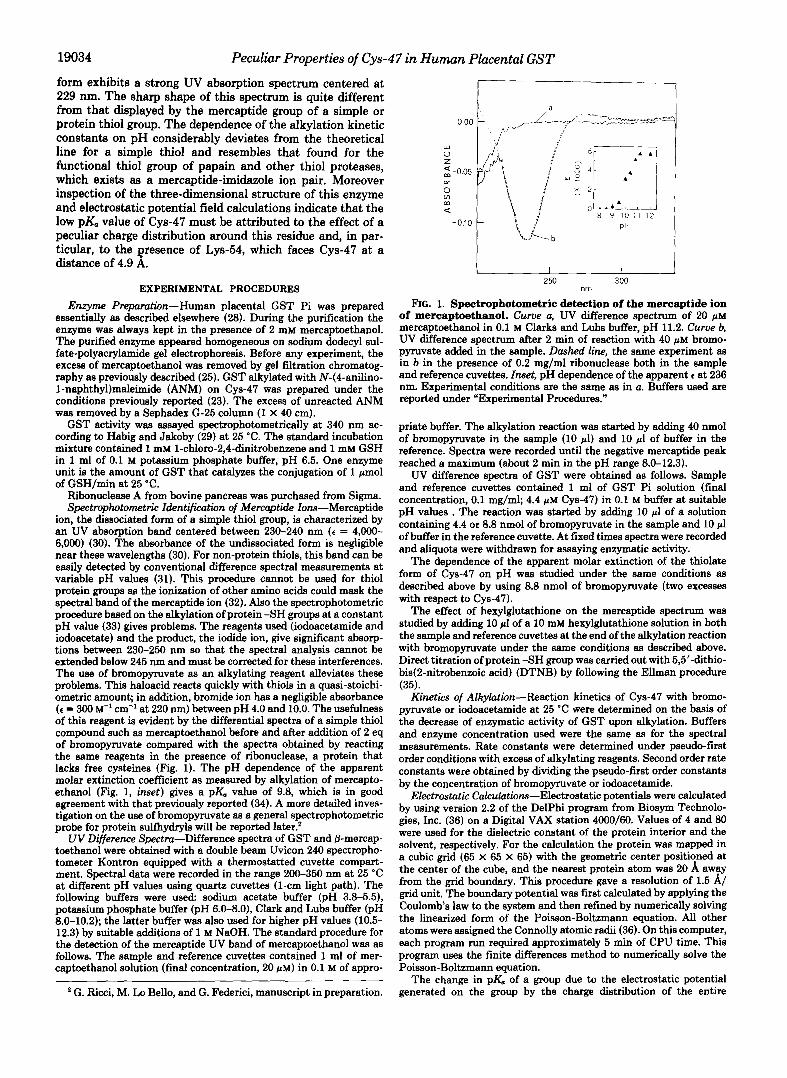
19034 Peculiar Properties of Cys-47 in Human Placental GST form exhibits a strong UV absorption spectrum centered at 229 nm. The sharp shape of this spectrum is quite different from that displayed by the mercaptide group of a simple or protein thiol group. The dependence of the alkylation kinetic constants on pH considerably deviates from the theoretical line for a simple thiol and resembles that found for the functional thiol group of papain and other thiol proteases, which exists as a mercaptide-imidazole ion pair. Moreover inspection of the three-dimensional structure of this enzyme and electrostatic potential field calculations indicate that the low pK. value of Cys-47 must be attributed to the effect of a peculiar charge distribution around this residue and, in par- ticular, to the resence of Lys-54, which faces Cys-47 at a distance of 4.9 x .
EXPERIMENTAL PROCEDURES
Enzyme Preparation-Human placental GST Pi was prepared essentially as described elsewhere (28). During the purification the enzyme was always kept in the presence of 2 mM mercaptoethanol. The purified enzyme appeared homogeneous on sodium dodecyl sul- fate-polyacrylamide gel electrophoresis. Before any experiment, the excess of mercaptoethanol was removed by gel filtration chromatog- raphy as previously described (25). GST alkylated with N-(a-anilino- 1-naphthy1)maleimide (ANM) on Cys-47 was prepared under the conditions previously reported (23). The excess of unreacted ANM was removed by a Sephadex G-25 column (1 X 40 cm).
cording to Habig and Jakoby (29) a t 25 "C. The standard incubation GST activity was assayed spectrophotometrically a t 340 nm ac-
mixture contained 1 mM l-chloro-2,4-dinitrobenzene and 1 mM GSH in 1 ml of 0.1 M potassium phosphate buffer, pH 6.5. One enzyme unit is the amount of GST that catalyzes the conjugation of 1 pmol of GSH/min at 25 "C.
Ribonuclease A from bovine pancreas was purchased from Sigma. Spectrophotometric Identifiation of Mercaptide Ions-Mercaptide
ion, the dissociated form of a simple thiol group, is characterized by an W absorption band centered between 230-240 nm (e = 4,000- 6,000) (30). The absorbance of the undissociated form is negligible near these wavelengths (30). For non-protein thiols, this band can be easily detected by conventional difference spectral measurements at variable pH values (31). This procedure cannot be used for thiol protein groups as the ionization of other amino acids could mask the spectral band of the mercaptide ion (32). Also the spectrophotometric procedure based on the alkylation of protein -SH groups at a constant pH value (33) gives problems. The reagents used (iodoacetamide and iodoacetate) and the product, the iodide ion, give significant absorp- tions between 230-250 nm so that the spectral analysis cannot be extended below 245 nm and must be corrected for these interferences. The use of bromopyruvate as an alkylating reagent alleviates these problems. This haloacid reacts quickly with thiols in a quasi-stoichi- ometric amount; in addition, bromide ion has a negligible absorbance ( C = 300 I"' cm" at 220 nm) between pH 4.0 and 10.0. The usefulness of this reagent is evident by the differential spectra of a simple thiol compound such as mercaptoethanol before and after addition of 2 eq of bromopyruvate compared with the spectra obtained by reacting the same reagents in the presence of ribonuclease, a protein that lacks free cysteines (Fig. 1). The pH dependence of the apparent molar extinction coefficient as measured by alkylation of mercapto- ethanol (Fig. 1, inset) gives a pK. value of 9.8, which is in good agreement with that previously reported (34). A more detailed inves- tigation on the use of bromopyruvate as a general spectrophotometric probe for protein sulfhydryls will be reported later.' UV Difference Spectra-Difference spectra of GST and 8-mercap-
toethanol were obtained with a double beam Uvicon 240 spectropho- tometer Kontron equipped with a thermostatted cuvette compart- ment. Spectral data were recorded in the range 200-350 nm at 25 "C at different pH values using quartz cuvettes (1-cm light path). The following buffers were used: sodium acetate buffer (pH 3.8-5.51, potassium phosphate buffer (pH 6.0-8.0), Clark and Lubs buffer (pH 8.0-10.2); the latter buffer was also used for higher pH values (10.5- 12.3) by suitable additions of 1 M NaOH. The standard procedure for the detection of the mercaptide UV band of mercaptoethanol was as follows. The sample and reference cuvettes contained 1 ml of mer- captoethanol solution (final concentration, 20 pM) in 0.1 M of appro-
' G. Ricci, M. Lo Bello, and G. Federici, manuscript in preparation.
250 nm
300
FrG. 1. Spectrophotometric detection of the mercaptide ion of mercaptoethanol. Curve a, uv difference spectnun of 20 phi mercaptoethanol in 0.1 M Clarks and Lubs buffer, pH 11.2. Curve b, UV difference spectrum after 2 min of reaction with 40 p~ bromo- pyruvate added in the sample. Dashed line, the same experiment as in b in the presence of 0.2 mg/ml ribonuclease both in the sample and reference cuvettes. Inset, pH dependence of the apparent c at 236 nm. Experimental conditions are the same as in a. Buffers used are reported under "Experimental Procedures."
priate buffer. The alkylation reaction was started by adding 40 nmol of bromopyruvate in the sample (10 pl) and 10 pl of buffer in the reference. Spectra were recorded until the negative mercaptide peak reached a maximum (about 2 min in the pH range 8.0-12.3).
UV difference spectra of GST were obtained as follows. Sample and reference cuvettes contained 1 ml of GST Pi solution (final concentration, 0.1 mg/mb 4.4 pM cys-47) in 0.1 M buffer at suitable pH values . The reaction was started by adding 10 pl of a solution containing 4.4 or 8.8 nmol of bromopyruvate in the sample and 10 pl of buffer in the reference cuvette. At fixed times spectra were recorded and aliquots were withdrawn for assaying enzymatic activity.
The dependence of the apparent molar extinction of the thiolate form of Cys-47 on pH was studied under the same conditions as described above by using 8.8 nmol of bromopyruvate (two excesses with respect to Cys-47).
The effect of hexylglutathione on the mercaptide spectrum was studied by adding 10 pl of a 10 mM hexylglutathione solution in both the sample and reference cuvettes at the end of the alkylation reaction with bromopyruvate under the same conditions as described above. Direct titration of protein -SH group was carried out with 5,5'-dithio- bis(2-nitrobenzoic acid) (DTNB) by following the Ellman procedure (35).
Kinetics of Alkylation-Reaction kinetics of Cys-47 with bromo- pyruvate or iodoacetamide at 25 "C were determined on the basis of the decrease of enzymatic activity of GST upon alkylation. Buffers and enzyme concentration used were the same as for the spectral measurements. Rate constants were determined under pseudo-first order conditions with excess of alkylating reagents. Second order rate constants were obtained by dividing the pseudo-first order constants by the concentration of bromopyruvate or iodoacetamide.
Electrostatie Calculations-Electrostatic potentials were calculated by using version 2.2 of the Delphi program from Biosym Technolo- gies, Inc. (36) on a Digital VAX station 4000/60. Values of 4 and 80 were used for the dielectric constant of the protein interior and the solvent, respectively. For the calculation the protein was mapped in a cubic grid (65 X 65 X 65) with the geometric center positioned at the center of the cube, and the nearest protein atom was 20 A away from the grid boundary. This procedure gave a resolution of 1.5 A/ grid unit. The boundary potential was first calculated by applying the Coulomb's law to the system and then refined by numerically solving the linearized form of the Poisson-Boltzmann equation. All other atoms were assigned the Connolly atomic radii (36). On this computer, each program run required approximately 5 min of CPU time. This program uses the finite differences method to numerically solve the Poisson-Boltzmann equation.
The change in pK, of a group due to the electrostatic potential generated on the group by the charge distribution of the entire

Peculiar Properties of Cys-47 in Human Placental GST 19035 molecule has been calculated using the equation
ApK, = Aq A+ f2.303 (Eq. 1)
where Aq is the change of the charge of the ionizable group and A* represents the change, in kT/e, of the potential at that group. In practice, the pK., shift between a free cysteine and Cys-47 was determined assuming the intrinsic pKa of this residue is identical to the p& of a free cysteine and calculating the contribution of the molecule to the electrostatic energy of the sulfur atom of Cys-47. The absolute pK., of Cys-47 was then calculated using the equation
= ~Kint + 4% 0%. 2)
where pZ& is the actual pK. of the residue in the protein, pKht is the intrinsic p& of the residue, and ApK. is given by Eq. 1. This approximation is rigorously valid only when the group investigated is the only titratable group in the pH range analyzed, otherwise the pK, of the group is affected by the ionization state of all the ionizable groups in the protein (37). The calculations were performed at two pH value conditions (4.0 and 7.0) and two different ionic strengths. The ionization states of titratable residues were calculated based on the following p& values: Asp = 4.5, Glu = 4.6, Lys = 10.4, Arg = 12.0, His = 6.2 (38).
RESULTS AND DISCUSSION
Spectral Properties of Humon Placental GST-When GST is reacted with a stoichiometric amount of bromopyruvate (1:l bromopyruvate/subunit) at pH 7.0, a fast inactivation occurs to less than 10% of the original activity (Fig. 2, inset). Direct titration of the thiol protein groups with DTNB indi- cates that bromopyruvate alkylated one of the two fast titrat- able -SH groups/subunit at this pH value (25). During the alkylation process differential UV spectrometry reveals the disappearance of an atypical spectral band with a maximum centered at 229 nm (Fig. 2) and an apparent c of 7500 M-' cm-'. Moreover a slight perturbation between 250 and 300 nm, belonging to aromatic residues and indicative of confor- mational changes (19, 25), is also observed. The fall of the absorbance at 229 nm parallels the inactivation pattern (Fig. 2, inset). The use of 2 eq of bromopyruvate does not change appreciably the extinction coefficient, and it does not modify the extent of inactivation although a second sulfhydryl is alkylated as checked by titration with DTNB. Therefore the spectral band must be related to the modification of the most reactive sulfhydryl group of this transferase, and Cys-47 is
0 00
-0.04 t I I 1 2
250 nm 300
FIG. 2. UV difference spectra of GST after reaction with bromopyruvate. 2.2 pM GST (4.4 p~ Cys-47) in 0.1 M potassium phosphate buffer, pH 7.0, was placed in the sample and reference cuvettes, and 4.4 pM (final concentration) bromopyruvate was added in the sample compartment. Curue a, time zero; curves b, c, d, and e, after 10, 20, 30, and 60 min of reaction, respectively. Inset, time course of GST inactivation (A) and absorbance change at 229 nm (m) under the conditions described above.
the most probable candidate. It is well known that any alkyl- ation of this residue yields rapid inactivation (21-24), and often it is the sole sulfhydryl group that can be modified as observed in the reaction of GST Pi with N-ethylmaleimide (22) or ANM (23), a fluorescent derivative of N-ethylmaleim- ide. Under our conditions the ANM-treated enzyme gives no spectral perturbations at 229 nm when reacted with 2 eq of brornopyruvate. In addition the enzyme inactivated with 1 eq of bromopyruvate does not covalently bind the fluorescent ANM (data not shown).
The above results suggest that Cys-47, the target of the alkylation with bromopyruvate, should be present in the thiolate form at pH 7.0, and therefore it should have an unusually low pK. value. To verify this possibility the pH dependence of the apparent molar extinction coefficient has been explored between pH 3.8 and 8.0. We could not perform any experiment below pH 3.8 because an irreversible, spon- taneous inactivation occurs below this pH value. The experi- mental titration curve is reported in Fig. 3 together with its best fit that has been obtained for a simple thiol with a pKa = 4.2. The possible acidic nature of Cys-47 has already been hypothesized as an explanation of its high reactivity even at neutral pH values (39), but no titration data have been available up to now. This unusually low pK. value of the Cys- 47 sulfhydryl group resembles that calculated for protein cysteines with a deprotonation assisted by an imidazole group as it occurs in papain (40), thiosubtilisin (33), 3-phosphoglyc- eraldehyde dehydrogenase (41), and others (42). The idea that the deprotonation of Cys-47 could be similarly assisted by some protein group is also suggested by the following. The maximum of the absorption spectrum is centered at a wave- length slightly lower than that found for free thiols (30). The shape of this peak is very sharp when compared with that of a free thiol (see Fig. 1). Eventually, the apparent molar absorbance of 7,500 is higher than that of a free thiolate ion. Although no spectral data are available below 240 nm for protein mercaptides, some useful suggestions can be gained from previous studies on enzymes provided of a mercaptide ion pair system (33). It has been reported that the spectrum of the mercaptide ion of thiosubtilisin linked with a positively charged group (ion pair with His) exhibits a lower absorption
8000 I I I I I I I I
6000 1 P IC
L 4000 U 0
2000 I A
A O
0 A
0 0
A
Q
A 0 L A , I I I I
1 2 3 4 5 6 7 8 PH
FIG. 3. pH dependence of the apparent molar extinction of the spectral band at 229 nm. 2.2 p~ GST was placed in the sample and reference cuvettes in the presence of 0.1 M buffer at suitable pH values. The reaction was started by addition of 8.8 PM bromopymvate in the sample. 0, apparent e values calculated at the end of the reaction when both the absorbance at 229 nm and the activity values (always less of 10%) became constant. A, theoretical e values calcu- lated for a mercaptide with a pK. = 4.2.

19036 Peculiar Properties of Cys-47 in Human Placental GST than that of GS- at 250 nm. In noting that this absorption is much lower than that of mercaptoacetate (33), it appears that the presence of a positive charge near the thiol group causes a blue shift of the mercaptide spectrum in proteins. This is confirmed by the observation that the protonation of the amino group of cysteine causes a shift of the mercaptide absorption maximum from 236-238 nm to 230-232 nm (31). Therefore the GST spectrum after bromopyruvate reaction, which shows a maximum at 229 nm, is consistent with the UV spectral band belonging to a mercaptide with a positive charge located near the sulfur atom. Furthermore the shape of a mercaptide UV signal is related to electronic transitions combined with a multiplicity of transitions between rotational and vibrational levels in the ground state and the excited state. It is reasonable that a protein mercaptide ion stabilized and made rigid by some protein ligand may produce a sharper spectrum. This could be the case with GST provided that the side chains of other amino acids form additive bonds with cys-47.
We were interested in determining the influence of hexyl- glutathione binding on the thiolate spectral band, given that binding of GSH and GSH-analogs has been shown to cause conformational changes (19). Since S-hexylglutathione strongly protects Cys-47 from the bromopyruvate attack we added this GSH analog at the end of inactivation in both the test and the reference samples. Under these conditions any conspicuous change of the thiolate band, as tested by differ- ential spectroscopy, can be due to the effect of substrate binding in the reference sample, where the thiolate is still present. After reaction of GST with bromopyruvate either at pH 4.0 and 7.0 (two critical pH values where the mercaptide ion is present at about 50 and loo%, respectively), the differ- ential spectra were recorded. A saturating amount of hexyl- glutathione (0.1 mM) was then added to the test and reference samples. In both experimental sets (pH 4.0 and 7.0) the negative absorption peak at 229 nm increases to about 20- 25% without any change in the shape or wavelength of the maximum. Whatever the reason for this small increase in absorbance, the result clearly indicates that the mercaptide of Cys-47 is still present in the hexylglutathione-GST com- plex without appreciable change of the pKa value.
It must be stressed that no differences in the spectral and acid base properties have been found when the alkylation of GST with bromopyruvate was performed at pH 4.0 and 7.0 in the presence of a lower ionic strength (0.02 M potassium phosphate or sodium acetate buffers).
Kinetic Data of GST Alkylation-The apparent kinetic con- stants for alkylation of a simple thiol are directly related to the dissociation of the -SH group since it has been demon- strated that only the mercaptide ion reacts at an appreciable rate (43,44). Therefore for a simple thiol a linear dependence of the log of the apparent second order kinetic constants on pH must be observed according to: Kapp = K ~ ( R s - ) . (1/(1 + (H+)/Ka(RSH))). The reactivity of a protein mercaptide stabi- lized as an ion-pair is generally lower than that of the free mercaptide but still measurable (41). In this case Kapp is the sum of the contributions of both the reactive species according
(H+)/Ko(,P))) (1/(1 + K.(mH)/(H+))). In this case the depend- ence of the log of the apparent alkylation constant on pH follows a double sigmoid curve, which allows the calculation of two distinct PI(, values: one for the free mercaptide form and the second due to the deprotonation of the mercaptide ion pair system. Such peculiar kinetic behavior has been found for the alkylation of papain (40), thiosubtilisin (45), and D- glyceraldehyde-3-phosphate dehydrogenase (41). The depend-
to: Kapp = K~(Fzs- )*(~/ (~ + (H+)/Ka(RSH))) + K W I P ) ( ~ / ( ~ +
ence of the bromopyruvate inactivation second order constant on pH for GST was evaluated using the conditions reported under "Experimental Procedures.'' The high reactivity of bromopyruvate led to some problems in calculating Klim, and the reported value of 13,140 s" M" was estimated on the basis of the average of three experiments at each pH value in the plateau region over pH 10.0. As shown in Fig. 4, at acidic pH values the dependence of the alkylation rate constants on pH consistently deviates from the theoretical line obtained from the alkylation of a simple thiol with a Kb = 13,140 s" M" and pKa = 9.3. The experimental points are best fitted with a theoretical curve describing a reaction involving a mercaptide ion pair system with Khm = 20 and pKa = 3.9 in equilibrium with a free mercaptide with Klh = 13,140 and pK. = 9.2. A similar behavior has been observed by using the less reactive iodoacetamide as the alkylating reagent (data not shown). In this case the experimental points are best fitted with a theoretical line due to the alkylation of a mercaptide ion pair with pKa = 4.0 and Klim = 0.02 s-l M" in equilibrium with a free mercaptide with Kb = 78 s-l M" and pK. = 9.8. The enhanced reactivity of Cys-47 always observed at pH 4.0 that yields a deviation from the theoretical double sigmoid curve has not been investigated. At this pH value probably other factors, for example, protein conformation changes may favor the alkylation reaction.
Scheme I represents a possible ionization equilibria scheme involving Cys-47 (P-SH) and a protein basic residue (B), that agree with the above findings.
P-SH + BH+'-, P-S- BH+ PKI = 3.9
(ion pair)
+ H+, pKz = 9.4-9.8 P-S- + B + 2H+ SCHEME I
Structural and Electrostatic Analysis-An assisted depro- tonation of a thiol protein group leading to a lowering of its apparent pK. is possible in several ways: (a) by hydrogen bond formation; ( b ) by simple electrostatic effect due to a number of positive charges near the sulfur atom; ( c ) by ion pair formation; ( d ) by general acid base reaction. An exami- nation of the three-dimensional structure of GST complexed with hexylglutathione (5) may clarify the cause of the peculiar spectral, acid base, and kinetic properties displayed by Cys- 47. This amino acid is pointing into a hydrophobic cleft of
- 2
3 4 5 6 7 8 9 1 0 1 1 PH
FIG. 4. pH dependence of rate constants of the alkylation of GST by bromopyruvate. Alkylation conditions at variable pH values were as reported under "Experimental Procedures." 0, theo- retical points calculated for a simple thiol with pZG = 9.3 and K b = 13,140 M" s-'; 0, theoretical points calculated for a mercaptide stabilized as an ion pair with & ~ p ) = 3.9 and Kb(~p) = 78 "' s-', Ko(s-) = 9.3 and K ~ c s - ) = 13,140 M" 8-'; A, experimental points.

Peculiar Properties of Cys-47 in Human Placental GST 19037
domain I of the subunit surrounded by a number of hydro- phobic residues including Val-6, Leu-43, Trp-38, Leu-52, Tyr- 63, and the aliphatic portions of the Lys-54 side chain. The sulfur atom of Cys-47 is in hydrogen-bonding distance with the main chain carbonyls of Leu-43 and Gln-51 (see Table I). The latter residue forms a number of important contacta with the glutathione inhibitor. Although not directly interacting with Cye-47, Trp-38 also forms important interactions with hexylglutathione. However, these residues cannot stabilize the conversion of Cys-47 into a thiolate ion since the carbonyl oxygen is a bad proton acceptor. Therefore the participation of a hydrogen bond in the deprotonation of Cys-47 is unlikely to occur. However the influence of "incipient" hydrogen bonds, for instance with the hydroxyl group of Tyr-63 (Cys- S--HO-Tyr distance about 4.2 A) cannot be absolutely ruled out in the stabilization of the thiolate form of Cys-47.
To evaluate the hypothesis that the lowering of the pK, value of the Cys-47 residue could be determined by electro- static effecta, the electrostatic potential value on the sulfur atom of Cys-47 has been calculated under different conditions by using the program DelPhi, which uses a macroscopic approach to numerically solve the Poisson-Boltzmann equa- tion.
It must be pointed out that the approach used is completely reliable only as far as pK, shift calculations are concerned, while determination of absolute p& values requires the si- multaneous calculation of the ionization state of each ioniz- able group of the protein at every pH value. We have assumed that the intrinsic pK, of this group in the protein is identical to that of the isolated amino acid in solution. With this assumption the pK, values are calculated by using Eq. 3.
pZ& = pKk + (A@)/2.303 (Eq. 3)
where Aq is the change of the charge of the residue, and + is the potential generated by the entire molecule upon the ti- trating group. Estimates of the p& differences between Cys- 47 in the protein and as a free amino acid, calculated at pH values 7.0 and 4.0, are reported in Table 11. Moreover, since the c-amino group of the Lys-54 residue is only 4.9 A away from the sulfur atom of Cys-47, the calculations have been carried out also by varying the protonation state of Lys-54. The resulta indicate that Lys-54 plays a major role in deter- miniig the pK, shift of Cys-47 (see also Fig. 5). It is important to stress that neutralization of other residues close to Cys-47 such as Lye-127 and Lys-44 has negligible effects (Table II). The resulta obtained from the calculations performed at dif- ferent pH values as well as at different ionic strength values do not greatly differ from each other, indicating that the contribution from other titritable residues is negligible and confirming that Lys-54 is the one mainly responsible for the unusually low pK, value of Cys-47. Finally another interesting observation given by cry&dlographic data is that the temper- ature factor of the r-nitrogen atom of Lys-54 is about 50% lower than the average value found for all other lysines of each subunit. This is an indication of the low mobility of this
TABLE I Residues within hydrogen-bonding distance of Cys-47
Atom Residue Atom Residue Distance A
N cys-47 0 Leu-43 2.99 N cys-47 0 Lys-44 3.40 SG CYS-47 0 Leu-43 3.18 SG CYS-47 0 Gln-51 3.34 0 CYS-47 N TyI-49 3.32 0 CYS-47 N G$-50 3.37
TABLE I1 p& of Cys-47 residue of GST at two different pH vdues and upon
neutralization of the surrounding lysine residues PELa
Distancen pH 4 PH 7
0.15 Mb 0.02 Mb 0.15 Id 0.02 d A
Cysteine 9.5 9.5 9.5 9.5 Wild type 3.5 2.6 2.8 3.4 qLY.&(=w 4.9 9.3 8.5 8.6 9.4 4Lym44 = w 7.9 4.7 3.9 4.0 4.9 ~ L ~ I Z ~ = V 15.7 3.6 2.7 2.9 3.8
"The distances given are between the sulfur atom of Cys-47 and the e-nitrogen atom of the surrounding lysine residues. In the case of Lye-127, the distance is between Lye-127 of one subunit and Cys-47 of the other subunit, since both these residues of the same subunit are too far to have any significant electrostatic effect.
The simulations have been performed at the highest and lowest ionic strength values used in the spectrophotometric and kinetic experiments.
E q is the net charge on the e-nitrogen of the lysine residues.
L" -4"'
h
\
FIG. 5. mmmn moas or m e region of GST surrounding Cys- 47. The side chains of Cys-47 and Lys-54 are represented as dotted spheres with the van der Waals atomic radius, colored by atom type (white, carbon; blue, nitrogen; red, oxygen; yellow, sulfur).
amino group mainly due to a hydrogen bond with the side chain of Tyr-63 and, possibly, by a further electrostatic inter- action with the thiolate group of Cys-47.
CONCLUSIONS
It is well documented that GST exerts a deprotonating action on the bound GSH at the active site by lowering its pK, value from 9.0 to about 6.0-6.8 (18, 19). It is surprising to fiid a second deprotonating center near the active site that acts on the sulfur atom of Cys-47. All the above experimental data strongly support the idea that Cys-47 undergoes an assisted deprotonation. The strong absorption band at 229, which disappears upon selective alkylation of Cys-47 (Fig. 2), has been reasonably assigned to the mercaptide ion of this amino acid. The dependence of the apparent molar extinction on pH indicates a very low pK, value of about 4.0 (Fig. 3). The improved differential spectrophotometric procedure based on the use of the high reactive bromopyruvate allows us to visualize the entire mercaptide band up to 220 nm (Fig. 2). The wavelength of the maximum and the sharp shape of this peak are indicative of the presence of both a nearby

19038 Peculiar Properties of Cys-47 in Human Placental GST
positive charge and one or more bonds that restrict the number of vibrational and rotational levels for electronic transitions of the mercaptide electrons. The dependence of the alkylation rate constants on pH adds further insight. The consistent deviation of the experimental points at neutral and acidic pH values from that theoretically calculated for an ideal thiol would explain the unusually high reactivity of Cys- 47 toward alkylating reagents under these pH conditions. The alkylation data can be explained by a model in which there is an equilibrium between a free mercaptide with pK, = 9.3-9.8 and a mercaptide ion pair with a pKa = 3.9 (see Fig. 4 and Scheme I). The three-dimensional structure of the hexylglu- tathione-GST complex has provided an insight into under- standing the peculiar properties of Cys-47 in the uncomplexed enzyme, noting that the binding of hexylglutathione to the active site of GST does not change the spectral and acid base properties of Cys-47. The crystallographic data show that the side chains of neighboring residues Lys-54 and Tyr-63 are close enough (within a 5-A sphere of Cys-47) to play a possible contributing role (Fig. 5). The protonated amino group of lysine may promote the release of the proton from Cys-47. The hydroxyl group of Tyr-63, although too far for a strong hydrogen bond (4.2 A from the sulfur atom), may still stabilize the anion by coordination with the sulfur atom by an incipient hydrogen bond. In this case the synergic action of these two nearby amino acid residues determines the overall atypical spectroscopic and kinetic behavior of Cys-47. Alternatively, Lys-54 may promote the formation and stabilization of a mercaptide ion pair. The deprotonating action of Lys-54 is supported by the electrostatic calculations of the pKa value of Cys-47. The theoretical pK, value of 3.5 agrees well with that experimentally obtained by spectral and kinetic experiments. Moreover the assignment of a neutral charge to Lys-54 shifts the theoretical pKa value from 3.5 to 9.5, indicating that the unusual acidic properties of Cys-47 are mainly due to Lys-54. As expected, neutralization of Lys-44 and Lys-127 (which are located 7.9 and 15.7 A from Cys-47, respectively) produced only small increases in pKa value.
Based on our results, we can argue that (a) the well known high reactivity of Cys-47 at physiological pH values is mainly due to an assisted deprotonation of this residue caused by the protonated Lys-54 and, possibly, by an incipient hydrogen bond with Tyr-63 and ( b ) the well known protective effect of substrate analogs against alkylating compounds acting on Cys-47 cannot be explained in terms of an increase of the pKa value of this residue but is probably due to a lowered acces- sibility of the thiolate ion.
What is the importance of the mercaptide ion pair system involving Cys-47 in GST? We may suggest that Cys-47 acts as a modulation site of the enzyme activity in vivo. The mercaptide ion pair seems to be involved in the maintenance of the structural characteristics of the G-site. The observa- tions that Cys-47, Lys-54, and Tyr-63 are highly conserved amino acids in the aligned sequences of all known Pi isoen- zymes (rat and mouse liver, human placenta, and pig lung) and that replacement of Cys-47 by site-directed mutagenesis lowers the affinity for GSH supports this idea. On the other hand the dramatic inactivation always observed during the alkylation of Cys-47 could be mainly due to the disturbance of Trp-38 and Gln-51, both found in the hydrophobic pocket of surrounding Cys-47, but also forming important hydrogen- bonding interactions with the glutathione in the G-site.
Site-directed mutagenesis experiments are underway to de-
fine more precisely the relative importance of Lys-54 and Tyr- 63 toward the activation of Cys-47 and in the maintenance of a proper active site structure of GST.
The possibility that the ion pair Cys-Lys may be present in other enzymes and adopts a structural stabilization or cata- lytic activation role is under investigation.
Acknowledgments-We thank Prof. M. Paci for helpful discussions of the manuscript and the Department of Obstetrics and Ginecology- Hospital "Figlie di S. Camillo" for providing biological material.
REFERENCES 1. Jakoby, W. B., and Habig, W. H. (1980) in Enzymatic Basis of Detoxification
(Jakoby, W. B., ed) Vol. 2, pp. 63-94, Academic Press, New York 2. Mannervik, B., Alin, P., Guthenberg, C., Jensson, H., Tahir, M. K., War-
holm, M., and Jornvall, H. (1985) Proc. Natl. A c d . Sci. U. S.A. 82, 7202-72M
3. Meyer, D. J., Coles, B., Pemble, S. E., Gilmore, K. S., Fraser, G. M., and
4. Reinemer P., Dirr, H. W Ladenstein, R. Shaffer, J., Gallay, O., and
5. Reinemer, P., Dirr, H. W., Ladenstein, R., Huber, R., Lo Bello, M., Federici,
6. Stenberg, G., Board, P. G., and Mannemk, B. (1991) FEBS Lett. 293,
Ketterer, B. (1991) Biochem. J. 274,409-414
Huber, R. (1991) EMBO). 10,1997-2006
G. and Parker, M. W. (1992) J. MOL BWL 227,214-226 lF( !LlKF(
7. Kong, K. H. Nishida M., Inoue, H., and Takahashi, K. (1992) Biochem.
8. Liu, Sf, ghang, P., Ji, X., Johnson, W. W., Gilliiand, G. L., and Armstrong,
9. Kong, K. H., Takasu, K., Inoue, H., and Takahashi, K. (1992) Biochem.
"I "_ Bu, h s. Res. Comiun. 182,1122-1129
R. N. (1992) J. Bwl. Chem. 267,4296-4299
10. Kolm, R. H., Sroga, G. E., and Mannervik, B. (1992) Biochem. J. 286, 11. Stenberg, G.. Board, P. G., Carlberg, I., and Mannervik, B. (1991) Biochem.
12. Zhang, P., Graminski, G. F., and Armstrong, R. N. (1991) J. BWL Chem.
13. Wang, R. W., Newton, D. J., Pickett, C. B., and Lu, A. Y. H. (1991) Arch.
14. Wang, R. W., $fewton, D. J., Pickett, C. B., and Lu, A. Y. H. (1992) Arch.
15. Nishihira, J., Ishickhi, T., Sakai, M., Nishi, S., and Kumazaki, T. (1992)
16. Widersten, M., Kolm, R. H., Bjornestedt, R., and Mannervik, B. (1992)
17. Manoharan, T. H., Gulick, A. M., Reinemer, P., Dirr, H. W., Huher, R.,
18. Chen, W. J., Graminski, G. F., and Armstrong, R. N. (1988) Biochemistry
19. Graminski, G. F., Kubo, Y., and Armstrong, R. N. (1989) Biochemistry 28,
20. Hatayama, I., Satoh, K., and Sato, K. (1990) Nucleic Acids Res. 18,4606 21. Ricci. G.. Del Boccio, G.. Pennelli. A., Aceto. A.. Whitehead, E. P., and
Biophys. Res. Commun. 184,194-197
537-540
J. 274,549-555 266,19475-19479
Biochem. BW hys. 286,574-578
Biochem. Bioph s 297,86-91
Biochem. Bwphys. Res. Commun. 186,1069-1077
Biuchem. J. 286,377-381 and Fahl, W. E. (1992) J. Mol. Bwl. 226,319-322
27,647-654
3562-3568
22. Tamai, K., Satoh, K., Tsuchida, S., Hatayama, I., Maki, T., and Sato, K.
23. Lo Bello. M.. Petruzzelli. R.. De Stefano. E., Tenedini, C., Barra, D., and
Federici, G. (1989) 3. BWl. Chem. 264,5462-5467
(1990) Biochem. Biophys. Res. Commun. 167.331-338
Federici, G . (1990) FEBS iett. 263,389-391 . . . .
24. Caccuri. A. M.. Petruzzelli. R.. Polizio. F.. Federici. G.. and Desideri. A. (1992) Arch. k c h e m . Bwphys. 297,'119-122
25. Ricci, G., Del Boccio, G., Pennelli, A., Lo Bello, M., Petruzzelli, R., Caccuri, A. M., Barra, D., and Federici, G. (1991) J. BWL Chem. 266, 21409-
. .
21415 26. Tamai K., Shen, H. Tsuchida, S., Hatayama, I., Satoh, K., Yasui, A.,
Oikiwa, A., and Sa'to, K. (1991) Biochem. Biophys. Res. Commun. 179. 7 0 1 ~ 7 0 7
27. Kong, K. H., Inoue, H., and Takahashi, K. (1991) BWchem. Biophys. Res.
28. Parker, M. W., Lo Bello, M., and Federici, G. (1990) J. MOL BWl. 213,
I"" ."I
Commun. 181,748-755
29. Habig, W. H., and Jakoby, W., B. (1981) Methods Enzyn~l . 77,39.3-405 30. Jocelyn, P. C. (1972) Bmhemlstry of SHgroup, pp. 49-51, Academic Press,
31. Benesch, R. E., and Benesch, R. (1955) J. Am. Chem. Soc. 77,5877-5881 32. Donovan, J. W. (1964) Biochemistry 3,67-74 33. Polgar, L. (1974) FEBS Lett. 38,187-190 34. Irvj-ig, J., Nelander, L., and Wadso, I. (1964) Acta Chem. S c a d . 18, 769-
221-222
New York
35. Ellman G. L. (1959) Arch. Biochem. BW hys 82,70-77 36. Delphi ;er. 2.2 (1992) BIOSYM Technof&i& Inc.,San Die o CA 37. Sharp, K. A., and Honlg. B. (1990) Annu. Reu. Bwphys. lfwhhys. Chem.
'1'14
19,301-332 38. Tanford, C. (1962) Adu. Protein Chem. 39. Desideri, A,, Caccuri, A. M., Polizio, F.,
40. Polgar, L. (1973) Eur. J. Biochem. 33, J. BWZ. Chem. 266,2063-2066
41. Pol ar, L. (1975) Eur. J. Biochem. 61, 42. Sahfman, L., and Williams, C. H., Jr.
Bastoni, R., 17,69-165
and Federici, G.
63-71 104-109
(1989) J. BWL Chem. 264, 8038
43. Herman, P. (1961) Chem. Ber. 94,442-448 44. Bednar, R. A. (1990) Biochemistry 29,3684-3690 45. Halasz, P., and Polgar, L. (1977) Eur. J. Biochem. 79,491-494
(1991)
8033-