the occurrence of heart-block in coronary artery thrombosis
TRANSCRIPT

THE OCCURRENCE OF HEART-BLOCK IN CORONARY ARTERY THROMBOSIS”
DAVID BALL, M.D.
NEW YORK, N. Y.
T HE occurrence of heart-block during the course of an acute coronary
artery thrombosis is rare. The case to be reported, which has been studied in great detail with frequent serial electrocardiograms, revealed an early complete heart-block, which in a retrograde fashion went through the stages of partial heart-block, gradually returning to normal sinus rhythm, with complete recovery.
Acute coronary artery thrombosis usually occurs in individuals with a normal cardiac rhythm and in the majority of cases the rhythm re- mains regular. However, various arrhythmias can occur with the on- set, and during the early stages of an acute coronary closure. The fre- quency with which disturbances in rhythm may be detected will depend largely upon how often the patient is examined, since many of the ir- regularities are transient and often cause few if any symptoms.
Premature contractions, both ventricular and auricular, are observed during the early stages of an attack and are of little or no significance. Paroxysmal auricular fibrillation occurs more frequently. Levine1 ob- servecl it in 34 instances of his series of 145 cases. He did not find that this arrhythmia materially altered the prognosis of the case. Occasional- ly permanent auricular fibrillation remains. Auricular flutter is rarely seen, and in one case personally observed, the patient recovered. A more uncommon but important disturbance in rhythm is the development of paroxysmal ventricular tachycardia. This condition has been well de- scribed by Herrmann,’ Robinson,3 and Levine,4 and Levine, Stevens, and Fu1ton.j The prompt recognition of this disturbance and institution of proper therapy may prove life saving.
Complete A-V dissociation during an attack of acute coronary artery thrombosis can occur and has been observed. Levine1 observed it only twice in his series of 145 cases, an incidence of 1.37 per cent. Both cases proved fatal. One of the earliest reports of transient complete he,art- block in coronary thrombosis with recovery was that of .Frothinghams in 1927.
The presence of complete heart-block during the early stage of a coronary closure has not been adequately explained. A careful study of the case to be reported together with an analysis of the cases of heart- block in coronary thrombosis described in the literature furnishes a key to the explanation for its occurrence.
*From the Medical Service of Dr. A. A. Epstein, Beth Israel Hospital, New York.
327
328 TIIE AMERICAS HEART JOURNAL
While heart-block in coronary closure is uncommon, it is even more rare to obser\-e all of its stages in one individual in whom recovery has taken place. ‘l’he report of such a condition with frequent serial electro- cardiograms is presented.
CASE REPORT
A. S., B. I. H. No. 44517, male, aged forty-six years, walked into the out-patient
department of the Beth Israel Hospital, and presented the following history: Two
days previously, while walking home from work, he suddenly experienced a burning sensation in the region of the lower sternum and epigastrium. He continued to
walk and when he reached his home he vomited profusely and broke out into a cold
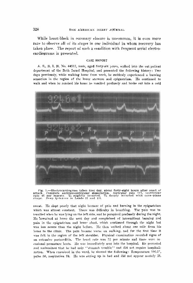
Fig. t.-Electrocardiogram taken first day, about forty-eight hours after onset of attack. Complete auriculoventricular dissociation. Auricular rate 117, ventricular rate 45 ner minute. Tn slightly inverted. T3 deeply inverted with cove-plane *haye. Deep Q-waves in Leads II and III.
sxveat. He slept poorly that night because of pain and burning in the epigastrium
which was almost constant. There was diffic,ulty in breathing. The pain was in-
tensified when he was lying on the left side, and he perspired profusely during the night.
He ‘remained at home the next day and complained of intermittent burning and
pain in the cpigaatrium and lolver chest, which continued through the night but
was less severe than the night before. He then walked about one milt from his
home to the clinic. The pain brcamc worse on walking, and for the first time it
was felt in the region of the left shoulder. Physical examination revealed signs of
an extensive pericarditis. The Ileart rate was 72 per minute and there were oc-
casional premature beats. He was immediately sent into the hospital. He protested
and maintained that he had only “st.omach trouble” and did not require hospitali-
zation. When cxaminrd in the ward, he showed the following: Temperature 100.?“, pulse 50, respiration 24. He was sitting up in bed and did not appear acutely ill.
BALL : HEART-BLOC% IS COROKARY ARTERY THROMBOSIS 329
He was only slightly dyspneic and perspired moderately. There was slight cyanosis
of the lips, and he complained only of very slight pain behind the lower sternum.
Pressure over the styloid process (Libman’s test) indicat.ed an individual who was
markedly hyposensitive to pain stimuli. The apex beat was not visible nor palpable,
but the heart was slightly enlarged to the left on percussion. The rhythm was regular
and the rate 50 per minute. A typical loud, leathery to-and-fro pericardial fric-
tion rub was heard over almost the entire precordial area. The first heart sound
was impure and at times was heard with a varying degree of intensity due to the
varying time relationship between auricular and ventricular contraction as occurs
in complete heart-block. A third sound, distinctly grating in quality, was constantl?
heard in about the middle of each long diastolic pause. It was at first thought that
it was either an auricular or a perieardial sound. The final impression was that
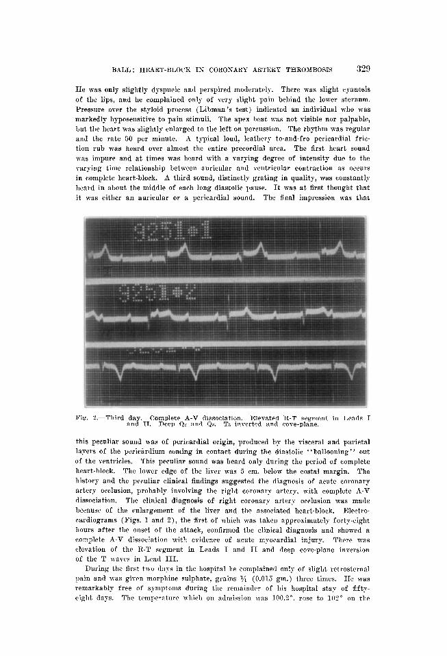
Fig. ?.-Third :;dy.IIComplete A-V dissociation. Elevated R-T segment in Leads I . Deep Q? and Qa. T3 inverted and cove-plane.
this peculiar sound was of pericardial origin, produced bv the visceral and parietal
layers of the pcrieirdium coming in contact during the diastolic “ballooning” out
of the ventricles. This peculiar sound was heard only during the period of complete
heart-block. The lower edge of the liver was 5 cm. below the costal margin. The
history and the peculiar clinical findings suggested the diagnosis of acute coronary
artery occlusion, probably involving the right coronary artery, with complete A-V
dissociation. The clinical diagnosis of right coronary artery occlusion was made
because of the enlargement of the liver and the associated heart-block. Electro-
cardiograms (Figs. 1 and O), the first of which was t,aken approximately forty-eight
hours after the onset of the attack, confirmed the clinical diagnosis and showed a
complete A-V dissociation with evidence of acute mpocardial injury. There was
(llevation of the R-T segment in Leads I and II and deep cove-plane inversion
of the T xaves in Lead III.
During the first two da?s in the hospital he complained only of slight retrosternal
pain and was giI--cn morphine sulphate, grains I/it (0.015 gm.) three times. He was remarkably free of symptoms during the remainder of his hospital stap of fiftp-
eight days. The temperature which on admission TWS 113n.2~, rose to 10’1” on the

330 THE AafE~1~2.4~ HEAR.T JOURNAL
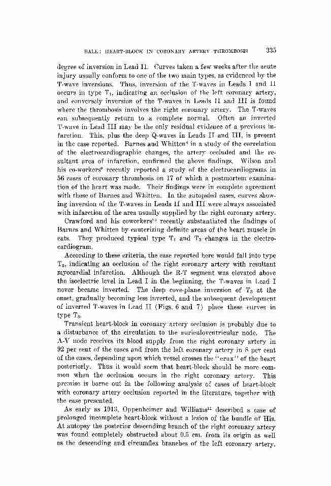
Fig. 3.-Seventh day. Two-to-one block. R-T segment Lead I almost isoelectric. Ta less inverted.
Fig. 4.-Eighth day. Two-to-one block in Lead I. Varying two-to-one and one-to one conduction in Lead II. Two-to-one block in Lend III.

BALL : IWART-BLOCK IN CORONARY ARTERY THROMBOSIS 331
second day and gradually declined, becoming normal on the sixth day. The white
blood count on admission was 16,050 with 82 per cent polynuclear leucocytes. The
sedimentation rate leas 42 per cent (normal 3 to 10 per cent). The leucocytosis per-
sisted for three weeks and the sedimentation rate became normal (5 per cent) on the twenty-seventh day. The blood pressure varied between 92 and 136 mm. of mercury
systolic, and 68 to 90 mm. diastolic. The blood Wassermann and urine examina-
tions were negative. The liver, which was at first enlarged, gradually receded and
could no longer be felt after the seventh day. The complete heart-block lasted
for six days. During this period, the ventricular rate (electrocardiographically)
varied between 45 and 60 per minute. Clinically, the heart rate dropped as low
as 42 per minute. On the seventh day, the electrocardiogram (Fig. 3) showed a
two-to-one heart-block with a ventricular rate of 53 per minute, and because of this
Fig. S.--Ninth day. Normal sinus rhythm. P-R interval measures 0.32 sec.
a return to normal sinus rhythm was anticipated. The next day (eighth day) the
cardiac rhythm was irregular. The electrocardiogram (Fig. 4) showed an arrhythmia
due to a varying two-to-one block and one-to-one A-V conduction. The rhythm be-
came regular the next day (ninth day). Electrocardiogram (Fig. 5) showed a normal sinus rhythm with a rate of 82 per minute. The P-R interval measured 0.22 second, the last remaining evidence of the heart-block. On the next day (tenth day)
the electrocardiogram showed a normal P-R interval and normal sinus rhythm re-
mained permanently established. The pericardial friction rub mas heard distinctly
for fifteen days.
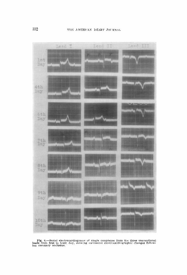
Figs. 6 and 7 show single complexes of serial electrocardiograms taken from
the day of admission until six months later. They show at first the elevated R-T
segment in Leads I and II and deep cove-plane inversion of T,. The R-T interval in Leads I and II gradually became isoelectric with inversion of the
T-waves in Lead II and lesser inversion of T,. The T-waves in Lead I
332 TIIE AMERIC’AN IlEART .JOL-RN.\I,
Fig. 6.-Serial electrocardiograms of single complexes from the three conventional leads from first to tenth day, slio\ving surrrssive electrocardiographic changes follow- ing coronary occlusion.

BALL : HEART-BLOCK IS CORONARY ARTERY TIIROMROSIS 333
Fig. ‘I.--Serial electrocardiograms of single complexes from the three conventional leads from twelfth day to six months, showing successive electrocardiographic changes following coronary occlusion.

334 THE AMERICAN HEART JOURNiiL
never became inverted. All of the electrocardiograms showed deep Q-waves
in Leads II and III. The teleroentgenogram showed slight enlargement of the left
ventricle. After the patient was discharged from thr hospital he was not seen for
five months. He then stated that for the preceding four months he had been working
daily pressing shirt8 eight to nine hours a day, and felt perfectly well. Blood pres-
sure was 180/110 mm. The heart was slightly enlarged to the left. The rhythm was
regular and the rate 80 per minute. The heart sounds at the apex were of good
quality, and the second aortic sound was modrratcly accentuated. Both lungs were clear and the liver xas not palpable. The electrocardiogram (Fig. 8) now showed
very little evidence of previous cardiac damage. It is significant to note that the
Q-waves in Leads II and III were still deep. On the basis of the enlargement of
the left ventricle and elevated blood prt~suw, it is assumed that this patient had a
Fig. 8.-Electrocardiogram taken six months after the acute attack. 9: and 63 are still deeply inverted. Tz slightiy upright and TB inverted.
hypertension for some time and the first evidence of vascular discase was the oc-
currence of an acute coronary artery occlusion.
DISCUSSION
In many cases of coronary artery t,hrombosis one can, from the electro- cardiograms, determine whether the occlusion involves the left or the right coronary artery, as was first shown by Parkinson and Bedford’ in 1928. They carefully described the successive changes in the electro- cardiogram following cardiac infarction and divided the curves into two main groups : type T1 and type Tz.. A definite sequence of changes in the R-T segment and T-waves occurs. The initial change is an elevation of the R-T segment from the isoelectric level followed later by an in- version of the T-waves in Lead I or III, but never in both, and a lesser
BALL : HEART-BLOCK IN CORONARY ARTERY TIIROl~BOSIG 335
degree of inversion in Lead II. Curves taken a few weeks after the acute injury usually conform to one of the two main types, as evidenced by the T-wave inversions. Thus, inversion of the T-waves in Leads I and II occurs in type T1, indicating an occlusion of the left coronary artery, and conversely inversion of the T-waves in Leads II and III is found where the thrombosis involves the right coronary artery. The T-waves can subsequently return to a complete normal. Often an inverted T-wave in Lead III may be the only residual evidence of a previous in- farction. This, plus the deep Q-waves in Leads II and III, is present in the case reported. Barnes and Whitten in a study of the correlation of the electrocardiographic changes, the artery occluded and the re-
sultant area of infarct.ion, confirmed the above findings. Wilson and his co-workers@ recently reported a study of the electrocardiograms in 56 cases of coronary thrombosis on 17 of which a postmortem examina- tion of the heart was made. Their findings were in complete agreement with those of Barnes and Whitten. In the atitopsied eases, curves show- ing inversion of the T-waves in Leads II and III were always associated with infarction of the area usually supplied by the right coronary artery.
Crawford and his coworkerslo recently substantiated the findings of Barnes and Whitten by cauterizing definite areas of the heart muscle in cats. They produced typical type T1 and T3 changes in the electro- cardiogram.
According to these criteria, the case reported here would fall into type Ts, indicating an occlusion of the right coronary artery with resultant myocardial infarction. Although the R-T segment was elevated above the isoelectric level in Lead I in the beginning, the T-waves in Lead I never became inverted. The deep cove-plane inversion of T3 at the onset, gradually becoming less inverted, and the subsequent development of inverted T-waves in Lead II (Figs. 6 and 7) place these curves in type Ta.
Tra.nsient heart-block in coronary artery occlusion is probabIy due to a disturbance of the circulation to the auriculoventricular node. The A-V node receives its blood supply from the right coronary artery in 92 per cent of the cases and from the left coronary artery in 8 per cent of the cases, depending upon which vessel crosses the “crux” of the heart posteriorly. Thus it would seem that heart-block should be more corn-
mon when the occlusion occurs in the right coronary artery. This premise is borne out in the following analysis of cases of heart-block with coronary artery occlusion reported in the literature, together with the case presented.
As early as 1913, Oppenheimer and Williamsll described a case of prolonged incomplete heart-block without a lesion of the bundle of His. At autopsy the posterior descending branch of the right coronary artery was found completely obstructed about 0.5 cm. from its origin as well as the descending and circumflex branches of the left coronary artery.

336 THE AMERICAN HEART JOURN.IL
Histological study revealed sclerosis and marked stenosis of the arter;\ to the A-V node. Serial sections of the bundle of His did not disclose any lesion to explain the block. They stated in conclusion that “complete heart.-block without anatomic lesions in the A-V system may possibly be of neurogenic or of circulatory origin, or it may be ascribed to chemical agents, to asphyxia, or to some hindrance to the passage of impulses from the terminal arborizations of the conduction system to the ven- tricular musculature.” Later in discussing a paper by Kugel, Oppen- heimer stated that since the original observation. he had had several instances in which it seemed probable that the heart-block was due to circulatory disturbance.
Parkinson and Bedford’ observed one case of transient heart-block in their series of cases. The electrocardiograms o-f this case conformed to the typical type Ta.
HansenI reported a case of complete A-V dissociation occurring on the fift,h day during an acute attack of coronary art,ery occlusion with death a few days later. The electrocardiograms were of type Ta with a
deep Qs. Postmortem examination was not obtained. Sandersl” observed a fatal case of coronary artery thrombosis with
complete heart-block and a relative ventricular tachycardia. The auricu- lar rate was 120 and the ventricular rate ‘70 per minute. Electrocardio- grams were of the type TJ. On postmortem examination, a thrombus was found obstructing the right coronary artery with an infarction involv- ing the greater portion of the outer wall of the right ventricle extending to the apex and to the anterior and posterior portions of the inter- ventricular septum.
Dr. &I. A. RothschildI has kindly furnished me with a remarkable case from his private practice. This was a man of fifty-three years who had a severe attack of precordial pain and dyspnea while riding horse- back. Clinically he had suffered an attack of acute coronary artery thrombosis. The electrocardiogram showed a complete A-V dissociation with inversion of the T-waves in Leads II and III, typical type Ta. The heart-block soon disappeared and the electrocardiogram subse- quently became entirely normal. Four years later he developed a second attack of coronary artery thrombosis without heart-block and died. The electrocardiogram at this time was of t,he type T1. This case is very important since it furnishes an interesting experiment in the same individual. The clinical and electrocardiographic evidence during the first attack would indicate that at this t,ime he had a thrombosis of the right coronary with heart-block, while his fatal attack was electrotardio- graphically a left coronary occlusion without heart-block.
FrothinghamC observed transient complete heart-block in a case of typical coronary closure with complete recovery. In his case also, the P-R interval was prolonged when normal sinus rhythm first appeared. The electrocardiograms conformed to type Ts.
RALL : HEART-BLOCK IN CORONARY ARTERY THROMBOSIS 337
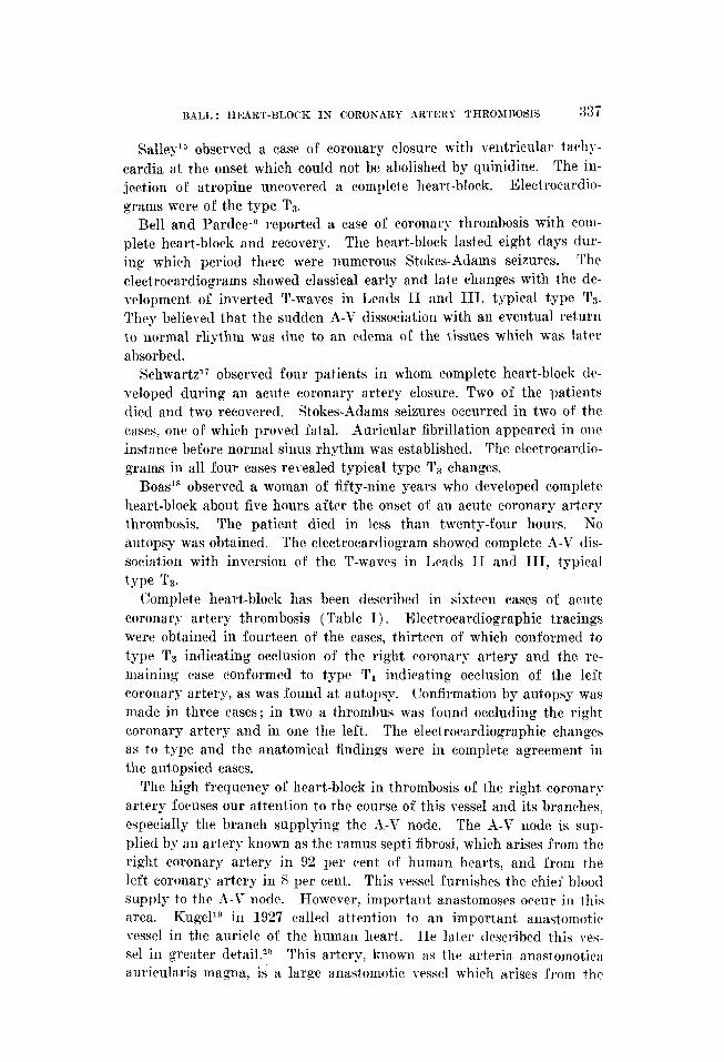
Salleylj observed a case of coronary closure with ventricular tachp-
cardia at the onset which could not be abolished by quinidine. The in- jection of atropine uncovered a complete heart-block. Electrocardio-
grams were of the type Ts. Bell and Pardeel” reported a case of coronary thrombosis with com-
plete heart-block and recovery. The heart-block lasted eight days dur- ing which period there were numerous Stokes-Adams seizures. The
electrocardiograms showed classical early and late changes with the de- velopment of inverted T-waves in Leads II and III, typical type T3. They believed that the sudden A-V dissociation with an eventual return to normal rhythm was due to an edema of t,he tissues which was later absorbed.
Schwartz17 observed four patients in whom complete heart-block de- veloped during an acute coronary artery closure. Two of the patients died and two recovered. Stokes-Adams seizures occurred in two of the cases, one of which proved fatal. Auricular fibrillation appeared in one instance before normal sinus rhythm was established. The electrocardio- grams in all four cases revealed typical type T3 changes.
Boas’” observed a woman of fifty-nine years who developed complet,e heart-block about five hours after the onset ol’ an acute coronary artery thrombosis. The patient died in less than twenty-four hours. No autopsy was obtained. The electrocardiogram showed complete A-V dis- sociation with inversion of the T-waves in Leads II and III, typical
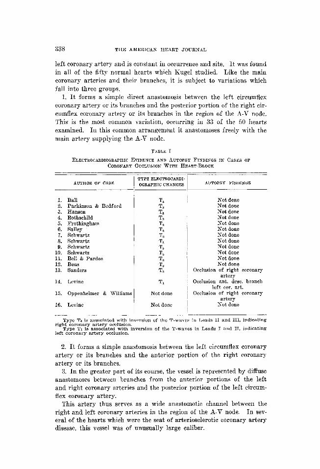
type 7’3. Complete heart-block has been described in sixteen cases of acute
coronary artery thrombosis (Table I). Electrocardiographic tracings were obtained in fourteen of the cases, thirteen of which conformed to type T3 indicat,ing occlusion of the right coronary artery and the re- maining case conformed to type T1 indicating occlusion of the left coronary artery, as was found at autopsy. Confirmation by autopsy was made in three cases ; in two a thrombus was found occluding the right coronary artery and in one the left. The electrocardiographic changes as to type and the anatomical findings were in complete agreement in the autopsied cases.
The high frequency of heart-block in thrombosis of the right coronar) artery focuses our attention to the course of this vessel and its branches, especially the branch supplying the 8-V node. The A-V node is sup- plied by an artery known as the ramus septi fibrosi, which arises from the right coronary artery in 92 per cent of human hearts, and from the left coronary artery in 8 per cent. This vessel furnishes the chief blood supply to the A-V node. However, important anastomoses occur in this area. KugeP in 1927 called attention to an important anastomotic vessel in the auricle of the human heart. He later described this ves- se1 in areater detail X) b . This artery, known as the arteria anastomoticw auricularis magna, is a large anastomotic vessel which arises from the
338 THE AMERICAN HEART JOURNAL
left coronary artery and is constant in occurrence and site. It was found
in all of the fifty normal hearts which Kugel studied. Like the main coronary arteries and their branches, it is subject to variations which fall into three groups.
1. It forms a simple direct anastomosis between the left circumflex coronary artery or its branches and the posterior portion of the right eir- cumflex coronary artery or its branches in the region of the A-V node. This is the most common variat,ion, occurring in 33 of the 50 hearts examined. In this common arrangement it anastomoses freely with the main artery supplying the A-V node.
T.4BLE 1
ELECTROCARDIOGRAPHIC EVIDENCE AND AUTOPSY FINDINGS IN CASES OF CORONARY OCCLUSION WITH HEART-BLOCK
AUTHOR OF CASE TYPEELECTROCARDI- OQRAPHICCHANGES AUTOPSY FINDINGS
1. Ball 2. Parkinson & Bedford 3. Hansen 4. Rothschild 5. Frothingham 6. Salley 7. Schwartz 8. Schwartz 9. Schwartz
10. Schwartz 11. Bell & Pardee 12. Boas 13. Sanders
T, T3 T, T3 T, T.9 T3 T, T, T, T, T, T,
14. Levine
15. Oppenheimer & Williams
16. Levine
Tl
Not done
Not done
Not done Not done Not done Not done Not done Not done Not done Not done Not done Not done Not done Not done
Occlusion of right coronary artery
Occlusion ant. desc. branch left car. art.
Occlusion of right coronary artery
Not done
Type Ts is associated with inversion of the T-waves in Leads II and III, indicating right coronary artery occlusion.
Type T1 is associated with inversion of the T-waves in Leads I and II, indicating left coronary artery occlusion.
2. It forms a simple anastomosis between the left circumflex coronary artery or its branches and the anterior portion of the right coronary artery or its branches.
3. In the greater part of its course, the vessel is represented by diffuse anastomoses between branches from the anterior portions of the left and right coronary arteries and the posterior portion of t,he left circum- flex coronary artery.
This artery thus serves as a wide anastomotic channel between the right and left coronary arteries in the region of the A-V node. In sev- eral of the hearts which were the seat of arteriosclerotic coronary artery disease, this vessel was of unusually large caliber.

BALL : HEART-BLOCK IN CORONARY ARTERY THROMBOSIS 339
We can therefore assume that where the anastomotic artery of Kugel forms a rich, collateral blood supply to the region of the A-V node as in Group 1, heart-block either is not likely to occur or may be very tran- sient, when t.he right coronary artery is occluded proximal t.o the branch supplying the A-V node.
However, where the collateral blood supply to the A-V node is not very rich as in the third group variation, heart-block is probably more liable to occur. In this case, occlusion of the right coronary artery, proximal to the ramus septi fibrosi, would temporarily cut off the maiu blood supply to the node and cause sufficient. &hernia to prevent some or all of the stimuli from the auricles from passing through the A-V node and into the bundle of His. Partial or complete A-V dissociation with a supraventricular type of ventricular complex would then result. The impulses producing t.he iclioventricular rhythm arise either in the lowermost portion of the node or in the upper part of the main stem of the bundle of His. This is illustrated by the fact that in all of the electrocardiographic tracings obtained in the reported cases of heart- block in coronary artery thrombosis, the ventricular complexes are al- ways of the supraventricular type. If the patient survives the initial shock and goes on to recovery, the collateral blood supply to the A-V node, mainly through the anastomotic artery of Kugel probably comes into play and gradually supplies sufficient blood to the region of the A-V node so that it again assumes its normal physiological function.
Geraudel” has furnished anatomical evidence in support of the above explanation. He demonstrated that stenosis and partial occlusion of the artery supplying the A-V node probably explained the occurrence of partial and complete heart-block in cases where a lesion of the bundle of His or its branches could not be found. By means of serial sections, he carefully examined the entire conduction system and the circulation to the A-V node in three cases of partial and complete A-V dissociation. The A-V node, bundle of His, and its branches were entirely free from any demonstrable lesion or injury in all three instances. However, a study of the artery supplying the A-V node revealed singular findings. In one ease t.he nodal vessel arose from the left coronary artery and was almost completely obstructed by a zone of proliferative endarteritis. In
the remaining two cases the artery to the A-V node arose from the right coronary, and in both instances the vessel showed marked narrowing with almost comp1et.e obliteration just above its point, of origin. It
may be significant that in all three cases the stenosing lesion in the nodal artery was found just at or proximal to its point of origin from the main coronary artery. In the first case the left coronary artery crossed the “crux” of the heart and supplied the artery to the A-V node, whereas in the other two cases in which the nodal artery arose from the right coronary artery, this main vessel was found to cross the “crux.” This is in accord with the studies of Gross.?”
340 THE AMER~(‘AX HEART JOURlY.41,
Jellick, Cooper, and Ophuls.‘” in 1906, described a case of Stokes- Adams syndrome occurring fourteen days before death in an individual suffering from acute epididpmitis and septicemia. ’ ’ Postmortem exami- nation of the heart demonstrated anemic necrosis of the muscular septum in the region of the bundle of His consequent on a recpnt thrombosis of its nutrient arteries. ”
r\Teuhof’” described a case of complete heart-block and auricular fibril- lation in a woman of eighty-three years who died in heart failure. At autopsy the coronary artery was found thickened and sclerosed although slightly patulons (he does not state which coronary) and the artery supplying the A-V node was completely calcified.
Carter and McEachern’” reported a case of recurrent complete heart- block in an individual with general and coronary arteriosclerosis. They too regarded the sudden and frecluent shifting from normal sinus rhythm to complete heart-block as being dependent upon vascular sclerosis with a deficient blood supply to the A-V node.
Levine’s case (Case l-4, Table 11 was the only instance in which the left coronary artery alone was involved, or rather where the right coronary artery was not at, all involved, and probably falls into the 8 per cent group in which the artery to the I’L-V node arises from the left coronary artery after crossing the “crux” of the heart.
PLINICAL SIGSIFICANCE
The clinical differentiation between right and left coronary artery occlusion has engaged the attention of many clinicians. The work of numerous investigators.‘, s* g. IF shows that infarction of that portion of the myocardium usually supplied by the right coronary artery is asso- ciated with inversion of the T-waves in Leads II and III, whereas in- farction of the myocardium supplied by the left coronary artery is asso- ciated with inversion of the T-waves in TJeads T and IT. The appear- ance of complete heart-block durin g an attack of acute coronary artery thrombosis points to involvement of the right coronary artery in about 93 per cent of the eases. If in addition to this we have other clinical signs such as rapid enlargement of the liver, as emphasized by Libman, the clinical diagnosis of right coronary artery occlusion can bc made with greater certainty.
SUMMARY AND CONCLUSIONS
A case of transient complete heart-block occurring during an attack of acute coronary artery thrombosis is described in detail. Changes in the ventricular portion of serial electrocardiograms conform to type TB, indicating myocardial damage as the result of occlusion of the right coronary artery.
The transient nature of A-V dissociation during an attack of coronary artery thrombosis has been explained on the basis of the peculiar anat-

BALL : HEART-BLOCK IN COROKARP ARTERY THROJIBOSIS 341
om- of the blood supply to the A-V node. Permanent heart-block with-
out any demonstrable lesions of the node or main st,em may be explained on the same basis.
The observations on the case presented and a review of similar cases reported in the literature indicate that in patients xyith coronary artery occlusion and complete heart-block, the right coronary artery is involved in approximately 93 per cent of the cases and the left in 7 per cent. The presence of complete A-V dissociation is therefore believed to be a valuable diagnostic criterion in the clinical cliff erentiation between right and left coronary artery thrombosis.
I am greatly indebted to Dr. Marcus A. Rothschild, Dr. Sidney P. Schwartz, and
Dr. Ernst P. Boas for furnishing me with clinical data and electrocardiograms of
cases which they observed but hare not published.
5.
6.
7.
8.
9.
I@.
11.
1“ -.
13.
14. 15.
16.
17. 18. 19.
20.
21.
REFERENCES
Levine, S. A.: Coronary Thrombosis : Its Various Clinical Features, Baltimore, 1929, Williams & Wilkins Co.
Herrmann, G. R.: Thrombosis of the Coronary Arteries With Tachycardia, J. Missouri State M. Assn. 8: 406. 1920.
Robinson, G. C., and Herrmann, G: R.: Paroxysmal Tachycardia of Ventricular Origin and Its Relation to Coronary Occlusion, Heart 8: 59, 1921.
Levine, S. A., and Stevens, W. B.: The Therapeutic Value of Quinidine in Coro- nary Thrombosis Complicated by Ventricular Tachycardia, AX HEART J. 3: 253, 1928.
Idem and Fulton, M. N.: The Effect of Quinidine Sulphate on Ventricular Tachycardia, J. A. M. A. 92: 1162, 1929.
Frothingham, C. : A Case of Coronary Thrombosis, M. Clin. Sorth America 10: 1357, 1927.
Parkinson, j., and Bedford, D. E.: Successive Changes in the Electrocardiogram After Cardiac Infarction, Heart 14: 195, 1928.
Barnes. A. R.. and Whitten.’ M. B.: Studv of the R-T Interval in Mvocardial In- farction, Aal. HEART J. 5: 142, 1929. ”
Wilson, F. N., Barker, P. S., MacLeod, A. G., and Klostermver, L. L.: The Elec- trocardiogram in Coronary Thrombosis, Proc. Sot. gxper. Biol. Sr Xed. 29: 1009. 1932.
Crawford, Jl H., Roberts, G. H., Abramson, D. T., and Cardwell, J. C.: Localiza- tion of Experimental Ventricular Mvocardial Lcnions br the Electrocardio- gram, Snr. HEART J. 7: 627, 1932. ”
Oppenheimer, B. S., and Williams, H. B.: Prolonged Complete Heart-Block Without Lesion of the Bundle of His. Pro?. Ser. ExDer. Biol. & Med. 10: 87. 1913.
Hansen, 0. S.: A Case of Coronary Thrombosis With Temporary Comnlete Heart-Blork, AK HEART J. 7: 3867 1932.
. I
Sanders, A. 0.: Coronary Thrombosis With Complete Heart-Block and Relative Vent&ular Tachvcardia. AN. HEART J. 6: 820: 1931.
Rothschild, M. A. : tiPersonal communication. ’ Salley, S. M.: An TJnusual Atropine Effect on Ventricular Tachpcardia, Am. J.
M. Se. 133: 456, 1932. Bell, 9.. and Pardee. H. E. B.: Coronarr Thrombosis, J. A. M. A. 94: 1533,
1930.’ Schwartz, S. P.: Personal communication. Boas. E. P.: Personal communication. Kugel, M. A.: An Important Anastomotic Vessel in the Auricle of the Human
Heart, Proc. N. Y. Path. Soe. Nov. 10, 1927. Arch. Path. and Lab. Med. 5: 3.55, 1928.
Idem : Anatomical Studies on the Coronary Artrrics and Their Branches, RX HEART J. 3: 360, 1928.
GBraudel, Emile : The Mechanism of the Heart and Its Anomalies, translation, Baltimore, 1930, Williams 8: Wilkins Co.

342 THE AMERICAN HEART JOURNAL
22. Gross, Louis: The Blood Supply to the Heart in Its Anatomical and Clinical Aspects, New York, 1921, Paul B. Hoeber, Inc.
23. Jellick, E. O., Cooper, M. D., and Ophuls, W.: The Sdams-Stokes Syndrome and the Bundle of His. J. A. M. A. 46: 955. 1906.
24. Neuhof, Seliam: A kase of Heart-Block and Auricular Fibrillation With Post- mortem Specimen: Comment on the Etiology of Fibrillation, Am. J. M. SC. 165: 34, 1923.
-_
25. Carter, E. P., and McEachern, D.: Recurrent Complete Heart-Block, Bull. Johns IIopkins Hosp. 49: 337, 1931.