the nu cleus pulposus microenvironment in the interver
TRANSCRIPT

J Guerrero et al. The nucleus pulposus microenvironment
707 www.ecmjournal.org
Abstract
The intervertebral disc (IVD) is a complex tissue, and its degeneration remains a problem for patients, without significant improvement in treatment strategies. This mostly age-related disease predominantly affects the nucleus pulposus (NP), the central region of the IVD. The NP tissue, and especially its microenvironment, exhibit changes that may be involved at the outset or affect the progression of IVD pathology. The NP tissue microenvironment is unique and can be defined by a variety of specific factors and components characteristic of its physiology and function. NP progenitor cell interactions with their surrounding microenvironment may be a key factor for the regulation of cellular metabolism, phenotype, and stemness. Recently, cell-transplantation approaches have been investigated for the treatment of degenerative disc disease, highlighting the need to better understand if and how transplanted cells can give rise to healthy NP tissue. Hence, understanding all the components of the NP microenvironment seems to be critical to better gauge the success and outcomes of approaches for tissue engineering and future clinical applications. Knowledge about the components of the NP microenvironment, how NP progenitor cells interact with them, and how changes in their surroundings can alter their function is summarised. Recent discoveries in NP tissue engineering linked to the microenvironment are also reviewed, meaning how crosstalk within the microenvironment can be adjusted to promote NP regeneration. Associated clinical problems are also considered, connecting bench-to-bedside in the context of IVD degeneration.
Keywords: Tissue engineering, regenerative medicine, intervertebral disc, nucleus pulposus, cell therapy, microenvironment, clinical research.
*Address for correspondence: Julien Guerrero, Tissue Engineering for Orthopaedics and Mechanobiology (TOM), Department for BioMedical Research (DBMR) of the Faculty of Medicine of the University of Bern, University of Bern, Bern, Switzerland.Telephone number: +41 316329883 Email: [email protected]
Copyright policy: This article is distributed in accordance with Creative Commons Attribution Licence (http://creativecommons.org/licenses/by-sa/4.0/).
European Cells and Materials Vol. 41 2021 (pages 707-738) DOI: 10.22203/eCM.v041a46 ISSN 1473-2262
The nuCleus pulposus miCroenvironmenT in The inTerverTebrAl disC: The founTAin of youTh?
J. Guerrero1,*, S. Häckel2, A.S. Croft1, S. Hoppe2, C.E. Albers2 and B. Gantenbein1,2
1 Tissue Engineering for Orthopaedics & Mechanobiology (TOM), Department for BioMedical Research (DBMR) of the Faculty of Medicine of the University of Bern, University of Bern, Bern, Switzerland
2 Department of Orthopaedic Surgery & Traumatology, Inselspital, Bern University Hospital, University of Bern, Bern, Switzerland
list of Abbreviations
2D 2-dimensional3D 3-dimensionalADSC adipose-derived stromal cellAF annulus fibrosusAMPC allogeneic mesenchymal precursor cellAPI recombinant human growth and differentiation factor-5 (aka. rhGDF-5)ASC adipose stromal cellBMAC bone marrow aspirate concentrateBMP bone morphogenetic proteinBMSCs bone marrow/mesenchymal stem/ stromal cellsCCL-5 chemokine (C-C motif)
CD202b angiopoietin-1 receptor CD24 cluster of differentiation 24CEP cartilaginous endplateCFU-f colony forming units-fibroblastCT computed tomographyCTGF connective tissue growth factorDDD degenerative disc disease DPQ Dallas pain questionnaireECM extracellular matrixEQ5D standardised measure of health-related quality of life developedES embryonic stemFGF fibroblast growth factorFRI functional rating indexGAG glycosaminoglycan

708 www.ecmjournal.org
J Guerrero et al. The nucleus pulposus microenvironment
GD2 disialoganglioside 2GDF growth and differentiation factorGLUT-1 glucose transporter 1GMP good manufacturing practiceHIF-1α hypoxia-inducible factor 1αIDCT injectable disc cell therapyIDD intervertebral disc degenerationIGF insulin growth factorIL interleukiniPSC induced pluripotent stem cellIVD intervertebral discJOABPEQ Japanese orthopaedic association back pain evaluation questionnaireLBP low-back painLDD lumbar disc degeneration/diseaseMB methylene blueMMP matrix metalloproteinaseMODISC Modic I discopathiesMPC mesenchymal precursor cellMRI magnetic resonance imagingMSC mesenchymal stromal cellMSV GMP)-compliant expanded bone marrow MSC (MSV, PEI Num. 10-134)Nanog homeobox protein NANOG NASS North American Spine SocietyNF-κB nuclear factor kappa-light-chain- enhancer of activated B cellsNotch1 notch homolog 1, translocation- associatedNP nucleus pulposusNRS numerical rating scaleOct4 octamer-binding transcription factor 4ODI Oswestry disability indexPASS patient acceptable symptom statePDGF platelet derived growth factor PEG polyethylene glycolpNIPAM poly(N-isopropylacrylamide)PROMIS patient-reported outcomes measurement information systemPRP platelet-rich plasmaRCT randomised-controlled trialrhGDF-5 recombinant human growth and differentiation factor-5RMQ Roland-Morris questionnaireSAPH self-assembling peptide hydrogelSF short formSIRT1 NAD-dependent deacetylase sirtuin-1SIRT6 NAD-dependent deacetylase sirtuin-6SLRP small leucine-rich proteoglycanSOX9 SRY-box transcription factor 9TAA triamcinolone acetonideTGF-β1 transforming growth factor- β1Tie2 TEK receptor tyrosine kinaseTNF-α tumour necrosis factor-αVAS visual analogue scaleVAST viable allograft supplemented disc regeneration treatment WPAI work productivity and activity index
introduction
The IVD can be defined as a joint between adjacent vertebral bodies. It is composed of three primary tissues, the NP, the AF, and the CEP (Urban and Roberts, 2003). The cells within each of the regions of the IVD can not only be subjected to physical and
biochemical stimuli from their ECM but also from their surrounding microenvironment (i.e. non-ECM related) (Baer et al., 2003; Cao et al., 2009; Cao et al., 2011; Hsieh et al., 2005; Hwang et al., 2014; Jackson et al., 2011a; Korecki et al., 2008). Moreover, it is well documented that disc degeneration first occurs in the NP region of the IVD (Wang et al., 2014; Zhao et al., 2007). In this context, this full microenvironment (i.e. ECM and non-ECM related factors) of the IVD, and especially the NP microenvironment is believed to play a critical role in the regulation and maintenance of this tissue. It can potentially improve the repair and regeneration of the vertebral column joint (Fig. 1). However, the understanding of the NP microenvironment of the IVD is still incomplete. In this review, papers reporting NP cell interactions with their surrounding microenvironment have been summarised to help improve future development of tissue engineering and regenerative medicine approaches. The first section covers what has been have learned from the current literature on:• the “genera l” compos i t ion o f the NP
microenvironment with the description of essential components (non-ECM and ECM-related),
• how NP cells interact with their native microenvironment,
• how changes in the surrounding, particularly due to degeneration or disease, can alter NP cells.
• the state-of-the-art for tissue engineering and regenerative medicine approaches using this knowledge to treat IVD degeneration and LBP.
• clinical trials involving and targeting the NP microenvironment and how all the available expertise was used to bring bench and bedside closer together.
The “general” composition of the np microenvironment
non-eCm-related factorsThe IVD is an avascular tissue. Therefore, the supply of nutrition to the disc primarily occurs through diffusion from its surrounding vasculature (Jackson et al., 2011b). The variation between cellular consumption rates and nutrient transport (passive or active) leads to concentration gradients of these metabolites and nutrients throughout the IVD. Consequently, this markedly affects the viability, proliferation, and function of cells, and collectively will alter any subsequent potential regeneration or repair. In human lumbar, thoracic, and cervical discs, oxygen levels vary considerably and do not appear to correlate with ageing or the degree of severity of pathological disease such as disc degeneration. Oxygen concentrations decrease from the AF across the disc inner structure (between 19.5 % and 0.65 %) with average normoxic levels in the central part of the NP being between 0.5 % and 10 % of oxygen

J Guerrero et al. The nucleus pulposus microenvironment
709 www.ecmjournal.org
(Bartels et al., 1998; Buckley et al., 2018). These levels are mainly determined by the transport through the CEP, but also by the cellular density within the tissue, and the cellular consumption rates. Another critical point of the microenvironment is the presence/absence of the most used “fuel” in the body, glucose. It is well recognised that NP cell viability is reduced if the microenvironment is glucose deficient. However, a low oxygen concentration within the microenvironment has not been shown to be detrimental to NP cell viability. Glucose level does play a critical role as a limiting factor for disc-cell survival (Urban et al., 2004). A mathematical model has predicted a decrease in glucose concentrations, from around 5 mmol/L in the AF to approximately 0.8 mmol/L, in the NP tissue part of healthy IVDs (Selard et al., 2003) that can even fall below critical levels with more substantial calcification of the CEP tissue (Jackson et al., 2011b). Importantly, cell death can already occur when cells are subjected to glucose concentrations below 0.5 mmol/L for more than 3 d (Horner and Urban, 2001). Additionally, low cell viability correlates with low glucose concentrations, which has been shown in scoliotic discs (Bibby et al., 2002). Another important parameter in the NP tissue microenvironment is the pH. The pH ranges from 2 to 6, which is mainly due to the local production of lactic acid, as a result of glycolysis by the IVD cells (Bartels et al., 1998). Several in vivo measurements revealed pH ranges from 5.7 to 7.5 (with a median at 7.0) (Bez et al., 2018; Gilbert et al., 2016; Nachemson, 1969). It is well established that the pH can significantly influence cell survival, negatively affect matrix synthesis rates
(Horner and Urban, 2001; Ohshima and Urban, 1992), but may also increase the expression of pro-inflammatory cytokines and pain-related factors in the context of LBP (Gilbert et al., 2016). Furthermore, scientists found that rates of metabolism (e.g. oxygen consumption) were sensitive and coupled in a non-linear way with the pH present in the microenvironment of bovine discs (Bibby et al., 2005). The physiological mechanical loading stress of the IVD tissue leads to constant exposure of the IVD cells to high osmolarity and, therefore, microenvironmental osmotic changes (Sadowska et al., 2018). Aggrecan, as a cartilage-specific proteoglycan is essential for maintaining hydration and hence osmotic pressure in the IVD (Urban and Roberts, 2003). Negatively charged GAGs regulate the ionic balance of the IVD ECM (Johnson et al., 2014). Aggrecan is linked to sulphated GAGs (e.g. keratan and chondroitin sulphate). They can create a negative charge and hence bind water within the tissue. If this is transposed to the tissue level, the generation of osmotic pressure in the IVD creates a water “flow” into the NP tissue. This water intake also leads to the load-bearing ability and the crucial swelling pressure of the IVD (Erwin and Hood, 2014; Urban and Maroudas, 1981; Urban et al., 1979; Urban and Roberts, 2003). In a healthy state, the extracellular osmolarity can vary from around 430 mOsm/L (iso-osmotic pressure) to around 496 mOsm/L (hyper-osmotic pressure) (Ishihara et al., 1997; van Dijk et al., 2011). These values are in a physiological range for IVD cells but would be considered too high or too variable if compared to the osmotic pressure that most mammalian cells experience (Appelboom
fig. 1. The fountain of youth. How the NP microenvironment and how factors involved in it can implement tissue engineering approaches for the regeneration of the IVD and therefore enable future clinical improvement.
Intervertebral discs NP tissue microenvironment(fountain of youth)
Implementation in tissueengineering strategies
Regenerative medicine approaches usingmicroenvironment components

710 www.ecmjournal.org
J Guerrero et al. The nucleus pulposus microenvironment
et al., 1956; Brocker et al., 2012). As a reference, the osmotic pressure of healthy blood ranges between 280 mOsm/L and 320 mOsm/L (Neidlinger-Wilke et al., 2012). In a diseased state (e.g. degenerated), the IVD osmolarity can even decrease so much that it reaches values of around 300 mOsm/L (hypo-osmotic pressure), due to a loss of proteoglycans (Wuertz et al., 2007). As a result, IVD hydration can be compromised, and fibrosis may occur. However, this hypo-osmotic pressure would be considered physiological for cells of other tissues, especially in older people (Hooper et al., 2015). Nevertheless, stem-cell-related research and associated publications discussing high osmolarities are still limited. Tao et al. (2013) pioneered the analysis of the effect of osmotic pressure on IVD progenitor cells. They found that high osmolarity could not only decrease the viability and proliferation of IVD cells but also affect the expression levels of collagen type II, SOX-9 transcription factor, and aggrecan in NP progenitor cells (Tao et al., 2013). Additionally, a study showed the effects of high osmolarity on human ADSCs in the context of IVD regeneration (Liang et al., 2012a). They concluded that high osmolarity in the IVD microenvironment is a deleterious factor that affects the survival and biological behaviour of ADSCs, making them less suitable for clinical applications. A recent bioinformatic analysis identified significant gene biomarkers of LBP caused by changes in the osmotic pressure of NP cells, providing a reference for future in-depth research (Zhao et al., 2020). In the context of regenerative medicine or tissue engineering, all 4 components of the microenvironment described above (oxygen, glucose, pH, and osmolarity) seem to have a different impact on the IVD cells (Fig. 2). On the one hand, oxygen concentration appears to have a pivotal role in regulating the biosynthesis and phenotype of
targeted cells for therapeutic applications (Naqvi and Buckley, 2015). On the other hand, low glucose concentrations, osmolarity, and low pH levels can impair the survival and biological behaviour of progenitor cells (Li et al., 2012; Naqvi and Buckley, 2016; Tao et al., 2013; Wuertz et al., 2009). All those parameters (non-ECM related) should be taken into account in the development of future approaches to tissue engineering and regenerative medicine targeting IVD and especially NP tissue.
eCm-related factorsThe IVD ECM can be characterised as a scaffold rich in molecules that offer structural and biochemical support to the resident cells (Fig. 3). The structural, and indeed functional requirements, determine the mechanical properties of the ECM, which depend on the protein composition of the matrix – particularly the percentage and type of several collagens and elastin fibres (Mercuri et al., 2014; Sivan et al., 2014a). Furthermore, the biochemical and physiological relevance of these properties is illustrated by a mutual/reciprocal crosstalk between ECM and cells. Cells are capable of sensing surrounding ECM stiffness thanks to integrin-mediated interactions with the matrix (Gilchrist et al., 2007). The mechanical properties of the ECM are then interpreted and can affect cell motility, proliferation, differentiation, and apoptosis (Newell et al., 2019; Peng et al., 2020). Hence, knowledge of the precise composition of IVD tissues is critical to the understanding of their physiology and physiopathology. For example, the most common pathological state of the IVD (degenerated) is often closely related to alterations in the composition and structure of the ECM. Degeneration of the IVD tissue is well known to be linked with excessive matrix catabolism (Roughley, 2004). The protein composition of human IVD tissues
fig. 2. summary of non-eCm-related components and their ranges within the np microenvironment. It can be characterised by 4 main aspects: (i) the oxygen level, (ii) the osmolarity, (iii) the pH, and (iv) the glucose concentration.
Extracellular matrix
Oxyg
en, o
smol
arity
, pH,
glu
cose
microenvironmentNP tissue
Growth factors(mmol/L)
[0.8]
[5.7] [5.7]

J Guerrero et al. The nucleus pulposus microenvironment
711 www.ecmjournal.org
will now be examined, to better estimate the amount of ECM proteins present both in a healthy and disease state. In terms of percentage, the ECM is mainly composed of water (70-90 % of wet weight), followed by proteoglycans (65 % of dry weight), and collagen type II (15-20 % of dry weight) (Eyre and Muir, 1977; Gower and Pedrini, 1969). Other minor components (though potentially critical functionally) of the NP ECM include elastin, small proteoglycans, other collagens (types III, VI, IX) (Melrose et al., 2001; Roberts et al., 1991; Yu, 2002), and laminins (Chen et al., 2009; Gilchrist et al., 2007; Nettles et al., 2004). IVD-ECM contains 385 ± 35 μg of collagen (all types) per mg of dry tissue. Its abundance is not significantly affected by age or sex. But, the amount of collagen is slightly different between the AF and NP, with the AF tissue having significantly more collagen than NP tissue. All publications on the topic, conclude that the dry weight of the NP tissue has more proteoglycans than AF tissue (Choi, 2009; Mwale et al., 2011). Concerning other main components of the IVD ECM, the total amount of proteoglycans in human tissue is 144 ± 16 μg per mg of dry tissue, and for elastin the amount is approximately 18 ± 4.1 μg per mg of dry tissue (McKee et al., 2019). However, in a pathological context (e.g. scoliosis or disc degeneration), changes to ECM composition can occur, particularly in the percentage of collagen (McKee et al., 2019). Furthermore, an increase in elastin is also observed in degenerated IVD (McKee et al., 2019). The protein composition of the ECM is a powerful regulator of cellular behaviour (Yue, 2014). Consequently, failure to adequately mimic the native extracellular environment during in vitro cell culture processes may lead to altered proliferation, adhesion, migration, polarity, differentiation, and apoptosis of the targeted cells. In the context of a clinical trial, in vitro results may not translate into the human patient (Edmondson et al., 2014). To become closer to the
native microenvironment, culturing on ECM-coated dishes, within 2D or even 3D culture, on biomimetic scaffolds, or organ-like/organoids culture has become a more and more common practice to overcome these unwanted artificial effects (Edmondson et al., 2014; Guerrero et al.; Huch and Koo, 2015). By precisely summarising an “ingredient list” for the microenvironment it should be possible to recreate models that mimic it, and further improve knowledge through more efficient in vitro tools.
how np progenitor cells interact with their microenvironment
IVD degeneration does not generally occur because of a single factor, but rather because of a variety of them. Usually, it is attributed to a complex interplay between environmental, genetic factors and mechanical damage (Dudli et al., 2012; Kalichman and Hunter, 2008; Kamper et al., 2016; Livshits et al., 2011; Mayer et al., 2013; Silva and Holguin, 2020). However, each of these factors leads to a final typical result of imbalance between the anabolic and catabolic microenvironment of the NP tissue in favour of catabolism. During disc degeneration or just simply during ageing, significant changes can occur in the ECM composition and structure and, therefore, have an impact on IVD cell behaviour. All these changes that drastically affect the IVD microenvironment lead to an important decrease in cell density, and cell clustering (Boos et al., 2002; Hastreiter et al., 2001; Johnson et al., 2001; Johnson and Roberts, 2003; Liebscher et al., 2011; Roberts et al., 2006). In the NP tissue, progenitor cells are embedded in a tissue-specific microenvironment that significantly influences the their biological and metabolic processes (Hu et al., 2018). Low oxygen tension, hypertonicity, low pH, and reduced
Oxyg
en, o
smol
arity
, pH,
glu
cose
Extracellular matrix
NP tissuemicroenvironment
Growth factors
elastin and laminin
fig. 3. summary of eCm-related components and their ranges within np microenvironment. It can be characterised by 4 main aspects: (i) water, (ii) proteoglycans, (iii) collagen type II, and (iv) other minor components.

712 www.ecmjournal.org
J Guerrero et al. The nucleus pulposus microenvironment
nutrient supply are characteristics of the specialised IVD microenvironment. The level of specialisation and complexity of this microenvironment could be challenging for cell survival and health of endogenous progenitor or future implanted cells (Sakai and Andersson, 2015). An indigenous progenitor cell population that expresses an endothelial specific marker, i.e. Tie2 (angiopoietin-1 receptor or CD202b), has been identified in human and murine NPs (Sakai et al., 2012). These Tie2+ cells were identified as the precursors of NP cells that further differentiate and start to express other surface markers, including GD2 and CD24 (Sakai et al., 2012). Moreover, Tie2+ cells have the ability of cell renewal (Sakai et al., 2012), and multi-potency with differentiation towards neurogenic, osteogenic, chondrogenic, and adipogenic lineages. The number of Tie2 positive cells in the human (Sakai et al., 2012) and mouse (Bach et al., 2016) NPC populations also decreased, not only during ageing but also with the progression of IVD degeneration. The specific factors responsible for an in vivo decrease of NP cell clustering and a reduced percentage of NP progenitor cells with age, or during a degenerative state, are not fully understood. One of the advanced explanations is that cell mortality associated with decreased nutrient oxygen and glucose transport in IVD induces the decrease of both cell clustering and the percentage of Tie2+ cells present in NP tissue (Urban and Roberts, 2003). Others have demonstrated that NP progenitor cells harvested from patients with degenerated IVDs still have similar cell colony-forming ability, proliferation rate, cell cycle, and trilineage differentiation properties to bone marrow stromal cells (Li et al., 2017a). Even NP progenitor cells derived from degenerated IVDs still retain their regeneration ability. They may be a promising cell candidate for cell-based regenerative medicine and tissue engineering for IVD degeneration. These results suggest that Tie2+ NPCs could play a crucial role in IVD regeneration, knowing that – even if their microenvironment is disrupted – they do not seem to be affected (Sakai et al., 2012). However, even if Tie2+ NPCs seems to be the “ideal candidate”, future strategies for NP tissue engineering and regenerative medicine should not only be focused on those cells as their numbers are very limited (around 1 %) in human IVD tissue (Hu et al., 2018).
how changes in their surroundings can alter np cells
Influences of non-ECM related factors on IVD progenitor cellsOne intrinsic characteristic of progenitor disc cells is that they reside under hypoxic conditions due to a blood-supply shortage in the IVD and especially in the NP tissue. Under low-oxygen levels, the NP
progenitor cells proliferate better than ASC, and their potential to differentiate on the chondrogenic lineage is increased (Li et al., 2013). Physiological hypoxia (2-5 %) seems to be the normal and the physiological environment for optimal functioning of IVD progenitor cells (Chen et al., 2014; Gantenbein et al., 2014; Stoyanov et al., 2011). However, invasion of blood vessels through fissures is generally observed during the process of IVD degeneration, which potentially increases the oxygen concentration and consequently aggravates the physiological hypoxic microenvironment of the IVD progenitor cells even further (Freemont et al., 2002; Nerlich et al., 2007). Therefore, in the context of cell therapy, restoring the hypoxic microenvironment may favour the use of NP progenitor cells. E x c e s s i v e m e c h a n i c a l l o a d i n g a n d impaired biomechanics can be another crucial microenvironmental factor influencing NP progenitor behaviour (Chu et al., 2018; Desmoulin et al., 2020). For example, several studies showed an essential correlation between the presence and the severity of disc degeneration in obese or overweight adults (Lidar et al., 2012; Samartzis et al., 2012). Nevertheless, at a cellular level, the biomechanical factors display a wide range of impacts on the biological functions of NP progenitor cells. Recently, in vitro studies have shown that static compression stress induces mitochondrial apoptosis in NP progenitor cells (Li et al., 2018; Yuan et al., 2018). Apart from the induction of apoptosis and senescence, stress stimuli seem to be critical for the normal functioning of progenitor cells (Hosseini et al., 2015; Kobayashi et al., 1989; Zhao et al., 2015). As an illustration of this, fluid-shear stress can give rise to overexpression of ECM components synthesised by AF progenitor cells (e.g. Collagen type I and MMP-1) (Chou et al., 2016) – demonstrating the “double-edged sword” effects of applied forces on IVD cells and especially NP progenitor cells. To investigate these effects further, studies should focus on exploring various methods to protect IVD progenitor cells from excessive mechanical loading-induced dysfunction and increased cell death. Optimising mechanical stimulation of in vitro cultured IVD progenitor cells (also using Tie2+) could be used to produce a matrix mimicking the microenvironment and to explore in more detail the relationship between ECM composition, mechanical forces, and cell behaviour. Another non-ECM-related factor that can drastically influence NP progenitor cells (endogenous or implanted) seems to be osmolarity. Nevertheless, publications associating osmolarity and NP progenitor cells are still limited. Tao et al. (2013) pioneered the investigation of the influence of osmolarity on IVD progenitor cells. They found that high osmolarity in NP progenitor cells could decrease the viability, proliferation, and gene expression levels of not only the transcription factor SOX-9 but also aggrecan, and collagen type II, the two main components of the NP ECM.

J Guerrero et al. The nucleus pulposus microenvironment
713 www.ecmjournal.org
Regarding the influences of non-ECM related factors on NP progenitor cells, it is necessary to examine the microenvironment pH. To have a comparison with already well-described cells, ASCs were used. Under low pH the proliferation and viability of the NP progenitor cells are better than those of ASCs (Han et al., 2014). More recently, it was shown that the acid-sensing ion channels in NP progenitor cells play a vital role in low-pH induced apoptosis, and the downregulation of stem-cell-related genes (e.g. Oct4, Nanog, Jagged, Notch1) and ECM synthesis (e.g. aggrecan, SOX-9, collagen type I and collagen type II) (Liu et al., 2017). It is also known, in connection with Diabetes mellitus or hyperglycaemia, that an excess of glucose has negative effects upon NP progenitor cell biology (Liu et al., 2020). A comparison of NP progenitor cells cultured in low- or high-glucose media demonstrated that high glucose concentration significantly decreases cell proliferation, colony-formation ability, migration, and wound-healing capability. Moreover, NP progenitor cells from high-glucose culture also show significantly decreased expressions of a variety of markers, including SIRT1, SIRT6, HIF-1α, GLUT-1, and caspase-3. Altogether, these data show that high levels of glucose might drastically impact NP progenitor cell behaviour (Liu et al., 2020).
Influences of ECM related factors on IVD progenitor cellsWith the degeneration process of IVDs, the imbalance between ECM anabolism and catabolism intensifies the tissue dysfunction (Feng et al., 2016; Kepler et al., 2013). When investigating NP progenitor cells in vitro, the importance of the ECM in the original tissue is not usually considered. However, the crosstalk and interaction between cells and the ECM are essential in affecting not only the morphology and phenotype but also the function of progenitor cells, as shown in NP tissue (Guilak et al., 2009; Rastogi et al., 2009). Perlecan, the common component of many stem cell niches, is produced by progenitor cells located in the NP tissue (Brown et al., 2018; Schlotzer-Schrehardt et al., 2007). It was previously found to play a decisive role in the chondrogenic differentiation of the IVD mesenchymal progenitor cells (Shu et al., 2013; Smith et al., 2010). Most importantly, changes to ECM mechanical strength in degenerated discs are transmitted to the cell membrane and consequently, by means of perlecan or other components of stem cell niches, activate the IVD progenitor cells (Subramony et al., 2013; Wang and Chen, 2013). The Piezo1 ion channel, which acts as a mechanosensor between the ECM and cells, is found in human NP cells to be involved in the pathway that starts with mechanical force sensing and results in cell apoptosis. The signal is mediated through mitochondrial dysfunction and endoplasmic reticulum stress (Li et al., 2017b). Nevertheless, many other aspects of mechanical sensing could potentially affect the behaviour of progenitor cells. How these
IVD progenitor cells respond to them will require further investigation. Specific components of the ECM seem to have a greater impact than the others on NP cells. Laminin recently attracted the attention of researchers, as it is present in healthy NP tissue, but absent in degenerating NP tissue (Chung and Mercurio, 2004; Foldager et al., 2016; Foldager et al., 2014; Mercurio et al., 2001; Sun et al., 2017). Moreover, laminin receptors (α6β1 and α6β4 integrins) are known to improve cell survival in response to microenvironmental changes such as low oxygen tension or serum-deprivation (Gu et al., 2002), both relevant to NP tissue (Chen et al., 2009). Another critical aspect of the microenvironment that can drastically influence the behaviour of NP progenitor cells is the inflammatory process occurring during IVD degeneration. Cytokines, such as IL-1β and TNF-α, are generally considered to be the key mediators of the IVD degenerative process and LBP (Burke et al., 2002; Risbud and Shapiro, 2014; Wang et al., 2020b). They are also upregulated in the degenerative IVDs, and they are closely related to various pathological IDD processes, including the inflammatory response, matrix destruction, cellular senescence, autophagy, apoptosis, pyroptosis, and proliferation. Conversely, IL-1β suppression prevents disc degeneration (Genevay et al., 2009; Le Maitre et al., 2007). Therefore, anti-IL-1β and anti-TNF-α therapies may have the potential to stop this vicious cycle present in disc degeneration and LBP. Knowing that IL-1β and TNF-α inhibition have the potential to alleviate IDD, an in-depth understanding of the role of IL-1β and TNF-α in IDD will benefit the development of new treatment methods for disc degeneration. The microenvironment of the native and, by extension, of degenerated IVD remains an important factor in the development of effective regenerative therapies (Baumgartner et al., 2021), presenting a challenge not only for the application of cell therapies but also for interventions based on growth factors, or other biologicals such as drugs, and biomaterials.
Tissue engineering and regenerative medicine for np tissue
A common way to improve or replace the function of biological tissue by tissue engineering is to use a scaffold (as a vehicle or a tissue replacement) in combination with cells that possess a regenerative potential, and/or additional components (chemicals, growth factors, oxygen priming, etc.). In the context of IVD tissue engineering, both the biological and mechanical properties of the scaffold will affect the biocompatibility within IVD tissue. Mimicking the microenvironment in the closest way possible will determine the in vivo function and regenerative potential of progenitor cells and determine the efficiency of IVD regeneration (Choi and Harley, 2012;

714 www.ecmjournal.org
J Guerrero et al. The nucleus pulposus microenvironment
Engler et al., 2007; Hsieh and Twomey, 2010; Seliktar, 2012). Utilising the natural molecules present in the IVD ECM could be an excellent starting point for the generation of scaffolding material, but synthetic polymers also seem to be efficient. Moreover, macromolecules present in the microenvironment could also be implemented or functionalised into an engineered scaffold in order to be even more similar to NP tissue. All those strategies will be presented in the following section.
related to non-eCm factorsAs discussed in the previous section, the components of the microenvironment from native and degenerative IVDs are well known to drive and affect the survival, behaviour, and fate of endogenous or transplanted NP cells. In the following part, non-ECM factors used for tissue engineering approaches for NP regeneration are described (Table 1). One of the key components of the IVD microenvironment described above is the low pH. An acidic environment is vital for the maintenance of NP cells’ activities. However, changes in the acidity of the environment on human NP cells can be detrimental. To overcome this problem, it has been shown that NP progenitor cells cultured in presence of amiloride, an acid-sensing ion channel blocker, could significantly improve their proliferation, expression of stem cell-related genes, and functional genes (Liu et al., 2017). As described above, the variation of osmolarity (osmotic concentration) can have a significant impact on the function and behaviour of NP cells. GAG has a key role in regulating osmolarity, therefore targeting this component of the microenvironment could be a critical element in improving tissue engineering approaches for NP repair and regeneration. For this reason, Sivan et al. (2014b) decided to develop a biomimetic GAG analogue-engineered scaffold based on sulphonate-containing polymers. The in vivo delivery of this biomimetic GAG, which can polymerise in situ, was used in a porcine and bovine degenerate explant model and, as a result, demonstrated the ability of the implanted hydrogel to restore NP tissue stiffness. Hydrogels or engineered scaffolds mimicking GAG function in vivo while maintaining disc hydration and height could be a solution for NP tissue repair or helping its regeneration by restoring part of the native microenvironment. Another critical point for NP tissue engineering could be that cells in vitro primed in a low oxygen environment could then also promote an NP phenotype in vivo. As an example, Feng et al. (2014), cultured NP bovine cells for 3 weeks in a 3D environment with different oxygen tension levels (2 % to 20 %). They demonstrated that, even after 8 weeks of in vivo implantation, the hypoxic priming of NP cells resulted in the maintenance of GAG, collagen type II, aggrecan, and SOX-9 expression compared to normoxic priming. The maintenance of the NP phenotype was therefore achieved if cells
were originally cultured under hypoxic conditions before implantation. In summary, combining hypoxia with a relevant scaffold could enhance NP function after implantation. Moreover, hypoxia induction of NP cells should be applied to most of the strategies to successfully regenerate NP tissue for IVD repair. Researchers also found that SLRPs – bioactive components of the ECM – were associated with fibrillogenesis, cellular growth and apoptosis, as well as tissue remodelling of the IVD. They can support the survival of IVD progenitor cells under hypoxic conditions by the activation of specific hypoxia-inducible factors (Chen et al., 2017). As a result, agents that can stimulate the production of SLRPs (e.g. biglycan or decorin) might help NP progenitor cells to survive under hypoxic conditions (Huang et al., 2013) and could become critical components for the niche, providing new approaches to use the biology of the microenvironment as a toolbox (Rajasekaran et al., 2021). Recently, a new approach was developed for the sequential release of chemokines followed by growth factors, using a delivery system based on polysaccharide microbeads (Frapin et al., 2020). In the pullulan-based scaffold, CCL-5 was first used to attract cells with a regenerative potential into NP tissue. After this first phase of recruitment, TGF-β1 and GDF-5 were chosen as growth factors to induce the synthesis of an ECM rich in collagen type II and aggrecan, in vitro. The authors then proceeded to ex vivo experiments using the same delivery system in degenerated ovine IVDs. As a result, after histological analysis, they observed an increase of NP cellularity and more collagen type II, and aggrecan. Moreover, the amount of Tie2+ positive cells (NP progenitor) was higher compared to the control condition. The sequential delivery of macromolecules to repair and regenerate the ECM could be a strategy for functionalised engineered scaffolds in the future. Concerning the GAG, trying to increase their expression in vivo after implantation can be one element to target – by the use of an engineered scaffold, as already described. However, trying to reduce its degradation in vivo can also be a way to improve the success rate after implantation. In this context, several in vitro studies have shown that glucosamine has an inhibitory effect on the degeneration of proteoglycans, providing the potential of a NP tissue engineering application in the context of IVD degeneration (Ilic et al., 2003; Munteanu et al., 2002; Samiric et al., 2006). A novel deacetylated poly-N-acetyl glucosamine hydrogel, as a potential therapy for treating NP degeneration, has also been developed (Gorapalli et al., 2012). Using primary human cells, they showed an increase in cell viability, metabolic activity, proteoglycan synthesis, and ECM protein expression (aggrecan and collagen type II). This hydrogel has promising in vitro characteristics and motivates further in vivo evaluation as a potential therapy for NP degeneration of the IVD.

J Guerrero et al. The nucleus pulposus microenvironment
715 www.ecmjournal.org
Target strategyComponent/growth factor
Cells/ material results Conclusion reference
ph/acidityBlocking of
acid-sensing ion channel
Amiloride Human NP-MSCs
Improvement of NP proliferation,
expression of stem cell-related genes, and
functional genes
The amiloride may improve IVD degeneration by
improving the activities of NP-MSCs
Liu et al., 2017
osmolarity Increase the GAG content
Biomimetic GAG analogue
engineered scaffold
Bovine and porcine IVD/sulphonate-containing polymers
Appropriate fixed charge density,
hydration and osmotic responsiveness
Can maintain disc hydration and height under the high and
variable compressive loads encountered in
vivo
Sivan et al., 2014b
oxygen levelHypoxic cells
priming prior to implantation
3 weeks of priming at
2 % O2
Bovine NP cells
Maintenance of GAG, collagen type II,
aggrecan, and SOX-9 expression compared to normoxic priming
Hypoxia enhanced the NP phenotype under
experimental conditions both in vitro and in vivo
Feng et al., 2014
Cell expansion/
eCm neosynthesis
Sequential delivery of chemokines and
growth factors
Chemokine (C-C motif)
ligand 5 followed by TGF-β1 and
GDF-5
Human ADCs/
Pullulan-based
scaffold
Proliferation of NP progenitor and induction of ECM synthesis, in vitro
The sequential delivery is efficient for the repair
and regeneration of NP’s ECM
Frapin et al., 2020
GAG degradation
Inhibitory effect on the degeneration of
proteoglycans
Deacetylated poly-N-acetyl glucosamine
Human disc cells/hydrogel
Increase in cell viability, metabolic
activity, proteoglycan synthesis, and ECM protein expression
Potential therapy for NP degeneration
Gorapalli et al., 2012
GAG degradation
Chondroprotective supplementation
Glucosamine hydrochloride
and chondroitin sulphate A
Human NP cells/
combination of alginate and CaCl2
Increase in collagen type II content
Potential for a chondroprotective
supplemented injectable scaffold in degenerative
disc
Foss et al., 2014
modulation of progenitor cells survival
under hypoxia
Implementation with native niche
components
SLRPs/biglycan/decorin
Tissue-specific IVD progenitor cells from
healthy Rhesus monkey
Reduction of the susceptibility of
progenitor cells to hypoxia-induced
apoptosis by promoting the
activation/stabilisation of HIF-1α and HIF-2α
SLRPs are niche components of
progenitor cells in IVD homeostasis,
providing new insights in progenitor cell biology and niche
factors under a hypoxic microenvironment
Huang et al., 2013
Table 1a. Tissue engineering strategies with non-eCm related factors for np. Microenvironment modulation.
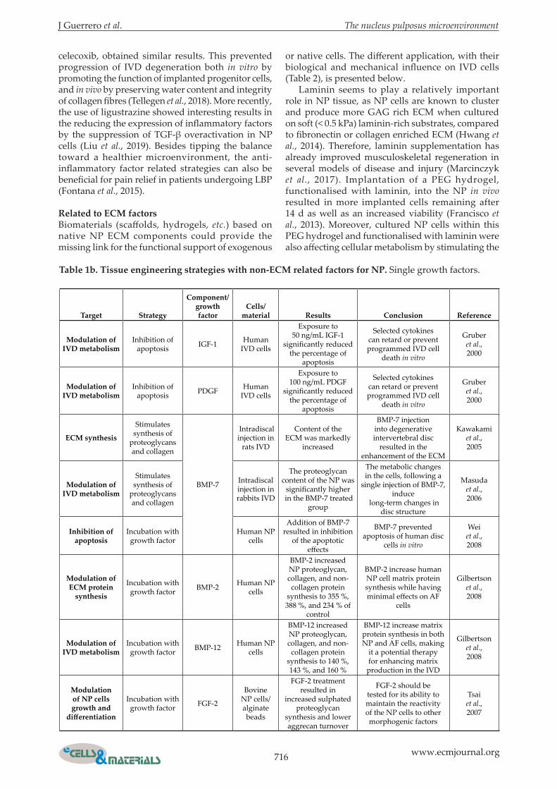
The fabrication/development of scaffolds with chondroprotective supplementation could be an interesting approach to avoid straying any further from the microenvironment of the NP tissue, in case of degeneration. Using a combination of alginate and CaCl2 (in a w/v ratio of 2 % and 0.025 mol/L, respectively), supplemented with glucosamine hydrochloride and chondroitin sulphate A, Foss et al. (2014) showed an improvement in both mechanical and biochemical properties of the engineered hydrogels. However, although the promising results come from a 28-day study with human NP cells, they are only in vitro. Therefore, in vivo studies appear as a necessity to confirm their validation for future applications in the field of regenerative medicine as applied to NP tissue. Recently, many factors that are able to influence the native microenvironment by modulating IVD metabolism in vivo were listed (Yang and Li, 2009). Growth factors could be used as a biological treatment choice for intervertebral disc degeneration (Paesold
et al., 2007). Studies using IGF-1, PDGF, or BMP-7 for their effects on apoptosis inhibition in IVD cells can also be listed (Gruber et al., 2000; Wei et al., 2008). However, the purpose of adding single growth factors (Chujo et al., 2006; Cui et al., 2008; Gilbertson et al., 2008; Hodgkinson et al., 2020; Kawakami et al., 2005; Li et al., 2004; Masuda et al., 2006; Tsai et al., 2007; Walsh et al., 2004) or a combination of them (Akyuva et al., 2019; Henry et al., 2017; Hou et al., 2016; Liang et al., 2012b; Sun et al., 2021) is mostly to stimulate the IVD metabolism through GAG, collagen or non-collagen synthesis. For successful implantation of tissue engineering scaffolds and/or cells within the NP tissue, another angle of attack could be to reduce the inflammation to get closer to the IVD anabolism and native microenvironment. To reach this target, the efficacy of intradiscal controlled release of TAA, an anti-inflammatory drug, in a preclinical degenerated IVD model has been evaluated (Rudnik-Jansen et al., 2019). Another team, using the same approach but using
716 www.ecmjournal.org
J Guerrero et al. The nucleus pulposus microenvironment
celecoxib, obtained similar results. This prevented progression of IVD degeneration both in vitro by promoting the function of implanted progenitor cells, and in vivo by preserving water content and integrity of collagen fibres (Tellegen et al., 2018). More recently, the use of ligustrazine showed interesting results in the reducing the expression of inflammatory factors by the suppression of TGF-β overactivation in NP cells (Liu et al., 2019). Besides tipping the balance toward a healthier microenvironment, the anti-inflammatory factor related strategies can also be beneficial for pain relief in patients undergoing LBP (Fontana et al., 2015).
related to eCm factorsBiomaterials (scaffolds, hydrogels, etc.) based on native NP ECM components could provide the missing link for the functional support of exogenous
or native cells. The different application, with their biological and mechanical influence on IVD cells (Table 2), is presented below. Laminin seems to play a relatively important role in NP tissue, as NP cells are known to cluster and produce more GAG rich ECM when cultured on soft (< 0.5 kPa) laminin-rich substrates, compared to fibronectin or collagen enriched ECM (Hwang et al., 2014). Therefore, laminin supplementation has already improved musculoskeletal regeneration in several models of disease and injury (Marcinczyk et al., 2017). Implantation of a PEG hydrogel, functionalised with laminin, into the NP in vivo resulted in more implanted cells remaining after 14 d as well as an increased viability (Francisco et al., 2013). Moreover, cultured NP cells within this PEG hydrogel and functionalised with laminin were also affecting cellular metabolism by stimulating the
Target strategy
Component/growth factor
Cells/material results Conclusion reference
modulation of ivd metabolism
Inhibition of apoptosis IGF-1 Human
IVD cells
Exposure to 50 ng/mL IGF-1
significantly reduced the percentage of
apoptosis
Selected cytokines can retard or prevent programmed IVD cell
death in vitro
Gruber et al., 2000
modulation of ivd metabolism
Inhibition of apoptosis PDGF Human
IVD cells
Exposure to 100 ng/mL PDGF
significantly reduced the percentage of
apoptosis
Selected cytokines can retard or prevent programmed IVD cell
death in vitro
Gruber et al., 2000
eCm synthesis
Stimulates synthesis of
proteoglycans and collagen
BMP-7
Intradiscal injection in
rats IVD
Content of the ECM was markedly
increased
BMP-7 injection into degenerative intervertebral disc
resulted in the enhancement of the ECM
Kawakami et al., 2005
modulation of ivd metabolism
Stimulates synthesis of
proteoglycans and collagen
Intradiscal injection in rabbits IVD
The proteoglycan content of the NP was
significantly higher in the BMP-7 treated
group
The metabolic changes in the cells, following a
single injection of BMP-7, induce
long-term changes in disc structure
Masuda et al., 2006
inhibition of apoptosis
Incubation with growth factor
Human NP cells
Addition of BMP-7 resulted in inhibition
of the apoptotic effects
BMP-7 prevented apoptosis of human disc
cells in vitro
Wei et al., 2008
modulation of eCm protein
synthesis
Incubation with growth factor BMP-2 Human NP
cells
BMP-2 increased NP proteoglycan, collagen, and non-
collagen protein synthesis to 355 %, 388 %, and 234 % of
control
BMP-2 increase human NP cell matrix protein synthesis while having minimal effects on AF
cells
Gilbertson et al., 2008
modulation of ivd metabolism
Incubation with growth factor BMP-12 Human NP
cells
BMP-12 increased NP proteoglycan, collagen, and non-
collagen protein synthesis to 140 %, 143 %, and 160 %
BMP-12 increase matrix protein synthesis in both NP and AF cells, making
it a potential therapy for enhancing matrix
production in the IVD
Gilbertson et al., 2008
modulation of np cells growth and
differentiation
Incubation with growth factor FGF-2
Bovine NP cells/alginate beads
FGF-2 treatment resulted in
increased sulphated proteoglycan
synthesis and lower aggrecan turnover
FGF-2 should be tested for its ability to maintain the reactivity of the NP cells to other morphogenic factors
Tsai et al., 2007
Table 1b. Tissue engineering strategies with non-eCm related factors for np. Single growth factors.

J Guerrero et al. The nucleus pulposus microenvironment
717 www.ecmjournal.org
Target strategyComponent/growth factor Cells/material results Conclusion reference
multiple growth factors
potentiation of exogenous
cells
Incubation with growth
factorBMP-2/PRP
Human BMSCs/Rabbit IVD/PRP
gel
The discs treated with BMP2-transduced BMSCs exhibited relatively well-
preserved NP structure
The combined use of these two agents
can significantly promote repair of the degenerated discs in
vivo
Hou et al., 2016
modulation of ivd
metabolism
Time control release of
growth factorBMP-2/IGF-1
Human IVD cells/polyvinyl alcohol-based
polymeric scaffold
Increased in the number of cells and the degree of ECM
development
Alternative method for intervertebral disc
administration of growth factors
Akyuva et al., 2019
modulation of ivd
metabolism
The in situ injection of
growth factors targeting
intervertebral disc
degenerative process
TGF-β1 and GDF-5
Human ADCs/Pullulan
microbeads into a cellulose-based
hydrogel
Sustained release of both growth factors,
for up to 28 d
Biphasic system may be a promising candidate for the development of an innovative bioactive delivery system for IVD regenerative medicine
Henry et al., 2017
modulation of ivd
metabolism
Incubation with growth
factors
FGF-2/dexamethasone
Rat MSCs/3D poly(lactide-co-glycolide) constructs/
heparin/poly(L-lysine) nanoparticles
Expression of disc-matrix proteins was significantly higher and of osteogenic
differentiation marker was decreased
Microspheres could be used as a scaffold to
improve cells growth and differentiating into
NP like cells
Liang et al., 2012b
regeneration of ivd
Anatomically correct IVD
scaffold3D Printing
CTGF/TGF-β3/polydopamine
nanoparticles/rat BMSCs
Fabricated and implanted IVD
scaffold exhibited a zone-specific matrix that displayed the
corresponding histological and immunological
phenotypes of IVD
Clinical application potential of the dual
growth factors-releasing IVD scaffold
fabricated by 3D bioprinting
Sun et al., 2021
Anti-inflammatory strategies
reduction of inflammation
Intradiscal delivery of
nonsteroidal anti-
inflammatory drug
Celecoxib (selective COX-
2 inhibitor)
Preclinical canine IVD
model/polyesteramide microspheres
No evidence of adverse effects on CT and MRI or macroscopic
evaluation of IVDs
Intradiscal controlled release of celecoxib
from polyesteramide microspheres
prevented progression of IVD degeneration
Tellegen et al., 2018
reduction of inflammation
Prolonging corticosteroid presence by controlled
release from biomaterials
TAA
Preclinical canine IVD
model/poly(esteramide)
microsphere
The low dosage of TAA microspheres
significantly reduced nerve growth factor immunopositivity in
degenerated NP tissue
Potential applicability for pain relief,
although beneficial effects were absent on
tissue degeneration
Rudnik-Jansen et al., 2019
modulation of ivd
metabolism
Reduce the expression of inflammatory
factors and TGF-β1
Ligustrazine (LIG)
Lumbar spinal instability mouse
model
A dose of 10-5 M LIG could
attenuated Smad2/3 phosphorylation
in IVD ex vivo and suppressed pSmad2/3,
CCN2, and ACAN expression in NP cells
in vitro
Ligustrazine could prevents IDD by
suppression of TGF-β overactivation in NP
cells
Liu et al., 2019
Table 1c. Tissue engineering strategies with non-eCm related factors for np. Multiple growth factors and anti-inflammatory strategies.

718 www.ecmjournal.org
J Guerrero et al. The nucleus pulposus microenvironment
Target strategyComponent/growth factor
Cells/material results Conclusion reference
laminin related
laminin
Scaffold with the ability to promote
or maintain an immature NP cell
phenotype
Photo-crosslinkable poly(ethylene
glycol)-laminin 111
(PEG-LM111) hydrogel
Immature porcine NP
cells
Promotion of cell clustering and increased levels of sGAG
production
LM111-functionalised hydrogels may
promote or maintain the expression of specific markers
characteristic of an immature NP cell
phenotype
Francisco et al., 2014
laminin
Biomaterials that retain delivered cells, promote cell survival,
and maintain or promote an NP
cell phenotype in vivo
Injectable, laminin-111
functionalised poly(ethylene
glycol)
Porcine NP cells
Higher NP cell retention in
cultured IVD explants within a PEG-LM111
carrier compared to cells in liquid
suspension
Injectable laminin-functionalised
biomaterial may be an easy to use carrier for
delivering cells to the IVD
Francisco et al., 2013
Collagen related
Collagen type ii
Delivery system with genipin as
the cross-linking agent
Collagen type II/chondroitin
sulphate composite hydrogel
RatADSCs/rat coccygeal
vertebrae degeneration
model
Disc height, water content,
ECM synthesis, and structure of the degenerated NP were partly
restored
Delivery system uses minimally
invasive approaches to promote the regeneration of degenerated NP
Zhou et al., 2018
Collagen type ii
Influence the bioactivity of
transplanted cells
Hydrogels composed of a mix of collagen types I and II
Human ADSCs
Expressions of SOX9, aggrecan,
and collagen type II were increased in a collagen type
II dependent manner
Collagen type II significantly
ameliorates human ADSC differentiation
into NP cells and promotes ECM
synthesis
Tao et al.,2016
Collagen type ii
Mimic the NP microenvironment
and promote differentiation
Microgels of collagen type II and hyaluronan
Rabbit ADSCs
Higher expression of
collagen type II, aggrecan, SOX9, and low levels of
collagen type I
By tuning microgels’ properties, it is
possible to influence cells phenotype
and differentiation ability
Fontana et al., 2014
Cells carrier
Administration using minimal
invasive surgery
Alginate-collagen type I composite
porous scaffold
Porcine MSCs and
AF cells
Alginate-collagen porous scaffolds supported cell
proliferation and ECM deposition
Advantages of incorporating
collagen to enhance cell migration and
proliferation in porous scaffolds
Guillaume et al., 2015
restore native eCm
Repair the degenerative environment
Collagen type II - hyaluronan - chondroitin-
6-sulphate tri-copolymer
construct
Rabbit nucleotomy IVD model
Narrowing of the intervertebral
disc space was significantly
retarded by the cell-scaffold
hybrids implantation
Maintenance of disc height and
restoration using NP cell-seeded tri-
copolymer implants
Huang et al., 2011
recreate the
resilient and
hydrophilic nature of the eCm
Construction of a chemically
stabilised composite hydrogel
Elastin/GAG/collagen type I
Human ADSCs
Cytocompatible and support the differentiation
towards an NP cell-like phenotype
Successful for host cell infiltration and active remodelling after implantation
Mercuri et al., 2014
Table 2a. Tissue engineering strategies with eCm related factors for np. Laminin and collagen related.
J Guerrero et al. The nucleus pulposus microenvironment
719 www.ecmjournal.org
Target strategyComponent/growth factor
Cells/material results Conclusion reference
sAph related
mimicking the natural
eCm for cell delivery
3D injectable hydrogel
Short self-assembling
peptide hydrogels
Bovine NP cells/Graphene
oxide
Promote high cell viability
and retained cell metabolic activity in
3D over the 7 d of culture
These hybrid hydrogels harbour
significant potential as injectable
scaffolds for the in vivo delivery of NP
cells
Ligorio et al., 2019
mimicking the natural
eCm for cell delivery
3D injectable hydrogel
Short self-assembling
peptide hydrogels
Bovine NP cells
Upregulation of KRT8, KRT18, FOXF1, higher
cell viability, and increase in aggrecan and collagen type II
deposition
SAPH had comparable strength to the native tissue,
was injectable, restored the IVD cell phenotype and stimulated deposition of
appropriate matrix components
Wan et al., 2016
mimicking the natural
eCm
A link N nanofibre scaffold
Short self-assembling
peptide hydrogels
Rabbit NP cells/mixing
peptide solution of RLN and RADA16
Scaffold exhibited little cytotoxicity and
promoted NP cells adhesion
Functionalised nanofibre scaffold
had excellent biocompatibility
and bioactivity with rabbit NP cells
Wang et al., 2012
decellularised eCm
Creation of an
appropriate micro-
environment for long term cell survival
Decellularised ECM Hydrogel Bovine NP/rat
ADSCs
Significant increase in NP marker genes expression without
the addition of exogenous biological
factors
Decellularised NP hydrogel
has low toxicity and inducible
differentiation, could serve as a bio-scaffold, bio-
carrier, and three-dimensional culture
system
Yu et al., 2020
rescue the degenerated
ivdDecellularised
ECMBiological scaffold
Porcine NP/human MSCs
Good cytocompatibility ex vivo and decelerated the degeneration of
the IVD in vivo
Naturally-derived ECM material that could induce MSCs
into NP cells
Xu et al., 2019
replace degenerated
ivdsDecellularisation
IVDs
Natural biological scaffold
Bovine IVD
Efficient cells removal and GAG
retainment after decellularisation
Possible to create a cell-free human
IVD biological scaffold with
attached bone using decellularisation
technology
Norbertczak et al., 2020
Treat disc degeneration
Use of matrix niche factor for exogenous cells differentiation
NP cell-derived acellular
matrix and collagen micro-encapsulation
Rabbit degenerative disc/human
MSCs
Acellular matrix supported MSC
survival and matrix production, and up-regulated the gene expression of NPC markers including
collagen type II and glypican 3
Potential application of the NPC-derived matrix microsphere as a favourable cell
carrier
Yuan et al., 2013
ivd repair
Instruct the regenerative cell
component to produce tissue-
specific ECM
Injectable biomimetic disc derived
self-assembled ECM hydrogels
Chondroitin sulphate
and collagen type II/
porcine nasal chondrocytes
Increase in GAG production
Inclusion of chondroitin sulphate within material aids
the preservation of a rounded cell morphology and
enhances their ability to synthesise
NP-like matrix
Borrelli and
Buckley, 2020
Table 2b. Tissue engineering strategies with eCm related factors for np. SAPH related and decellularised ECM.

720 www.ecmjournal.org
J Guerrero et al. The nucleus pulposus microenvironment
Target strategyComponent/growth factor
Cells/material results Conclusion reference
Natural scaffolds
restore functions of both the Af
and np
Biphasic biomaterial
Silk protein for the AF and fibrin/hyaluronic
acid gels for the NP
Porcine AF cells/porcine
articular chondrocytes
Formation of AF band NP-tissue like within the hydrogel after 6
weeks of culture
This biphasic scaffold was
effective in the formation of the total IVD in vitro
Park et al., 2012
restore physiological functionality of damaged
ivd
Biomimetic scaffold
Alginate hydrogel
encapsulating cells
Rabbit NP cells/dorsal
space of nude mice
NP cells colonised in the alginate hydrogel
NP cells colonised the biomimetic
scaffold and form ECM similar to native NP tissue
Du et al., 2019
mimics the mechanical
properties of the human np tissue
Thermo-sensitive hydrogel
Chitosan-basedBovine NP
cells/human lumbar IVDs
Mechanical properties similar to the human
NP tissue
Provided suitable environment to
maintain NP cells alive and active,
and induced production of GAG by the encapsulated
cells
Alinejad et al., 2019
mimics the mechanical
properties of the human np tissue
Chitosan hydrogel
Chitosan hydrogel
Nude mice/New Zealand
rabbit NP cells
Mechanical property meets the requirement
of the normal NP
Cells on the tissue engineered NP
expressed ECM, which indicated
that the cells maintained their
biological function
Yuan et al., 2019
regeneration of ivd micro-
environment
Acellular and cellular tissue-
engineering strategies
Ionic- and photo-
crosslinked methacrylated
gellan gum hydrogel
Rat lung fibroblasts (L929 cells)
Non-cytotoxic in vitro
Promising biomaterials to be used in IVD
tissue-engineering strategies
Silva- Correia
et al., 2011
substitute the inner ivd part
Injectable hydrogel
Ionic- (iGG-MA) and photo-
crosslinked (phGG-MA)
methacrylated gellan gum hydrogels
Human BMSCs
and nasal chondrocytes
No cytotoxicity with MSCs and nasal
chondrocytes, and no inducion of pro-
inflammatory responses in endothelial cells
Potential use of modified gellan
gum-based hydrogel as a
suitable material in NP tissue engineering
Tsaryk et al., 2017
np regeneration
Injectable biomaterials
act as carriers of growth
factors
Gelatine methacryloyl microspheres
Rat ADSCs/GDF-5
Enhancement of the in vitro differentiation of cells into NP-like
phenotypes
Attenuation of in vivo degeneration
of rat IVD, maintenance of NP tissue integrity and acceleration of ECM
synthesis
Xu et al., 2020
enhancing intra-np residence
time of therapeutic
drugs
Bind with the intra-NP negatively
charged groups
Positively charged
avidin grafted branched dextran
nanostructures
Bovine NP
Month-long retention of cationic nanostructures
within the NP following intra-discal
administration
Way for effective clinical translation
of potential therapeutics
Wagner et al., 2020
minimally invasive repair of
degenerative ivd
Injectable tissue
engineered NP
Combination of PRP gel
scaffold and cells
Rabbit ADSCs
The level of GAG, gene expression of HIF-1α,
aggrecan, collagen type II was higher when cells
were seeded with the scaffold
Feasible method for construction of autologous
injectable tissue engineered NP
Zhang et al., 2020
Synthetic scaffolds
increase survival and function of
np cells
Tissue stiffness
3D matrices of varying degrees of stiffness
Porcine NP cells
Matrices with a low shear storage
modulus (G’ = 1 kPa) promoted significantly
proliferation and chondrogenic differentiation
Effect of the matrix modulus on the fate
of NP progenitor cells
Navaro et al., 2015
Table 2c. Tissue engineering strategies with eCm related factors for np. Natural and synthetic scaffolds.

J Guerrero et al. The nucleus pulposus microenvironment
721 www.ecmjournal.org
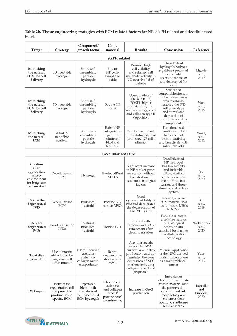
expression of N-cadherin and cytokeratin 8 (Francisco et al., 2014). Both studies provide evidence that the use of laminin in engineered scaffolds could positively influence NP cell proliferation and function and should be studied in more detail to improve NP tissue engineering strategies. Another vital component of IVD ECM that can be used as scaffolding material is collagen and particularly collagen type II (Zhou et al., 2018). This main ECM component promotes the differentiation of NP progenitor cells, especially into the chondrogenic lineage (Tao et al., 2016). When collagen type II is used at high concentrations for the scaffolding material, ASCs express a high level of collagen type II, aggrecan, SOX9, and low levels of collagen type I in response (Fontana et al., 2014; Tao et al., 2016). Collagen (mainly type II) should be used as the only and main component of the scaffold. Its implementation at a significant level should be enough to generate advantages such as enhancing cell migration and proliferation in porous scaffolds, which could be used for implemented tissue repair strategies (Guillaume et al., 2015; Huang et al., 2011; Sakai and Grad, 2015; Sarker et al., 2015; Yang and Li, 2009). Merging/combining most of the components of the NP microenvironment – such as elastin, collagen, and GAG – within a unique engineered scaffold/hydrogel could be a huge step towards successful NP tissue engineering applications. A chemically-stabilised elastin-GAG-collagen composite hydrogel shows great in vivo biocompatibility potential, with host cell infiltration and active remodelling of the NP tissue, after 4 weeks of in vivo subcutaneous implantation in rats (Mercuri et al., 2014). Studies should now investigate the feasibility of this composite hydrogel for the replacement and regeneration of the NP. Very recently, short SAPHs have attracted significant interest as they can mimic the natural ECM of the targeted tissue, holding significant promise for the ab initio design of the cellular microenvironment (Ligorio et al., 2019). Moreover, SAPHs are beneficial mechanically, as they present a strength comparable to the native NP tissue, are injectable, can restore the IVD cell phenotype, and stimulate the deposition of appropriate ECM components (Wan et al., 2016). These hydrogels open up new possibilities as they can be perfectly integrated into the NP microenvironment and offer the possibility of being customised by functionalisation (Wang et al., 2012). Another approach, that has appeared recently, is the use of decellularised matrices instead of combining several main components. These matrices preserve the NP ECM biological components and microstructure and, therefore, should perfectly mimic the NP microenvironment (Yu et al., 2020; Yuan et al., 2013). A protocol for the decellularisation of porcine NP tissue has been developed, which results in a decrease in IVD degeneration following in vivo implantation (Xu et al., 2019). This, naturally derived, bioactive ECM material could serve as a
potential treatment option for degenerated discs (Borrelli and Buckley, 2020). Nevertheless, it still needs to be investigated in a human setting or for GMP rules. However, very recently and for the first time, the creation of decellularised whole human IVD with attached bone was achieved (Hodgkinson et al., 2020). Further investigations are required before this technology can be taken forward as an implantable regenerative solution for IVD replacement in clinical applications. In conclusion, key components of the NP microenvironment from an IVD play an important role in ongoing and future approaches for NP tissue engineering. The use of components that are already present in the native microenvironment seem to have a high rate of success. However, the development of scaffolds based on natural materials such as silk (Park et al., 2012), alginate (Du et al., 2019), chitosan (Alinejad et al., 2019; Yuan et al., 2019) or gellan gum (Silva-Correia et al., 2011; Tsaryk et al., 2017) also show good results but are more different from native components. Interesting and positive effects demonstrated by those different strategies, to promote IVD and NP regeneration, rely on the concept of optimally mimicking the microenvironment requiring treatment, and remain to be further elucidated (Guilak et al., 2009). Although synthetic scaffolds can provide functional support for progenitor cell transplantation, their mechanical influence on IVD progenitor cells should not be neglected (Melrose, 2016). The elasticity of a scaffold can influence the differentiation of progenitor cells (Zhu et al., 2016); for example, a stiff synthetic matrix can promote osteogenesis and inhibit chondrogenesis of NP progenitor cells (Navaro et al., 2015). Therefore, biomaterials should be designed with appropriate elasticity and stiffness to facilitate progenitor cell function.
The clinical problem – still in between bench and bedside
LBP compromises a complex of symptoms with varying underlying pathologies, including micro- and macro-instability, disturbing of neurological structures, and degeneration of facet joints, inflammatory conditions, neoplasm, and deformity. A widely accepted concept considers the IVD itself as the generator of pain, which is referred to as discogenic pain. However, the underlying biochemical or physical patho-mechanisms of LBP related solely to the IVD remain mostly unclear. Moreover, many patients with radiological abnormalities of the IVD do not suffer any clinical symptoms (Boden et al., 1990). Several in vitro studies found an upregulation of inflammatory markers such as TNF-α and IL-1β in patients with suspected discogenic LBP. Histological studies revealed the ingrowth of nerves and vessels into the IVD. This could be the beginning of a degenerative cascade (Mosley et al., 2017), with pain

722 www.ecmjournal.org
J Guerrero et al. The nucleus pulposus microenvironment
signals transmitted continuously into the central nervous system. Thus, patients perceive pain because of the IVD-degeneration progression. Despite these observations from in vitro and preclinical studies, the clinical diagnosis of discogenic pain is difficult. A possible approach to the diagnostic scheme for patients with suspected discogenic LBP is provocative discography. After puncturing the AF with a thin needle, saline and fluoroscopic agents are injected, increasing intradiscal pressure. Subsequently, the provocation of typical pain confirms the diagnosis of discogenic LBP. Drawbacks of provocative discography include the absence of objective findings with the positive result relying purely on subjective perception and response of the individual patient. Moreover, several studies demonstrated an acceleration of IVD degeneration following discography, likely as a result of iatrogenic AF injury during the procedure (Carragee et al., 2009; Cuellar et al., 2016). Therefore, the use of provocative discography as a diagnostic test has diminished recently. In cases where discogenic pain is suspected to be the underlying cause of LBP in a clinical routine, patient treatment focuses on the management of symptoms and not on addressing the underlying pathomechanisms. First-line therapy of patients with pain related to IVD degeneration consists of conservative treatment modalities, including physiotherapy and pain management with non-steroid anti-inflammatory drugs, opioids, or epidural steroid injections (Airaksinen et al., 2006). If conservative management fails, surgical treatment is often performed. The most common surgical intervention of degenerative disc disease is IVD removal, replacement with implants, and fusion using bone grafting to permanently connect two or more vertebrae of the spine. Based on laboratory and preclinical findings, IVD-maintaining surgical approaches have been
increasingly studied recently. Attempts to address IVD regeneration invasively comprise a variety of techniques that specifically aim at modulating the degeneration cascade of the IVD (Fig. 4). Access to the IVD to apply these techniques is either achieved by intradiscal injection or minimally-invasive or open-surgical approaches. However, only a few methods have made their way into clinical practice. A brief overview of ongoing clinical trials of the most promising strategies will now be provided, focussing on the ECM, and concentrating upon cell-based therapies, growth factors, and biomaterials.
Cell-based therapies
The cell-based therapy approach (Table 3) utilises the transplantation of cells into the IVD by puncturing the AF. The injected cells release anti-inflammatory factors/secretome and produce ECM (Wangler et al., 2019). Theoretically, this leads to IVD regeneration, with reduced pain and improved function, for the patient. There are mainly 5 categories of cell sources used to treat IVD degeneration: (1) IVD-derived cells, (2) chondrocyte-like cells, (3) MSCs, (4) iPSCs, and (5) ES cells (Sakai and Schol, 2017). By far, most published studies and ongoing clinical trials focus on MSCs (Table 3). However, any clinical evidence for positive effects still remains unclear. What has already been shown is that most of these procedures are safe (Orozco et al., 2011; Wei et al., 2014). The most often used donor site of autologous MSCs is the iliac crest, meaning an additional invasive procedure for the patient. The harvested cells are then either directly injected or grown in the lab and subsequently reinjected into the IVD. A meta-analysis of 6 studies (using either MSC or chondrocytes) showed an improvement of pain in the ODI as an outcome parameter for function (Wu et al., 2018).
fig. 4. overview of the most recent applications used for clinical trials in the context of degenerative ivds and lbp. Clinical therapies can be divided into 3 different strategies: (i) cell-based with the use of IVD-derived cells, chondrocyte-like cells, MSCs, iPSCs or ES cells, (ii) growth factors-based with the use of GDF-5 or PRP and to finish, (iii) biomaterials-based with the use of Gelstix™ hydrogel, Biostat Biologx® fibrin sealant and VAST.

J Guerrero et al. The nucleus pulposus microenvironment
723 www.ecmjournal.org
Cells studyreference/
Web ref. ninclusion
criteriastudy design material outcome
follow up Conclusion/status
Clinical trial number
AsCs
Autologous adipose derived stem cell therapy for intervertebral
disc
Han et al., 2014 and
Kumar et al., 2017
1019 and 70
years Pfirrmann’s grade III–IV
Single centre,
single-arm, phase I
Intradiscal injection
combined hyaluronic
acid derivative and AT-MSCs
tissue fill
AS, ODI, Short Form-36 (SF-36),
and imaging (lumbar
spine X-ray imaging and
MRI)
12 months
Combined implantation of
AT-MSCs and HA derivative in chronic
discogenic LBP is safe and tolerable.
However, the efficacy of combined AT-MSCs
and HA should be investigated in a
randomised controlled trial in a larger
population
NCT02338271
bmsCs
Efficacy of intradiscal injection
of BM-MSC RESPINE
Web ref. 26 112
18 to 60 years Pfirrmann’s
score modified
Griffith et al. (2007) grade
4 to 7
RCT, multicentre
Intradiscal injection,
cell dose 20 ± 5 × 10⁶ cells suspended in 2 mL of
HypoThermosol isotonic
transport solution
VAS pain, ODI
12 months
No results published yet
Recruiting (until December 2021)
NCT03737461
Treatment of DDD with allogenic
mesenchymal stem cells (MSV)
Web ref. 4 and
Noriega et al., 2017
24
18 to 75 years Disc
degeneration with loss of
min 20 % disc height
Phase I-II trial
BMSCs expanded using the Valladolid
IBGM procedure
VAS pain, ODI, SF-12
12 months
Allogeneic MSC therapy may be a valid alternative
for the treatment of degenerative disc
disease that is more logistically convenient
than the autologous MSC treatment. The
intervention is simple, does not require
surgery, provides pain relief, and significantly improves disc quality
NCT01860417
Bone marrow concentrate intradiscal injection
for chronic discogenic LBP
Web ref. 15 60
18 to 55 years Chronic low
back pain abnormal disc pathology in MRI or CT
RCTIntradiscal injection of
bone marrow concentrate
VAS pain, ODI
12 months
No results published yet.
Recruiting (until January 2021)
NCT03340818
Single intradiscal injection of
BMACWeb ref. 3 20
18 to 60 years lumbar
disc height loss < 50 % (Modified Pfirrman
grade ≤ 7) in MRI
Prospective study, single centre
Autologous BMAC is a cellular rich fraction of
bone marrow aspirate
VAS pain, ODI, Cell viability testing,
CFU-f assay
12 months
No results published yet
Recruiting (until September 2021)
NCT03912454
IVD repair by autologous
mesenchymal bone marrow
cells
Web ref. 15 and
Orozco et al., 2011
10
18 to 60 years lumbar
disc height loss < 50 % (Modified Pfirrman
grade ≤ 7) in MRI
Phase I trial
Autologous expanded bone marrow MSC injected into
the NP
VAS pain, ODI, SF-36,
MRI 12
months
MSC therapy may be a valid alternative treatment for chronic back pain caused by
DDD. Advantages over current gold standards
include simpler and more conservative
intervention without surgery,
preservation of normal biomechanics, and same or better pain
relief
NCT03340818 EudraCT
2008-001191-68
mpCs
MPCsWeb ref. 11
and Amirdelfan et
al., 2021100
18 years and older L1-S1 Disc
degeneration confirmed by
MRI
RCT, multicentre
Immuno-selected, culture-
expanded, nucleated,
allogeneic adult MPCs when
combined with hyaluronic acid
VAS pain, ODI, WPAI,
MRI36
months
Evidence that intra-discal injection of
MPCs could be a safe, effective, durable and
minimally invasive therapy for subjects who have chronic
LBP associated with moderate DDD
NCT01290367
Rexlemestrocel-L alone or
combined with hyaluronic acid in subjects with
chronic LBP
Web ref. 12 404
18 years and older
L1 to S1 modified
Pfirrmann score 3 to 6
(MRI)
RCT, multicentre
Mesoblast’s rexlemestrocel-L
alone or combined with
hyaluronic acid/AMPCs
VAS pain, ODI
24 months Active, not recruiting NCT02412735
discogenic cells
Safety and preliminary
efficacy of IDCT, a treatment for symptomatic
LDD
Web ref. 8 and
Silverman et al., 2020
38
18 to 75 years L3 to S1
symptomatic disc
degeneration
RCT, multicentre
Injection of IDCT
discogenic cells + sodium hyaluronate
vehicle
Adverse events, VAS pain, ODI, JOABPEQ
2months
Preliminary Conclusion: That
intradiscal injection of discogenic cells may be a viable treatment for human DDD. The
cells produce ECM that may rebuild the
depleting tissue within degenerating discs.
The cells do not pose any significant safety
concerns
NCT03955315
Web ref. 7 and
Silverman et al., 2020
60Adverse
events, VAS pain, ODI,
24 months NCT03347708
Autologous stem cells
A single dose of BRTX 100 for
patients with chronic LDD
Web ref. 23 99
18 to 60 years Modified Pfirrmann
score of 2 to 6 on MRI
RCT
Autologous hypoxic
cultured MSCs collected from the
patient’s bone marrow and
co-administered with an
autologous biomaterial
carrier (human platelet lysate)
VAS pain, ODI
12 months
Not yet recruiting. Estimated study
completion August 2023
NCT04042844
Table 3. recent clinical trials addressing cell-based materials for disc regeneration.

724 www.ecmjournal.org
J Guerrero et al. The nucleus pulposus microenvironment
A meta-regression analysis identified a significant preference for MSC over chondrogenic cells (Schol and Sakai, 2019; Wu et al., 2018). Because of the need for high-quality clinical data, the RESPINE-project was funded by the European Union’s funding programme for research and innovation, Horizon 2020. The project started in 2017 and aimed at verifying the efficacy of an allogeneic intervertebral MSC-based therapy by providing a high-level clinical trial, including 112 patients with pain related to disc degeneration. No results have yet been published.
Growth factors
Injection of growth factors could support homeostasis of IVD cells and, therefore, enhance disc regeneration. In addition to many in vitro and animal studies (Kennon et al., 2018) there are only a few ongoing clinical trials (Table 4). A multicentre, randomised, blinded, and placebo-controlled trial, provided by DePuy Synthes, investigated the effect of rhGDF-5. The results, posted on clinicaltrails.org (Web ref. 17), showed a higher dosage of rhGDF-5 (2.0 mg/disc) with an improvement in pain and quality of life measurements but it also had a higher risk profile with more (serious) adverse events (Kennon et al., 2018). As a combination of different growth factors, PRP that has been already studied extensively for cartilage regeneration, has also been studied for LBP. Even though it seems to be a safe procedure (Levi et al., 2016; Tuakli-Wosornu et al., 2016), results from a prospective, non-randomised trial revealed a relatively low-success rate (pain and ODI) under 50 % (Levi et al., 2016). Moreover, a prospective, randomised-controlled study on intradiscal PRP injection showed an improvement of pain and function after a one-year follow-up (Tuakli-Wosornu et al., 2016). Nevertheless, adequately designed randomised controlled trials are still needed to clarify the safety as well as long-term benefits of intradiscal PRP injection (Buser et al., 2019).
drugs
With the various mechanisms associated with IVD degeneration in mind, some current research and clinical applications have focused on different approaches for pain treatment and/or reversing disc degeneration. One aspect is the application of drugs. In addition to analgesics, which with their low molecular weight are often used as a first resort to relieve LBP associated with IVD degeneration, various novel treatments are also being evaluated in preclinical models (Cherif et al., 2020; Cunha et al., 2020; Hu et al., 2019; Wang et al., 2020a). Among these, inflammatory cytokine and enzyme inhibitors are frequently used as therapeutic agents, but so far these agents have shown no or only limited effectiveness
and an increased risk of adverse events (Tryfonidou et al., 2020). However, several clinical trials are ongoing targeting different critical points of the diseased microenvironment (Table 5).
biomaterials
One major problem that must be addressed for regenerative strategies of the IVD is the loss of the NP water content. The dehydration of the IVD tissue is an early sign of disc degeneration, seen as a so-called “black disc”, with low water content and, therefore, a dark instead of a light NP, in MRI images (Pfirrmann et al., 2001). Consequently, hydrophilic materials such as hydrogels that might compensate for the decreased water content have been extensively studied both in research (Buckley et al., 2018) and clinical trials (Table 6). The origin of the hydrogels can either be natural or synthetic. Natural hydrogels (including alginate, collagen or fibrin-clot) can serve as a cell-carriers (e.g. for MSCs) by providing a 3D cytocompatible environment. However, their inferior mechanical properties are a significant disadvantage (Frauchiger et al., 2017; Peroglio et al., 2013). Synthetic hydrogels, on the other hand, have better mechanical properties. Such gels are, for example, the thermo-responsive hydrogel pNIPAM, PEG or the very promising group of silk scaffolds. Frauchiger et al. (2017) reviewed the different hydrogels in detail. For clinical use, such a formulation needs to be injectable and has to be applied intradiscally by a minimally invasive approach. However, there are only a few studies on such injectable materials for humans (e.g. GelStix™, an intradiscal admitted hydrogel) (Ceylan et al., 2019). A randomised, placebo-controlled, multicentre study indicated a significant reduction of pain, function, and high patient satisfaction (Ceylan et al., 2019). Also, internal disruption of the disc can be found in degenerated IVDs. Therefore, a clinical study investigated the intradiscal injection of fibrin sealant (Biostat Biologx®) (Yin et al., 2014). A prospective placebo-controlled pilot study with a two-year follow-up demonstrated a safe application and improved pain and function (Yin et al., 2014). The VAST clinical trial aimed to safely supplement tissue loss to degenerate IVDs. By injection of a supplemental viable disc matrix, improved pain, and function at one-year was achieved (Beall et al., 2020). Despite all these increasing efforts to investigate degenerative processes in the IVD and straight-forward clinical trials, translation into everyday clinical practice with a direct impact on patient management is sparse. Moreover, care must be taken to measure the outcome parameters. Most of the studies mentioned above did not include any radiographic, especially MRI imaging, in their outcome measures. Therefore, it is not possible to assume the influence on the IVD microenvironment and especially restoration of the ECM.

J Guerrero et al. The nucleus pulposus microenvironment
725 www.ecmjournal.org
Com
pone
nts
stud
yr
efer
ence
/W
eb re
f.n
incl
usio
n cr
itera
stud
y de
sign
mat
eria
lo
utco
me
follo
w
upC
oncl
usio
n/st
atus
Clin
ical
tria
l nu
mbe
r
prp
PRP
for L
BPW
eb re
f. 13
3020
to 6
0 ye
ars
LDD
con
firm
ed
by M
RISi
ngle
cen
tre
Intr
adis
cal i
njec
tion
of P
RPVA
S pa
in, O
DI,
4 m
onth
sN
o re
sults
pub
lishe
d ye
t. C
ompl
eted
(Mar
201
9)N
CT0
3197
415
Intr
a-di
scal
Inje
ctio
n of
PRP
fo
r LBP
(MO
DI-P
RP)
Web
ref.
2512
6
18 to
60
year
s lu
mba
r (st
ages
<
5 of
Pfir
-rm
ann’
s sc
ore)
in
MRI
RCT,
sin
gle
cent
re
Inje
ctio
n of
2 m
L of
au
tolo
gous
PRP
into
th
e m
edia
n po
rtio
n of
the
susp
ecte
d di
sc
unde
r rad
iogr
aphi
c gu
idan
ce.
OD
I, RM
Q, P
ASS
, VA
S pa
in, E
Q5D
, SF
-36
3-12
m
onth
sN
o re
sults
pub
lishe
d ye
t Re
crui
ting
(unt
il Se
p 20
22)
NC
T037
1252
7
Lum
bar i
ntra
disc
al P
RP
inje
ctio
ns: A
pro
spec
tive,
do
uble
-blin
d, ra
ndom
ised
co
ntro
lled
stud
y
Tuak
li-W
osor
nu
et a
l., 2
016
47
Gra
de 3
or 4
an
nula
r fiss
ure
as d
eter
min
ed b
y di
scog
raph
y.
Con
cord
ant p
ain
on d
isco
grap
hy
RCT
Sing
le in
ject
ions
of
auto
logo
us P
RP in
to
sym
ptom
atic
deg
en-
erat
ive
IVD
s
FRI,
NRS
for p
ain,
SF
-36,
mod
ified
N
ASS
Out
com
e Q
uest
ionn
aire
2 m
onth
s
Intr
adis
cal P
RP s
how
ed
sign
ifica
nt im
prov
emen
ts in
FRI
, N
RS B
est P
ain,
and
NA
SS p
atie
nt
satis
fact
ion
scor
es o
ver 8
wee
ks
com
pare
d w
ith c
ontr
ols.
Tho
se
who
rece
ived
PRP
mai
ntai
ned
sign
ifica
nt im
prov
emen
ts in
FRI
sc
ores
N/A
Intr
adis
cal P
RP in
ject
ion
for
chro
nic
disc
ogen
ic L
BPLe
vi et
al.,
20
1622
18 y
ears
and
ol
der
posi
tive
disc
og-
raph
y
Pros
pect
ive
Tria
l
Inje
ctio
n of
1.5
mL
of a
utol
ogou
s PR
P in
ject
ed in
to th
e di
scVA
S pa
in, O
DI
6 m
onth
s
Prel
imin
ary
6 m
onth
find
ings
, us
ing
stri
ct c
ateg
oric
al s
ucce
ss
crite
ria,
for i
ntra
disc
al P
RP
as a
trea
tmen
t for
pre
sum
ed
disc
ogen
ic L
BP
Ster
ling
IRB,
Tr
ial N
o. 4
143-
001S
rhG
df-
5
Intr
adis
cal r
hGD
F-5
phas
e I/
II c
linic
al tr
ial
Web
ref.
1632
18 y
ears
and
ol
der
L3/L
4 to
L5/
S1
disc
ogen
ic p
ain
confi
rmed
by
disc
ogra
phy
Phas
e I/I
Ia,
mul
ticen
tre
Intr
adis
cal i
njec
tion
of A
PI (r
hGD
F-5)
, 0.
25 m
g an
d 1
mg
Neu
rolo
gica
l as-
sess
men
t, ad
vers
e ev
ents
, OD
I, VA
S pa
in, S
F-36
,
12
mon
ths
Com
plet
ed 2
016,
no
peer
-re
view
ed re
sults
ava
ilabl
e
NC
T008
1381
3
Intr
adis
cal r
hGD
F-5
for t
he
trea
tmen
t of e
arly
-sta
ge L
DD
Web
ref.
1940
Intr
adis
cal i
njec
tion
of A
PI (r
hGD
F-5)
, 1.
0 m
g or
2.0
mg
NC
T011
5892
4
Intr
adis
cal r
hGD
F-5
(sin
gle
adm
inis
trat
ion)
for t
he tr
eat-
men
t of e
arly
-sta
ge L
DD
Web
ref.
1724
RCT,
mul
ti-ce
ntre
Intr
adis
cal i
njec
tion
of A
PI (r
hGD
F-5)
, 1.
0 m
g or
2.0
mg
NC
T011
2400
6
Intr
adis
cal r
hGD
F-5
for t
he
trea
tmen
t of e
arly
-sta
ge L
DD
Web
ref.
1831
Intr
adis
cal i
njec
tion
of A
PI (r
hGD
F-5)
, 1.
0 m
gN
CT0
1182
337
Tabl
e 4.
rec
ent c
linic
al tr
ials
add
ress
ing
grow
th-f
acto
rs fo
r dis
c re
gene
ratio
n.

726 www.ecmjournal.org
J Guerrero et al. The nucleus pulposus microenvironment
stud
yr
efer
ence
/W
eb re
f.n
incl
usio
n cr
itera
stud
y de
sign
mat
eria
lo
utco
me
follo
w
upC
oncl
usio
n/st
atus
Clin
ical
tria
l nu
mbe
r
AM
G01
03 in
sub
ject
s w
ith c
hron
ic
disc
ogen
ic lu
mba
r bac
k pa
inW
eb re
f. 2
2518
and
75
year
s L1
and
S1
Pfirr
man
n sc
ore
of 3
or
4 o
n M
RI
RCT,
m
ultic
entr
e
Sing
le in
trad
isca
l inj
ectio
n of
A
MG
0103
(D
ecoy
olig
onuc
leot
ides
aga
inst
N
F-κB
)
Safte
y, V
AS
pain
, OD
I, RM
DQ
, SF-
36,
imag
ing
(x-r
ay,
MRI
)
6 m
onth
sA
ctiv
e, n
ot y
et
recr
uitin
gN
CT0
3263
611
Ass
essm
ent o
f the
effi
cacy
of
an
intr
adis
cal i
njec
tion
of
cort
icoi
ds in
Mod
ic I
disc
opat
hies
. (M
OD
ISC
)W
eb re
f. 24
5018
and
80
year
s LB
P as
soci
ated
with
M
odic
I di
scop
athy
(M
RI)
RCT,
sin
gle
cent
rePa
tient
s w
ill re
ceiv
e an
intr
adis
cal
inje
ctio
n of
hyd
roco
rtan
cyl.
VAS
pain
, SF-
36, D
PQ6
mon
ths
Com
plet
ed–
no re
sults
pu
blis
hed
NC
T016
9413
4
Effec
t of a
balo
para
tide
on L
DD
Web
ref.
2710
918
yea
rs a
nd o
lder
M
odifi
ed P
firrm
ann
scor
e of
2-3
(MRI
)RC
TIn
ject
ion
of 8
0 μg
aba
lopa
ratid
e (p
arat
hyro
id h
orm
one)
su
bcut
aneo
usly
onc
e da
ily fo
r 90
d
NRS
pai
n,
PRO
MIS
-29
scor
e, M
RI12
m
onth
sN
ot y
et re
crui
ting
NC
T037
0892
6
A s
ingl
e as
cend
ing
dose
stu
dy o
f sa
fety
and
tole
rabi
lity
of S
TA36
3 co
mpa
red
to p
lace
bo in
15
patie
nts
with
chr
onic
dis
coge
nic
LBP
Web
ref.
22
15
20 to
60
year
s
L3/4
to L
5/S1
dis
c de
gene
ratio
n Pfi
rrm
ann
grad
e II
-III
RCT
Intr
adis
cal i
njec
tion
of d
iffer
ent
dose
s of
STA
363
(S-la
ctic
aci
d) in
a
volu
me
of 1
.5 m
LVA
S pa
in, O
DI,
MRI
3 m
onth
sC
ompl
eted
– n
o re
sults
pub
lishe
dN
CT0
3055
845
Effica
cy a
nd s
afet
y of
STA
363
at
2 co
ncen
trat
ions
(60
mg/
mL
and
120
mg/
mL)
com
pare
d to
pla
cebo
in
pat
ient
s w
ith c
hron
ic d
isco
geni
c LB
P
126
18 y
ears
and
old
er
disc
deg
ener
atio
n Pfi
rrm
ann
grad
e 2
to 3
on
MRI
at L
2/3
to L
5/S1
RCT,
m
ultic
entr
eSi
ngle
intr
adis
cal i
njec
tion
of
STA
363
into
1 o
r 2 IV
Ds
NRS
pai
n, O
DI,
EQ5D
, MRI
6 m
onth
sO
ngoi
ngEu
draC
T 2
019-
0049
43-5
4W
eb re
f. 29
Safe
ty, t
oler
abili
ty a
nd e
ffica
cy
of Y
H14
618
follo
win
g si
ngle
in
trad
isca
l inj
ectio
n in
pat
ient
s W
eb re
f. 5
48
20 y
ears
and
old
er
L1-S
1 di
sc d
egen
erat
ion
degr
ee 2
~3 m
odifi
ed
Thom
pson
cl
assi
ficat
ion
(MRI
)
RCT
YH 1
4618
is a
syn
thet
ic p
eptid
e ac
ting
as a
tran
sfor
min
g gr
owth
fact
or β
(TG
Fβ) r
ecep
tor
anta
goni
st
safe
ty a
nd to
l-er
abili
ty, V
AS
pain
, OD
I3
mon
ths
Com
plet
edN
CT0
1526
330
Clin
ical
tria
l of Y
H14
618
in
patie
nts
with
deg
ener
ativ
eW
eb re
f. 6
326
19 Y
ears
and
old
er
L1/L
2 - L
5/S1
di
sc d
eger
nara
tion
Pfirr
man
n gr
ade
2 to
4
RCT
VAS
pain
, OD
I, EQ
5D6
mon
ths
Com
plet
ed –
no
resu
lts p
ublis
hed
yet
NC
T023
2001
9
A s
tudy
of t
he s
afet
y, to
lera
bilit
y,
and
phar
mac
okin
etic
s of
SM
0469
0 in
ject
able
sus
pens
ion
follo
win
g si
ngle
intr
adis
cal i
njec
tion
in
subj
ects
with
DD
D
Web
ref.
106
25 to
55
year
s L4
/L5
or L
5/S1
di
scog
enic
pai
nPh
ase
I, no
n-ra
ndom
ired
Sing
le in
trad
isca
l inj
ectio
n of
0.
03 m
g, 0
.07
mg
or 0
.15
mg
SM04
690
(Wnt
pat
hway
inhi
bito
r)
unde
r fluo
rosc
opic
gui
danc
e
Safe
ty a
nd
tole
rabi
lity
6 m
onth
sTe
rmin
ated
(Stu
dy
stop
ped
due
to
busi
ness
reas
ons)
NC
T032
4639
9
Intr
adis
cal M
B in
ject
ion
trea
tmen
t fo
r chr
onic
dis
coge
nic
LBP.
A
pro
spec
tive
clin
ical
ser
ies
follo
wed
by
a ra
ndom
ised
pl
aceb
o-co
ntro
lled
clin
ical
tria
l
Web
ref.
10
and
Kim
et a
l., 2
012
65
18 a
nd 6
5 ye
ars
di
scog
raph
y To
dd
Wetz
el c
lass
ifica
tion
grad
e 2
and
3 an
d a
prov
oked
pai
n of
at
leas
t NRS
5
Phas
e I,
non-
rand
omis
edIn
trad
isca
l met
hyle
ne b
lue
inje
ctio
n (m
ethy
lthio
nium
ch
lori
de)
NRS
pai
n, a
d-ve
rse
even
ts12
m
onth
s
The
intr
adis
cal
MB
inje
ctio
n is
a
shor
t-ter
m e
ffect
ive
min
imal
ly in
vasi
ve
trea
tmen
t ind
icat
ed
for d
isco
geni
c ba
ck
pain
but
it m
ay lo
se it
s eff
ectiv
enes
s lo
ng-
term
.
NC
T032
4639
9 Eu
draC
T 20
10-
0220
25-1
5
Tabl
e 5.
Rec
ent c
linic
al tr
ials
add
ress
ing
diff
eren
t dru
gs fo
r dis
c re
gene
ratio
n.
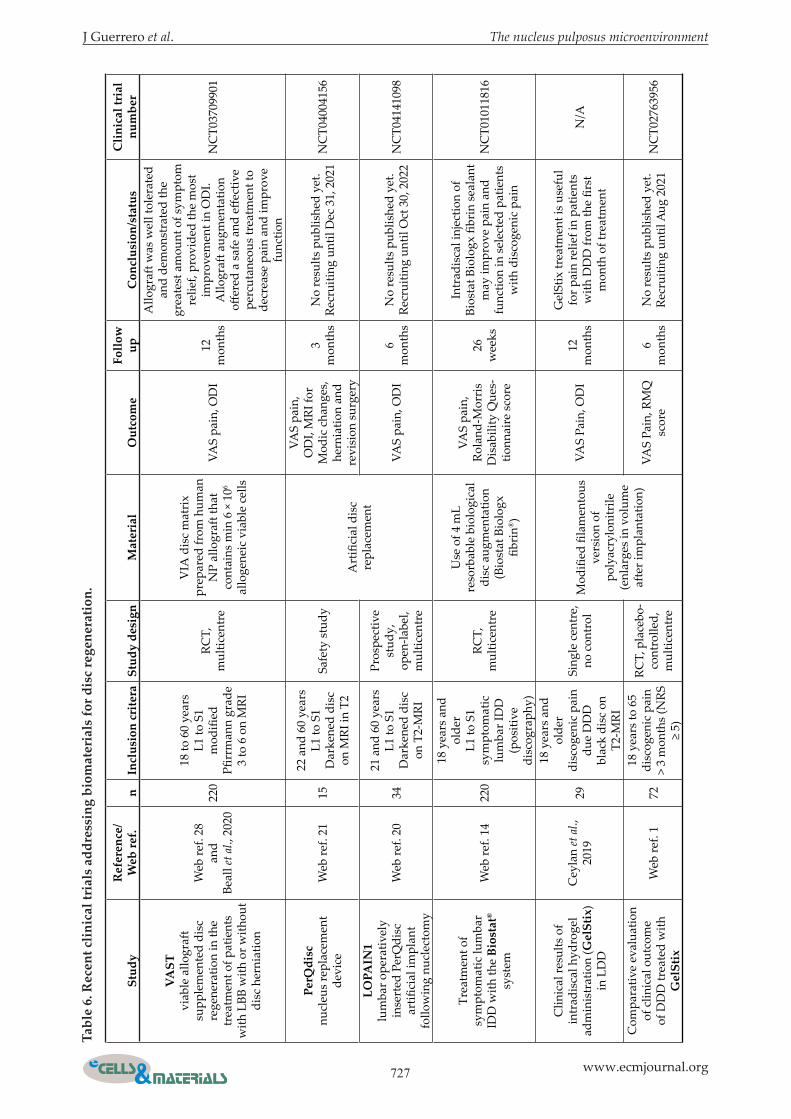
J Guerrero et al. The nucleus pulposus microenvironment
727 www.ecmjournal.org
stud
yr
efer
ence
/ W
eb re
f.n
incl
usio
n cr
itera
stud
y de
sign
mat
eria
lo
utco
me
follo
w
upC
oncl
usio
n/st
atus
Clin
ical
tria
l nu
mbe
r
vA
sT
viab
le a
llogr
aft
supp
lem
ente
d di
sc
rege
nera
tion
in th
e tr
eatm
ent o
f pat
ient
s w
ith L
BB w
ith o
r with
out
disc
her
niat
ion
Web
ref.
28
and
Beal
l et a
l., 2
020
220
18 to
60
year
s L1
to S
1 m
odifi
ed
Pfirr
man
n gr
ade
3 to
6 o
n M
RI
RCT,
m
ultic
entr
e
VIA
dis
c m
atri
x pr
epar
ed fr
om h
uman
N
P al
logr
aft t
hat
cont
ains
min
6 ×
106
allo
gene
ic v
iabl
e ce
lls
VAS
pain
, OD
I 12
m
onth
s
Allo
graf
t was
wel
l tol
erat
ed
and
dem
onst
rate
d th
e gr
eate
st a
mou
nt o
f sym
ptom
re
lief,
prov
ided
the
mos
t im
prov
emen
t in
OD
I. A
llogr
aft a
ugm
enta
tion
offer
ed a
saf
e an
d eff
ectiv
e pe
rcut
aneo
us tr
eatm
ent t
o de
crea
se p
ain
and
impr
ove
func
tion
NC
T037
0990
1
perQ
disc
nu
cleu
s re
plac
emen
t de
vice
Web
ref.
2115
22 a
nd 6
0 ye
ars
L1 to
S1
Dar
kene
d di
sc
on M
RI in
T2
Safe
ty s
tudy
Art
ifici
al d
isc
repl
acem
ent
VAS
pain
, O
DI,
MRI
for
Mod
ic c
hang
es,
hern
iatio
n an
d re
visi
on s
urge
ry
3 m
onth
sN
o re
sults
pub
lishe
d ye
t. Re
crui
ting
until
Dec
31,
202
1N
CT0
4004
156
lopA
in1
lum
bar o
pera
tivel
y in
sert
ed P
erQ
disc
ar
tifici
al im
plan
t fo
llow
ing
nucl
ecto
my
Web
ref.
2034
21 a
nd 6
0 ye
ars
L1 to
S1
Dar
kene
d di
sc
on T
2-M
RI
Pros
pect
ive
stud
y,
open
-labe
l, m
ultic
entr
e
VAS
pain
, OD
I6
mon
ths
No
resu
lts p
ublis
hed
yet.
Recr
uitin
g un
til O
ct 3
0, 2
022
NC
T041
4109
8
Trea
tmen
t of
sym
ptom
atic
lum
bar
IDD
with
the
bios
tat®
sy
stem
Web
ref.
1422
0
18 y
ears
and
ol
der
L1 to
S1
sym
ptom
atic
lu
mba
r ID
D
(pos
itive
di
scog
raph
y)
RCT,
m
ultic
entr
e
Use
of 4
mL
reso
rbab
le b
iolo
gica
l di
sc a
ugm
enta
tion
(Bio
stat
Bio
logx
fib
rin®
)
VAS
pain
, Ro
land
-Mor
ris
Dis
abili
ty Q
ues-
tionn
aire
sco
re
26
wee
ks
Intr
adis
cal i
njec
tion
of
Bios
tat B
iolo
gx fi
brin
sea
lant
m
ay im
prov
e pa
in a
nd
func
tion
in s
elec
ted
patie
nts
with
dis
coge
nic
pain
NC
T010
1181
6
Clin
ical
resu
lts o
f in
trad
isca
l hyd
roge
l ad
min
istr
atio
n (G
elst
ix)
in L
DD
Cey
lan
et a
l.,
2019
29
18 y
ears
and
ol
der
disc
ogen
ic p
ain
due
DD
D
blac
k di
sc o
n T2
-MRI
Sing
le c
entr
e,
no c
ontr
olM
odifi
ed fi
lam
ento
us
vers
ion
of
poly
acry
loni
trile
(e
nlar
ges
in v
olum
e af
ter i
mpl
anta
tion)
VAS
Pain
, OD
I12
m
onth
s
Gel
Stix
trea
tmen
t is
usef
ul
for p
ain
relie
f in
patie
nts
with
DD
D fr
om th
e fir
st
mon
th o
f tre
atm
ent
N/A
Com
para
tive
eval
uatio
n of
clin
ical
out
com
e of
DD
D tr
eate
d w
ith
Gel
stix
Web
ref.
172
18 y
ears
to 6
5 di
scog
enic
pai
n >
3 m
onth
s (N
RS
≥ 5)
RCT,
pla
cebo
-co
ntro
lled,
m
ultic
entr
e
VAS
Pain
, RM
Q
scor
e6
mon
ths
No
resu
lts p
ublis
hed
yet.
Recr
uitin
g un
til A
ug 2
021
NC
T027
6395
6
Tabl
e 6.
rec
ent c
linic
al tr
ials
add
ress
ing
biom
ater
ials
for d
isc
rege
nera
tion.

728 www.ecmjournal.org
J Guerrero et al. The nucleus pulposus microenvironment
Conclusions
Healthy IVDs and especially their NP tissue are physiologically hypoxic, acidic, have a high osmolarity, and are poor in nutrient supplementation. These characteristics make this a very specific microenvironment in the human body but also sufficiently inhospitable for future implanted cells, if needed. This complex microenvironment can encounter many modifications/adjustments of non-ECM-related and ECM-related factors that, as a result, affect NP cells – inducing function impairment. During disc degeneration, NP cells that have lost their ability to form functional and stable cell-cell and cell-ECM crosstalk may also lose their ability to produce specific ECM and ECM-related proteins. This contributes to the inability of the NP tissue to regenerate and self-repair and, therefore, the IVD itself. The NP cells are in perpetual interaction with the surrounding microenvironment and receive cues that help to regulate and maintain the NP phenotype through the behaviour of NP progenitor cells. Perfect knowledge of all the components of this microenvironment and how they interact with NP progenitor cells, could ultimately lead to the development of more efficient strategies able to reverse or at least slow down IVD degeneration. Being able to mimic, thus study and understand NP cell-ECM crosstalk gave rise to many new tissue engineering strategies with different rates of success. Partially recreating the microenvironment with one of the main ECM components (e.g. collagen type II, fibronectin, laminin, elastin, or GAG) did not show useful results for IVD repair or regeneration, as compared to engineered-scaffolds that combine many of them into one biomaterial. However, the best current approach –with outstanding potential for intervertebral disc regeneration – seems to be decellularised NP tissue, able to highly preserve structures and significant biochemical components of the native tissue. There has been a dramatic improvement in the understanding and “mastering” in the use of potential key factors of the NP microenvironment for regenerative medicine approaches in the previous decade. The implementation of already existing culture methods, with key factors such as hypoxic conditions or ECM related proteins, close the gap towards successful in vivo applications. All the components needed for the “reconstruction” as well as the regeneration of the NP tissue seem to be already present within the IVD microenvironment. Therefore, the microenvironment of the NP tissue can be rightly considered as its own fountain of youth in the treatment of degenerative disease and LBP. Understanding which factors are present in the IVD microenvironment and how they govern the IVD behaviour has not only scientific merit but also a huge translational value. New strategies, with the ability to harness these components/factors, have started
to emerge and could provide a valid therapeutic option for future clinical management to treat DDD and more generally LBP. Future studies to further clarify the characteristics and functions of the IVD microenvironment in more detail and to mimic its function will facilitate IVD tissue engineering and the discovery of novel approaches for IVD regenerative medicine.
Acknowledgements
This study was supported by the H2020 project “iPSpine” under the grant agreement #825925 and the Swiss National Science Project #310030E_192674 / 1, both assigned to B.G. Further funds were received from the Swiss Society of Orthopaedics (SGOT), the clinical trials unit of Bern, University Hospital Bern, and by an Eurospine Task Force Research grant #2019_22 assigned to C.E.A.
All authors indicated no potential conflict of interest.
Authors’ contributions
J. Guerrero: study conception and design and drafted the manuscript. S. Häckel drafted the manuscript. A.S. Croft drafted and edited the manuscript. S. Hoppe drafted the manuscript. C.E. Albers drafted the manuscript and acquired funding. B. Gantenbein drafted and edited the manuscript and acquired funding.
references
Airaksinen O, Brox JI, Cedraschi C, Hildebrandt J, Klaber-Moffett J, Kovacs F, Mannion AF, Reis S, Staal JB, Ursin H, Zanoli G, Pain CBWGoGfCLB (2006) Chapter 4. European guidelines for the management of chronic nonspecific low back pain. Eur Spine J 15 suppl 2: S192-S300. Akyuva Y, Kaplan N, Yilmaz I, Ozbek H, Sirin DY, Karaaslan N, Guler O, Ates O (2019) Delivering growth factors through a polymeric scaffold to cell cultures containing both nucleus pulposus and annulus fibrosus. Turk Neurosurg 29: 180-193. Alinejad Y, Adoungotchodo A, Grant MP, Epure LM, Antoniou J, Mwale F, Lerouge S (2019) Injectable chitosan hydrogels with enhanced mechanical properties for nucleus pulposus regeneration. Tissue Eng Part A 25: 303-313. Amirdelfan K, Bae H, McJunkin T, DePalma M, Kim K, Beckworth WJ, Ghiselli G, Bainbridge JS, Dryer R, Deer TR, Brown RD (2021) Allogeneic mesenchymal precursor cells treatment for chronic low back pain associated with degenerative disc

J Guerrero et al. The nucleus pulposus microenvironment
729 www.ecmjournal.org
disease: a prospective randomized, placebo-controlled 36-month study of safety and efficacy. Spine J 21: 212-230. Appelboom JW, Brodsky WA, Dennis WH, Diamond I, Miley JF, Rehm WS (1956) The freezing point depression of mammalian tissues in relation to the question of osmotic activity of cell fluid. J Gen Physiol 40: 183-199. Bach FC, Zhang Y, Miranda-Bedate A, Verdonschot LC, Bergknut N, Creemers LB, Ito K, Sakai D, Chan D, Meij BP, Tryfonidou MA (2016) Increased caveolin-1 in intervertebral disc degeneration facilitates repair. Arthritis Res Ther 18: 59. DOI: 10.1186/s13075-016-0960-y. Baer AE, Laursen TA, Guilak F, Setton LA (2003) The micromechanical environment of intervertebral disc cells determined by a finite deformation, anisotropic, and biphasic finite element model. J Biomech Eng 125: 1-11. Bartels EM, Fairbank JC, Winlove CP, Urban JP (1998) Oxygen and lactate concentrations measured in vivo in the intervertebral discs of patients with scoliosis and back pain. Spine (Phila Pa 1976) 23: 1-8. Baumgartner L, Wuertz-Kozak K, Le Maitre CL, Wignall F, Richardson SM, Hoyland J, Ruiz Wills C, Gonzalez Ballester MA, Neidlin M, Alexopoulos LG, Noailly J (2021) Multiscale regulation of the intervertebral disc: achievements in experimental, in silico, and regenerative research. Int J Mol Sci 22: 703. DOI: 10.3390/ijms22020703. Beall DP, Wilson GL, Bishop R, Tally W (2020) VAST Clinical trial: safely supplementing tissue lost to degenerative disc disease. Int J Spine Surg 14: 239-253. Bez M, Zhou Z, Sheyn D, Tawackoli W, Giaconi JC, Shapiro G, Ben David S, Gazit Z, Pelled G, Li D, Gazit D (2018) Molecular pain markers correlate with pH-sensitive MRI signal in a pig model of disc degeneration. Sci Rep 8: 17363. DOI: 10.1038/s41598-018-34582-6. Bibby SR, Fairbank JC, Urban MR, Urban JP (2002) Cell viability in scoliotic discs in relation to disc deformity and nutrient levels. Spine (Phila Pa 1976) 27: 2220-2228. Bibby SR, Jones DA, Ripley RM, Urban JP (2005) Metabolism of the intervertebral disc: effects of low levels of oxygen, glucose, and pH on rates of energy metabolism of bovine nucleus pulposus cells. Spine (Phila Pa 1976) 30: 487-496. Boden SD, Davis DO, Dina TS, Patronas NJ, Wiesel SW (1990) Abnormal magnetic-resonance scans of the lumbar spine in asymptomatic subjects. A prospective investigation. J Bone Joint Surg Am 72: 403-408. Boos N, Weissbach S, Rohrbach H, Weiler C, Spratt KF, Nerlich AG (2002) Classification of age-related changes in lumbar intervertebral discs: 2002 Volvo Award in basic science. Spine (Phila Pa 1976) 27: 2631-2644. Borrelli C, Buckley CT (2020) Injectable disc-derived ECM hydrogel functionalised with chondroitin
sulfate for intervertebral disc regeneration. Acta Biomater 117: 142-155. Brocker C, Thompson DC, Vasiliou V (2012) The role of hyperosmotic stress in inflammation and disease. Biomol Concepts 3: 345-364. Brown S, Matta A, Erwin M, Roberts S, Gruber HE, Hanley EN, Jr., Little CB, Melrose J (2018) Cell clusters are indicative of stem cell activity in the degenerate intervertebral disc: can their properties be manipulated to improve intrinsic repair of the disc? Stem Cells Dev 27: 147-165. Buckley CT, Hoyland JA, Fujii K, Pandit A, Iatridis JC, Grad S (2018) Critical aspects and challenges for intervertebral disc repair and regeneration-harnessing advances in tissue engineering. JOR Spine 1: e1029. DOI: 10.1002/jsp2.1029. Burke JG, Watson RW, McCormack D, Dowling FE, Walsh MG, Fitzpatrick JM (2002) Intervertebral discs which cause low back pain secrete high levels of proinflammatory mediators. J Bone Joint Surg Br 84: 196-201. Buser Z, Chung AS, Abedi A, Wang JC (2019) The future of disc surgery and regeneration. Int Orthop 43: 995-1002. Cao L, Guilak F, Setton LA (2009) Pericellular matrix mechanics in the anulus fibrosus predicted by a three-dimensional finite element model and in situ morphology. Cell Mol Bioeng 2: 306-319. Cao L, Guilak F, Setton LA (2011) Three-dimensional finite element modeling of pericellular matrix and cell mechanics in the nucleus pulposus of the intervertebral disk based on in situ morphology. Biomech Model Mechanobiol 10: 1-10. Carragee EJ, Don AS, Hurwitz EL, Cuellar JM, Carrino JA, Herzog R (2009) 2009 ISSLS prize winner: does discography cause accelerated progression of degeneration changes in the lumbar disc: a ten-year matched cohort study. Spine (Phila Pa 1976) 34: 2338-2345. Ceylan A, Asik I, Ozgencil GE, Erken B (2019) Clinical results of intradiscal hydrogel administration (GelStix) in lumbar degenerative disc disease. Turk J Med Sci 49: 1634-1639. Chen J, Jing L, Gilchrist CL, Richardson WJ, Fitch RD, Setton LA (2009) Expression of laminin isoforms, receptors, and binding proteins unique to nucleus pulposus cells of immature intervertebral disc. Connect Tissue Res 50: 294-306. Chen JW, Li B, Yang YH, Jiang SD, Jiang LS (2014) Significance of hypoxia in the physiological function of intervertebral disc cells. Crit Rev Eukaryot Gene Expr 24: 193-204. Chen L, Liao J, Klineberg E, Leung VY, Huang S (2017) Small leucine-rich proteoglycans (SLRPs): characteristics and function in the intervertebral disc. J Tissue Eng Regen Med 11: 602-608. Cherif H, Bisson DG, Mannarino M, Rabau O, Ouellet JA, Haglund L (2020) Senotherapeutic drugs for human intervertebral disc degeneration and low back pain. Elife 9. DOI: 10.7554/eLife.54693.

730 www.ecmjournal.org
J Guerrero et al. The nucleus pulposus microenvironment
Choi JS, Harley BA (2012) The combined influence of substrate elasticity and ligand density on the viability and biophysical properties of hematopoietic stem and progenitor cells. Biomaterials 33: 4460-4468. Choi YS (2009) Pathophysiology of degenerative disc disease. Asian Spine J 3: 39-44. Chou PH, Wang ST, Yen MH, Liu CL, Chang MC, Lee OK (2016) Fluid-induced, shear stress-regulated extracellular matrix and matrix metalloproteinase genes expression on human annulus fibrosus cells. Stem Cell Res Ther 7: 34. DOI: 10.1186/s13287-016-0292-5. Chu G, Shi C, Lin J, Wang S, Wang H, Liu T, Yang H, Li B (2018) Biomechanics in annulus fibrosus degeneration and regeneration. Adv Exp Med Biol 1078: 409-420. Chujo T, An HS, Akeda K, Miyamoto K, Muehleman C, Attawia M, Andersson G, Masuda K (2006) Effects of growth differentiation factor-5 on the intervertebral disc-in vitro bovine study and in vivo rabbit disc degeneration model study. Spine (Phila Pa 1976) 31: 2909-2917. Chung J, Mercurio AM (2004) Contributions of the alpha6 integrins to breast carcinoma survival and progression. Mol Cells 17: 203-209. Cuellar JM, Stauff MP, Herzog RJ, Carrino JA, Baker GA, Carragee EJ (2016) Does provocative discography cause clinically important injury to the lumbar intervertebral disc? A 10-year matched cohort study. Spine J 16: 273-280. Cui M, Wan Y, Anderson DG, Shen FH, Leo BM, Laurencin CT, Balian G, Li X (2008) Mouse growth and differentiation factor-5 protein and DNA therapy potentiates intervertebral disc cell aggregation and chondrogenic gene expression. Spine J 8: 287-295. Cunha C, G QT, Ribeiro-Machado C, C LP, Ferreira JR, Molinos M, S GS, Barbosa MA, R MG (2020) Modulation of the in vivo inflammatory response by pro- versus anti-inflammatory intervertebral disc treatments. Int J Mol Sci 21: 1730. DOI: 10.3390/ijms21051730. Desmoulin GT, Pradhan V, Milner TE (2020) Mechanical aspects of intervertebral disc injury and implications on biomechanics. Spine (Phila Pa 1976) 45: E457-E464. Du L, Yang Q, Zhang J, Zhu M, Ma X, Zhang Y, Wang L, Xu B (2019) Engineering a biomimetic integrated scaffold for intervertebral disc replacement. Mater Sci Eng C Mater Biol Appl 96: 522-529. Dudli S, Haschtmann D, Ferguson SJ (2012) Fracture of the vertebral endplates, but not equienergetic impact load, promotes disc degeneration in vitro. J Orthop Res 30: 809-816. Edmondson R, Broglie JJ, Adcock AF, Yang L (2014) Three-dimensional cell culture systems and their applications in drug discovery and cell-based biosensors. Assay Drug Dev Technol 12: 207-218. Engler AJ, Sweeney HL, Discher DE, Schwarzbauer JE (2007) Extracellular matrix elasticity directs stem cell differentiation. J Musculoskelet Neuronal Interact 7: 335.
Erwin WM, Hood KE (2014) The cellular and molecular biology of the intervertebral disc: a clinician’s primer. J Can Chiropr Assoc 58: 246-257. Eyre DR, Muir H (1977) Quantitative analysis of types I and II collagens in human intervertebral discs at various ages. Biochim Biophys Acta 492: 29-42. Feng C, Liu H, Yang M, Zhang Y, Huang B, Zhou Y (2016) Disc cell senescence in intervertebral disc degeneration: causes and molecular pathways. Cell Cycle 15: 1674-1684. Feng G, Li L, Hong Y, Liu H, Song Y, Pei F, Ma PX, Gong Q, Gupte MJ (2014) Hypoxia promotes nucleus pulposus phenotype in 3D scaffolds in vitro and in vivo: laboratory investigation. J Neurosurg Spine 21: 303-309. Foldager CB, Toh WS, Christensen BB, Lind M, Gomoll AH, Spector M (2016) Collagen type IV and laminin expressions during cartilage repair and in late clinically failed repair tissues from human subjects. Cartilage 7: 52-61. Foldager CB, Toh WS, Gomoll AH, Olsen BR, Spector M (2014) Distribution of basement membrane molecules, laminin, and collagen type IV, in normal and degenerated cartilage tissues. Cartilage 5: 123-132. Fontana G, See E, Pandit A (2015) Current trends in biologics delivery to restore intervertebral disc anabolism. Adv Drug Deliv Rev 84: 146-158. Fontana G, Thomas D, Collin E, Pandit A (2014) Microgel microenvironment primes adipose-derived stem cells towards an NP cells-like phenotype. Adv Healthc Mater 3: 2012-2022. Foss BL, Maxwel l TW, Deng Y (2014) Chondroprotective supplementation promotes the mechanical properties of injectable scaffold for human nucleus pulposus tissue engineering. J Mech Behav Biomed Mater 29: 56-67. Francisco AT, Hwang PY, Jeong CG, Jing L, Chen J, Setton LA (2014) Photocrosslinkable laminin-functionalized polyethylene glycol hydrogel for intervertebral disc regeneration. Acta Biomater 10: 1102-1111. Francisco AT, Mancino RJ, Bowles RD, Brunger JM, Tainter DM, Chen YT, Richardson WJ, Guilak F, Setton LA (2013) Injectable laminin-functionalized hydrogel for nucleus pulposus regeneration. Biomaterials 34: 7381-7388. Frapin L, Clouet J, Chedeville C, Moraru C, Samarut E, Henry N, Andre M, Bord E, Halgand B, Lesoeur J, Fusellier M, Guicheux J, Le Visage C (2020) Controlled release of biological factors for endogenous progenitor cell migration and intervertebral disc extracellular matrix remodelling. Biomaterials 253: 120107. DOI: 10.1016/j.biomaterials.2020.120107. Frauchiger DA, Tekari A, Woltje M, Fortunato G, Benneker LM, Gantenbein B (2017) A review of the application of reinforced hydrogels and silk as biomaterials for intervertebral disc repair. Eur Cell Mater 34: 271-290. Freemont AJ, Watkins A, Le Maitre C, Baird P, Jeziorska M, Knight MT, Ross ER, O’Brien JP,

J Guerrero et al. The nucleus pulposus microenvironment
731 www.ecmjournal.org
Hoyland JA (2002) Nerve growth factor expression and innervation of the painful intervertebral disc. J Pathol 197: 286-292. Gantenbein B, Calandriello E, Wuertz-Kozak K, Benneker LM, Keel MJ, Chan SC (2014) Activation of intervertebral disc cells by co-culture with notochordal cells, conditioned medium and hypoxia. BMC Musculoskelet Disord 15: 422. DOI: 10.1186/1471-2474-15-422. Genevay S, Finckh A, Mezin F, Tessitore E, Guerne PA (2009) Influence of cytokine inhibitors on concentration and activity of MMP-1 and MMP-3 in disc herniation. Arthritis Res Ther 11: R169. DOI: 10.1186/ar2858. Gilbert HTJ, Hodson N, Baird P, Richardson SM, Hoyland JA (2016) Acidic pH promotes intervertebral disc degeneration: acid-sensing ion channel -3 as a potential therapeutic target. Sci Rep 6: 37360. DOI: 10.1038/srep37360. Gilbertson L, Ahn SH, Teng PN, Studer RK, Niyibizi C, Kang JD (2008) The effects of recombinant human bone morphogenetic protein-2, recombinant human bone morphogenetic protein-12, and adenoviral bone morphogenetic protein-12 on matrix synthesis in human annulus fibrosis and nucleus pulposus cells. Spine J 8: 449-456. Gilchrist CL, Chen J, Richardson WJ, Loeser RF, Setton LA (2007) Functional integrin subunits regulating cell-matrix interactions in the intervertebral disc. J Orthop Res 25: 829-840. Gorapalli D, Seth A, Vournakis J, Whyne C, Akens M, Zhang A, Demcheva M, Qamirani E, Yee A (2012) Evaluation of a novel poly N-acetyl glucosamine (pGlcNAc) hydrogel for treatment of the degenerating intervertebral disc. Life Sci 91: 1328-1335. Gower WE, Pedrini V (1969) Age-related variations in proteinpolysaccharides from human nucleus pulposus, annulus fibrosus, and costal cartilage. J Bone Joint Surg Am 51: 1154-1162. Griffith JF, Wang YX, Antonio GE, Choi KC, Yu A, Ahuja AT, Leung PC (2007) Modified Pfirrmann grading system for lumbar intervertebral disc degeneration. Spine (Phila Pa 1976) 32: E708-E712. Gruber HE, Norton HJ, Hanley EN, Jr. (2000) Anti-apoptotic effects of IGF-1 and PDGF on human intervertebral disc cells in vitro. Spine (Phila Pa 1976) 25: 2153-2157. Gu J, Fujibayashi A, Yamada KM, Sekiguchi K (2002) Laminin-10/11 and fibronectin differentially prevent apoptosis induced by serum removal via phosphatidylinositol 3-kinase/Akt- and MEK1/ERK-dependent pathways. J Biol Chem 277: 19922-19928. Guerrero J, Häckel S, Croft AS, Albers CE, Gantenbein B (2020) The effects of 3D culture on the expansion and maintenance of nucleus pulposus progenitor cell multipotency. JOR Spine 4:e1131. DOI 10.1002/jsp2.1131. Guilak F, Cohen DM, Estes BT, Gimble JM, Liedtke W, Chen CS (2009) Control of stem cell fate
by physical interactions with the extracellular matrix. Cell Stem Cell 5: 17-26. Guillaume O, Naqvi SM, Lennon K, Buckley CT (2015) Enhancing cell migration in shape-memory alginate-collagen composite scaffolds: In vitro and ex vivo assessment for intervertebral disc repair. J Biomater Appl 29: 1230-1246. Han B, Wang HC, Li H, Tao YQ, Liang CZ, Li FC, Chen G, Chen QX (2014) Nucleus pulposus mesenchymal stem cells in acidic conditions mimicking degenerative intervertebral discs give better performance than adipose tissue-derived mesenchymal stem cells. Cells Tissues Organs 199: 342-352. Hastreiter D, Ozuna RM, Spector M (2001) Regional variations in certain cellular characteristics in human lumbar intervertebral discs, including the presence of alpha-smooth muscle actin. J Orthop Res 19: 597-604. Henry N, Clouet J, Fragale A, Griveau L, Chedeville C, Veziers J, Weiss P, Le Bideau J, Guicheux J, Le Visage C (2017) Pullulan microbeads/Si-HPMC hydrogel injectable system for the sustained delivery of GDF-5 and TGF-beta1: new insight into intervertebral disc regenerative medicine. Drug Deliv 24: 999-1010. Hodgkinson T, Gilbert HTJ, Pandya T, Diwan AD, Hoyland JA, Richardson SM (2020) Regenerative response of degenerate human nucleus pulposus cells to GDF6 stimulation. Int J Mol Sci 21: 7143. DOI: 10.3390/ijms21197143. Hooper L, Abdelhamid A, Ali A, Bunn DK, Jennings A, John WG, Kerry S, Lindner G, Pfortmueller CA, Sjostrand F, Walsh NP, Fairweather-Tait SJ, Potter JF, Hunter PR, Shepstone L (2015) Diagnostic accuracy of calculated serum osmolarity to predict dehydration in older people: adding value to pathology laboratory reports. BMJ Open 5: e008846. DOI: 10.1136/bmjopen-2015-008846. Horner HA, Urban JP (2001) 2001 Volvo award winner in basic science studies: effect of nutrient supply on the viability of cells from the nucleus pulposus of the intervertebral disc. Spine (Phila Pa 1976) 26: 2543-2549. H o s s e i n i M S , Ta f a z z o l i - S h a d p o u r M , Haghighipour N, Aghdami N, Goodarzi A (2015) The synergistic effects of shear stress and cyclic hydrostatic pressure modulate chondrogenic induction of human mesenchymal stem cells. Int J Artif Organs 38: 557-564. Hou Y, Shi G, Shi J, Xu G, Guo Y, Xu P (2016) Study design: in vitro and in vivo assessment of bone morphogenic protein 2 combined with platelet-rich plasma on treatment of disc degeneration. Int Orthop 40: 1143-1155. Hsieh AH, Twomey JD (2010) Cel lular mechanobiology of the intervertebral disc: new directions and approaches. J Biomech 43: 137-145. Hsieh AH, Wagner DR, Cheng LY, Lotz JC (2005) Dependence of mechanical behavior of the murine

732 www.ecmjournal.org
J Guerrero et al. The nucleus pulposus microenvironment
tail disc on regional material properties: a parametric finite element study. J Biomech Eng 127: 1158-1167. Hu B, He R, Ma K, Wang Z, Cui M, Hu H, Rai S, Wang B, Shao Z (2018) Intervertebral disc-derived stem/progenitor cells as a promising cell source for intervertebral disc regeneration. Stem Cells Int 2018: 7412304. DOI: 10.1155/2018/7412304. Hu Y, Huang L, Shen M, Liu Y, Liu G, Wu Y, Ding F, Ma K, Wang W, Zhang Y, Shao Z, Cai X, Xiong L (2019) Pioglitazone protects compression-mediated apoptosis in nucleus pulposus mesenchymal stem cells by suppressing oxidative stress. Oxid Med Cell Longev 2019: 4764071. DOI: 10.1155/2019/4764071. Huang B, Zhuang Y, Li CQ, Liu LT, Zhou Y (2011) Regeneration of the intervertebral disc with nucleus pulposus cell-seeded collagen II/hyaluronan/chondroitin-6-sulfate tri-copolymer constructs in a rabbit disc degeneration model. Spine (Phila Pa 1976) 36: 2252-2259. Huang S, Leung VY, Long D, Chan D, Lu WW, Cheung KM, Zhou G (2013) Coupling of small leucine-rich proteoglycans to hypoxic survival of a progenitor cell-like subpopulation in rhesus macaque intervertebral disc. Biomaterials 34: 6548-6558. Huch M, Koo BK (2015) Modeling mouse and human development using organoid cultures. Development 142: 3113-3125. Hwang PY, Chen J, Jing L, Hoffman BD, Setton LA (2014) The role of extracellular matrix elasticity and composition in regulating the nucleus pulposus cell phenotype in the intervertebral disc: a narrative review. J Biomech Eng 136: 021010. Ilic MZ, Martinac B, Handley CJ (2003) Effects of long-term exposure to glucosamine and mannosamine on aggrecan degradation in articular cartilage. Osteoarthritis Cartilage 11: 613-622. Ishihara H, Warensjo K, Roberts S, Urban JP (1997) Proteoglycan synthesis in the intervertebral disk nucleus: the role of extracellular osmolality. Am J Physiol 272: C1499-1506. Jackson AR, Huang CY, Brown MD, Gu WY (2011a) 3D finite element analysis of nutrient distributions and cell viability in the intervertebral disc: effects of deformation and degeneration. J Biomech Eng 133: 091006. DOI: 10.1115/1.4004944. Jackson AR, Huang CY, Gu WY (2011b) Effect of endplate calcification and mechanical deformation on the distribution of glucose in intervertebral disc: a 3D finite element study. Comput Methods Biomech Biomed Engin 14: 195-204. Johnson WE, Eisenstein SM, Roberts S (2001) Cell cluster formation in degenerate lumbar intervertebral discs is associated with increased disc cell proliferation. Connect Tissue Res 42: 197-207. Johnson WE, Roberts S (2003) Human intervertebral disc cell morphology and cytoskeletal composition: a preliminary study of regional variations in health and disease. J Anat 203: 605-612. Johnson ZI, Shapiro IM, Risbud MV (2014) Extracellular osmolarity regulates matrix homeostasis
in the intervertebral disc and articular cartilage: evolving role of TonEBP. Matrix Biol 40: 10-16. Kalichman L, Hunter DJ (2008) The genetics of intervertebral disc degeneration. Familial predisposition and heritability estimation. Joint Bone Spine 75: 383-387. Kamper M, Hamann N, Prein C, Clausen-Schaumann H, Farkas Z, Aszodi A, Niehoff A, Paulsson M, Zaucke F (2016) Early changes in morphology, bone mineral density and matrix composition of vertebrae lead to disc degeneration in aged collagen IX -/- mice. Matrix Biol 49: 132-143. Kawakami M, Matsumoto T, Hashizume H, Kuribayashi K, Chubinskaya S, Yoshida M (2005) Osteogenic protein-1 (osteogenic protein-1/bone morphogenetic protein-7) inhibits degeneration and pain-related behavior induced by chronically compressed nucleus pulposus in the rat. Spine (Phila Pa 1976) 30: 1933-1939. Kennon JC, Awad ME, Chutkan N, DeVine J, Fulzele S (2018) Current insights on use of growth factors as therapy for intervertebral disc degeneration. Biomol Concepts 9: 43-52. Kepler CK, Ponnappan RK, Tannoury CA, Risbud MV, Anderson DG (2013) The molecular basis of intervertebral disc degeneration. Spine J 13: 318-330. Kim SH, Ahn SH, Cho YW, Lee DG (2012) Effect of intradiscal methylene blue injection for the chronic discogenic low back pain: one year prospective follow-up study. Ann Rehabil Med 36: 657-664. Kobayashi S, Shimamoto T, Taniguchi O, Hashimoto H, Hirose S (1989) A case report of Klinefelter’s syndrome associated with progressive systemic sclerosis. Nihon Naika Gakkai Zasshi 78: 854-855. Korecki CL, MacLean JJ, Iatridis JC (2008) Dynamic compression effects on intervertebral disc mechanics and biology. Spine (Phila Pa 1976) 33: 1403-1409. Kumar H, Ha DH, Lee EJ, Park JH, Shim JH, Ahn TK, Kim KT, Ropper AE, Sohn S, Kim CH, Thakor DK, Lee SH, Han IB (2017) Safety and tolerability of intradiscal implantation of combined autologous adipose-derived mesenchymal stem cells and hyaluronic acid in patients with chronic discogenic low back pain: 1-year follow-up of a phase I study. Stem Cell Res Ther 8: 262. DOI: 10.1186/s13287-017-0710-3. Le Maitre CL, Hoyland JA, Freemont AJ (2007) Interleukin-1 receptor antagonist delivered directly and by gene therapy inhibits matrix degradation in the intact degenerate human intervertebral disc: an in situ zymographic and gene therapy study. Arthritis Res Ther 9: R83. DOI: 10.1186/ar2282. Levi D, Horn S, Tyszko S, Levin J, Hecht-Leavitt C, Walko E (2016) Intradiscal platelet-rich plasma injection for chronic discogenic low back pain: preliminary results from a prospective trial. Pain Med 17: 1010-1022. Li H, Liang C, Tao Y, Zhou X, Li F, Chen G, Chen QX (2012) Acidic pH conditions mimicking

J Guerrero et al. The nucleus pulposus microenvironment
733 www.ecmjournal.org
degenerative intervertebral discs impair the survival and biological behavior of human adipose-derived mesenchymal stem cells. Exp Biol Med (Maywood) 237: 845-852. Li H, Tao Y, Liang C, Han B, Li F, Chen G, Chen Q (2013) Influence of hypoxia in the intervertebral disc on the biological behaviors of rat adipose- and nucleus pulposus-derived mesenchymal stem cells. Cells Tissues Organs 198: 266-277. Li X, Leo BM, Beck G, Balian G, Anderson GD (2004) Collagen and proteoglycan abnormalities in the GDF-5-deficient mice and molecular changes when treating disk cells with recombinant growth factor. Spine (Phila Pa 1976) 29: 2229-2234. Li XC, Tang Y, Wu JH, Yang PS, Wang DL, Ruan DK (2017a) Characteristics and potentials of stem cells derived from human degenerated nucleus pulposus: potential for regeneration of the intervertebral disc. BMC Musculoskelet Disord 18: 242. DOI: 10.1186/s12891-017-1567-4. Li XF, Leng P, Zhang Z, Zhang HN (2017b) The Piezo1 protein ion channel functions in human nucleus pulposus cell apoptosis by regulating mitochondrial dysfunction and the endoplasmic reticulum stress signal pathway. Exp Cell Res 358: 377-389. Li Z, Chen S, Ma K, Lv X, Lin H, Hu B, He R, Shao Z (2018) CsA attenuates compression-induced nucleus pulposus mesenchymal stem cells apoptosis via alleviating mitochondrial dysfunction and oxidative stress. Life Sci 205: 26-37. Liang C, Li H, Tao Y, Zhou X, Li F, Chen G, Chen Q (2012a) Responses of human adipose-derived mesenchymal stem cells to chemical microenvironment of the intervertebral disc. J Transl Med 10: 49. Liang CZ, Li H, Tao YQ, Zhou XP, Yang ZR, Xiao YX, Li FC, Han B, Chen QX (2012b) Dual delivery for stem cell differentiation using dexamethasone and bFGF in/on polymeric microspheres as a cell carrier for nucleus pulposus regeneration. J Mater Sci Mater Med 23: 1097-1107. Lidar Z, Behrbalk E, Regev GJ, Salame K, Keynan O, Schweiger C, Appelbaum L, Levy Y, Keidar A (2012) Intervertebral disc height changes after weight reduction in morbidly obese patients and its effect on quality of life and radicular and low back pain. Spine (Phila Pa 1976) 37: 1947-1952. Liebscher T, Haefeli M, Wuertz K, Nerlich AG, Boos N (2011) Age-related variation in cell density of human lumbar intervertebral disc. Spine (Phila Pa 1976) 36: 153-159. Ligorio C, Zhou M, Wychowaniec JK, Zhu X, Bartlam C, Miller AF, Vijayaraghavan A, Hoyland JA, Saiani A (2019) Graphene oxide containing self-assembling peptide hybrid hydrogels as a potential 3D injectable cell delivery platform for intervertebral disc repair applications. Acta Biomater 92: 92-103. Liu J, Tao H, Wang H, Dong F, Zhang R, Li J, Ge P, Song P, Zhang H, Xu P, Liu X, Shen C (2017) Biological behavior of human nucleus pulposus
mesenchymal stem cells in response to changes in the acidic environment during intervertebral disc degeneration. Stem Cells Dev 26: 901-911. Liu S, Cheng Y, Tan Y, Dong J, Bian Q (2019) Ligustrazine prevents intervertebral disc degeneration via suppression of aberrant TGFbeta activation in nucleus pulposus cells. Biomed Res Int 2019: 5601734. DOI: 10.1155/2019/5601734. Liu Y, Li Y, Nan LP, Wang F, Zhou SF, Wang JC, Feng XM, Zhang L (2020) The effect of high glucose on the biological characteristics of nucleus pulposus-derived mesenchymal stem cells. Cell Biochem Funct 38: 130-140. Livshits G, Popham M, Malkin I, Sambrook PN, Macgregor AJ, Spector T, Williams FM (2011) Lumbar disc degeneration and genetic factors are the main risk factors for low back pain in women: the UK twin spine study. Ann Rheum Dis 70: 1740-1745. Marcinczyk M, Elmashhady H, Talovic M, Dunn A, Bugis F, Garg K (2017) Laminin-111 enriched fibrin hydrogels for skeletal muscle regeneration. Biomaterials 141: 233-242. Masuda K, Imai Y, Okuma M, Muehleman C, Nakagawa K, Akeda K, Thonar E, Andersson G, An HS (2006) Osteogenic protein-1 injection into a degenerated disc induces the restoration of disc height and structural changes in the rabbit anular puncture model. Spine (Phila Pa 1976) 31: 742-754. Mayer JE, Iatridis JC, Chan D, Qureshi SA, Gottesman O, Hecht AC (2013) Genetic polymorphisms associated with intervertebral disc degeneration. Spine J 13: 299-317. McKee TJ, Perlman G, Morris M, Komarova SV (2019) Extracellular matrix composition of connective tissues: a systematic review and meta-analysis. Sci Rep 9: 10542. DOI: 10.1038/s41598-019-46896-0. Melrose J (2016) Strategies in regenerative medicine for intervertebral disc repair using mesenchymal stem cells and bioscaffolds. Regen Med 11: 705-724. Melrose J, Ghosh P, Taylor TK (2001) A comparative analysis of the differential spatial and temporal distributions of the large (aggrecan, versican) and small (decorin, biglycan, fibromodulin) proteoglycans of the intervertebral disc. J Anat 198: 3-15. Mercuri J, Addington C, Pascal R, 3rd, Gill S, Simionescu D (2014) Development and initial characterization of a chemically stabilized elastin-glycosaminoglycan-collagen composite shape-memory hydrogel for nucleus pulposus regeneration. J Biomed Mater Res A 102: 4380-4393. Mercurio AM, Bachelder RE, Chung J, O’Connor KL, Rabinovitz I, Shaw LM, Tani T (2001) Integrin laminin receptors and breast carcinoma progression. J Mammary Gland Biol Neoplasia 6: 299-309. Mosley GE, Evashwick-Rogler TW, Lai A, Iatridis JC (2017) Looking beyond the intervertebral disc: the need for behavioral assays in models of discogenic pain. Ann N Y Acad Sci 1409: 51-66. Munteanu SE, Ilic MZ, Handley CJ (2002) Highly sulfated glycosaminoglycans inhibit aggrecanase

734 www.ecmjournal.org
J Guerrero et al. The nucleus pulposus microenvironment
degradation of aggrecan by bovine articular cartilage explant cultures. Matrix Biol 21: 429-440. Mwale F, Ciobanu I, Giannitsios D, Roughley P, Steffen T, Antoniou J (2011) Effect of oxygen levels on proteoglycan synthesis by intervertebral disc cells. Spine (Phila Pa 1976) 36: E131-138. Nachemson A (1969) Intradiscal measurements of pH in patients with lumbar rhizopathies. Acta Orthop Scand 40: 23-42. Naqvi SM, Buckley CT (2015) Extracellular matrix production by nucleus pulposus and bone marrow stem cells in response to altered oxygen and glucose microenvironments. J Anat 227: 757-766. Naqvi SM, Buckley CT (2016) Bone marrow stem cells in response to intervertebral disc-like matrix acidity and oxygen concentration: implications for cell-based regenerative therapy. Spine (Phila Pa 1976) 41: 743-750. Navaro Y, Bleich-Kimelman N, Hazanov L, Mironi-Harpaz I, Shachaf Y, Garty S, Smith Y, Pelled G, Gazit D, Seliktar D, Gazit Z (2015) Matrix stiffness determines the fate of nucleus pulposus-derived stem cells. Biomaterials 49: 68-76. Neidlinger-Wilke C, Mietsch A, Rinkler C, Wilke HJ, Ignatius A, Urban J (2012) Interactions of environmental conditions and mechanical loads have influence on matrix turnover by nucleus pulposus cells. J Orthop Res 30: 112-121. Nerlich AG, Schaaf R, Walchli B, Boos N (2007) Temporo-spatial distribution of blood vessels in human lumbar intervertebral discs. Eur Spine J 16: 547-555. Nettles DL, Richardson WJ, Setton LA (2004) Integrin expression in cells of the intervertebral disc. J Anat 204: 515-520. Newell N, Carpanen D, Grigoriadis G, Little JP, Masouros SD (2019) Material properties of human lumbar intervertebral discs across strain rates. Spine J 19: 2013-2024. Norbertczak HT, Ingham E, Fermor HL, Wilcox RK (2020) Decellularized intervertebral discs: a potential replacement for degenerate human discs. Tissue Eng Part C Methods 26: 565-576. Noriega DC, Ardura F, Hernandez-Ramajo R, Martin-Ferrero MA, Sanchez-Lite I, Toribio B, Alberca M, Garcia V, Moraleda JM, Sanchez A, Garcia-Sancho J (2017) Intervertebral disc repair by allogeneic mesenchymal bone marrow cells: a randomized controlled trial. Transplantation 101: 1945-1951. Ohshima H, Urban JP (1992) The effect of lactate and pH on proteoglycan and protein synthesis rates in the intervertebral disc. Spine (Phila Pa 1976) 17: 1079-1082. Orozco L, Soler R, Morera C, Alberca M, Sanchez A, Garcia-Sancho J (2011) Intervertebral disc repair by autologous mesenchymal bone marrow cells: a pilot study. Transplantation 92: 822-828. Paesold G, Nerlich AG, Boos N (2007) Biological treatment strategies for disc degeneration: potentials and shortcomings. Eur Spine J 16: 447-468.
Park SH, Gil ES, Cho H, Mandal BB, Tien LW, Min BH, Kaplan DL (2012) Intervertebral disk tissue engineering using biphasic silk composite scaffolds. Tissue Eng Part A 18: 447-458. Peng Y, Huang D, Liu S, Li J, Qing X, Shao Z (2020) Biomaterials-induced stem cells specific differentiation into intervertebral disc lineage cells. Front Bioeng Biotechnol 8: 56. DOI: 10.3389/fbioe.2020.00056. Peroglio M, Eglin D, Benneker LM, Alini M, Grad S (2013) Thermoreversible hyaluronan-based hydrogel supports in vitro and ex vivo disc-like differentiation of human mesenchymal stem cells. Spine J 13: 1627-1639. Pfirrmann CW, Metzdorf A, Zanetti M, Hodler J, Boos N (2001) Magnetic resonance classification of lumbar intervertebral disc degeneration. Spine (Phila Pa 1976) 26: 1873-1878. Rajasekaran S, Soundararajan DCR, Tangavel C, Nayagam SM, Sri Vijayanand KS, Sunmathi R, Matchado MS, Muthurajan R, Shetty AP, Kanna RM, Dharmalingam K (2021) Uncovering molecular targets for regenerative therapy in degenerative disc disease: do small leucine-rich proteoglycans hold the key? Spine J 21: 5-19. Rastogi A, Thakore P, Leung A, Benavides M, Machado M, Morschauser MA, Hsieh AH (2009) Environmental regulation of notochordal gene expression in nucleus pulposus cells. J Cell Physiol 220: 698-705. Risbud MV, Shapiro IM (2014) Role of cytokines in intervertebral disc degeneration: pain and disc content. Nat Rev Rheumatol 10: 44-56. Roberts S, Evans H, Trivedi J, Menage J (2006) Histology and pathology of the human intervertebral disc. J Bone Joint Surg Am 88 suppl 2: 10-14. Roberts S, Menage J, Duance V, Wotton S, Ayad S (1991) 1991 Volvo award in basic sciences. Collagen types around the cells of the intervertebral disc and cartilage end plate: an immunolocalization study. Spine (Phila Pa 1976) 16: 1030-1038. Roughley PJ (2004) Biology of intervertebral disc aging and degeneration: involvement of the extracellular matrix. Spine (Phila Pa 1976) 29: 2691-2699. Rudnik-Jansen I, Tellegen A, Beukers M, Oner F, Woike N, Mihov G, Thies J, Meij B, Tryfonidou M, Creemers L (2019) Safety of intradiscal delivery of triamcinolone acetonide by a poly(esteramide) microsphere platform in a large animal model of intervertebral disc degeneration. Spine J 19: 905-919. Sadowska A, Kameda T, Krupkova O, Wuertz-Kozak K (2018) Osmosensing, osmosignalling and inflammation: how intervertebral disc cells respond to altered osmolarity. Eur Cell Mater 36: 231-250. Sakai D, Andersson GB (2015) Stem cell therapy for intervertebral disc regeneration: obstacles and solutions. Nat Rev Rheumatol 11: 243-256. Sakai D, Grad S (2015) Advancing the cellular and molecular therapy for intervertebral disc disease. Adv Drug Deliv Rev 84: 159-171.

J Guerrero et al. The nucleus pulposus microenvironment
735 www.ecmjournal.org
Sakai D, Nakamura Y, Nakai T, Mishima T, Kato S, Grad S, Alini M, Risbud MV, Chan D, Cheah KS, Yamamura K, Masuda K, Okano H, Ando K, Mochida J (2012) Exhaustion of nucleus pulposus progenitor cells with ageing and degeneration of the intervertebral disc. Nat Commun 3: 1264. DOI: 10.1038/ncomms2226. Sakai D, Schol J (2017) Cell therapy for intervertebral disc repair: Clinical perspective. J Orthop Translat 9: 8-18. Samartzis D, Karppinen J, Chan D, Luk KD, Cheung KM (2012) The association of lumbar intervertebral disc degeneration on magnetic resonance imaging with body mass index in overweight and obese adults: a population-based study. Arthritis Rheum 64: 1488-1496. Samiric T, Ilic MZ, Handley CJ (2006) Sulfated polysaccharides inhibit the catabolism and loss of both large and small proteoglycans in explant cultures of tendon. FEBS J 273: 3479-3488. Sarker B, Hum J, Nazhat SN, Boccaccini AR (2015) Combining collagen and bioactive glasses for bone tissue engineering: a review. Adv Healthc Mater 4: 176-194. Schlotzer-Schrehardt U, Dietrich T, Saito K, Sorokin L, Sasaki T, Paulsson M, Kruse FE (2007) Characterization of extracellular matrix components in the limbal epithelial stem cell compartment. Exp Eye Res 85: 845-860. Schol J, Sakai D (2019) Cell therapy for intervertebral disc herniation and degenerative disc disease: clinical trials. Int Orthop 43: 1011-1025. Selard E, Shirazi-Adl A, Urban JP (2003) Finite element study of nutrient diffusion in the human intervertebral disc. Spine (Phila Pa 1976) 28: 1945-1953. Seliktar D (2012) Designing cell-compatible hydrogels for biomedical applications. Science 336: 1124-1128. Shu C, Hughes C, Smith SM, Smith MM, Hayes A, Caterson B, Little CB, Melrose J (2013) The ovine newborn and human foetal intervertebral disc contain perlecan and aggrecan variably substituted with native 7D4 CS sulphation motif: spatiotemporal immunolocalisation and co-distribution with Notch-1 in the human foetal disc. Glycoconj J 30: 717-725. Silva-Correia J, Oliveira JM, Caridade SG, Oliveira JT, Sousa RA, Mano JF, Reis RL (2011) Gellan gum-based hydrogels for intervertebral disc tissue-engineering applications. J Tissue Eng Regen Med 5: e97-107. Silva MJ, Holguin N (2020) Aging aggravates intervertebral disc degeneration by regulating transcription factors toward chondrogenesis. FASEB J 34: 1970-1982. Silverman LI, Dulatova G, Tandeski T, Erickson IE, Lundell B, Toplon D, Wolff T, Howard A, Chintalacharuvu S, Foley KT (2020) In vitro and in vivo evaluation of discogenic cells, an investigational cell therapy for disc degeneration. Spine J 20: 138-149.
Sivan SS, Hayes AJ, Wachtel E, Caterson B, Merkher Y, Maroudas A, Brown S, Roberts S (2014a) Biochemical composition and turnover of the extracellular matrix of the normal and degenerate intervertebral disc. Eur Spine J 23 suppl 3: S344-353. Sivan SS, Roberts S, Urban JP, Menage J, Bramhill J, Campbell D, Franklin VJ, Lydon F, Merkher Y, Maroudas A, Tighe BJ (2014b) Injectable hydrogels with high fixed charge density and swelling pressure for nucleus pulposus repair: biomimetic glycosaminoglycan analogues. Acta Biomater 10: 1124-1133. Smith SM, Shu C, Melrose J (2010) Comparative immunolocalisation of perlecan with collagen II and aggrecan in human foetal, newborn and adult ovine joint tissues demonstrates perlecan as an early developmental chondrogenic marker. Histochem Cell Biol 134: 251-263. Stoyanov JV, Gantenbein-Ritter B, Bertolo A, Aebli N, Baur M, Alini M, Grad S (2011) Role of hypoxia and growth and differentiation factor-5 on differentiation of human mesenchymal stem cells towards intervertebral nucleus pulposus-like cells. Eur Cell Mater 21: 533-547. Subramony SD, Dargis BR, Castillo M, Azeloglu EU, Tracey MS, Su A, Lu HH (2013) The guidance of stem cell differentiation by substrate alignment and mechanical stimulation. Biomaterials 34: 1942-1953. Sun B, Lian M, Han Y, Mo X, Jiang W, Qiao Z, Dai K (2021) A 3D-bioprinted dual growth factor-releasing intervertebral disc scaffold induces nucleus pulposus and annulus fibrosus reconstruction. Bioact Mater 6: 179-190. Sun Y, Wang TL, Toh WS, Pei M (2017) The role of laminins in cartilaginous tissues: from development to regeneration. Eur Cell Mater 34: 40-54. Tao Y, Zhou X, Liu D, Li H, Liang C, Li F, Chen Q (2016) Proportion of collagen type II in the extracellular matrix promotes the differentiation of human adipose-derived mesenchymal stem cells into nucleus pulposus cells. Biofactors 42: 212-223. Tao YQ, Liang CZ, Li H, Zhang YJ, Li FC, Chen G, Chen QX (2013) Potential of co-culture of nucleus pulposus mesenchymal stem cells and nucleus pulposus cells in hyperosmotic microenvironment for intervertebral disc regeneration. Cell Biol Int 37: 826-834. Tellegen AR, Rudnik-Jansen I, Beukers M, Miranda-Bedate A, Bach FC, de Jong W, Woike N, Mihov G, Thies JC, Meij BP, Creemers LB, Tryfonidou MA (2018) Intradiscal delivery of celecoxib-loaded microspheres restores intervertebral disc integrity in a preclinical canine model. J Control Release 286: 439-450. Tryfonidou MA, de Vries G, Hennink WE, Creemers LB (2020) “old drugs, new tricks” - local controlled drug release systems for treatment of degenerative joint disease. Adv Drug Deliv Rev 160: 170-185. Tsai TT, Guttapalli A, Oguz E, Chen LH, Vaccaro AR, Albert TJ, Shapiro IM, Risbud MV

736 www.ecmjournal.org
J Guerrero et al. The nucleus pulposus microenvironment
(2007) Fibroblast growth factor-2 maintains the differentiation potential of nucleus pulposus cells in vitro: implications for cell-based transplantation therapy. Spine (Phila Pa 1976) 32: 495-502. Tsaryk R, Silva-Correia J, Oliveira JM, Unger RE, Landes C, Brochhausen C, Ghanaati S, Reis RL, Kirkpatrick CJ (2017) Biological performance of cell-encapsulated methacrylated gellan gum-based hydrogels for nucleus pulposus regeneration. J Tissue Eng Regen Med 11: 637-648. Tuakli-Wosornu YA, Terry A, Boachie-Adjei K, Harrison JR, Gribbin CK, LaSalle EE, Nguyen JT, Solomon JL, Lutz GE (2016) Lumbar intradiskal platelet-rich plasma (PRP) injections: a prospective, double-blind, randomized controlled study. PM R 8: 1-10; quiz 10. DOI: 10.1016/j.pmrj.2015.08.010. Urban JP, Maroudas A (1981) Swelling of the intervertebral disc in vitro. Connect Tissue Res 9: 1-10. Urban JP, Maroudas A, Bayliss MT, Dillon J (1979) Swelling pressures of proteoglycans at the concentrations found in cartilaginous tissues. Biorheology 16: 447-464. Urban JP, Roberts S (2003) Degeneration of the intervertebral disc. Arthritis Res Ther 5: 120-130. Urban JP, Smith S, Fairbank JC (2004) Nutrition of the intervertebral disc. Spine (Phila Pa 1976) 29: 2700-2709. van Dijk B, Potier E, Ito K (2011) Culturing bovine nucleus pulposus explants by balancing medium osmolarity. Tissue Eng Part C Methods 17: 1089-1096. Wagner EK, Vedadghavami A, Jacobsen TD, Goel SA, Chahine NO, Bajpayee AG (2020) Avidin grafted dextran nanostructure enables a month-long intra-discal retention. Sci Rep 10: 12017. DOI: 10.1038/s41598-020-68351-1. Walsh AJ, Bradford DS, Lotz JC (2004) In vivo growth factor treatment of degenerated intervertebral discs. Spine (Phila Pa 1976) 29: 156-163. Wan S, Borland S, Richardson SM, Merry CLR, Saiani A, Gough JE (2016) Self-assembling peptide hydrogel for intervertebral disc tissue engineering. Acta Biomater 46: 29-40. Wang B, Wu Y, Shao Z, Yang S, Che B, Sun C, Ma Z, Zhang Y (2012) Functionalized self-assembling peptide nanofiber hydrogel as a scaffold for rabbit nucleus pulposus cells. J Biomed Mater Res A 100: 646-653. Wang J, Huang L, Huang Y, Jiang Y, Zhang L, Feng G, Liu L (2020a) Therapeutic effect of the injectable thermosensitive hydrogel loaded with SHP099 on intervertebral disc degeneration. Life Sci: 118891. DOI: 10.1016/j.lfs.2020.118891. Wang SZ, Rui YF, Lu J, Wang C (2014) Cell and molecular biology of intervertebral disc degeneration: current understanding and implications for potential therapeutic strategies. Cell Prolif 47: 381-390. Wang Y, Che M, Xin J, Zheng Z, Li J, Zhang S (2020b) The role of IL-1beta and TNF-alpha in intervertebral disc degeneration. Biomed Pharmacother 131: 110660. DOI: 10.1016/j.biopha.2020.110660.
Wang YK, Chen CS (2013) Cell adhesion and mechanical stimulation in the regulation of mesenchymal stem cell differentiation. J Cell Mol Med 17: 823-832. Wangler S, Menzel U, Li Z, Ma J, Hoppe S, Benneker LM, Alini M, Grad S, Peroglio M (2019) CD146/MCAM distinguishes stem cell subpopulations with distinct migration and regenerative potential in degenerative intervertebral discs. Osteoarthritis Cartilage 27: 1094-1105. Wei A, Brisby H, Chung SA, Diwan AD (2008) Bone morphogenetic protein-7 protects human intervertebral disc cells in vitro from apoptosis. Spine J 8: 466-474. Wei A, Shen B, Williams L, Diwan A (2014) Mesenchymal stem cells: potential application in intervertebral disc regeneration. Transl Pediatr 3: 71-90. Wu T, Song HX, Dong Y, Li JH (2018) Cell-based therapies for lumbar discogenic low back pain: systematic review and single-arm meta-analysis. Spine (Phila Pa 1976) 43: 49-57. Wuertz K, Godburn K, Iatridis JC (2009) MSC response to pH levels found in degenerating intervertebral discs. Biochem Biophys Res Commun 379: 824-829. Wuertz K, Urban JP, Klasen J, Ignatius A, Wilke HJ, Claes L, Neidlinger-Wilke C (2007) Influence of extracellular osmolarity and mechanical stimulation on gene expression of intervertebral disc cells. J Orthop Res 25: 1513-1522. Xu H, Sun M, Wang C, Xia K, Xiao S, Wang Y, Ying L, Yu C, Yang Q, He Y, Liu A, Chen L (2020) GDF5-GelMA injectable microspheres laden with adipose-derived stem cells for disc degeneration repair. Biofabrication. DOI: 10.1088/1758-5090/abc4d3. Xu J, Liu S, Wang S, Qiu P, Chen P, Lin X, Fang X (2019) Decellularised nucleus pulposus as a potential biologic scaffold for disc tissue engineering. Mater Sci Eng C Mater Biol Appl 99: 1213-1225. Yang X, Li X (2009) Nucleus pulposus tissue engineering: a brief review. Eur Spine J 18: 1564-1572. Yin W, Pauza K, Olan WJ, Doerzbacher JF, Thorne KJ (2014) Intradiscal injection of fibrin sealant for the treatment of symptomatic lumbar internal disc disruption: results of a prospective multicenter pilot study with 24-month follow-up. Pain Med 15: 16-31. Yu J (2002) Elastic tissues of the intervertebral disc. Biochem Soc Trans 30: 848-852. Yu L, Sun ZJ, Tan QC, Wang S, Wang WH, Yang XQ, Ye XJ (2020) Thermosensitive injectable decellularized nucleus pulposus hydrogel as an ideal biomaterial for nucleus pulposus regeneration. J Biomater Appl: 35: 182-192. Yuan C, Pu L, He Z, Wang J (2018) BNIP3/Bcl-2-mediated apoptosis induced by cyclic tensile stretch in human cartilage endplate-derived stem cells. Exp Ther Med 15: 235-241. Yuan D, Chen Z, Xiang X, Deng S, Liu K, Xiao D, Deng L, Feng G (2019) The establishment and biological assessment of a whole tissue-engineered

J Guerrero et al. The nucleus pulposus microenvironment
737 www.ecmjournal.org
intervertebral disc with PBST fibers and a chitosan hydrogel in vitro and in vivo. J Biomed Mater Res B Appl Biomater 107: 2305-2316. Yuan M, Yeung CW, Li YY, Diao H, Cheung KMC, Chan D, Cheah K, Chan PB (2013) Effects of nucleus pulposus cell-derived acellular matrix on the differentiation of mesenchymal stem cells. Biomaterials 34: 3948-3961. Yue B (2014) Biology of the extracellular matrix: an overview. J Glaucoma 23: S20-23. Zhang Z, Ma J, Ren D, Li F (2020) A possible injectable tissue engineered nucleus pulposus constructed with platelet-rich plasma and ADSCs in vitro. J Orthop Surg Res 15: 311. DOI: 10.1186/s13018-020-01840-1. Zhao C, Quan X, He J, Zhao R, Zhang Y, Li X, Sun S, Ma R, Zhang Q (2020) Identification of significant gene biomarkers of low back pain caused by changes in the osmotic pressure of nucleus pulposus cells. Sci Rep 10: 3708. DOI: 10.1038/s41598-020-60714-y. Zhao CQ, Wang LM, Jiang LS, Dai LY (2007) The cell biology of intervertebral disc aging and degeneration. Ageing Res Rev 6: 247-261. Zhao YH, Lv X, Liu YL, Zhao Y, Li Q, Chen YJ, Zhang M (2015) Hydrostatic pressure promotes the proliferation and osteogenic/chondrogenic differentiation of mesenchymal stem cells: The roles of RhoA and Rac1. Stem Cell Res 14: 283-296. Zhou X, Wang J, Fang W, Tao Y, Zhao T, Xia K, Liang C, Hua J, Li F, Chen Q (2018) Genipin cross-linked type II collagen/chondroitin sulfate composite hydrogel-like cell delivery system induces differentiation of adipose-derived stem cells and regenerates degenerated nucleus pulposus. Acta Biomater 71: 496-509. Zhu C, Li J, Liu C, Zhou P, Yang H, Li B (2016) Modulation of the gene expression of annulus fibrosus-derived stem cells using poly(ether carbonate urethane)urea scaffolds of tunable elasticity. Acta Biomater 29: 228-238.
Web references
1. Anestesiologia DmPMV, Rijnstate Ziekenhuis A, the Netherlands, Lugano ORd (2016) Comparative evaluation of clinical outcome of DDD treated with the GelStix Study. https://ClinicalTrials.gov/show/NCT02763956. [18-02-2021]. 2. AnGes I (2018) AMG0103 in subjects with chronic discogenic lumbar back pain. https://ClinicalTrials.gov/show/NCT03263611. [18-02-2021].. 3. Bone, Rouge JCoB (2019) A prospective study of clinical outcomes following a single intradiscal injection of BMAC for single level discogenic LBP. https://ClinicalTrials.gov/show/NCT03912454. [18-02-2021].. 4. Celular RdT, Citospin, Valladolid Uo (2013) Treatment of degenerative disc disease with allogenic mesenchymal stem cells (MSV). https://ClinicalTrials.gov/show/NCT01860417. [18-02-2021].
5. Corporation Y (2012) Safety, tolerability, and efficacy of YH14618 in patients with degenerative disc disease. https://ClinicalTrials.gov/show/NCT01526330. [18-02-2021]. 6. Corporation Y (2015) Clinical trial of YH14618 in patients with degenerative disc disease. https://ClinicalTrials.gov/show/NCT02320019. [18-02-2021]. 7. DiscGenics I (2018) Study to evaluate the safety and preliminary efficacy of IDCT, a treatment for symptomatic lumbar intervertebral disc degeneration. https://ClinicalTrials.gov/show/NCT03347708. [18-02-2021]. 8. DiscGenics I (2019) Study to evaluate the safety and preliminary efficacy of IDCT, a treatment for symptomatic lumbar disc degeneration. https://ClinicalTrials.gov/show/NCT03955315. [18-02-2021]. 9. Han I, University C, Hospital BC (2015) Autologous adipose derived stem cell therapy for intervertebral disc degeneration. https://ClinicalTrials.gov/show/NCT02338271. [18-02-2021]. 10. LLC S (2017) A study of the safety, tolerability, and pharmacokinetics of SM04690 injectable suspension following single intradiscal injection in subjects with degenerative disc disease. https://ClinicalTrials.gov/show/NCT03246399. [18-02-2021]. 11. Mesoblast L (2011) Safety and preliminary efficacy study of MPCs in subjects with lumbar back pain. https://ClinicalTrials.gov/show/NCT01290367. [18-02-2021]. 12. Mesoblast L, Quintiles I (2015) Placebo-controlled study to evaluate rexlemestrocel-l alone or combined with hyaluronic acid in subjects with chronic LBP. https://ClinicalTrials.gov/show/NCT02412735. [18-02-2021]. 13. Pei-Yuan Lee M, Corporation AB, Hospital SCM (2016) PRP for LBP. https://ClinicalTrials.gov/show/NCT03197415. [18-02-2021]. 14. Spinal Restoration I (2010) Treatment of symptomatic lumbar internal disc disruption with the Biostat® System. https://ClinicalTrials.gov/show/NCT01011816. [18-02-2021]. 15. Spine A, Physicians S (2018) Bone marrow concentrate intradiscal injection for chronic discogenic low back pain. https://ClinicalTrials.gov/show/NCT03340818. [18-02-2021]. 16. Spine D (2008) Intradiscal rhGDF-5 phase I/II clinical trial. https://ClinicalTrials.gov/show/NCT00813813. [18-02-2021]. 17. Spine D (2010) A multicentre, randomised, double-blind, placebo controlled, clinical trial to evaluate the safety, tolerability and preliminary effectiveness of 2 doses of intradiscal rhGDF-5 (single administration) for the treatment of early-stage lumbar disc degeneration. https://ClinicalTrials.gov/show/NCT01124006. [18-02-2021]. 18. Spine D, Janssen Korea L, Korea (2010) A clinical trial to evaluate the safety, tolerability and preliminary effectiveness of single administration intradiscal rhGDF-5 for the treatment of early-stage lumbar disc degeneration. https://ClinicalTrials.gov/show/NCT01182337. [18-02-2021].

738 www.ecmjournal.org
J Guerrero et al. The nucleus pulposus microenvironment
19. Spine D, Ltd J-CP (2010) A clinical trial to evaluate the safety, tolerability and preliminary effectiveness of single administration intradiscal rhGDF-5 for the treatment of early-stage lumbar disc degeneration. https://ClinicalTrials.gov/show/NCT01158924. [18-02-2021]. 20. Technologies SS (2019a) Lumbar operatively inserted perqdisc artificial implant following nuclectomy. https://ClinicalTrials.gov/show/NCT04141098. [18-02-2021]. 21. Technologies SS (2019b) Safety study for an artificial disc replacement to treat chronic low back pain. https://ClinicalTrials.gov/show/NCT04004156. [18-02-2021]. 22. Therapeutics S (2017) A single ascending dose study of safety and tolerability of STA363 compared to placebo in 15 patients with chronic discogenic low back pain. https://ClinicalTrials.gov/show/NCT03055845. [18-02-2021]. 23. Therapies B (2021) A single dose of BRTX 100 for patients with chronic lumbar disc disease (cLDD). https://ClinicalTrials.gov/show/NCT04042844. [18-02-2021]. 24. University Hospital M (2012) Assessment of the efficacy of an intradiscal injection of corticoids in modic i discopathies. https://ClinicalTrials.gov/show/NCT01694134. [18-02-2021]. 25. University Hospital M (2018) Intra-discal injection of PRP for low back pain. https://ClinicalTrials.gov/show/NCT03712527. [18-02-2021]. 26. University Hospital M, Studien IZK, Network ECRI, Département de l’information médicale CdM, Centre National de la Recherche Scientifique F, Montpellier U, S.A.S U-B, National University of Ireland G, Ireland, Valladolid Uo, Citospin, Hospital RU, APHP, University CB-M, BG Klinikum Bergmannstrost H, Germany, Hospital NU, Tissular IdTR, Navarra Uo (2019) Efficacy of intradiscal injection of BM-MSC in subjects with chronic low back pain (LBP) due to lumbar degenerative disc disease (DDD) unresponsive. https://ClinicalTrials.gov/show/NCT03737461. [18-02-2021]. 27. University JH (2021) Effect of abaloparatide on lumbar disc degeneration. https://ClinicalTrials.gov/show/NCT03708926. [18-02-2021].
28. Vivex Biomedical I (2017) Viable allograft supplemented disc regeneration in the treatment of patients with low back pain. https://ClinicalTrials.gov/show/NCT03709901. [18-02-2021]. 29. https://www.clinicaltrialsregister.eu/ctr-search/trial/2019-004943-54/ES. [18-02-2021].
discussion with reviewer
Jivko stoyanov: In your review article you well describe the possible cell sources that can be used for the repair of degenerative disc disease (providing several references to published studies and clinical trials). If you had to choose one therapeutic cell type, which one would you select and why?Authors: Various cell types are described in this review and have been used for IVD regeneration including IVD-derived cells, chondrocytes-like cells, mesenchymal stromal cells (MSCs), iPSCs, or ES cells. However, MSCs isolated from bone marrow or from adipose tissue have immunomodulatory functions, are more easily accessible for sampling, and can differentiate into cartilage. Therefore, they should be considered, on my point of view, as a potentially ideal cell source for IVD regeneration.
Jivko stoyanov: Would you point the top three factors of the NP microenvironment on which regenerative medicine approach should be concentrated in the next decade?)Authors: From our point of view and according to the large literature cited in this review, the top three factors/components that must be taken from the NP microenvironment for regenerative purposes are the following ones. First, the use of hypoxic conditions to pre-accommodate (prime) the cells used in the approach, secondly a biomaterial able to mimic mechanical and physiological aspects of the NP, ideally decellularised ECM, and thirdly the possible addition of the most promising growth factor, GDF-5.
editor’s note: The Guest Editor responsible for this paper was Zhen Li.