the nervous system - vanderbilt university · introduction the nervous system “our nervous system...
TRANSCRIPT
1
Chapter I
INTRODUCTION
The Nervous system
“Our nervous system developed for one sole purpose, to maintain
our lives and satisfy our needs. All our reflexes serve this purpose.
This makes us utterly egotistic. With rare exceptions people are
really interested in one thing only: themselves. Everybody, by
necessity, is the center of his own universe.”-- Albert Szent-Györgyi
The nervous system is a network of specialized cells that allows an organism to
be aware of itself and its environment and allows the organism to interact with its
environment. The nervous system is traditionally broken up into 2 systems: the
central nervous system (CNS) and the peripheral nervous system (PNS). The
peripheral nervous system is further subdivided into the autonomic nervous
system (ANS) and the somatic nervous system (SNS) . The ANS, often referred
to as the visceral nervous system, receives sensory information from the internal
body organs and acts as a control center by which the body can maintain
homeostasis. The ANS generally controls the body’s involuntary functions such
as heart rate, digestion, respiration rate, perspiration, urination, sexual arousal
and pupil size (Tortora and Anagnostakos, 1990).

2
The SNS, also called the voluntary nervous system, is the second
component of the peripheral nervous system. The SNS controls all voluntary
body movements through innervation of skeletal muscle and reception of external
stimuli such as skin and sense organs (Tortora and Anagnostakos, 1990). This
allows the body to physically respond to changes in the environment. Although,
both branches of the peripheral nervous system are important in maintaining
homeostasis and responding to environmental changes, the PNS alone cannot
elicit a response. The PNS requires that its sensory information be wired through
the CNS in order to illicit a motor response.
Our ability to integrate sensory information and respond to stimuli is a
direct result of the control center of the body, the CNS. The CNS consists of the
brain and spinal cord, and is responsible for regulating organ function, higher
thought, and body movements (Fig. 1). The CNS functions as a relay station,
where afferent information is passed through the spinal cord into the brain where
the sensory information is processed and an appropriate efferent response is
elicited.
Development of the Central Nervous System
Neurulation
After gastrulation, an embryo has a defined body axis as well as three defined
germ layers: endoderm, mesoderm and ectoderm. Shortly after the specification
of the three germ layers, the notochord, a mesodermal rod like structure, which
defines the body axis in early embryos, begins secretion of the signaling

3
Figure 1. The Central nervous system. The central nervous system (CNS) is one of the two principal divisions of the body's nervous system. Consisting of the brain(1) and the spinal cord(3), the CNS(2) is the control center for the entire
nervous system.
4
molecule Sonic hedgehog (Shh). Shh an antagonist of the growth factor bone
morphogenic protein (BMP) restricts BMP expression from the overlying
ectoderm to form the neural ectoderm or neural plate. This neural plate
undergoes a series of precise morphogenic movements in which the neural plate
converges and intercalates to form the hollow neural tube. The process by which
this occurs is termed neurulation. If these morphogenic movements are not
precise, neural tube defects like spina bifida can occur. Spina bifida is a
congenital birth defect that happens when the neural tube fails to close and
portions of the spinal cord reside outside the vertebral column (Kondo et al.,
2009).
Although the end result of neurulation may be the same, the process by
which various organisms undergo neurulation may be different. For example,
frog, rabbit, mouse and chick undergo a t.wofold neurulation process
characterized as primary and secondary neurulation, whereas neurulation in
some vertebrates like zebrafish has been characterized only as secondary
neurulation (Copp et al., 2000; Copp et al., 2003). During primary neurulation, the
neural plate invaginates, while the lateral ends of the neural plate converge
towards each other. As the lateral ends of the neural plate meet they
downregulate expression of E-cadherin, an epithelial cell adhesion marker and
begin expressing N-cadherin and N-CAM, neural cell adhesion molecules (Copp
et al., 2003; Hong and Brewster, 2006; Lowery and Sive, 2004). After the neural
tube fuses, the neural crest cells migrate away from the dorsal neural tube and
the non neural ectoderm slides over the neural tube and fuses. Secondary

5
neurulation refers to the process in which the neural plate ectodermn condenses
to form a neural tube, instead of the neural plate rolling. (Copp et al., 2003;
Lowery and Sive, 2004).
Unlike in birds and mammals, zebrafish neurulation initially begins with the
formation of the neural keel a solid rod like structure that was initially thought to
be a mass of mesenchymal cells. The lumen or central canal forms through a
secondary process known as cavitation. During cavitation, the ventral cells of the
neural keel downregulate cell adhesion markers and begin to separate in a
ventral to dorsa fashion creating a central canal throughout the length of the CNS
(Lowery and Sive, 2004; Schmitz et al., 1993). Adding to the confusion, in
zebrafish the neuroectoderm does not express any of the classical polarized
epithelial cell markers such as zona occludins 1 (ZO-1) or occludins (Aaku-
Saraste et al., 1996; Geldmacher-Voss et al., 2003). Despite the lack of epithelial
cell markers, lineage tracing and time-lapse imaging provides evidence that
these columnar neural plate cells move and behave like an epithelium (Papan
and Campos-Ortega, 1994). Hence, zebrafish neurulation may have more in
common with primary neurulation than was previously thought.
Dorsal-ventral patterning of the Central Nervous System
No matter how an organism constructs its neural tube, the end result is a neural
tube consisting of naïve neuroepithelial cells that require specific genetic
instructions in order for them to differentiate and populate the CNS. Because the
CNS is composed of the brain and spinal cord, both of which have their own
6
specific mechanisms of dorsoventral patterning, I will focus on dorsoventral
patterning of the spinal cord as it is better understood and more relevant to my
research.
Dorsoventral patterning of the spinal cord is the process by which
signaling molecules and transcription factors pattern the spinal cord so that cell
specification and cell distribution occurs within spatially restricted domains. After
neurulation, the primitive neural tube is induced by high levels of Shh secreted by
the notochord to form the floor plate. The floor plate is a group of non neuronal
ventral midline cells, that express and secrete the morphogen Shh. Concomitant
with Shh secretion, the dorsal midline cells, begin expressing and secreting the
TGF growth factor, Bone morphogenic protein (BMP) (Altman and Bayer, 1984;
Placzek et al., 1991; van Straaten and Hekking, 1991; van Straaten et al., 1985;
Yamada et al., 1991) (Fig. 2). BMP and Shh are distributed in opposing
gradients, which establishes different concentrations of the two morphogens
along the dorsal ventral axis of the spinal cord (Altman and Bayer, 1984; Jessell,
2000; Lee and Jessell, 1999; Mekki-Dauriac et al., 2002). This differential
concentration of the two morphogens creates discrete domains where specific
transcription factors are activated based on the amount of Shh and BMP present.
This allows for specific cell types to be specified within certain areas along the
dorsal ventral axis of the spinal cord (Rowitch, 2004) (Fig. 2). Loss and gain of
function experiments revealed that these domains can regulate their adjacent
domains and that disruption of domain specific transcription factors or signaling
molecules leads to an expansion or a loss of an adjacent domain. Therefore to

7
Figure 2. Dorsal-ventral patterning of the spinal cord. Opposing
concentration gradients of Sonic hedgehog (Shh), and Bone Mophogenic Protein (BMP) create distinct and discrete domains within the spinal cord. These domains express unique combinations of homeobox domain proteins, that give
rise to distinct neural cell types.

8
maintain normal number and distribution of neural cells it is important that the
spinal cord is patterned correctly along its dorsal ventral axis.
Ventral spinal cord domains
The ventral spinal cord is composed of five distinct precursor domains: p0, p1,
p2, pMN and p3 (Fig. 2). Each of these domains is patterned by the
concentration of Shh they receive. Just lateral to the ventral midline floor plate
cells is the p3 domain. The homeobox domain protein Nkx2.2 is expressed in this
domain and restricts pax6 expression dorsally. In the p3 domain, Nkx2.2
expression specifies v3 interneurons, perineurial glia and a subset of
oligodendrocytes (Briscoe et al., 1999; Kim et al., 2008a; Kim et al., 2008b;
Kucenas et al., 2008). Dorsally adjacent to the p3 domain is the progenitor motor
neuron domain (pMN), which gives rise to motor neurons, adult proliferative
precursors and most oligodendrocytes. The pMN domain expresses the basic
helix loop helix transcription factor Olig2, which has been shown to be important
in both motor neuron and oligodendrocyte specification (Park et al., 2002;
Rowitch, 2004). Just dorsal to the pMN domain are the p2, p1 and p0 domains.
These domains give rise mainly to interneurons with each domain specifying a
specific subtype of interneurons (Batista et al., 2008; Burrill et al., 1997; Ericson
et al., 1996; Ericson et al., 1997) (Fig. 2).

9
Major Cell types of the Central Nervous System
The central nervous system consists of neurons and glial cells. Neurons
comprise about half the volume of the CNS and glial cells account for the
rest. Together these two populations of cells function to maintain CNS
homeostasis by receiving signals from throughout the body, interpreting these
signals and then coordinating the body’s response.
Neurons
In 1923, Santiago Ramon y Cajal, the father of modern neuroscience, described
the idea of neurons as the fundamental unit of the nervous system, which he
called the neuron theory. While much has changed in the field of neuroscience,
his neuron theory still remains accurate (Lopez-Munoz et al., 2006). Neurons are
a major class of cells in the nervous system. In vertebrates, neurons are found in
the brain, the spinal cord and in the nerves and ganglia of the peripheral nervous
system. They have excitable membranes, which allow them to generate and
propagate electrical impulses. There are three types of neurons in the vertebrate
body: sensory neurons, interneurons and motor neurons. Although the size and
functions of a neuron may differ, their overall morphology remains the same.
They all have an axon, a cell body and dendrites. The axon is the part of the
neuron that responsible for conducting and propagating nerve electrical
impulses. A cell body or soma contains the nucleus and other organelles. The
dendrites are the branched projections of the cell body that receives and
transmits electrical impulses from other neurons

10
Motor neurons
Our ability to move our body within our environment is due directly to the
functions of motor neurons. Motor neurons are born in the CNS but project their
axons into the PNS, where they innervate muscle directly. Zebrafish undergo a
two stage neurogenesis. Primary neurogenesis starts at around 9-10 hpf when
the primary motor neurons are formed (Kimmel et al., 1994; Myers et al., 1986).
At about 16 hpf the first secondary motor neurons appear (Myers et al., 1986).
These two classes of motor neurons differ in the size, shape and position in the
spinal cord. The early born primary motor neurons have larger somata and
axons, and are usually found more dorsal to the secondary motor neurons
(Myers et al., 1986). The zebrafish primary motor neurons are divided into four
subtypes: CaP, RoP, MiP and VaP. Three of these primary motor neuron
subtypes are named for their positions within the hemisegment: CaP-caudal
primary motor neuron, RoP- rostral primary motor neuron and MiP- middle
primary motor neuron (Bernhardt et al., 1990; Hutson and Chien, 2002;
Westerfield et al., 1986). The fourth primary motor neuron type, variable primary
motor neurons (VaP), appears transiently and is normally eliminated via
apoptosis (Westerfield et al., 1986).
In the pMN domain the transcription factor Olig2 and the LIM homeobox
genes islet1 and islet2 are important in specifying primary motor neurons
(Hutchinson and Eisen, 2006; Park et al., 2002; Takebayashi et al., 2002). Loss
of function experiments, using anti-sense morpholino oligoneucleotides, showed

11
that reducing olig2 expression in the spinal cord produced fewer primary motor
neurons. Conversely, RNA overexpression of olig2 increased the number of
primary motor neurons in the spinal cord (Park et al., 2002). Similar loss of
function experiments showed that in the absence of islet1, primary motor neuron
numbers were reduced (Hutchinson and Eisen, 2006).
Hedgehog signaling has also been shown to play an important role in
specifying motor neurons. Using the smoothened mutants (smu), which lack
functional Hedgehog signaling protein Smoothened and are devoid of Hedgehog
signaling, primary motor neurons are reduced and secondary motor neurons are
absent (Chen et al., 2001; Lewis and Eisen, 2001). Evidence suggests that the
relatively few primary motor neurons that are specified in smu mutants are the
result of lingering activity of the maternal Smoothened protein, because embryos
which completely lacked Hedgehog signaling were devoid of primary motor
neurons (Lewis and Eisen, 2001).
Sensory Neurons
Sensory neurons are nerve cells within the nervous system responsible for
converting external stimuli from the organism's environment into internal
electrical impulses. Sensory nerves take in and communicate information about
temperature, pressure, pain and position. Some sensory neurons respond to
tactile stimuli and can activate motor neurons in order to achieve muscle
contraction. Such connections between sensory and motor neurons underlie
motor reflex loops and several forms of involuntary behavior, including pain
12
avoidance. In humans, such reflex circuits are commonly located in the spinal
cord.
Rohon-Beard Neurons
For animals that develop outside their mothers the ability to respond to light and
other external stimuli early during development is important. Rohon-Beard (RB)
neurons are the first sensory neurons to develop in anamniote vertebrates and
they control embryonic and early larval senses and movements (Clarke et al.,
1984a; Williams et al., 2000). They develop along the dorsal spinal cord,
beginning in the hindbrain and terminating in the caudal spinal cord. RB neurons
are known by their large cell body as well as their characteristic morphology.
Each RB neuron extends axons rostrally towards the hindbrain and caudally with
the spinal cord (Bernhardt et al., 1990; Metcalfe et al., 1990). They also extend a
peripheral axon that exits the spinal cord, and terminates its free nerve ending in
the skin. As the sensory neurons of the dorsal root ganglion develop they
functionally replace the RB neurons, and as a consequence the RB neurons
undergo apoptosis (Williams et al., 2000). However, some organisms like the
newt and lamprey maintain their RB neurons into adulthood (Clarke et al., 1984b;
Nakao and Ishizawa, 1987).
Glia
In 1856, German pathologist, Rudolph Virchow observed a non neuronal
component to the nervous system. He called this non neuronal nervous tissue
glia, which when translated means putty or glue. He hypothesized that glia

13
functioned as a sort of connective tissue that provided rigidity and support to the
nervous system (Webster and Astrom, 2009). For most of the 20th century glia
were described as providing physical and structural support for neurons, and
although the supportive function ascribed to glia was correct, scientists now
believe glia to be more dynamic than once thought. Glial cells insulate neurons,
maintain homeostasis, remove cellular debris from the nervous system, and
serve as adult stem cells (Lledo et al., 2008; Streit, 2000; Streit et al., 1988;
Webster and Astrom, 2009).
Radial Glia
Radial glia are a type of glia that are defined by two characteristics. Firstly, they
extend a radial process from their cell bodies which make contact with the pial
surface, and secondly, like most glia they express glial fibrillary acid protein
(GFAP). They are derived from polarized neuroepithelial cells early in
development, and like neuroepithelial cells they are also polarized, having apical-
basal cell polarity (Temple, 2001). Just prior to the end of embryonic
development, a portion of radial glia tranform into astrocytes in the adult
mammalian CNS (Cepko et al., 1993; Halliday and Cepko, 1992; Noctor et al.,
2001; Walsh and Cepko, 1993). Historically radial glia were thought to have a
supportive role in CNS development, in which they serve as a scaffold for
migrating neurons. However, emerging data argues that radial glia are
neurogenic, gliogenic and give rise to adult neural stem cells in the mammalian
and zebrafish CNS (Anthony et al., 2004; Malatesta et al., 2000; Merkle et al.,
2004; Park et al., 2007).

14
Using retroviral labeling of radial glia in embryonic mice, it has been
shown in vivo that radial glia can divide asymmetrically producing neurons (Ever
and Gaiano, 2005; Fishell and Kriegstein, 2003; Gotz et al., 2002). In the mouse
striatum, radial glia derived from neuroepithelial cells give rise to adult stem cells
(Gates et al., 1995; Mao et al., 2001; Sundholm-Peters et al., 2004). These adult
stem cells proliferate in the adult brain and give rise to neurons in the olfactory
bulb and the hippocampus (Ihrie and Alvarez-Buylla, 2008). Lineage tracing
experiments show that these striatial radial glia can give rise to astrocytes,
oligodendrocytes and many neuronal subtypes. In the zebrafish spinal cord, the
olig2+ expressing radial glia display many of the characteristics of post embryonic
stem cells. They appear to divide asymmetrically and give rise to
oligodendrocytes but not neurons (Merkle et al., 2004; Park et al., 2007).
Oligodendrocytes
Oligodendrocytes are the myelinating cells of the CNS and they function to
increase the rate of which an electrical impulse can travel along an axon in the
central nervous system. Oligodendrocytes achieve this by wrapping their plasma
membranes around an axon and producing a lipid rich material known as myelin
(Simons and Trotter, 2007). Myelin insulates the axon and allows for electrical
impulses to jump between areas of myelination, which speeds up the rate of
which the electrical impulse travels. This jumping of electrical impulses is
technically referred to as saltatory conduction. As an embryo develops the need
for myelination increases, as these electrical impulses are now required to travel
longer distances.

15
Most oligodendrocytes are born in the ventral spinal cord, in the pMN
domain after primary motor neurons are specified. Loss and gain of function
experiments have shown that the Olig family of genes are required for
specification of oligodendrocytes. In mice, loss of Olig gene function resulted in a
dramatic reduction in the number of oligodendrocytes in the spinal cord (Lu et al.,
2002; Park and Moon, 2002; Zhou and Anderson, 2002). Conversely,
overexpression of olig2 lead to an increase in oligodendrocytes (Park et al.,
2002; Park and Moon, 2002; Zhou and Anderson, 2002). The continued
expression of olig2 may be critical for oligodendrocyte development, because
unlike neurons that downregulate olig2 expression shortly after specification,
OPCs express olig2 during all stages of oligodendrocyte development.
Specification of spinal cord OPCs is also regulated by Delta-Notch
signaling. Delta-Notch is a highly conserved cell to cell signaling pathway that
has been shown to be important in several cell regulatory processes like cell
differentiation. In mutants lacking Delta-Notch signaling, excess motor neurons
are specified at the expense of OPCs. In these mutants, the amount of cell
division is reduced when compared to wild type suggesting that in the absence of
Delta-Notch signaling, the proliferative OPCs exit the cell cycle and prematurely
differentiate. In addition to controlling cell cycle exit Delta-Notch signaling
regulates the levels of olig2 expression in the spinal cord. Regulation of olig2
expression is critical for specification of OPCs. In Delta-Notch signaling mutants,
olig2 expression is absent from the developing CNS, however, ngn1, which
specifies motor neurons in the pMN domain, is increased (Yang et al., 2006).

16
These data argue that Delta-Notch signaling is required to maintain pMN
precursors so that they are able to produce OPCs later in development.
In the zebrafish spinal cord, OPCs are specified at 36 hpf when they begin
expressing the transcription factor and OPC specification marker Sox10 . At this
stage OPCs are immature and have a bipolar morphology. At around 50 hpf
spinal cord OPCs begin migrating out of the ventral olig2 domain. As they
migrate OPCs mature and change their morphology. Their cell bodies are no
longer rounded and take on a more elongated appearance. They also extend
more filopodial-like processes and begin searching out their target axons. At 72
hpf the OPCs mature to oligodendrocytes and begin wrapping their target
neurons. Shortly after wrapping oligodendrocytes begin myelinating axons. This
stage is marked by their expression of the myelin genes myelin basic protein
(mbp) and proteolipid protein (plp/dm20).
Pathogenesis
In the absence of properly functioning oligodendrocytes, conduction of electrical
signals throughout the CNS is impaired leading to loss of motor skills, sensations
and cognition. Multiple sclerosis, Devic’s disease and optic neuritis are some
CNS demyelinating diseases in which oligodendrocytes lose their ability to wrap
axons. Demyelinating diseases refers to any disease or condition that results in
damage to the myelin sheets. The term demyelinating disease refers to the
condition rather than the cause, as there are several demyelinating disease,
many of which have their own etiology.

17
Multiple sclerosis is an autoimmune disease, characterized by the
inflammatory demyelination of neurons (Hewagama and Richardson, 2009). In
multiple sclerosis, the body recognizes myelin basic protein and proteolipid
protein as foreign and mounts an immune response against the myelin sheath
leading to the formation of lesions or plaques (Hewagama and Richardson, 2009;
Korn, 2008; Weiner, 2009). Multiple sclerosis affects women more than men with
onset of the disease presenting between 15 and 50 years of age(Korn, 2008).
Although this disease was first indentified over a century ago, the etiology of the
disease still remains unclear. The prevailing hypothesis is that genetically
susceptible individuals get infected with a virus. This hypothesis proposes that
the body mounts a response to the viral antigens, which are similar to myelin
protein genes (Hewagama and Richardson, 2009; Korn, 2008; Weiner, 2009).
The inability of the body’s immune cells to distinguish between viral antigens and
self myelin proteins lead to an inflammatory attack on the oligodendrocytes.
Evidence suggests that the body tries to remyelinate axons but is unsuccessful.
Therefore, by understanding the mechanisms that regulates oligodendrocyte
specification and differentiation scientist are hopeful that they will be able to
provide effective therapeutic strategies.
Stem Cells
Stem cells are classically defined as a type of undifferentiated cells that can self-
renew and give rise to many different cell types (Fig. 3). They are believed to
function as a sort of internal repair system, dividing essentially without limit to
replenish diseased or dying cells for the life of the organism. In addition, they

18
function to maintain the normal turnover of regenerative organs, such as blood,
skin or intestinal tissues. Stem cells have the ability to go through numerous
cycles of cell division while maintaining an undifferentiated or naive state.
Secondly, stem cells are classified based on their potency (Alison and Islam,
2009; Alison et al., 2002). Potency refers to the differentiation potential of the
stem cell, or the capacity of the stem cell to differentiate into specialized cell
types. Most stem cells are classified as pluripotent or multipotent. Pluripotent
stem cells have the ability to differentiate into nearly all cell types, whereas
multipotent stem cells have a more limited cell fate potential (Alison and Islam,
2009; Alison et al., 2002). Multipotent stem cells can differentiate into a number
of cell types, but only those cell types associated with a particular organ system.
Currently, scientists have categorized stem cells into two broadly defined
groups: embryonic stem cells and adult stem cells. Embryonic stem cells (ES
cells) are stem cells derived from the inner cell mass of an early stage of
mammalian embryo known as a blastocyst. ES cells are pluripotent, as they are
able to differentiate into all derivatives of the three primary germ layers. Because
they are pluripotent, scientists are hopeful that ES cells may be used for tissue
transplantation in the future.
Adult stem cells are undifferentiated cells, found throughout the body after
embryonic development, that replenish dying cells and regenerate damaged
tissues. Scientific interest in adult stem cells has centered on their ability to self-
renew indefinitely and generate all the cell types of the organ from which they

19
Figure 3. Stem Cells have the ability to self-renew and differentiate. Stem cells divide asymmetrically. Asymmetric cell division produces 2 daughter cells with different properties. Stem cells divide asymmetrically to give rise to two
distinct daughter cells: a copy of themselves and a cell that is programmed to differentiate into another cell type.

20
originate. Adult stem cells are found within discrete tissue pockets called niches.
These stem cell niches provide support and maintenance of the adult stem cell
by regulating their proliferation and differentiation (Walker et al., 2009). The adult
stem cells in these niches are believed to be specified early in development and
proliferate undifferentiated until they receive the proper signals to initiate
differentiation (Walker et al., 2009). Currently, the mechanisms that specify these
adult stem cells early in development are not clearly understood.
At the end of neurulation, the mammalian CNS is made up of a
pseudostratified epithelium where bipolar neuroepithial cells span the entire
thickness of the neural tube. Neuroepithelial cells represents a population of
embryonic precursor cells that give rise to all cell types of the developing CNS.
These neural precursors proliferate to increase their numbers, and undergo a
characteristic movement of the nucleus (interkinetic nuclear migration) between
the basal and the apical surface (Baye and Link, 2008; Sauer, 1936). The
position of the nucleus is determined by its progression through the cell cycle. At
mitosis, the nucleus resides at the apical surface near the ventricle. During G1
the nucleus begins moving towards the basal surface and reaches its most basal
location in S phase. During G2, the neuroepithelial nucleus begins its migration
back to the apical membrane (Baye and Link, 2008). Because, proliferation of
these neuroepithelial cells is not synchronized, it is possible for these cells to be
in different phases of the cell cycle at any given time. This asynchronous
proliferation results in the epithelium being pseudostratified (Sauer, 1936).
Although interkinetic nuclear migration has been established in the mammalian

21
CNS, at present no evidence argues for this occurring in the zebrafish brain and
spinal cord. However, there is evidence for interkinetic nuclear migration
occurring in retinal development (Baye and Link, 2007; Baye and Link, 2008).
After several rounds of cell divisions, lineage tracing experiments
demonstrated that many neuroepithelial cells are bi-potent progenitors that give
rise first to neurons and then to radial glial cells (Williams and Price, 1995).
Already at this early stage some neuroepithelial cells give rise exclusively to
neurons or glial cells implying a certain degree of lineage heterogeneity
(McCarthy et al., 2001; Williams and Price, 1995). Eventually as development
continues, radial glial cells become the predominant precursor population after
the onset of neurogenesis. In the mouse cortex, the transition of neuroepithelial
to radial glial cells appears to occur around embryonic day E10. This transition is
characterized by expression of astroglial markers such as GLAST and BLBP and
an alteration in tight junctional coupling (Aaku-Saraste et al., 1996; Malatesta et
al., 2003). The basic-helix–loop–helix (bHLH) Hes transcription factors are
important for the transition of neuroepithelial cells to radial glia. These
downstream targets of Notch signaling are crucial for the transition of
neuroepithelial cells to radial glia, as mice deficient in Hes1 and Hes5 show
normal neuroepithelial cells at E8 but impaired radial glial cell differentiation at
E9.5 (Hatakeyama et al., 2004). Therefore it seems that Notch signaling
mediated by Hes transcription factors is not required by neuroepithelial cells or
may be occurring via a molecular mechanisms independent of Hes-mediated
transcription prior to E9, because no defects are obvious in the Hes1, 3, 5 triple

22
mutants at this stage (Hatakeyama et al., 2004). It is only at the onset of
neurogenesis, when radial glial cells appear, that Hes-mediated transcription
becomes essential for radial glial maintenance. Likewise in zebrafish, conditional
inhibition of Notch signaling after the onset of neurogenesis resulted a reduction
in the number of radial glia (Kim et al., 2008b). These data suggest, that although
neuroepithial cells and radial glia may have similar morphological characteristics,
there are molecular differences between neuroepithelial and radial glial cells.
After embryogenesis, these radial glial cells are destined to one of two
fates. The cell fate decision seems to be predicated on the type of organism the
radial glial cells inhabit. In lower vertebrates like reptiles, birds, fish and
amphibians, where neurogenesis persists in a rather widespread fashion in the
adult brain, radial glial cells with access to the ventricle can produce neurons,
oligodendrocytes, and astrocytes, or persist into adulthood (Adolf et al., 2006;
Alvarez-Buylla, 1990; Alvarez-Buylla and Nottebohm, 1988; Chapouton et al.,
2006; Jia and Halpern, 1998; Stevenson and Yoon, 1981). However, in the
mammalian CNS, where adult neurogenesis occurs in discrete pockets, radial
glial cells within these pockets transition to stem cells with astrocyte
characteristics (Alvarez-Buylla and Lim, 2004). These astrocyte like adult stem
cells are the source of new neurons in the adult mammalian brain (Alvarez-Buylla
et al., 2001; Alvarez-Buylla and Lim, 2004).
Adult neurogenesis occurs in the subventricular zone (SVZ), of the
forebrain lateral ventricles, and in the subgranular zone (SGZ), of the dentate
gyrus, within the hippocampus (Doetsch and Hen, 2005). The SVZ is a layer of

23
dividing cells that are found along the lateral walls of the lateral ventricle.
Neurons born in the SVZ are initially specified as neuroblasts that feed into the
rostral migratory stream leading to the olfactory bulb, where they differentiate into
two kinds of inhibitory neurons: granule and periglomerular cells (Doetsch and
Hen, 2005). In the subgranular zone of the hippocampus, adult neural stem cells
generate neurons and glia that contribute to the granular layer of the dentate
gyrus (Cameron et al., 1993; Namba et al., 2005). It has been hypothesized that
the new neurons generated in the hippocampus may play a critical role in
learning and memory processing. As individuals age, their learning and memory
capacity is reduced. It has been proposed that this reduction is due to a decrease
of adult neurogenesis in the subgranular zone of the hippocampus (Taupin and
Gage, 2002).
Stem Cell Maintenance
Scientists hypothesize that early in development, adult stem cell precursors are
deposited into their stem cell niche (Slack, 2008). This niche provides them with
support that allows them to slowly divide and remain undifferentiated until
required. There are two strategies by which stem cells can self renew, while at
the same time producing cells that differentiate: symmetric and asymmetric
division. Stem cells can divide symmetrically to generate two similar daughter
cells and expand the stem cell pool or asymmetrically to self-renew and generate
differentiating daughter cells. The proper balance between symmetric and
asymmetric division is critical for the generation and subsequent repair of tissues.
24
Asymmetric cell division is a process by which a cell divides to generate
two daughter cells that are molecularly different at birth. Such divisions have
already been shown as a means to generate cellular diversity during
development and it provides an attractive strategy as to how stem cells can
maintain the balance of the competing needs of self-renewal and differentiation
(Morrison and Kimble, 2006). Neural stem cells have the potential to divide
symmetrically, producing two identical daughter cells, or asymmetrically,
producing two different daughter cells. Neural stem cells may also undergo
symmetric differentiative divisions, depleting the stem cell pool and producing
two developmentally restricted precursors or post-mitotic progeny. Proliferative
symmetric divisions serve to rapidly increase a progenitor pool, whereas
asymmetric divisions are important for differentiation, as they generate daughter
cells that can differ in size and cell fate (Morrison and Kimble, 2006). Vertebrate
neuroepithelial cells, which are considered to be neural stem cells, initially
undergo several rounds of proliferative symmetric divisions to increase their
number. Later, during neurogenesis, asymmetric divisions are observed in the
ventricular zone of the developing cortex, when neuroepithelial cells directly
generate neurons and self-renew (Gotz and Huttner, 2005). Differentiative
symmetric divisions were also observed in the subventricular zone of the
developing cortex, where intermediate progenitors can give rise to two post-
mitotic cells (Gotz and Huttner, 2005; Noctor et al., 2004). Control of stem cell
division and differentiation is thought to be controlled by cell polarity and
asymmetric cell division.

25
Cell polarity
Cell polarity, which is fundamental to many aspects of cell and developmental
biology, is involved in the processes of differentiation, proliferation and
morphogenesis in both unicellular and multicellular organisms. There are several
different planes of polarization that are necessary for proper development, but I
will only discuss two: apical-basal polarity and planar cell polarity, as they both
play an important role in CNS development. Planar cell polarity (PCP) helps to
organize cells within the plane of the epithelium. The PCP pathway was first
recognized in Drosophila melanogaster, where it regulates organization of
ommatidia in the compound eye and hairs and bristles on wings and legs (Adler,
2002). Within the cell, PCP pathway regulates localization of signaling complexes
that regulate asymmetric organization of the actin cytoskeleton. Key members of
the PCP pathway include transmembrane receptor Frizzled (Fz) and its
downstream effector Dishevelled (Dsh), which regulates the actin cytoskeleton,
and the Jun kinase pathway. Other important PCP pathway proteins include the
transmembrane protein Van Gogh/Strabismus (Vang/Stbm) and its binding
partner Prickle (Pk) (Fig. 4). Together they function to regulate localization and
activity of the Fz/Dsh protein complex (Klein and Mlodzik, 2005; Komiya and
Habas, 2008) (Fig. 4).
The PCP pathway is also required in vertebrates. It functions in the
regulation of body hair orientation, polarization of the sensory epithelia within the
inner ear, and organized cellular movements during convergent extension, a

26
Figure 4. The Planar Cell Polarity Pathway. Evolutionarily conserved planar
cell polarity (PCP) signaling pathway regulates diverse cellular behaviors during invertebrate and vertebrate development. The D. melanogaster PCP pathway
mediates cell polarity in the plane of epithelia. In vertebrates, the PCP pathway controls distinct convergence and extension movements. In X. laevis mesoderm
it controls mediolateral intercalation as well as dorsally directed neuroectoderm intercalation and in zebrafish, the PCP pathway controls lateral mesoderm
anterior and dorsally directed migration.

27
process critical in gastrulation and neural tube formation (Jessen et al., 2002;
Ueno and Greene, 2003)(Fig. 4). During convergent extension, cells intercalate
in a polarized fashion, bring lateral structures closer together, and at the same
time elongate the anterior-posterior body axis (Fig. 4). Many vertebrate mutants
of the key genes involved in PCP pathway display convergent extension defects
resulting in abnormal gastrulation and neural tube development (Copp et al.,
2003; Jessen et al., 2002). In mammalian and Xenopus embryos, this often
results in neural tube closure defects like spina bifida, and in zebrafish embryos,
the loss of function of the critical PCP pathway gene stbm leads to defects in
convergent extension resulting in a dramatic shortening of the body axis (Jessen
et al., 2002; Park and Moon, 2002).
Apical-Basal Polarity
Apical-basal polarity is a fundamental property of all eukaryotic cells. As an
organism develops, regulation of cell polarity is essential for the spatial and
temporal regulation of cell and tissue morphogenesis. For example, polarization
of epithelial cells is an essential component in their ability to form an epithelial
barrier as well as many other critical cellular functions. Cell polarity is also
involved in the regulation of cell identity. During asymmetric cell division, cell fate
identity relies on the polarized distribution and unequal segregation of cell fate
determinants at mitosis. The polarized distribution of key regulatory molecules
not only underlies the generation of differentiated cell types in developing tissues
but also controls the initiation of the anterior posterior body axis in flies and
worms (Suzuki and Ohno, 2006). Finally, cell polarity is a key aspect of cell

28
differentiation, underlying cell migration and morphogenesis. As scientists have
continued to work with polarity proteins, a signaling complex of three proteins,
Par3, Par6 and atypical protein kinase C (aPKC), has emerged as a central
player in the mechanisms that regulate apical-basal cell polarity in the different
cell types of various organisms.
The first apical-basal polarity genes were discovered in C. elegans, during
a genetic screen for maternal-effect mutations affecting the unequal partitioning
of polar granules to the posterior cell during the asymmetric division of the one-
cell embryo (Kemphues et al., 1988). Two of the genes discovered, par-3 and
par-6, encode Post synaptic density protein /Discs-large/ZO1 (PDZ) domain
proteins that co-localize at the anterior pole of the embryo (Etemad-Moghadam
et al., 1995; Hung and Kemphues, 1999). A third protein, aPKC, was later shown
to bind Par3 and to co-localize with Par3 and Par6, leading to the proposal that
the three proteins form a complex (Izumi et al., 1998).
In C. elegans the Par complex of proteins functions to establish anterior
posterior polarity and regulates the first asymmetric division after fertilization.
Upon fertilization, the microtubule-nucleating activity of the sperm asters acts to
exclude the Par3/Par6/aPKC complex from the posterior cortex of the cell
(Pellettieri and Seydoux, 2002; Sadler and Shakes, 2000). In Drosophila Par-6,
aPKC and Par-3 proteins also form a complex that is known to regulate several
developmental processes. For example, the Par3/Par6/aPKC complex regulates
apical–basal polarity of epithelial cells in the embryo, polarized migration of
border cells in the egg chamber and establishment of asymmetry in mitotic

29
neuroblasts (Wodarz, 2002). In Drosophila, these three proteins appear to also
co-localize together, suggesting that, like their C. elegans homologues, the
Drosophila proteins Par-6 and aPKC, together with Par3, act as a signaling
complex (Wodarz, 2001; Wodarz, 2002; Wodarz et al., 2000). In vertebrates,
homologues of Par6, aPKC (aPKCl and aPKCz) and of Par3 (ASIP) have also
been identified. Together, these three proteins were shown to co-localize at tight
junctions. These three proteins have also been shown to be important in the
establishment and maintenance of apical–basal polarity in epithelia, as studies in
cultured epithelial cells indicate that the Par3/Par6/aPKC complex promotes the
formation of epithelial tight junctions. Lastly, Par6 and aPKC have also been
described in vitro as regulators of cell polarity in migrating primary rat astrocytes.
In these cells, Par3 does not appear to co-localize with Par6 and aPKC and is
therefore not part of the complex (Etienne-Manneville and Hall, 2001).
In the embryonic Drosophila CNS, the Par-aPKC complex has been
shown to be important in regulating cell fate through asymmetric cell divisions.
Neuroblasts, a population of Drosophila CNS precursor cells, delaminate from
the neural ectoderm and are polarized expressing aPKC, Par3, and Par6 in the
apical membrane (Doe, 2008; Rolls et al., 2003; Rolls and Doe, 2004) (Fig. 5).
The Par-aPKC complex proteins that direct neuroblast polarity are expressed
and localized in epithelial cells, where they act to establish apico-basal polarity.
The neuroblast inherits these proteins prior to delamination (Rolls et al., 2003).
After neuroblasts delaminate, their mitotic spindle rotates 90°, so that subsequent

30
Figure 5. The Par-aPKC complex regulates asymmetric neuroblast divisions in the Drosophila CNS. After delaminating from the neuroepithelium, the neuroblast localizes aPKC, Par-6 and Par-3 to its apical membrane (red).
This evolutionarily conserved apical complex is known as the aPKC-Par complex and is responsible for segregating proteins like Numb, Miranda and Prospero to the basal side of the cell (purple). This polarized neuroblast divides
asymmetrically to give rise to another neuroblast and a ganglion mother cell (green). The ganglion mother cell differentiates and gives rise to neurons.

31
divisions are oriented along the apico-basal axis of the embryo (Kaltschmidt and
Brand, 2002; Kaltschmidt et al., 2000). Neuroblasts divide asymmetrically and
these unequal divisions produce larger apical daughter cells that retain the stem-
cell-like properties of a neuroblast and smaller, fate-committed basal daughter
cells called ganglion mother cells (GMCs) that produce neurons (Lee et al.,
2006a; Lee et al., 2006b; Rolls et al., 2003). Spindle rotation and the subsequent
asymmetric segregation of cell-fate determinants to the basal GMC are
dependent on the Par-aPKC complex at the apical cortex of the neuroblast.
In the Drosophila neuroblast, the Par-aPKC complex binds Inscuteable
(Insc) through Par3, and Insc, in turn, recruits Partner of Inscuteable (Pins) and
Locomotion defective (Loco), which are two GDP-dissociation inhibitors (GDIs)
that interact with the heterotrimeric G protein subunit, G i. (Kraut et al., 1996; Yu
and David, 2004; Yu et al., 2002). Through loss of function and over expression
experiments, G i and Pins have been shown to be important proteins in
regulating spindle orientation and asymmetric cell divisions (Nipper et al., 2007).
The apical complex is important in directing cell-fate determinants to the
basal cortex. The homeodomain protein Prospero (Pros) and pros RNA, as well
as the phosphotyrosine-binding domain protein Numb, are localized as basal
crescents and segregate to the basal GMC (Betschinger et al., 2003; Doe et al.,
1991; Uemura et al., 1989; Vaessin et al., 1991). Of all the proteins localized to
the basal side of the neuroblast, only Pros appears to act as a cell-fate
determinant in the GMC, whereas Numb plays a role later in development, when
the GMC divides, to discriminate between the sibling neurons (Buescher et al.,

32
1998; Spana and Doe, 1995). Asymmetric segregation of Pros protein and pros
RNA is mediated by the adaptor protein Miranda (Mira). Once Pros is segregated
to the GMC, Mira is degraded, thereby releasing Pros from the cortex (Ikeshima-
Kataoka et al., 1997; Matsuzaki et al., 1998). Pros can then enter the nucleus,
where it has been thought to specify GMC identity by promoting the expression
of GMC-specific genes and repressing the expression of neuroblast-specific
genes (Choksi et al., 2006).
Development of zebrafish as a model System.
Danio rerio, commonly referred to as the zebrafish, is a relatively young scientific
model organism. Its rise as a model organism began at the University of Oregon,
Eugene, when George Streisinger recognized the need for a vertebrate model in
which to study developmental genetic mutations. In the late 1960s Streisinger
began working with this tropical freshwater fish as he saw its potential to “study
features of the organization and embryological development of the vertebrate
nervous system through the use of mutant strains” (Grunwald and Eisen, 2002).
While working with zebrafish he recognized that they were relatively easy to
maintain in a laboratory and that its fecundity would be beneficial genetically as a
single female can produce over a hundred embryos in a single clutch. Zebrafish
develop externally from their mothers and during early development, embryos
are transparent. These two characteristics make zebrafish an excellent model for
live imaging. Zebrafish also develop rapidly as a fertilized embryo can develop
into a free swimming larva within 72 hours.

33
In 1993, two large scale forward genetic screens began in Tübingen,
Germany and Boston, Massachusetts. In a forward genetic screen a physical trait
or phenotype is first identified and then through genetic mapping, the mutated
gene is positionally cloned. In the relatively short time of three years, the results
from these screens were published in a landmark December issue of
Development in 1996. So important and numerous were the results that the
entire issue was dedicated to the screen. From this screen approximately 4,000
embryonic mutant phenotypes were recovered and identified, one of which, heart
and soul (has-/-), is the basis for my thesis. The results from this screen solidified
zebrafish as a viable vertebrate genetic model, as it showed that many of the
known molecular mechanisms were conserved in zebrafish.

34
CHAPTER II
THE APICAL POLARITY PROTEIN PRKCI IS NECESSARY FOR
MAINTENANCE OF SPINAL CORD PRECURSORS IN ZEBRAFISH
Abstract
During spinal cord development, precursor cells transition from proliferative
divisions to differentiative divisions. Traditionally proliferative divisions, which
increase cell numbers, are thought to be symmetric, whereas differentiative
divisions are thought to occur both by symmetric and asymmetric divisions.
Currently, the mechanisms that control this differentiative cell division fate remain
to be defined. However, studies conducted using atypical protein kinase C
(aPKC) suggests that aPKC has a conserved function in controlling cell division
orientation. In this study, we look at the role aPKC may play in maintaining
precursor division in the zebrafish spinal cord. Through time-lapse imaging and
loss of function studies we were able to show that aPKC does regulate precursor
division in the zebrafish spinal cord, and in its absence excess oligodendrocyte
precursor cells (OPCs) are specified at the possible expense of adult precursors.
Introduction
In the developing central nervous system of vertebrates, regulation of cell
division influences the balance of neural precursors, neurons and glia. During
early stages of neural development, precursors undergo symmetric, proliferative
divisions to expand the precursor population. Later, precursors exhibit

35
asymmetric, self-renewing divisions to produce one precursor and one
differentiated cell or symmetric, differentiative divisions to produce two progeny
fated to exit the cell cycle. Therefore, mechanisms that regulate the number and
type of symmetric versus asymmetric divisions influence brain size and cell
composition.
In Drosophila, asymmetric division of embryonic neuroblasts to produce
progeny having different fates is regulated by cell polarity and orientation of cell
cleavage. An evolutionarily conserved complex of proteins consisting of
Par3/Bazooka, Par6 and atypical Protein Kinase C (aPKC) is localized to the
apical membrane of epithelial neuroectodermal cells and thereby to the apical
membrane of neuroblasts as they delaminate from the ectoderm (Petronczki and
Knoblich, 2001; Schober et al., 1999; Wodarz, 2001; Wodarz, 2002; Wodarz et
al., 2000). Par complex proteins act through the tumor suppressor Lethal giant
larvae to exclude other proteins, including the cell fate determinants Prospero
and Numb, from apical membrane, limiting their localization to basal membrane
(Betschinger et al., 2003; Plant et al., 2003; Yamanaka et al., 2003). As
neuroblasts delaminate from the epithelium, Inscuteable and a cassette of
heterotrimeric G protein signaling factors are recruited to the apical membrane
and orient the mitotic spindle perpendicular to the plane of the epithelium,
resulting in cleavage that is orthogonal to the axis of apicobasal polarity (Kraut
and Campos-Ortega, 1996; Kraut et al., 1996; Schaefer et al., 2000; Yu et al.,
2000). Consquently, Prospero and Numb are segregated to the basal progeny
cell, which becomes a Ganglion Mother Cell (GMC) fated to divide once to

36
produce two neurons. The apical progeny cell, lacking Propero and Numb,
remains as a neuroblast (Wodarz and Huttner, 2003).
Regulation of the plane in which neuroepithelial precursors divide by
apicobasal polarity cues in the CNS of vertebrate embryos could provide an
effective means for regulating symmetric versus asymmetric divisions. Consistent
with this possibility, some investigations have described analyses of fixed tissue
and live cell imaging that reveal both planar division (division in the plane of the
epithelium, also known as vertical cleavage) and orthogonal division (division
perpendicular to the plane of the epithelium, also known as horizontal cleavage)
(Chenn and McConnell, 1995; Haydar et al., 2003; Sanada and Tsai, 2005).
Additionally, Par complex proteins are localized to the apical membrane of
neuroepithelial cells (Afonso and Henrique, 2006; Geldmacher-Voss et al., 2003;
Imai et al., 2006; Kovac et al., 2007; Manabe et al., 2002; von Trotha et al.,
2006), similar to localization of homologous proteins in the Drosophila
neuroepithelium. Several other studies, however, indicate that most divisions in
the vertebrate neuroectoderm are planar during both proliferative and neurogenic
phases of neural development (Geldmacher-Voss et al., 2003; Kosodo et al.,
2004; Lyons et al., 2003; Morin et al., 2007; Noctor et al., 2008). Therefore, the
relationship between cell division pattern and cell fate in the vertebrate CNS
remains unclear.
In the spinal cord, the ventral pMN precursor domain, defined by
expression of the transcription factor-encoding gene Olig2, gives rise first to
motor neurons and later to oligodendrocyte progenitor cells (OPCs), which

37
migrate throughout the spinal cord, divide and differentiate as myelinating
oligodendrocytes (Adam et al., 2000; Masahira et al., 2006; Park et al., 2004;
Takebayashi et al., 2000; Zhou et al., 2000). Our analysis of the clonal progeny
of olig2+ neural plate cells in zebrafish implied the existence of asymmetric
divisions that give rise to motor neurons, OPCs and some ventral interneurons
(Park et al., 2004). Near the end of embryogenesis remaining olig2+ precursors
adopt radial glial characteristics and are maintained as slowly dividing OPC
precursors into adulthood (Park et al., 2007). We found that Notch signaling is
required continuously during development to maintain olig2+ precursors and
regulate the numbers of precursors specified for motor neurons and
oligodendroctye fates (Kim et al., 2008a; Shin et al., 2003) but we are still
uncertain about the exact mechanisms that maintain and specify olig2+
precursors and the potential role of cell polarity.
In this study we investigated the role of apical cell polarity in spinal cord
precursor maintenance and specification using the heart and soul (has) mutation,
which disrupts function of aPKC (Horne-Badovinac et al., 2001). Time-lapse
imaging revealed that in has mutant embryos neuroepithelial cells gradually
switch from planar to oblique divisions. Concomitant with this switch in cell
division pattern is a loss of apical character, loss of neuroepithelial precursors
and formation of excess neurons and OPCs. We conclude that planar cell
division, directed by apically localized aPKC , is required for maintenance of
neuroepithelial precursors.

38
Experimental Procedures
Fish Husbandry
Embryos were produced by pair-wise mating and kept at 28.50C in egg water or
embryo medium. Embryos were staged to hours post fertilization (hpf) or days
post fertilization (dpf) according to established zebrafish guidelines (Kimmel et
al., 1995). Embryos that were to be used for live imaging, immunocytochemistry,
or in situ hybridization were treated in 0.003% phenylthiourea (PTU) in egg water
to block pigmentation. The experiments conducted in this paper used the
following strains of zebrafish: AB, hasm567 (Stainier et al., 1996), Tg(olig2:egfp)
(Shin et al., 2003), and Tg(h2afv:gfp) (Pauls et al., 2001).
Immunocytochemistry
Embryos and larvae were fixed in 4% antibody fix (4% paraformaldehyde, 8%
sucrose, 1x PBS) overnight at 40C. After fixing, the embryos were embedded in
1.5% agar/ 5% sucrose blocks and placed in 30% sucrose/PBS solution to
equilibrate overnight. The blocks were then frozen over 2-methylbutane chilled by
liquid nitrogen. We collected 10-12 m sections on superfrost microscope slides
using a cryostat microtome. The sections were rehydrated in 1x PBS for 30 min.
and then blocked in 2% BSA/sheep serum in 1x PBS for 30 min. before
incubating with primary antibody overnight at 4oC. For fluorescent detection of
antibody labeling, we used Alexa Fluor 488, Alexa Fluor 568, Alexa Fluor 647
goat anti-mouse or goat anti-rabbit conjugates (1:500, Molecular Probes). The

39
primary antibodies used included rabbit anti-aPKC (#sc-216, 1:200, Santa Cruz
Biotechnology, Inc.), mouse anti-Islet (clone # 39.4D5, 1:1,000, Developmental
Studies Hybridoma Bank (DSHB), Iowa City, IA), rabbit anti-phospho-Histone-H3
(# 06570, 1:1000, Upstate Biotechnology, Charlottesville, VA), rabbit anti-Sox10
(1:1,000) (Park et al., 2005), mouse anti-ZO-1 (#33-9100, 1:200, Invitrogen) and
mouse anti-ZRF-1 (1:500, University of Oregon Monoclonal Antibody Facility).
In situ hybridization
Embryos and larvae were fixed in 4% paraformaldehyde overnight at 40C and
stored in methanol at -200C. After linearizing plasmids with the appropriate
restriction enzymes, anti-sense cRNA was transcribed using Roche DIG-labeling
reagents and T3, T7 or SP6 RNA polymerases (New England Biolabs). cldnk
(Noctor et al., 2001) was identified in a microarray screen for oligodendrocytes-
specific genes and will be described elsewhere. sox19b was originally named
sox31 (Girard et al., 2001). After processing embryos for in situ RNA
hybridization embryos were embedded in agar and sectioned as described
above. Sections were rehydrated in 1X PBS for 30 min. then covered with 75%
glycerol. Images were obtained using a Retiga EXI camera attached to a Zeiss
Axiovert 200 microscope equipped with Openlab software.
In vivo time-lapse imaging
Embryos were raised in egg water containing PTU and at the appropriate stages
manually dechorionated using watchmaker forceps. Embryos were anesthetized

40
using 3-aminobenzoic acid ethyl ester (Tricaine) and mounted laterally or dorsally
in 35 mm glass bottom petri dishes containing 0.8% low-melting temperature
agarose. Confocal time-lapse movies were obtain by using a 40X oil immersion
objective mounted on a Zeiss Axiovert 200 microscope equipped with a
PerkinElmer spinning disk confocal system. Z-stack images were obtained every
3-5 min and compiled into a Quicktime movie using Volocity software
(Improvision). Widefield time-lapse movies were obtained using a 40X objective
on a Zeiss Axiovert 200 microscope equipped with a Retiga EXI camera. Z-
stacked images were obtained every 10-12 min. Embryos were maintained at
28.50C using a heated stage chamber during imaging.
Angle of division measurements
PTU-treated Tg(h2afv:gfp) and Tg(h2afv:gfp);has-/- embryos were mounted
dorsally at 27 hpf in low melting point agarose. Z-stack images were obtained at
the level of the central canal every 3-5 minutes. These images were then
compiled into a Quicktime time-lapse movie using Volocity. Movies were
analyzed using Openlab software. Going frame by frame, we tracked dividing
cells at the central canal into telophase and then drew a line parallel to the
separated chromatids. With the central canal as a reference, we measured the
angle between the line and the central canal, using a program called Screen
Protractor (Iconico.com).

41
Results
PrkCi is Required for Maintenance of Apical Polarity and Adherens
Junctions in the Spinal Cord Neuroepithelium We initiated an analysis of zebrafish spinal cord neuroepithelial polarity by
labeling transverse sections with an antibody that recognizes a carboxyl terminal
epitope common to PrkCi and Protein kinase C, zeta (PrkCz)
(Cui et al., 2007; Horne-Badovinac et al., 2001). At 24 hours post fertilization
(hpf) and continuing through 48 hpf PrkCi/z proteins were localized to apical cell
membranes contacting the spinal cord medial septum and central canal (Fig.
1A,B). By 72 hpf, when most spinal cord cell divisions have ceased (Park et al.,
2007), PrkCi/z proteins were diminished at the medium septum, but retained
around the central canal (Fig. 1C). Zonula Occludins-1 (ZO-1) antibody, which
recognizes a protein associated with apical neuroepithelial adherens junctions
(Aaku-Saraste et al., 1996; Hurd et al., 2003; Manabe et al., 2002), revealed a
similar pattern of localization (Fig. 1G-I).
In the Drosophila CNS, apical localization of Par/aPKC complexes is
dependent on aPKC function (Wodarz et al., 2000). Consistent with this, targeted
mutation of Prkci in mice results in loss of neuroepithelial adherens junctions
within the neocortex (Imai et al., 2006). To investigate whether the apical polarity
of zebrafish neuroepithelial cells similarly requires PrkCi function, we examined
embryos homozygous for the m567 allele of heart and soul (has), which express
a truncated, inactive PrkCi protein (Horne-Badovinac et al., 2001). We first
assessed the amount and localization of PrkC proteins. Because the hasm567

42
allele eliminates the antibody epitope from PrkCi, any labeling evident in mutant
embryos represents PrkCz and maternally expressed PrkCi (Horne-Badovinac et
al., 2001). At 24 hpf, PrkC localization in has–/– embryos was indistinguishable
from wild-type embryos (Fig. 1D). However, by 48 hpf, has–/– embryos had
diminished levels of PrkC. Particularly, anti-PrkC labeling was nearly absent from
the medial septum and revealed a smaller central canal (Fig. 1E). Additionally,
PrkC was absent in many sections (data not shown), raising the possibility that
the central canal is discontinuous along the length of the spinal cord in mutant
embryos. At 72 hpf, diminished levels of PrkC were similarly localized to a
smaller, apparently discontinuous central canal (Fig. 1F).
ZO-1 immunocytochemistry revealed a localization pattern identical to that
of PrkC in has–/– embryos. At 24 hpf, ZO-1 appeared normal in mutant embryos
(Fig. 1J), indicating that in the absence of PrkCi, PrkCz is sufficient to localize
adherens junction proteins. By contrast, at 48 and 72 hpf ZO-1 was mostly
absent from the medial septum and outlined a small, discontinuous central canal
(Fig. 1K,L). Taken together, these data indicate that apical polarity of spinal
neuroepithelial cells is initially normal in the absence of zygotically encoded
PrkCi function but that apical polarity, adherens junctions and central canal
integrity gradually degrade.

43
Fig. 1. Zebrafish spinal cord cells have apical polarity, which requires PrkCi function. All panels show transverse sections through trunk spinal cord, dorsal up. Dashed circle marks the perimeter of the spinal cord. Arrowheads and arrows indicate central canal and medial septum, respectively. A-F: Sections labeled with anti-PrkCi/z antibody. PrkC is localized to the medial septum and central canal of wild-type embryos at 24 and 48 hpf (A,B). At 72 hpf, PrkC is absent from the medial septum but remains around the central canal of wild-type larvae (C). At 24 hpf, PrkC localization is normal in has–/– embryos (D). However, by 48 hpf very little PrkC is evident at the medial septum and, although PrkC remains around the central canal, the central canal is reduced in size (E) or entirely absent (not shown). Sections of 72 hpf has–/– larvae similarly reveal PrkC localization around an abnormally small and discontinuous central canal (F). The apical protein ZO-1 has a similar localization pattern to PrkC in wild-type and has–/– embryos and larvae (G-L). Scale bar = 20 μM.

44
PrkCi is Required to Maintain Planar Divisions of Spinal Cord Precursors
In zebrafish, cell divisions within the medial neural plate are oriented in the
mediolateral plane of the neuroepithelium (Concha and Adams, 1998;
Geldmacher-Voss et al., 2003). As the neural plate condenses to form the neural
keel and then neural rod, cell divisions remain perpendicular to the
anteroposterior axis and become orthogonal to the plane of the neuroepithelium
(Geldmacher-Voss et al., 2003). Upon formation of the neural tube, cell divisions
rotate 900 so that they occur within the plane of the neuroepithelium
(Geldmacher-Voss et al., 2003; Lyons et al., 2003). As a prelude to our
investigation of neural cell polarity and fate, we performed our own analysis of
cell division orientation, but focused on a later period of development than
previous studies. To mark dividing cells we used the transgenic reporter
Tg(h2afv:gfp), which expresses EGFP fused to a histone protein (Pauls et al.,
2001). We imaged embryos from a dorsal view, focusing on cells that line the
central canal starting at 27 hpf and continuing until 33 hpf. This corresponds to
the period during which most spinal cord neurogenesis is completed and
precedes OPC formation (Park et al., 2002; Shin et al., 2003). Consistent with
previous reports of cell division patterns in both zebrafish and chick neural tube
(Geldmacher-Voss et al., 2003; Lyons et al., 2003; Morin et al., 2007), divisions
of cells bordering the central canal were always planar, parallel to the central
canal (Fig. 2A,C,E).

45
Fig. 2. PrkCi maintains planar divisions of cells that divide along the central canal. A-D: Frames captured from time-lapse movies, from a dorsal view, of wild-type and has–
/– embryos carrying the Tg(h2afv:egfp) transgene. Numbers in upper right corners indicate time elapsed from beginning of imaging at 27 hpf. Dashed circles outline dividing cells, arrows point to the central canal and bi-directional arrows indicate orientation of the mitotic spindle and angle of division. A,B: In both wild-type and has–/– embryos, divisions that occur before 30 hpf are planar. The central canal is less distinct in has–/– embryos than in wild type. C,D: Divisions after 30 hpf remain planar in wild-type embryos (C) but become oblique in has–/– embryos (D). The central canal is indistinct and appears to have been replaced by cells. E,F: Quantification of angles of division in wild-type (E) and has–/– embryos (F). In wild type most division planes are within 150 of the plane of the epithelium, indicated by the central canal. has–/– embryos have
numerous divisions greater than 150. Scale bar = 24 M.

46
Time-lapse imaging revealed the orientation of cell divisions changes with
time in has mutant embryos. Prior to 30 hpf, cells bordering the central canal
divide similarly to those in wild-type (Fig. 2B). Beginning at about 30 hpf,
however, most divisions were oblique to the plane of the epithelium, with some
nearly perpendicular (Fig. 2D,E). At the same time, the central canal became
less distinct, with the space occupied by spinal cord cells (Fig. 2D). Therefore,
loss of PrkCi function results in disruption of planar division and a breakdown of
the neuroepithelium.
Loss of PrkCi Function Causes Formation of Excess OPCs Without Affecting Motor Neuron Formation
The above data reveal that PrkCi is required for maintenance of apical polarity
and planar divisions of spinal cord precursors. Because cell polarity and division
pattern often influence cell fate, we assessed formation of neurons and glia using
molecular markers. We first labeled transverse sections of wild-type and has–/–
spinal cords with anti-Hu antibody, which marks all newly formed neurons
(Marusich et al., 1994). No apparent differences in the distribution of Hu+
neurons was evident between wild-type and mutant embryos at 24, 48 and 72
hpf (Fig. 3A-F). To quantify the number of neurons, we labeled sections of 72 hpf
wild-type and mutant larvae with anti-Hu antibody and a nuclear stain and
determined the proportion of spinal cord cells that had neuronal identity. This
revealed little difference in the number of neurons formed by larval stage (data
not shown). This phenotype is similar to that of embryos deficient for Notch
signaling in which precursors prematurely exit the cell cycle and differentiate as

47
neurons except that in the latter excess neurons are evident at the earliest
stages of neurogenesis (Appel and Eisen, 1998; Appel et al., 2001; Itoh et al.,
2003; Shin et al., 2003). Therefore, we examined Isl+ motor neurons, Rohon-
Beard sensory neurons and interneurons, which are mostly formed by about 48
hpf. No obvious differences in the number and distribution of Isl+ neurons was
apparent between wild type and mutant through 72 hpf (Fig. 3G-L) and
quantification of Isl+ motor neurons revealed no statistically signficant differences
at 48 and 72 hpf (Fig. 3Q). We also examined Isl+ motor neurons, Rohon-Beard
sensory neurons and interneurons, which are mostly formed by about 48 hpf. No
obvious differences in the number and distribution of Isl+ neurons was apparent
between wild type and mutant through 72 hpf (Fig. 3G-L) and quantification of
Isl+ motor neurons revealed no statistically significant differences at 48 and 72
hpf (Fig. 3Q). Therefore, formation of the earliest-formed spinal cord neurons is
not apparently affected by absence of PrkCi function, consistent with our
observation that PrkCz localization, apical polarity and cell division pattern
appear normal during early neurogenesis.
To investigate formation of OPCs, we labeled sections with anti-Sox10
antibody, which serves as a specific marker of OPCs and differentiating
oligodendroctyes (Kuhlbrodt et al., 1998; Park et al., 2005). At 48 hpf, soon after
specification of spinal cord OPCs begins, the number and distributon of OPCs
was indistinguishable between wild-type and has mutant embryos (data not
shown). However, by 72 hpf, both the number and distribution of OPCs was
altered in has–/– spinal cords relative to wild type (Fig. 3M,N). In particular, mutant

48
Fig. 3. Loss of PrkCi function produces excess OPCs. All images are of transverse sections through trunk spinal cord, dorsal up. Outlined circle marks the perimeter of the spinal cord. A-F: The number and distribution of neurons, marked by anti-Hu labeling, are similar in wild-type and has-/- embryos at 24, 48 and 72 hpf (A-F). G-L: Isl+ motor neurons (brackets), interneurons (solid arrowheads) and Rohon-Beard sensory neurons (open arrowheads) are similar in wild-type and has-/- embryos and larvae through 72 hpf. M,N: Anti-Sox10 labeling reveals excess OPCs in 72 hpf has-/- larva (N) compared to wild type (M). O,P: cldnk RNA expression marking differentiating oligodendrocytes. In wild-type, cldnk+ cells (arrows) are at the pial surface in dorsal and ventral spinal cord (O). has-/- larvae have excess cldnk+ cells and some occupy ectopic positions in medial spinal cord (P). Q: Quantification of spinal cord Isl+ motor neurons in the spinal cord between wild-type and has-/- embryos at 48 and 72 hpf. R: Quantification of spinal cord Sox10+ OPCs. Error bars represent s.e.m. Statistical significance was determined using
Student’s t-test. Scale bar = 20 M.

49

50
larvae had 1.4-fold more OPCs than wild type (Fig. 3R) and some OPCs were
abnormally positioned within medial regions of the spinal cord (Fig. 3N). We also
performed in situ RNA hybridization to detect expression of cldnk, which
specifically marks differentiating oligodendrocytes (N. Takada and B. Appel,
unpublished data). This similarly revealed an excess of oligodendrocytes in
has–/– spinal cords relative to wild type (Fig. 3O,P). Additionally, cldnk+ cells
sometimes occupied medial spinal cord, where they are normally not found.
These data indicate that PrkCi function limits both the formation and
differentiation of oligodendrocyte lineage cells.
PrkCi Function and Apical Polarity are Required to Maintain Spinal Cord Precursors In zebrafish, slowly-dividing olig2+ spinal cord precursors that have radial glial
characteristics and continuously give rise to new OPCs are maintained through
late embryonic stage into adulthood (Park et al., 2007). One possible explanation
for the excess OPC phenotype of has mutant larvae, therefore, is that, in the
absence of PrkCi function, olig2+ cells lose precursor characteristics and develop
as oligodendroctyes. Consistent with this, whereas olig2+ radial fibers are evident
by 72 hpf in transverse sections of wild-type Tg(olig2:egfp) larvae (Fig. 4A-C),
similar processes were rare in has–/–;Tg(olig2:egfp) larvae (Fig. 4D-E). We next
labeled sections with anti-Zrf-1 antibody, which marks spinal cord radial glia
(Metcalfe et al., 1990). Through 48 hpf, Zrf-1+ fibers were similar in wild-type and
has mutant embryos (Fig. 4G,H,J,K). However, by 72 hpf, whereas GFAP+ radial
glia were distributed uniformly throughout the spinal cord of wild-type larvae (Fig.

51
Fig. 4. PrkCi is required to maintain ventral spinal cord cells with precursor characteristics. All images are of transverse sections through trunk spinal cord, dorsal up. Outlined circle marks the perimeter of the spinal cord. A-F: EGFP expression driven by the Tg(olig2:egfp) promoter. In wild-type embryos, EGFP marks motor neurons and pMN precursors through 48 hpf (A,B). By 72 hpf, EGFP+ fibers (arrowheads), marking precursors that persist into larval stage, become evident. EGFP expression appears normal in has–/– embryos at 24 and 48 hpf (D,E). At 72 hpf, few EGFP+ radial fibers are evident (F). Asterisks mark dorsally migrated OPCs. G-L: Zrf-1 immunocytochemistry to label radial glial fibers. In wild-type embryos and larvae, radial fibers are distributed uniformly through the spinal cord (G-I). In ventral spinal cord, the apical membrane of some radial glia surround the central canal (arrowheads). In has mutants (J-L), radial fibers initially appear normal but by 72 hpf a gap appears in ventral spinal cord (bracket) and the central canal is reduced or absent. M,N: sox19b RNA expression. At 72 hpf in wild type, sox19b expression marks cells near the central canal (arrow) (M). At 72 hpf sox19b is not expressed at its normal position in has–/– larva (bracket). Instead, ventral
and dorsal spinal cord cells express sox19b. Scale bar = 20 M.

52

53
4I), radial glia were consistently absent from the spinal cord just dorsal to
remnants of the central canal (Fig. 4L). We next used in situ RNA hybridization to
detect expression of sox19b, which marks cells of the medial spinal cord that are
likely dividing precursors (Fig. 4M-O). Similar to Zrf-1, sox19b expression
appeared normal through 48 hpf in has mutant embryos (Fig. 4P,Q). By 72 hpf,
sox19b expression is limited to cells near the central canal in wild-type larvae
(Fig. 4O). By contrast, sox19b expression was absent from a comparable
position in has mutant larvae but was maintained in more dorsal and ventral cells
(Fig. 4R). Taken together, these data indicate that loss of PrkCi function results
in loss of spinal cord precursors near the central canal during late
embryogenesis.
To further investigate the relationship between apical polarity and spinal
cord precursor division, we labeled embryos and larvae carrying the
Tg(olig2:egfp) transgene with antibody specific to Phospho-Histone H3 (anti-
PH3), which labels cells in M phase. At 2 dpf, numerous olig2+ PH3+ cells were
evident along the central canal of wild type embryos (Fig. 5A). By 3 dpf, the
number of PH3+ cells was substantially decreased (Fig. 5B), consistent with our
previous data showing a significant decline in the number of dividing cells in the
spinal cord between 2 and 3 dpf (Park et al., 2007). In has mutant embryos, the
central canal, outlined by olig2+ cells, was discontinuous with remaining pockets
of central canal surrounded by rosettes of olig2+ cells (Fig. 5C,D). Notably, PH3+
cells were evident, but appeared only within the rosettes. As in wild type, 3 dpf

54
Fig. 5. Loss of PrkCi function causes a transient excess and then deficit of dividing spinal cord precursors. A-D: Images, from dorsal view and focused on the trunk spinal cord, of wild-type and has–/– embryos and larvae carrying the Tg(olig2:egfp) reporter and labeled with anti-PH3 antibody to mark M phase cells. In wild type most mitotic cells (arrowheads) are adjacent to the central canal (arrow). More cells divide at 2 dpf (A) than at 3 dpf (B). In has–/– embryos (C,D) many EGFP+ cells form rosettes (brackets) surrounding discontinuous portions of central canal (asterisks). PH3+ are usually only found within rosettes. E-H: Images, from dorsal view and focused on the trunk spinal cord, of wild-type and has–/– embryos and larvae labeled with anti-PH3 antibody (red) and anti-ZO-1 antibody (blue) to mark apical membrane. In wild type (E,F), ZO-1 localization outlines a continuous central canal closely associated with PH3+ mitotic cells. In has–/– embryos and larvae (G,H) ZO-1 labeling is discontinuous but mitotic cells are nearly always associated with remaining apical membrane. I: Quantification of PH3+ cells at 2 and 3 dpf over a 288 μm length of trunk spinal cord. Error bars represent s.e.m. Statistical significance was determined using Student’s t-test.

55
has mutant larvae had fewer PH3+ cells than at 2 dpf. We also use ZO-1 labeling
to examine the distribution of dividing cells. As expected, PH3+ cells in wild type
had apical polarity and were associated with the central canal (Fig. 5E,F). 2 dpf
has mutant embryos had patches of apical membrane, consistent with the
discontinuous nature of the central canal, and PH3+ cells were nearly always
associated with ZO-1 labeling (Fig. 5G). We noticed that the density of PH3+ cells
seemed to be greater in mutant embryos than in wild type. In fact, whereas wild-
type embryos had 3.9 PH3+ cells within a 288 m length of trunk spinal cord,
mutant embryos had 5.4 PH3+ cells over the same distance (Fig. 5I). At 3 dpf, the
gaps in ZO-1 labeling of mutant larvae were larger and more frequent than at 2
dpf (Fig. 5H) and mutant larvae had fewer M phase cells (Fig. 5I). Therefore,
has–/– mutant embryos have a transient increase and then deficit of dividing
spinal cord precursors.
Our data raise the possibility that excess OPCs arise in has–/– mutant
embryos due to misallocation of spinal cord precursors to an oligodendrocyte
fate. To test this, we performed time-lapse imaging of wild-type and has mutant
embryos carrying the Tg(olig2:egfp) transgene. Over a 10 hour period,
approximately 1.5 fold more OPCs migrated from ventral spinal cord into dorsal
spinal cord in has mutant embryos relative to wild type (Fig. 6A-C). Migrating
OPCs did not divide more frequently in has mutant embryos than in wild type.
Therefore, excess OPCs arise in has mutant embryos from specification of
excess pMN precursors for oligodendrocyte fate.

56
Fig. 6. Loss of PrkCi function causes formation of excess OPCs from pMN precursors. Panels show frames captured from time-lapse videos of wild-type and has–
/– embryos carrying the Tg(olig2:egfp) reporter. Dorsal is up and time elapsed from beginning of imaging at 50 hpf is indicated in upper right corners of panels. Brackets mark pMN precursor domain in ventral spinal cord. A: Wild-type embryo. Arrowheads mark OPCs migrating dorsally from the ventral pMN precursor domain. B: has–/– embryo. More OPCs (arrowheads) emerge from the pMN domain than in wild type over a similar period of time. C: Quantification of dorsally migrating OPCs. Data were obtained from time-lapse movies of 6 wild-type and 5 has–/– embryos. Error bars represent s.e.m. Statistical significance was determined using Student’s t-test.

57
Discussion
During development, neural precursors divide both to produce new precursors
and to give rise to differentiating neurons and glial cells. In vertebrates, most
precursors transform into differentiated cell types by early postnatal stages but,
at least in some regions of the CNS, others gain characteristics of stem cells
(Gotz and Huttner, 2005; Merkle and Alvarez-Buylla, 2006). Therefore, the
mechanisms that determine whether precursors undergo self-renewing or
differentiative divisions play key roles in neural development and homeostasis.
Extensive investigations of the development of the Drosophila nervous
system have uncovered mechanisms for regulation of cell division and cell fate
that provide a compelling model for vertebrate neural development (Doe, 2008).
The nervous system of the fly embryo is produced from a sheet of ventral
neuroectoderm. Divisions of neuroectodermal cells occur within the plane of the
epithelium and the resulting progeny cells thereby remain within it. By virtue of a
lateral inhibition mechanism mediated by Notch signaling, a select number of
neuroectodermal cells are specified as neuroblasts, which begin to delaminate
from the epithelium in the basal direction (Egger et al., 2008). Par complex
proteins, which are localized to the apical membrane of neuroectodermal cells,
remain with apical membrane of the neuroblasts as they delaminate (Yu et al.,
2006). The apical localization of Par complex proteins serves to both restrict
proteins that specify Ganglion Mother Cell (GMC) fate to basal membrane and,
by organizing a complex of proteins including Inscuteable, Pins and
heterotrimeric G proteins, to direct a 900 rotation of the mitotic spindle (Egger et
al., 2008; Wu et al., 2008; Yu et al., 2006). Subsequent division of the neuroblast

58
occurs perpendicular to the plane of the epithelium resulting in distribution of
GMC fate determinants to the basal progeny cell but not to the apical cell, which
remains as a neuroblast. In embryos the GMCs then generally divide once to
produce a pair of differentiated neurons.
In the neural tube of vertebrate embryos, the mitotic apparatus of dividing
neuroepithelial cells is apically positioned so that divisions occur close to the
lumen (Hollyday, 2001). As cells exit the mitotic cycle, they lose contact with the
apical, lumenal surface and migrate basally where they differentiate as neurons
or glia. Following the Drosophila example, divisions within the plane of the
epithelium might then produce progeny that remain as neuroepithelial precursors
whereas divisions that are perpendicular could be asymmetric, producing one
apically positioned precursor cell and one basally positioned differentiated cell.
Consistent with this, imaging of living cells in slices of ferret brain revealed both
planar and perpendicular divisions as well as some intermediate divisions
(Chenn and McConnell, 1995). Imaging of slices from embryonic mouse cortex
revealed similar mitotic patterns accompanied by rapid migration of basal
progeny produced by perpendicular divisions (Haydar et al., 2003). However,
several other studies now indicate that the majority of precursor divisions in the
neural tube and retina of vertebrate embryos are planar (Das et al., 2003;
Geldmacher-Voss et al., 2003; Konno et al., 2008; Kosodo et al., 2004; Lyons et
al., 2003; Morin et al., 2007; Noctor et al., 2008). Our time-lapse imaging of
zebrafish spinal cord precursors revealed a complete absence of perpendicular
divisions, even during a period when many neurons and OPCs are formed.

59
Therefore, the simple model in which asymmetric neural cell fates result from
perpendicular divisions seemingly cannot account for the many neurons and glia
that arise during development. Careful examination of neural precursor divisions
revealed that some result in unequal segregation of apical membrane to progeny
cells, which correlated with asymmetric fate (Konno et al., 2008; Kosodo et al.,
2004). Whether slight differences in the amount of apical membrane produces
asymmetric fates via asymmetric distribution of fate determinants and whether
these differences can fully account for the decision to remain as a precursor or
differentiate remain unknown.
Maintenance of adherens junctions is associated with maintenance of
some stem cell cell populations (Fuchs et al., 2004; Song et al., 2002) and could
provide a mechanism by which asymmetric segregation of apical membrane to
progeny cells influences fate. Vertebrate embryos localize Par complex proteins
to apical adherens junctions of the neuroepithelium (Afonso and Henrique, 2006;
Geldmacher-Voss et al., 2003; Imai et al., 2006; Kovac et al., 2007; Manabe et
al., 2002; von Trotha et al., 2006) and these are lost from neuroepithelial cells as
they differentiate. Therefore, Par complex and adherens junction associated
proteins might be neccesary for maintaining proliferative neuropithelial
precursors. This idea has been tested by examining mice deficient for Prkci
function in Nestin+ neural precursors (Imai et al., 2006). Neuroeptihelial character
and adherens junctions were lost from mutant mice but, surprisingly, the total
number of dividing cells and neurons were normal at E15.5 suggesting that, at
least in this instance, Prkci is not necessary for neurogenesis.

60
Similar conclusions have been made from some studies testing the
functions of vertebrate proteins homologous to fly proteins that orient the mitotic
spindle. In both chick and mouse, disruption of LGN, a G protein regulator
homologous to Drosophila Pins, during early periods of neurogenesis
randomized the normally planar orientation of neuroepithelial cell divisions
without overtly altering neurogenesis but did cause a basal displacement of
dividing cells (Konno et al., 2008; Morin et al., 2007). By contrast, another study
showed a higher frequency of non-planar divisions at slightly later stages of
neurogenesis and found that blocking G signaling and function of the Pins
homolog AGS3 increases the number of planar divisions that produce two
postmitotic neurons (Sanada and Tsai, 2005).
Our own investigation now reveals that loss of PrkCi function and
adherens junctions in zebrafish results in the loss of substantial numbers of
spinal cord precursors during late embryogenesis, with concomitant increases in
OPCs. In wild type embryos, ZO-1 labeling reveals adherens junctions at the
medial septum and surrounding the central canal of the spinal cord during
neurogenic stages. ZO-1 is gradually lost from the medial septum as
neurogenesis subsides at late embryonic stage but persists around the central
canal, an active site of cell division, into juvenile stage (Park et al., 2007). In has
mutant embryos, ZO-1 localization initially appeared normal, probably due to
Prkcz function. Accordingly, neural precursor divisions resembled wild type,
remaining within the plane of the neuroepithelium, and neurons were formed in
apparently normal number. However, ZO-1 disappeared prematurely from the
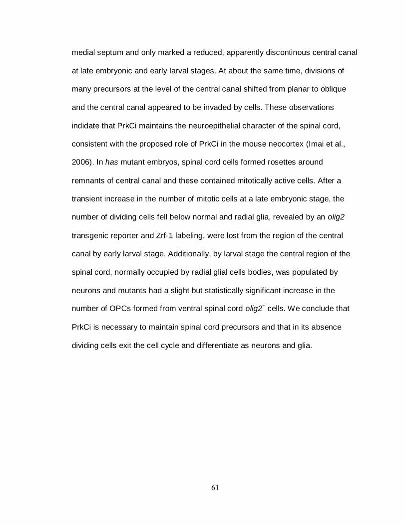
61
medial septum and only marked a reduced, apparently discontinous central canal
at late embryonic and early larval stages. At about the same time, divisions of
many precursors at the level of the central canal shifted from planar to oblique
and the central canal appeared to be invaded by cells. These observations
indidate that PrkCi maintains the neuroepithelial character of the spinal cord,
consistent with the proposed role of PrkCi in the mouse neocortex (Imai et al.,
2006). In has mutant embryos, spinal cord cells formed rosettes around
remnants of central canal and these contained mitotically active cells. After a
transient increase in the number of mitotic cells at a late embryonic stage, the
number of dividing cells fell below normal and radial glia, revealed by an olig2
transgenic reporter and Zrf-1 labeling, were lost from the region of the central
canal by early larval stage. Additionally, by larval stage the central region of the
spinal cord, normally occupied by radial glial cells bodies, was populated by
neurons and mutants had a slight but statistically significant increase in the
number of OPCs formed from ventral spinal cord olig2+ cells. We conclude that
PrkCi is necessary to maintain spinal cord precursors and that in its absence
dividing cells exit the cell cycle and differentiate as neurons and glia.

62
CHAPTER III
CDC42 SIGNALING REGULATES MIGRATION OF THE NKX2.2A+ SUBSET OF
ZEBRAFISH OPCS
Introduction
The myelinating cells of the CNS, oligodendrocytes, wrap and insulate axons to
allow for rapid propagation of electrical impulses over long distances.
Oligodendrocytes are specified in the pMN domain of the ventral spinal cord.
After specification, oligodendrocytes must migrate to their target axons located
throughout the CNS. Although oligodendrocytes function so intimately with
neurons, they often are not specified near their interacting neuron and may have
to travel long distances to myelinate an axon. The mechanisms that control
oligodendrocyte migration remain to be clearly defined.
Oligodendrocytes are initially specified as an immature oligodendrocyte
progenitors cells (OPCs). These OPCs migrate out of the pMN domain to occupy
positions in the dorsal and ventral spinal cord. As they migrate, OPCs mature
and eventually become oligodendrocytes. Part of this maturation process
involves changing their morphology as well as regulating expression of different
transcription factors. When an OPC is fully mature, the oligodendrocyte ceases
migration and begins ensheathing axons and at a later point begins producing
myelin. It is becoming increasingly clear that several mechanisms may be
involved in oligodendrocyte migration.

63
Currently, our understanding of OPC migration is very limited and most
work focuses on in vitro migration. In 2006, Kirby et al performed in vivo studies
in which they investigated OPC migration and wrapping. From these experiments
we learned that OPC migration is a dynamic and highly variable process that
may involve the constant remodeling of the actin cytoskeleton. We also learned
that migrating OPCs extend and retract highly protrusive filopodial like processes
that appear to sense their surrounding environment and direct OPC migration.
Because the Rho family of GTPase is important in regulating filopodial formation
and OPC migration appears to receive migratory information from these filopodial
like processes, we hypothesized that members of the Rho family of proteins
played a role in OPC migration.
The Rho family of small GTPases is made up of about 20 intracellular
signaling molecules, many of which have been shown to be important in
regulating the actin cytoskeleton. These Rho family GTPases function by
shuttling between a GTP-bound active form and a GDP-bound inactive form. The
shuttling between these two states is controlled by two sets of proteins, guanine
nucleotide exchange factors (GEFs) and GTPase activating proteins (GAPs)
(Heasman and Ridley, 2008) (Fig. 1). In their GTP bound active form Rho
GTPases can interact with and activate downstream effectors leading to
regulation of intracellular processes such as vesicle transport, cell cycle
progression, microtubule rearrangements and gene expression. Rho-GTPases
can also lead to stimulation of certain processes like migration, neuronal
development, cell adhesion and cell division (Heasman and Ridley, 2008).
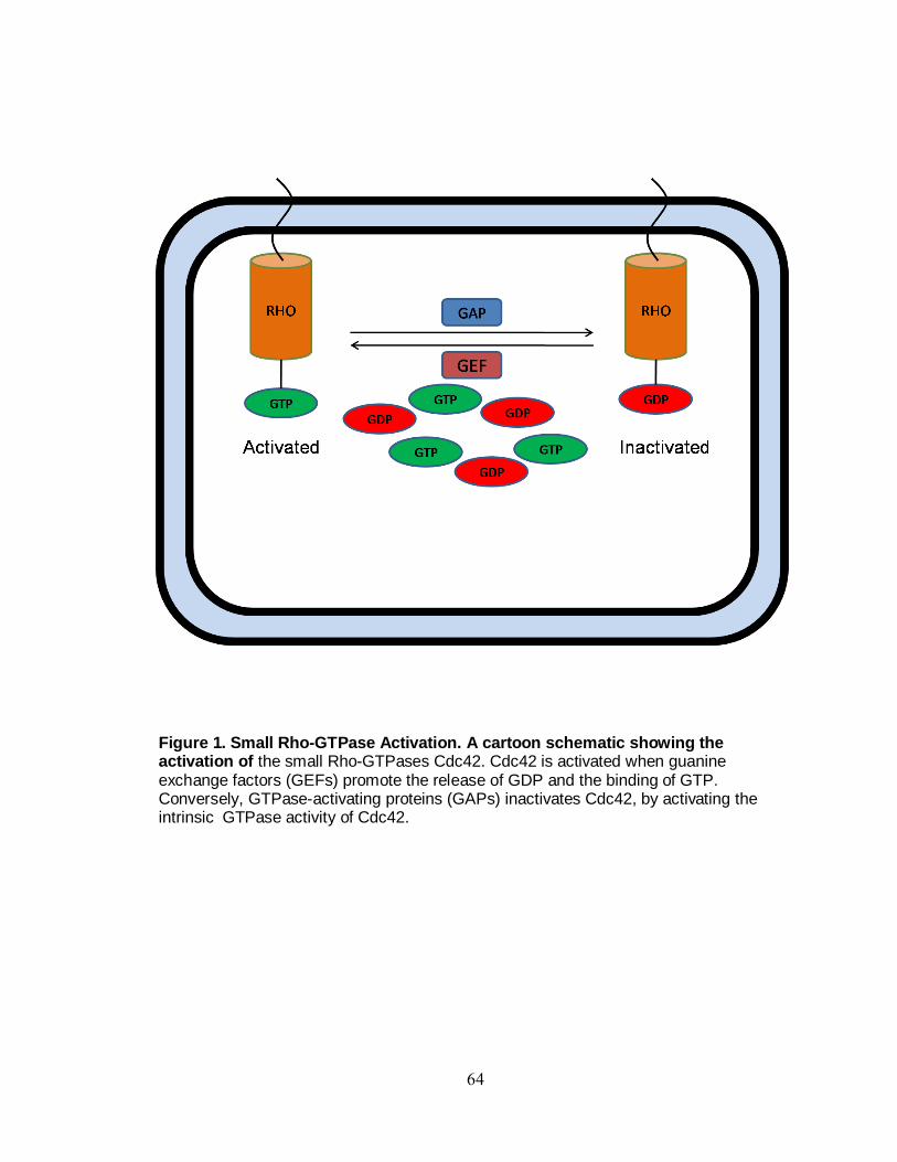
64
Figure 1. Small Rho-GTPase Activation. A cartoon schematic showing the activation of the small Rho-GTPases Cdc42. Cdc42 is activated when guanine exchange factors (GEFs) promote the release of GDP and the binding of GTP. Conversely, GTPase-activating proteins (GAPs) inactivates Cdc42, by activating the intrinsic GTPase activity of Cdc42.

65
Cdc42 is an evolutionarily conserved small Rho family GTPase that functions in
regulating cell polarity and the remodeling of the actin cytoskeleton. It was
originally detected in the yeast Saccharomyces cerevisiae as a mutation that
caused defects in budding and cell polarity (Adams et al., 1990; Johnson and
Pringle, 1990). Since then Cdc42 has been shown to function in a variety of
cellular processes like cell migration, cell polarity and cellular actin dynamics
(Etienne-Manneville et al., 2005; Hall, 2005; Yang and Zheng, 2007).
In many cell types Cdc42 is important in regulating apical polarity by
recruiting members of the apical polarity complex, Par6/PrkCi ,to the apical
membrane by binding to the semi-CRIB domain of Par6 (Garrard et al., 2003).
Although the partitioning defective protein, Par3, function is part of the
evolutionarily conserved Par/PrkCi complex, its role in Cdc42 signaling remains
unclear (Atwood et al., 2007; Welchman et al., 2007). In vitro cell migration
assays on cultured astrocytes show a need for Cdc42 in cell migration and
filopodial formation, as astrocytes fail to migrate in its absence (Etienne-
Manneville et al., 2005). In addition, highly filopodial process like axon growth
cone projection and neurite extension require Cdc42 for proper pathfinding and
extension (Kim et al., 2002). Because Cdc42 seems to be required for migration
and filopodial extensions in highly migratory cells, we hypothesized that Cdc42
be required for OPC migration in vivo. The following experiments were designed
to test this hypothesis.

66
Experimental Procedures
Fish Husbandry
Embryos were produced by pair-wise mating and kept at 28.5oC in egg water or
embryo medium. Embryos were staged to hours post fertilization (hpf) or days
post fertilization (dpf) according to established zebrafish guidelines (Kimmel et
al., 1995). Embryos that were to be used for live imaging, immunohistochemistry,
or in situ hybridization were treated in 0.003% phenylthiourea (PTU) in egg water
to block pigmentation. The experiments conducted in this chapter used the
following strains of zebrafish were used: AB, Tg(nkx2.2a:megfp) (Kirby et al.,
2006), Tg(sox10(7.2):mrfp) (Kucenas et al., 2008) and Tg(hsp70:cdc42(N17)).
Immunocytochemistry
Embryos and larvae were fixed in 4% antibody fix (4% paraformaldehyde, 8%
sucrose, 1x PBS) overnight at 4oC. After fixing, the embryos were embedded in
1.5% agar/ 5% sucrose blocks and placed in 30% sucrose/PBS solution to
equilibrate overnight. The blocks were then frozen over 2-methylbutane chilled by
liquid nitrogen. We collected 10-12 m sections on superfrost microscope slides
using a cryostat microtome. The sections were rehydrated in 1x PBS for 30 min.
and then blocked in 2% BSA/sheep serum in 1x PBS for 30 min. before
incubating with primary antibody overnight at 4oC. For fluorescent detection of
antibody labeling, we used Alexa Fluor 488, Alexa Fluor 568 goat anti-mouse or
goat anti-rabbit conjugates (1:500, Molecular Probes). The primary antibodies
67
used included rabbit anti-PrkCi (#sc-216, 1:200, Santa Cruz Biotechnology, Inc.),
mouse anti-Islet (clone # 39.4D5-s, 1:1,000, Developmental Studies Hybridoma
Bank (DSHB), Iowa City, IA), rabbit anti-Sox10 (1:1,000) (Park et al., 2005), and
mouse anti-Myc (1:200, Fisher) and mouse anti-ZRF-1 (1:500, DSHB).
In vivo time-lapse imaging
Embryos were raised in egg water containing PTU and at the appropriate stages
manually dechorionated using watchmaker forceps. Embryos were anesthetized
using 3-aminobenzoic acid ethyl ester (Tricaine) and mounted laterally or dorsally
in 35 mm glass bottom petri dishes containing 0.8% low-melting temperature
agarose. Confocal time-lapse movies were obtain by using a 40X oil immersion
objective mounted on a Zeiss Axiovert 200 microscope equipped with a
PerkinElmer spinning disk confocal system. Z-stack images were obtained every
15 min and compiled into a Quicktime movie using Volocity software
(Improvision).
Heat-shock induction
To induce expression of dominant negative Cdc42(N17),
Tg(hsp70:cdc42(N17)myc) heterozygous fish were crossed either with
Tg(nkx2.2:megfp) or Tg(sox10(7.2):mrfp) fish and the resultant embryos raised in
egg water containing PTU at 28.5 C. Beakers containing PTU egg water were
allowed to come to temperature in a water bath set at 40 C. Embryos younger
than 36 hpf were heat shocked in their chorions. Embryos were heat shocked for
68
30 min and then transferred to a 28.5 incubator to come back down to
temperature. Transgenic embryos identified by Myc expression and morphology
were selected for analysis.
Results
Loss of PrkCi function disrupts OPC distribution in the spinal cord.
In situ hybridizations using claudink (cldk) anti-sense riboprobe revealed that
OPCs in has-/- embryos, which lack functional PrkCi, have OPCs that are
ectopically located in the medial spinal cord (Fig. 2 B). In wildtype embryos,
OPCs were absent from the medial septum. (Fig.2 A). Similarly, antibody labeling
using the OPC specification marker Sox10, also revealed ectopic OPCs (Fig. 2
C,D). Zebrafish OPC migration is a highly dynamic process in which OPCs
extend and retract their filopodial like processes until they encounter a target
axon to wrap and myelinate. In the absence of PrkCi a subset of OPCs may lose
their ability to migrate normally. To investigate this further, we decided to look at
signaling pathways in which PrkCi may function. Because OPC migration is such
a dynamic process, we hypothesized that OPC migration requires the constant
remodeling and reorganization of the actin cytoskeleton so that the migrating
OPC can achieve polarized directed migration. To investigate this hypothesis, we
focused on the small rho GTPase Cdc42, which has been shown to have an
evolutionarily conserved role in cytoskeletal remodeling.

69
Phenotypic characterization of dominant negative Cdc42(N17) embryos.
Because the Rho-GTPase Cdc42 is important in several developmental
processes, it was important for us to have temporal control of Cdc42 signaling.
To achieve this, we disrupted Cdc42 using a transgenic zebrafish line that
expresses a dominant negative form of Cdc42 (Cdc42(N17))
under the control of the heat shock promoter. Dominant negative Cdc42(N17)
has a threonine to asparagine substitution on residue 17, which abolishes its
affinity for GTP and lowers its affinity for GDP (Luo et al., 1994). When
expressed at high levels, Cdc42(N17) binds GEFs thereby preventing them from
activitating endogenous Cdc42 (Luo et al., 1994). Heatshocking wild-type
embryos at 24, 36 and 48 hpf had no affect on their morphology at 3 dpf (Fig. 3
A). By contrast, Tg(hsp70:cdc42(N17)) embryos that were heatshocked at 24, 36
and 48 hpf and analyzed at 3 dpf had dorsally curved tails, small eyes, cardiac
edema, and some forebrain and hindbrain necrosis (Fig. 3 B). This necrosis did
not extend to the rest of the CNS because the spinal cord was necrosis free (Fig.
3 B). The Tg(hsp70:cdc42(N17)myc) transgenic line was designed so that a Myc
epitope tag is expressed at the C-terminal end of the dominant negative
Cdc42(N17) protein. Therefore, we performed antibody labeling on embryos that
were heatshocked to determine the expression of the Cdc42(N17) protein in the
spinal cord using an antibody against Myc. Cdc42(N17)+ embryos heat shocked
at 24 hpf and fixed 4 hours later showed Myc expression in all cells of the spinal
cord and the surrounding

70
Figure 2. PrkCi may control oligodendrocyte progenitor cell migration. Dashed circle demarcates the spinal cord border. Arrowheads indicate the OPCs, and asterisks denote ectopically positions OPCs. A,B: In situ hybridization using cldnk riboprobe shows an increase in the number of OPCs, as well ectopic OPCs in the medial septum. C,D: Sections labeled with anti-Sox10 antibody. Sox10 labeling also showed ectopic OPCs.

71
Figure 3. Expression of Cdc42(N17) perturbs embryo morphology. A. Tg(hsp70:Cdc42(N17))- embryos maintain a wildtype morphology after heatshocking at 24, 48. and 72 hpf. B. Tg(hsp70:Cdc42(N17))+ embryos have reduced eyes, dorsally curved tails, cardiac edema and some head necrosis after heatshocking at 24, 48 and
72 hpf.

72
muscle (data not shown). Similar expression was observed in Cdc42(N17)+
embryos that were heatshocked at 48 and 72 hpf (data not shown). Cdc42(N17)-
embryos heat shocked at similar time points lacked Myc expression in the spinal
cord at all time points. Myc labeling throughout the embryo demonstrated that
heat shocking Tg(hsp70:cdc42(N17)) embryos drove expression of the
Cdc42(N17)-Myc fusion protein. This suggests that Cdc42 function is being
inhibited; however biochemical analysis would be needed to determine that
conclusively.
Disruption of Cdc42 function affects OPC migration but not specification.
We have shown that the loss of PrkCi function results in the formation of excess
OPCs and loss of neural precursors. Cdc42 plays an important role in stabilizing
PrkCi to the apical membrane. Because apical polarity proteins are dependent on
each other for proper localization to the apical membrane, disruption in Cdc42
may affect apical protein localization. Therefore, we reasoned that expression of
dominant negative Cdc42 protein might produce a phenotype similar to that of
loss of PrkCi function. To test this we performed antibody labeling on 3 dpf
Cdc42(N17)- and Cdc42(N17)+ embryos that were heat shocked at 24, 48 and 72
hpf. The 3 dpf time point was chosen because in has-/- embryos we observed the
biggest phenotypical differences at 3 dpf. Using an antibody against PrkC we
observed that PrkC is localized around the central canal at 3 dpf in Cdc42(N17)-
embryos (Fig. 4A). In heatshocked Cdc42(N17)+ embryos we observed that PrkC
is also localized around the central canal. However in Cdc42(N17)+ embryos we

73
observed residual localization of PrkC in medial septum (Fig. 4B). In wildtype
embryos PrkC is localized the medial septum until 48 hpf when we begin to
observe gaps in PrkC localization. By 72 hpf there is no localization of PrkC in
the medial septum.
Because disruption of PrkCi in the spinal cord resulted in a loss of neural
precursors, we decided to looked at the patterning and location of the radial glia
in the spinal cord. ZRF-1 labeling in the spinal cord of 3 dpf heat shocked
Cdc42(N17)- and heat shocked Cdc42(N17)+ embryos showed the characteristic
radial glia morphology in which cell body makes contact with the apical
membrane of the spinal cord and the radial process makes contact with the pial
surface (Fig. 4 C,D). In Cdc42(N17)+ embryos the radial glia still extend from the
apical to the pial surface, however, their radial fibers appear more disorganized
but there is no apparent loss of radial glia in the medial spinal cord similar to has-
/- embryos (Fig. 4 D). To determine if specification of neuronal subtypes were
affected, we next looked at the number of Islet+ cells in the absence of normal
Cdc42 function. At 3 dpf Islet antibody labeling showed no difference in the
number of ventral Islet+ cells between wild-type and has-/- spinal cords(Fig. 4
E,F). Similar to the PrkCi loss of function data, there was also no difference in the
number of ventral Islet+ cells in heat shocked Cdc42(N17)+ when compared to
Cdc42(N17)- embryos (Fig. 4 I).
To investigate OPC specification, we performed immunocytochemistry
using anti-Sox10 antibody to mark OPCs. This revealed no statistical significance
differences in the number of Sox10+ cells in Cdc42(N17)- and Cdc42(N17)+

74
Figure 4. Loss of Cdc42 function produces ectopic OPCs but does not affect apical polarity. All panels show transverse sections through trunk spinal cord. Dashed circle demarcates the perimeter of the spinal cord. A,B: Sections labeled with anti-PrkCi antibody. PrkCi is localized to the central canal of the spinal cord of wild-type embryos at 3 dpf (open arrowhead) (A). In Tg(hsp70:Cdc42(N17)myc)+ embryos heatshocked at 24, 48 and 72 hpf PrkCi is localized to the central canal (open arrowhead) and a small portion of the medial septum (bracket) (B). C,D: Anti-ZRF-1 labeling revealed normal number and distribution of radial glia in the spinal cord at 3dpf. In the Cdc(N17)+ embryos the radial processes of the radial glia were less organized (D). E,F: Anti-Islet antibody labeling revealed normal numbers of motor neurons and Rohon-Beard sensory neurons were similar in wild-type and CdcN17)+ embryos at 3 dpf. G,H: Anti-Sox10 antibody labeling reveal no difference in the number of Sox10+ cells in wild-type and Cdc42(N17)+ spinal cords at 3dpf. Cdc42(N17)+ embryos did however, have ectoicly positioned OPCs in their spinal cord at 3dpf (open arrowhead)(H). Quantification of spinal cord Islet+ cells. Quantification of spinal cord Sox10+ OPCs. Error bars represent
s.e.m. Statiscal significance was determined using Student’s t-test. Scale bar = 20 m

75
spinal cords at 3dpf (Fig. 4 G,H J). Although the number of OPCs was unaffected
by expression of dominant negative Cdc42, the positions of the OPCs were
disrupted. OPCs were ectopically positioned in the medial spinal cords of
Tg(hsp70l:Cdc42(N17) embryos following heat shock (Fig. 4H), similar to the
abnormal positions of OPCs in has-/- embryos.
nkx2.2a+ OPCs fail to migrate properly when Cdc42 signaling is disrupted.
Sox10 antibody labeling revealed ectopic OPCs in the absence of functional
Cdc42. One interpretation of these data is that Cdc42 is required for the proper
migration of OPCs. To test this, we decided to look at OPC migration in the
absence of Cdc42 using time-lapse microscopy. To achieve this goal, we heat
shocked Tg(nkx2.2a:megfp) embryos at 24, 36 and 48 hpf and began imaging
laterally mounted embryos at 50 hpf for a period of 24 h. The Tg(nkx2.2:megfp)
zebrafish line allows us to observe migration of OPCs and perineurial glia in the
zebrafish spinal cord (Kirby et al., 2006; Kucenas et al., 2008).
During the time-lapse movies we observed that shortly after 50 hpf
nkx2.2a+ OPCs begin to extend and retract membrane processes into the dorsal
spinal cord (Fig. 5). Eventually the OPC cell bodies followed their projections and
migrated out of the ventral nkx2.2 domain. As the time-lapse continues, OPCs
were observed migrating throughout the spinal cord in the general direction of the
dorsal longitudinal fascicle (DLF) (Fig. 5). This process is repeated over several
hours as more OPCs migrate and populate the dorsal spinal cord. Just prior to 72

76
Figure 5. nkx2.2+ OPC migration in the zebrafish spinal cord. Frames captured from time-lapse movies, from a lateral view, of Tg(nkx2.2a:megfp) embryos. Numbers in upper right corners indicate time elapsed from beginning of imaging at 50 hpf. nkx2.2a+ OPCs send fine filopodial like processes out of the ventral nkx2.2a domain (arrows). Eventually the nkx2.2a+ OPCs migrate out of the ventral nkx2.2a domain and take up positions within the dorsal spinal cord (closed arrowheads).

77
Figure 6. nkx2.2+ OPC migration is perturbed in Tg(hsp:Cdc42(N17))+ embryos. Frames captured from time-lapse movies, from a lateral view, of Tg(nkx2.2a:megfp);(hsp:Cdc42(N17))+ embryos. Numbers in upper right corners indicate time elapsed from beginning of imaging at 50 hpf. (A) Unlike in wild-type embryos, the nkx2.2+ OPCs in Tg(hsp:Cdc42(N17))+ embryos fail to extend any fine filopdial like processes out the ventral nkx2.2a+ domain. As the time-lapse imaging begins, rounded nkx2.2a+ cell are observed the ventral nkx2.2a+ domain (open arrowhead). As the movie progresses, rounded nkx2.2a+ cells can bee seen migrating through the dorsal spinal cord. These cells fail to extend any filopodial like processes and are never observed wrapping axons. (B) As the time-lapse imaging continues, filopodilial processes are observed coming out of the ventral spinal cord.

78
hpf some of the nkx2.2a+ cells stop migrating and begin to wrap the axons of the
DLF (Fig. 5). In Tg(nkx2.2a:megfp;(hsp70:Cdc42(N17)myc))+ embryos heat
shocked at 24, 36 and 48 hpf a different pattern of migration was observed. In
the nkx2.2 domain, rounded nkx2.2a+ cells were seen moving throughout the
domain (Fig. 6 A). As time elapsed these rounded nkx2.2a+ cells, which lack any
filopodial like projections, migrated out of the nkx2.2 domain to occupy positions
in the dorsal spinal cord (Fig. 6 A). As the time-lapse continued, we observed
that the number of dorsally migrated nkx2.2a+ OPCs was much less in the Cdc42
(N17)+ embryos than in control embryos. In addition, as the experiment
progressed, we observed much more activity of the nkx2.2a+ cells in the nkx2.2a
domain. Their morphology changed from being rounded and took on an oblong
shape, similar to the morphology of nkx2.2a+ observed in control embryos.
Eventually highly protrusive nkx2.2a+OPCs can be seen migrating out of the
ventral nkx2.2a domain (Fig. 6 B). This suggests that when Cdc42 signaling is
blocked, Nkx2.2a+ OPCs are unable to pathfind out of the ventral nkx2.2a
domain.
Sox10+ OPCs maintain normal morphology and migration in the absence of
Cdc42 signaling. In the ventral spinal cord, the nkx2.2 and olig2 domains can give rise to OPCs. In
the mouse and chick spinal cord, both populations of OPCs eventually begin to
express both Nkx2.2 and Olig2. However, in the zebrafish spinal cord there are
two populations of OPCs: nkx2.2a-olig2+ and nkx2.2a+olig2+ OPCs, although both
populations express the OPCs marker Sox10. From my time-lapse experiments,

79
we determined that the number of nkx2.2a+ OPCs were reduced, however
antibody labeling showed no difference in the average number of Sox10+ OPCs
in the spinal cord between heat shocked control and Cdc42(N17)+ embryos (Fig.
4 G,H,J). Because nkx2.2a+ OPCs represent a portion of the Sox10 expressing
OPCs in the spinal cord, we decided to look at the morphology and migration of
all the OPCs in the spinal cord using Tg(sox10(7.2):mrfp) transgenic line. After
heatshocking Tg(sox10(7.2):mrfp) embryos at 24, 36 and 48 hpf we laterally
mounted embryos and began imaging at 50 hpf.
At the start of the time-lapse, Sox10+ cells are seen in the ventral part of
the spinal cord where they are specified, however as the time-lapse continues
Sox10+ OPCs can be seen extending and retracting filopodial like projections as
they prepare to migrate out of the ventral domain. Shortly after 50 hpf the Sox10+
OPCs begin to take a dorsal migratory path, generally in the direction of the
filopdial protrusions (Fig. 7). In addition to their dorsal migration, these Sox10+
OPCs move laterally searching out a position in which to settle. Just prior to 72
hpf Sox10+ OPCs stop migrating and can be seen wrapping target axons in the
dorsal spinal cord (Fig. 7).
Time-lapse movies of Tg(sox10(7.2):mrfp);(hsp70:Cdc42(N17)myc))+
embryos heat shocked at 24, 36 and 48 hpf showed a similar phenotype to
control embryos. Similar to control embryo, Sox10+ OPCs began migrating out of
the ventral spinal cord shortly after 50 hpf in Tg(sox10(7.2):mrfp);
(hsp70:Cdc42(N17)myc))+ embryos (Fig. 8). OPCs appeared to have a

80
Figure 7. Sox10+ OPC migration in the zebrafish spinal cord. Frames captured from time-lapse movies, from a lateral view, of Tg(sox10(7.2):mrfp) embryos. Numbers in upper right corners indicate time elapsed from beginning of imaging at 50 hpf. Sox10+ OPCs send fine filopodial like processes out of the ventral spinal cord. Eventually the Sox10+ OPCs migrate out of the ventral domain and take up positions within the dorsal spinal cord (arrowheads).

81
Figure 8. Sox10+ OPCs migrate normally in the absence of Cdc42 function. Frames captured from time-lapse movies, from a lateral view, of Tg(sox10(7.2):mrfp);(hsp70:cdc42(n17))+ embryos. Numbers in upper right corners indicate time elapsed from beginning of imaging at 50 hpf. Sox10+ OPCs send fine filopodial like processes out of the ventral spinal cord similar to control embryos. Eventually the Sox10+ OPCs migrate out of the ventral domain and take up positions within the dorsal spinal cord (arrowheads) and begin to wrap axons (open arrowheads).

82
morphology similar to control embryos and appeared to exhibit normal behavior
patterns as the OPCs extended and retracted their filopodial like projections
before settling into position to begin wrapping axons at around 72 hpf (Fig. 8). In
addition, no Sox10+ OPCs displayed a rounded morphology similar to what was
observed in heat shocked Tg(nkx2.2a:megfp;(hsp70:Cdc42(N17)myc))+
embryos, strongly suggesting that the nkx2.2a+ cells may not be specified as
OPCs in the absence of Cdc42 (Fig. 8).
Discussion
Conduction of electrical impulses along the axons requires proper and uniform
myelination of axons. Early in development, oligodendrocytes are specified in
ventral spinal cord and are often required to migrate out of the ventral spinal cord
to meet target axons. Disruption of OPC migration means that axons are not
myelinated and that electrical impulses are not transmitted efficiently leading to
disruption of motor and cognitive skills. As more research is done, it is becoming
clear that no single pathway or secreted factor may control OPC migration. In
fact OPC migration may be a concerted dance in which OPCs have the ability to
respond to the various signals they encounter.
Prior to migration, a cell becomes polarized, by reorganizing its cellular
architecture, such that the microtubule-organizing center (MTOC), microtubules
and Golgi apparatus are positioned in front of the nucleus. This process is called
polarization and is dependent on Cdc42 and the Par complex proteins. RNA
interference of Cdc42 in the one cell Caenorhabditis elegans embryos resulted in

83
embryos that could not localize Par6, Par3 and PrkCi to the apical side of the
embryo. The embryos fail to become polarized and resemble par6, par3 and
PrkCi mutant embryos (Gotta et al., 2001). In addition, Cdc42 is critical for the
recruitment and activation of the Par complex proteins to initial cell-cell contacts,
resulting in local activation of PAR polarity complex, which is essential for the
formation of tight junctions, maturation of the apical junctional complex and
establishment of cell polarity (Ohno, 2001; Yamanaka et al., 2001). From our
data, we observed that in the absence of the apical polarity Par complex protein
PrkCi, OPCs are found in ectopic positions in the medial spinal cord. Similarly,
when Cdc42 signaling was disrupted OPCs failed to migrate properly and were
found in ectopic locations within the medial spinal cord. However, unlike
PrkCi mutants, embryos expressing the dominant negative form of Cdc42 did not
have mislocalization of Par complex proteins, excess OPCs or loss of precursors.
In addition the number and morphology of Islet+ cells were comparable to
wildtype and and heat shocked controls. This suggests that Cdc42 may regulate
OPC migration by signaling through PrkCi. However, unlike in single cell C.
elegans embryos and polarized epithelia, Cdc42 may function with PrkCi in a
pathway that does not affect cellular polarity.
As OPCs migrate, they extend fine filopdial like process that appear to
sense the environment and direct the OPC. Total internal reflection (TIRF)
microscopy showed that Cdc42, Par6 and PrkCi are expressed in the leading
edge of cells during polarized migration (Etienne-Manneville and Hall, 2001).
Inhibition of Cdc42 by microinjection of a dominant negative at leading edge in

84
migrating astrocytes resulted in a failure of the MTOC to become polarized.
Cdc42 has an evolutionarily conserved role in regulating the formation of filopdia.
In vitro time-lapse microscopy imaging of cultured cells lines shows that Cdc42 is
important for filopodia formation, as quiescent cell lines began to initiate filopdia
formation, shortly after the addition of exogenous Cdc42 (Etienne-Manneville and
Hall, 2001; Kozma et al., 1995; Nobes and Hall, 1995). Conversely, when
cultured cells were microinjected with the dominant negative, cells were unable
to form filopdia (Etienne-Manneville and Hall, 2001; Kozma et al., 1995; Nobes
and Hall, 1995). Our results show that when Cdc42 function is impaired in
Tg(nkx2.2a:megfp);(hsp:Cdc42(N17)) embryos, the highly migratory and
filopodial nkx2.2a+ OPCs fail to migrate and appear to drift randomly. In addition,
these nkx2.2a+ OPCs have a rounded morphology and fail to extend any
filopodial like protrusions. This argues that Cdc42 may be required for formation
of filopodial like protrusions in the nkx2.2a+ OPCs. The randomness of the
migration of the nkx2.2a+ OPCs suggests that without their filopodial like
protrusions the migratory nkx2.2a+ OPCs are incapable of sensing and
responding to their environment. Because, filopdia have been ascribed to
function in a sensory and exploratory capacity (Hall, 2005). Interestingly, as the
time-lapse experiments continued, the ventral nkx2.2a+ lost their rounded
morphologies, and cells began to extend more filopodial like processes into the
dorsal spinal cord. Eventually, some nkx2.2a+ OPCs even migrated out of the
ventral nkx2.2 domain and occupied positions in the dorsal spinal cord. Reporter
protein expression levels have been shown to spike 2-4 hours after heat shock

85
induction and that these expression levels gradually decrease so that by 12 hour
post heat shock induction reporter protein expression levels are modestly higher
than control embryos (Adam et al., 2000). Therefore, one explanation for the
increase in filopodial activity and migration of the nkx2.2a+ OPCs is that as time
elapses, the levels of Cdc42(N17) protein decrease and eventually endogenous
Cdc42 protein is able to out compete the dominant negative.
In the ventral spinal cord, OPCs are specified in the pMN and p3 domain.
OPCs specified in the pMN domain express the basic helix loop helix
transcription factor Olig2, whereas OPCs specified in the p3 domain express the
transcription factor Nkx2.2 (Rowitch, 2004). Cell specified in these domains
receive differential concentrations of the morphogens shh and bmp and therefore
up or down regulate different combinations of genes. In the chick spinal cord,
antibody labeling experiements show that most migratory Nkx2.2+ OPCs become
Olig2+ shortly after beginning migration. However, in the mouse spinal cord,
OPCs express both Nkx2.2 and Olig2 prior to initiating migration. In the zebrafish
spinal cord, the relationship between nkx2.2 and olig2 becomes more
complicated. In zebrafish, there are two distinct populations of OPCs:
Nkx2.2a+Olig2+ and Nkx2.2-Olig2+, both of which express the OPC specification
marker Sox10.
Our Sox10 antibody data showed no statistical difference in the number of
OPCs in the spinal cord at 3 dpf. However, our time-lapse movies and whole
mount imaging data showed fewer dorsally migrated nkx2.2a+ cells in the spinal
cord of 3 dpf Tg(nkx2.2:megfp)(hsp70:Cdc42(N17)) embryos as compared to
86
control embryos. One explanation for these results is that in the absence of the
nkx2.2a+ OPCs, the nkx2.2a- population of OPCs may compensate for the
deficiency. This idea of replacement/compensation is not unheard as laser
ablation experiments show that when OPCs are lost, the remaining OPCs can
divide and migrate to replace lost OPCs (Kirby et al., 2006). To investigate this
possibility, we performed time-lapse imaging on heat shocked
Tg(sox10(7.2):mrfp) and Tg(sox10(7.2):mrfp);(hsp70:Cdc42(N17)+ embryos.
From these experiments, we learned that the Sox10+ OPCs in
Tg(sox10(7.2):mrfp);(hsp70:Cdc42(N17)+ embryos appear to migrate normally
and OPC behaviors were similar to control time-lapse movies. In the
Cdc42(N17)+ embryos the OPCs extend and retract their fine filopodial like
process as they move in and out of the dorsal spinal cord and eventually begin
wrapping axons.
Cdc42 has a conserved role in regulating cell polarity and actin
cytoskeleton dynamics across many species. By perturbing Cdc42 function I was
able to demonstrate that Cdc42 may have a novel role in regulating the migration
of the nkx2.2a+ subset of OPCs in the zebrafish spinal cord. Although the
mechanistic basis for this defect in nkx2.2a+ OPC migration phenotype is
unknown, I have provided an excellent starting point for those types of
experiments to progress.

87
CHAPTER IV
NOTCH SIGNALING MAY BE REQUIRED FOR THE MAINTENANCE OF THE
POSTEMBRYONIC PRECURSORS IN THE SPINAL CORD
Introduction
Because the likelihood of recovery from a neurodegenerative disease or a major
CNS insult is extremely low, scientists in the field came to the erroneous
conclusion that “adult” or postembryonic neurogenesis does not occur in the
mammalian brain. This fallacy was dispelled by work done by the Altman lab, in
which they used thymidine analogs to show that constitutive proliferation does
occur in the hippocampus and subventricular zone of the adult mammalian brain
(Altman, 1962; Altman, 1963; Emsley et al., 2005; Temple, 2001). However, this
work was slow to gain acceptance, because many in the scientific community did
not believe the data. The subventricular zone of the lateral ventricle and the
subgranular zone of the hippocampus are the two best characterized areas of
adult proliferation and neurogenesis in mammals (Emsley et al., 2005).
However, postembryonic proliferative cells have been isolated and cultured from
the optic nerve, corpus callosum, spinal cord and hypothalamus, as well as other
regions of the brain. Cells derived from the subventricular zone and the
subgranular zone are highly migratory and often travel long distances to reach
their targets. The proliferative cells of the subventricular zone enter the rostral
migratory stream and travel to the olfactory bulb where they differentiate to

88
become interneurons (Aguirre and Gallo, 2004; Doetsch and Hen, 2005; Lim et
al., 1997). The proliferative cells of the subgranular zone travel a shorter distance
as they differentiate to become hippocampal granule neurons (Aguirre and Gallo,
2004; Doetsch and Hen, 2005; Lim et al., 1997). From these experiments, we
now understand that the CNS is not a static organ system and is capable of
neural regeneration. These postembryonic changes may coincide with distinct
periods of development. In many organisms, the transition from embryonic to
postembryonic stages is followed by dramatic changes in body plan and
morphology. These dramatic transitions are generally referred to as
metamorphosis. However, many organism, especially mammal do not undergo
dramatic metamorphological changes to their morphology.
Zebrafish, for example, do not undergo dramatic changes in their body
plan and morphology while undertaking metamorphosis. So slight are the
changes that some argue that metamorphosis does not occur and that these
slight morphological changes are just transition phases (Balon, 1990; Brown,
1997; Youson, 1988). However, some point to the changes in zebrafish
pigmentation, scales and fin shape at discrete times in development to suggest
that zebrafish undergo some form of metamorphosis (Brown, 1997; Parichy,
2003; Quigley and Parichy, 2002). Besides expression levels of various thyroid
hormones, very little is known about the changes that are occurring in the
zebrafish embryo as it transitions from embryonic stages to the postembryonic
stages. Many questions remain to be answered for example are there any
cellular or morphological changes occurring with the specific organ system that

89
can be attributed to changes in gene expression during this stage of
development? If so, do these changes in gene expression and morphology
represent metamorphological changes.
In embryonic development, there are several genes and signaling
pathways that play important roles in regulating and patterning the various
tissues of the developing embryo. One such signaling pathway, Notch, has been
shown to be required for many aspects of development, such as nervous system
development. Notch signaling, which get its name from the notched wing
phenotype described in Drosophila mutants, is an evolutionarily conserved
pathway and has been described to function in many organisms (Artavanis-
Tsakonas et al., 1999). Notch signaling is generally ascribed to important in
regulating binary cell fate decisions through lateral inhibition. Lateral inhibition is
the process by which cells all with the same developmental potential, differentiate
into 2 different populations of cells. In lateral inhibition, cell that achieve one cell
fate begin to up-regulate Notch which prohibits neighboring cells from achieving
a similar fate, creating cellular diversity within a population (Artavanis-Tsakonas
et al., 1999).
The Notch family of receptors is composed of a type1 single pass
transmembrane receptor protein. The Notch receptor generally consists of a
large extracellular domain (NECD) and a smaller intracellular domain (NICD)
(Ehebauer et al., 2006). Activation of Notch signaling requires binding of a Notch
ligand, which activates a cascade of proteolytic cleavages. The Notch ligands are

90
Figure 1. The Notch signaling pathway. A cartoon schematic showing the Notch signaling pathway. Binding of the Notch ligand to the Notch receptor triggers g-secretase cleavage of the intracellular domain of the Notch receptor (NICD). NICD then translocates to the nucleus where it activate Notch target genes.

91
also type1 single pass transmembrane proteins and are members of the DSL
(Delta/Serrate/Lag-2) family of proteins (Ehebauer et al., 2006). Because both
the receptor and the ligand are both transmembrane proteins, activation of Notch
signaling requires cell-to-cell contact. When the Notch ligand binds to the Notch
receptor, the Notch receptor is cleaved through -secretase activity, which results
in the release of the NICD. The NICD then translocates to the nucleus where it
can alter gene expression of Notch target genes (Ehebauer et al., 2006) (Fig. 1).
Much of what is known about Notch signaling occurs in the early embryo, where
Notch signaling is regulating early cell fate decisions. Recent insights and
discoveries into adult neurogenesis and adult stem cells show that proliferative
and differentiative divisions are occurring in the postembryonic and adult CNS
(Amrein and Lipp, 2009; Zupanc, 2008). Still, questions as to the mechanisms
that regulate these postembryonic populations remain to be answered. Are the
same mechanisms that were important in regulating cell proliferation and
differentiation of the precursor population in the embryonic CNS important in
regulating these precursor cell populationin the postembryonic or adult CNS?
Experimental Procedures
Fish Husbandry
Embryos were produced by pair-wise mating and kept at 28.5oC in egg water or
embryo medium. Embryos were staged to hours post fertilization (hpf) or days
post fertilization (dpf) according to established zebrafish guidelines (Kimmel et

92
al., 1995). Embryos that were to be used for live imaging, immunohistochemistry,
or in situ hybridization were treated in 0.003% phenylthiourea (PTU) in egg water
to block pigmentation. The experiments conducted in this chapter used the AB
strain.
Immunocytochemistry
Embryos and larvae were fixed in 4% antibody fix (4% paraformaldehyde, 8%
sucrose, 1x PBS) overnight at 4oC. After fixing, the embryos were embedded in
1.5% agar/ 5% sucrose blocks and placed in 30% sucrose/PBS solution to
equilibrate overnight. The blocks were then frozen over 2-methylbutane chilled by
liquid nitrogen. We collected 10-12 m sections on superfrost microscope slides
using a cryostat microtome. The sections were rehydrated in 1x PBS for 30 min.
and then blocked in 2% BSA/sheep serum in 1x PBS for 30 min. before
incubating with primary antibody overnight at 4oC. For fluorescent detection of
antibody labeling, we used Alexa Fluor 488, Alexa Fluor 568 goat anti-mouse or
goat anti-rabbit conjugates (1:500, Molecular Probes). The primary antibodies
used included rabbit anti-Sox10 (1:1,000) (Park et al., 2005) mouse anti-HuC
(1:100, Molecular Probes) mouse anti-PCNA (1:200, #ab15497 Abcam)and
mouse anti-ZRF-1 (1:500, DSHB).
Antigen Retrieval
All Immunohistochemical labeling with PCNA antibody required this step to
increase better binding of the antibody to the antigen. Sections of 15 and 17 dpf

93
embryos were transferred to a Coplin jar containing 10 mM sodium citrate buffer
(pH 8.5). The Coplin jar was then placed in a 100 C water bath for 30 minutes.
After 30 minutes the Coplin jar was removed and allowed to come to room
temperature on the bench. When sections were cooled immunohistochemical
labeling was done as described above.
In situ hybridization on sections
Embryos and larvae were fixed in 4% paraformaldehyde overnight at 4oC. After
linearizing plasmids with the appropriate restriction enzymes, anti-sense cRNA
was transcribed using Roche DIG-labeling reagents and T3, T7 or SP6 RNA
polymerases (New England Biolabs). For the experiments contained within this
paper the following anti-sense RNA probes were used: notch1a and dld. After
fixing, the embryos were embedded in 1.5% agar/ 5% sucrose RNase free blocks
and placed in 30% sucrose/PBS RNase free solution to equilibrate overnight.
The blocks were then frozen over 2-methylbutane chilled by liquid nitrogen. We
collected 10-12 m sections on superfrost microscope slides using a cryostat
microtome. Sections were rehydrated in 1X PBS for 30 min. then covered with
75% glycerol. Images were obtained using a Retiga EXI camera attached to a
Olympus Novius EX70 microscope equipped with Openlab software.
DAPT Treatment
To block Notch signaling, DAPT (N-[N-(3,5-Difluorophenacetyl-L-alanyl]-S-
phenylglycine-t-butyl ester; Calbiochem, La Jolla, CA) treatments were

94
performed as previously described (Geling et al., 2002). DAPT was reconstituted
with DMSO to make a stock concentration of 10 M. For experiments, aliquots
were diluted to 100 M in EM. Larval fish were placed in the DAPT solution at 15
and 17 dpf and incubated overnight at 28.5°C. Control embryos were incubated
in an equivalent concentration (1%) of DMSO in EM and incubated overnight.
Results
The rate of cell division in the spinal cord fluctuates during development.
Until recently postembryonic neurogenesis was thought to be absent in the CNS.
Studies performed on the adult mammalian and avian brains show that it does
occur and that it may be a way of regenerating neurons in the adult brain. The
spinal cord, like the brain, is often thought of as a fully differentiated tissue with
little regenerative capacity. I wanted to determine if the spinal cord contained
pockets of cells that could proliferate leading to neurogenesis in a juvenile to
adult fish, similar to what is observed in the adult brain of other models. To do
this we employed a strategy that would label mitotic cells. During mitosis a cell
must replicate its DNA in a semi conservative fashion. Using the existing strands
of DNA as a template, the appropriate complementary base is added creating
two copies of the genome. To determine the rate of cell division we used the
cell’s replication machinery to label cells with BrdU, a thymine analog, at stages
ranging from 1 dpf to 30 dpf.

95
Figure 2. Dividing cells of the zebrafish spinal cord. All panels show transverse section through trunk spinal cord. Dashed circle denotes the spinal cord boundary. A-G. Embryos, larvae and juveniles treated with BrdU. Sections labeled with anti-BrdU antibody. H. Graph showing the rate of cell divisions in the zebrafish spinal cord.

96
At 1 dpf cell division in the spinal cord occurred rapidly and many cells
were BrdU+. On average there were approximately 42.9 Brdu+ cells per section
(Fig. 2A, H). At 3, 6 and 7 dpf cell divisions decreased dramatically and on
average the number of BrdU+ cells per section was 4.9, 2.3 and 1.9 respectively
(Fig. 2 B,C,D,H). At 10 dpf the number of cell divisions in the spinal cord began
to increase and the average number of BrdU+ cells per section was 2.5 (Fig. 2
H). At 17, 20 and 30 dpf the increase in spinal cord cell divisions continued with
an average of 4.2, 8 and 15.5 cells per section respectively (Fig. 2 E,F,G,H).
Interestingly, early in development, cell division occur randomly throughout the
spinal cord; however, as the embryo develops and transitions into a juvenile, cell
divisions appear to be restricted to the ventricular zone of the spinal cord.
Post-embryonic cell divisions may give rise to radial glia and OPCs but not neurons.
Adult neurogenesis occurs in distinct niches within the adult mammalian brain. In
these niches the adult stem cells differentiate and under in vivo conditions give
rise only to neurons (Alvarez-Buylla and Lim, 2004). And although under extreme
exogenous conditions the fate of the adult neural stem cells can be altered, the
default cell fate are hippocampal neurons or olfactory bulb neurons. (Hack et al.,
2005; Jackson et al., 2006; Jessberger et al., 2008). As previously described,
most of the postembryonic cell divisions appear to be limited to the ventricular
zone of the spinal cord. We were interested in determining the fate of these
dividing ventricular zone cells. To achieve this we pulsed postembryonic juvenile
embryos with the S-phase marker BrdU. I used BrdU as a lineage tracer,

97
because DNA replicates semi conservatively so any incorporated BrdU would be
passed on in successive divisions.
Using Zrf-1 antibody which labels the radial fibers and cell membranes of
the radial glia, we observed BrdU+ cells along the ventricle of the spinal cord at
17 dpf. These BrdU+ cells were outlined with the Zrf-1 positive membranes (Fig.
3 A) (arrowheads). Similar labeling was observed at 20 and 30 dpf suggesting
that BrdU+ cells were giving rise to rise to radial glia or that the radial glia were
dividing giving rise to other cell types. Next I wanted to determine if the BrdU
positive cell were giving rise to any other cell type. Using an antibody to Sox10,
an OPC specification marker, I observed BrdU and Sox10 double labeled
cells at 30dpf. Next I wanted to determine if these postembryonic proliferative
cells gave rise to neurons as is the norm in the adult mammalian brain. Using the
pan neuronal marker HuC, I was unable to identify any HuC and BrdU double
labeled cells in the spinal cord at 15 and 17 dpf. This suggested that the
proliferative cells in the spinal cord do not give rise to neurons, unlike what is
observed in the adult mammalian brain (Alvarez-Buylla et al., 2001; Alvarez-
Buylla and Lim, 2004; Alvarez-Buylla and Lois, 1995).

98
Figure 3. Radial glia and OPCs arise from post embryonic proliferative cells. All panels show transverse sections through trunk spinal cord. All panels are labeled with anti-BrdU antibody after being treated with BrdU. Dashed circle demarcates the perimeter of the spinal cord. A-C: Sections labeled with anti-ZRF-1 antibody. ZRF-1 labels the cell membranes of radial glia. Arrowheads indicate radial glia labeled with ZRF-1 and Brdu. D: Sections labeled with anti-Sox10 antibody, which labels the OPCs. Arrowheads indicate OPCs labeled with Sox10 and Brdu. E,F: Sections labeled with anti-Hu antibody. Hu labels all post mitotic neurons. Arrowheads indicate BrdU+ cells that do not colocalize with BrdU. All post mitotic neurons were devoid of BrdU staining.
Scale bar = 20 m.

99
Notch signaling may regulate postembryonic proliferation in the zebrafish spinal cord.
As was described earlier, divisions within the spinal cord transition from a rapidly
dividing period to a quiescent period, and back to a period of rapid division.
During the rapidly dividing periods, cells were exiting the cell cycle and
differentiating. I wanted to determine what mechanisms behind this
transformation. Loss of function and ablation studies have shown that Notch
signaling is important in regulating binary cell fate decision as well as regulating
differentiation through lateral inhibition (Shin et al., 2007). During neural
development, Notch signaling maintains precursor cells in the undifferentiated
state and in its absence, precursor cells exited the cell cycle and differentiate. In
zebrafish, blocking Notch signaling resulted in early born neurons at the expense
of the undifferentiated precursors. I wanted to determine if expression of Notch
signaling proteins played a role in regulating postembryonic proliferation. To do
this, I looked at expression of various delta and notch genes at various
embryonic and postembryonic stages.
Of the many delta and notch genes we investigated, only 2 genes had an
interesting pattern of expression: deltad and notch1a. deltad (dld) expression at 5
dpf is largely absent and found in random punctate expression throughout the
spinal cord. At 10 dpf dld is still randomly expressed throughout the spinal cord
similar to its expression at 5 dpf. At 20 and 30 dpf dld expression increases and
is expressed strongly in the ventricular zone of the spinal cord (Fig 4 A-D).
notch1a expression at 5 dpf is expressed in a diffuse pattern around the spinal
cord. At 10 dpf a similar diffuse pattern of notch1a expression is observed,

100
however at 20 and 30 dpf expression of notch1a increases and is restricted to
the cells of the ventricular zone (Fig 4 E-H). Both the timing and pattern of
expression of dld and notch1a coincided with the timing and pattern of cell
division in the postembryonic spinal cord.
Inhibiting Notch signaling decreases postembryonic cell division.
After observing that increases in the levels of dld and notch1a gene expression
coincided with increases in the number of postembryonic cell divisions, I wanted
to determine if Notch signaling affected cell divisions in the postembryonic spinal
cord. I tested this by knocking down Notch signaling using the chemical inhibitor
N-[N-(3,5-Difluorophenacetyl-L-alanyl)]-S-phenylglycine t-butyl ester (DAPT).
DAPT is a gamma secretase inhibitor that blocks Notch signaling by inhibiting the
cleavage and nuclear translocation of the Notch intracellular domain (NICD).
Earlier, I determined that postembryonic proliferation in the zebrafish spinal cord
begins to increase at around 15 dpf. Therefore, I reasoned that if Notch signaling
was important for increasing the rate of cell divisions in the postembryonic
zebrafish spinal cord, then blocking it at this time would have the most dramatic
result. At 15 dpf the average number of PCNA+ cells in the spinal cord averaged
4 cells per section in the DMSO treated controls and 1.8 cells per section in the
DMSO treated postembryonic larvae (Fig. 5 A,B,C). Two days later, at 17 dpf, an
average of 3.1 PCNA positive cells per section were found in the DMSO control
and 1.9 cells per section in the DAPT treated postembryonic embryos. These

101
Figure 4. dld and notch1a RNA expression is up regulated during postembryonic stages. All panels show transverse sections through trunk spinal cord at 5,10, 20 and 30 dpf. A-D: In situ hybridization using dld riboprobe shows an increase in RNA expression in the cells of the medial septum during late postembryonic periods. dld RNA expression is diffuse at 5 and 10 dpf however by 20 and 30 dpf dld RNA expression is present in the cell lining the proliferative zone of the spinal cord. E-H: In situ hybridization using notch1a riboprobe shows an increase in gene expression in the cells of the medial septum during late postembryonic periods. Similar to dld RNA expression, notch1a RNA is absent from the spinal cord at 5 and 10 dpf but is highly expressed in the cells of the
proliferative zone at 20 and 30 dpf. Scale bar = 20 m

102
Figure 5. Delta-Notch signaling regulates postembryonic proliferation in the spinal cord. All panels show transverse sections through trunk spinal cord at 15 dpf. A,B:Sections labeled with an anti-PCNA antibody. Embryos treated with DAPT, a gamma secretase inhibitor that block Delta-Notch signaling, have reduced numbers of proliferative cells. C. Quantification of spinal cord PCNA+ proliferative cells between control and DAPT treated embryos. Statistical significance was determined using
Student’s t-test. Scale bar = 20 m

103
data argues strongly for a role in Delta-Notch signaling in regulating
postembryonic proliferation in the zebrafish spinal cord.
Discussion
As an organism ages, injured and dying cells are replaced as needed. Proper
maintenance of this homeostasis allows for the organ systems in the body to
function without any noticeable interruption. This was thought to be true of most
organ systems except for the nervous system. Ramon y Cajal argued that the
adult nervous system was composed entirely of terminally differentiated cells that
ceased proliferating after embryogenesis (Lopez-Munoz et al., 2006). In his
theory, the cells comprising the nervous system were all specified shortly after
birth, and replacement of dying or injured cells did not occur via postembryonic
neurogenesis (Lopez-Munoz et al., 2006). Although evidence for postembryonic
neurogenesis was found in the reptilian brain as early as 1950, it was not until
Altman and colleagues published a series of papers reporting [H3]-thymidine
evidence for new neurons in various regions of adult rats, including the dentate
gyrus of the hippocampus, neocortex and olfactory bulb that the long held dogma
of no postembryonic proliferation in the nervous system was debunked (Altman,
1962; Altman, 1963; Altman and Das, 1965; Alvarez-Buylla and Lois, 1995). At
present, very little is known about the molecular mechanisms controlling
postembryonic proliferation and regulating the transition of precursor cells from a
quiescent state to an actively proliferating state. In addition, cell fate
determination of these proliferative cells is not well defined. Here we identified a
population of post embryonic proliferative cells in the zebrafish spinal cord, and

104
investigated the role of the Delta-Notch signaling pathway in regulating these
post embryonic proliferative cells.
Since the 1960’s, the discovery of postembryonic neurogenesis in the
adult mammalian brain, numerous studies have documented widespread
neurogenesis in non-mammals, such as fish, amphibians, reptiles and birds
(Alvarez-Buylla and Nottebohm, 1988; Chapouton et al., 2006; Grandel et al.,
2006; Kaslin et al., 2008; Lopez-Garcia et al., 1988; Raymond and Easter, 1983).
Our data shows that in addition to areas of proliferation in the adult brain, there
are areas of postembryonic proliferation, in zebrafish spinal cord. Understanding
the mechanisms that control postembryonic proliferation in the spinal cord is
important, as studies looking at cell specification and differentiation in the brain
and spinal cord, shows that these two areas regulate cell specification and
differentiation differently.
One of the key features of zebrafish as a model system is its rapid
external development. A fertilized zebrafish embryo can develop from a single
cell embryo to a free swimming larva within 72 hours (Kimmel et al., 1995). This
rapid development allowed us to investigate rates of proliferation in the zebrafish
spinal cord over several stages of development. Through BrdU incorporation
experiments, we found that at 1 dpf the spinal cord is undergoing a period of
widespread rapid cell divisions. This rapid rate of cell divisions is followed by a
dramatic decrease in cell divisions at 2 and 3 dpf, towards the end of
embryogenesis. At around 15 dpf there is a dramatic increase in the rate of cell

105
division in the spinal cord. The increase in number of postembryonic proliferative
cell divisions
In the mammalian brain, postembryonic proliferative cells give rise to
neurons and oligodendrocytes. These newborn cells integrate themselves into
the CNS to form functional circuits. To determine the fate of these proliferative
postembryonic spinal cord cells in zebrafish, we used BrdU as a lineage tracer.
Our data showed that the dividing cells of the postembryonic spinal cord gave
rise to oligodendrocytes and radial glia. However, contrary to what is thought to
occur, our data showed that no new neurons are specified from the
postembryonic proliferative cells.
Because Notch signaling plays an important role in maintaining the proliferative
precursor population, we hypothesized that they may also play a role during the
postembryonic stages. (Shin et al., 2007). Our in situ data shows that notch1a
and dld transcripts are absent from the spinal cord during the quiescent
postembryonic period. However, during the proliferative postembryonic periods,
cells along the medial septum began expressing notch1a and dld transcripts. The
temporal and spatial expression of both genes suggested a role for Notch
signaling in during postembryonic proliferation. Using DAPT, we found that
larvae, in which Notch signaling was disrupted, had fewer postembryonic
proliferative cells in their spinal cords. By inhibiting Notch signaling at the onset
of postembryonic proliferation, we were able to determine that Notch signaling
does play a role in maintaining the postembryonic proliferative precursors in the
zebrafish spinal cord.

106
CHAPTER V
CONCLUSIONS AND FUTURE DIRECTIONS
Development of the spinal cord from a single layer of neuroepithelium is a
complex process that involves the coordination of several processes including
cell migration, cell specification and cell differentiation. Proper patterning of the
spinal cord requires that these processes are spatially and temporally regulated,
so that genes are turned on or off when and where required. Any disruption in
these processes can affect the overall morphology of the spinal cord. In this
thesis I show that in the absence of prkci, an apical polarity gene, zebrafish
spinal cord precursor cells fail to maintain parallel precursor divisions. As a
consequence, OPCs are specified in excess at the expense of the precursor
population. In addition, I show that Cdc42 plays a role in the migration of the
Nkx2.2a+ OPC population in the zebrafish spinal cord. Lastly, I argue for the role
of Notch signaling in maintaining the postembryonic proliferative cells in the
spinal cord.
The apical polarity protein PrkCi is necessary for maintenance of spinal cord precursors in zebrafish.
Oligodendrocytes, the myelinating cells of the vertebrate CNS, play an important
role in transmitting electrical impulses along the axons. In their absence,
cognitive and motor functions are impaired. Demyelinating diseases affect more
than two million people worldwide, and represent a significant world health

107
burden. By understanding the mechanisms that control how these cells develop,
scientist are hopeful that they can prevent or cure demyelinating diseases.
I showed using the zebrafish spinal cord as a model that PrkCi, the
zebrafish homologue to aPKC is important in specifying OPCs in normal
numbers, by maintaining parallel precursor divisions. Through various loss of
function experiments, aPKC has been shown to regulate cell specification in
Drosophila and C. elegans. In this thesis, I show that in the zebrafish spinal
cord, PrkCi has a similar function, as it regulates specification of zebrafish OPCs.
In addition, aPKC regulates establishment and maintenance of apical-basal
polarity in polarized epithelia in Drosophila, C. elegans and mammals. This role
for aPKC is conserved in zebrafish, because in the absence of functional PrkCi
apical basal polarity is disrupted in the zebrafish spinal cord. By performing
antibody labeling experiments, using an antibody against PrkC, I showed that
both PrkCi and PrkCz are expressed in the apical membranes of the spinal cord
cells at the medial septum and around the central canal. In has-/- embryos I
demonstrated that PrkCz was unable to compensate for the function of PrkCi.
Localization of apical markers, like ZO-1, was diminished at the medial septum
and around the central canal, showing that apical polarity is disrupted in has
mutants. Although the Par-aPKC complex is an evolutionary conserved complex,
accumulating evidence has also revealed diversity and complexity in the means
by which different cells utilize the PAR-aPKC complex to establish polarity.
aPKC regulates asymmetric cell divisions in the Drosophila CNS. When
aPKC is present, these asymmetric cell divisions occur perpendicular to plain of

108
the overlying epithelia and produces a nueroblast and a GMC. However, in the
absence of aPKC, orientation of these divisions are randomized and produces 2
ganglion mother cells of equal potential. In C. elegans, aPKC-Par complex
controls the asymmetric division of the fertilized 1- cell embryo. Disruption of
apkc-par complex genes results in a fertilized embryo that fails to divide
asymmetrically and produces two daughter cells of equal size and possible equal
potential (Watts et al., 1996). In this thesis, I showed that in the zebrafish spinal
cord, PrkCi regulates precursor cell divisions orientation, but in a different
manner than in Drosophila and C. elegans. Time-lapse imaging experiments
allowed me to show that PrkCi maintained parallel divisions in the presumptive
precursor cells around the central canal. Using Tg(h2afv:egfp) embryos, I
observed that the dividing cells at the central canal surface divided parallel to the
plane of the central canal. Similar time-lapse experiments, using
Tg(h2afv:egfp);has-/- embryos, which lack expression of functional PrkCi, showed
that in the absence of PrkCi, division around the central canal could occur
parallel or non parallel. This is unique, as stem cells and their resultant precursor
cells are generally thought to undergo asymmetric proliferative divisions, in which
they give rise to another precursor cell and a differentiated cell. These
asymmetric proliferative divisions are thought to occur perpendicular to the plane
of the epithelia. I also showed that initiation of the non-parallel divisions occurred
around 30 hpf, which occurs just prior to OPC specification. This randomization
of cell divisions may lead to a loss of precursor cell maintenance and an increase
in differentiated cell types.

109
In addition, through loss of function experiment, I determined, that as a
consequence of these non-parallel cell divisions at the central canal, a significant
excess of OPCs were found in the spinal cord of has-/- embryos. However, early
born cells like motor neurons and Rohon Beard cells were unaffected. This is
different than what occurs in the Drosophila CNS; because when aPKC is absent
from the Drosophila CNS, the neuroblasts divide symmetrically and give rise to
excess GMCs, which go on to differentiate into excess neurons. As a
consequence of dividing symmetrically, the number of neuroblasts is reduced. In
situ hybridizations, antibody labeling and whole mount imaging, demonstrated
that in the absence of PrkCi, precursor cell population was reduced, and that the
reduction in precursor cells give rise to the excess OPCs.
Through time-lapse imaging, of Tg(olig2:egfp) and Tg(olig2:egfp);has-/-
embryos, we provided evidence that the excess OPCs in has-/- embryos were
due to increased specification of OPCs. By using time-lapse imaging, we were
able to discern whether the excess OPCs in the has-/- spinal cords were due to
increased cell proliferation in the dorsal spinal cord or due to increased OPC
specification in the ventral olig2 domain. In the time-lapse movies, I saw that in
has-/- embryos, excess OPCs were specified in the ventral olig2 domain and
these excess OPCs migrated out into the dorsal spinal cord. It also showed that
the excess OPCs were not because of increased OPC proliferation in the dorsal
spinal cord.
In summary, I have shown that in the absence of PrkCi precursor cells fail
to maintain parallel divisions, and excess OPCs are specified at the expense of

110
the precursor cells; however many outstanding questions remained to be
answered. For example, does angle of divisions predict cell fate? In the
Drosophila CNS, neuroblasts divide asymmetrically giving rise to two daughter
cells of different cell fates. In this thesis, I was unable to show any direct
evidence that established a connection between angle of division and cell fate.
This was largely due to the lack of available tools. Because our Tg(h2afv:egfp)
line labeled all the cell nuclei, it was impossible for us to determine the cell fates
of a newly divided daughter cells once they migrated away from the central
canal. One approach around this obstacle would be to inject either h2afv:egfp
DNA or RNA at low concentrations into single cell transgenic embryos. Possible
transgenic embryos for injection include: Tg(sox10(7.2):mrfp), Tg(olig2:dsred)
and Tg(islet1:dsred). My goal with this experiment would be to create a transient
transgenic that has mosaic expression of GFP in a few cells at the central canal.
By labeling only a few cells it would allow us to track newly divided daughter cells
as they mature and turn on cell specification genes like islet, sox10 and olig2.
PrkCi has been shown to be part of the evolutionarily conserved Par-
aPKC complex that regulates cell polarity and asymmetric divisions. In this
thesis, I was unable to show definitively that phenotypes observed in the
absence of PrkCi were direct results of disruption in the Par-aPKC complex. As
members of the Par-aPKC complex are dependent on each other for proper
localization to the apical membrane, disrupting one gene should affect the
formation and function of the complex. In future experiments, I would suggest
knocking down par-3 and par-6 expression by injection of morpholino antisense

111
oligonucleotides. If the phenotypes observed in PrkCi deficient embryos were the
result of a loss of Par-aPKC complex signaling, disruption of other complex
members should produce a similar phenotype. Morpholinos that disrupt Par-3
expression in zebrafish exist and the zebrafish homologue to par-6 has already
been cloned, therefore disruption of par-3 and par-6 should be simple to achieve
(Munson et al., 2008; Wei et al., 2004).
Although, I was able to show an overall reduction in precursor cells, I was
unable to provide quantitative data for this reduction. To rectify this, I would
perform whole-mount immunohistochemisty on Tg(olig2:egfp) and
Tg(olig2:egfp)has-/- embryos using an antibody against ZRF-1, which labels the
radial glia and the postembryonic proliferative precursors. Then the number of
double labeled postembryonic proliferative cells could be counted and analyzed.
Cdc42 signaling regulates migration of the nkx2.2a+ subset of zebrafish OPCs.
Cell migration is an essential process during many phases of development and
adult life. It drives embryonic morphogenesis and contributes to tissue repair and
regeneration (Keller, 2005; Luster et al., 2005). It also orchestrates disease
progression in cancer, mental retardation, atherosclerosis, and arthritis (Adiguzel
et al., 2009; Emma, 2009; Yamaguchi et al., 2005). Understanding the
mechanism by which cells migrate may lead to the development of novel
therapeutic strategies that can aide in disease prevention or cures. OPCs are a
population of glial cells that migrate toward their target axons. Failure of OPC
and axons to connect can lead to diseases that impair cognitive and motor

112
function. Advances in transgenesis and the transparent nature of zebrafish have
made it possible to study the migration of migratory cells like OPCs in vivo.
Cdc42 has a conserved role in regulating cell polarity and the actin
cytoskeleton in many eukaryotic organisms. Cdc42 has been shown to have a
role in yeast budding, epithelial polarity, migratory polarity and fate specification
during cell division. Cdc42-knockout mice are embryonic lethal and die before
embryonic day 7.5 (Chen et al., 2000). Because Cdc42 plays such an important
role in many early developmental pathways, other approaches to disrupting
Cdc42 function were needed. For example, in many cell types, both constitutively
active Cdc42 and dominant-negative Cdc42 have been shown to effective in
disrupting Cdc42 function by affecting the formation of highly dynamic finger-like
actin-rich protrusions known as filopodia (Gupton and Gertler, 2007). Through
heat shocking experiments I observed that expression of dominant negative
Cdc42(N17)-Myc in zebrafish produces embryos with dorsally curved tails,
cardiac edema and with no apparent necrosis in the spinal cord. Heat shocked
wildtype embryos appear normal and did appear to exhibit any abnormal
phenotypes as a result of heatshocking. Myc labeling confirmed the expression
of the Cdc42(N17)-Myc fusion protein throughout the entire embryo. This shows
that heatshocking may be an effective way of inhibiting Cdc42 signaling.
Cell polarity is fundamental to many cellular processes, including
migration, differentiation and morphogenesis. Cdc42 seems to function primarily
through the polarity protein Par-6 and thereby with Par-3 and atypical protein
kinase C (aPKC) isoforms to induce polarity in several different animal models

113
(Etienne-Manneville, 2004; Goldstein and Macara, 2007). Cdc42 and the aPKC-
Par complex play important roles in the formation and maintenance of polarity in
epithelial cells. Epithelial cells have a polarized morphology that is characterized
by an asymmetrical distribution of proteins, which form distinct apical and basal
domains. These distinct apical and basal domains allow for directed transport
across the epithelial layer while at the same time acting as a barrier to certain
small solutes and ions. Constitutively active and dominant-negative Cdc42 affect
the polarized trafficking of proteins to the apical and basal domains (Etienne-
Manneville, 2004; Macara, 2004). The localization of PAR6–aPKC to the apical
domain is dependent on Cdc42 and is required for targeting of apical proteins
(Wu et al., 2006). Through loss of function experiments, aPKC-Par complex
proteins fail to maintain their apical epithelial localization in the absence of
Cdc42, eventually leading to a breakdown of the epithelia (Wu et al., 2006). In
the zebrafish spinal cord, blocking Cdc42 signaling produced ectopically
positioned OPCs similar to what is seen in the absence of PrkCi. However, unlike
what was observed in has mutants, blocking of Cdc42 signaling did not affect
apical polarity, as the localization of apical polarity proteins PrkC and ZO-1 was
normal. In addition the number of Sox10+ OPCs was similar between control and
Cdc42(N17)+ embryos. This also suggests that Cdc42 and PrkCi may be
functioning together, but through a pathway that does not affect apical polarity.
This is not a unique role for Cdc42 as studies have shown that Cdc42 can
regulate cell polarity by either not interacting with the aPKC-Par complex or by

114
interacting with selective members of the aPKC-Par complex(Etienne-Manneville
et al., 2005; Gomes et al., 2005).
Timelapse imaging of heat shocked Tg(nkx2.2a:megfp)(hsp:cdc42(n17)-
myc) embryos showed that the Nkx2.2a+ OPCs fail to migrate properly in the
absence of Cdc42 signaling. However, timelapse imaging of
Tg(sox10(7.2):mrfp)(hsp:cdc42(n17)-myc) under the same conditions, revealed
that the Sox10+ OPCs migrate normally and in normal numbers. This suggests
that the migration of various populations of OPCs may be regulated differently.
Our antibody and time-lapse data argues for a role of Cdc42 in the
migration of the Nkx2.2a+ OPCs. However, our data also shows that in the
absence of Cdc42 function, the number of Sox10+ OPCs in the spinal cord is
comparable to wild type. In addition, our time-lapse data on
Tg(sox10(7.2):mfrp)(hsp:cdc42(N17)-myc)+ embryos shows that the Sox10+
OPCs migrate normally out of the ventral spinal cord and in normal numbers.
This suggests that Cdc42 regulates migration of the Nkx2.2a+ Olig2+ Sox10+
OPCs and that the Nkx2.2a- Olig2+ Sox10+ OPC can compensate for the
Nkx2.2a+ Olig2+ Sox10+ OPCs. To test this hypothesis, I propose doing time
lapse imaging on heatshocked Tg(sox10(7.2):mfrp)(nkx2.2a:megfp) and
Tg(sox10(7.2):mfrp)(hsp:cdc42(N17)-myc)+ embryos. If the Nkx2.2a- Olig2+
Sox10+ OPCs are compensating for the Nkx2.2a+ Olig2+ Sox10+ OPCs in the
absence of normal Cdc42 function, you would expect to see only Nkx2.2a- Olig2+
Sox10+ OPCs.

115
Taken together these data show that Cdc42 may be important in either the
specification and/or migration of the nkx2.2a+ subset of OPCs. This could
potentially be a novel function for Cdc42 in oligodendrocytes. Current studies
show that Cdc42 plays an important role in many aspects of glial cell migration
and development. For example, Cdc42 mutant mice have myelin sheaths that
are thinner and more disorganized. This wrapping deficiency appears to be the
result of a defect in removing cytoplasm as the oligodendrocyte plasma
membranes wrap around the axon (Thurnherr2006). In addition, Cdc42 has been
shown to be important in Schwann cell proliferation and myelination as well as rat
astrocyte migration.(Benninger2007etienne-manville2006).
From our experiments in this thesis, I showed that heat shocked
Tg(hsp:cdc42(N17)-myc) embryos had distinct morphological phenotypes, and
expressed the dominant negative fusion protein Cdc42(N17)-Myc in all cells of
the spinal cord. Although I identified the presence of the dominant negative I
never tested the efficacy of the dominant negative. Heatshocking of
Tg(hsp:cdc42(N17)-myc) embryos leads to the expression of a dominant
negative Cdc42 that contains a asparagine to threonine substitution mutation at
amino acid 17 (Heasman and Ridley, 2008). The dominant negative theoretically
blocks endogenous Cdc42 signaling by out competing endogenous Cdc42 for
binding to GEFs. The N17 mutation allows the binding of GEFs but does not
allow the subsequent activation of Rho GTPase signaling. To test the efficacy of
the dominant negative, I propose performing GEF activity assays. After
heatshocking Tg(hsp:cdc42(N17)-myc) embryos, lysates can be made of control

116
and Cdc42(N17) embryos to test for the ability of the GEF to activate the release
of GDP.
One caveat to our experiments is that heat shocking expresses a
dominant negative that binds and sequesters GEFs. Several small Rho-GTPases
can use the same GEF for activation of signaling (Heasman and Ridley, 2008).
By sequestering GEF activity, I may be blocking more than one small Rho-
GTPase. Therefore the results interpreted may not be due solely to the effects of
inhibition of Cdc42 signaling.
Notch signaling may be required for the maintenance of the postembryonic precursors in the spinal cord.
Until recently, the CNS has long been thought of as a static organ system
incapable of regeneration. This CNS plasticity appears to be evolutionarily
conserved as many areas of adult neurogenesis or postembryonic proliferation
have been identified across species. In the mammalian CNS, newborn
postembryonic cells migrate to various brain regions, replace older ones and
connect to existing circuits. Evidence suggests that this replacement is related to
acquisition of new information. Therefore, postembryonic proliferation and
replacement can be seen as a form of brain plasticity that enables organisms to
adjust to environmental changes like learning and memory. In this thesis I
identified a population of postembryonic proliferative precursors in the zebrafish
spinal cord that appears to be maintained through Notch signaling. In addition,
these precursors appear to be giving rise to radial glia and oligodendrocytes but
not neurons.

117
By performing BrdU incorporation experiments, I was able to identify areas
of postembryonic cell proliferation along the medial septum in the zebrafish
spinal cord. There are two distinct periods of cell division. The first occurs during
embryogenesis and is followed by a quiescent transition period during early larval
stages. The second stage occurs at about 15 dpf where I observed an increase
in cell divisions during the later larval stages. Cell proliferation in the larval stages
is maintained into adulthood.
Using BrdU as a lineage tracer, I was able to identify that these
postembryonic cells give rise to OPCs and possibly radial glia. However, at all
time points examined, I was unable to detect any neurons that expressed the pan
neuronal marker Hu and the cell proliferation marker Brdu, suggesting that these
postembryonic proliferative cells do not give rise to neurons in the postembryonic
spinal cord.
Because Notch signaling has been shown to be important in maintaining
precursor cells, I looked at expression of notch genes at various stages in
development. In situ hybrizations revealed that the temporal and spatial gene
expression pattern of notch1a and dld were similar to the temporal and spatial
pattern of cell division. To investigate this, I blocked Notch signaling using the
chemical inhibitor DAPT. Antibody labeling, using an antibody PCNA as a cell
division marker, revealed that in the absence of Notch signaling, the number of
postembryonic proliferative cells was decreased. One pitfall to this experiment is
that DAPT blocks all g-secretase activity within the embryo, which makes it
impossible to know for sure if the affects are solely to a loss of Notch signaling.

118
To address this, I would use a transgenic line that expresses a dominant
negative form of suppressor of hairless under the control of the heat shock
promoter. This would make it possible for us to block Notch signaling in a time
dependant fashion.
119
REFERENCES CITED
Aaku-Saraste, E., Hellwig, A. and Huttner, W. B. (1996). Loss of occludin and functional tight junctions, but not ZO-1, during neural tube closure--remodeling of the neuroepithelium prior to neurogenesis. Dev Biol 180, 664-79.
Adam, A., Bartfai, R., Lele, Z., Krone, P. H. and Orban, L. (2000). Heat-
inducible expression of a reporter gene detected by transient assay in zebrafish. Exp Cell Res 256, 282-90.
Adams, A. E., Johnson, D. I., Longnecker, R. M., Sloat, B. F. and Pringle, J. R. (1990). CDC42 and CDC43, two additional genes involved in budding and the establishment of cell polarity in the yeast Saccharomyces cerevisiae. J Cell Biol
111, 131-42.
Adiguzel, E., Ahmad, P. J., Franco, C. and Bendeck, M. P. (2009). Collagens in the progression and complications of atherosclerosis. Vascular Medicine 14,
73-89.
Adler, P. N. (2002). Planar signaling and morphogenesis in Drosophila. Dev Cell
2, 525-35.
Adolf, B., Chapouton, P., Lam, C. S., Topp, S., Tannh‰user, B., Str‰hle, U., Gˆtz, M. and Bally-Cuif, L. (2006). Conserved and acquired features of adult neurogenesis in the zebrafish telencephalon. Developmental Biology 295, 278-
293.
Afonso, C. and Henrique, D. (2006). PAR3 acts as a molecular organizer to define the apical domain of chick neuroepithelial cells. J Cell Sci 119, 4293-304.
Aguirre, A. and Gallo, V. (2004). Postnatal neurogenesis and gliogenesis in the olfactory bulb from NG2-expressing progenitors of the subventricular zone. J Neurosci 24, 10530-41.
Alison, M. R. and Islam, S. (2009). Attributes of adult stem cells. J Pathol 217,
144-60.
Alison, M. R., Poulsom, R., Forbes, S. and Wright, N. A. (2002). An introduction to stem cells. J Pathol 197, 419-23.
Altman, J. (1962). Are new neurons formed in the brains of adult mammals? Science 135, 1127-8.

120
Altman, J. (1963). Autoradiographic investigation of cell proliferation in the brains of rats and cats. Anat Rec 145, 573-91.
Altman, J. and Bayer, S. A. (1984). The development of the rat spinal cord. Adv Anat Embryol Cell Biol 85, 1-164.
Altman, J. and Das, G. D. (1965). Autoradiographic and histological evidence of postnatal hippocampal neurogenesis in rats. J Comp Neurol 124, 319-35.
Alvarez-Buylla, A. (1990). Mechanism of neurogenesis in adult avian brain. Experientia 46, 948-55.
Alvarez-Buylla, A., Garcia-Verdugo, J. M. and Tramontin, A. D. (2001). A unified hypothesis on the lineage of neural stem cells. Nat Rev Neurosci 2, 287-
93.
Alvarez-Buylla, A. and Lim, D. A. (2004). For the Long Run: Maintaining
Germinal Niches in the Adult Brain. 41, 683-686.
Alvarez-Buylla, A. and Lois, C. (1995). Neuronal stem cells in the brain of adult vertebrates. Stem Cells 13, 263-72.
Alvarez-Buylla, A. and Nottebohm, F. (1988). Migration of young neurons in adult avian brain. Nature 335, 353-4.
Amrein, I. and Lipp, H. P. (2009). Adult hippocampal neurogenesis of mammals: evolution and life history. Biol Lett 5, 141-4.
Anthony, T. E., Klein, C., Fishell, G. and Heintz, N. (2004). Radial glia serve as neuronal progenitors in all regions of the central nervous system. Neuron 41,
881-90.
Appel, B. and Eisen, J. S. (1998). Regulation of neuronal specification in the zebrafish spinal cord by Delta function. Development 125, 371-80.
Appel, B., Givan, L. A. and Eisen, J. S. (2001). Delta-Notch signaling and lateral inhibition in zebrafish spinal cord development. BMC Dev Biol 1, 13.
Artavanis-Tsakonas, S., Rand, M. D. and Lake, R. J. (1999). Notch Signaling: Cell Fate Control and Signal Integration in Development. Science 284, 770-776.
Atwood, S. X., Chabu, C., Penkert, R. R., Doe, C. Q. and Prehoda, K. E. (2007). Cdc42 acts downstream of Bazooka to regulate neuroblast polarity through Par-6 aPKC. J Cell Sci 120, 3200-6.
Balon, E. K. (1990). Epigenesis of an epigeneticist : the development of some alternative concepts on the early ontogeny and evolution of fishes. Guelph
Ichthyology Reviews 1, 1-48.

121
Batista, M. F., Jacobstein, J. and Lewis, K. E. (2008). Zebrafish V2 cells develop into excitatory CiD and Notch signalling dependent inhibitory VeLD interneurons. Developmental Biology 322, 263-275.
Baye, L. M. and Link, B. A. (2007). Interkinetic nuclear migration and the selection of neurogenic cell divisions during vertebrate retinogenesis. J Neurosci
27, 10143-52.
Baye, L. M. and Link, B. A. (2008). Nuclear migration during retinal development. Brain Research 1192, 29-36.
Bernhardt, R. R., Chitnis, A. B., Lindamer, L. and Kuwada, J. Y. (1990). Identification of spinal neurons in the embryonic and larval zebrafish. J Comp Neurol 302, 603-16.
Betschinger, J., Mechtler, K. and Knoblich, J. A. (2003). The Par complex directs asymmetric cell division by phosphorylating the cytoskeletal protein Lgl. Nature 422, 326-30.
Briscoe, J., Sussel, L., Serup, P., Hartigan-O'Connor, D., Jessell, T. M.,
Rubenstein, J. L. R. and Ericson, J. (1999). Homeobox gene Nkx2.2 and specification of neuronal identity by graded Sonic hedgehog signalling. Nature
398, 622-627.
Brown, L. D. (1997). The metamorphosis: conversion in historical context. Bull N Y Acad Med 74, 238-47.
Buescher, M., Yeo, S. L., Udolph, G., Zavortink, M., Yang, X., Tear, G. and Chia, W. (1998). Binary sibling neuronal cell fate decisions in the Drosophila
embryonic central nervous system are nonstochastic and require inscuteable-mediated asymmetry of ganglion mother cells. Genes Dev 12, 1858-70.
Burrill, J. D., Moran, L., Goulding, M. D. and Saueressig, H. (1997). PAX2 is expressed in multiple spinal cord interneurons, including a population of EN1+ interneurons that require PAX6 for their development. Development 124, 4493-
503.
Cameron, H. A., Woolley, C. S., McEwen, B. S. and Gould, E. (1993).
Differentiation of newly born neurons and glia in the dentate gyrus of the adult rat. Neuroscience 56, 337-44.
Cepko, C. L., Ryder, E. F., Austin, C. P., Walsh, C. and Fekete, D. M. (1993). Lineage analysis using retrovirus vectors. Methods Enzymol 225, 933-60.
Chapouton, P., Adolf, B., Leucht, C., Tannhauser, B., Ryu, S., Driever, W. and Bally-Cuif, L. (2006). her5 expression reveals a pool of neural stem cells in the adult zebrafish midbrain. Development 133, 4293-303.

122
Chen, F., Ma, L., Parrini, M. C., Mao, X., Lopez, M., Wu, C., Marks, P. W., Davidson, L., Kwiatkowski, D. J., Kirchhausen, T. et al. (2000). Cdc42 is
required for PIP2-induced actin polymerization and early development but not for cell viability. Current Biology 10, 758-765.
Chen, W., Burgess, S. and Hopkins, N. (2001). Analysis of the zebrafish smoothened mutant reveals conserved and divergent functions of hedgehog activity. Development 128, 2385-2396.
Chenn, A. and McConnell, S. K. (1995). Cleavage orientation and the asymmetric inheritance of Notch1 immunoreactivity in mammalian neurogenesis. Cell 82, 631-41.
Choksi, S. P., Southall, T. D., Bossing, T., Edoff, K., de Wit, E., Fischer, B.
E., van Steensel, B., Micklem, G. and Brand, A. H. (2006). Prospero Acts as a Binary Switch between Self-Renewal and Differentiation in Drosophila†Neural†Stem Cells. Developmental Cell 11, 775-789.
Clarke, J. D., Hayes, B. P., Hunt, S. P. and Roberts, A. (1984a). Sensory physiology, anatomy and immunohistochemistry of Rohon-Beard neurones in embryos of Xenopus laevis. J Physiol 348, 511-25.
Clarke, J. D., Hayes, B. P., Hunt, S. P. and Roberts, A. (1984b). Sensory
physiology, anatomy and immunohistochemistry of Rohon-Beard neurones in embryos of Xenopus laevis. J Physiol 348, 511-525.
Concha, M. L. and Adams, R. J. (1998). Oriented cell divisions and cellular morphogenesis in the zebrafish gastrula and neurula: a time-lapse analysis. Development 125, 983-94.
Copp, A., Cogram, P., Fleming, A., Gerrelli, D., Henderson, D., Hynes, A., Kolatsi-Joannou, M., Murdoch, J. and Ybot-Gonzalez, P. (2000). Neurulation and neural tube closure defects. Methods Mol Biol 136, 135-60.
Copp, A. J., Greene, N. D. and Murdoch, J. N. (2003). The genetic basis of mammalian neurulation. Nat Rev Genet 4, 784-93.
Cui, S., Otten, C., Rohr, S., Abdelilah-Seyfried, S. and Link, B. A. (2007).
Analysis of aPKClambda and aPKCzeta reveals multiple and redundant functions during vertebrate retinogenesis. Mol Cell Neurosci 34, 431-44.
Das, T., Payer, B., Cayouette, M. and Harris, W. A. (2003). In vivo time-lapse imaging of cell divisions during neurogenesis in the developing zebrafish retina. Neuron 37, 597-609.
Doe, C. Q. (2008). Neural stem cells: balancing self-renewal with differentiation. Development 135, 1575-87.

123
Doe, C. Q., Chu-LaGraff, Q., Wright, D. M. and Scott, M. P. (1991). The prospero gene specifies cell fates in the drosophila central nervous system. Cell
65, 451-464.
Doetsch, F. and Hen, R. (2005). Young and excitable: the function of new neurons in the adult mammalian brain. Curr Opin Neurobiol 15, 121-8.
Egger, B., Chell, J. M. and Brand, A. H. (2008). Insights into neural stem cell biology from flies. Philos Trans R Soc Lond B Biol Sci 363, 39-56.
Ehebauer, M., Hayward, P. and Martinez-Arias, A. (2006). Notch Signaling Pathway. Sci. STKE 2006, cm7-.
Emma, J. B. (2009). Involvement of the cytoskeletal elements in articular cartilage homeostasis and pathology. International Journal of Experimental Pathology 90, 1-15.
Emsley, J. G., Mitchell, B. D., Kempermann, G. and Macklis, J. D. (2005). Adult neurogenesis and repair of the adult CNS with neural progenitors, precursors, and stem cells. Prog Neurobiol 75, 321-41.
Ericson, J., Morton, S., Kawakami, A., Roelink, H. and Jessell, T. M. (1996). Two critical periods of Sonic Hedgehog signaling required for the specification of motor neuron identity. Cell 87, 661-73.
Ericson, J., Rashbass, P., Schedl, A., Brenner-Morton, S., Kawakami, A.,
van Heyningen, V., Jessell, T. M. and Briscoe, J. (1997). Pax6 controls progenitor cell identity and neuronal fate in response to graded Shh signaling. Cell 90, 169-80.
Etemad-Moghadam, B., Guo, S. and Kemphues, K. J. (1995). Asymmetrically distributed PAR-3 protein contributes to cell polarity and spindle alignment in early C. elegans embryos. Cell 83, 743-752.
Etienne-Manneville, S. (2004). Cdc42 - the centre of polarity. J Cell Sci 117,
1291-1300.
Etienne-Manneville, S. and Hall, A. (2001). Integrin-Mediated Activation of Cdc42 Controls Cell Polarity in Migrating Astrocytes through PKC[zeta]. Cell 106,
489-498.
Etienne-Manneville, S., Manneville, J.-B., Nicholls, S., Ferenczi, M. A. and Hall, A. (2005). Cdc42 and Par6-PKC{zeta} regulate the spatially localized association of Dlg1 and APC to control cell polarization. J. Cell Biol. 170, 895-
901.
Ever, L. and Gaiano, N. (2005). Radial 'glial' progenitors: neurogenesis and signaling. Curr Opin Neurobiol 15, 29-33.

124
Fishell, G. and Kriegstein, A. R. (2003). Neurons from radial glia: the consequences of asymmetric inheritance. Curr Opin Neurobiol 13, 34-41.
Fuchs, E., Tumbar, T. and Guasch, G. (2004). Socializing with the neighbors: stem cells and their niche. Cell 116, 769-78.
Garrard, S. M., Capaldo, C. T., Gao, L., Rosen, M. K., Macara, I. G. and Tomchick, D. R. (2003). Structure of Cdc42 in a complex with the GTPase-binding domain of the cell polarity protein, Par6. Embo J 22, 1125-33.
Gates, M. A., Thomas, L. B., Howard, E. M., Laywell, E. D., Sajin, B.,
Faissner, A., Gotz, B., Silver, J. and Steindler, D. A. (1995). Cell and molecular analysis of the developing and adult mouse subventricular zone of the cerebral hemispheres. J Comp Neurol 361, 249-66.
Geldmacher-Voss, B., Reugels, A. M., Pauls, S. and Campos-Ortega, J. A. (2003). A 90{degrees} rotation of the mitotic spindle changes the orientation of mitoses of zebrafish neuroepithelial cells. Development 130, 3767-3780.
Geling, A., Steiner, H., Willem, M., Bally-Cuif, L. and Haass, C. (2002). A
gamma-secretase inhibitor blocks Notch signaling in vivo and causes a severe neurogenic phenotype in zebrafish. EMBO Rep 3, 688-94.
Girard, F., Cremazy, F., Berta, P. and Renucci, A. (2001). Expression pattern of the Sox31 gene during zebrafish embryonic development. Mech Dev 100, 71-
3.
Goldstein, B. and Macara, I. G. (2007). The PAR Proteins: Fundamental Players in Animal Cell Polarization. Developmental Cell 13, 609-622.
Gomes, E. R., Jani, S. and Gundersen, G. G. (2005). Nuclear Movement Regulated by Cdc42, MRCK, Myosin, and Actin Flow Establishes MTOC Polarization in Migrating Cells. Cell 121, 451-463.
Gotta, M., Abraham, M. C. and Ahringer, J. (2001). CDC-42 controls early cell polarity and spindle orientation in C. elegans. Current Biology 11, 482-488.
Gotz, M., Hartfuss, E. and Malatesta, P. (2002). Radial glial cells as neuronal
precursors: a new perspective on the correlation of morphology and lineage restriction in the developing cerebral cortex of mice. Brain Res Bull 57, 777-88.
Gotz, M. and Huttner, W. B. (2005). The cell biology of neurogenesis. Nat Rev Mol Cell Biol 6, 777-88.
Grandel, H., Kaslin, J., Ganz, J., Wenzel, I. and Brand, M. (2006). Neural stem cells and neurogenesis in the adult zebrafish brain: origin, proliferation dynamics, migration and cell fate. Dev Biol 295, 263-77.

125
Grunwald, D. J. and Eisen, J. S. (2002). Headwaters of the zebrafish [mdash] emergence of a new model vertebrate. Nat Rev Genet 3, 717-724.
Gupton, S. L. and Gertler, F. B. (2007). Filopodia: The Fingers That Do the Walking. Sci. STKE 2007, re5-.
Hack, M. A., Saghatelyan, A., de Chevigny, A., Pfeifer, A., Ashery-Padan, R., Lledo, P.-M. and Gotz, M. (2005). Neuronal fate determinants of adult olfactory bulb neurogenesis. Nat Neurosci 8, 865-872.
Hall, A. (2005). Rho GTPases and the control of cell behaviour. Biochem Soc
Trans 33, 891-5.
Halliday, A. L. and Cepko, C. L. (1992). Generation and migration of cells in the developing striatum. Neuron 9, 15-26.
Hatakeyama, J., Bessho, Y., Katoh, K., Ookawara, S., Fujioka, M., Guillemot,
F. and Kageyama, R. (2004). Hes genes regulate size, shape and histogenesis of the nervous system by control of the timing of neural stem cell differentiation. Development 131, 5539-5550.
Haydar, T. F., Ang, E., Jr. and Rakic, P. (2003). Mitotic spindle rotation and mode of cell division in the developing telencephalon. Proc Natl Acad Sci U S A
100, 2890-5.
Heasman, S. J. and Ridley, A. J. (2008). Mammalian Rho GTPases: new insights into their functions from in vivo studies. Nat Rev Mol Cell Biol 9, 690-701.
Hewagama, A. and Richardson, B. (2009). The genetics and epigenetics of autoimmune diseases. J Autoimmun.
Hollyday, M. (2001). Neurogenesis in the vertebrate neural tube. Int J Dev
Neurosci 19, 161-73.
Hong, E. and Brewster, R. (2006). N-cadherin is required for the polarized cell behaviors that drive neurulation in the zebrafish. Development 133, 3895-3905.
Horne-Badovinac, S., Lin, D., Waldron, S., Schwarz, M., Mbamalu, G.,
Pawson, T., Jan, Y., Stainier, D. Y. and Abdelilah-Seyfried, S. (2001). Positional cloning of heart and soul reveals multiple roles for PKC lambda in zebrafish organogenesis. Curr Biol 11, 1492-502.
Hung, T. J. and Kemphues, K. J. (1999). PAR-6 is a conserved PDZ domain-containing protein that colocalizes with PAR-3 in Caenorhabditis elegans embryos. Development 126, 127-35.

126
Hurd, T. W., Gao, L., Roh, M. H., Macara, I. G. and Margolis, B. (2003). Direct interaction of two polarity complexes implicated in epithelial tight junction assembly. Nat Cell Biol 5, 137-42.
Hutchinson, S. A. and Eisen, J. S. (2006). Islet1 and Islet2 have equivalent
abilities to promote motoneuron formation and to specify motoneuron subtype identity. Development 133, 2137-2147.
Hutson, L. D. and Chien, C.-B. (2002). Wiring the zebrafish: axon guidance and synaptogenesis. Current Opinion in Neurobiology 12, 87-92.
Ihrie, R. A. and Alvarez-Buylla, A. (2008). Cells in the astroglial lineage are neural stem cells. Cell Tissue Res 331, 179-91.
Ikeshima-Kataoka, H., Skeath, J. B., Nabeshima, Y.-i., Doe, C. Q. and Matsuzaki, F. (1997). Miranda directs Prospero to a daughter cell during Drosophila asymmetric divisions. Nature 390, 625-629.
Imai, F., Hirai, S.-i., Akimoto, K., Koyama, H., Miyata, T., Ogawa, M., Noguchi, S., Sasaoka, T., Noda, T. and Ohno, S. (2006). Inactivation of
aPKC{lambda} results in the loss of adherens junctions in neuroepithelial cells without affecting neurogenesis in mouse neocortex. Development 133, 1735-
1744.
Itoh, M., Kim, C. H., Palardy, G., Oda, T., Jiang, Y. J., Maust, D., Yeo, S. Y., Lorick, K., Wright, G. J., Ariza-McNaughton, L. et al. (2003). Mind bomb is a
ubiquitin ligase that is essential for efficient activation of Notch signaling by Delta. Dev Cell 4, 67-82.
Izumi, Y., Hirose, T., Tamai, Y., Hirai, S.-i., Nagashima, Y., Fujimoto, T., Tabuse, Y., Kemphues, K. J. and Ohno, S. (1998). An Atypical PKC Directly Associates and Colocalizes at the Epithelial Tight Junction with ASIP, a Mammalian Homologue of Caenorhabditis elegans Polarity Protein PAR-3. J. Cell Biol. 143, 95-106.
Jackson, E. L., Garcia-Verdugo, J. M., Gil-Perotin, S., Roy, M., Quinones-Hinojosa, A., VandenBerg, S. and Alvarez-Buylla, A. (2006). PDGFR±-Positive B Cells Are Neural Stem Cells in the Adult SVZ that Form Glioma-like
Growths in Response to Increased PDGF Signaling. 51, 187-199.
Jessberger, S., Toni, N., Clemenson Jr, G. D., Ray, J. and Gage, F. H. (2008).
Directed differentiation of hippocampal stem/progenitor cells in the adult brain. Nat Neurosci 11, 888-893.
Jessell, T. M. (2000). Neuronal specification in the spinal cord: inductive signals and transcriptional codes. Nat Rev Genet 1, 20-9.

127
Jessen, J. R., Topczewski, J., Bingham, S., Sepich, D. S., Marlow, F., Chandrasekhar, A. and Solnica-Krezel, L. (2002). Zebrafish trilobite identifies new roles for Strabismus in gastrulation and neuronal movements. Nat Cell Biol
4, 610-5.
Jia, C. and Halpern, M. (1998). Neurogenesis and migration of receptor neurons in the vomeronasal sensory epithelium in the opossum, Monodelphis domestica. J Comp Neurol 400, 287-97.
Johnson, D. I. and Pringle, J. R. (1990). Molecular characterization of CDC42, a Saccharomyces cerevisiae gene involved in the development of cell polarity. J
Cell Biol 111, 143-52.
Kaltschmidt, J. A. and Brand, A. H. (2002). Asymmetric cell division: microtubule dynamics and spindle asymmetry. J Cell Sci 115, 2257-2264.
Kaltschmidt, J. A., Davidson, C. M., Brown, N. H. and Brand, A. H. (2000).
Rotation and asymmetry of the mitotic spindle direct asymmetric cell division in the developing central nervous system. Nat Cell Biol 2, 7-12.
Kaslin, J., Ganz, J. and Brand, M. (2008). Proliferation, neurogenesis and regeneration in the non-mammalian vertebrate brain. Philos Trans R Soc Lond B Biol Sci 363, 101-22.
Keller, R. (2005). Cell migration during gastrulation. Current Opinion in Cell Biology 17, 533-541.
Kemphues, K. J., Priess, J. R., Morton, D. G. and Cheng, N. (1988). Identification of genes required for cytoplasmic localization in early C. elegans embryos. Cell 52, 311-320.
Kim, H., Kim, S., Chung, A. Y., Bae, Y. K., Hibi, M., Lim, C. S. and Park, H. C.
(2008a). Notch-regulated perineurium development from zebrafish spinal cord. Neurosci Lett 448, 240-4.
Kim, H., Shin, J., Kim, S., Poling, J., Park, H. C. and Appel, B. (2008b). Notch-regulated oligodendrocyte specification from radial glia in the spinal cord of zebrafish embryos. Dev Dyn 237, 2081-9.
Kim, M. D., Kolodziej, P. and Chiba, A. (2002). Growth Cone Pathfinding and Filopodial Dynamics Are Mediated Separately by Cdc42 Activation. J. Neurosci.
22, 1794-1806.
Kimmel, C. B., Ballard, W. W., Kimmel, S. R., Ullmann, B. and Schilling, T. F. (1995). Stages of embryonic development of the zebrafish. Dev Dyn 203, 253-
310.

128
Kimmel, C. B., Warga, R. M. and Kane, D. A. (1994). Cell cycles and clonal strings during formation of the zebrafish central nervous system. Development
120, 265-76.
Kirby, B. B., Takada, N., Latimer, A. J., Shin, J., Carney, T. J., Kelsh, R. N.
and Appel, B. (2006). In vivo time-lapse imaging shows dynamic oligodendrocyte progenitor behavior during zebrafish development. Nat Neurosci
9, 1506-11.
Klein, T. J. and Mlodzik, M. (2005). Planar cell polarization: an emerging model points in the right direction. Annu Rev Cell Dev Biol 21, 155-76.
Komiya, Y. and Habas, R. (2008). Wnt signal transduction pathways. Organogenesis 4, 68-75.
Kondo, A., Kamihira, O. and Ozawa, H. (2009). Neural tube defects: prevalence, etiology and prevention. Int J Urol 16, 49-57.
Konno, D., Shioi, G., Shitamukai, A., Mori, A., Kiyonari, H., Miyata, T. and Matsuzaki, F. (2008). Neuroepithelial progenitors undergo LGN-dependent
planar divisions to maintain self-renewability during mammalian neurogenesis. Nat Cell Biol 10, 93-101.
Korn, T. (2008). Pathophysiology of multiple sclerosis. J Neurol 255 Suppl 6, 2-
6.
Kosodo, Y., Roper, K., Haubensak, W., Marzesco, A. M., Corbeil, D. and Huttner, W. B. (2004). Asymmetric distribution of the apical plasma membrane during neurogenic divisions of mammalian neuroepithelial cells. Embo J 23,
2314-24.
Kovac, J., Oster, H. and Leitges, M. (2007). Expression of the atypical protein
kinase C (aPKC) isoforms iota/lambda and zeta during mouse embryogenesis. Gene Expr Patterns 7, 187-96.
Kozma, R., Ahmed, S., Best, A. and Lim, L. (1995). The Ras-related protein Cdc42Hs and bradykinin promote formation of peripheral actin microspikes and filopodia in Swiss 3T3 fibroblasts. Mol. Cell. Biol. 15, 1942-1952.
Kraut, R. and Campos-Ortega, J. A. (1996). inscuteable,A Neural Precursor Gene ofDrosophila,Encodes a Candidate for a Cytoskeleton Adaptor Protein. Developmental Biology 174, 65-81.
Kraut, R., Chia, W., Jan, L. Y., Jan, Y. N. and Knoblich, J. A. (1996). Role of inscuteable in orienting asymmetric cell divisions in Drosophila. Nature 383, 50-5.

129
Kucenas, S., Takada, N., Park, H. C., Woodruff, E., Broadie, K. and Appel, B. (2008). CNS-derived glia ensheath peripheral nerves and mediate motor root development. Nat Neurosci 11, 143-51.
Kuhlbrodt, K., Herbarth, B., Sock, E., Hermans-Borgmeyer, I. and Wegner, M. (1998). Sox10, a novel transcriptional modulator in glial cells. J Neurosci 18,
237-50.
Lee, C. Y., Andersen, R. O., Cabernard, C., Manning, L., Tran, K. D., Lanskey, M. J., Bashirullah, A. and Doe, C. Q. (2006a). Drosophila Aurora-A kinase inhibits neuroblast self-renewal by regulating aPKC/Numb cortical polarity and spindle orientation. Genes Dev 20, 3464-74.
Lee, C. Y., Robinson, K. J. and Doe, C. Q. (2006b). Lgl, Pins and aPKC regulate neuroblast self-renewal versus differentiation. Nature 439, 594-8.
Lee, K. J. and Jessell, T. M. (1999). The specification of dorsal cell fates in the vertebrate central nervous system. Annu Rev Neurosci 22, 261-94.
Lewis, K. E. and Eisen, J. S. (2001). Hedgehog signaling is required for primary motoneuron induction in zebrafish. Development 128, 3485-3495.
Lim, D. A., Fishell, G. J. and Alvarez-Buylla, A. (1997). Postnatal mouse
subventricular zone neuronal precursors can migrate and differentiate within multiple levels of the developing neuraxis. Proc Natl Acad Sci U S A 94, 14832-6.
Lledo, P.-M., Merkle, F. T. and Alvarez-Buylla, A. (2008). Origin and function of olfactory bulb interneuron diversity. Trends in Neurosciences 31, 392-400.
Lopez-Garcia, C., Molowny, A., Garcia-Verdugo, J. M. and Ferrer, I. (1988). Delayed postnatal neurogenesis in the cerebral cortex of lizards. Developmental Brain Research 43, 167-174.
Lopez-Munoz, F., Boya, J. and Alamo, C. (2006). Neuron theory, the cornerstone of neuroscience, on the centenary of the Nobel Prize award to Santiago Ramon y Cajal. Brain Res Bull 70, 391-405.
Lowery, L. A. and Sive, H. (2004). Strategies of vertebrate neurulation and a re-evaluation of teleost neural tube formation. Mech Dev 121, 1189-97.
Lu, Q. R., Sun, T., Zhu, Z., Ma, N., Garcia, M., Stiles, C. D. and Rowitch, D. H.
(2002). Common developmental requirement for Olig function indicates a motor neuron/oligodendrocyte connection. Cell 109, 75-86.
Luo, L., Liao, Y. J., Jan, L. Y. and Jan, Y. N. (1994). Distinct morphogenetic functions of similar small GTPases: Drosophila Drac1 is involved in axonal outgrowth and myoblast fusion. Genes Dev 8, 1787-802.
130
Luster, A. D., Alon, R. and von Andrian, U. H. (2005). Immune cell migration in inflammation: present and future therapeutic targets. Nat Immunol 6, 1182-1190.
Lyons, D. A., Guy, A. T. and Clarke, J. D. W. (2003). Monitoring neural progenitor fate through multiple rounds of division in an intact vertebrate brain. Development 130, 3427-3436.
Macara, I. G. (2004). Parsing the Polarity Code. Nat Rev Mol Cell Biol 5, 220-
231.
Malatesta, P., Hack, M. A., Hartfuss, E., Kettenmann, H., Klinkert, W.,
Kirchhoff, F. and Gˆtz, M. (2003). Neuronal or Glial Progeny: Regional Differences in Radial Glia Fate. 37, 751-764.
Malatesta, P., Hartfuss, E. and Gotz, M. (2000). Isolation of radial glial cells by fluorescent-activated cell sorting reveals a neuronal lineage. Development 127,
5253-63.
Manabe, N., Hirai, S., Imai, F., Nakanishi, H., Takai, Y. and Ohno, S. (2002). Association of ASIP/mPAR-3 with adherens junctions of mouse neuroepithelial cells. Dev Dyn 225, 61-9.
Mao, L., Lau, Y. S., Petroske, E. and Wang, J. Q. (2001). Profound
astrogenesis in the striatum of adult mice following nigrostriatal dopaminergic lesion by repeated MPTP administration. Brain Res Dev Brain Res 131, 57-65.
Marusich, M. F., Furneaux, H. M., Henion, P. D. and Weston, J. A. (1994). Hu neuronal proteins are expressed in proliferating neurogenic cells. J Neurobiol 25,
143-55.
Masahira, N., Takebayashi, H., Ono, K., Watanabe, K., Ding, L., Furusho, M., Ogawa, Y., Nabeshima, Y.-i., Alvarez-Buylla, A., Shimizu, K. et al. (2006).
Olig2-positive progenitors in the embryonic spinal cord give rise not only to motoneurons and oligodendrocytes, but also to a subset of astrocytes and ependymal cells. Developmental Biology 293, 358-369.
Matsuzaki, F., Ohshiro, T., Ikeshima-Kataoka, H. and Izumi, H. (1998). miranda localizes staufen and prospero asymmetrically in mitotic neuroblasts and epithelial cells in early Drosophila embryogenesis. Development 125, 4089-4098.
McCarthy, M., Turnbull, D. H., Walsh, C. A. and Fishell, G. (2001).
Telencephalic Neural Progenitors Appear To Be Restricted to Regional and Glial Fates before the Onset of Neurogenesis. J. Neurosci. 21, 6772-6781.
Mekki-Dauriac, S., Agius, E., Kan, P. and Cochard, P. (2002). Bone morphogenetic proteins negatively control oligodendrocyte precursor specification in the chick spinal cord. Development 129, 5117-30.

131
Merkle, F. T. and Alvarez-Buylla, A. (2006). Neural stem cells in mammalian development. Curr Opin Cell Biol 18, 704-9.
Merkle, F. T., Tramontin, A. D., GarcÃa-Verdugo, J. M. and Alvarez-Buylla, A. (2004). Radial glia give rise to adult neural stem cells in the subventricular zone. Proceedings of the National Academy of Sciences of the United States of America 101, 17528-17532.
Metcalfe, W. K., Myers, P. Z., Trevarrow, B., Bass, M. B. and Kimmel, C. B. (1990). Primary neurons that express the L2/HNK-1 carbohydrate during early development in the zebrafish. Development 110, 491-504.
Morin, X., Jaouen, F. and Durbec, P. (2007). Control of planar divisions by the G-protein regulator LGN maintains progenitors in the chick neuroepithelium. Nat
Neurosci 10, 1440-8.
Morrison, S. J. and Kimble, J. (2006). Asymmetric and symmetric stem-cell divisions in development and cancer. Nature 441, 1068-1074.
Munson, C., Huisken, J., Bit-Avragim, N., Kuo, T., Dong, P. D., Ober, E. A.,
Verkade, H., Abdelilah-Seyfried, S. and Stainier, D. Y. R. (2008). Regulation of neurocoel morphogenesis by Pard6[gamma]b. Developmental Biology 324, 41-
54.
Myers, P. Z., Eisen, J. S. and Westerfield, M. (1986). Development and axonal outgrowth of identified motoneurons in the zebrafish. J Neurosci 6, 2278-89.
Nakao, T. and Ishizawa, A. (1987). Development of the spinal nerves of the larval lamprey: IV. Spinal nerve roots of 21-mm larval and adult lampreys, with
special reference to the relation of meninges with the root sheath and the perineurium. J Comp Neurol 256, 386-99.
Namba, T., Mochizuki, H., Onodera, M., Mizuno, Y., Namiki, H. and Seki, T. (2005). The fate of neural progenitor cells expressing astrocytic and radial glial markers in the postnatal rat dentate gyrus. Eur J Neurosci 22, 1928-41.
Nipper, R. W., Siller, K. H., Smith, N. R., Doe, C. Q. and Prehoda, K. E. (2007). Galphai generates multiple Pins activation states to link cortical polarity and spindle orientation in Drosophila neuroblasts. Proc Natl Acad Sci U S A 104,
14306-11.
Nobes, C. D. and Hall, A. (1995). Rho, rac, and cdc42 GTPases regulate the assembly of multimolecular focal complexes associated with actin stress fibers, lamellipodia, and filopodia. Cell 81, 53-62.
Noctor, S. C., Flint, A. C., Weissman, T. A., Dammerman, R. S. and Kriegstein, A. R. (2001). Neurons derived from radial glial cells establish radial units in neocortex. Nature 409, 714-20.

132
Noctor, S. C., Martinez-Cerdeno, V., Ivic, L. and Kriegstein, A. R. (2004). Cortical neurons arise in symmetric and asymmetric division zones and migrate through specific phases. Nat Neurosci 7, 136-44.
Noctor, S. C., Martinez-Cerdeno, V. and Kriegstein, A. R. (2008). Distinct behaviors of neural stem and progenitor cells underlie cortical neurogenesis. J Comp Neurol 508, 28-44.
Ohno, S. (2001). Intercellular junctions and cellular polarity: the PAR-aPKC complex, a conserved core cassette playing fundamental roles in cell polarity. Curr Opin Cell Biol 13, 641-8.
Papan, C. and Campos-Ortega, J. A. (1994). On the formation of the neural keel and neural tube in the zebrafishDanio (Brachydanio) rerio. Development
Genes and Evolution 203, 178-186.
Parichy, D. M. (2003). Pigment patterns: fish in stripes and spots. Current
Biology 13, R947-R950.
Park, H.-C., Boyce, J., Shin, J. and Appel, B. (2005). Oligodendrocyte
Specification in Zebrafish Requires Notch-Regulated Cyclin-Dependent Kinase Inhibitor Function. J. Neurosci. 25, 6836-6844.
Park, H. C., Mehta, A., Richardson, J. S. and Appel, B. (2002). olig2 is required for zebrafish primary motor neuron and oligodendrocyte development. Dev Biol 248, 356-68.
Park, H. C., Shin, J. and Appel, B. (2004). Spatial and temporal regulation of ventral spinal cord precursor specification by Hedgehog signaling. Development
131, 5959-69.
Park, H. C., Shin, J., Roberts, R. K. and Appel, B. (2007). An olig2 reporter
gene marks oligodendrocyte precursors in the postembryonic spinal cord of zebrafish. Dev Dyn 236, 3402-7.
Park, M. and Moon, R. T. (2002). The planar cell-polarity gene stbm regulates cell behaviour and cell fate in vertebrate embryos. Nat Cell Biol 4, 20-5.
Pauls, S., Geldmacher-Voss, B. and Campos-Ortega, J. A. (2001). A zebrafish histone variant H2A.F/Z and a transgenic H2A.F/Z:GFP fusion protein for in vivo studies of embryonic development. Dev Genes Evol 211, 603-10.
Pellettieri, J. and Seydoux, G. (2002). Anterior-Posterior Polarity in C. elegans and Drosophila--PARallels and Differences. Science 298, 1946-1950.
Petronczki, M. and Knoblich, J. A. (2001). DmPAR-6 directs epithelial polarity and asymmetric cell division of neuroblasts in Drosophila. Nat Cell Biol 3, 43-9.
133
Placzek, M., Yamada, T., Tessier-Lavigne, M., Jessell, T. and Dodd, J. (1991). Control of dorsoventral pattern in vertebrate neural development: induction and polarizing properties of the floor plate. Development Suppl 2, 105-
22.
Plant, P. J., Fawcett, J. P., Lin, D. C., Holdorf, A. D., Binns, K., Kulkarni, S. and Pawson, T. (2003). A polarity complex of mPar-6 and atypical PKC binds, phosphorylates and regulates mammalian Lgl. Nat Cell Biol 5, 301-8.
Quigley, I. K. and Parichy, D. M. (2002). Pigment pattern formation in zebrafish: a model for developmental genetics and the evolution of form. Microsc Res Tech
58, 442-55.
Raymond, P. A. and Easter, S. S., Jr. (1983). Postembryonic growth of the
optic tectum in goldfish. I. Location of germinal cells and numbers of neurons produced. J Neurosci 3, 1077-91.
Rolls, M. M., Albertson, R., Shih, H. P., Lee, C. Y. and Doe, C. Q. (2003). Drosophila aPKC regulates cell polarity and cell proliferation in neuroblasts and epithelia. J Cell Biol 163, 1089-98.
Rolls, M. M. and Doe, C. Q. (2004). Baz, Par-6 and aPKC are not required for axon or dendrite specification in Drosophila. Nat Neurosci 7, 1293-5.
Rowitch, D. H. (2004). Glial specification in the vertebrate neural tube. Nat Rev Neurosci 5, 409-419.
Sadler, P. L. and Shakes, D. C. (2000). Anucleate Caenorhabditis elegans sperm can crawl, fertilize oocytes and direct anterior-posterior polarization of the 1-cell embryo. Development 127, 355-66.
Sanada, K. and Tsai, L. H. (2005). G protein betagamma subunits and AGS3
control spindle orientation and asymmetric cell fate of cerebral cortical progenitors. Cell 122, 119-31.
Sauer, F. C. (1936). The interkinetic migration of embryonic epithelial nuclei. Journal of Morphology 60, 1-11.
Schaefer, M., Shevchenko, A. and Knoblich, J. A. (2000). A protein complex containing Inscuteable and the Galpha-binding protein Pins orients asymmetric cell divisions in Drosophila. Curr Biol 10, 353-62.
Schmitz, B., Papan, C. and Campos-Ortega, J. A. (1993). Neurulation in the anterior trunk region of the zebrafish Brachydanio rerio. Development Genes and
Evolution 202, 250-259.

134
Schober, M., Schaefer, M. and Knoblich, J. A. (1999). Bazooka recruits Inscuteable to orient asymmetric cell divisions in Drosophila neuroblasts. Nature
402, 548-51.
Shin, J., Park, H. C., Topczewska, J. M., Mawdsley, D. J. and Appel, B.
(2003). Neural cell fate analysis in zebrafish using olig2 BAC transgenics. Methods Cell Sci 25, 7-14.
Shin, J., Poling, J., Park, H.-C. and Appel, B. (2007). Notch signaling regulates neural precursor allocation and binary neuronal fate decisions in zebrafish. Development 134, 1911-1920.
Simons, M. and Trotter, J. (2007). Wrapping it up: the cell biology of myelination. Current Opinion in Neurobiology 17, 533-540.
Slack, J. M. W. (2008). Origin of Stem Cells in Organogenesis. Science 322,
1498-1501.
Song, X., Zhu, C. H., Doan, C. and Xie, T. (2002). Germline stem cells anchored by adherens junctions in the Drosophila ovary niches. Science 296,
1855-7.
Spana, E. P. and Doe, C. Q. (1995). The prospero transcription factor is
asymmetrically localized to the cell cortex during neuroblast mitosis in Drosophila. Development 121, 3187-3195.
Stainier, D. Y., Fouquet, B., Chen, J. N., Warren, K. S., Weinstein, B. M., Meiler, S. E., Mohideen, M. A., Neuhauss, S. C., Solnica-Krezel, L., Schier, A. F. et al. (1996). Mutations affecting the formation and function of the cardiovascular system in the zebrafish embryo. Development 123, 285-292.
Stevenson, J. A. and Yoon, M. G. (1981). Mitosis of radial glial cells in the optic tectum of adult goldfish. J. Neurosci. 1, 862-875.
Streit, W. J. (2000). Microglial response to brain injury: a brief synopsis. Toxicol
Pathol 28, 28-30.
Streit, W. J., Graeber, M. B. and Kreutzberg, G. W. (1988). Functional plasticity of microglia: a review. Glia 1, 301-7.
Sundholm-Peters, N. L., Yang, H. K., Goings, G. E., Walker, A. S. and Szele,
F. G. (2004). Radial glia-like cells at the base of the lateral ventricles in adult mice. J Neurocytol 33, 153-64.
Suzuki, A. and Ohno, S. (2006). The PAR-aPKC system: lessons in polarity. J Cell Sci 119, 979-87.

135
Takebayashi, H., Nabeshima, Y., Yoshida, S., Chisaka, O., Ikenaka, K. and Nabeshima, Y. (2002). The basic helix-loop-helix factor olig2 is essential for the development of motoneuron and oligodendrocyte lineages. Curr Biol 12, 1157-
63.
Takebayashi, H., Yoshida, S., Sugimori, M., Kosako, H., Kominami, R., Nakafuku, M. and Nabeshima, Y. (2000). Dynamic expression of basic helix-loop-helix Olig family members: implication of Olig2 in neuron and oligodendrocyte differentiation and identification of a new member, Olig3. Mech Dev 99, 143-8.
Taupin, P. and Gage, F. H. (2002). Adult neurogenesis and neural stem cells of the central nervous system in mammals. J Neurosci Res 69, 745-9.
Temple, S. (2001). The development of neural stem cells. Nature 414, 112-7.
Tortora, G. J. and Anagnostakos, N. P. (1990). Principles of anatomy and
physiology. New York: Harper & Row.
Uemura, T., Shepherd, S., Ackerman, L., Jan, L. Y. and Jan, Y. N. (1989).
numb, a gene required in determination of cell fate during sensory organ formation in Drosophila embryos. Cell 58, 349-360.
Ueno, N. and Greene, N. D. (2003). Planar cell polarity genes and neural tube closure. Birth Defects Res C Embryo Today 69, 318-24.
Vaessin, H., Grell, E., Wolff, E., Bier, E., Jan, L. Y. and Jan, Y. N. (1991). prospero is expressed in neuronal precursors and encodes a nuclear protein that is involved in the control of axonal outgrowth in Drosophila. Cell 67, 941-953.
van Straaten, H. W. and Hekking, J. W. (1991). Development of floor plate, neurons and axonal outgrowth pattern in the early spinal cord of the notochord-deficient chick embryo. Anat Embryol (Berl) 184, 55-63.
van Straaten, H. W., Hekking, J. W., Thors, F., Wiertz-Hoessels, E. L. and Drukker, J. (1985). Induction of an additional floor plate in the neural tube. Acta Morphol Neerl Scand 23, 91-7.
von Trotha, J. W., Campos-Ortega, J. A. and Reugels, A. M. (2006). Apical localization of ASIP/PAR-3:EGFP in zebrafish neuroepithelial cells involves the oligomerization domain CR1, the PDZ domains, and the C-terminal portion of the protein. Dev Dyn 235, 967-77.
Walker, M. R., Patel, K. K. and Stappenbeck, T. S. (2009). The stem cell niche. J Pathol 217, 169-80.
Walsh, C. and Cepko, C. L. (1993). Clonal dispersion in proliferative layers of developing cerebral cortex. Nature 362, 632-5.

136
Watts, J. L., Etemad-Moghadam, B., Guo, S., Boyd, L., Draper, B. W., Mello, C. C., Priess, J. R. and Kemphues, K. J. (1996). par-6, a gene involved in the
establishment of asymmetry in early C. elegans embryos, mediates the asymmetric localization of PAR-3. Development 122, 3133-3140.
Webster, H. and Astrom, K. E. (2009). Gliogenesis: historical perspectives, 1839-1985. Adv Anat Embryol Cell Biol 202, 1-109.
Wei, X., Cheng, Y., Luo, Y., Shi, X., Nelson, S. and Hyde, D. R. (2004). The zebrafish Pard3 ortholog is required for separation of the eye fields and retinal lamination. Developmental Biology 269, 286-301.
Weiner, H. L. (2009). The challenge of multiple sclerosis: how do we cure a chronic heterogeneous disease? Ann Neurol 65, 239-48.
Welchman, D. P., Mathies, L. D. and Ahringer, J. (2007). Similar requirements for CDC-42 and the PAR-3/PAR-6/PKC-3 complex in diverse cell types. Dev Biol
305, 347-57.
Westerfield, M., McMurray, J. V. and Eisen, J. S. (1986). Identified motoneurons and their innervation of axial muscles in the zebrafish. J. Neurosci.
6, 2267-2277.
Williams, B. P. and Price, J. (1995). Evidence for multiple precursor cell types in the embryonic rat cerebral cortex. Neuron 14, 1181-8.
Williams, J. A., Barrios, A., Gatchalian, C., Rubin, L., Wilson, S. W. and Holder, N. (2000). Programmed Cell Death in Zebrafish Rohon Beard Neurons Is Influenced by TrkC1/NT-3 Signaling. Developmental Biology 226, 220-230.
Wodarz, A. (2001). Cell polarity: no need to reinvent the wheel. Curr Biol 11,
R975-8.
Wodarz, A. (2002). Establishing cell polarity in development. Nat Cell Biol 4,
E39-44.
Wodarz, A. and Huttner, W. B. (2003). Asymmetric cell division during neurogenesis in Drosophila and vertebrates. Mech Dev 120, 1297-309.
Wodarz, A., Ramrath, A., Grimm, A. and Knust, E. (2000). Drosophila atypical protein kinase C associates with Bazooka and controls polarity of epithelia and neuroblasts. J Cell Biol 150, 1361-74.
Wu, P. S., Egger, B. and Brand, A. H. (2008). Asymmetric stem cell division: lessons from Drosophila. Semin Cell Dev Biol 19, 283-93.

137
Wu, X., Quondamatteo, F., Lefever, T., Czuchra, A., Meyer, H., Chrostek, A., Paus, R., Langbein, L. and Brakebusch, C. (2006). Cdc42 controls progenitor cell differentiation and beta-catenin turnover in skin. Genes Dev 20, 571-85.
Yamada, T., Placzek, M., Tanaka, H., Dodd, J. and Jessell, T. M. (1991).
Control of cell pattern in the developing nervous system: polarizing activity of the floor plate and notochord. Cell 64, 635-47.
Yamaguchi, H., Wyckoff, J. and Condeelis, J. (2005). Cell migration in tumors. Current Opinion in Cell Biology 17, 559-564.
Yamanaka, T., Horikoshi, Y., Sugiyama, Y., Ishiyama, C., Suzuki, A., Hirose, T., Iwamatsu, A., Shinohara, A. and Ohno, S. (2003). Mammalian Lgl forms a protein complex with PAR-6 and aPKC independently of PAR-3 to regulate epithelial cell polarity. Curr Biol 13, 734-43.
Yamanaka, T., Horikoshi, Y., Suzuki, A., Sugiyama, Y., Kitamura, K.,
Maniwa, R., Nagai, Y., Yamashita, A., Hirose, T., Ishikawa, H. et al. (2001). PAR-6 regulates aPKC activity in a novel way and mediates cell-cell contact-induced formation of the epithelial junctional complex. Genes Cells 6, 721-31.
Yang, L. and Zheng, Y. (2007). Cdc42: a signal coordinator in hematopoietic stem cell maintenance. Cell Cycle 6, 1445-50.
Yang, X., Tomita, T., Wines-Samuelson, M., Beglopoulos, V., Tansey, M. G., Kopan, R. and Shen, J. (2006). Notch1 signaling influences v2 interneuron and motor neuron development in the spinal cord. Dev Neurosci 28, 102-17.
Youson, J. H. (1988). First metamorphosis. San Diego: Academic Press.
Yu, F. and David, P. S. (2004). Analysis of the Roles of Pins and Heterotrimeric G Proteins in Asymmetric Division of Drosophila Neuroblasts. In Methods in
Enzymology, vol. Volume 389, pp. 364-382: Academic Press.
Yu, F., Kuo, C. T. and Jan, Y. N. (2006). Drosophila neuroblast asymmetric cell division: recent advances and implications for stem cell biology. Neuron 51, 13-
20.
Yu, F., Morin, X., Cai, Y., Yang, X. and Chia, W. (2000). Analysis of partner of inscuteable, a novel player of Drosophila asymmetric divisions, reveals two distinct steps in inscuteable apical localization. Cell 100, 399-409.
Yu, F., Ong, C. T., Chia, W. and Yang, X. (2002). Membrane Targeting and Asymmetric Localization of Drosophila Partner of Inscuteable Are Discrete Steps Controlled by Distinct Regions of the Protein. Mol. Cell. Biol. 22, 4230-4240.
Zhou, Q. and Anderson, D. J. (2002). The bHLH transcription factors OLIG2 and OLIG1 couple neuronal and glial subtype specification. Cell 109, 61-73.

138
Zhou, Q., Wang, S. and Anderson, D. J. (2000). Identification of a novel family of oligodendrocyte lineage-specific basic helix-loop-helix transcription factors. Neuron 25, 331-43.
Zupanc, G. K. (2008). Adult neurogenesis and neuronal regeneration in the brain of teleost fish. J Physiol Paris 102, 357-73.