the nervous system - south sevier high schoolsensory neuron - the neuron located in the gray matter...
TRANSCRIPT

The NERVOUS SystemThe NERVOUS System

What exactly is “Mad Hatter” disease?

Functions of the Functions of the Nervous SystemNervous System
�� Sensory Sensory
–– senses stimuli from both within the body senses stimuli from both within the body and from the external environmentand from the external environment
�� Integrative Integrative
–– analyzes, interprets, and stores information analyzes, interprets, and stores information about the stimuli it has receives from the about the stimuli it has receives from the sensory portion of the nervous systemsensory portion of the nervous system
�� MotorMotor
–– responds to stimuli by some type of actionresponds to stimuli by some type of action
�� muscular contractionmuscular contraction
�� glandular secretionglandular secretion

Divisions of the Divisions of the Nervous SystemNervous System
�� Central Nervous System (CNS)Central Nervous System (CNS)
�� Peripheral Nervous System (PNS)Peripheral Nervous System (PNS)
�� Somatic Nervous System (SNS)Somatic Nervous System (SNS)
–– VoluntaryVoluntary
�� Autonomic Nervous System (ANS)Autonomic Nervous System (ANS)
–– InvoluntaryInvoluntary
�� Sympathetic DivisionSympathetic Division
�� Parasympathetic DivisionParasympathetic Division

Nervous System SchematicNervous System Schematic

The Central Nervous SystemThe Central Nervous System
�� Consists of the brain and the spinal Consists of the brain and the spinal cordcord
�� Sorts incoming sensory informationSorts incoming sensory information
�� Generates thoughts and emotionsGenerates thoughts and emotions
�� Forms and stores memoriesForms and stores memories
�� Stimulates muscle contractionsStimulates muscle contractions
�� Stimulates glandular secretionsStimulates glandular secretions

The Peripheral The Peripheral Nervous SystemNervous System
�� Connects sensory receptors, muscles, Connects sensory receptors, muscles, and glands in the peripheral parts of the and glands in the peripheral parts of the body to the central nervous systembody to the central nervous system
�� Consists of cranial and spinal nervesConsists of cranial and spinal nerves
�� Afferent Neurons (Sensory)Afferent Neurons (Sensory)
–– conduct nerve impulses from sensory conduct nerve impulses from sensory receptors toward the CNSreceptors toward the CNS
�� Efferent Neurons (Motor)Efferent Neurons (Motor)
–– conduct nerve impulses from the CNS to conduct nerve impulses from the CNS to muscles and glandsmuscles and glands
The Somatic Nervous SystemThe Somatic Nervous System
�� Made up of sensory neurons that Made up of sensory neurons that convey information from the cutaneous convey information from the cutaneous and special sense receptors in the head, and special sense receptors in the head, body wall, and extremities to the CNSbody wall, and extremities to the CNS
�� Also contains the motor neurons from Also contains the motor neurons from the CNS that conduct impulses to the the CNS that conduct impulses to the skeletal musclesskeletal muscles

The Autonomic The Autonomic Nervous SystemNervous System
�� Contains sensory neurons mainly from Contains sensory neurons mainly from the viscera that convey information to the viscera that convey information to the CNSthe CNS
�� Contains the efferent neurons that Contains the efferent neurons that conduct impulses to smooth muscle, conduct impulses to smooth muscle, cardiac muscle, and glandscardiac muscle, and glands
�� Unconscious controlUnconscious control
�� Two divisions of the ANSTwo divisions of the ANS
–– Sympathetic Division Sympathetic Division -- stimulatory effectstimulatory effect
–– Parasympathetic Division Parasympathetic Division -- inhibitory inhibitory effecteffect

NeuronsNeurons
�� The nerve cells responsible for the The nerve cells responsible for the special functions of the nervous systemspecial functions of the nervous system
–– sensingsensing -- rememberingremembering -- thinkingthinking
–– controlling muscle activitycontrolling muscle activity
–– controlling glandular secretionscontrolling glandular secretions
�� Synapse Synapse -- the functional relay points the functional relay points between two neurons or between a between two neurons or between a neuron and an effector organneuron and an effector organ
–– Neuromuscular JunctionNeuromuscular Junction
–– Neuroglandular JunctionNeuroglandular Junction

Parts of A NeuronParts of A Neuron
�� Cell Body (Soma or Perikaryon)Cell Body (Soma or Perikaryon)
–– nucleus, cytoplasm, organelles of a neuronnucleus, cytoplasm, organelles of a neuron
�� Dendrites Dendrites -- tapered, highly branched tapered, highly branched processes protruding from the cell bodyprocesses protruding from the cell body
–– usually very shortusually very short
–– AFFERENT FUNCTIONAFFERENT FUNCTION
�� Axons Axons -- long, thin, cylindrical processlong, thin, cylindrical process
–– usually myelinatedusually myelinated
–– EFFERENT FUNCTIONEFFERENT FUNCTION

NeuronNeuron

NeuronsNeurons

NeurogliaNeuroglia
�� Nervous system cells that support, Nervous system cells that support, nurture and protect the neuronsnurture and protect the neurons
�� Types of Neuroglia found in the CNSTypes of Neuroglia found in the CNS
–– AstrocytesAstrocytes
–– OligodendrocytesOligodendrocytes
–– MicrogliaMicroglia
–– Ependymal CellsEpendymal Cells
�� Types of Neuroglia found in the PNSTypes of Neuroglia found in the PNS
–– Neurolemmocytes (Schwann Cells)Neurolemmocytes (Schwann Cells)

AstrocytesAstrocytes
�� StarStar--shaped cells with many processesshaped cells with many processes
�� Participate in metabolism of Participate in metabolism of neurotransmittersneurotransmitters
�� Maintain K+ balance for generation of Maintain K+ balance for generation of nervous impulsesnervous impulses
�� Participate in brain developmentParticipate in brain development
�� Help form the blood brain barrierHelp form the blood brain barrier
�� Provide a link between neurons and Provide a link between neurons and blood vesselsblood vessels

AstrocyteAstrocyte

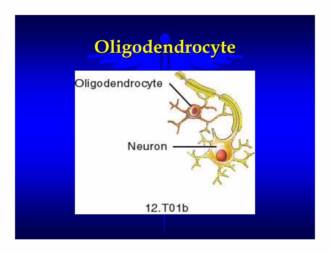
OligodendrocytesOligodendrocytes
�� Small cells with few processesSmall cells with few processes
�� Form a supporting network around the Form a supporting network around the neurons by twining around neurons neurons by twining around neurons and producing a lipid and protein and producing a lipid and protein wrapping around the neurons wrapping around the neurons (myelin sheath)(myelin sheath)
OligodendrocyteOligodendrocyte

MicrogliaMicroglia
�� Small phagocytic cells that protect the Small phagocytic cells that protect the central nervous system by engulfing central nervous system by engulfing and invading microbes and invading microbes
�� Clears away debris from dead cellsClears away debris from dead cells

MicrogliaMicroglia
Ependymal CellsEpendymal Cells
�� Neuroglia cells that line the brain Neuroglia cells that line the brain ventriclesventricles
�� Line the central canal of the spinal cordLine the central canal of the spinal cord
�� Helps form and circulate cerebral spinal Helps form and circulate cerebral spinal fluidfluid

Ependymal CellsEpendymal Cells

Neuroglia of the PNSNeuroglia of the PNS
�� Schwann Cells Schwann Cells -- NeurolemmocytesNeurolemmocytes
–– Cells responsible for producing the myelin Cells responsible for producing the myelin sheaths around the PNS neuronssheaths around the PNS neurons
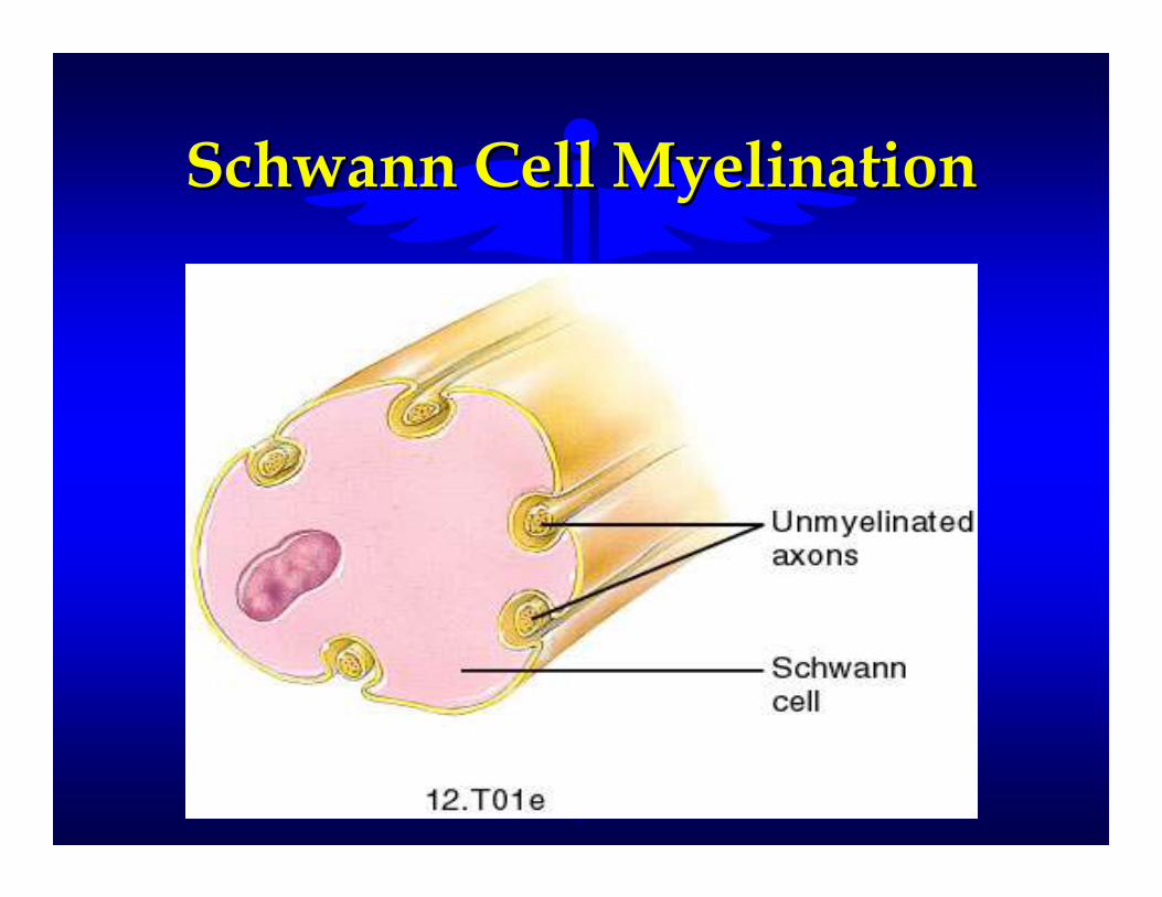
Schwann Cell MyelinationSchwann Cell Myelination

Schwann CellSchwann Cell(Neurolemmocyte)(Neurolemmocyte)

MyelinationMyelination
�� The process of developing or producing The process of developing or producing a Myelin Sheatha Myelin Sheath
�� Insulates the axon of a neuronInsulates the axon of a neuron
�� Increases the speed of nerve impulse Increases the speed of nerve impulse conductionconduction
–– CNS CNS -- oligodendrocytesoligodendrocytes
–– PNS PNS -- neurolemmocytes (Schwann Cells)neurolemmocytes (Schwann Cells)
�� Diseases such as TayDiseases such as Tay--Sachs disease and Sachs disease and Multiple Sclerosis involve destruction Multiple Sclerosis involve destruction of the myelin sheaths around the nerveof the myelin sheaths around the nerve

MyelinationMyelination
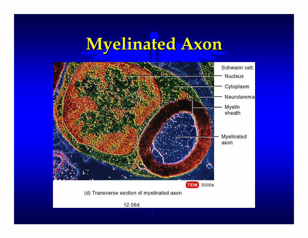
Myelinated AxonMyelinated Axon

Unmyelinated AxonUnmyelinated Axon

NeurophysiologyNeurophysiology
The transmission of nerve The transmission of nerve (electrical) impulses from (electrical) impulses from
nervous tissue to other nervous nervous tissue to other nervous tissue, organs, glands, and tissue, organs, glands, and
muscles.muscles.

Neuron Membrane PotentialNeuron Membrane Potential

Neuron Action PotentialNeuron Action Potential

Transmission of Nerve Transmission of Nerve ImpulsesImpulses
�� An electrical event due to movement of An electrical event due to movement of ions across a membraneions across a membrane
�� Also called an action potentialAlso called an action potential
–– Lasts about 1 msec (1/1000 of a second)Lasts about 1 msec (1/1000 of a second)
–– Dependent upon diameter of the axonDependent upon diameter of the axon
�� larger diameter axons larger diameter axons -- 0.4 msec (1/2500 sec)0.4 msec (1/2500 sec)
–– 2500 impulses per second2500 impulses per second
�� smaller diameter axons smaller diameter axons -- 4 msec (1/250 sec)4 msec (1/250 sec)
–– 250 impulses per second250 impulses per second

All or None PrincipleAll or None Principle
�� Ff depolarization reaches a threshold, Ff depolarization reaches a threshold, an action potential (impulse) is an action potential (impulse) is conductedconducted
�� Each action potential (impulse) is Each action potential (impulse) is conducted at maximum strength unless conducted at maximum strength unless there are toxic materials within the cell there are toxic materials within the cell or the membrane has been disruptedor the membrane has been disrupted

Neuron ImpulseNeuron Impulse
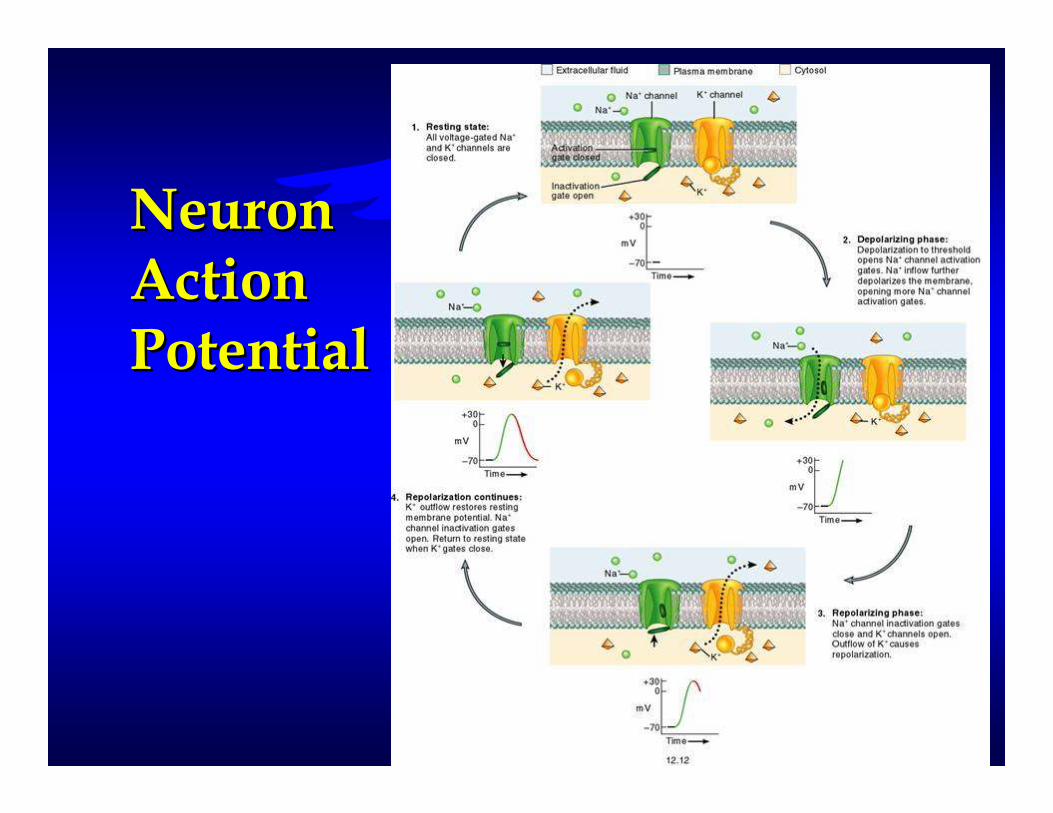
Neuron Neuron Action Action PotentialPotential
Types of Impulse Types of Impulse ConductionConduction
�� Continuous Conduction Continuous Conduction -- step by step step by step depolarization of each sequential, depolarization of each sequential, adjacent area of of the nerve cell adjacent area of of the nerve cell membranemembrane
–– typical of unmyelinated nerve fiberstypical of unmyelinated nerve fibers
–– type of action potential in muscle fiberstype of action potential in muscle fibers
�� Saltatory Conduction Saltatory Conduction -- the jumping of the jumping of an action potential across specialized an action potential across specialized neurofibril nodes along the axonneurofibril nodes along the axon
–– Nodes of RanvierNodes of Ranvier
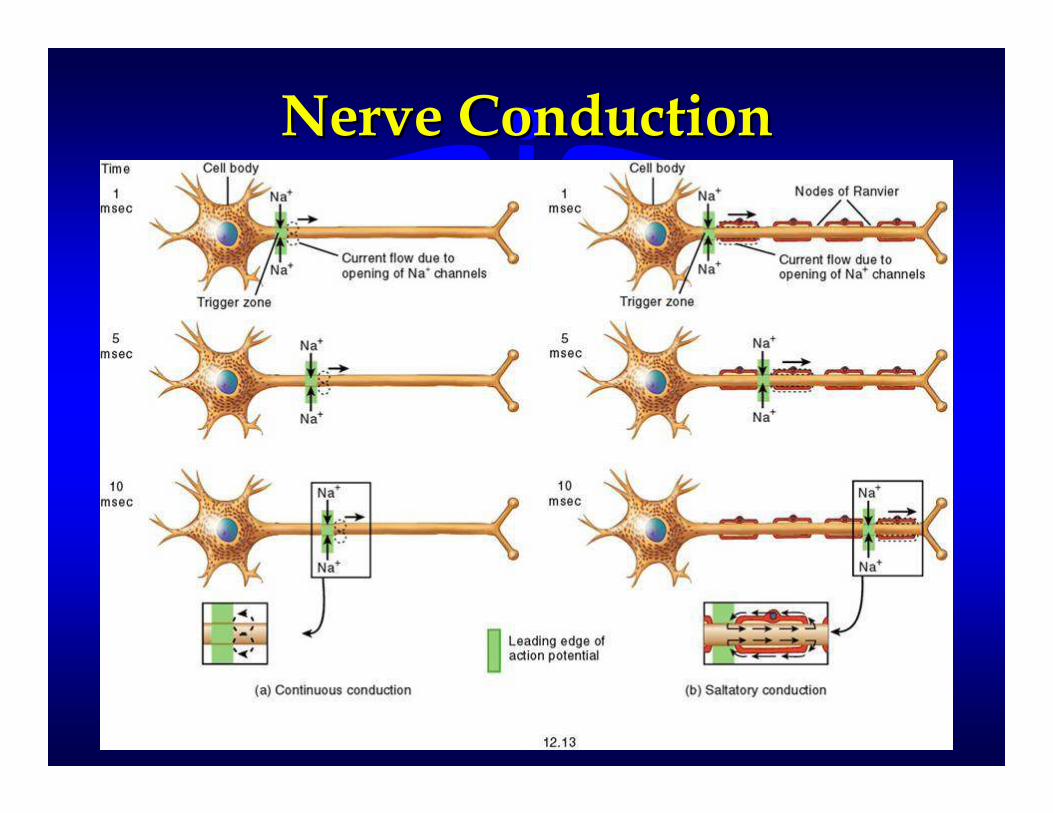
Nerve ConductionNerve Conduction

Gray and White MatterGray and White Matter
�� White Matter White Matter -- the aggregation of the aggregation of myelinated processes from many myelinated processes from many neurons neurons
–– Visible upon freshly dissected brain or Visible upon freshly dissected brain or spinal tissuespinal tissue
–– White color is due to myelinationWhite color is due to myelination
�� Gray Matter Gray Matter -- unmyelinated nerve cell unmyelinated nerve cell bodies, axons, dendrites, ganglia, and bodies, axons, dendrites, ganglia, and axon terminalsaxon terminals
–– Appears gray because of lack of myelinAppears gray because of lack of myelin

Gray and White MatterGray and White Matter

Protection and Coverings Protection and Coverings of the Brainof the Brain
�� Protected by the cranial bones and the Protected by the cranial bones and the cranial meningescranial meninges
–– Dura Mater Dura Mater -- outer layerouter layer
–– Arachnoid Arachnoid -- middle layermiddle layer
–– Pia Mater Pia Mater -- inner layerinner layer
�� Also protected by cerebrospinal fluidAlso protected by cerebrospinal fluid
–– fluid that nourishes and protects the brain fluid that nourishes and protects the brain and spinal cordand spinal cord
–– continuously circulates through the continuously circulates through the subarachnoid space around the brain and subarachnoid space around the brain and throughout the cavities within the brainthroughout the cavities within the brain

Meninges of the BrainMeninges of the Brain
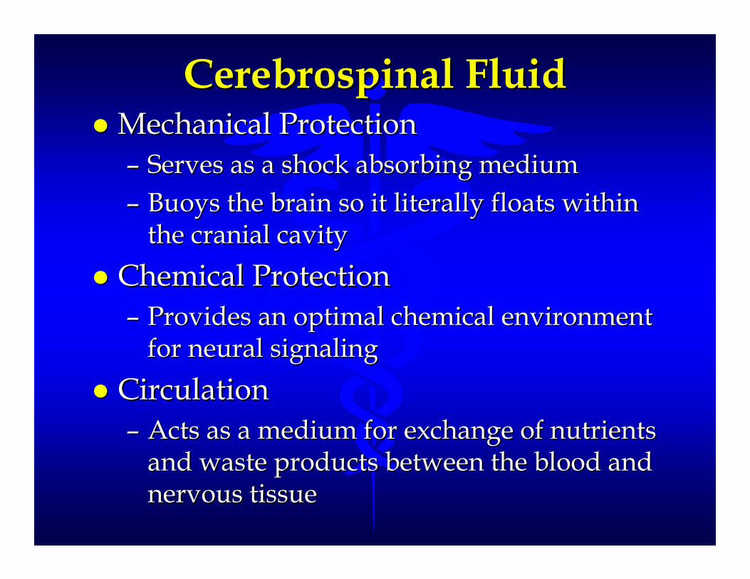
Cerebrospinal FluidCerebrospinal Fluid�� Mechanical ProtectionMechanical Protection
–– Serves as a shock absorbing mediumServes as a shock absorbing medium
–– Buoys the brain so it literally floats within Buoys the brain so it literally floats within the cranial cavitythe cranial cavity
�� Chemical ProtectionChemical Protection
–– Provides an optimal chemical environment Provides an optimal chemical environment for neural signalingfor neural signaling
�� Circulation Circulation
–– Acts as a medium for exchange of nutrients Acts as a medium for exchange of nutrients and waste products between the blood and and waste products between the blood and nervous tissuenervous tissue

Transmission of Nerve Transmission of Nerve Impulses at SynapsesImpulses at Synapses
�� Most nervous conduction is from Most nervous conduction is from neuron to neuron (interneurons neuron to neuron (interneurons -- 90%)90%)
�� Types of SynapsesTypes of Synapses
–– Axon to dendriteAxon to dendrite
–– Axon to somaAxon to soma
–– Axon to axonAxon to axon
�� Two ways to transmit impulses across a Two ways to transmit impulses across a synapsesynapse
–– Electrical SynapsesElectrical Synapses
–– Chemical SynapsesChemical Synapses

MeningesMeninges
�� Connective tissue covering found Connective tissue covering found around the brain and spinal cord around the brain and spinal cord
�� Three layered membraneThree layered membrane
–– Dura Mater Dura Mater -- outer most layer outer most layer
�� dense irregular connective tissuedense irregular connective tissue
–– Arachnoid Arachnoid -- middle layermiddle layer
�� spider web arrangement of collagen fibersspider web arrangement of collagen fibers
–– Pia Mater Pia Mater -- inner most meningesinner most meninges
�� very delicate layer of thin tissuevery delicate layer of thin tissue

Spinal CordSpinal CordProtectiveProtectiveCoveringsCoverings
�� Dura MaterDura Mater
�� ArachnoidArachnoid
�� Pia MaterPia Mater

ReflexesReflexes�� Fast, predictable, automatic responses Fast, predictable, automatic responses
to changes in the environment that help to changes in the environment that help maintain homeostasismaintain homeostasis
�� Somatic Reflexes Somatic Reflexes -- involve skeletal involve skeletal musclesmuscles
�� Visceral (Autonomic) Reflexes Visceral (Autonomic) Reflexes -- involve involve responses of smooth muscles, the heart, responses of smooth muscles, the heart, and glandsand glands
�� Involve the spinal nervesInvolve the spinal nerves

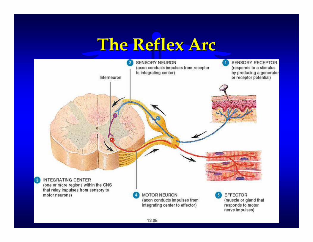
The Reflex Arc The Reflex Arc
�� A response by the body involving only A response by the body involving only the body segment being affected and the body segment being affected and the spinal cord the spinal cord
–– Brain does not have to be involvedBrain does not have to be involved
�� Receptor Receptor -- the distal end of a sensory the distal end of a sensory neuron (dendrite)neuron (dendrite)
–– Responds to a specific stimulusResponds to a specific stimulus
�� a change in internal or external environmenta change in internal or external environment
–– Triggers a nerve impulseTriggers a nerve impulse

�� Sensory Neuron Sensory Neuron -- the neuron located in the neuron located in the gray matter of the spinal cordthe gray matter of the spinal cord
–– conducts impulses from the receptor to the conducts impulses from the receptor to the spinal cordspinal cord
�� Integrating Center Integrating Center -- a region within the a region within the CNS (spinal cord or brain) that CNS (spinal cord or brain) that interprets the information from the interprets the information from the sensory neuron and initiates an sensory neuron and initiates an appropriate responseappropriate response
�� Motor Neurons Motor Neurons -- the neurons arising the neurons arising from the integrating center that relay a from the integrating center that relay a nerve impulse to the part of the body nerve impulse to the part of the body that will respond to the stimulusthat will respond to the stimulus

�� Effector Effector -- the part of the body that the part of the body that responds to the motor nerve impulse responds to the motor nerve impulse (usually a muscle or a gland)(usually a muscle or a gland)
–– Effector Effector -- skeletal muscle skeletal muscle -- somatic reflexsomatic reflex
–– Effector Effector -- cardiac, smooth muscle, or gland cardiac, smooth muscle, or gland --visceral reflexvisceral reflex
The Reflex ArcThe Reflex Arc

Reflex Arc ExamplesReflex Arc Examples�� Stretch Reflex Stretch Reflex -- results in the results in the
contraction of a muscle if it has been contraction of a muscle if it has been stretched suddenlystretched suddenly
�� Tendon Reflex Tendon Reflex -- results in the results in the contraction of a muscle when a tendon contraction of a muscle when a tendon is stretched suddenlyis stretched suddenly
�� Flexor (Withdrawal) Reflex Flexor (Withdrawal) Reflex -- sudden sudden contraction and removal of a body contraction and removal of a body segment as a result of a pain stimulussegment as a result of a pain stimulus

Tendon ReflexTendon Reflex

WithdrawalWithdrawalReflexReflex�� also calledalso called
Flexor/WithdrawalFlexor/Withdrawal
ReflexReflex

Anatomy of the Autonomic Nervous Anatomy of the Autonomic Nervous SystemSystem
Slide 7.73Copyright © 2003 Pearson Education, Inc. publishing as Benjamin Cummings
Figure 7.25

The BRAINThe BRAIN

Sensory and Motor Areas of the Sensory and Motor Areas of the
Cerebral CortexCerebral Cortex
Slide 7.31Copyright © 2003 Pearson Education, Inc. publishing as Benjamin Cummings
Figure 7.14

The BRAINThe BRAIN
�� One of the largest organs in the bodyOne of the largest organs in the body
�� Controls all mental functions Controls all mental functions
�� Component of the CNSComponent of the CNS
�� Composed of over 100 billion neuronsComposed of over 100 billion neurons
�� Comprises 2Comprises 2--3% of body weight3% of body weight
�� Utilizes over 20% of body’s energyUtilizes over 20% of body’s energy

Major Divisions of the Major Divisions of the BRAINBRAIN
�� CEREBRUM CEREBRUM -- occupies most of the occupies most of the cranium and is divided into right and cranium and is divided into right and left halves called hemispheresleft halves called hemispheres
�� CEREBELLUM CEREBELLUM -- the posteriorthe posterior--inferior inferior portion of the brainportion of the brain
�� BRAIN STEM BRAIN STEM -- consists of the medulla consists of the medulla oblongata, the pons, and the midbrainoblongata, the pons, and the midbrain
–– it is continuous with the spinal cordit is continuous with the spinal cord
�� DIENCEPHALON DIENCEPHALON -- located above the located above the brainstem, composed primarily of the:brainstem, composed primarily of the:
–– ThalamusThalamus -- HypothalamusHypothalamus


The BrainThe Brain

VentriclesVentricles
�� Cavities within the brainCavities within the brain
�� Lateral ventricles (2) Lateral ventricles (2) -- located within located within each hemisphere in the cerebrumeach hemisphere in the cerebrum
�� Third ventricle Third ventricle -- a vertical slit between a vertical slit between the lateral ventricles and inferior to the the lateral ventricles and inferior to the right and left halves of the thalamusright and left halves of the thalamus
�� Fourth ventricle Fourth ventricle -- space between the space between the brainstem and the cerebellumbrainstem and the cerebellum

Ventricles of the BrainVentricles of the Brain

Choroid PlexusChoroid Plexus
�� Network of capillaries in the walls of Network of capillaries in the walls of the ventriclesthe ventricles
�� Covered with ependymal cells that Covered with ependymal cells that form the cerebrospinal fluidform the cerebrospinal fluid
�� These ependymal cells are so close These ependymal cells are so close together they form the bloodtogether they form the blood--brain brain barrier.barrier.
–– Selectively permeable barrierSelectively permeable barrier
–– Protects the brain and spinal cord from Protects the brain and spinal cord from potentially harmful substances in the bloodpotentially harmful substances in the blood

Flow ofFlow ofCerebroCerebro--SpinalSpinalFluidFluid

Flow ofFlow ofCerebroCerebro--Spinal Spinal FluidFluid

Blood Supply to the BrainBlood Supply to the Brain
�� One of the most metabolically active One of the most metabolically active organs in the bodyorgans in the body
�� Makes up only 2Makes up only 2--3% of body weight but 3% of body weight but uses about 20% of available Ouses about 20% of available O22 at restat rest
�� Well supplied with OWell supplied with O22 and nutrientsand nutrients
�� Only nutritional source for brain Only nutritional source for brain metabolic activity is glucosemetabolic activity is glucose
�� Capillaries in the brain are much less Capillaries in the brain are much less leaky than other capillaries in the body leaky than other capillaries in the body and form a blood brain barrierand form a blood brain barrier

The Brain StemThe Brain Stem
�� The most inferior portion of the brainThe most inferior portion of the brain
�� Connects the brain to the spinal cordConnects the brain to the spinal cord
�� Composed of Three AreasComposed of Three Areas
–– The Medulla OblongataThe Medulla Oblongata
–– The PonsThe Pons
–– The MidbrainThe Midbrain

The Medulla OblongataThe Medulla Oblongata�� Most inferior portion of the brain stemMost inferior portion of the brain stem
�� Connects the brain stem to the spinal Connects the brain stem to the spinal cordcord
�� Respiratory CenterRespiratory Center
–– Adjusts rhythm and depth of breathingAdjusts rhythm and depth of breathing
�� Cardiovascular CenterCardiovascular Center
–– Regulates heart rate and contraction forceRegulates heart rate and contraction force
–– Influences vasoconstriction and Influences vasoconstriction and vasodilationvasodilation
�� Also controls coughing, vomiting, Also controls coughing, vomiting, swallowing, and hiccuppingswallowing, and hiccupping

The Medulla OblongataThe Medulla Oblongata

The Medulla OblongataThe Medulla Oblongata

The PonsThe Pons
�� Lies superior to the medulla oblongataLies superior to the medulla oblongata
�� Together with the respiratory center in Together with the respiratory center in the medulla helps control respirationthe medulla helps control respiration

The PonsThe Pons

The MidbrainThe Midbrain
�� Superior to the ponsSuperior to the pons
�� Connects the brain stem to the Connects the brain stem to the diencephalondiencephalon

The MidbrainThe Midbrain

Pons and MidbrainPons and Midbrain

The DiencephalonThe Diencephalon
�� Area of the brain containing the:Area of the brain containing the:
–– ThalamusThalamus
–– HypothalamusHypothalamus

The ThalamusThe Thalamus
�� Oval structure that makes up 80% of the Oval structure that makes up 80% of the diencephalondiencephalon
�� Comprised of a pair of oval masses Comprised of a pair of oval masses (mostly gray matter)(mostly gray matter)
�� Principle Principle relay stationrelay station between the between the various sections of the brain various sections of the brain

The ThalamusThe Thalamus

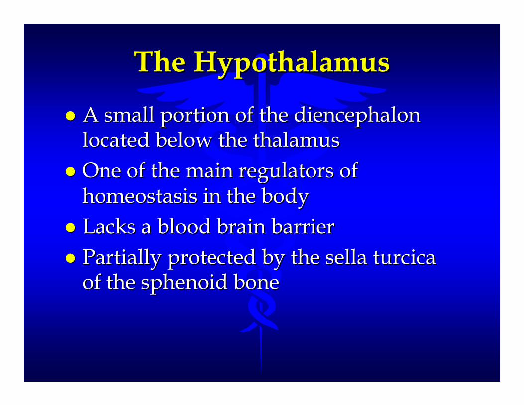
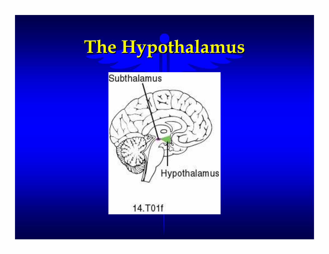
The HypothalamusThe Hypothalamus
�� A small portion of the diencephalon A small portion of the diencephalon located below the thalamuslocated below the thalamus
�� One of the main regulators of One of the main regulators of homeostasis in the bodyhomeostasis in the body
�� Lacks a blood brain barrierLacks a blood brain barrier
�� Partially protected by the sella turcica Partially protected by the sella turcica of the sphenoid boneof the sphenoid bone

Functions of the Functions of the HypothalamusHypothalamus
�� coordinates Nervous System and coordinates Nervous System and Endocrine System activities to maintain Endocrine System activities to maintain HomeostasisHomeostasis
–– Thirst, Hunger, SatietyThirst, Hunger, Satiety
–– Sleep Patterns and Waking StatesSleep Patterns and Waking States
–– Sex Drive, Maturation, Aggression, and Sex Drive, Maturation, Aggression, and RageRage
–– influences movement of food through the influences movement of food through the Gastrointestinal TractGastrointestinal Tract
–– production and secretion of hormones That production and secretion of hormones That control other Endocrine Glandscontrol other Endocrine Glands
The HypothalamusThe Hypothalamus

HypothalamusHypothalamus

The CerebrumThe Cerebrum�� Largest division of the brainLargest division of the brain
�� Occupies most of the craniumOccupies most of the cranium
�� Accounts for 85% of brain massAccounts for 85% of brain mass
�� Divided into right and left hemispheresDivided into right and left hemispheres
–– Longitudinal FissureLongitudinal Fissure
–– Corpus CallosumCorpus Callosum
�� Cerebral cortex Cerebral cortex -- the outer surface area the outer surface area of the cerebrumof the cerebrum
–– Composed mainly of gray matterComposed mainly of gray matter
–– Contains billions of neuronsContains billions of neurons

The CerebrumThe Cerebrum


Lobes of the CerebrumLobes of the Cerebrum�� Named after the bones that cover themNamed after the bones that cover them
–– Frontal LobeFrontal Lobe
–– Parietal LobeParietal Lobe
–– Temporal LobeTemporal Lobe
–– Occipital LobeOccipital Lobe
Frontal LobeFrontal Lobe
�� Motor AreasMotor Areas
–– Controls movement of voluntary skeletal Controls movement of voluntary skeletal musclesmuscles
�� Association AreasAssociation Areas
–– Carry on high level intellectual processingCarry on high level intellectual processing
�� Problem Solving Problem Solving -- Reasoning Reasoning -- PlanningPlanning
�� Concentration Concentration -- Memory Memory -- BehaviorBehavior
�� Emotions Emotions -- Expressions Expressions

Parietal LobeParietal Lobe
�� Sensory AreasSensory Areas
–– Interprets sensations such as:Interprets sensations such as:
–– touch touch -- pressure pressure -- pain on the surface of the pain on the surface of the skinskin
�� Association AreasAssociation Areas
–– Understanding of speechUnderstanding of speech
–– Using words to express thoughts and Using words to express thoughts and feelingsfeelings

Temporal LobeTemporal Lobe
�� Sensory AreasSensory Areas
–– Hearing and balanceHearing and balance
�� Association AreasAssociation Areas
–– Interpret sensory experiencesInterpret sensory experiences
–– Memory of visual scenes Memory of visual scenes -- music music -- smells smells and other complex sensory patterns and other complex sensory patterns
Occipital LobeOccipital Lobe
�� Sensory AreasSensory Areas
–– Visual processing and interpretationVisual processing and interpretation
�� Association AreasAssociation Areas
–– Combines visual images with sensory Combines visual images with sensory experienceexperience

The CerebellumThe Cerebellum

Cerebellum and BrainstemCerebellum and Brainstem

The CerebellumThe Cerebellum�� Second largest portion of the brainSecond largest portion of the brain
�� Occupies the inferior and posterior Occupies the inferior and posterior aspects of the cranial cavityaspects of the cranial cavity
�� Processes sensory informationProcesses sensory information
–– BalanceBalance -- CoordinationCoordination
–– Maintains postural equilibriumMaintains postural equilibrium


Nervous System Disorders Nervous System Disorders and and
Homeostatic ImbalancesHomeostatic Imbalances

Alzheimer’s Disease (AD)Alzheimer’s Disease (AD)
�� Disabling neurological disorder that Disabling neurological disorder that effects about 11% of the populationeffects about 11% of the population
�� Fourth leading cause of brain death Fourth leading cause of brain death among the elderlyamong the elderly
�� A chronic, organic, mental disorder, a A chronic, organic, mental disorder, a form of preform of pre--senile dementia due to senile dementia due to atrophy of neurons of the frontal and atrophy of neurons of the frontal and occipital lobes occipital lobes
�� AD patients usually die from AD patients usually die from complications due to being bedriddencomplications due to being bedridden

Amyotrophic Lateral Amyotrophic Lateral Sclerosis (ALS)Sclerosis (ALS)
�� Also known as Lou Gehrig’s DiseaseAlso known as Lou Gehrig’s Disease
�� A relatively rare neurological disorderA relatively rare neurological disorder
�� A syndrome marked by muscular weakness A syndrome marked by muscular weakness and atrophy with spasticity and hyperflexion and atrophy with spasticity and hyperflexion due to degeneration of the motor neurons of due to degeneration of the motor neurons of the spinal cord, medulla, and cortexthe spinal cord, medulla, and cortex
�� A degenerative diseaseA degenerative disease
�� No known cureNo known cure

Bacterial MeningitisBacterial Meningitis
�� Infection of the meninges by the Infection of the meninges by the bacterium Haemophilus Influenzaebacterium Haemophilus Influenzae
�� Usually affects children under age 5Usually affects children under age 5
�� Symptoms include severe headaches Symptoms include severe headaches and feverand fever
�� Can lead to brain damage and even Can lead to brain damage and even death if not treateddeath if not treated

Cerebral Palsy (CP)Cerebral Palsy (CP)
�� A group of motor disorders due to loss A group of motor disorders due to loss of muscle controlof muscle control
�� Caused by damage to the motor areas Caused by damage to the motor areas of the brain during fetal development, of the brain during fetal development, birth, or infancybirth, or infancy
�� About 70% of CP individuals are About 70% of CP individuals are somewhat mentally retarded due to the somewhat mentally retarded due to the inability to hear well or speak fluentlyinability to hear well or speak fluently
�� Not a progressive disease but the Not a progressive disease but the symptoms are irreversiblesymptoms are irreversible

EpilepsyEpilepsy
�� Short, recurrent, periodic, attacks of Short, recurrent, periodic, attacks of motor, sensory, or psychological motor, sensory, or psychological malfunctionmalfunction
�� Characterized by seizures which can Characterized by seizures which can result in involuntary skeletal muscle result in involuntary skeletal muscle contraction, loss of muscle control, contraction, loss of muscle control, inability to sense light, noise, and smell, inability to sense light, noise, and smell, and loss of consciousnessand loss of consciousness
�� Most epileptic seizures are idiopathicMost epileptic seizures are idiopathic

Multiple Sclerosis (MS)Multiple Sclerosis (MS)
�� The progressive destruction of the The progressive destruction of the myelin sheaths of neurons of the CNSmyelin sheaths of neurons of the CNS
�� The sheaths deteriorates to The sheaths deteriorates to sclerosesscleroses
–– hardened scars or plaqueshardened scars or plaques
�� “short circuits” nerve transmission“short circuits” nerve transmission
�� Cause is unknownCause is unknown
–– May be a type of an autoimmune diseaseMay be a type of an autoimmune disease
�� No known cureNo known cure
�� Progressive loss of function with Progressive loss of function with intermittent periods of remissionintermittent periods of remission

Parkinson’s Disease (PD)Parkinson’s Disease (PD)
�� A progressive disorder of the CNS that A progressive disorder of the CNS that usually affects individuals over 60usually affects individuals over 60
�� Cause is unknown but a toxic Cause is unknown but a toxic environmental factor is suspectedenvironmental factor is suspected
�� Chemical basis of the disease appears to Chemical basis of the disease appears to be to little dopamine and too much Ach be to little dopamine and too much Ach
�� Treatment includes increasing levels of Treatment includes increasing levels of dopamine and decreasing Achdopamine and decreasing Ach
–– Difficult because dopamine does not cross Difficult because dopamine does not cross the blood brain barrier the blood brain barrier
�� A chronic nervous disease A chronic nervous disease characterized by a fine, slowly characterized by a fine, slowly spreading tremor, muscle weakness and spreading tremor, muscle weakness and rigidity, and a peculiar gait rigidity, and a peculiar gait
�� Other causes may include brain damage Other causes may include brain damage at birth, metabolic disturbances, at birth, metabolic disturbances, infections, toxins, vascular infections, toxins, vascular disturbances, head injuries, and tumors disturbances, head injuries, and tumors and abscesses of the brainand abscesses of the brain
�� Usually can be controlled with drug Usually can be controlled with drug therapytherapy
–– GABA GABA -- gamma aminobutyric acidgamma aminobutyric acid

�� Symptoms include muscle tremor, Symptoms include muscle tremor, muscle rigidity, bradykinesia, muscle rigidity, bradykinesia, hypokinesia or dyskinesia, speech and hypokinesia or dyskinesia, speech and walking impairmentwalking impairment
�� Attempting to transplant fetal nervous Attempting to transplant fetal nervous tissue into the damaged area of the tissue into the damaged area of the brain of some Parkinson’s Disease brain of some Parkinson’s Disease patientspatients
Cerebral Vascular AccidentCerebral Vascular Accident(CVA) (CVA) -- StrokeStroke
�� The most common brain disorderThe most common brain disorder
�� Characterized by slurred speech, loss of Characterized by slurred speech, loss of or blurred vision, dizziness, weakness, or blurred vision, dizziness, weakness, paralysis of a limb or hemiplegia, coma, paralysis of a limb or hemiplegia, coma, and deathand death
�� Ischemic CVA Ischemic CVA -- due to lack of blood due to lack of blood supply to a particular area of the brainsupply to a particular area of the brain
�� Hemorrhagic CVA Hemorrhagic CVA -- due to the rupture due to the rupture of a blood vessel in the brainof a blood vessel in the brain

Risk Factors for StrokeRisk Factors for Stroke
�� hypertensionhypertension
�� heart diseaseheart disease
�� smokingsmoking
�� diabetesdiabetes
�� atherosclerosisatherosclerosis
�� hyperlipidemiahyperlipidemia
�� obesityobesity
�� excessive alcohol intakeexcessive alcohol intake

SensationsSensationsand and
Special SensesSpecial Senses

SensesSenses
Specialized structures of the Specialized structures of the
nervous system which provide nervous system which provide information about the information about the
environment in which we live to environment in which we live to help maintain homeostasishelp maintain homeostasis

Functions of Functions of Special Senses Special Senses
�� Sensory Sensory -- monitoring the body and the monitoring the body and the external environment for changing external environment for changing conditionsconditions

Sensory PathwaysSensory Pathways
�� All pathways begin with a receptor and All pathways begin with a receptor and the sensory information is transmitted the sensory information is transmitted to the CNSto the CNS
�� Always begins with a stimulusAlways begins with a stimulus
–– change in the environmentchange in the environment
ReceptorsReceptors
�� Structures which provide feedback Structures which provide feedback about the environmentabout the environment
�� Are impulse specificAre impulse specific–– Only respond to one type of stimulusOnly respond to one type of stimulus
�� Many have sensory function Many have sensory function adaptationsadaptations–– May end as bare dendrites or be a complex May end as bare dendrites or be a complex
organorgan

VisionVision
�� The most complex of the special sensesThe most complex of the special senses
–– Over 70% of the sensory receptors in the Over 70% of the sensory receptors in the body are photoreceptors for sightbody are photoreceptors for sight
�� Visual organs, the eyes are supported Visual organs, the eyes are supported by a number of accessory structures and by a number of accessory structures and internal organsinternal organs
–– Dependent upon photoreceptors in the Dependent upon photoreceptors in the eyeseyes

The EyeThe Eye
Accessory Structures Accessory Structures of the Eyeof the Eye
�� Eyelids Eyelids -- protects the anterior surfaceprotects the anterior surface
–– Conjunctiva Conjunctiva -- the mucous membrane of the eyelid the mucous membrane of the eyelid
–– Helps moisten and lubricate the eyeballHelps moisten and lubricate the eyeball
�� Lacrimal Apparatus Lacrimal Apparatus -- secretes tearssecretes tears
–– lacrimal glandlacrimal gland -- lacrimal saclacrimal sac
–– lacrimal canalslacrimal canals -- nasolacrimal ductnasolacrimal duct
–– moistens and lubricates the eyeballmoistens and lubricates the eyeball
–– fights against infection (enzymes in tears)fights against infection (enzymes in tears)
�� Extrinsic Muscles of the Eyeball (6)Extrinsic Muscles of the Eyeball (6)
–– skeletal muscles that move the eyeballskeletal muscles that move the eyeball
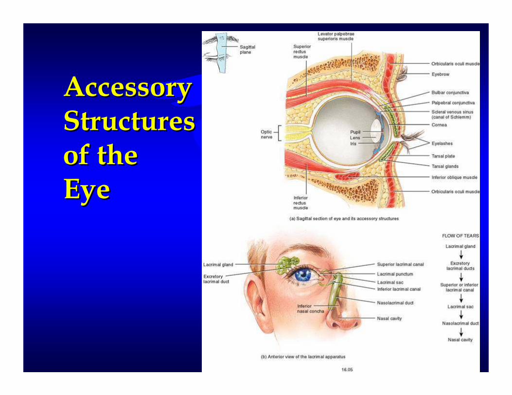
Accessory Accessory StructuresStructuresof theof theEyeEye

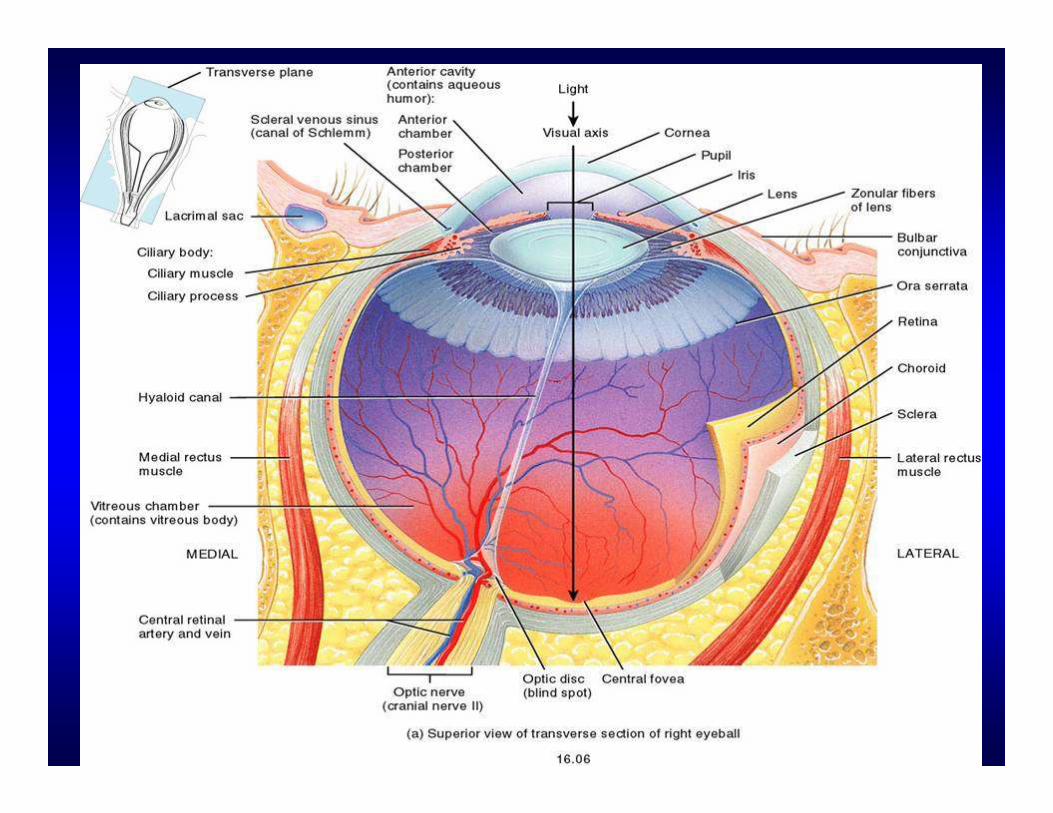
Structure of the EyeStructure of the Eye
�� The wall consists of three layers of The wall consists of three layers of tissue or tunicstissue or tunics
�� Fibrous Tunic Fibrous Tunic -- outer layerouter layer
�� Vascular Tunic Vascular Tunic -- middle layermiddle layer
�� Nervous Tunic Nervous Tunic -- inner layerinner layer
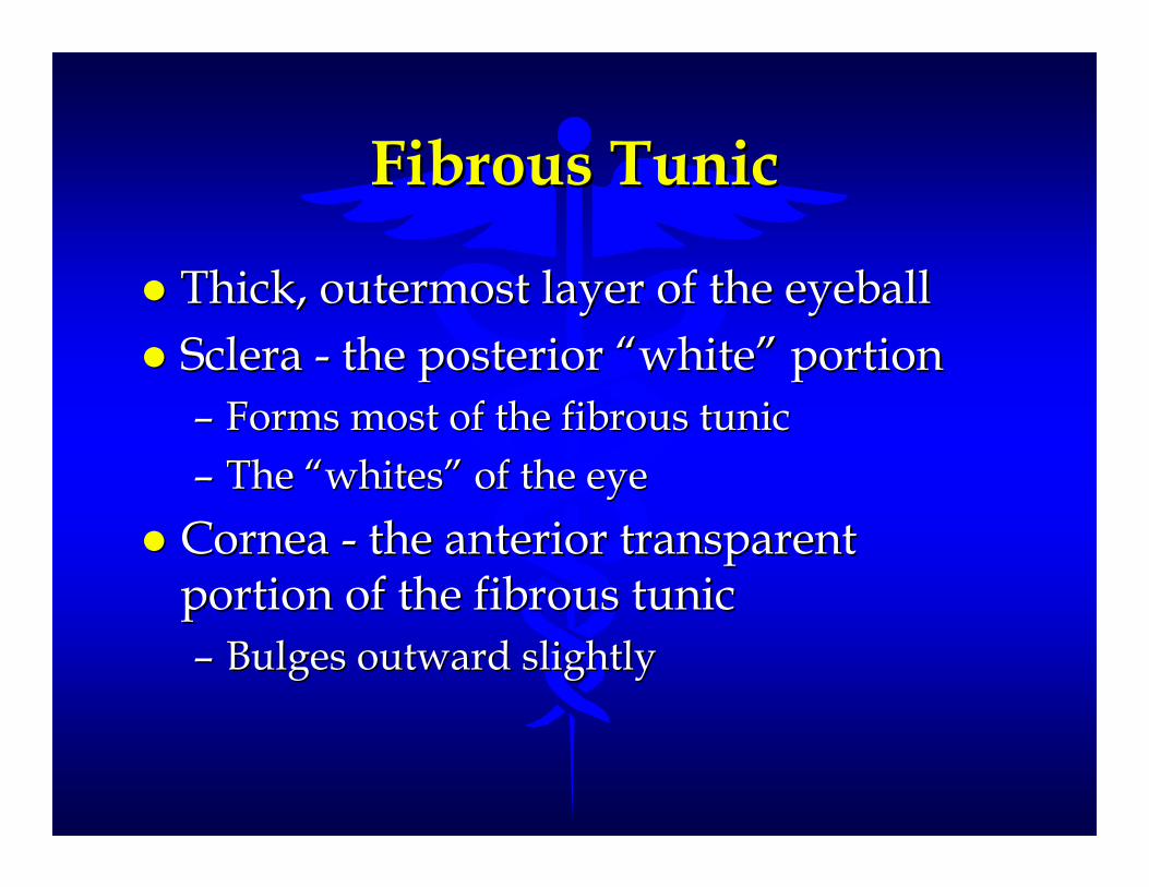
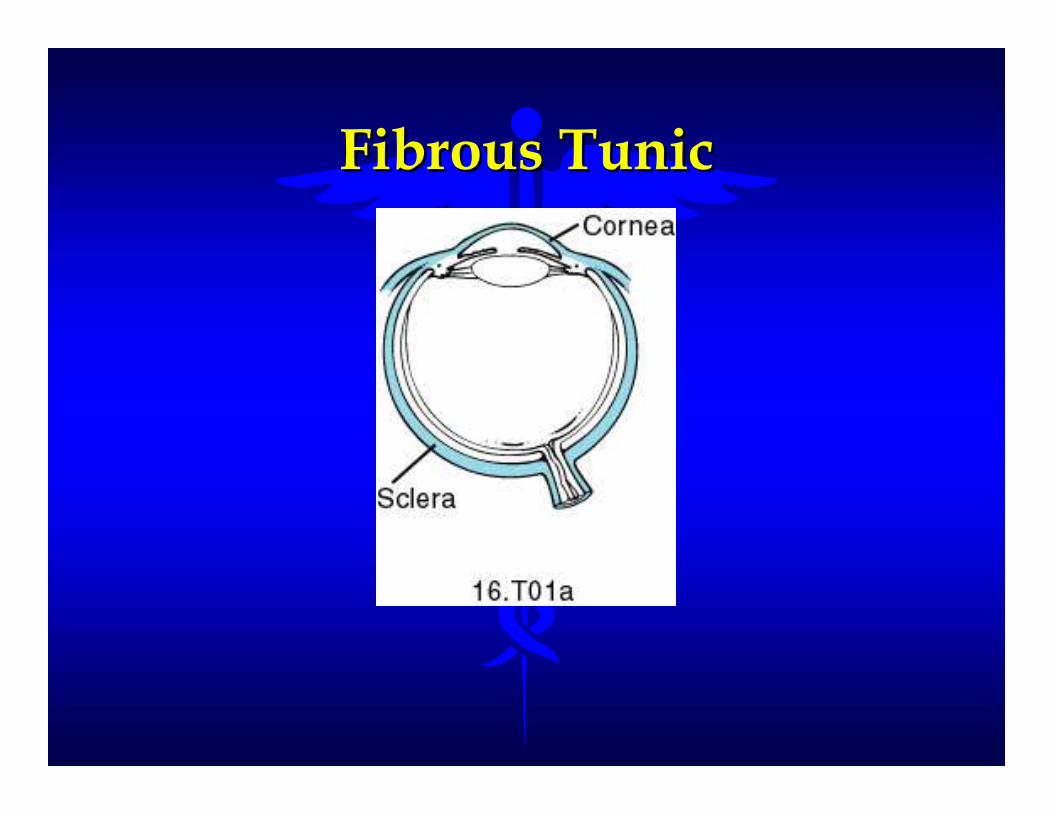
Fibrous TunicFibrous Tunic
�� Thick, outermost layer of the eyeballThick, outermost layer of the eyeball
�� Sclera Sclera -- the posterior “white” portionthe posterior “white” portion
–– Forms most of the fibrous tunicForms most of the fibrous tunic
–– The “whites” of the eyeThe “whites” of the eye
�� Cornea Cornea -- the anterior transparent the anterior transparent portion of the fibrous tunicportion of the fibrous tunic
–– Bulges outward slightlyBulges outward slightly
Fibrous TunicFibrous Tunic

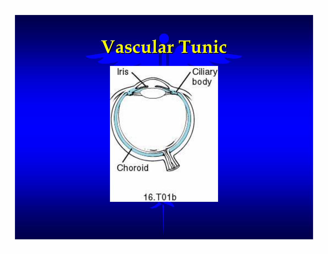
Vascular TunicVascular Tunic
�� Extremely vascularExtremely vascular
�� Supplies blood to numerous structures Supplies blood to numerous structures of the eyeof the eye
–– ChoroidChoroid -- Ciliary BodyCiliary Body
–– IrisIris -- LensLens
Vascular TunicVascular Tunic

�� Choroid Choroid -- posterior, thin portion of the posterior, thin portion of the vascular tunicvascular tunic
–– A thin, dark brown membrane that lines most of A thin, dark brown membrane that lines most of the internal surface of the sclerathe internal surface of the sclera
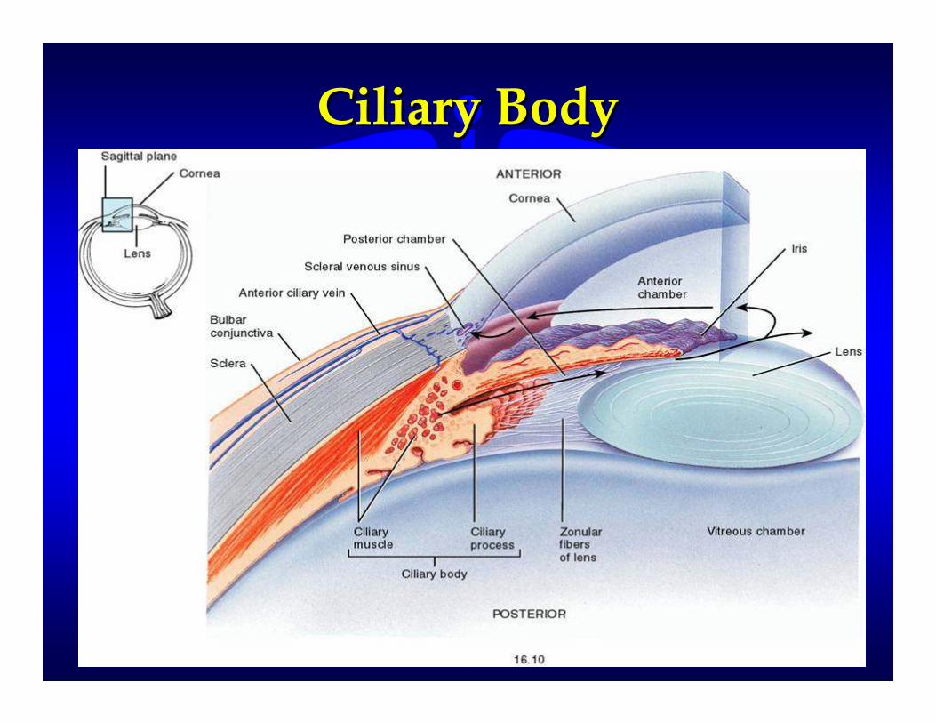
�� Ciliary Body Ciliary Body -- anterior, thick portion of the anterior, thick portion of the vascular tunicvascular tunic
–– Thickest part of the vascular tunicThickest part of the vascular tunic
–– Consists of smooth muscle fibersConsists of smooth muscle fibers
–– Attaches to the lens by ligamentsAttaches to the lens by ligaments
–– Changes the thickness and shape of the lens.Changes the thickness and shape of the lens.
Ciliary BodyCiliary Body

�� Iris Iris -- anterior, colored portion of the anterior, colored portion of the Vascular TunicVascular Tunic–– contraction of it’s smooth muscle accounts contraction of it’s smooth muscle accounts
for dilation or constriction of the Pupils for dilation or constriction of the Pupils (openings to the inner cavities of the eyes)(openings to the inner cavities of the eyes)
�� Lens Lens -- special tissue which focuses and special tissue which focuses and directs light entering the eyedirects light entering the eye–– suspended by the Ciliary Bodysuspended by the Ciliary Body
–– located behind the Irislocated behind the Iris
–– alteration of the shape of the lens to alteration of the shape of the lens to accommodate for near or far vision accommodate for near or far vision focusing (Accommodation) focusing (Accommodation)

The LensThe Lens

Iris Iris –– Pupil DiameterPupil Diameter

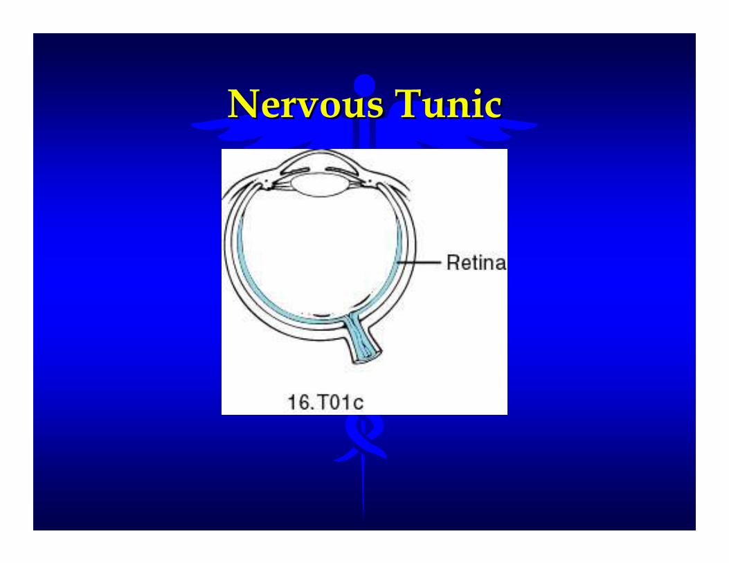
Nervous TunicNervous Tunic
�� The inner layer of the eyeThe inner layer of the eye
�� Retina Retina -- a thin fragile layer of neurons that a thin fragile layer of neurons that forms the inner lining of the eyeball’s forms the inner lining of the eyeball’s posterior wallposterior wall–– Lines the posterior cavity and contains the Lines the posterior cavity and contains the
photoreceptor cells (rods and cones), bipolar photoreceptor cells (rods and cones), bipolar neurons, and ganglion cellsneurons, and ganglion cells
�� Optic Nerve Optic Nerve -- axons and ganglion cells axons and ganglion cells –– Transmits images to the occipital lobe of the brain Transmits images to the occipital lobe of the brain
for interpretation of what we seefor interpretation of what we see
Nervous TunicNervous Tunic

Rods and ConesRods and Cones
�� Rods Rods -- elongated cylindrical dendrites elongated cylindrical dendrites that are sensitive to varying light that are sensitive to varying light conditionsconditions
–– Allows us to see under varying light Allows us to see under varying light intensities (night vision)intensities (night vision)
�� Cones Cones -- dendrites with tapered endsdendrites with tapered ends
–– Color sensitiveColor sensitive
–– Determines the “sharpness” of visionDetermines the “sharpness” of vision

Rods Rods andandConesCones

Rods and ConesRods and Cones
Other Structures of the Other Structures of the Nervous TunicNervous Tunic
�� Optic Disc Optic Disc -- blind spot where the optic blind spot where the optic nerve exits the retinanerve exits the retina
�� Fovea Centralis Fovea Centralis -- an area of the retina an area of the retina containing many cone cellscontaining many cone cells
–– the area of sharpest visionthe area of sharpest vision

RetinaRetina
Elements of Vision in the EyeElements of Vision in the Eye
�� Vision spectrum of the eyeVision spectrum of the eye–– only detect three colorsonly detect three colors
–– RedRed -- GreenGreen -- BlueBlue
�� Aspects of vision of the eyeAspects of vision of the eye–– colorcolor
–– motionmotion
–– formform
–– depthdepth
RefractionRefraction
�� the “bending” of light rays as it travels the “bending” of light rays as it travels through the eyethrough the eye
�� the pathway of light as it travels through the the pathway of light as it travels through the eyeeye
�� influenced by:influenced by:
–– shape of the lensshape of the lens
–– shape and thickness of the corneashape and thickness of the cornea
–– amount and consistency of the Aqueous and amount and consistency of the Aqueous and Vitreous HumorVitreous Humor

RefractionRefraction

VisionVisionAbnormalitiesAbnormalities
Physiology of VisionPhysiology of Vision
�� Rods and cones convert light waves Rods and cones convert light waves into a series of signals that results in the into a series of signals that results in the generation of an action potential in the generation of an action potential in the ganglion cellsganglion cells–– Both rods and cones contain pigments that Both rods and cones contain pigments that
decompose when exposed to lightdecompose when exposed to light
–– The decomposition of the pigments is what The decomposition of the pigments is what generates the action potentialgenerates the action potential
Visual PathwaysVisual Pathways
�� From the rods and cones, the nervous From the rods and cones, the nervous impulse is passed on to bipolar neurons and impulse is passed on to bipolar neurons and then on to ganglion cellsthen on to ganglion cells
�� Axons from the ganglion cells extend out of Axons from the ganglion cells extend out of the eye and converge to from the optic nervethe eye and converge to from the optic nerve
�� The optic nerves cross behind the eye at an The optic nerves cross behind the eye at an area known as the optic chiasmaarea known as the optic chiasma
�� The optic nerve terminates at the thalamusThe optic nerve terminates at the thalamus

�� Visual impulses from the thalamus are Visual impulses from the thalamus are transmitted by other neurons to the transmitted by other neurons to the occipital lobe of the cerebral cortex occipital lobe of the cerebral cortex where the impulses are interpreted as where the impulses are interpreted as the sense of sight.the sense of sight.

Visual PathwayVisual Pathway
Hearing Hearing
�� Dependent upon special organs within Dependent upon special organs within the earthe ear
�� The ears are also associated with The ears are also associated with maintaining equilibrium and balancemaintaining equilibrium and balance
�� Three Regions of the EarsThree Regions of the Ears–– Outer EarOuter Ear
–– Middle EarMiddle Ear
–– Inner EarInner Ear

The EarThe Ear

Outer EarOuter Ear
�� Direct sound waves toward the Direct sound waves toward the eardrumeardrum
�� Auricle Auricle -- the outer appendagethe outer appendage
�� Auditory Canal Auditory Canal -- a tube that extends a tube that extends into the temporal boneinto the temporal bone

The Outer EarThe Outer Ear

Middle EarMiddle Ear

Middle EarMiddle Ear
�� An airAn air--filled space within the temporal filled space within the temporal bonebone
�� Tympanic Cavity Tympanic Cavity -- contains the contains the auditory ossicles auditory ossicles
–– Smallest bones in the bodySmallest bones in the body
�� Malleus (hammer)Malleus (hammer)
�� Incus (anvil)Incus (anvil)
�� Stapes (stirrup)Stapes (stirrup)
�� Auditory (Eustachian) Tube Auditory (Eustachian) Tube -- a tube a tube from the middle ear to the pharynxfrom the middle ear to the pharynx
–– Allows for pressure equalization between Allows for pressure equalization between the middle ear and the atmosphere the middle ear and the atmosphere
�� Tympanic Membrane (Eardrum) Tympanic Membrane (Eardrum) -- thin, thin, semitransparent membrane separating semitransparent membrane separating the outer and the middle earthe outer and the middle ear
–– Vibrates in response to sound waves Vibrates in response to sound waves striking itstriking it
–– The vibrations are then transmitted to the The vibrations are then transmitted to the auditory ossicles auditory ossicles

Middle Ear StructuresMiddle Ear Structures

�� The tympanic membrane and auditory The tympanic membrane and auditory ossicles convert sound waves into ossicles convert sound waves into mechanical movement within the mechanical movement within the middle ear and then transmit that middle ear and then transmit that motion to the “oval window” motion to the “oval window”
�� The oval window opens into the The oval window opens into the cochlea of the inner earcochlea of the inner ear
�� Within the inner ear the vibrations of Within the inner ear the vibrations of the stapes causes the fluid within the the stapes causes the fluid within the inner ear to move stimulating the inner ear to move stimulating the receptors for hearingreceptors for hearing

The Three Regions of the The Three Regions of the Inner EarInner Ear
�� Formed by the canals of the bony labyrinth Formed by the canals of the bony labyrinth and the series of sacs of the membranous and the series of sacs of the membranous labyrinthlabyrinth
�� Involved in both the sense of hearing and the Involved in both the sense of hearing and the maintenance of balance and equilibriummaintenance of balance and equilibrium
�� CochleaCochlea
�� VestibuleVestibule
�� Semicircular Canals Semicircular Canals

The Inner EarThe Inner Ear

Inner Ear StructuresInner Ear Structures
�� The Semicircular Canals The Semicircular Canals -- three loops that lie three loops that lie at right angles to each other at right angles to each other
�� The Vestibule The Vestibule -- the chamber between the the chamber between the cochlea and the semicircular canalscochlea and the semicircular canals
–– Both the semicircular canals and the vestibule are Both the semicircular canals and the vestibule are involved with maintaining balance or equilibriuminvolved with maintaining balance or equilibrium
�� The Cochlea The Cochlea -- shape resembles a snail shellshape resembles a snail shell
–– Contains the organs of hearing (Corti)Contains the organs of hearing (Corti)
�� Receptor cells that move in response to endolymph Receptor cells that move in response to endolymph motion motion
�� Releases neurotransmitters that stimulate nerve impulsesReleases neurotransmitters that stimulate nerve impulses

The CochleaThe Cochlea

Organ of CortiOrgan of Corti

Cross Section of CochleaCross Section of Cochlea
Inner Ear (Labyrinth)Inner Ear (Labyrinth)
�� Consists of a winding, complicated series of Consists of a winding, complicated series of passageways or canalspassageways or canals
�� Bony Labyrinth Bony Labyrinth -- a series of canals within the a series of canals within the temporal bonetemporal bone
–– Contains perilymphContains perilymph
�� Membranous Labyrinth Membranous Labyrinth -- an internal series of an internal series of sacs and tubessacs and tubes
–– Contains endolymphContains endolymph
–– Conforms to the bony labyrinth shapeConforms to the bony labyrinth shape
–– Also helps form the shape of the three regions of Also helps form the shape of the three regions of the inner earthe inner ear

Vestibulocochlear NerveVestibulocochlear Nerve

Nerve PathwaysNerve Pathways
�� Sound waves cause the tympanic membrane Sound waves cause the tympanic membrane to vibrateto vibrate
�� The vibration of the tympanic membrane The vibration of the tympanic membrane causes the stapes to move back and forthcauses the stapes to move back and forth
�� Movement of the stapes back and forth Movement of the stapes back and forth pushes the oval window in and out pushes the oval window in and out producing waves in the perilymph of the producing waves in the perilymph of the inner earinner ear

�� Pressure waves in the perilymph push Pressure waves in the perilymph push the vestibular membrane inward the vestibular membrane inward increasing the pressure of the increasing the pressure of the endolymph within the cochlear ductendolymph within the cochlear duct
�� The hair cells in the Organ of Corti The hair cells in the Organ of Corti convert the motion of the endolymph to convert the motion of the endolymph to the release of neurotransmittersthe release of neurotransmitters
�� These neurotransmitters stimulate a These neurotransmitters stimulate a nerve impulse in a sensory branch of nerve impulse in a sensory branch of the Vestibulocochlear Nerve (CN #VIII)the Vestibulocochlear Nerve (CN #VIII)

�� The impulse is then transferred through The impulse is then transferred through the midbrain and the thalamus and the midbrain and the thalamus and finally terminates in the temporal lobe finally terminates in the temporal lobe of the cerebral cortex where the sound of the cerebral cortex where the sound is interpretedis interpreted

Physiology of HearingPhysiology of Hearing

Nervous System Disorders Nervous System Disorders and and
Homeostatic ImbalancesHomeostatic Imbalances

Alzheimer’s Disease (AD)Alzheimer’s Disease (AD)
�� Disabling neurological disorder that Disabling neurological disorder that effects about 11% of the populationeffects about 11% of the population
�� Fourth leading cause of brain death Fourth leading cause of brain death among the elderlyamong the elderly
�� A chronic, organic, mental disorder, a A chronic, organic, mental disorder, a form of preform of pre--senile dementia due to senile dementia due to atrophy of neurons of the frontal and atrophy of neurons of the frontal and occipital lobes occipital lobes
�� AD patients usually die from AD patients usually die from complications due to being bedriddencomplications due to being bedridden

Amyotrophic Lateral Amyotrophic Lateral Sclerosis (ALS)Sclerosis (ALS)
�� Also known as Lou Gehrig’s DiseaseAlso known as Lou Gehrig’s Disease
�� A relatively rare neurological disorderA relatively rare neurological disorder
�� A syndrome marked by muscular weakness A syndrome marked by muscular weakness and atrophy with spasticity and hyperflexion and atrophy with spasticity and hyperflexion due to degeneration of the motor neurons of due to degeneration of the motor neurons of the spinal cord, medulla, and cortexthe spinal cord, medulla, and cortex
�� A degenerative diseaseA degenerative disease
�� No known cureNo known cure
Bacterial MeningitisBacterial Meningitis
�� Infection of the meninges by the Infection of the meninges by the bacterium Haemophilus Influenzaebacterium Haemophilus Influenzae
�� Usually affects children under age 5Usually affects children under age 5
�� Symptoms include severe headaches Symptoms include severe headaches and feverand fever
�� Can lead to brain damage and even Can lead to brain damage and even death if not treateddeath if not treated

Cerebral Palsy (CP)Cerebral Palsy (CP)
�� A group of motor disorders due to loss A group of motor disorders due to loss of muscle controlof muscle control
�� Caused by damage to the motor areas Caused by damage to the motor areas of the brain during fetal development, of the brain during fetal development, birth, or infancybirth, or infancy
�� About 70% of CP individuals are About 70% of CP individuals are somewhat mentally retarded due to the somewhat mentally retarded due to the inability to hear well or speak fluentlyinability to hear well or speak fluently
�� Not a progressive disease but the Not a progressive disease but the symptoms are irreversiblesymptoms are irreversible
EpilepsyEpilepsy
�� Short, recurrent, periodic, attacks of Short, recurrent, periodic, attacks of motor, sensory, or psychological motor, sensory, or psychological malfunctionmalfunction
�� Characterized by seizures which can Characterized by seizures which can result in involuntary skeletal muscle result in involuntary skeletal muscle contraction, loss of muscle control, contraction, loss of muscle control, inability to sense light, noise, and smell, inability to sense light, noise, and smell, and loss of consciousnessand loss of consciousness
�� Most epileptic seizures are idiopathicMost epileptic seizures are idiopathic

Multiple Sclerosis (MS)Multiple Sclerosis (MS)
�� The progressive destruction of the The progressive destruction of the myelin sheaths of neurons of the CNSmyelin sheaths of neurons of the CNS
�� The sheaths deteriorates to The sheaths deteriorates to sclerosesscleroses
–– hardened scars or plaqueshardened scars or plaques
�� “short circuits” nerve transmission“short circuits” nerve transmission
�� Cause is unknownCause is unknown
–– May be a type of an autoimmune diseaseMay be a type of an autoimmune disease
�� No known cureNo known cure
�� Progressive loss of function with Progressive loss of function with intermittent periods of remissionintermittent periods of remission

Parkinson’s Disease (PD)Parkinson’s Disease (PD)
�� A progressive disorder of the CNS that A progressive disorder of the CNS that usually affects individuals over 60usually affects individuals over 60
�� Cause is unknown but a toxic Cause is unknown but a toxic environmental factor is suspectedenvironmental factor is suspected
�� Chemical basis of the disease appears to Chemical basis of the disease appears to be to little dopamine and too much Ach be to little dopamine and too much Ach
�� Treatment includes increasing levels of Treatment includes increasing levels of dopamine and decreasing Achdopamine and decreasing Ach
–– Difficult because dopamine does not cross Difficult because dopamine does not cross the blood brain barrier the blood brain barrier

�� A chronic nervous disease A chronic nervous disease characterized by a fine, slowly characterized by a fine, slowly spreading tremor, muscle weakness and spreading tremor, muscle weakness and rigidity, and a peculiar gait rigidity, and a peculiar gait
�� Other causes may include brain damage Other causes may include brain damage at birth, metabolic disturbances, at birth, metabolic disturbances, infections, toxins, vascular infections, toxins, vascular disturbances, head injuries, and tumors disturbances, head injuries, and tumors and abscesses of the brainand abscesses of the brain
�� Usually can be controlled with drug Usually can be controlled with drug therapytherapy
–– GABA GABA -- gamma aminobutyric acidgamma aminobutyric acid

�� Symptoms include muscle tremor, Symptoms include muscle tremor, muscle rigidity, bradykinesia, muscle rigidity, bradykinesia, hypokinesia or dyskinesia, speech and hypokinesia or dyskinesia, speech and walking impairmentwalking impairment
�� Attempting to transplant fetal nervous Attempting to transplant fetal nervous tissue into the damaged area of the tissue into the damaged area of the brain of some Parkinson’s Disease brain of some Parkinson’s Disease patientspatients

Cerebral Vascular AccidentCerebral Vascular Accident(CVA) (CVA) -- StrokeStroke
�� The most common brain disorderThe most common brain disorder
�� Characterized by slurred speech, loss of Characterized by slurred speech, loss of or blurred vision, dizziness, weakness, or blurred vision, dizziness, weakness, paralysis of a limb or hemiplegia, coma, paralysis of a limb or hemiplegia, coma, and deathand death
�� Ischemic CVA Ischemic CVA -- due to lack of blood due to lack of blood supply to a particular area of the brainsupply to a particular area of the brain
�� Hemorrhagic CVA Hemorrhagic CVA -- due to the rupture due to the rupture of a blood vessel in the brainof a blood vessel in the brain

Risk Factors for StrokeRisk Factors for Stroke
�� hypertensionhypertension
�� heart diseaseheart disease
�� smokingsmoking
�� diabetesdiabetes
�� atherosclerosisatherosclerosis
�� hyperlipidemiahyperlipidemia
�� obesityobesity
�� excessive alcohol intakeexcessive alcohol intake

Clinical TermsClinical TermsDiseases and DisordersDiseases and Disorders

AmetropiaAmetropia
�� Myopia Myopia -- nearsightednessnearsightedness–– Imaged focused in front of the retinaImaged focused in front of the retina
�� Presbyopia Presbyopia -- a defect in vision in a defect in vision in advancing age involving loss of advancing age involving loss of accommodation or recession of near accommodation or recession of near point (results in farsightedness)point (results in farsightedness)
�� Hyperopia Hyperopia -- farsightednessfarsightedness–– Image focused in back of the retinaImage focused in back of the retina

CataractsCataracts
�� Abnormal loss of transparency of the Abnormal loss of transparency of the lenslens
�� Vision becomes blurry or cloudyVision becomes blurry or cloudy
�� Can be removed and have an artificial Can be removed and have an artificial lens insertedlens inserted
�� Most often occurs to individuals over Most often occurs to individuals over the age of 50. Exposure to sunlight and the age of 50. Exposure to sunlight and smoking increases the risk.smoking increases the risk.

�� Conjunctivitis Conjunctivitis -- inflammation of the inflammation of the conjunctiva, the mucous membrane that conjunctiva, the mucous membrane that lines the eyelid and is reflected to the lines the eyelid and is reflected to the eyeball. Also known as “Pink Eye”eyeball. Also known as “Pink Eye”
�� Strabismus Strabismus –– “cross“cross--eyed”eyed”

GlaucomaGlaucoma
�� A group of eye diseases characterized by A group of eye diseases characterized by elevated intraocular pressure in the eye elevated intraocular pressure in the eye resulting in atrophy of the optic nerve which resulting in atrophy of the optic nerve which may lead to blindnessmay lead to blindness
�� Caused by an obstruction of the outflow of Caused by an obstruction of the outflow of the aqueous and vitreous humorthe aqueous and vitreous humor
�� Minor cases can be treated with eye dropsMinor cases can be treated with eye drops
�� More severe cases may require a surgical More severe cases may require a surgical incision into the iris of the eyeincision into the iris of the eye

Macular DegenerationMacular Degeneration
�� The destruction or tearing away of the retina The destruction or tearing away of the retina from the back of the eyefrom the back of the eye
�� Commonly occurs in the region of the retina Commonly occurs in the region of the retina known as the macula luteaknown as the macula lutea
�� Can be caused by:Can be caused by:
–– Vascular diseases (diabetes)Vascular diseases (diabetes)
–– Chronic increased pressure (glaucoma)Chronic increased pressure (glaucoma)
–– Sudden blow or impact to the head or eye Sudden blow or impact to the head or eye (Detached Retina) (Detached Retina)

VertigoVertigo
�� A condition of dizziness and spatial A condition of dizziness and spatial disorientationdisorientation
�� In some individuals it is due to heights In some individuals it is due to heights or fear of high places or fear of high places
�� A spinning sensation that may result in A spinning sensation that may result in loss of balance and equilibrium loss of balance and equilibrium
TinnitusTinnitus
�� Ringing or tinkling sounds or Ringing or tinkling sounds or sensations in the earsensations in the ear

Middle Ear InfectionMiddle Ear Infection
�� Infection of the tympanic membrane or Infection of the tympanic membrane or other structures associated with the other structures associated with the middle ear (Otitis Media) middle ear (Otitis Media)

DeafnessDeafness
�� Loss of the ability to hearLoss of the ability to hear
�� Conductive Deafness: deafness Conductive Deafness: deafness resulting from any condition that resulting from any condition that prevents sound waves from being prevents sound waves from being transmitted to the auditory receptorstransmitted to the auditory receptors
�� Sensorineural Deafness: deafness due to Sensorineural Deafness: deafness due to defective function of the cochlea, organ defective function of the cochlea, organ of Corti, or the auditory nerveof Corti, or the auditory nerve
