the musculoskeletalsystem - mrs. wilson's...
TRANSCRIPT

© 2015 Thompson Educational Publishing, Inc. 1
THE MUSCULOSKELETAL SYSTEM

The Musculoskeletal System
© 2015 Thompson Educational Publishing, Inc. 2
• The musculoskeletal
system consists of bones,
joints, and muscles that
provide support, and
stability to a body, thus
giving humans (and many
other animal species) the
ability to move.
• There are approximately
640 muscles in the
human body.
• Composes half our body
weight.
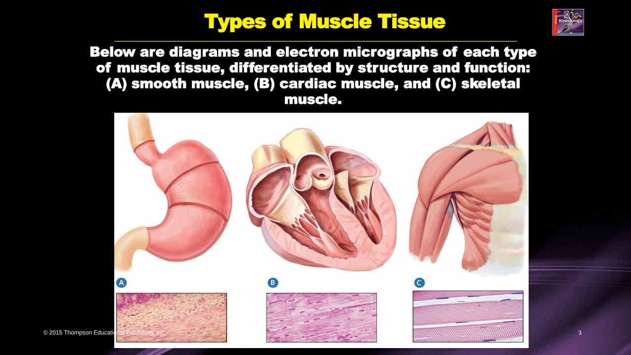
Types of Muscle Tissue
Below are diagrams and electron micrographs of each type
of muscle tissue, differentiated by structure and function:
(A) smooth muscle, (B) cardiac muscle, and (C) skeletal
muscle.
© 2015 Thompson Educational Publishing, Inc. 3
Smooth Muscles and Cardiac Muscles
© 2015 Thompson Educational Publishing, Inc. 4
Muscle tissue refers to a collection of cells that shorten during
contraction.
• Smooth Muscles. Surrounding the body’s internal organs,
including the blood vessels, hair follicles, and the urinary, genital,
and digestive tracts, are smooth muscles. Smooth (non-striated)
muscle tissue contracts more slowly than skeletal muscles, but
can remain contracted for longer periods of time. They are also
involuntary.
• Cardiac Muscles. As the name suggests, cardiac muscles are
found in only one place in the body—the heart. They are
responsible for creating the action that pumps blood from the
heart to the rest of the body. Cardiac muscles are involuntary
muscles because they are not controlled consciously, and are
instead directed to act by the autonomic nervous system.
Striated.

Skeletal Muscles
© 2015 Thompson Educational Publishing, Inc. 6
Skeletal Muscles. These muscles are the type of
muscles that are attached to the bones (by tendons
and other tissues).
• They are the most prevalent muscle type in the human
body—they comprise 30 to 40 percent of human body
weight.
• Skeletal muscles are “voluntary”—humans have conscious
control over their skeletal muscles; that is, the brain can
tell them what to do.
• Skeletal muscle tissue is referred to as striated, or
striped, because of its appearance under a microscope
as a series of alternating light and dark stripes.
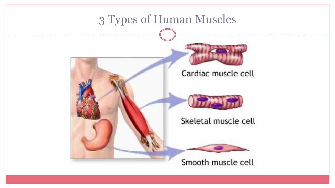
3 Types of Human Muscles

How Skeletal Muscles Are Named
© 2015 Thompson Educational Publishing, Inc. 8
Muscles are typically named after their action, location,
shape, direction of the fibres, number of divisions/heads, or
the points of attachment.
Types of Muscle Contraction
© 2015 Thompson Educational Publishing, Inc. 9
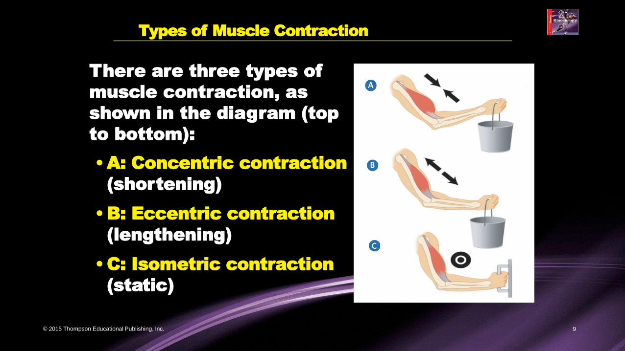
There are three types of
muscle contraction, as
shown in the diagram (top
to bottom):
•A: Concentric contraction
(shortening)
•B: Eccentric contraction
(lengthening)
•C: Isometric contraction
(static)
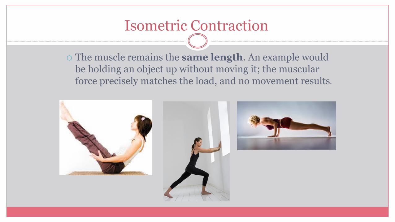
Isometric Contraction
The muscle remains the same length. An example would be holding an object up without moving it; the muscular force precisely matches the load, and no movement results.
Agonist and Antagonist Muscle Pairs
© 2015 Thompson Educational Publishing, Inc. 11
Muscles pull. They never push.
Skeletal muscles are typically arranged
as opposing pairs.
•The muscle primarily responsible for
movement of a body part is referred to as
the agonist muscle.
•The muscle that counteracts the agonist,
lengthening when the agonist muscle
contracts, is called the antagonist muscle.

Muscle Teamwork
Agonist (prime mover)
The muscle or group of muscles producing the desired effect
Eg. Bicep curl-biceps brachii is the agonist
Antagonist
• The muscle or group of
muscles opposing the action
• Eg. Bicep curl-triceps brachii is
the antagonist
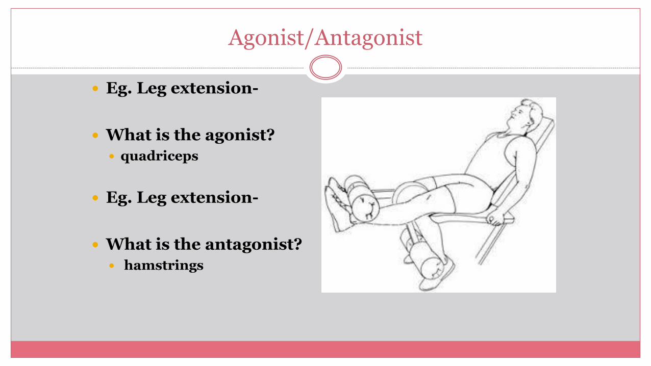
Agonist/Antagonist
Eg. Leg extension-
What is the agonist?
quadriceps
Eg. Leg extension-
What is the antagonist?
hamstrings
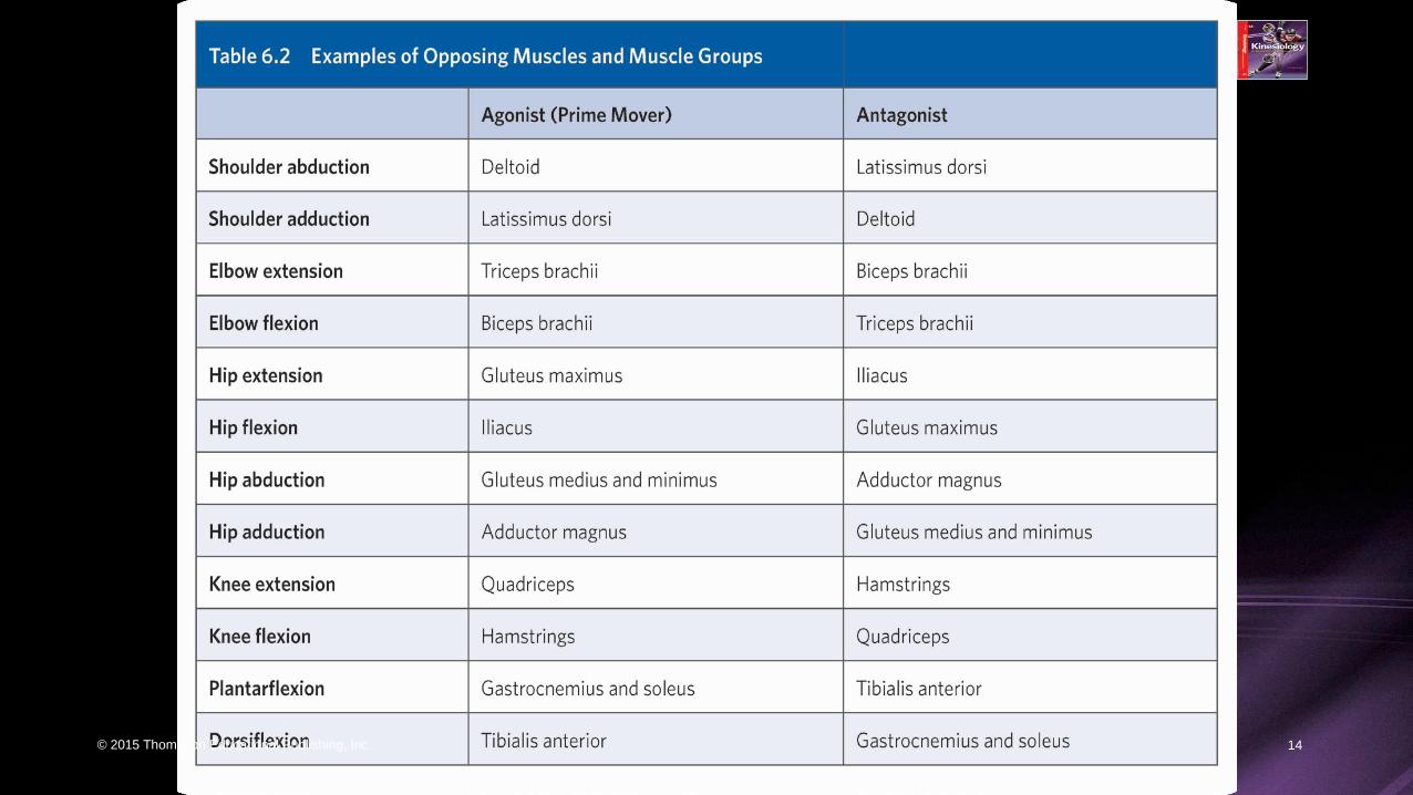
Opposing Muscles and Muscle Groups
© 2015 Thompson Educational Publishing, Inc. 14
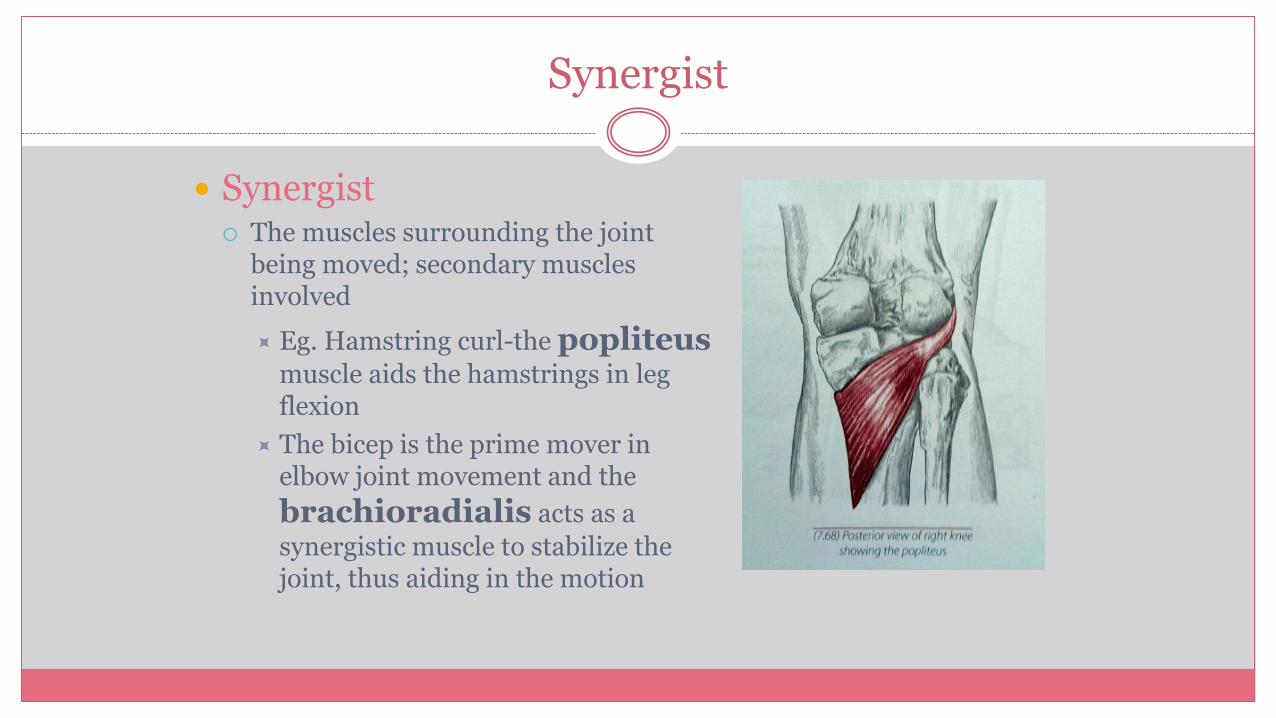
Synergist
Synergist The muscles surrounding the joint
being moved; secondary muscles involved
Eg. Hamstring curl-the popliteusmuscle aids the hamstrings in leg flexion
The bicep is the prime mover in elbow joint movement and the
brachioradialis acts as a
synergistic muscle to stabilize the joint, thus aiding in the motion
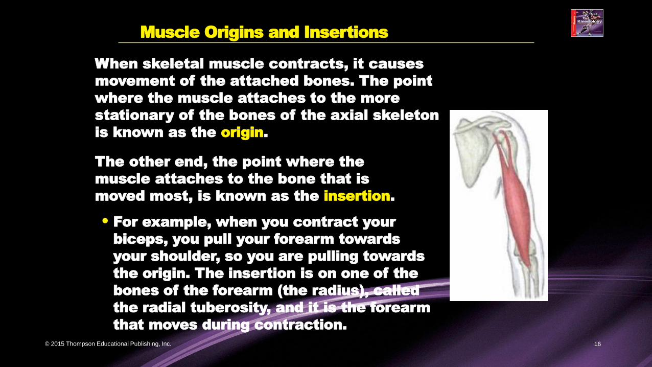
Muscle Origins and Insertions
© 2015 Thompson Educational Publishing, Inc. 16
When skeletal muscle contracts, it causes
movement of the attached bones. The point
where the muscle attaches to the more
stationary of the bones of the axial skeleton
is known as the origin.
The other end, the point where the
muscle attaches to the bone that is
moved most, is known as the insertion.
• For example, when you contract your
biceps, you pull your forearm towards
your shoulder, so you are pulling towards
the origin. The insertion is on one of the
bones of the forearm (the radius), called
the radial tuberosity, and it is the forearm
that moves during contraction.

Delayed Onset Muscle Soreness (DOMS)
• Microscopic tearing deep within the muscle fibres
• Most frequent when you begin a wt tr program, change
routine, dramatically increase the duration or intensity of
xcise routine
• May last several hours to several days after xcise session
• Felt in first 24 hours, peak 24-72 hours, disappears 5-7
days later


Lesson 6.3
© 2015 Thompson Educational Publishing, Inc. 19
THE NEUROMUSCULAR SYSTEM
~ ~ ~
TOPICS COVERED IN THIS LESSON
• The Anatomy of Skeletal Muscle
• The Sliding Filament Theory
How Muscles Attach to Bones
© 2015 Thompson Educational Publishing, Inc. 20
Skeletal muscle is attached to the
bone either indirectly (via tendons)
or directly (when the outer
membrane of the muscle attaches
to the outer membrane of the bone).
•The most common of the two
ways in which muscles attach
to bones is the indirect method
(i.e., via tendons).

Attachments
Direct attachment
Collagen fibers of epimysium are continuous with periosteum of bone
Indirect attachment
Collagen fibers of epimysium continue as a strong, fibrous tendon that merges with the periosteum
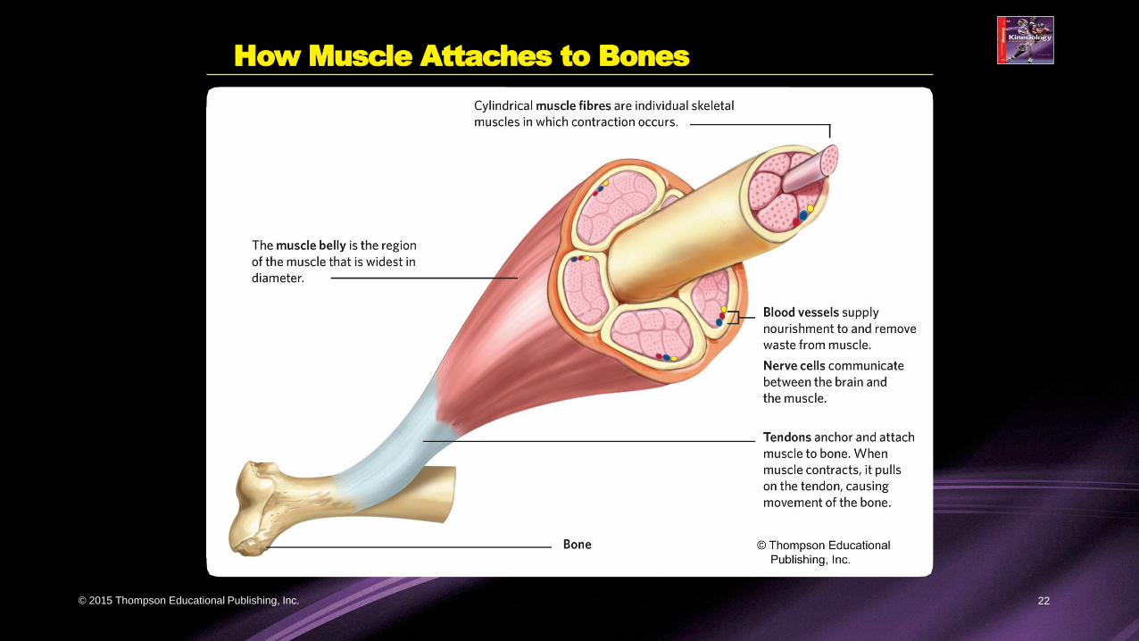
How Muscle Attaches to Bones
© 2015 Thompson Educational Publishing, Inc. 22

Indirect Attachment