the muscular system biol 105 lecture packet 11 chapter 6
TRANSCRIPT

The Muscular System
Biol 105
Lecture Packet 11
Chapter 6
Copyright © 2009 Pearson Education, Inc.
Outline
I. Characteristics of muscles
II. Three types of muscle
III. Functions of muscles
IV. Structure of skeletal muscles
V. Mechanics of muscle contraction
VI. Energy sources for muscle contraction
Copyright © 2009 Pearson Education, Inc.
Muscular System
Remember there were different types of muscle: cardiac, smooth and skeletal.
All muscle cells are elongated and therefore are called muscle fibers.
All muscle tissues contract.
Muscles contain muscle cells (called muscle fibers), connective tissue, blood vessels, and nerves
Copyright © 2009 Pearson Education, Inc.
1. Smooth muscle
2. Cardiac muscle
3. Skeletal muscle
11-2
Types of Muscles

Copyright © 2009 Pearson Education, Inc.
Smooth muscles are involuntary muscles found in the walls of many internal organs (digestive tract, respiratory system, blood vessels).
Function to aid in the function of other organs
11-2
Smooth muscle

Copyright © 2009 Pearson Education, Inc.
Cardiac muscles are involuntary muscles found only in the heart wall.
Functions by contracting to force blood from the heart into the arteries
11-2
Cardiac muscle
Copyright © 2009 Pearson Education, Inc.
Skeletal muscle are voluntary muscles attached to the skeleton.
Usually work in pairs
11-2
Skeletal muscle

11-2


Copyright © 2009 Pearson Education, Inc.
Skeletal Muscles Work in Pairs
Most skeletal muscles are antagonistic pairs.
One muscle contracts, the other relaxes
Muscles are attached to the bone by tendons
Skeletal muscles are usually attached to two bones on opposite sides of a joint
Copyright © 2009 Pearson Education, Inc.
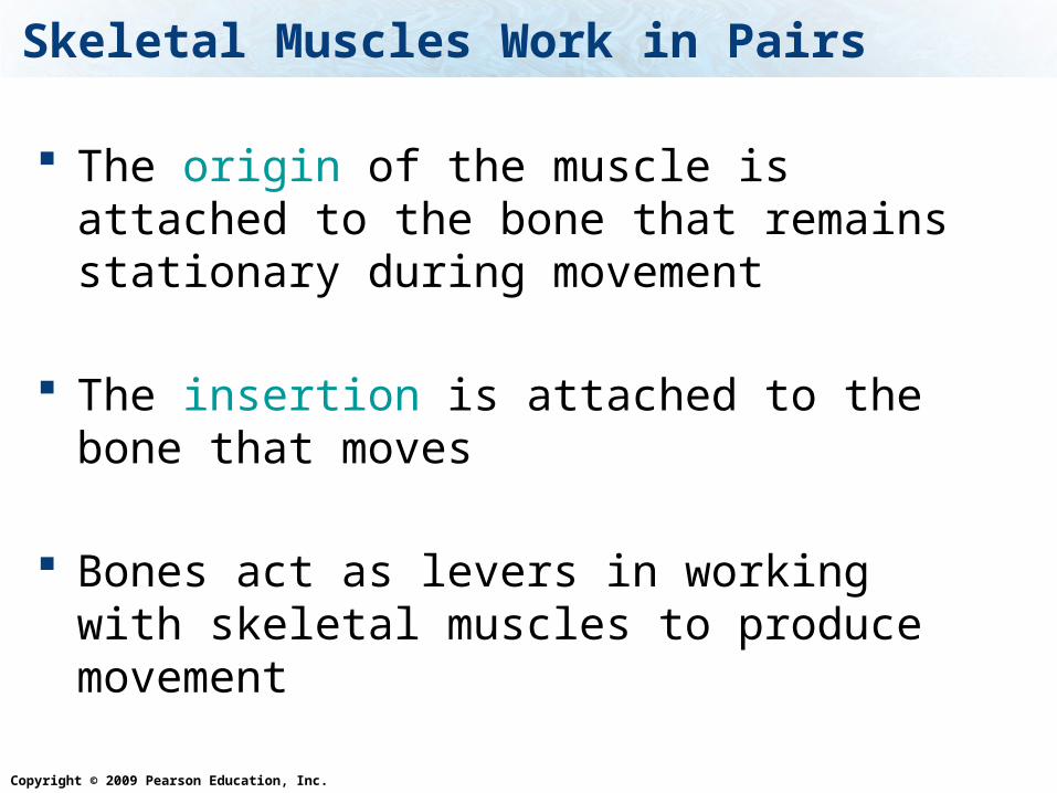
Skeletal Muscles Work in Pairs
The origin of the muscle is attached to the bone that remains stationary during movement
The insertion is attached to the bone that
moves
Bones act as levers in working with skeletal muscles to produce movement

Copyright © 2009 Pearson Education, Inc.
Skeletal Muscles Work in Pairs
Figure 6.1a
(a) Flexion
The relaxed tricepsis stretched.
The biceps contracts and pulls the forearm up, flexing the arm.
Origin of muscle:attachment of muscle to less moveable bone
Insertion of muscle:attachment of muscle to more moveable bone

Copyright © 2009 Pearson Education, Inc.
Functions of Skeletal Muscles
1. Support the body – maintain our posture
2. Movement of bones, and other tissues
3. Help maintain a constant body temperature – generates heat
4. Helps move blood through the veins and lymphatic fluid through the lymphatic vessels
5. Help to protect vital organs and stabilize joints

Copyright © 2009 Pearson Education, Inc.
Smooth muscles are under this kind of control
1. Voluntary
2. Involuntary
Voluntary
Involunta
ry
50%50%

Copyright © 2009 Pearson Education, Inc.
Smooth muscles are found in
1. The heart
2. Digestive tract
3. Attached to bones
The heart
Digestive
tract
Attached to
bones
33% 33%33%

Copyright © 2009 Pearson Education, Inc.
Structure of Skeletal Muscles
Muscles are covered by connective tissue called fascia.
A muscle contains bundles of skeletal muscle fibers (muscle cells), the bundles are called fascicles. These bundles are covered by connective tissue.
Blood vessels and nerves are between the fascicles.

Copyright © 2009 Pearson Education, Inc.
Structure of Skeletal Muscles
Figure 6.3a–b
(b) A light micrograph of a longitudinal view of skeletal muscle cells
Skeletal muscle consists of many bundles of muscle cells.
A muscle cellconsists of manymyofibrils.
A bundle of muscle cells is called a fascicle.
(a) A section of a skeletal muscle
The striped (striated) appearance of a skeletal muscle cell is due to the regular arrangement of myofilaments.

Copyright © 2009 Pearson Education, Inc.
Sarcomeres
Figure 6.3b–c
(b) A light micrograph of a longitudinal view of skeletal muscle cells
(c) A diagram and electron micrograph of a myofibril
Z line
One sarcomere
The striped (striated) appearance of a skeletal muscle cell is due to the regular arrangement of myofilaments.

Copyright © 2009 Pearson Education, Inc.
A bundle of muscle cells is called:
1. Fascicles
2. Fascia
3. Muscle Fibers
Fasci
cles
Fasci
a
Muscl
e Fibers
33% 33%33%
Copyright © 2009 Pearson Education, Inc.
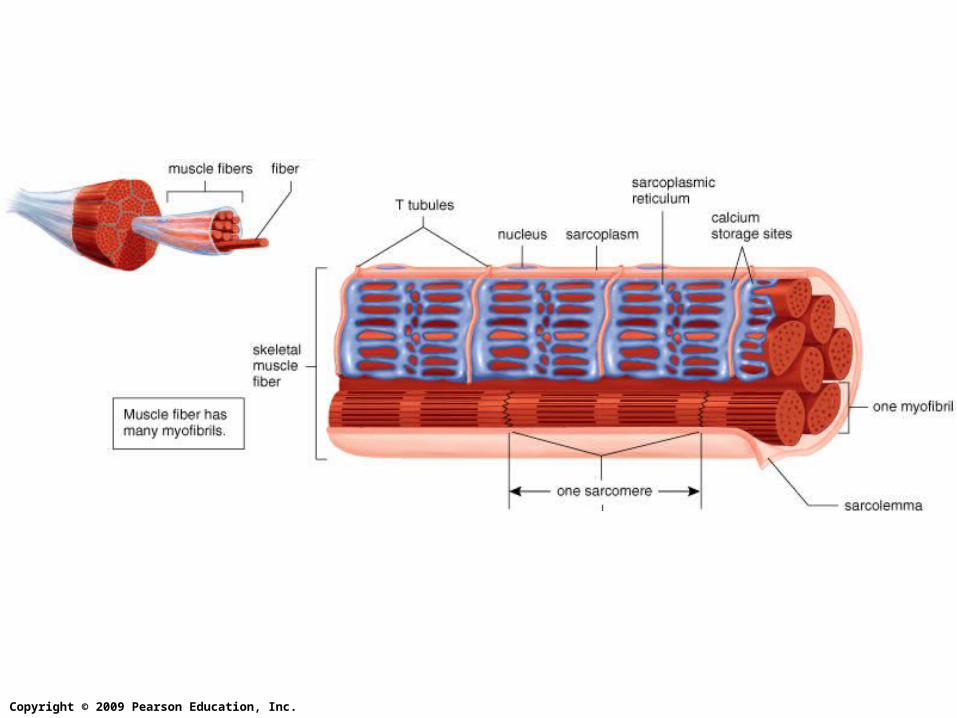
Muscle Cells
Muscle cells are long cells called muscle fibers.
The muscle fiber is composed of long thin myofibrils


Copyright © 2009 Pearson Education, Inc.
a. T tubule b. Sarcoplasmic reticulum
c. myofibril
d. Z linee. sarcomere f. sarcolemma

Copyright © 2009 Pearson Education, Inc.
Muscle Cells cont
Myofibrils are bundles of myofilaments that contracts.
Myofilaments are made of actin and myosin filaments.
When muscle fibers are stimulated to contract, myofilaments slide past one another, causing sarcomeres to shorten.
Copyright © 2009 Pearson Education, Inc.
Muscle Cell Components
Muscle cells (muscle fibers) have many of the same components as typical cells have but some of their components have different names

Copyright © 2009 Pearson Education, Inc.
Muscle Cell Components
Sarcolemma – plasma membrane (cell membrane)
Sarcoplasm – similar to cytoplasm, contains large amount of stored glycogen and myoglobin.
Myoglobin is an oxygen binding protein similar to hemoglobin, but found only in muscles
Sarcoplasmic reticulum – similar to endoplasmic reticulum, one of its functions is to store Ca2+
Copyright © 2009 Pearson Education, Inc.
Muscle Cell Components
Muscle cells (muscle fibers) also have unique features:
Multiple nuclei
Transverse tubules (T tubules) – extensions of the sarcolemma that come into contact with the sarcoplasmic reticulum.
Copyright © 2009 Pearson Education, Inc.
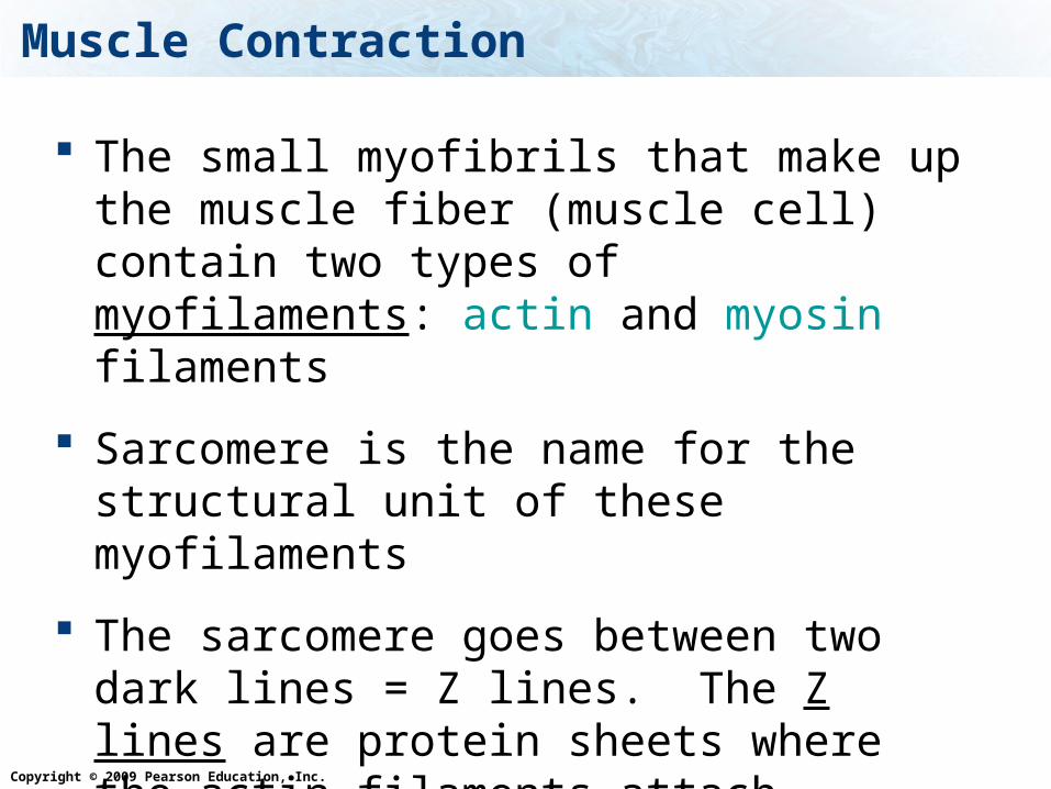
Muscle Contraction
The small myofibrils that make up the muscle fiber (muscle cell) contain two types of myofilaments: actin and myosin filaments
Sarcomere is the name for the structural unit of these myofilaments
The sarcomere goes between two dark lines = Z lines. The Z lines are protein sheets where the actin filaments attach

Copyright © 2009 Pearson Education, Inc.
Sarcomeres
Figure 6.3c–d
(c) A diagram and electron micrograph of a myofibril
(d) A sarcomere, the contractile unit of a skeletal muscle, contains actin and myosin myofilaments.
Z line
Z line
Z line
Actin
Myosin
One sarcomere
One sarcomere

Copyright © 2009 Pearson Education, Inc.
The two myofilaments are:
Actin filaments: Thin filaments that formed by two intertwining strands of the protein actin.
Myosin filaments: Thick filaments of the protein myosin shaped like a golf club, with a round “head”.
Myofilaments – actin and myosin

Copyright © 2009 Pearson Education, Inc.
The myosin heads can bind and detach from the thin actin filament. When bound it creates cross-bridges.
When the muscle is stimulated, these filaments slide past each other, making the sarcomere to shorten
Myofilaments – actin and myosin

Copyright © 2009 Pearson Education, Inc.
Muscle Contraction cont
A neuron signals the muscle to contract
The myosin heads attach to the actin then pull the actin toward the center of the sarcomere
Then the myosin heads detach

Copyright © 2009 Pearson Education, Inc.
Sarcomeres
Figure 6.4
Copyright © 2009 Pearson Education, Inc.
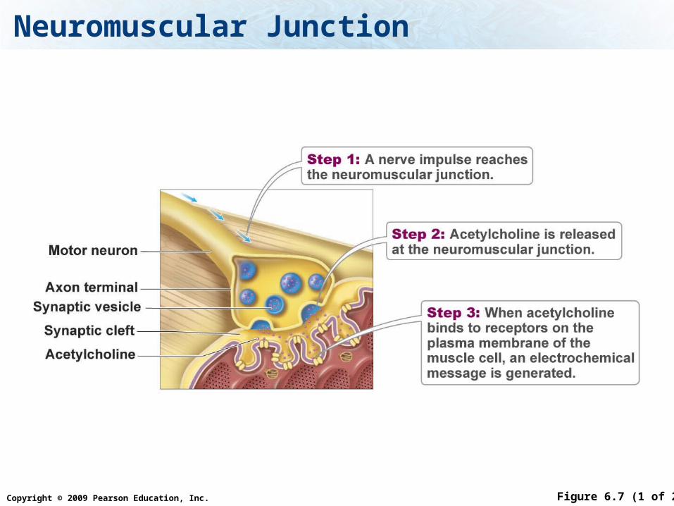
Neuromuscular Junction
Figure 6.7 (1 of 2)

Copyright © 2009 Pearson Education, Inc.
Steps of Muscle Contraction
1. Action potentials are transmitted through the neurons.
2. At the end of the neurons neurotransmitters are released
3. Neurotransmitters bind to receptor on the sarcolemma

Copyright © 2009 Pearson Education, Inc.
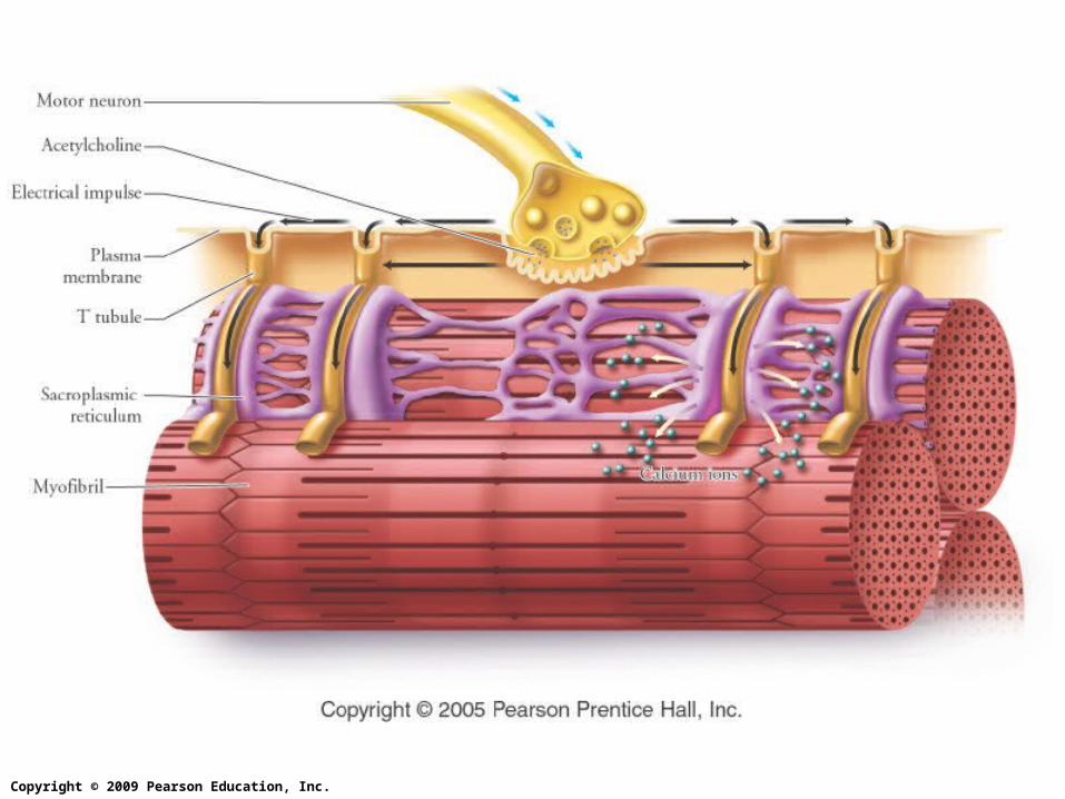
Steps of Muscle Contraction
4. The receptors are ion channels that open
5. An action potential travels through the T tubules
6. The action potential goes to the sarcoplasmic reticulum
7. The sarcoplasmic reticulum releases Ca2+.

Copyright © 2009 Pearson Education, Inc.
Steps of Muscle Contraction
8. The calcium binds to the troponin on the actin filament
9. This opens up binding site for the myosin to attach
10.Now the myosin binds to the actin
11. ATP is needed for the myosin to slide past the actin

Copyright © 2009 Pearson Education, Inc.
Sarcomeres
Figure 6.6 (1 of 2)

Copyright © 2009 Pearson Education, Inc.
Sarcomeres
Figure 6.6 (2 of 2)
Copyright © 2009 Pearson Education, Inc.
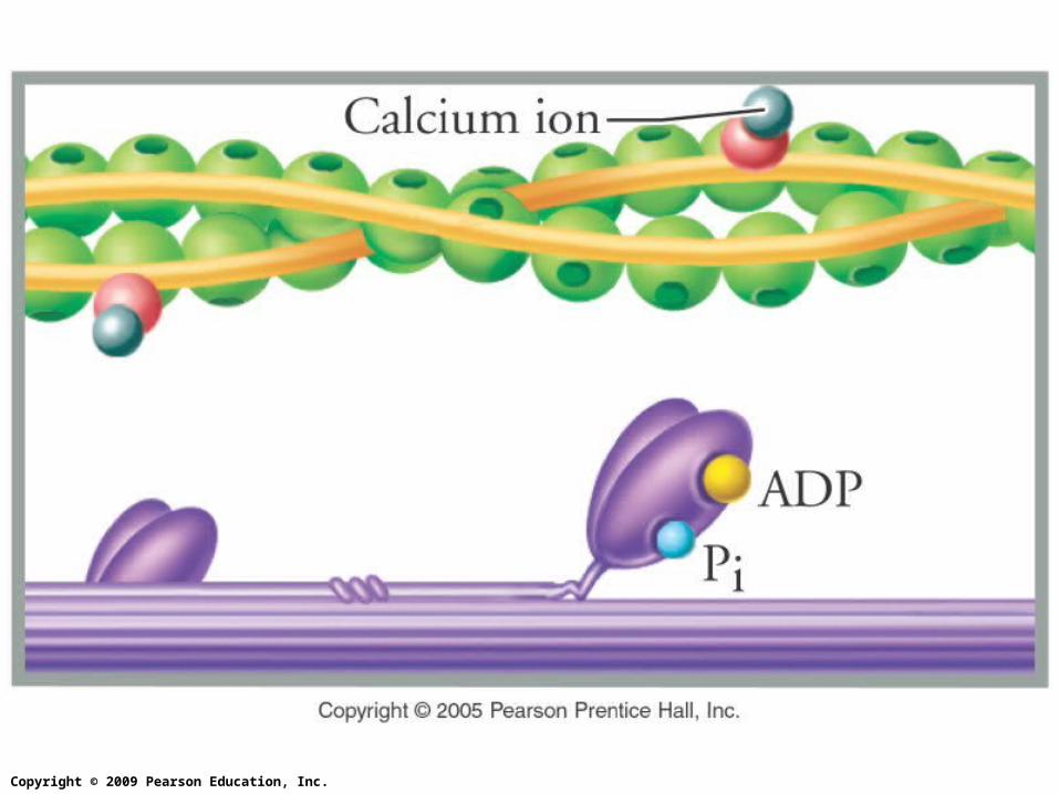
Tropomyosin-troponin complex
The tropomyosin-troponin complex is attached to the actin filament.
Calcium binds to the troponin, causing a shift in the complex, opening the sites for myosin to attach.
Muscle Contraction Video

Copyright © 2009 Pearson Education, Inc.
ATP is needed for the myofilaments to slide past each other


Copyright © 2009 Pearson Education, Inc.
What is the an oxygen binding protein found only in muscles?
1. Myosin
2. Actin
3. Hemoglobin
4. Myoglobin
Myosin
Actin
Hemoglobin
Myoglobin
25% 25%25%25%

Copyright © 2009 Pearson Education, Inc.
What ion is required for the myofilaments to bind to each other?
1. Potassium
2. Calcium
3. Chloride
4. Sodium
Potassi
um
Calcium
Chloride
Sodium
25% 25%25%25%

Copyright © 2009 Pearson Education, Inc.
Where is the calcium stored?
1. Nucleus
2. Sarcolemma
3. Sarcoplasmic reticulum
Nucleus
Sarcolemma
Sarcoplasm
ic re
ticulum
33% 33%33%

Copyright © 2009 Pearson Education, Inc.
a. T tubule b. Sarcoplasmic reticulum
c. myofibril
d. Z linee. sarcomere f. sarcolemma

Copyright © 2009 Pearson Education, Inc.
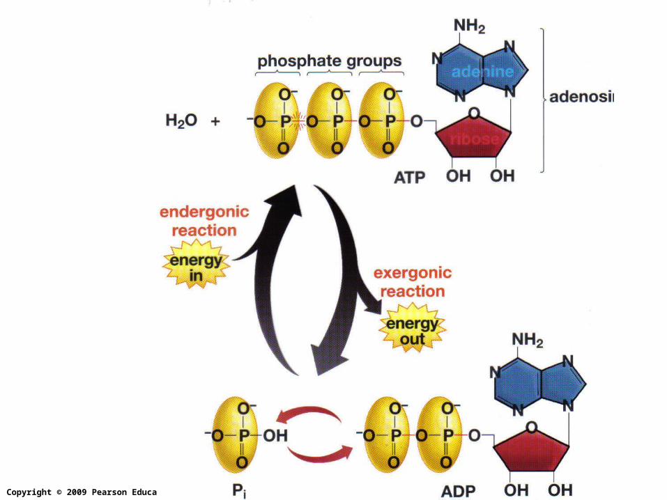
ATP
ATP is the currency. Like money in the bank.
The bonds between the phosphate groups are high energy bonds
Copyright © 2009 Pearson Education, Inc.
The Energy Source
Muscle contractions take a lot of energy in the form of ATP.
Muscles get their ATP from three sources:
1. The breakdown of creatine phosphate 2. Cellular respiration 3. Fermentation

Copyright © 2009 Pearson Education, Inc.
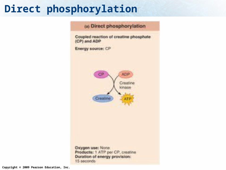
1. Creatine Phosphate
Creatine phosphate regenerates ADP to make ATP
This gives quick energy for a few seconds (up to 30 sec)
Only 1 ATP is produced per creatine phosphate
Oxygen is not needed.
When a muscle is resting, the ATP in turn regenerates creatine phosphate.
Copyright © 2009 Pearson Education, Inc.
Direct phosphorylation

Copyright © 2009 Pearson Education, Inc.
2. Cellular Respiration
In the mitochondria, glucose is broken down to produce ATP.
Remember that oxygen is needed on the electron transport chain to produce the ATP.
Carbon dioxide is produced as a waste product during the Krebs cycle step in cellular respiration
Can provide energy for hours. Produces 36 ATP per glucose molecule Can use glucose as well as fatty acids and amino
acids for energy source

Copyright © 2009 Pearson Education, Inc.
3. Fermentation
This is when the cell only uses glycolysis, and glucose is broken down to lactic acid.
Since the Krebs cycle and the electron transport chain is skipped, no oxygen is required.
No CO2 is produced as a waste produce but lactic acid is produced
Can provide energy for 30 – 60 sec 2 ATP produced per glucose molecule
Copyright © 2009 Pearson Education, Inc.
ATP Comes from Many Sources
Figure 6.10

Copyright © 2009 Pearson Education, Inc.
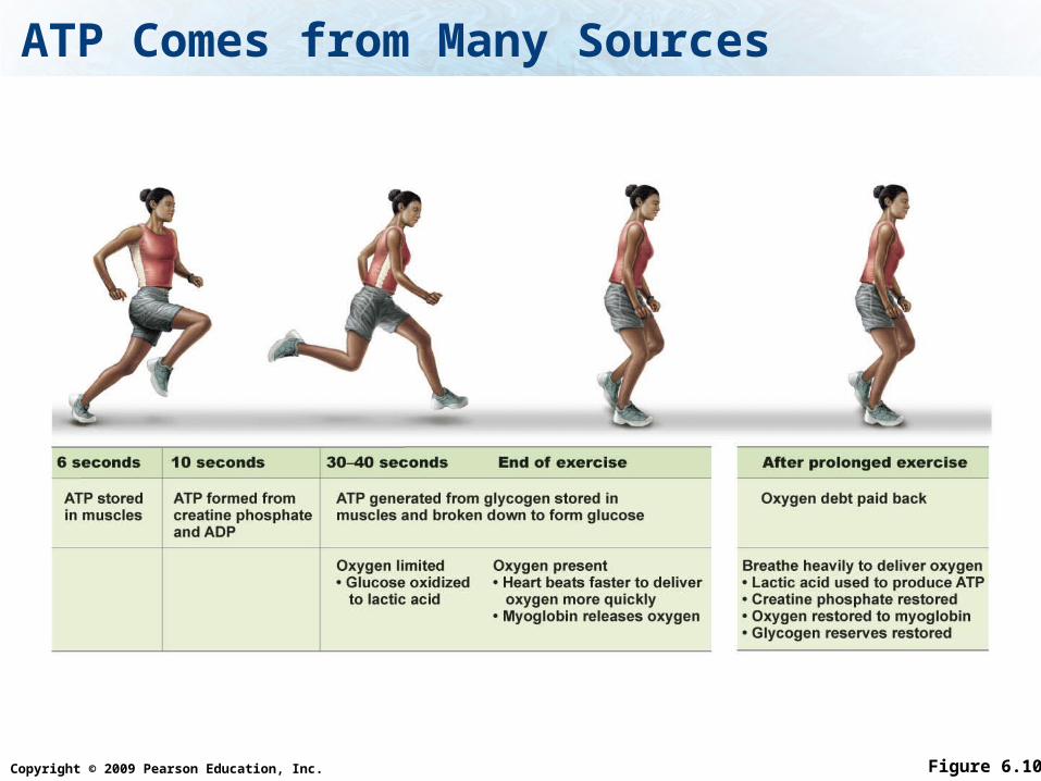
ATP Comes from Many Sources
Figure 6.10 (1 of 2)
6 seconds 10 seconds 30–40 seconds
ATP stored in muscles
ATP formed from creatine phosphate and ADP
ATP generated from glycogen stored in muscles and broken down to form glucose
Oxygen limited• Glucose oxidized to lactic acid
Copyright © 2009 Pearson Education, Inc.
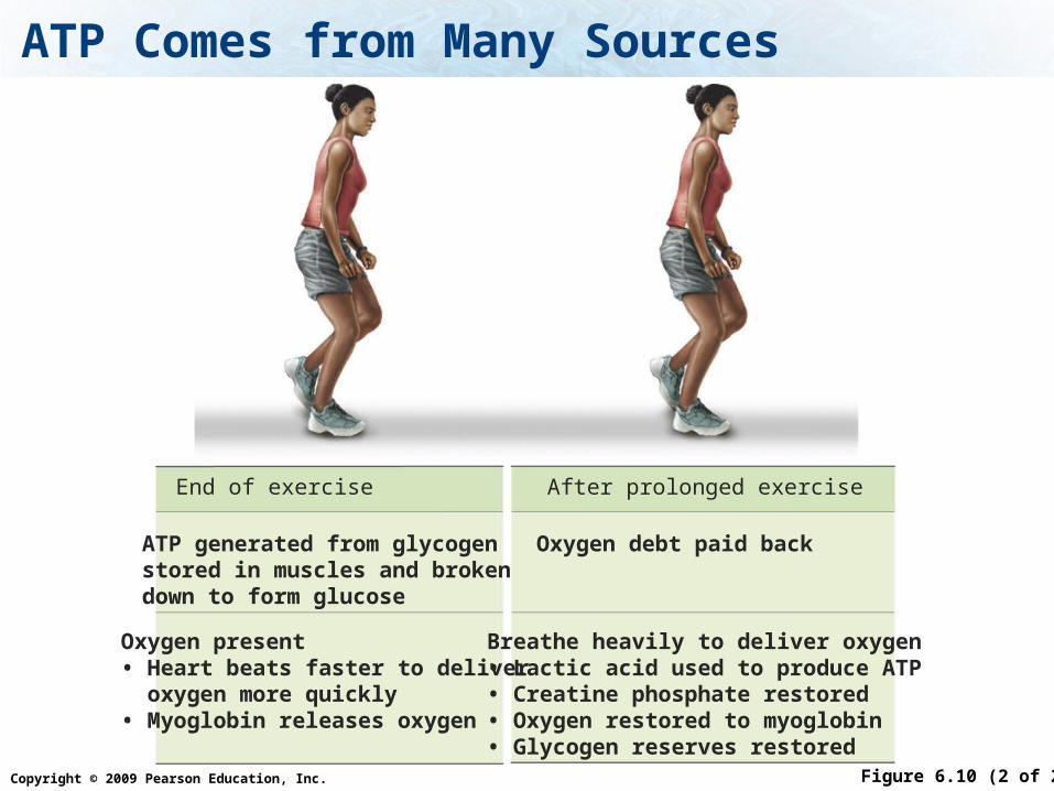
ATP Comes from Many Sources
Figure 6.10 (2 of 2)
End of exercise After prolonged exercise
ATP generated from glycogen stored in muscles and broken down to form glucose
Oxygen debt paid back
Breathe heavily to deliver oxygen• Lactic acid used to produce ATP• Creatine phosphate restored• Oxygen restored to myoglobin• Glycogen reserves restored
Oxygen present• Heart beats faster to deliver oxygen more quickly• Myoglobin releases oxygen
Copyright © 2009 Pearson Education, Inc.
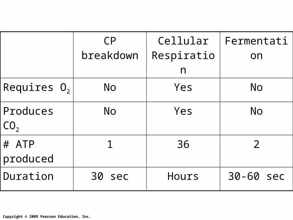
CP breakdown
Cellular Respiration
Fermentation
Requires O2 No Yes No
Produces CO2 No Yes No
# ATP produced
1 36 2
Duration 30 sec Hours 30-60 sec

Copyright © 2009 Pearson Education, Inc.
Which energy source would a long distance runner mainly use on a run that lasted for hours?
1. Fermentation
2. Cellular respiration
3. Creatine Phosphate
Ferm
entation
Cellular r
espira
tion
Creatine Phosp
hate
33% 33%33%

Copyright © 2009 Pearson Education, Inc.
Which energy source would a sprinter use in the first 5 seconds of the race?
1. Fermentation
2. Cellular respiration
3. Creatine Phosphate
Ferm
entation
Cellular r
espira
tion
Creatine Phosp
hate
33% 33%33%

Copyright © 2009 Pearson Education, Inc.
Important Concepts
What are the three types of muscles, where are they found, are they under vol. or invol. control
What are the functions of skeletal, cardiac and smooth muscles
How do skeletal muscles work in pairs?
What is the structure and the components of a muscle, and of a muscle cell (muscle fiber) and the functions of the muscle cell components.

Copyright © 2009 Pearson Education, Inc.
Important Concepts
What is the function of tendons?
What stimulates a muscle to contract
Be able to describe the steps of how the message is transmitted from the neuron to the myofilaments
What is the role of Ca2+.
What happens when the message is received by the myofilaments?

Copyright © 2009 Pearson Education, Inc.
Important Concepts
What are the components of the muscle fibers, their functions, be able to identify them in an illustration, including: myofibrils, sarcomeres, Z lines, the myofilaments - actin and myosin filaments, cross-bridges, sarcolemma, sarcoplasm, sarcoplasmic reticulum, T-tubules
What are the components and the function of the tropomyosin-troponin complex

Copyright © 2009 Pearson Education, Inc.
Important Concepts
What are the three energy sources for muscle contraction, which require oxygen, which produce carbon dioxide, how many ATP are produced, how long can it provide energy

Copyright © 2009 Pearson Education, Inc.
Definitions
muscle fibers, myoglobin, fascia, fascicles, myofibrils, sarcomere, involuntary, voluntary, origin, insertion