the most important secondary structural elements of proteins are: a. α-helix b. pleated-sheet...
TRANSCRIPT

The most important secondary structural elements of proteins are:
A. α-Helix
B. Pleated-sheet structures
C. β Turns
•The most common secondary structures are
the α-helix, the β-conformation, and turns.
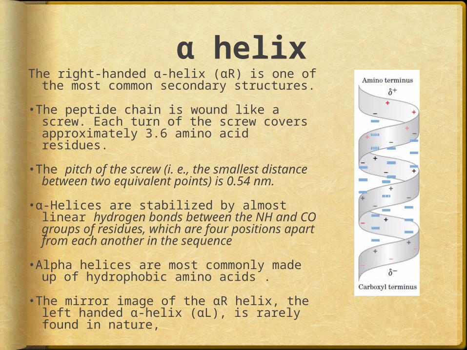
α helixThe right-handed α-helix (αR) is one of the
most common secondary structures.
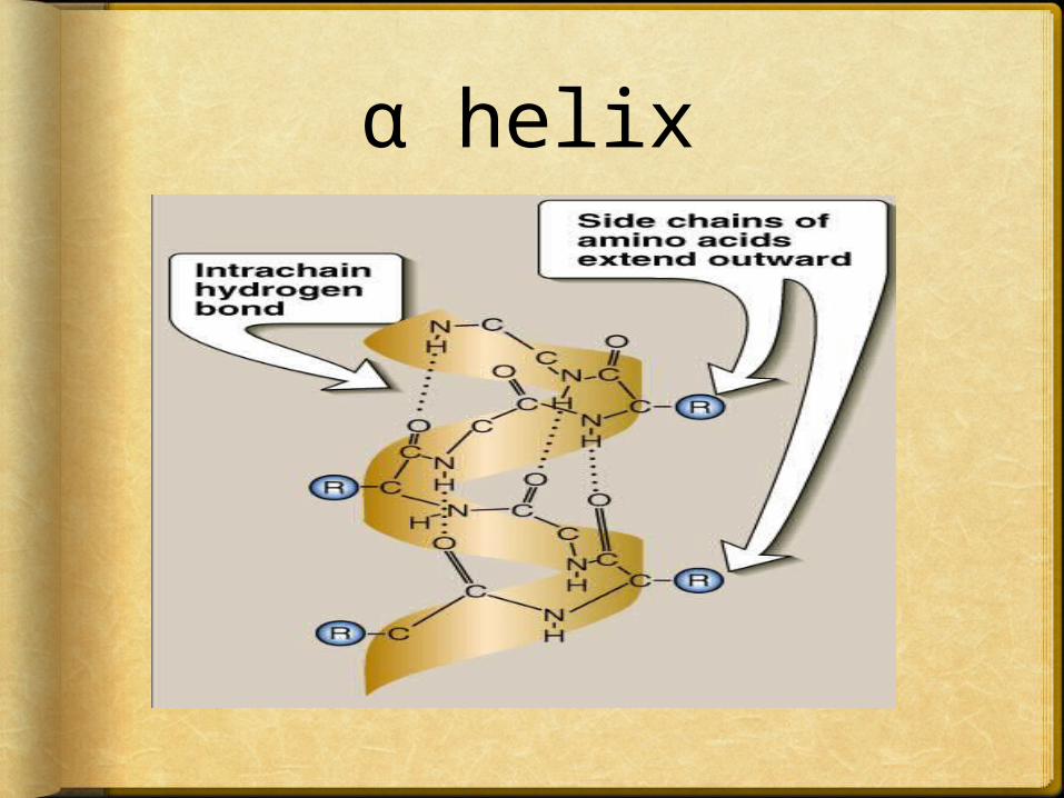
•The peptide chain is wound like a screw. Each turn of the screw covers approximately 3.6 amino acid residues.
•The pitch of the screw (i. e., the smallest distance between two equivalent points) is 0.54 nm.
•α-Helices are stabilized by almost linear hydrogen bonds between the NH and CO groups of residues, which are four positions apart from each another in the sequence
•Alpha helices are most commonly made up of hydrophobic amino acids .
•The mirror image of the αR helix, the left handed α-helix (αL), is rarely found in nature,

PITCH
α helix

The CO group of one amino acid is hydrogen bonded to the NH group of the amino acid four residues away

Amino acids that can not be
found in an α-helix Proline: the nitrogen atom In prolineis part
of rigid ring and the rotation around N-αC bond is not possible . In addition , the nitrogen atom of a proline residue in peptide linkage has no substitutent hydrogen to form hydrogen bond with other residues . So it inserts a kink in the chain, which interferes with the smooth, helical structure.
A .A residue with charged R-groups : if there large numbers of charged a.a. (e.g. glu, asp, his, lys, or arg) they disrupt the helix by forming ionic bonds, or by electrostatically repelling or attracting each other
3-Amino acids with bulky side chains: such as trp, or a.a., such as val or ile, that branch at the β-carbon (the first carbon in the R-group, next to the α-carbon) can interfere with formation of the α-helix if they are present in large numbers and close to each others.
Examples of α-helix
Alpha helices are found in almost all proteins to various extents.
•Some proteins are entirely α –helix eg
α -keratin fibrous protein in hair.
•Other proteins have different amount of α -helix e.g. hemoglobin has 80% α -helix
•Some proteins have no α -helices eg β–keratin in silk
α keratinα-keratins is an example of a protein constructed
almost exclusively of α-helices .
•They are fibrous protein , insoluble and resistant to stretching .They form tough fibers and found in hair , nails ,wool, claws, hooves, and much of the outer layer of skin and are also constituents of intermediate filaments of the cytoskeleton in certain cells .
•α-keratin is rich in cysteine which provides covalent disulphide cross-links between adjacent polypeptide chains .

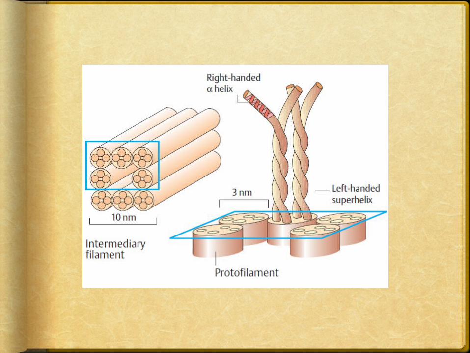
The -keratin helix is a right-handed α-helix .
•Two strands of -keratin, oriented in parallel
(with their amino termini at the same end), are wrapped about each other to form a supertwisted coiled coil.
•The helical path of the supertwists is left-handed, opposite in sense to the α-helix.
•The surfaces where the two helices touch are made up of hydrophobic amino acid residues, their R groups meshed together in a regular interlocking pattern.
This permits a close packing of the polypeptide chains within the left-handed supertwist.
•So , α-keratin is rich in the hydrophobic residues Ala, Val, Leu, Ile, Met, and Phe.
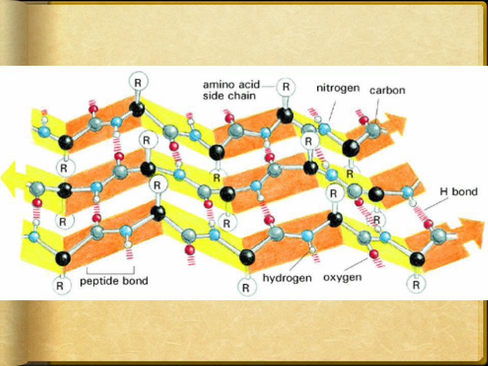
β-pleated sheetThe backbone of the polypeptide chain is
extended into a zigzag rather than helical structure
•The zigzag polypeptide chains can be arranged side by side to form a structure resembling a series of pleats
•hydrogen bonds are formed between adjacent segments of polypeptide chain.
•The individual segments that form a sheet are usually nearby on the polypeptide chain, but can also be quite distant from
each other in the linear sequence of the polypeptide;
they may even be segments in different polypeptide chains.
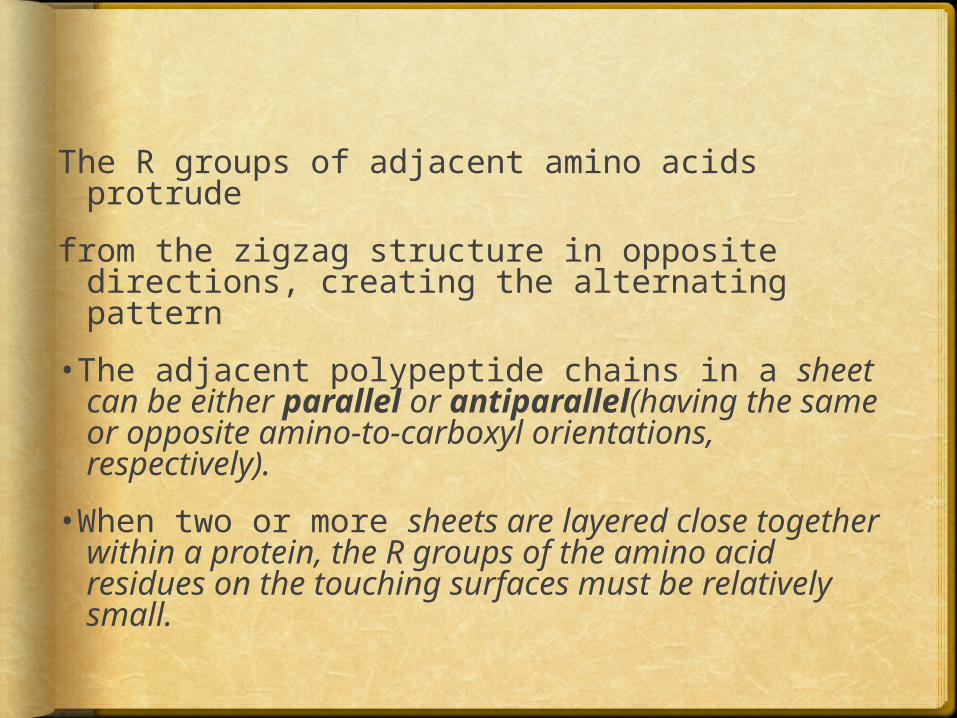
The R groups of adjacent amino acids protrude
from the zigzag structure in opposite directions, creating the alternating pattern
•The adjacent polypeptide chains in a sheet can be either parallel or antiparallel(having the same or opposite amino-to-carboxyl orientations, respectively).
•When two or more sheets are layered close together within a protein, the R groups of the amino acid residues on the touching surfaces must be relatively small.

The pleated sheet:
•Holds proteins in a parallel arrangement with hydrogen bonds.•Has R groups that extend above and below the sheet.•Is typical of fibrous proteins such as silk.
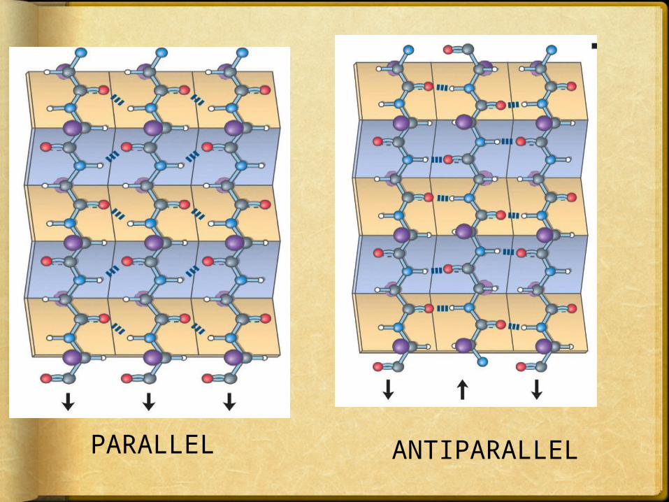
PARALLEL ANTIPARALLEL

Limitations to the kinds of
amino acids that can occur in
the β-structure R-groups of the amino acid residues on the contact surfaces must be relatively small .
• If the R-groups are bulky or have like charges , the pleated sheets can not exist because of R-group interaction .
• Structural units comprising from 2-5 parallel or anti parallel β-sheets are especially common .

Example of β-pleated sheet
structure
β-keratins contain 100% β-pleated sheet.
• Silk fibroin , a member of a class of β-keratins, consist almost entirely of antiparallel β-sheets
• Fibroin and other β-keratins are rich in amino acids having relatively small R-groups , particularly glycin and alanine .
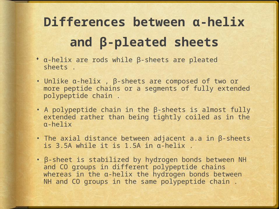
Differences between α-helix
and β-pleated sheets α-helix are rods while β-sheets are pleated sheets .
• Unlike α-helix , β-sheets are composed of two or more peptide chains or a segments of fully extended polypeptide chain .
• A polypeptide chain in the β-sheets is almost fully extended rather than being tightly coiled as in the α-helix
• The axial distance between adjacent a.a in β-sheets is 3.5A while it is 1.5A in α-helix .
• β-sheet is stabilized by hydrogen bonds between NH and CO groups in different polypeptide chains whereas in the α-helix the hydrogen bonds between NH and CO groups in the same polypeptide chain .
β Turns β Turns are often found at sites where the
peptide chain changes direction.
β-Bends reverse the direction of a polypeptide chain, helping it to form a compact, globular shape.
They are usually found on the surface of protein molecules
These are sections in which four amino acid residues are arranged in such a way that the course of the chain reverses by about 180°into the opposite direction.

β-Bends are generally composed of four amino acids, one of which may be proline the amino acid that causes a “kink” in the polypeptide chain. Glycine, the amino acid with the smallest R-group, is also frequently found in β-bends
are stabilized by hydrogen bonds between residues 1 and 4.
β Turns are often located between the individual strands of antiparallel pleated sheets, or between strands of pleated sheets and α helices


•The peptide groups of the central two residues in β turns do not participate in any hydrogen bonding.
•Gly and Pro residues often occur in β turns, the former because it is small and flexible, the latter because peptide bonds involving the imino nitrogen of proline readily assume the cis configuration.