the morphological effects of asthma, as well as
TRANSCRIPT
1341
Int. J. Morphol.,29(4):1341-1350, 2011.
The Morphological Effects of Asthma, As Well AsConventional and Alternative Asthma Therapies on Parietal
and Chief Cells in the Stomach of BALB/c Mice
Los Efectos Morfológicos del Asma, Así Como de las Terapias Convencionales y Alternativas delAsma sobre las Células Parietales y Principales en el Estómago de Ratones BALB/c
*Warren Antonio Vieira; *Hester Magdalena Oberholzer & ** Etheresia Pretorius
VIEIRA, W. A.; OBERHOLZER, H. M. & PRETORIUS, E. The morphological effects of asthma, as well as conventional andalternative asthma therapies on parietal and chief cells in the stomach of BALB/c mice. Int. J. Morphol., 29(4):1341-1350, 2011.
SUMMARY: Scientific literature, although limited in this area, supports the hypothesis that asthma, by means of selectiveleukocyte trafficking between the various mucosal and glandular sites of the body, can have the same pathophysiological effects on thestomach as the airways. This study aimed to determine if asthma, in the absence and presence of various asthma therapies (Hydrocortisoneand Modul8TM), imparted any morphological alteration on the stomach parietal and chief cells. The BALB/c murine asthmatic mousemodel was the model of choice in this study. The asthma induction protocol as well as the asthma therapies were proved to be effectivewith the aid of bronchial lavage fluid leukocyte quantification. Fundic and pyloric biopsies were extracted at termination and assessed bymeans of transmission electron, scanning electron and light microscopy. The extracted fundic and pyloric biopsies revealed asthmaalone induced parietal cell hypertrophy (increase in parietal cell size P < 0.000100 in both stomach regions) and chief cell hyper functioning.The use of Hydrocortisone and Modul8TM, as a therapy to correct the perceived gastric alterations were dismal; only in the case of fundicparietal cells were both treatments able to compensate for the hypertrophic effect caused by asthma, while in the pylorus parietal cellasthma- induced hypertrophy was only compensated for by Modul8TM.
KEY WORDS: Fundus; Pylorus; Hypertrophy; Hypersecretion; TEM.
INTRODUCTION
Asthma is a well-known disease of the lungs, andalthough it affects other organs and systems, little researchhas focused on its effects on the gastrointestinal tract (GIT).The GIT plays an important role in the immune functioning,and due to continual exposure to antigens. The gut-associatedlymphoid tissue (GALT) of the stomach, like that of the restof the GIT, is essential in preventing harmful antigen uptakewhile still allowing the absorption of nutrient molecules(Awang-Hazmi et al., 2007). The GALT of the stomach iscomposed of numerous components including diffuselymphatic tissues, intraepithelial lymphocytes, epitheliumand eosinophils.
Selective trafficking of various leukocytes betweenthe various glandular and mucosal sites, known to occur inanimals but largely unstudied in the case of humans, ishypothesized to allow bronchial asthma the ability to indu-
ce the same pathophysiological effects on the stomach asthe respiratory tissues (Wallaert et al., 1995; Bernsand etal., 2003; Rudzik et al., 1975; Nepomnyashchikh et al., 2004;Nauta et al., 2008).
A defining histopathological feature of therespiratory tissues amongst asthmatics is the increasedinfiltration of leukocytes, predominantly eosinophils andallergen-specific CD4+ Th2 cells (Nauta et al.; Warner &Knight, 2008; Kay, 1988; Sumi & Hamid, 2007; Lloyd &Rankin, 2003; D’Ambrosio et al., 2003; Gwinn et al.,2006; Kelly et al., 2007). The entire stomach mucosaduring asthma, as in the respiratory tissues, has been notedto show an increased influx of lymphocytes; however, nostudies to date have determined if these invadinglymphocytes are allergen-specific CD4+ Th2 cells(Nepomnyashchikh et al.).
* Department of Anatomy, Faculty of Health Sciences, University of Pretoria, South Africa.** Department of Physiology, Faculty of Health Sciences, University of Pretoria, South Africa.

1342
As enhanced leukocyte infiltration into a vascularizedtissue is a common feature of inflammation, the stomach ofasthmatic patients may be in a state of inflammation (Kay;Rang et al., 2003).
Goblet cell hyperplasia and increased mucusproduction is commonly described within the airways ofasthmatics along with an alteration in the composition ofthe released mucus (Rogers, 2002; Fahy, 2002; Mauad etal., 2007). Under asthmatic conditions, mucin proteinexpression appears to be altered, with the mucus itself beingfound to be more adhesive and less fluid in nature (Fahy;Gordon; Mauad et al.; Sumi & Hamid; Warner & Knight).Literature suggests a mucin expression alteration within thefundus of asthmatics as well.
The fundal surface epithelium mucin granules havebeen seen to possess a polymorphic nature based on theseverity of the individual’s asthma. Severe to moderateasthmatics possess mucin granules with an electrontransparent nature when viewed with a transmissionelectron microscope (TEM) while mild to moderateasthmatics mucin granules possess a osmophilic nature(Nepomnyashchikh et al.). Moderate to severe asthmaticshold a large number of mixt-cells, in possession of a largenumber of mucin granules, within their fundal glands. Theoccurrence of these mixt-cells may either be the result ofdelayed glandular differentiation or due to the inhibitionof protein synthesis due to stomach degeneration andemergency compensation through increased mucusproduction (Nepomnyashchikh et al.). In the pylorus, onthe other hand, surface epithelium cells possess round,electron dense granules within moderate asthmatics, whileits endocrine cells undergo hyperplasia (Nepomnyashchikhet al.).
From the above assessment of literature, theassessment of the effect of asthma on the stomach has notbeen carried out in great detail. Much detail still needs to begathered as to the reason for the perceived phenomena aswell as determining if the perceived alterations are indeed aresult of bronchial asthma.
This study therefore aims to explore themorphological interactions of asthma on the stomach parietaland chief cells, through the use of the clinically relevantBALB/c asthmatic murine model. Furthermore, this studyaimed to determine, if any, the effects which various asthmatherapies (Hydrocortisone, a conventional asthma therapy,and Modul8TM (Manufacturer: Natel Neutratec SA (Pty) Ltd;Johannesburg, South Africa), a homeopathicimmunomodulator employed as an asthma remedy) had inthis situation.
MATERIAL AND METHOD
Implementing the asthmatic BALB/c mouse ani-mal model. Six-week-old (female) BALB/c mice (each ofaverage weight 20g) were maintained in the University ofPretoria Biomedical Research Centre and providedOvalbumin (OVA)-free food and water ad libitum, All ex-perimental protocols complied with the requirements andstandards of the University of Pretoria’s Animal Use andCare Committee and the WHO/UNESCO. The ethicalclearance number for this study is 151/2006. Lab AnimalTechnologists monitored the animals every day for clinicalsigns or behavioral abnormalities, of which none wereobserved.
Mice were divided into the following experimentalgroups: six control animals, six asthmatic animals receivingno treatment, six asthmatic animals treated withphysiological comparable levels of Hydrocortisone (100mg/kg) and six asthmatic animals treated with the homeopathicproduct Modul8TM (10%). Implementation of the BALB/casthmatic mouse model involved sensitization, nebulizationand treatment of animals on specific days as indicated inTable I.
Sensitization of the animals involved theintraperitoneal injection of 25mg ovalbumin (OVA) (GradeV; Sigma Aldrich) and 2mg Al(OH)
3 dissolved in 0.5ml of
0.9% saline solution. All animals in the asthmatic and twotreatment groups were sensitized.
Nebulization of the animals with 1% OVA in PBSwas done using an inhalation exposure system (IES) (Glas-COL® Inhalation Exposure System, Model 099C A4212,Terre Haute, Indiana). Standard protocol was applied for theIES. Nebulization involved placing the six animals from thesame experimental group inside a stainless steel wire meshbasket, which is divided into five equal compartments. Eachcompartment therefore held the six animals from the sameexperimental group. One complete cycle in the IES includeda preheat cycle of 15 minutes followed by the nebulizationwith the OVA for 60 minutes, a cloud decay cycle for 15minutes and a decontamination cycle also 15 minutes as perstandard protocol. Mice were nebulized twice daily.
Treatment procedures involved in this study wereconducted by veterinary technicians. This involved theadministration of Modul8TM and hydrocortisone to theasthmatic animals. Solu-CortefTM 100mg Hydrocortisone(Pfizer Laboratories (Pty) Limited), dissolved in sterile PBSwas injected intra-peritoneally, while Modul8™ (NatelHealthcare SA (PTY) Ltd.) was administered orally on the
VIEIRA, W. A.; OBERHOLZER, H. M. & PRETORIUS, E. The morphological effects of asthma, as well as conventional and alternative asthma therapies on parietal and chief cells in the stomachof BALB/c mice. Int. J. Morphol., 29(4):1341-1350, 2011.

1343
specific treatment days as indicated in Table I. Modul8™ isa complex homeopathic immunomodulator that enhancesthe immunity of an individual and consists of a mixture ofdifferent substances in different concentrations. Thefollowing substances are found in this product: Aconitumnapellus (D20), Arsenicum album (D18), Asafoetida (D20),Calcarea carbonica (D16), Conium maculatum (DH17),Ipecacuanha (D13), Phosphorus (D20), Rhus toxicodendron(D17), silicea (D20), sulfur (D24), Thuja occidentalis (D19),alcohol (0.2%, v/v) and purified water (44.050 ml). Theproduct is available in 50 ml bottles on which the declaredpotencies (D) are expressed. The latter is an indication of anindex of 10-fold dilution. The dosage of Modul8™ wascalculated from the adult human dosage and a dosage of28ml/kg was administered once daily via oral gavage as itallows for administration of a controlled amount ofcompound. The adult dosage of 100mg/kg body weighthydrocortisone was converted to the average mass of amouse, which was taken at 20g. The hydrocortisone wasadministered via intraperitoneal injection, as was indicatedon the product itself and since it is one of the most commonroutes of administration in mice.
BAL technique. On the day of termination, a small skinincision was made in the skin of each mouse in the ventralregion of the trachea. This was followed by blunt dissectionto expose the trachea. A small transverse incision was madebelow the larynx through which a 21G venous catheter wasemployed to inject 0.3ml of saline into the trachea andaspirated with a syringe. The BAL fluid was collected andpooled for the individual groups. The samples were thencentrifuged for two minutes at 1000rpm and used to preparehistological smears. The smears were stained with RapidHematological Stain and viewed with a Nikon Optiphot lightmicroscope. A hundred cells were counted per slide andstatistically analyzed.
Tissue for TEM. Biopsies from the pylorus and fundus werefixed in a 2.5% glutaraldehyde/formaldehyde solutionimmediately after extraction, for one hour, and subsequentlyrinsed with 0.075M phosphate buffer solution (PBS) threetimes for 15 minutes. The biopsies were exposed to thesecondary fixative, 0.5% osmium tetroxide, for one hourafter which the biopsies were rinsed with 0.075M PBS once
again for three times for 15 minutes. The biopsies were thendehydrated in 30%, 50%, 70%, 90% and three changes ofabsolute ethanol. The biopsies were embedded in quetolresin, from which ultra-thin sections were cut using adiamond knife and contrasted with uranyl acetate and leadcitrate. The contrasted TEM sections were analyzed with aJOEL JEM 2100F transmission electron microscope.
Tissue for light microscopy. Biopsies from the pylorus andfundus were fixed in a 2.5% glutaraldehyde/formaldehydesolution immediately after extraction, for one hour, andsubsequently rinsed with 0.075M PBS three times for 15minutes. The biopsies were then dehydrated in 30%, 50%,70%, 90% and three changes of absolute ethanol. Thebiopsies were embedded in LR white resin and sectioned ata thickness of 1mm and stained with Toluidine Blue Osolution. The biopsies were analyzed with a Nikon Optiphotlight microscope.
Statistical analysis of data. The statistical comparisons wereconducted using the statistical program NCSS® at asignificance level of 0.05.
BAL fluid leukocyte counts. Three stained BAL fluidsmears were chosen at random, for each experimental group,for leukocyte differentiation and quantification. A hundredcells were quantified per slide at a 100x magnification. Eachleukocyte species (basophil, eosinophil, lymphocyte,monocyte and neutrophil) was considered separately betweenthe four experimental groups via one-way ANOVA’s withTukey-Kramer Multiple-Comparison Tests used for pos-hoccomparisons. For each experimental group the averagedifferential BAL fluid leukocyte numbers and theirassociated standard deviations were calculated.
Parietal cell size. 90 random parietal cells were measured,based on the principles of Feret’s minimal diameter, of threerandom mice from each experimental group, within boththe fundus and pylorus with the aid of Olympus Soft ImagingSolutions® software. The data was utilized for statisticalassessment of these cells sizes:
Between the four experimental groups by means of aKruskal-Wallis one-way ANOVA, in each region of thestomach, due to a lack of normality in the data’s distribution.The Tukey-Kramer Multiple-Comparison Test was utilizedfor pos-hoc comparisons to determine where significancelay.
Between the two regions of the stomach within eachexperimental group by means of the Mann-Whitney U test,due to the lack of normality in the data’s distribution. Thenull hypothesis for the control, asthma and Modul8TM tests
Table I. Implementation of the BALB/c asthmatic mouse model
VIEIRA, W. A.; OBERHOLZER, H. M. & PRETORIUS, E. The morphological effects of asthma, as well as conventional and alternative asthma therapies on parietal and chief cells in the stomachof BALB/c mice. Int. J. Morphol., 29(4):1341-1350, 2011.
Steps for Implementation andTreatment Days
Sensitization 0, 5
Nebulization 13, 14,15, 30, 31, 32
Treatment 15 – 18, 22 – 25, 36 – 39
Termination 42
1344
was that the fundic parietal cells, which appeared bigger tothe eye then those of the pylorus, were significantly largerthan that resident in the pylorus. The null hypothesis for theHydrocortisone test was that the pyloric parietal cells, whichappeared bigger to the eye then those of the fundus, werelarger than those resident in the fundus.
Feret’s minimum diameter is a measuring methodutilized to estimate the length or size of a cell or fiber (Briguetet al., 2004; Alamar et al., 2008). It is the minimum distancebetween two parallel tangents at opposite sides of the cell orfibers borders (Briguet et al.). This measuring method hasthe advantage of being insensitive to deviations from optimalcross-sectioning while still being reliable to quantifydetectable size differences (Briguet et al.).
RESULTS AND DISCUSSION
When comparing the different pathophysiologicalfeatures of the lungs and the stomach, great similarity is seen.Table II expresses the over lapping pathophysiologicalfeatures found to occur within the stomach and lungs andairways of human asthmatics, while Table III expresses allthe currently known pathological features associated withthe stomach in asthmatics.
Due to the information shown in Tables II and III, inthe current manuscript, a detailed ultrastructural investigationof the stomach cells (pariental and chief cells) wasperformed. Also, we compared the BAL fluid leukocytecounts between the different groups, in order to confirmasthma.
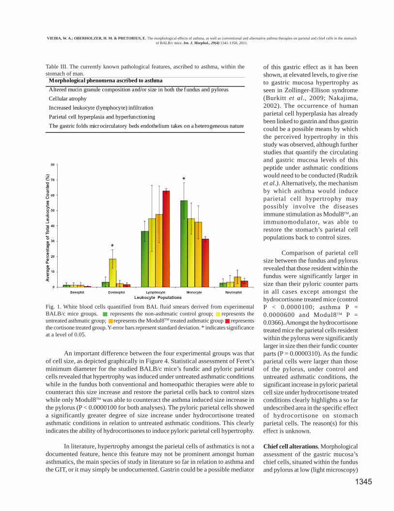
The averaged differential BAL fluid leukocyte countsof this study, for each experimental group at termination,were presented in Figure 1. From Figure 1 it can be seen
that the eosinophil levels increased significantly amongstthe untreated asthmatic mice in comparison to the non-asthmatic control mice (P = 0.00312), while both treatmentgroups showed eosinophil levels which were statisticallyindifferent from control levels but significantly lower thanthat of the untreated asthmatic mice. The untreated asthmaticmice of this study showed a five-fold increase in eosinophilnumber, a clear indication that asthma was indeed inducedamongst these mice. The collected BAL results for this studywere theoretically expected as untreated asthmatics havebeen reported to show an increase in BAL fluid eosinophillevels while the use of corticosteroids, such asHydrocortisone, have been shown to reduce the infiltrationof eosinophils amongst asthmatics (Gwinn et al.; Holgate,2008; Venge, 1988).
Hydrocortisone is one of the prominent treatmentsused in the battle against asthma due to its highly effectiveanti-inflammatory and immunosuppressive effects (Warner& Knight; Rang et al.; Garrod, 1958; Crompton, 2006).Unfortunately, due to it this therapy’s associated detrimentaleffects on the body, other therapy options are sought after inthe fight against asthma – one such option being the use ofhomeopathic immunomodulators. Modul8TM is a highlydiluted, non-toxic homeopathic remedy which has beensuggested for use by patients who experience diseases wheretheir immune system is compromised, such as asthma, dueto its ability to modulate the immune system naturally(Modul8 Inc., 2009).
Parietal cell alterations. Morphological assessment of thegastric mucosa’s parietal cells, situated within the fundusand pylorus at both low magnification (light microscopy)and high magnification (TEM), showed no appreciabledifferences between the four experimental groups, Figures2 (light microscopy) and 3 (TEM) reveal the morphology atlow and high magnification respectively of the differentexperimental groups pylorus parietal cells.
Morphological phenomena ascribed to asthma Lungs & Airways Stomach
Cellular atrophyþ
Surface epithelium
þSurface, pit, crypt and glandular
epithelium
Increased leukocyte infiltrationþ
Eosinophils, lymphocytes, mast cells,monocytes/macrophages and neutrophils
þLymphocytes
Mucus composition alterationsþ
Goblet cellsþ
Fundic surface epithelium
Table II. The overlapping morphological features, ascribed to asthma, within the lungs and airways and the stomach of man.
Symbol: = feature present.
VIEIRA, W. A.; OBERHOLZER, H. M. & PRETORIUS, E. The morphological effects of asthma, as well as conventional and alternative asthma therapies on parietal and chief cells in the stomachof BALB/c mice. Int. J. Morphol., 29(4):1341-1350, 2011.
þ
1345
An important difference between the four experimental groups was thatof cell size, as depicted graphically in Figure 4. Statistical assessment of Feret’sminimum diameter for the studied BALB/c mice’s fundic and pyloric parietalcells revealed that hypertrophy was induced under untreated asthmatic conditionswhile in the fundus both conventional and homeopathic therapies were able tocounteract this size increase and restore the parietal cells back to control sizeswhile only Modul8TM was able to counteract the asthma induced size increase inthe pylorus (P < 0.0000100 for both analyses). The pyloric parietal cells showeda significantly greater degree of size increase under hydrocortisone treatedasthmatic conditions in relation to untreated asthmatic conditions. This clearlyindicates the ability of hydrocortisones to induce pyloric parietal cell hypertrophy.
In literature, hypertrophy amongst the parietal cells of asthmatics is not adocumented feature, hence this feature may not be prominent amongst humanasthmatics, the main species of study in literature so far in relation to asthma andthe GIT, or it may simply be undocumented. Gastrin could be a possible mediator
of this gastric effect as it has beenshown, at elevated levels, to give riseto gastric mucosa hypertrophy asseen in Zollinger-Ellison syndrome(Burkitt et al., 2009; Nakajima,2002). The occurrence of humanparietal cell hyperplasia has alreadybeen linked to gastrin and thus gastrincould be a possible means by whichthe perceived hypertrophy in thisstudy was observed, although furtherstudies that quantify the circulatingand gastric mucosa levels of thispeptide under asthmatic conditionswould need to be conducted (Rudziket al.). Alternatively, the mechanismby which asthma would induceparietal cell hypertrophy maypossibly involve the diseasesimmune stimulation as Modul8TM, animmunomodulator, was able torestore the stomach’s parietal cellpopulations back to control sizes.
Comparison of parietal cellsize between the fundus and pylorusrevealed that those resident within thefundus were significantly larger insize than their pyloric counter partsin all cases except amongst thehydrocortisone treated mice (controlP < 0.0000100; asthma P =0.0000600 and Modul8TM P =0.0366). Amongst the hydrocortisonetreated mice the parietal cells residentwithin the pylorus were significantlylarger in size then their fundic counterparts (P = 0.0000310). As the fundicparietal cells were larger than thoseof the pylorus, under control anduntreated asthmatic conditions, thesignificant increase in pyloric parietalcell size under hydrocortisone treatedconditions clearly highlights a so farundescribed area in the specific effectof hydrocortisone on stomachparietal cells. The reason(s) for thiseffect is unknown.
Chief cell alterations. Morphologicalassessment of the gastric mucosa’schief cells, situated within the fundusand pylorus at low (light microscopy)
Morphological phenomena ascribed to asthma
Altered mucin granule composition and/or size in both the fundus and pylorus
Cellular atrophy
Increased leukocyte (lymphocyte) infiltration
Parietal cell hyperplasia and hyperfunctioning
The gastric folds microcirculatory beds endothelium takes on a heterogeneous nature
Table III. The currently known pathological features, ascribed to asthma, within thestomach of man.
Fig. 1. White blood cells quantified from BAL fluid smears derived from experimentalBALB/c mice groups. represents the non-asthmatic control group; represents theuntreated asthmatic group; represents the Modul8TM treated asthmatic group representsthe cortisone treated group. Y-error bars represent standard deviation. * indicates significanceat a level of 0.05.
VIEIRA, W. A.; OBERHOLZER, H. M. & PRETORIUS, E. The morphological effects of asthma, as well as conventional and alternative asthma therapies on parietal and chief cells in the stomachof BALB/c mice. Int. J. Morphol., 29(4):1341-1350, 2011.

1346
and high (TEM) magnification, showed asthma as well asthe employed treatments for the disease to have a significantimpact on these cells secretory functions. Figure 5 revealsthe morphology at low magnification of stomach fundic andpylorus chief cells, while Figure 6 reveals the morphologyof the stomach pylorus chief cells at high magnification forthe different experimental groups.
Under control conditions, the BALB/c mice held arelatively large amount of zymogenic granules within their
fundic and pyloric chief cells, clearly identifiable due to theirstaining with Toluidine Blue O solution. This is in line withtheory as gastric chief cells not only secrete pepsin inresponse to neuronal and hormonal stimuli but also secretea large basal amount of pepsinogen (Tani et al., 1989). Thisbase line secretion was noted to be greater in the pylorusthan in the fundus of the control mice, as can be seen inFigure 5. Under untreated asthmatic conditions there wasan increase in the amount of zymogenic granules presentwithin the gastric mucosa’s chief cells in comparison to the
Fig. 2. Light microscopic images of a longitudinal section of pyloric parietal cells, taken at 40x magnification, derived from the stomachof (A) a non-asthmatic control mouse, (B) an untreated asthmatic mouse, (C) a Hydrocortisone treated asthmatic mouse and (D) aModul8TM treated asthmatic mouse. The black line represents a scale bar of 50mm. Ca – Canaliculi, Pc – Parietal cells, LP – Laminapropria, PM – Plasma Membrane, Mt – Mitochondria, RER – Rough Endoplasmic Reticulum, N – Nucleus, Zc – Zymogenic cells, Nc –Neck cells, Zg – Zymogen granules.
VIEIRA, W. A.; OBERHOLZER, H. M. & PRETORIUS, E. The morphological effects of asthma, as well as conventional and alternative asthma therapies on parietal and chief cells in the stomachof BALB/c mice. Int. J. Morphol., 29(4):1341-1350, 2011.

1347
Fig. 3. Transmission electron microscopic images of a longitudinal section of pyloric parietal cells derived from the stomach of (A) anon-asthmatic control mouse, (B) an untreated asthmatic mouse, (C) a Hydrocortisone treated asthmatic mouse and (D) a Modul8TM
treated asthmatic mouse. The white scale bars represent 2µm. Ca – Canaliculi, Pc – Parietal cells, LP – Lamina propria, PM – PlasmaMembrane, Mt – Mitochondria, RER – Rough Endoplasmic Reticulum, N – Nucleus, Zc – Zymogenic cells, Nc – Neck cells, Zg –Zymogen granules.
Fig. 4. Average parietal cell size within thefundus and pylorus of the stomach of eachexperimental group. represents the non-asthmatic control group; represents theuntreated asthmatic group; represents thehydrocortisone treatment asthmatic group;represents the Modul8TM treatment asthmaticgroup. Y-error bars represent standarddeviation; * indicates significance for betweengroup comparison, for each stomach region,at a level of 0.05 and ~ indicates significancefor region comparison of the stomach, withineach group, at a level of 0.05.
VIEIRA, W. A.; OBERHOLZER, H. M. & PRETORIUS, E. The morphological effects of asthma, as well as conventional and alternative asthma therapies on parietal and chief cells in the stomachof BALB/c mice. Int. J. Morphol., 29(4):1341-1350, 2011.

1348
their respective control situations. The increasewas noted to be greater within the fundic chiefcells. Such alterations have not been documentedbefore in the literature.
Research, with the aid of canine androdent models, has shown that histamine is ableto induce a species specific secretion ofpepsinogen by the gastric chief cells(Wiederanders et al., 1960; Lopez-Diaz et al.,2006; Tani & Tanaka, 1990). In the case of ratchief cells, Lopez-Diaz et al. have shown thathistamine, possibly working through the chiefcells H2 receptors, induces a dose-dependentpepsinogen secretion. It is known that under anallergic response, such as in the case of asthma,the body releases a large amount of histaminewhich might be the cause of the perceivedincrease in gastric chief cell secretory activitywithin the experimental model, if the BALB/cmice chief cells respond as rats chief cells toincreased histamine levels (Nelson & Cox, 2005).
Hydrocortisone and Modul8TM inducedeven greater increases in the amount ofzymogenic granules present within theasthmatic mice chief cells in relation to the con-trol situation and untreated asthmatic situation,as expressed in Figure 5. In the case ofHydrocortisone this increase in zymogenicgranule production, above that induced by thedisease alone, is understandable and expected,as research has documented that corticosteroidsare able to induce the hypersecretion ofpepsinogen from these cells (Wiederanders etal.). Hydrocortisone also appears to have a sitespecific affect on the gastric mucosa, increasingthe number and size of the zymogenic granulesin the fundic chief cells more severely than thecorresponding pyloric cells. The reason forModul8TM inducing such an effect on zymogenicgranule secretion is unknown nor is it knownwhether it functions by a similar mechanism toHydrocortisone, in this respect, but it alsoappears to have a site specific affect on thegastric mucosa, increasing the number and sizeof the zymogenic granules in the pylorus chiefcells more severely than the correspondingfundic cells. Modul8TM and hydrocortisone canbe seen to both be ineffective treatments incompensating in the asthma, instead inducedhyperfunctioning of gastric chief cells isobserved.
Fig. 5. Light microscopic images of a longitudinal section, at variousmagnifications, of the fundic and pyloric chief cells, taken at 40x magnification,derived from the stomach of a (A) a non-asthmatic control mouse, (B) auntreated asthmatic mouse, (C) a Hydrocortisone treated asthmatic mouseand (D) a Modul8TM treated asthmatic mouse. The black line represents ascale bar of 50mm.
VIEIRA, W. A.; OBERHOLZER, H. M. & PRETORIUS, E. The morphological effects of asthma, as well as conventional and alternative asthma therapies on parietal and chief cells in the stomachof BALB/c mice. Int. J. Morphol., 29(4):1341-1350, 2011.

1349
CONCLUSION
Asthma does appear to significantly affect themorphology of the stomachs parietal and chief cells – parietalcell hypertrophy and chief cell hyperfunctioning – althoughthese features have not been documented thus far in literature.This may be due to the alterations noted being a speciesspecific phenotypic expression of the disease, due the specificprotocol utilized events or simply not assessed thus far inhumans.
The beneficial effects of using Hydrocortisone andModul8TM as a therapy to correct the perceived gastricalterations are not very strong as both were found toaggravate the situation more often than help. Hydrocortisonewas associated with aggravation of asthmatic induced pyloricparietal cell hypertrophy and fundic and pyloric chief cellhyperfunctioning. Modul8TM was associated with theaggravation of asthma induced pyloric and fundic chief cell
hyperfunctioning. This study also provides evidence thatHydrocortisone, and even Modul8TM, may have area specificeffects on the cells which reside within the gastric mucosa.Hydrocortisone appears to have a more detrimental effecton the pyloric parietal cells and fundic chief cells, whileModul8TM appears to have a greater detrimental effect onthe pyloric chief cells. The aspect of area specific effects ofHydrocortisone has apparently not been considered inliterature thus far, and would be an important avenue ofresearch in trying to solve some of the controversysurrounding Hydrocortisone and its detrimental effects on thegastric mucosa. The effects of Modul8TM on the stomach havenot been assessed in scientific literature. Modul8TM did showsome benefits as it was able to restore both the pyloric andfundic parietal cells back to control sizes under asthmaticconditions. Thus Modul8TM may provide a useful alternative,as complementary medicine, to more traditional treatmentregimes but further studies would need to be performed inorder to determine if this is the case, and how to circumventthe chief cell aggravation it appears to induce.
VIEIRA, W. A.; OBERHOLZER, H. M. & PRETORIUS, E. Los efectos morfológicos del asma, así como de las terapias convencio-nales y alternativas del asma sobre las células parietales y principales en el estómago de ratones BALB/c. Int. J. Morphol., 29(4):1341-1350, 2011.
RESUMEN: La literatura científica, aunque limitada en esta área, apoya la hipótesis de que el asma, por medio del tráficoselectivo de leucocitos entre los diferentes sitios y la mucosa glandular del cuerpo, puede tener los mismos efectos fisiopatológicos en elestómago y las vías respiratorias. Este estudio tuvo como objetivo determinar si el asma, en ausencia y presencia de diversos tratamientospara el asma (hidrocortisona y Modul8 TM), generó alguna alteración morfológica en las céluals parietales y principales del estómago. Elmodelo murino BALB/c del ratón asmático fue el modelo de elección en este estudio. El protocolo de inducción de asma, así como eltratamiento del asma demostró ser eficaz con la ayuda de lavado bronquial y cuantificación leucocitaria del fluido. Biopsias de lasregiones fúndica y pilórica fueron extraídas y evaluadas por medio de microscopía electrónica de transmisión, de barrido y de luz. Lasbiopsias extraídas de la región fúndica y pilórica revelaron que el asma solamente induce hipertrofia de las células parietales (aumentodel tamaño de las células parietales P <0,00001 en ambas regiones del estómago) e hiperfuncionamiento de las células principales. El usode hidrocortisona y Modul8 TM, como una terapia para corregir las alteraciones gástricas fue disimil, sólo en el caso de las célulasparietales fúndicas ambos tratamientos fueron capaces de compensar el efecto hipertrófico causado por el asma, mientras que en la célulaparietal pílorica la hipertrofia inducida por el asma solamente se vio compensada por Modul8TM.
PALABRAS CLAVE: Fondo; Píloro; Hipertrofia; Hipersecreción; TEM.
REFERENCES
Alamar, M. C.; Vanstreels, E; Oey, M. L; Molt?, E. & Nicola?, B.M. Micromechanical behaviour of apple tissue in tensile andcompression tests: Storage conditions and cultivar effect. J.Food Eng., 86(3):324-33, 2008.
Awang-Hazmi, A. J.; Zuki, A. B. Z.; Zamri-Saas, M. & Po, S. Theresponse of gut associated lymphoid tissues (GALT) flowingoral administration of P. Multocida B2 in rats. Vet On-Line,2007. Available in: http://www.priory.com/vet/peyers.htm.
Bernsand, M.; Ericsson, P.; Björkqvist, M.; Zhao, C. M.; Hakanson,R. & Norlen, P. Submucosal microinfusion of endothelin and
adreline mobilizes ECL-cell histamine in rat stomach and cau-ses mucosal damage: a microdialysis study. Br. J.Pharmacol.,140(4):707-17, 2003.
Briguet, A.; Courdier-Fruh, I.; Foster, M.; Meier, T. & Magyar, J.P. Histological parameters for the quantitative assessment ofmuscular dystrophy in the mdx-mouse. Neuromuscul. Disord.,14(10):675-82, 2004.
Burkitt, M. D.; Varro, A. & Pritchard, D. M. Importance of gastrinin the pathogenesis and treatment of gastric tumours. World J.Gastroenterol., 15(1):1-16, 2009.
VIEIRA, W. A.; OBERHOLZER, H. M. & PRETORIUS, E. The morphological effects of asthma, as well as conventional and alternative asthma therapies on parietal and chief cells in the stomachof BALB/c mice. Int. J. Morphol., 29(4):1341-1350, 2011.

1350
Crompton, G. A brief history of inhaled asthma therapy over the lastfifty years. Prim. Care Respir. J., 15(6):326-31, 2006.
D’Ambrosio, D.; Panina-Bordignon, P. & Sinigaglia, F. Chemokinereceptors in inflammation: an overview. J. Immunol. Methods,273(1-2):3-13, 2003.
Fahy, J. V. Goblet cell and mucin gene abnormalities in asthma. Chest,122(6):320S-6S, 2002.
Garrod, O. The pharmacology of cortisone, cortisol (hydrocortisone)and their new analogues. Postgrad. Med. J., 34(392):300-4, 1958.
Gordon, B. R. Asthma history and presentation. Otolaryngol. Clin.North Am., 41(2):375-85, vii-viii, 2008.
Gwinn, W. M.; Damsker, J. M.; Falahati, R.; Okwumabua, I;, Kelly-Welch, A.; Keegan, A. D.; Vanpouille, C.; Lee, J. J.; Dent, L. A.;Leitenberg, D.; Bukrinsky, M. I. & Constant, S. L. Novelapproach to inhibit asthma-mediated lung inflammation usinganti-CD147 intervention. J. Immunol., 177(7):4870-9, 2006.
Holgate, S. T. Pathogenesis of Asthma. Clin. Exp. Allergy, 38(6):872-97, 2008.
Kay, A. B. Leukocytes in asthma. Immunol. Invest., 17(8-9):679-705, 1988.
Kelly, M.; Hwang, J. M. & Kubes, P. Modulating leukocyterecruitment in inflammation. J. Allergy Clin. Immunol., 120(1):3-10, 2007.
Lopez-Diaz, L.; Hinkle, K. L.; Jain, R. N.; Zavros, Y.; Brunkan, C.S.; Keeley, T.; Eaton, K. A.; Merchant, J. L.; Chew, C. S. &Samuelson, L. C. Parietal cell hyperstimulation and autoimmunegastritis in cholera toxin transgenic mice. Am. J. Physiol.Gastrointest. Liver Physiol., 290(5):G970-9, 2006.
Lloyd, C. M. & Rankin, S. M. Chemokines in allergic airway disease.Curr. Opin. Pharmacol., 3(4):443-8, 2003.
Mauad, T.; Bel, E. H. & Sterk, P. J. Asthma therapy and airwayremodeling. J. Allergy Clin. Immunol., 120(5):997-1009, 2007.
Modul8 Inc. Modul8 – increase your immunity naturally, 2009.Available in: http://www.modul8sa.co.za/index.php.
Nakajima, T.; Konda, Y.; Izumi, Y.; Kanai, M.; Takeuchi, T. & Chi-ba, T. Gastrin interfers with the differentiation of gastric pit cellsand parietal cells. Aliment. Pharmacol. Ther., 16(Suppl. 2):3-9,2002.
Nauta, A. J.; Engels, F.; Knippels, L. M.; Garssen, J.; Nijkamp, F. P.;& Redegeld, F. A. Mechanism of allergy and asthma. Eur. J.Pharmacol., 585(2-3):354-60, 2008.
Nelson, D. L. & Cox, M. M. Lehninger, Principles of Biochemistry.4th edn. New York, W. H. Freeman and Company, 2005.
Nepomnyashchikh, G. I.; Chernyavskaya, G. M.; Aidagulova, S. V.& Korabel'nikov, D. I. Ultrastructural Changes in Cells of theGastric and Small Intestinal Mucosa during Bronchial Asthma.Bull. Exp. Bio. Med., 137(3):341-6, 2004.
Rang, H. P.; Dale, M. M.; Ritter, J. M. & Moore, P. K. Pharmacology.5th edn. Edinburgh, Churchill Livingstone, 2003.
Rogers, D. F. Airway goblet cell hyperplasia in asthma:Hypersecretory and anti-inflammatory? Clin. Exp. Allergy,32(8):1124-7, 2002.
Rudzik, R.; Clancy, R. L.; Perey, D. Y.; Day, R. P. & Bienenstock, J.Repopulation with IgA-containing cells of bronchial and intes-tinal lamina propria after transfer of homologous Peyer's patchand bronchial lymphocytes. J. Immunol., 114(5):1599-604, 1975.
Sumi, Y. & Hamid, Q. Airway remodeling in asthma. Allergol. Int.,56(4):341-8, 2007.
Tani, S.; Tanaka, T.; Kudo, Y. & Takahagi, M. Pepsinogen secretionfrom cultured rat gastric mucosal cells. Chem. Pharm. Bull.,37(8):2188-90, 1989.
Tani, S. & Tanaka, T. Direct inhibition of pepsingogen secretion fromrat gastric chief cells by somatostatin. Chem. Pharm. Bull.,38(8):2246-8, 1990.
Venge, P. Eosinophil. In: Barnes, P. J.; Rodger, I. W. & Thomson, N.C. (Eds.) Asthma: basic mechanisms and clinical management.3rd edn. San Diego, Academic Press, 1988. pp.141-57.
Wallaert, B.; Desreumaux, P.; Copin, M. C.; Tillie, I.; Benard, A.;Colombel, J. F.; Gosselin, B.; Tonnel, A. B. & Janin, A.Immunoreactivity for interleukin 3 and 5 and granulocyte/macrophage colony-stimulating factor of intestinal mucosa inbronchial asthma. J. Exp. Med., 182(6):1897-904, 1995.
Warner, S. M. & Knight, D. A. Airway modeling and remodeling inthe pathogenesis of asthma. Curr. Opin. Allergy Clin. Immunol.,8(1):44-8, 2008.
Wiederanders, R. E.; Classen, K. L.; Gobbel, W. G. Jr. & Doyle, M.M. The effect of cortisone acetate on gastric secretion. Ann. Surg.,152:119-28, 1960.
Correspondence to:Etheresia PretoriusDepartment of PhysiologyFaculty of Health SciencesUniversity of PretoriaPrivate Bag x3230007, ArcadaiSOUTH AFRICA Email: [email protected] Received: 15-07-2011Accepted: 05-09-2011
VIEIRA, W. A.; OBERHOLZER, H. M. & PRETORIUS, E. The morphological effects of asthma, as well as conventional and alternative asthma therapies on parietal and chief cells in the stomachof BALB/c mice. Int. J. Morphol., 29(4):1341-1350, 2011.