the mechanism of acute silver nitrate toxicity in ... · the mechanism of acute silver nitrate...
TRANSCRIPT

ELSEVIER Aquatic Toxicology 38 (1997) 145-163
The mechanism of acute silver nitrate toxicity in freshwater rainbow trout (Oncorhynchus mykiss) is inhibition of gill
Na’ and Cl- transport
Ian J. Morgan”, Raymond P. Henryb, Chris M. Wood”>*
“Biology Department, Mckiaster University, 1280 Main Street West, Hamilton, Ont. L8S 4KI. Canada ‘Department of Zoology and Wildlife Science, IO1 Carey Hall, Auburn Universitv,
Auburn, AL 36849-5414, USA
Accepted 30 September 1996
Abstract
Rainbow trout (Oncorhynchus mykiss) were exposed to 2 and 10 pg I-’ silver (as AgNOs) for up to 75 h in moderately hard freshwater. At 10 yg 1-l total Ag, branchial Na+ and Cl- influxes were inhibited by over 50% immediately and by almost 100% at 8 h, and showed no
signs of recovery over the duration of the experiment. Na+ and Cl- effluxes were much less affected. These changes in unidirectional fluxes resulted in a large net loss of both Na+ and Cll across the gills and a significant decrease in plasma [Na+] and [Cl-]. The effects of
exposure to 2 pg 1-l Ag on Na+ and Cl- transport were generally similar to those at 10 l.tg l-‘, but were of a lesser magnitude. Unidirectional Na+ fluxes recovered immediately following removal of silver, after 48 h exposure to 2 pg 1-l. Michaelis-Menten kinetic anal-
ysis demonstrated that the maximal rate of Na+ influx (Jmax) was significantly reduced after 48 h exposure to 2 pg 1-l Ag, whereas the affinity of the transport sites for Na+ (l/K,) was unaffected, indicating that the inhibition of Na+ influx by silver was of a non-competitive nature. Fish exposed to 10 pg I-’ Ag for 48 h also had significantly lower activities of the
branchial enzymes Na+/K+ ATPase (85% inhibition) and carbonic anhydrase (28% inhibi- tion). The results of this study suggest that a disturbance of branchial ionoregulation, as a result of inhibition of branchial enzymes involved in ion transport, is the principal mecha- nism of the physioIogica1 toxicity of silver nitrate to freshwater fish.
Keywords: Silver nitrate; Physiological toxicity; Osmoregulation; Na+ and Cl- transport; Rainbow trout; Oncorhynchus mykiss
*Corresponding author.
0166-445X/97/$17.00 0 1997 Elsevier Science B.V. All rights reserved.
PIISO166-445X(96)00835-1

146 1. J. Morgan et allAquatic Toxicology 3X (1997) 145.-163
1. Introduction
A number of studies have demonstrated that silver nitrate is highly toxic to
freshwater fish. Davies et al. (1978) recorded mean 96 h LC5e values for rainbow
trout (Oncorhynchus mykiss) of 0.06 uM and 0.12 uM (6.5 pg 1-l and 13.0 ug 1-l)
in soft and hard water, respectively. Similar values were obtained for rainbow trout by Nebeker et al. (1983) and Hogstrand et al. (1996). The 96 h LCsO of silver nitrate
for bluegill (Lepomis macrochirus), however, was somewhat higher at 0.56 uM
(60 pg 1-l) (Buccafusco et al., 1981). The toxicity of silver nitrate has been attrib-
uted to the presence of Ag+, the ‘free’ silver ion ; other dissolved forms of silver such as AgCl,.- and Ag(&Oa)- are much less toxic (LeBlanc et al., 1984; Hog- strand et al., 1997; Wood et al., 1996a, Wood et al., 1996b). Ag+ is readily com-
plexed by a number of ligands in freshwater such as Cl-, S2- and dissolved organic carbon (DOC) (Janes and Playle, 1995), and even under laboratory conditions Ag+
may account for less than 35% of the total silver present in a solution of silver
nitrate (Wood et al., 1996a). On this basis, Ag+ is more toxic than Al”+, Cd’+, Cu2+, Hg”+ and Zn2+.
Silver is discharged into the aquatic environment from a number of domestic, agricultural and industrial sources, primarily via sewage treatment plants. The silver in aquatic discharges may occur in a variety of chemical forms, though the con- centration of free ionic Ag+ is probably very low in most circumstances. For example, photoprocessing effluent is almost entirely in the form of silver thiosul-
phate (Ag2S203), which, though soluble, is highly stable and produces negligible Agf in solution (LeBlanc et al., 1984). The environmental concentration of Ag+ is likely to be further reduced by the scavenging of Ag+ by negatively charged ligands.
However, no suitable method has been developed for measuring very low concen- trations of Ag+ (below 0.2-0.5 ug 1-l ; Schildtkraut et al., 1995). For environmental
monitoring a conservative approach has therefore been adopted by regulatory agen- cies whereby toxicity data obtained from laboratory studies of AgNOa (i.e. free Agf) are applied to total recoverable silver concentrations (e.g. Warrington, 1995).
Recent work suggested that the principal toxic effect of Ag+ in freshwater fish is a severe ionoregulatory disturbance (Wood et al., 1996a). Branchial Na+ influx in adult rainbow trout was inhibited by approximately 40% immediately following exposure to 10 pg 1-l silver (as AgNOs), and both plasma [Na+] and [Cl-] de- creased steadily over 6 days. A significant decrease in relative plasma volume and an increase in haematocrit and plasma protein concentration were also observed, leading the investigators to suggest that ‘Ag-+ interferes with the mechanism(s) of net Naf and Cl- uptake at the gills, and causes death by secondary fluid volume disturbance, haemoconcentration, and eventual cardiovascular collapse’.
However, the above study did not measure the effects of silver nitrate on Na+ efflux and net flux or on Cl- fluxes, nor was the mechanistic origin of the iono-
regulatory disturbance ascertained. The aims of the present study, therefore, were to examine more closely the effects of silver nitrate on branchial unidirectional Nat and Cl- fluxes, and to investigate the mechanisms by which any disturbance oc- curred. Specifically, the effect of Ag’ on the activity of two branchial enzymes that

I.J. Morgun et al.lAquatic Toxicology 38 (1997) 145-163 141
may be involved in both Na+ and Cl- uptake-carbonic anhydrase (CA) and Na+l
K+ ATPase-was studied. A further aim of the present study was to compare the
mechanisms of Ag+ toxicity with those of other metals which are known to disrupt branchial osmoregulation in freshwater fish, such as cadmium, copper and zinc. The
present experiments exposed fish to two concentrations of silver (as AgNOs), 2 and 10 pg 1-l. The higher of these concentrations was chosen as this value is close to the
7 day LCsO for rainbow trout measured under similar conditions of aquatic chem-
istry (9.4 pg 1-l; Hogstrand et al., 1996) and has been shown to cause a suite of
physiological disturbances, particularly within the circulatory system and to osmo- regulation (Wood et al., 1996a), and 2 pg 1-l was ,used as a comparison to deter-
mine whether similar physiological effects would be seen at a lower concentration of silver.
2. Materials and methods
2.1. Experimental animals
Rainbow trout of 200-300 g were obtained from Spring Valley Trout Farm, Petersburg, Ontario. These were maintained indoors in circular glass-fibre tanks
(500 1 capacity) supplied with flowing, aerated, dechlorinated Hamilton tapwater. The approximate composition of the water was (mM) [Na+] 0.5, [Cl-] 0.7, [Ca’+] 1.0, [Mg2+] 0.2, [K+] 0.05; titratable alkalinity to approximately pH 4.0, 1.0; pH 7.8-8.0. The fish were fed ad libitum on a commercial trout diet until 3 days before
the experiments. All experiments were carried out at ambient temperature (1 l- 16°C).
2.2. The effects of silver nitrate on unidirectional Na+ and Cl- fluxes
Fish were transferred to individual black Perspex boxes (3 1 capacity) supplied
with flowing, aerated water and left to acclimate to the chambers for 48 h. The fish were exposed to 2 or 10 pg 1-l total silver (as AgN03) in a flow-through system for 75 h by addition of 32 mg 1-l AgN03 solution (in a light-shielded bottle) to a well- mixed header tank via a peristaltic pump. The unidirectional fluxes of Na+ and Cl- were measured using radioisotopes over 3 h under control conditions (i.e. post- acclimation but pre-exposure) and after O-3, 8-l 1, 24-27, 48-51 and 72-75 h of
silver exposure. For the duration of each measurement, the flow of water to the boxes was turned off and 2 pCi 1-l of 22Na and 5 pCi 1-l of 36C1 were added and
allowed to mix for 15 min. After equilibration and again after 3 h, a 50 ml water sample was taken and analysed for 22Na and 36C1 radioactivity, Na+ and Cl- concentration and total silver concentration (see below). At the end of the experi- ment a blood sample was obtained from each fish by caudal puncture. The blood was centrifuged at 9000g for 2 min and the plasma was removed and stored at -20°C for later analysis of Na+ and Cl- concentrations. As the fish were not fitted with indwelling catheters, blood samples were not available from these fish before

148 I. J. Morgun et al.lAquatic Toxicology 38 (1997) 145-163
the silver exposure. Therefore a second group of fish were placed in boxes and
exposed to normal freshwater only for a total of 123 h (48 h acclimation plus
75 h ‘exposure’ period) and plasma samples were obtained as above to act as controls.
A similar experiment was carried out to determine whether the effects of silver on
Naf fluxes were reversible, i.e. whether any recovery occurred on removal of the silver. Fish were acclimated to the experimental chambers for 48 h and then ex-
posed to 2 pg 1-l silver as above, for 48 h. The fish were then returned to clean (i.e.
non-silver-contaminated) water for a further 48 h (‘recovery’ period). Unidirectional
Na+ fluxes were measured under control conditions, at 8-10 h and 48-50 h of silver exposure, and at 24 h, lo-12 h, 2426 h and 48-50 h of ‘recovery’.
Water samples taken at the beginning and end of flux determinations and termi-
nal plasma samples were analysed for 22Na radioactivity by y-counting (Canberra-
Packard, Meridan, CT; Miniaxi ‘I), for “%l radioactivity by p-scintillation counting
(LKB Turka, Finland, Rackbeta), for [Na+] by atomic absorption spectrophotom-
etry (Varian, Palo Alto, CA, USA; AA1275), and for [Cl-] by the mercuric thio- cyanate method (Zall et al., 1956). Water samples were analysed for total [Ag] by
graphite furnace atomic absorption spectrophotometry (Varian AA1275 with GTA- 95 atomizer). The unidirectional fluxes of Na+ and Cl- (pmol kg-’ h-l) were
calculated as follows:
Influx. Jio” (cpmiion-cpm+on) V
IT, = MSA ‘YE
where cpmi ion and cpmf ion are respectively the initial and final radioactivity
(counts min-’ 1-l) of 22 Na or “‘%?l, MSA is the mean specific activity of Na+ or
Cl- (see below), V is the volume of the flux chamber (l), W is the weight of fish
(kg), and t is time (h). The mean specific activity was calculated from
where [ion]; and [ionIf are the initial and final concentration of Naf or Cl- @M).
Repeated radioisotope flux determinations may lead to significant internal accumu- lation of radioactivity. The efflux of this radioactivity during subsequent flux meas- urements (‘backflux’) would tend to reduce the calculated Jinion below the true values. Correction of Jilli”” for radioisotopic backflux was therefore made using the equation of Maetz (1956), when the internal specific activity of the terminal plasma sample was 10% or more of the external specific activity:
Corrected J:z” = V.{ (cpmiion-cpmfion)-MSA,,t.([ion]i-[ion]f)}
MSA,,,-MSAint. Wt
where MSAint and MSA,,, are the internal and external mean specific activity, respectively. The MSAirlt was calculated as

I.J. Morgan et al.lAquatic Toxicology 38 (1997) 145-163 149
~~~~~~ = C cPmi + C Trnf 2. I/int .ion,
where Xcpmi and Ecpmf are the summated 22Na or 36C1 radioactivity (counts min-l) accumulated by the fish from the water at the start and end of the flux period, respectively, Vint is the internal radiospace and ion, is the plasma concentration of Naf or Cl-. Vi,, was taken as 280.4 ml kg-’ and 252.7 ml kg-’ for Na+ and Cl-‘, respectively (Wood, 1988), and ion, was measured in the ter- minal plasma samples. To estimate Ccpmi and Ecpmf, the radioactivity remaining since the previous flux determination was calculated as
cpm left = (cpm fish-l) .exp( -Jz. W.MSAi,t .t/cpm fish-‘)
where cpm fish-‘, Joution and MSAint were as calculated at the end of the previous flux period.
Net fluxes and effluxes of Na+ and Cl- were calculated from
Net flux, Jz = ([ion]i-[ion]f).~
Efflux, Jz = net flu-influx
The mean unidirectional fluxes of Nat and Cl- were calculated and the values for each measurement during exposure to silver and ‘recovery’ were compared with their respective controls using a two-tailed, paired t-test (P < 0.05).
2.3. The effects of silver nitrate on the kinetics of Na+ influx
The effects of silver on sodium saturation kinetics were measured using the pro- tocol of Goss and Wood (1990). The external Na+ concentration was increased sequentially and the rate of Na+ influx was measured using 22Na at each concen- tration, such that the kinetic parameters of Naf influx could be determined, based on Michaelis-Menten kinetics (see below).
The fish were transferred to boxes similar to those described above but of a smaller volume (1.5 l), and left to acclimate for 48 h. The rate of Na+ influx was measured over 30 min at nominal external Na+ concentrations of 50, 150, 300, 600, 1200 and 2400 uM under control conditions and again after exposure to 2 ug 1-l silver (as AgNOs) for 48 h. Before each series of Na+ influx measurements, each box was flushed thoroughly with a Na+-free synthetic freshwater of an otherwise similar composition to normal Hamilton tapwater. The artificial freshwater was made from deionized water by addition of 1.0 mM CaCOs, 0.2 mM MgCOa and 0.35 mM MgCl2. The water was gassed with CO2 for 24 h to dissolve the relatively insoluble CaCOs, and then with air for 12 h to drive off excess COz. To achieve the required external Naf concentration the necessary volume of 1 M Na2S04 contain-

150 I.J. Morpn et ul.lAyuazic~ Tuicolo~~v 38 i 1997) 145-163
ing 3 uCi ml- ’ ‘*Na was added to the water and was allowed to mix for 15 min. A
25 ml water sample was taken at the start and end of each flux for measurement of “Na activity and Na+ concentration. At the end of each flux the water removed
owing to sampling (50 ml) was replaced with an equal volume of Na+-free water, and the appropriate volume of 22Na-labelled 1 M NaZS04 for the next flux meas-
urement was added. The rate of Na+ influx _I!“’ vs. external Nat concentration, [Na+lrx,, was plot-
ted for each individual fish Ender control and silver-exposed conditions and curves
were fitted to the data by computer (Fig-P, Biosoft) using the MichaelissMenten
equation :
J,xa = Jmix. INa+ I,,, 111 K,,, + Pa+],,,,
where J,,,,, is the apparent maximal rate of Na- transport and K,,, is the inverse of
the affinity of the transport system for Na+. The mean values of J,,,,, and K,,
during Ag exposure were compared statistically with those of the controls using a two-tailed, paired t-test (P < 0.05).
2.4. The @x-ts oj’silvrr nitrate on the activity of’hranchid enzymes
Fish were placed in boxes, allowed to acclimate for 48 h, and exposed to either
2 ug 1-l or 10 ug 1-l Ag (as AgN03) for 48 h as described above. At the end of the exposure the fish were killed with an overdose of MS222 buffered with NaHCOs. The gills were perfused free of erythrocytes using phosphate-free modified Cortland
saline (Perry et al., 1984) and were stored at -80°C. The activity of Na+/K+-ATPase in control and silver exposed gills was measured
using the method of Holliday (1985). Approximately 50 mg of gill filaments were homogenized in 5 ml of medium (250 mM sucrose, 6 mM EDTA, 20 mM imida- zole, pH 7.2) at 04°C with 30 strokes of a Teflon-in-glass homogenizer (Thomas Scientific, St. Laurent, Que.; Canada). The protein concentration in these homoge-
nates was measured using the method of Lowry et al. (1951) with bovine serum albumin as a standard. Phosphate liberated from ATP by Na+/K+-ATPase was measured in two media. The first medium contained optimal concentrations of all ions (LOO mM Na+, 30 mM K+, 10 mM Mg2+, 5 mM ATP and 20 mM imidazole; pH 7.2) and therefore the activity of all ATPases present in the crude homogenate was assayed. The second medium lacked K+ and contained 1 mM ouabain (130 mM Na+, 10 mM Mg 2+ 5 mM ATP, 1 mM ouabain and 20 mM imidazole; , pH 7.2) and therefore assayed the activity of all ATPases except Na+/K+-ATPase. The reaction was carried out for 30 min at 30°C and was stopped by addition of ice- cold Bonting’s colour reagent (178 mM FeS04, 8.1 mM ammonium molybdate in 580 mM HPSOJ) and the concentration of inorganic phosphate (Pi) liberated was measured calorimetrically at 700 nm as the reduced phosphomolybdate complex. The activity of Na+/K+-ATPase (expressed as umol Pi mg-l protein h-l) was cal- culated from the difference in Pi between the two media.

I.J. Morgan et aLlAquatic Toxicology 38 (1997) 145-163 151
Carbonic anhydrase (CA) activity (expressed in pmol CO2 hydrated mg-’ protein min-‘) was measured electrometrically using the delta pH method as de-
300 1 Sodium
-400 C 0 8 16 24 32 40 48 56 64 12
200
100
* * 0
-100
C 0 8 16 24 32 40 48 56 64 72
Time (h)
Fig. 1. The effects of exposure to 10 ug 1-r silver (as AgNOs) on the unidirectional fluxes of Na’ and
Cl- in the rainbow trout (0. mykiss). Values are means f SEM (N= 612). Positive values represent
movement into the fish (J& negative values represent movement out of the fish (J& and hatched
bars represent the net movement of the ion (J&, *Significantly different from the control (C) for Ji,
or JC,ut. +Significantly different from the control for JnCt (PC 0.05).

152 I.J. Morgan et aLlAquatic Toxicology 3X (1997) 145-163
scribed by Henry (1991). Approximately 0.25 g of frozen gill tissue was homogen-
ized (Omni, Gainesville, VA; Omni 1000) in 4 ml of medium (225 mM mannitol, 75 mM sucrose, 10 mM Trizma base, adjusted to pH 7.4 with 10% phosphoric acid)
and sonicated for 30 s at 40 W (Heat Systems, Farmingdale, NY; Micro-son). The
homogenate was centrifuged at 4°C for 20 min at 75OOg and the supernatant was assayed directly. Supernatant (50 ~1) was added to 6 ml medium (as above) and
rapidly stirred at 4°C. The reaction was started by the addition of 100 ~1 of COz- saturated water and the drop in pH was monitored with a null-point recording pH meter that was calibrated by the addition of 0.1 M HCI. The protein concentration
in these supernatants was determined by the Bradford Coomassie Brilliant blue dye
method (Bio-Rad, Richmond, CA, USA). The mean enzyme activities after expo- sure to 2 and 10 ug 1-r Ag were statistically compared with the controls using
Student’s two-tailed t-test (P < 0.05). The effects of silver nitrate on the two enzymes were also measured in vitro.
Erythrocyte-free gill tissue was obtained as above from fish that had been kept in
boxes for 48 h but had not been exposed to silver. Tissue homogenates were pre- pared and silver nitrate (in the appropriate assay medium) was added directly to the reaction tubes and allowed to equilibrate for 60 s before the initiation of the enzyme assay. The final silver concentrations in vitro were chosen such that the concen-
tration of silver that caused a 50% inhibition of enzyme activity (150) could be
determined.
3. Results
No mortalities occurred in any of the experiments.
3.1. The efects of silver on unidirectional Nat and Cl- fluxes
Exposure to 10 ug 1-l Ag resulted in a large and rapid inhibition of both Na+
Table 1
Plasma concentrations ([ion],,, mMf SEM: N=6-12) and change in total exchangeable internal pools
(A[ion]i. “/;I) of Na’ and Cl- in rainbow trout (Oncorhynchus mykiss) after 75 h exposure to 2 and
10 pg I--’ silver (as AgNO:s)
LAgI (Pg 1 -‘J [Na ’ I,, A[Na+],” [Cl I,’ A[Cl- 11”
Measured Estimated Measured Estimated
Control 151.2 (6.2) 130.2 (2.8)
2 127.5 (3.2) 15.7 18.1 114.0 (4.5) 12.4 15.7
10 nd 26.2 nd” 29.1
<“Total exchangeable internal pool=control [ion], X 42 mmol kgg’ (Na’) or 33 mmol kgg’ (Cl-); from Wood (1988).
‘No data; samples lost.
I.J. Morgan et aLlAquatic Toxicology 38 (1997) 14.5163 153
300
1 Sodium d 200 AS
100
7 r: 0 7
2 -100
-200
-400’ , I I I I I I I I I I C 0 8 16 24 32 40 48 56 64 12
300
Chloride 200
I:: C 0 8 16 24 32 40 48 56 64 12
Tie (h)
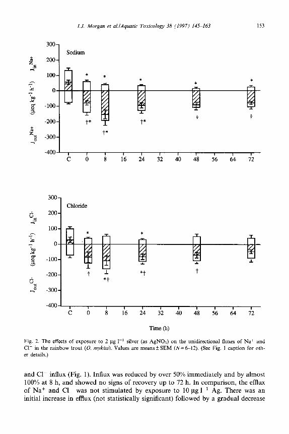
Fig. 2. The effects of exposure to 2 pg I-’ silver (as AgNOs) on the unidirectional fluxes of Na+ and Cl- in the rainbow trout (0. mykiss). Values are means k SEM (N= 6-12). (See Fig. 1 caption for oth- er details.)
and Cl- influx (Fig. 1). Influx was reduced by over 50% immediately and by almost 100% at 8 h, and showed no signs of recovery up to 72 h. In comparison, the efflux of Na+ and Cl- was not stimulated by exposure to 10 pg 1-l Ag. There was an initial increase in efflux (not statistically significant) followed by a gradual decrease

154 I.J. Morgan et al.lAquatic Toxicology 38 (1997) 145~-163
over time, so that at 72 h the Na’ and Cl- efflux was significantly lower than control rates. The net result of these changes in unidirectional fluxes was a dramatic
loss of ions from the fish (i.e. a significant negative net flux), and although this loss gradually attenuated, ionic balance did not fully recover over the period of the
exposure. Unfortunately, the terminal plasma samples from the fish exposed to
10 yg 1-l Ag were lost. However, the loss of Na+ and Cl- was estimated by taking
a time-weighted average value for the net measured Na+ or Cl- flux and multi- plying this by the total time of the exposure (Table 1). This resulted in an estimated
loss of 11 .O mmol kg-’ of Na+ and 9.6 mmol kggl of Cl-. The total exchangeable internal pools of Na+ and Cl- in rainbow trout kept in water of identical compo-
sition are approximately 42 mmol kg-’ and 33 mmol kg-l, respectively (Wood, 1988) and therefore the estimated loss of Na’ and Cl- was 26% and 29% of the
exchangeable pool, respectively. The effects of exposure to 2 pg 1-l Ag (as AgNOs) on Na+ and Cl- transport
were similar to those observed at 10 ug 1-l in that a significant inhibition of influx occurred, but of a lesser magnitude (Fig. 2). However, there was a significant
increase in Na+ and Cl- efflux over the first 24 h of this exposure. The fish exposed to 2 pg 1-l Ag had plasma Na+ and Cl- concentrations that were 15.7% and 12.4%
lower, respectively, than those of the control fish. As shown in Table 1, these measured losses were in reasonable agreement with the estimated losses (18.1%
and 15.7%~ respectively) based on branchial net ion fluxes, as above.
The effects of 2 pg I-’ Ag on branchial Na + fluxes were found to be rapidly
2Fg 1-l Ag “Recovery”
C 12 24 36 48 60 72 84 96
Time (h)
Fig. 3. Unidirectional Na+ fluxes in rainbow trout (0. mykiss) during 48 h exposure to 2 pg I-’ silver (as AgN03), and during recovery thereafter in clean freshwater. Values are means k SEM (N= 6). (See
Fig. 1 caption for other details.)
I.J. Morgan et al.lAquatic Toxicology 38 (1997) 145-163 155
600
1
OY I I I I I f
0 500 loo0 1500 zoo0 2500 3ooO
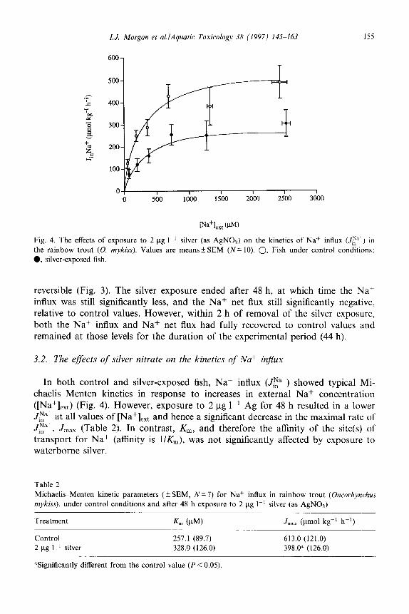
Fig. 4. The effects of exposure to 2 f.tg 1-r silver (as AgNOs) on the kinetics of Na’ influx (J,:” ) in
the rainbow trout (0. mykiss). Values are means+SEM (N= 10). 0. Fish under control conditions:
0, silver-exposed fish.
reversible (Fig. 3). The silver exposure ended after 48 h, at which time the Na+ influx was still significantly less, and the Na+ net flux still significantly negative, relative to control values. However, within 2 h of removal of the silver exposure,
both the Na+ influx and Na+ net flux had fully recovered to control values and remained at those levels for the duration of the experimental period (44 h).
3.2. The eflects of silver nitrate on the kinetics of Nat influx
In both control and silver-exposed fish, Naf influx (JE”+) showed typical Mi-
chaelis-Menten kinetics in response to increases in external Na+ concentration ([Na+],,,) (Fig. 4). However, exposure to 2 pg 1-l Ag for 48 h resulted in a lower JNa+ at all values of [Na+leXt J&+
and hence a significant decrease in the maximal rate of , .I,,, (Table 2). In contrast, K,,, and therefore the affinity of the site(s) of
&nsport for Naf (affinity is l/K,), was not significantly affected by exposure to waterborne silver.
Table 2
Michaelis-Menten kinetic parameters ( f SEM, N = 7) for Nat influx in rainbow trout (Oncorhynchus mykiss), under control conditions and after 48 h exposure to 2 pg 1-r silver (as AgNOs)
Treatment Kr, (PM)
Control 257.1 (89.7) 2 ug I-’ silver 328.0 (126.0)
*Significantly different from the control value (P < 0.05).
J,, (pm01 kgg’ hh’)
613.0 (121.0)
398.0” (126.0)

156 I. J. Morgan et al.lAyuatic Toxicology 38 11997) 145-163
2otxl B
1 lwo-
LOQO-
500-
o-
T
T
Fig. 5. The effects of 48 h exposure to 2 and IO ug 1~ ’ silver (as AgNO:s) in viva on the branchial ac-
tivity of (A) Na+/K+ ATPase and (B) carbonic anhydrase in the rainbow trout (0. mykiss). Values
are means + SEM (N = 6). ‘Significantly different from the control (P < 0.05).
.
Fig. 6. The effects of silver (as AgNOs) in vitro on the activity of branchial carbonic anhydrase (0)
and Na+/K- ATPase (0) in the rainbow trout (0. mykiss). Values are means f SEM (N= 6). In vivo
inhibition values from Fig. 5 (filled symbols) are included for comparison.

I. J. Morgan et aLlAquatic Toxicology 38 (1997) 145-163 157
3.3. The effects of silver nitrate on the activity of branchial enzymes
Although the mean activity of branchial Na+/K+ ATPase after 48 h of exposure to 2 pg 1-l Ag was approximately 50% lower than that under control conditions, this difference was not statistically significant (Fig. 5(A)). However, exposure to 10 pg 1-l Ag in vivo significantly inhibited branchial Na+/K+ ATPase activity by approximately 85%. Branchial carbonic anhydrase (CA) activity was inhibited in vivo by Ag but to a lesser degree than was Na+/K+ ATPase (Fig. 5(B)). Exposure to 2 pg 1-i Ag had no effect on CA activity, but fish exposed to 10 pg 1-l had a CA activity that was significantly lower than that of control fish by approximately 30%.
The estimated silver concentration that caused a 50% inhibition of Na+/K+ ATPase activity in vitro (150) was 670 ug ll’ (Fig. 6). The in vitro Z50 value for CA was 31 pg 1-l (Fig. 6). When AgNOs was replaced by NaNOs, no in vitro enzyme inhibition was observed (data not shown).
4. Discussion
Silver speciation analysis using the aquatic chemistry equilibrium programme MINEQL+ (Schecher and McAvoy, 1992) indicated that during exposure to 10 pg 1-l Ag (as AgNOs), three main species of silver were present: the free silver ion, Ag+ (34.5%) and two dissolved silver chloride species, AgCl, and AgC12- (60.2% and 5.2%, respectively). However, previous studies have demonstrated that silver chlorides are not toxic to freshwater fish (LeBlanc et al., 1984; Hog- strand et al., 1996), even at concentrations many times greater than those of the present experiments, and therefore the physiological effects observed during expo- sure to AgNOs in the present experiments can be attributed primarily to the free silver ion, Ag+. This concurs both with previous studies of silver toxicity in fresh- water (LeBlanc et al., 1984; Wood et al., 1996a, Wood et al., 1996b), and with work on the toxicity of other metals where the free metal ion has been found to be the most toxic form (Borgman, 1983; Luoma, 1983; Brezonik et al., 1991).
Exposure to waterborne AgNOs resulted in a severe disturbance of branchial Na+ and Cl- regulation. Fish exposed to both 2 and 10 pg 1-l Ag immediately began to lose ions to the surrounding water as a result of a severe, persistent inhibition of branchial influx rates and a lesser, temporary stimulation of branchial efflux rates. Although this loss of ions decreased over the duration of the experi- ment, this apparent ‘adaptation’ may have been due to a depletion of the exchange- able internal pools of Na+ and Cl- rather than an indication of any acclimation to silver. The fish exposed to 2 ug 1-l Ag lost 16% of their exchangeable Na+ pool over the 72 h of exposure whereas those exposed to 10 pg 1-l lost an estimated 28% of exchangeable Na+ This estimated loss is somewhat higher than that measured by Wood et al. (1996a), who observed a 19% decrease in plasma Na+ after 96 h of exposure to 10 ug 1-l Ag (as AgNOs). Nevertheless, a loss of plasma Na+ of over 30% is lethal in rainbow trout (McDonald et al., 1980) and hence a continuation of exposure to 10 pg 1-l Ag would have probably resulted in fish mortalities. Indeed,

158 I.J. Morgan et al.lAquatic Toxicology 38 (1997) 145-163
Wood et al. (1996a) reported that mortalities commenced after 6 days of exposure to 10 ug 1-r Ag, when plasma ion losses reached 30%. It can be concluded, there- fore, that disturbance of branchial ion transport is a major factor in the toxicity of
Ag+ to freshwater fish.
Sodium influx in freshwater fish is known to depend on the external (waterborne) Naf concentration in a manner well described by Michaelis-Menten kinetics (Avel-
la et al., 1987; Goss and Wood, 1990; Potts, 1994). This approach was therefore
used to analyse the nature of the inhibition of Na’ influx by Ag+. The values obtained for K,, and J,,, under control conditions in the present experiments showed good agreement with values in the literature (see Table 3 of Goss and
Wood (1990)). The significant decrease in J,,, and the lack of change in Km during silver exposure compared with the controls demonstrates that the inhibition of Naf
influx by Ag’ is non-competitive. This implies that the binding of Ag+ to a second site on the Na+ transport mechanism causes a conformational change in the specific
Na+ uptake site which is consequently rendered non-functional. There is therefore a reduction in the number of available Na+ transport sites and hence a reduction in V,,,,, whereas the affinity (l/K,,,) of the remaining functional Na+ sites is unaf-
fected . Janes and Playle (1995) demonstrated that silver binds readily to the surface of
the gills. This binding was successfully modelled on the assumption that Ag+ com- peted with other cations for negatively charged sites on the gill surface as postulated
by Pagenkopf (1983). Na+ and Cl- are thought to have separate sites and mech- anisms of uptake on the apical membrane of the gill epithelium (Evans, 1993; Goss
et al., 1995). External Na.+ is taken up in exchange for a proton (Hf) whereas external Cl- is taken up in exchange for internal bicarbonate ion (HCOs-). It is
unlikely that the external binding sites of the Cl-/HCOs- exchange mechanism would attract cations. The simultaneous inhibition of both sodium and chloride influx by silver nitrate therefore suggested that the site(s) of inhibition were not on the apical membrane of the gill epithelial cells. However, waterborne silver may cross the gill epithelium and accumulate in the blood (Wood et al., 1996a, Wood et
al., 1996b) and as the gills appeared to be the principal target organ of physiological silver toxicity, it was considered likely that the site(s) of inhibition of both Na+ and Cl- by Ag+ occurred within the cells of the branchial epithelium. Ag+ inhibits the
activity of several enzymes from fish in vitro (Jackim et al., 1970; Christensen, 1971; Christensen and Tucker, 1976) and therefore we studied the effects of silver exposure on the activity of two intracellular branchial enzymes that are involved in ion transport, Na+/K+ ATPase and carbonic anhydrase (CA).
Na+/K+ ATPase is located on the basolateral membranes of the ion-transporting cells of the branchial epithelium and actively transports Na+ from these cells into the blood (Wood, 1992; Evans, 1993). Although this enzyme is not thought to be directly involved in Cl- transport in freshwater fish, it has been shown that appli- cation of the specific Na+/K+ ATPase inhibitor, ouabain, reduces the influx of both Na+ and Cl- (Richards and Fromm, 1970; Battram et al., 1989). Carbonic anhy- drase occurs in the apical region of the gill epithelial cells, both internally and externally (Rahim et al., 1988) and catalyses the hydration of CO2 to produce

I. J. Morgan et al.lAquatic Toxicology 38 (1997) 145-163 159
H+, which may be exchanged for external Na+, and HCOs-, which may be ex- changed for external Cl-. Both Na+/K+ ATPase (Nechay and Saunders, 1984) and CA (Christensen and Tucker, 1976) have been shown to be inhibited by silver nitrate in vitro in other animals. Hence, a poisoning of one or both of these enzymes by Ag+ in vivo could result in an inhibition of Na+ and Cl- transport across the gills of rainbow trout.
Indeed, in vivo exposure to 10 pg 1-i silver for 48 h inhibited branchial Na+/K+ ATPase activity by 85% and inhibited CA activity by 28%. The greater inhibition of Na+/K+ ATPase activity suggested that a non-competitive inhibition of this enzyme was the primary cause of the ionregulatory disturbance observed during exposure to 10 ug 1-l Ag, though as CA activity was also significantly inhibited, the relative contribution of the two enzymes cannot be stated for certain. However, the Ag concentration that caused a 50% inhibition of Na+/K+ ATPase in vitro (15s) was 670 p.g 1-l (5.9 pM), much greater than the waterborne concentrations used in the present study. This implies that considerable accumulation of Ag within the gill tissues must have occurred during exposure to 10 pg 1-l Ag to cause the 85% inhibition of Na+/K+ ATPase activity that was observed. This suggestion is sup- ported by both Wood et al. (1996a) and Hogstrand et al. (1996), who recorded gill Ag concentrations of 275 ug kg-’ in 250450 g rainbow trout and 4.5 mg kg-’ in 14 g rainbow trout, respectively, after 6 days of exposure to 10 ug 1-l Ag (as
AgNOs). In contrast to Na+/K+ ATPase, the inhibition of CA by Ag in vitro was very
similar to that in vivo, which does not appear to support the above conclusion that Ag accumulated in the gill tissue during exposure to waterborne silver nitrate. This apparent anomaly may be explained, however, by the exact technique used to assay branchial CA. The gill tissue of rainbow trout has a very high activity of CA and hence the slopes of the assay curves used in the delta pH method (Henry, 1991) were very steep, which tended to magnify small differences in the raw data. To overcome this problem, it was necessary to dilute the gill homogenates several-fold. This had no effect on the silver concentration present in vitro, as the AgN03 was added post-dilution. However, in the in vivo assays the only silver present was that from the waterborne exposures, and so dilution of the homogenates could have reduced the actual silver concentration. The inhibition of CA recorded in vivo may therefore have been an underestimate of the actual inhibition, particularly if Ag is only a weak inhibitor of CA. The rapid recovery of Na+ fluxes when the silver exposure was removed indeed suggests that the binding of silver to the Na+/K+ ATPase and/or CA was weak and that the inhibition was reversible.
In comparison with other waterborne toxicants, the pattern of osmoregulatory disturbance in freshwater fish owing to Ag+, i.e. a reduction of branchial Na+ and Cl- influx, shows most similarity to that seen in fish exposed to environmental acidification (Hf) and to waterborne copper. In contrast, exposure to aluminium, cadmium or zinc causes only moderate disturbance to Naf and Cl- regulation at levels of water hardness and pH comparable with those of the present study (Wood, 1992; Heath, 1995). Environmental acidification causes a simultaneous inhibition of both Na+ and Cl- influx and a stimulation of Na+ and Cl- effluxes, which result in

160 I.J. Morgun et ul./Ayuuric Toxicology 38 (1997) 145 163
a decrease in plasma Na+ and Cl- (Wood, 1989). Plasma K+ and Ca”+ concen- trations, however, are unaffected by H+ (McDonald et al., 1980; Audet et al., 1988). This is precisely the pattern of plasma ion concentration changes that was
observed by Wood et al. (1996a) when rainbow trout were exposed to 10 l.tg 1-l Ag, and indeed Wood et al. (1996a) suggested that Ag+ acts as an apical membrane blocker of ion transport sites, a mechanism that is generally accepted for H+
toxicity (Wood, 1989). However, the present study suggests that Ag+ toxicity acts
internally within the gills, primarily by inhibition of branchial Na+/K+ ATPase and carbonic anhydrase. Low pH alone does not appear to appreciably inhibit Na+/K+
ATPase (Reite and Staurnes, 1987; Audet and Wood, 1993). The only studies to
observe lower Na+/K+ ATPase activities in acid-exposed fish were in smolting Atlantic salmon, Sulmo salur (Saunders et al., 1983; Johnston et al., 1984; Staurnes
et al., 1993), which have high ATPase activities in preparation for migration to seawater and are not comparable with the fish used in the present study. We there- fore conclude that the mechanisms of Ag+ toxicity are not the same as those of H+.
Copper also inhibits branchial Na+ influx (Lauren and McDonald, 1985; Reid and McDonald, 1988) and like Ag+, but in contrast to H+, has been clearly shown to inhibit branchial Na+/K+ ATPase (Lorz and McPherson, 1976; Lauren and
McDonald, 1987). However, copper is also an inhibitor of branchial Ca2+ ATPase (Shephard and Simkiss, 1978) and Ca2+ fluxes (Sayer et al., 1991) and causes a
decrease in plasma Ca” concentration (Pilgaard et al., 1994), which was not seen
during Ag+ exposure (Wood et al., 1996a). Therefore, although the physiological mechanisms of Ag+ toxicity share some features with both H+ and copper, there are also some important differences such that the full pattern of physiological
toxicity seems to be characteristic of Ag+ alone. The present definition of the physiological mechanisms of silver nitrate toxicity in
freshwater rainbow trout provides a well-defined biological response that may be
used to detect the presence of bioavailable Ag-+ under various conditions of water quality. Traditionally, [Ag+] has been measured by physico-chemical methods such
as silver electrodes and anodic stripping voltammetry, or has been calculated using computer programs such as MINEQL +. These methods provide only indirect ap-
proximations of the biological impact of the measured Ag+. Measurement of phys- iological (branchial Na+ influx) or biochemical (branchial Na’lK’ ATPase activity) parameters, however, gives a direct and immediate indication of a biological impact,
which is the important factor from a regulatory standpoint.
Acknowledgements
We thank Dr. Daland Juberg, Dr. Bob Cappel, Dr. Joe Gorsuch, Dr. Ken Robillard, Tom Bober of Eastman Kodak, and Tom Dufficy of the National Asso- ciation of Photographic Manufacturers (NAPM) for valuable input and liaison. This work was supported by a grant from the NAPM/Silver Coalition to C.M.W.

I.J. Morgan et aLlAquatic Toxicology 38 (1997) 145-163 161
References
Audet, C. and Wood, C.M., 1993. Branchial morphological and endocrine responses of rainbow trout
(Oncorhynchus mykiss) to a long-term sublethal acid exposure in which acclimation did not occur. Can.
J. Fish. Aquat. Sci., 50: 198-209.
Audet, C., Munger, RX and Wood, C.M., 1988. Long-term sublethal acid exposure in rainbow trout
(Salmo gairdneri) in soft water: effects on ion exchanges and blood chemistry. Can. J. Fish. Aquat.
Sci., 45: 1387-1398.
Avella, M., Masoni, A., Bornancin, M. and Mayer-Gostan, N., 1987. Gill morphology and sodium influx
in the rainbow trout (Salmo gairdneri) acclimated to artificial freshwater environments. J. Exp. Zool.,
241: 159-169.
Battram, J.C., Eddy, F.B., Chang, Y.J. and Fidler, J., 1989. Chloride transport by isolated gill cells of the
freshwater adapted rainbow trout (Salmo gairdnert]. Comp. Biochem. Physiol. A, 94: 439445.
Borgman, U., 1983. Metal speciation and toxicity of free metal ions to aquatic biota. In: J.O. Nriagu
(Editor), Aquatic Toxicology. Wiley, New York, pp. 47-72.
Brezonik, P.L., King, S.O. and Mach, C.E., 1991. The influence of water chemistry on trace metal
bioavailability and toxicity to aquatic organisms. In: M.C. Newman and A.W. McIntosh (Editors),
Metal Exotoxicology: Concepts and Applications. Lewis, Chelsea, MI, pp. l-31.
Buccafusco, R.J., Ells, S.J. and LeBlanc, G.A., 1981. Acute toxicity of priority pollutants to bluegill
(Lepomis macrochirus). Bull. Environ. Contam. Toxicol., 26: 446452.
Christensen, G.M., 1971, Effects of metal cations and other chemicals upon in vitro activity of two
enzymes in the blood plasma of white sucker, Catostomus commersoni (La&p&de). Chem. Biol Inter-
actions, 4: 351-361.
Christensen, G.M. and Tucker, J.H., 1976. Effects of selected water toxicants on the in vitro activity of
fish carbonic anhydrase. Chem. Biol. Interactions, 13: 181-192.
Davies, P.H., Goettl, Jr., J.P. and Sinley, J.R., 1978. Toxicity of silver to rainbow trout (Salmo gairdneri). Water Res., 12: 113-117.
Evans, D.H., 1993. Osmotic and ionic regulation. In: D.H. Evans (Editor), The Physiology of Fishes.
CRC Press, Boca Raton, FL, pp. 315-341.
Goss, G.G. and Wood, C.M., 1990. Na+ and Cl- uptake kinetics, diffusive effluxes and acidic equivalent
fluxes across the gills of rainbow trout. I. Responses to environmental hypoxia. J. Exp. Biol., 152: 521l
547.
Goss, G-G., Perry, S. and Laurent, P., 1995. Ultrastructural and morphometric studies on ion and acid--
base transport processes in freshwater fish. In: C.M. Wood and T.J. Shuttleworth (Editors), Cellular
and Molecular Approaches to Fish Ionic Regulation. Academic Press, San Diego, CA, pp. 257-
284.
Heath, A.G., 1995. Water Pollution and Fish Physiology, 2nd edn. Lewis, Boca Raton, FL, 359 pp.
Henry, R.P., 1991. Techniques for measuring carbonic anhydrase activity in vitro. The electrometric delta
pH and pH stat methods. In: S.J. Dodgson, R.E. Tashian, G. Gros and N.D. Carter (Editors), The
Carbonic Anhydrases. Plenum, New York, pp. 119~125.
Hogstrand, C., Galvez, F. and Wood, CM., 1996. Toxicity, silver accumulation and metallothionein
induction in freshwater rainbow trout during exposure to different silver salts. Environ. Toxicol.
Chem., 15: 1102-l 108.
Holliday, C.W., 1985. Salinity-induced changes in gill Na,K-ATPase activity in the mud fiddler crab, Uca pugnax. J. Exp. Zool., 233: 199-208.
Jackim, E., Hamlin, J.M. and Sonis, S., 1970. Effects of metal poisoning on five liver enzymes in the
killifish (Fundulus heteroclitus). J. Fish. Res. Board Can., 27: 383-390.
Janes, N. and Playle, R.C., 1995. Modeling silver binding to gills of rainbow trout (Oncorhynchus mykiss). Environ. Toxicol. Chem., 14: 1847-l 858.
Johnston, C.E., Saunders, R.L., Henderson, E.B., Harman, P.R. and Davidson, K., 1984. Chronic effects
of low pH on some physiological aspects of smoltification in Atlantic salmon (Salmo salw). Can. Tech.
Rep. Fish. Aquat. Sci., 1294, 7 pp.
Lauren, D.J. and McDonald, D.G., 1985. Effects of copper on branchial ionoregulation in the rainbow

162 I.J. Morgan et al,lAyuatic Toxicology 38 (1997) 145-163
trout, Salmo guirdneri Richardson. Modulation by waterhardness and pH. J. Comp. Physiol. 9, 155:
6355644.
Lauren, D.J. and McDonald, D.G., 1987. Acclimation to copper by rainbow trout; physiology. Can. J.
Fish. Aquat. Sci., 44: 999104.
LeBlanc, G.A., Mastone, J.D., Paradice, A.P., Wilson, B.P., Lockhart, Jr., H.B. and Robillard, K.A.,
1984. The influence of speciation on the toxicity of silver to fathead minnow (Pimephales promelas). Environ. Toxicol. Chem., 3: 3746.
Lorz, H.W. and McPherson, B.P., 1976. Effects of copper or zinc in fresh water on the adaptation to
seawater and ATPase activity, and the effects of copper on migratory disposition of coho salmon
(Oncorhynchus kisutch). J. Fish. Res. Board Can., 33: 202332030.
Lowry, O.H., Rosebrough, N.J., Farr, A.L. and Randall, R.J., 1951. Protein measurement with the Folin
phenol reagent. J. Biol. Chem., 193: 265.-275.
Luoma, S.N., 1983. Bioavailability of trace metals to aquatic organisms-a review. Sci. Total Environ.,
28: l-22.
Maetz. J., 1956. Les echanges de sodium chez les poissons Carassius aura&r L. Action d’un inhibiteur de
I’anhydrase carbonique. Physiologic, 48: 108551099.
McDonald, D.G., Hobe, H. and Wood, C.M., 1980. The influence of calcium on the physiological
responses of the rainbow trout Salmo gairdneri, to low environmental pH. J. Exp. Biol., 88: 1099131.
Nebeker, A.V., McAuliffe, C.K., Mshar, R. and Stevens, C.K., 1983. Toxicity of silver to steelhead and
rainbow trout, fathead minnows and Duphniu magna. Environ. Toxicol. Chem., 2: 95-104.
Nechay, B.R. and Saunders, J.P., 1984. Inhibition of adenosine triphosphatases in vitro by silver nitrate
and silver sulfadiazine. J. Am. Colt. Toxicol., 3: 37-42.
Pagenkopf, G.K., 1983. Gill surface interaction model for trace metal toxicity to fishes: role of complex-
ation, pH and water hardness. Environ. Sci. Technol., 17: 3422347.
Perry, S.F., Daxboeck, C., Ellis, A.G. and Smith, D.J., 1984. Perfusion methods for the study of gill
physiology. In: W.S. Hoar and D.J. Randall (Editors), Fish Physiology, Vol. X9. Academic Press,
New York, pp. 3255388.
Pilgaard, L., Malte, H. and Jensen, F.B., 1994. Physiological effects and tissue accumulation of copper in
freshwater rainbow trout (Oncorhynchus mykiss) under normoxic and hypoxic conditions. Aquat.
Toxicol., 29: 197. 212.
Potts, W.T.W., 1994. Kinetics of sodium uptake in freshwater animals: a comparison of ion-exchange
and proton pump hypotheses. Am. .I. Physiol., 266: R315 -R320.
Rahim, S.M., Delaunoy. J-P. and Laurent, P., 1988. Identification and immunocytochemical localization
of two different carbonic anhydrase isoenzymes in teleostean fish erythrocytes and gill epithelia. His-
tochemistry, 89: 451459.
Reid, S.D. and McDonald, D.G.. 1988. Effects of cadmium, copper and low pH on ion fluxes in the
rainbow trout Salmo gairdneri. Can. J. Fish. Aquat. Sci., 45: 244253.
Reite, O.B. and Staumes, M., 1987. Acidified water: effects on physiological mechanisms in the gills of
salmonids. In: Surface Water Acidification Programme, Midterm Review Conference, Bergen, Nor-
way, pp. 298-304.
Richards, B.D. and Fromm, P.O., 1970. Sodium uptake by isolated-perfused gills of rainbow trout
(Salmo gairdneri). Comp. Biochem. Physiol., 33: 303-310.
Saunders, R.L., Henderson, E.B. and Harmon, P.R.. 1983. Effects of low environmental pH on smolting of Atlantic salmon (Salmo sakar). Can. J. Fish. Aquat. Sci.. 40: 120331211.
Sayer, M.D.J.. Reader, J.P. and Morris, R.. 1991. Effects of six trace metals on calcium fluxes in brown
trout (Sulmo trutta L.) in soft water. J. Comp. Physiol. B, 161: 5377542.
Schecher, W.D. and McAvoy, D.C., 1992. MINEQL’; a software environment for chemical equilibrium
modelling. Comput. Environ. Urban Syst., 16: 65-76.
Schildtkraut. D.E., Davis, A.T., Robillard, K.A. and Twist, J.P., 1995. Measurements of active silver in
environmental samples by anodic stripping voltammetry: towards development of a robust analytical
method. In: A.W. Andren (Editor), Argentum II. Proc. 2nd Int. Conf. on the Transport, Fate and
Effects of Silver in the Environment, 669 August 1995. University of Wisconsin Press, Madison,
pp. 185. 193.

I.J. Morgan et al.lAquatic Toxicology 38 (1997) 145-163 163
Shephard, K. and Simkiss, K., 1978. The effects of heavy metal ions on Ca*+ ATPase extracted from fish
gills. Comp. Biochem. Physiol. B, 61: 69-72.
Staumes, M., Blix, P. and Reite, O.B., 1993. Effects of acid water and aluminum on Parr-smolt trans-
formation and seawater tolerance in Atlantic salmon, Salmo salar. Can. J. Fish. Aquat. Sci., 50: 1816
1827.
Warrington, P.D., 1995. Ambient water quality criteria for silver. Draft document. Water Quality
Branch, Environmental Protection Department, Ministry of Environment, Lands and Parks, Province
of British Columbia, Vancouver, 169 pp.
Wood, C.M., 1988. Acid-base and ionic exchanges at gills and kidney after exhaustive exercise in the
rainbow trout. J. Exp. Biol., 136: 461481.
Wood, C.M., 1989. The physiological problems of fish in acid waters. In: R. Morris, E.W. Taylor, D.J.A.
Brown and J.A. Brown (Editors), Acid Toxicity and Aquatic Animals. Cambridge University Press,
Cambridge, 282 pp.
Wood, CM., 1992. Flux measurements as indices of H+ and metal effects on freshwater fish. Aquat.
Toxicol., 22: 239-264.
Wood, C.M., Hogstrand, C., Galvez, F. and Munger, R.S., 1996a. The physiology of waterborne silver
toxicity in freshwater rainbow trout (Oncorhynchus mykim) I. The effects of ionic Ag+. Aquat. Tox-
icol., 35: 93-109.
Wood, CM., Hogstrand, C., Galvez, F. and Munger, R.S., 1996b. The physiology of waterborne silver
toxicity in freshwater rainbow trout (Oncorhynchus mykiss). II. The effects of silver thiosulphate.
Aquat. Toxicol., 35: 11 l-125.
Zall, D.M., Fisher, M.D. and Garner, Q.M., 1956. Photometric determination of chlorides in water.
Anal. Chem.. 28: 166551678.