the mechanical role of the cervix in pregnancy - loci.wisc.edu mechanical role of the cervix... ·...
TRANSCRIPT

The mechanical role of the cervix in pregnancy
Kristin M. Myers a,n, Helen Feltovich b,c, Edoardo Mazza d, Joy Vink e, Michael Bajka f,Ronald J. Wapner e, Timothy J. Hall c, Michael House g
a Department of Mechanical Engineering, Columbia University, New York, NY, USAb Department of Obstetrics and Gynecology, Intermountain Healthcare, Provo, UT, USAc Department of Medical Physics, University of Wisconsin, Madison, WI, USAd Department of Mechanical and Process Engineering, ETH Zurich, & EMPA Dübendorf, Switzerlande Department of Obstetrics and Gynecology, Columbia University Medical Center, New York, NY USAf Department of Obstetrics and Gynecology, University Hospital of Zurich, Switzerlandg Department of Obstetrics and Gynecology, Tufts Medical Center, Boston, MA, USA
a r t i c l e i n f o
Article history:Accepted 28 February 2015
Keywords:CervixPregnancyPreterm birth
a b s t r a c t
Appropriate mechanical function of the uterine cervix is critical for maintaining a pregnancy to term sothat the fetus can develop fully. At the end of pregnancy, however, the cervix must allow delivery, whichrequires it to markedly soften, shorten and dilate. There are multiple pathways to spontaneous pretermbirth, the leading global cause of death in children less than 5 years old, but all culminate in prematurecervical change, because that is the last step in the final common pathway to delivery. The mechanismsunderlying premature cervical change in pregnancy are poorly understood, and therefore current clinicalprotocols to assess preterm birth risk are limited to surrogate markers of mechanical function, such assonographically measured cervical length. This is what motivates us to study the cervix, for which wepropose investigating clinical cervical function in parallel with a quantitative engineering evaluation ofits structural function. We aspire to develop a common translational language, as well as generate arigorous integrated clinical-engineering framework for assessing cervical mechanical function at thecellular to organ level. In this review, we embark on that challenge by describing the current landscapeof clinical, biochemical, and engineering concepts associated with the mechanical function of the cervixduring pregnancy. Our goal is to use this common platform to inspire novel approaches to delineatenormal and abnormal cervical function in pregnancy.
& 2015 Published by Elsevier Ltd.
1. Introduction
The cervix is contiguous with the lower part of the uterus. Itsproximal portion is located in the abdomen and its distal portionin the vagina. It has a narrow central canal which runs along itsentire length, connecting the uterine cavity and the lumen of thevagina. The opening of this canal into the uterus is called theinternal os and the opening into the vagina the external os(Fig. 1A). During pregnancy, the primary biomechanical functionof the cervix is to maintain the fetus within the uterus. Thisrequires withstanding multiple forces from the uterus, includingthe weight of the growing fetus and amniotic sac, as well aspassive pressure from the uterine wall. Then, in a dramaticreversal of roles, the cervix markedly softens, shortens and dilatesto allow delivery of the fetus. Shortly after delivery, the cervix
reforms into its previous shape and consistency. How the cervixmanages these complex dynamic changes is an interesting andunderstudied biomechanics problem.
Critical problems can occur when the timing and extent of thebiomechanical changes are altered. Specifically, premature soft-ening, shortening and dilation, which may be considered earlymechanical failure, occurs in cases of spontaneous preterm birth(sPTB). The underlying pathophysiology of these changes is poorlyunderstood despite that preterm birth affects 15 million babiesannually, is the leading cause of childhood (o5 years old) death,and in 2013 was responsible for 1 million deaths (World HealthOrganization, 2014). The rate of preterm birth has signific-antly decreased by 2 decades of intense research effort into itspathophysiologies and associated molecular mechanisms. Webelieve that this lack of progress is partly due to lack of crosstalkbetween clinicians, engineers and basic scientists, and that pro-gress will require multidisciplinary collaboration between pre-viously distinct areas of expertise such as clinical obstetrics andengineering.
Contents lists available at ScienceDirect
journal homepage: www.elsevier.com/locate/jbiomechwww.JBiomech.com
Journal of Biomechanics
http://dx.doi.org/10.1016/j.jbiomech.2015.02.0650021-9290/& 2015 Published by Elsevier Ltd.
n Corresponding author.E-mail address: [email protected] (K.M. Myers).
Please cite this article as: Myers, K.M., et al., The mechanical role of the cervix in pregnancy. Journal of Biomechanics (2015), http://dx.doi.org/10.1016/j.jbiomech.2015.02.065i
Journal of Biomechanics ∎ (∎∎∎∎) ∎∎∎–∎∎∎
To begin that dialogue, here we provide an engineering frame-work for studying the mechanical function of the cervix duringpregnancy. The main steps include modeling the material behaviorof the cervix, characterizing the pelvic anatomy, capturing theappropriate contact conditions between the pelvic soft tissues, andunderstanding the relevant loading and boundary conditions.Accomplishing these tasks are a challenge because, in addition tounderstanding the basic material and anatomical parameters, onemust consider the changes that occur throughout pregnancy toaccommodate the growth and ultimate delivery of the fetus.However, overcoming these engineering challenges could lead toa better understanding of normal and pathological cervicaldeformation.
2. The clinical problem of preterm birth
2.1. Scope of the problem
Preterm birth is defined as delivery between 20 weeks and 36weeksþ6 days gestation. Medically indicated preterm birth mayresult from maternal factors (e.g. preeclampsia or placenta previa)or fetal factors (e.g. oligohydramnios or growth restriction).Spontaneous preterm birth (sPTB) was formerly divided into twogeneral categories, namely cervical dysfunction (cervical insuffi-ciency or cervical incompetence) or preterm labor (typically thoughtto be the result of intrauterine infection or bleeding). A morecurrent understanding is that sPTB from all causes can be seenas part of an extremely complex continuum involving multiplephenotypes (Barros et al., 2015; Solomon and Iams, 2014). Theetiology of sPTB is multifactorial, involving diverse precipitatingfactors such as infection and inflammation, bleeding, poor nutri-tion, demographics, stress, ethnicity and race, genetic predisposi-tions and many others, all presumably with individual, and over-lapping, molecular mechanisms (Gravett et al., 2010). A recentattempt to categorize phenotypes of preterm birth showed thatapproximately 25% of these births are neither medically indicatednor associated with any known phenotype (Barros et al., 2015).
Organizations such as the March of Dimes have recentlycelebrated a decline in the preterm birth rate (from 12.8% in2006 to 11.4% by 2013), but data from the Centers for DiseaseControl shows that the sPTB rate in 2012 was nearly identical tothat in 1997 (Martin et al., 2013; Schoen et al., in press; Solomonand Iams, 2014). Strategies to address known risk factors (e.g.genitourinary infection and poor nutrition) have been ineffective,as have drug therapies targeted against uterine contractions,infection, or inflammation (Gravett et al., 2010; Solomon andIams, 2014). The American College of Obstetricians and Gynecol-ogists and the Society for Maternal-Fetal Medicine promoteintramuscular progesterone treatment in patients with a history
of sPTB, and vaginal progesterone supplementation or cerclage (asuture tied around the cervix) in patients with a short cervix in thecurrent pregnancy (American College of Obstetricians andGynecologists, 2012; Society for Maternal-Fetal Medicine Publica-tions Committee, 2012). Importantly however, the decline inpreterm birth has been attributed primarily to provider education,which has resulted in fewer nonmedically indicated deliveries o39 weeks, fewer teenage pregnancies, less smoking in pregnancy,and fewer twin and triplet pregnancies (Schoen et al., in press;Solomon and Iams, 2014). Progesterone supplementation andcerclage in selected patients are “probably contributing” accordingto a 2015 review (Schoen et al., in press), but this is obviouslyunclear, as are the mechanisms by which these treatments work,which must contribute to the reason current interventions that areineffective in the vast majority of patients (American College ofObstetricians and Gynecologists, 2012; Conde-Agudelo et al., 2013;Grobman et al., 2012).
As stated recently by Norman and Shennan, the fact that 95% ofsPTB is intractable to current therapies suggests that substantialfurther research is needed (Norman and Shennan, 2013). An und-erstanding of the molecular mechanisms of the multiple pathwaysto sPTB is essential to the development of etiologic- and patient-specific interventions for patients at high risk and avoidance ofunnecessary and potentially detrimental treatment in those at lowrisk (Feltovich et al., 2012; Iams and Berghella, 2010). We think thecervix is the logical place to start this investigation becausecervical ripening is the last step before labor and delivery in thesPTB pathway, the final common denominator of a multitude ofoverlapping etiologies (Gracie et al., 2011; Gravett et al., 2010).
2.2. The role of the cervix in PTB and the importance of cervicalremodeling
Clinicians use terms such as softening, shortening, funneling,effacing, and dilating to describe the cervical deformation thatoccurs during pregnancy (Fig. 2). Collectively, these changes arecalled cervical remodeling and refer to both the tissue's intrinsicmaterial property changes and its resultant anatomical changes.Mouse models demonstrate a distinct separation between an earlyphase of remodeling that starts soon after conception and con-tinues through delivery (cervical softening) and a later phase, neardelivery, that involves rapid, marked softening and shortening(cervical ripening) (Akgul et al., 2012; Holt et al., 2011; Mahendroo,2012; Read et al., 2007; Timmons et al., 2010a; Word et al., 2007).This process is not yet well characterized in human pregnancy. Asin the mouse, cervical softening in human pregnancy begins early,a fact that was exploited to detect pregnancy as early as 6 weeks ofgestation in the 19th century before diagnostic blood and urinetests were developed. Recent in vivo mechanical interrogation ofthe cervix (Badir et al., 2013a; Parra-Saavedra et al., 2011) supports
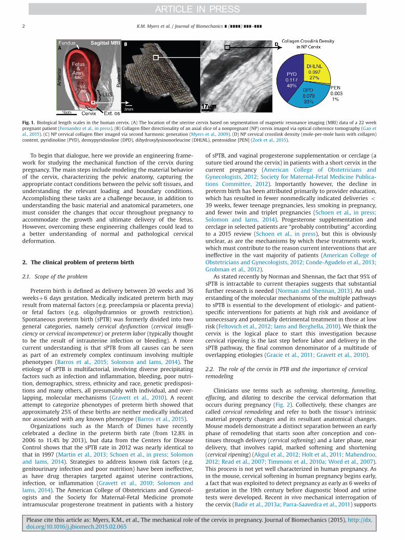
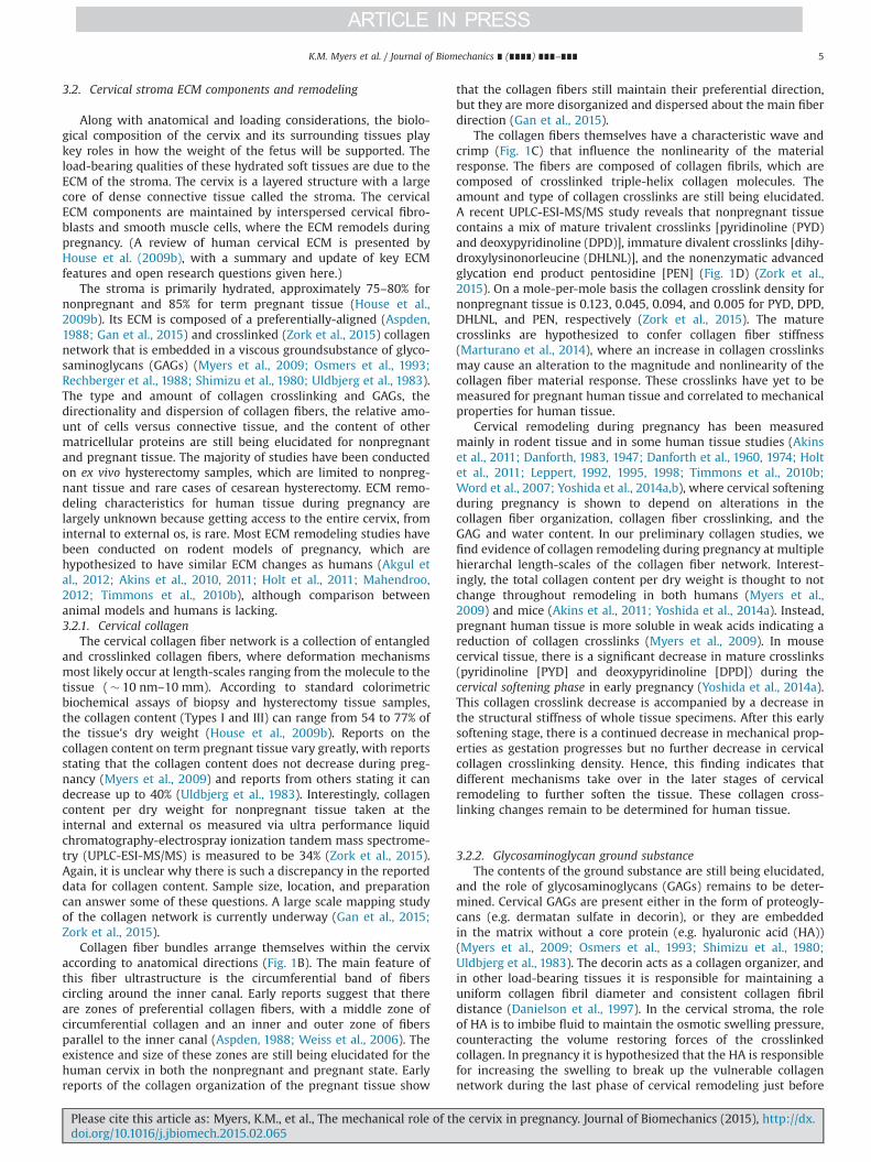
Fig. 1. Biological length scales in the human cervix. (A) The location of the uterine cervix based on segmentation of magnetic resonance imaging (MRI) data of a 22 weekpregnant patient (Fernandez et al., in press). (B) Collagen fiber directionality of an axial slice of a nonpregnant (NP) cervix imaged via optical coherence tomography (Gan etal., 2015). (C) NP cervical collagen fiber imaged via second harmonic generation (Myers et al., 2009). (D) NP cervical crosslink density (mole-per-mole basis with collagen)content, pyridinoline (PYD), deoxypyridinoline (DPD), dihydroxylysinonorleucine (DHLNL), pentosidine [PEN] (Zork et al., 2015).
K.M. Myers et al. / Journal of Biomechanics ∎ (∎∎∎∎) ∎∎∎–∎∎∎2
Please cite this article as: Myers, K.M., et al., The mechanical role of the cervix in pregnancy. Journal of Biomechanics (2015), http://dx.doi.org/10.1016/j.jbiomech.2015.02.065i

the clinical finding of early, progressive cervical softening (seeSection 4.2.2). Starting in mid-pregnancy, the normal humancervix begins to shorten until delivery, confirmed by longitudinalstudy of ultrasound cervical length (Iams et al., 1996). In addition,there is a clear relationship between cervical softening and theorganization and composition of its extracellular matrix (ECM);specifically, cervical softening and shortening relate to dysfunc-tional ECM remodeling (House et al., 2009b).
Despite this, clinical assessment of softening remains entirelysubjective; the clinician describes the cervix as soft, medium, orfirm (Bishop, 1964). Compounding the matter, they often use theterms softening and ripening and remodeling interchangeably. Thisimprecision obscures the clinician's ability to effectively describe apatient's clinical findings to another clinician, let alone someoneoutside of the obstetric profession. To the engineer, cervical soft-ness is a material property that must be defined with appropriateconstitutive equations and cervical shortening is a tissue deforma-tion that results from an evolving three-dimensional (3D) stressstate and the intrinsic remodeling of the materials constituents(Fernandez et al., in press; House et al., 2012, 2013; House andSocrate, 2006; Paskaleva, 2007). However, the engineer currentlyhas no effective means to apply these definitions to the clinicalsituation because of the language mismatch between cliniciansand basic scientists. This obscures the search for specific markersof possible pathogenic processes, such as abnormal tissue materialproperty changes or abnormal anatomical considerations. In otherwords, a common language seems fundamental to progresstoward understanding the problem of spontaneous preterm birth.
3. The multi-scale mechanical environment of pregnancy
Studying and characterizing reproductive organs in real-timethroughout gestation is understandably challenging. Pregnancy isa protected environment and accessing organs to measure eithergeometry or material properties during this time is difficult. Thereis a wide range of biologic length scales that determine thestructural response of the reproductive organs during pregnancy(Fig. 1A). These factors include features of the pelvic anatomy andthe hierarchal material characteristics of the cervix and itssurrounding tissues, from the collagen fibrils (" 10–500 nm)bundling together to form collagen fibers (" 1–500 μm, Fig. 1C)and the collagen fibers bundling together in a preferred anatomic
direction to form an overall tissue ultrastructure (" 1–10 mm,Fig. 1B).
Insight into the physiologic loads experienced during preg-nancy and the load-carrying capability of the cervix have beenderived from finite element models (Fernandez et al., in press;House et al., 2012, 2013; Mahmoud et al., 2013; Paskaleva, 2007),mechanical and biochemical studies of ex vivo tissue specimens(Conrad et al., 1980; Conrad and Ueland, 1976, 1979; Fernandezet al., 2013; Gan et al., 2015; Myers et al., 2008, 2010; Oxlund et al.,2010a,b; Petersen et al., 1991; Rechberger et al., 1988; Yao et al.,2014), in vivo mechanical and biochemical interrogations of thecervix (Badir et al., 2013a; Bauer et al., 2007; Feltovich et al., 2010,2012; Feltovich and Hall, 2013; Hee et al., 2014; House et al., 2005,2009; Hricak et al., 1990; Maldjian et al., 1999; Mazza et al., 2006,2013; Parra-Saavedra et al., 2011), and theoretical mechanics (Liaoet al., 2014; Myers and Ateshian, 2014; Paskaleva, 2007). At thepresent time, there is no single set of correlating geometric andmaterial property data from a single pregnant patient throughoutgestation.
3.1. Anatomy of the pregnant pelvis organ and physiologic loading
Knowledge of the 3D anatomy of the cervix and surroundingstructures is essential for a comprehensive understanding of thebiomechanical mechanisms leading to clinically-observed cervicalshortening (House et al., 2013). A short cervix, associated with anincreased risk of sPTB, is clinically defined as less than 25 mm(To et al., 2001). An MRI study of the 3D geometry of the pelvisillustrates the diverse range of the volumetric dimensions of theuterus and the cervix (House et al., 2009). The cervix is generally3 cm long and 2.5 cm in diameter, although these dimensions varyconsiderably between patients depending on age, number ofprevious births, and the menstrual cycle (Bauer et al., 2007). Thecervix and upper vagina are supported in the pelvis by the cardinaland uterosacral ligaments, which maintain the position of thecervix during uterine growth (Ramanah et al., 2012). Within theuterus, the fetal membranes are in direct contact with the superiorsurface of the cervix at the internal os. (Fig. 1A)
Both magnetic resonance imaging (MRI) and ultrasound (US)imaging studies suggest that the anatomical structure most esse-ntial to normal cervical function is the internal os. Sonographic imagesof the cervix reveal that cervical shortening invariably begins at theinternal os (Zilianti et al., 1995), where the cervix starts to dilate,leading to a funneled cervical canal (Fig. 2). Early finite element
Fig. 2. Cervical deformation patterns and clinical definitions: Cervical length is clinically measured as the portion of the cervix that is closed. Effacement progresses innormal pregnancy when the fetal head descends and shortens the cervix. Funneling is a pathologic condition related to an abnormal cervical deformation pattern when themembranes slip into the inner canal and the cervix prematurely shortens.
K.M. Myers et al. / Journal of Biomechanics ∎ (∎∎∎∎) ∎∎∎–∎∎∎ 3
Please cite this article as: Myers, K.M., et al., The mechanical role of the cervix in pregnancy. Journal of Biomechanics (2015), http://dx.doi.org/10.1016/j.jbiomech.2015.02.065i

analysis (FEA) demonstrate this clinically-observed deformation pat-tern showing that the internal os opposes the lateral pull of theuterine wall and the hydrostatic forces of the uterine cavity(Paskaleva, 2007; House et al., 2012, 2013; Fernandez et al., inpress). It is viewed that the internal os dilates first because tissuestresses are higher there compared with the external os (Houseet al., 2013), and the external os is not significantly loaded untilthe cervix has significantly shortened (Fernandez et al., in press).Building FEA models is difficult because of the complexity ofpregnancy-related anatomy, incomplete data on tissue propertiesand the difficulty in defining appropriate boundary conditionsand contact. A full validation of these FEA models are still needed,but hypotheses can still be made about the key mechanicalfactors that influence the load-carrying capability of the cervixduring pregnancy. These factors include: cervical material prop-erties, cervical geometry, fetal membrane properties and adhe-sion, static loading, and uterine contractions.
Cervical material properties: The load-bearing region of thecervix is its dense collagen-rich core, called the cervical stroma(see Section 3.2). Its mechanical load bearing properties arise fromthe hierarchal organization of its collagen network. The materialresponse of the cervical tissue to loading is nonlinear, anisotropic,and time-dependent (see Section 4). During pregnancy, the stromaundergoes a complex growth and remodeling process that isincompletely understood (Myers and Ateshian, 2014). The out-come of cervical remodeling is tissue that is orders of magnitudesofter during pregnancy compared with nonpregnant tissue(Table 1) (Conrad and Ueland, 1976; Myers et al., 2008, 2010;Rechberger et al., 1988; Yao et al., 2014). Premature softening isassociated with preterm shortening and sPTB, but it is not clearhow these material characteristics of the cervix influence itsmechanical function under in vivo loading conditions.
Cervical geometry: The 3D geometry of the cervix and theuterocervical angle has an important effect on the load distribu-tion and stretch pattern within the internal os (Fernandez et al., inpress; Paskaleva, 2007). Large anatomical changes occur with fetalgrowth and these changes are associated with changes in cervicallength, cervical volume, and uterocervical angle (House et al.,2009). A better understanding of relationships between anatomi-cal geometry and cervical loading has direct clinical relevance. For
example, modification of the uterocervical angle was postulated tobe the mechanism of PTB prevention for the Arabin pessary inrecent clinical trials (Goya et al., 2012).
Fetal membrane material properties and adhesion to the lower uterinesegment: An under-appreciated factor that influences the amount ofstress in the cervical tissue is the fetal membrane. The degree ofadhesion between the fetal membrane and lower uterine segment andthe material properties of the fetal membrane influence the amount ofload that is placed on the cervix (Fernandez et al., in press; Paskaleva,2007). Simulation reveals that load sharing occurs between themembrane and cervix when the interface is intact (Fernandez et al.,in press; Paskaleva, 2007) and when the stiffness of the fetalmembrane is high (Fernandez et al., in press). When the adhesion isdisrupted or if the fetal membrane stiffness decreases, cervical stressesare increased. We expect a better understanding of the mechanicalrole of the fetal membrane will help clarify (1) how the cervixshortens in response to fundal pressure (the dynamic cervix) and(2) the mechanics of membrane prolapse, which is observed in casesof premature cervical shortening.
Static loading: In the absence of the uterine contractions, thecervix is loaded by intrauterine pressure and gravity. In addition,growth of the amniotic sac results in tensile stresses from theuterine wall. These forces also depend on the support action ofpelvic floor structures and abdominal wall. We hypothesize thatstatic loading through a combination of uterine growth, hydro-static pressure and gravity are the dominant loads that causecervical shortening. Most patients with cervical shortening showno clinical signs of preterm uterine contractions. A better under-standing of these static loads is critically needed for a betterunderstanding of cervical deformation.
Uterine contractions: Although uterine contractility is clearlyimportant for preterm cervical changes, many patients withpreterm uterine contractions do not develop cervical shortening.In contrast, cervical shortening is often seen when preterm contr-actions are not prominent. An explanation for these contrastingobservations is lacking and highlights a significant knowledge gap.An important goal of future efforts will be study the relativecontributions of static loading and uterine contractility and theirrelative contribution to cervical shortening.
Table 1Cervical tissue tensile tangent moduli from uni-axial ex vivo mechanical tests. The strain level reported is location of the tangent moduli. PG¼pregnant, NP¼nonpregnant,ex.¼external, int.¼ internal, circ¼circumferential, long.¼ longitudinal, PGE2¼Prostaglandin E2, CI¼cervical insufficiency, instan¼ instantaneous, eq¼equilibrium.
Reference Specimen description Strain rate & Strain level Tangent modulus (MPa)
Conrad and Ueland, 1976 NP and PG Term Rate: 0.22 %/s NP: 0.055ex. os biopsy (5$10 mm) Level: 0.8 PG: 0.021
Conrad and Ueland (1979) PG Term Non-treated Rate: 0.22 %/s PG: 0.027PG Term Oxytocin- & PGE2-treated PG Oxytocin: 0.029ex. os biopsy PG PGE2: 0.015
Conrad et al. (1980) NP int. os Not given NP inner: 0.0643rectangular circ. strips NP middle: 0.0413in 3 radial zones NP outer: 0.0242
Rechberger et al. (1988) NP and PG at post partum Rate: 1.11 %/s NP inner: 40.3ex. os biopsy (2$2$12 mm) Level NP: 0.61 PG: 1.84
Level PG: 0.93Petersen et al. (1991) NP int. and ex. os Rate: 5.55 %/s NP int os circum: 4
biopsy taken in Strain: level: 0.7 NP int os long: 3.9circ. and long. directions NP ex. os circum: 3.6(1–2$10 mm) NP ex. os long: 3.2
Oxlund et al. (2010a,b) NP with and w/out history of CI Rate: 4.17 %/s NP no history: 5.33ex. os biopsy Level: 0.49 NP w/ history of CI: 5.41taken in long. direction (2$15 mm)
Myers et al. (2010) NP and PG Term Rate: 0.1 %/s NP instan: 0.837 eq. 0.554rectangular circ. strips (2$4$10 mm) Level NP: 0.25 PG instan: 0.003 eq. 0.003
Level PG: 0.35
K.M. Myers et al. / Journal of Biomechanics ∎ (∎∎∎∎) ∎∎∎–∎∎∎4
Please cite this article as: Myers, K.M., et al., The mechanical role of the cervix in pregnancy. Journal of Biomechanics (2015), http://dx.doi.org/10.1016/j.jbiomech.2015.02.065i
3.2. Cervical stroma ECM components and remodeling
Along with anatomical and loading considerations, the biolo-gical composition of the cervix and its surrounding tissues playkey roles in how the weight of the fetus will be supported. Theload-bearing qualities of these hydrated soft tissues are due to theECM of the stroma. The cervix is a layered structure with a largecore of dense connective tissue called the stroma. The cervicalECM components are maintained by interspersed cervical fibro-blasts and smooth muscle cells, where the ECM remodels duringpregnancy. (A review of human cervical ECM is presented byHouse et al. (2009b), with a summary and update of key ECMfeatures and open research questions given here.)
The stroma is primarily hydrated, approximately 75–80% fornonpregnant and 85% for term pregnant tissue (House et al.,2009b). Its ECM is composed of a preferentially-aligned (Aspden,1988; Gan et al., 2015) and crosslinked (Zork et al., 2015) collagennetwork that is embedded in a viscous groundsubstance of glyco-saminoglycans (GAGs) (Myers et al., 2009; Osmers et al., 1993;Rechberger et al., 1988; Shimizu et al., 1980; Uldbjerg et al., 1983).The type and amount of collagen crosslinking and GAGs, thedirectionality and dispersion of collagen fibers, the relative amo-unt of cells versus connective tissue, and the content of othermatricellular proteins are still being elucidated for nonpregnantand pregnant tissue. The majority of studies have been conductedon ex vivo hysterectomy samples, which are limited to nonpreg-nant tissue and rare cases of cesarean hysterectomy. ECM remo-deling characteristics for human tissue during pregnancy arelargely unknown because getting access to the entire cervix, frominternal to external os, is rare. Most ECM remodeling studies havebeen conducted on rodent models of pregnancy, which arehypothesized to have similar ECM changes as humans (Akgul etal., 2012; Akins et al., 2010, 2011; Holt et al., 2011; Mahendroo,2012; Timmons et al., 2010b), although comparison betweenanimal models and humans is lacking.3.2.1. Cervical collagen
The cervical collagen fiber network is a collection of entangledand crosslinked collagen fibers, where deformation mechanismsmost likely occur at length-scales ranging from the molecule to thetissue (" 10 nm–10 mm). According to standard colorimetricbiochemical assays of biopsy and hysterectomy tissue samples,the collagen content (Types I and III) can range from 54 to 77% ofthe tissue's dry weight (House et al., 2009b). Reports on thecollagen content on term pregnant tissue vary greatly, with reportsstating that the collagen content does not decrease during preg-nancy (Myers et al., 2009) and reports from others stating it candecrease up to 40% (Uldbjerg et al., 1983). Interestingly, collagencontent per dry weight for nonpregnant tissue taken at theinternal and external os measured via ultra performance liquidchromatography-electrospray ionization tandem mass spectrome-try (UPLC-ESI-MS/MS) is measured to be 34% (Zork et al., 2015).Again, it is unclear why there is such a discrepancy in the reporteddata for collagen content. Sample size, location, and preparationcan answer some of these questions. A large scale mapping studyof the collagen network is currently underway (Gan et al., 2015;Zork et al., 2015).
Collagen fiber bundles arrange themselves within the cervixaccording to anatomical directions (Fig. 1B). The main feature ofthis fiber ultrastructure is the circumferential band of fiberscircling around the inner canal. Early reports suggest that thereare zones of preferential collagen fibers, with a middle zone ofcircumferential collagen and an inner and outer zone of fibersparallel to the inner canal (Aspden, 1988; Weiss et al., 2006). Theexistence and size of these zones are still being elucidated for thehuman cervix in both the nonpregnant and pregnant state. Earlyreports of the collagen organization of the pregnant tissue show
that the collagen fibers still maintain their preferential direction,but they are more disorganized and dispersed about the main fiberdirection (Gan et al., 2015).
The collagen fibers themselves have a characteristic wave andcrimp (Fig. 1C) that influence the nonlinearity of the materialresponse. The fibers are composed of collagen fibrils, which arecomposed of crosslinked triple-helix collagen molecules. Theamount and type of collagen crosslinks are still being elucidated.A recent UPLC-ESI-MS/MS study reveals that nonpregnant tissuecontains a mix of mature trivalent crosslinks [pyridinoline (PYD)and deoxypyridinoline (DPD)], immature divalent crosslinks [dihy-droxylysinonorleucine (DHLNL)], and the nonenzymatic advancedglycation end product pentosidine [PEN] (Fig. 1D) (Zork et al.,2015). On a mole-per-mole basis the collagen crosslink density fornonpregnant tissue is 0.123, 0.045, 0.094, and 0.005 for PYD, DPD,DHLNL, and PEN, respectively (Zork et al., 2015). The maturecrosslinks are hypothesized to confer collagen fiber stiffness(Marturano et al., 2014), where an increase in collagen crosslinksmay cause an alteration to the magnitude and nonlinearity of thecollagen fiber material response. These crosslinks have yet to bemeasured for pregnant human tissue and correlated to mechanicalproperties for human tissue.
Cervical remodeling during pregnancy has been measuredmainly in rodent tissue and in some human tissue studies (Akinset al., 2011; Danforth, 1983, 1947; Danforth et al., 1960, 1974; Holtet al., 2011; Leppert, 1992, 1995, 1998; Timmons et al., 2010b;Word et al., 2007; Yoshida et al., 2014a,b), where cervical softeningduring pregnancy is shown to depend on alterations in thecollagen fiber organization, collagen fiber crosslinking, and theGAG and water content. In our preliminary collagen studies, wefind evidence of collagen remodeling during pregnancy at multiplehierarchal length-scales of the collagen fiber network. Interest-ingly, the total collagen content per dry weight is thought to notchange throughout remodeling in both humans (Myers et al.,2009) and mice (Akins et al., 2011; Yoshida et al., 2014a). Instead,pregnant human tissue is more soluble in weak acids indicating areduction of collagen crosslinks (Myers et al., 2009). In mousecervical tissue, there is a significant decrease in mature crosslinks(pyridinoline [PYD] and deoxypyridinoline [DPD]) during thecervical softening phase in early pregnancy (Yoshida et al., 2014a).This collagen crosslink decrease is accompanied by a decrease inthe structural stiffness of whole tissue specimens. After this earlysoftening stage, there is a continued decrease in mechanical prop-erties as gestation progresses but no further decrease in cervicalcollagen crosslinking density. Hence, this finding indicates thatdifferent mechanisms take over in the later stages of cervicalremodeling to further soften the tissue. These collagen cross-linking changes remain to be determined for human tissue.
3.2.2. Glycosaminoglycan ground substanceThe contents of the ground substance are still being elucidated,
and the role of glycosaminoglycans (GAGs) remains to be deter-mined. Cervical GAGs are present either in the form of proteogly-cans (e.g. dermatan sulfate in decorin), or they are embeddedin the matrix without a core protein (e.g. hyaluronic acid (HA))(Myers et al., 2009; Osmers et al., 1993; Shimizu et al., 1980;Uldbjerg et al., 1983). The decorin acts as a collagen organizer, andin other load-bearing tissues it is responsible for maintaining auniform collagen fibril diameter and consistent collagen fibrildistance (Danielson et al., 1997). In the cervical stroma, the roleof HA is to imbibe fluid to maintain the osmotic swelling pressure,counteracting the volume restoring forces of the crosslinkedcollagen. In pregnancy it is hypothesized that the HA is responsiblefor increasing the swelling to break up the vulnerable collagennetwork during the last phase of cervical remodeling just before
K.M. Myers et al. / Journal of Biomechanics ∎ (∎∎∎∎) ∎∎∎–∎∎∎ 5
Please cite this article as: Myers, K.M., et al., The mechanical role of the cervix in pregnancy. Journal of Biomechanics (2015), http://dx.doi.org/10.1016/j.jbiomech.2015.02.065i

dilation (Akgul et al., 2012). However, recent evidence shows thatthere is no change in cervical hydration in HA knock-out mice(Akgul et al., 2014).
4. Cervical tissue mechanical properties
Characterizing the material behavior of human cervical tissueusing either in vivo or ex vivo methodologies remains a challenge.Each methodology has advantages and drawbacks in terms of itsability to capture tissue remodeling characteristics of pregnanttissue or to obtain enough data to derive fully predictive 3Dconstitutive material equations. Animal models of pregnancyoffer opportunities to test gestation-timed, genetically-altered, orchemically-treated samples. However, it is unclear whether thefactors driving tissue remodeling in these animal models aresimilar enough to humans, e.g. the hormonal landscape and/orthe in vivo loading state of the cervix (Elovitz and Mrinalini, 2004).Given such challenges, constitutive model development andunderstanding the mechanical environment of pregnancy mustrely on several complementary approaches. Here we describe theconclusions gained from recent reports on the mechanical proper-ties of the human cervix, discuss the validation of multiple resultsfrom separate studies, and describe how these results can be usedtogether to draw conclusions about the role of the cervix inpregnancy.
4.1. Ex vivo tissue analysis
Ex vivo mechanical tests allow for control over definitions ofboundary and loading conditions such that careful analysis of thetissue's stress response to various loading scenarios can beassessed. Age is known to affect the mechanical properties ofnonpregnant cervical tissue. Spherical indentation tests reveal thatolder samples have a lower force response to compressive inden-tation (Yao et al., 2014), while in another study the uni-axialtensile response to loading for older tissue is higher (Oxlund et al.,2010b). Age is an important factor because most ex vivo studieshave been conducted on hysterectomy specimens, and the age ofthe hysterectomy patient is often older than that of a typicalpregnant patient. Mechanical testing data on pregnant tissuesamples are rare because collecting tissue samples is, of course,difficult. Additionally, pregnant tissue is fragile and tends todamage under specimen preparation and loading. Whole tissuesamples have been obtained and tested from cesarean hysterect-omy cases of patients with placenta accreta, which may not berepresentative of normal cervical tissue because accreta is abnor-mal growth of the placenta into the uterine wall, and it is notknown whether similar abnormalities exist within the cervix inthese cases.
Table 1 summarizes uni-axial tension results reported in theliterature for human cervical tissue. The tangent moduli arecalculated as the linear slope of the engineering stress versusstrain curve. Each study measured this tangent at different strainrates and levels of strain. Given that material response of cervicaltissue is nonlinear and time-dependent, pulling the tissue atvarious rates and measuring the tangent modulus at differentstrains will influence the modulus reported. Additionally, becausethe tissue is known to be anisotropic, the direction of testing alsoinfluences results. To compare, all results are converted to MPaand the strain rates and levels are listed. The tangent moduli inuni-axial tension for nonpregnant tissue are reported to be in therange 0.0242–40.3 MPa, and the percent change in the modulifrom nonpregnant to term ranges from %62% (Conrad and Ueland,1976) to %95% (Rechberger et al., 1988) and %99.5% (Myers et al.,2010). It is unclear why such a large range in reported moduli exist
for human cervical tissue. The variation in testing protocols, tissueage, and parity could explain the orders in magnitude in differencein the tangent tensile modulus between the studies. Hence,comparisons can only be made between samples of the samestudy and patients with similar obstetric backgrounds.
Taking these limitations into account, important key features ofthe material behavior have been discovered. Human tissue testsreveal that the dense collagenous core of the cervix is similar toother load-bearing soft tissues in that its material response toloading is anisotropic, nonlinear, and time-dependent. The mainfeatures of cervical tissue behavior are: (1) term pregnant tissue isorders of magnitude softer than nonpregnant tissue (Table 1),(2) pregnant tissue has a higher hydraulic permeability comparedto nonpregnant tissue (Fernandez et al., 2013), (3) the tensilestress response to deformation is larger than the compressionresponse (Myers et al., 2008, 2010, in press; Yao et al., 2014),(4) tissue displays a large amount of stress relaxation after a ramp-hold in deformation (Myers et al., 2008, 2010; Petersen et al., 1991;Yao et al., 2014), and (5) there is a large range in tissue propertiesbetween patients due to diverse obstetric backgrounds (as seen inTable 1) Myers et al., 2008, 2010; Oxlund et al., 2010a,b; Yao et al.,2014. Continued mechanical testing is needed to determine theextent of anisotropy, heterogeneity, and time-dependent visco- orporoelastic deformation mechanisms on the material behavior ofthe tissue at different length and time scales. Once these chal-lenges are overcome and an appropriate constitutive model isderived, datasets in the literature can be better interpreted andcompared.
4.2. In vivo tissue analysis
In vivo interrogation of the cervix during pregnancy allowscharacterization of longitudinal changes over the course of thegestation. The driving motivation for the development of in vivotools is both investigational and diagnostic in nature, where theultimate clinical goal is to uncover cervical biomarkers that arepredictive of sPTB. Before this clinical goal is met, engineeringchallenges must be overcome. These challenges include non-invasive design while sufficiently probing the tissue and inter-pretation of results given complex material behavior and bound-ary conditions. Here we review the latest in vivo mechanicaltesting data on human cervical tissue, and discuss the advancesof mechanical aspiration and ultrasound on the study of cervicalmaterial characterization.
4.2.1. Balloon inflation – EndoFlipThe most recent mechanical testing data for pregnant cervical
tissue reports the inflation-displacement response of 5 earlypregnant and 6 term pregnant patients, where the inner canal ofthe cervix was pressurized using a balloon inflation device calledthe EndoFlip (Hee et al., 2014). Because of the positioning of thedevice in the inner canal, it was able to capture the deformationbehavior from the internal to the external os. The pressure-displacement results are interpreted based on a fiber compositematerial model that assumes three preferentially aligned fiberfamilies homogeneously distributed throughout a thick-walledcylinder (Liao et al., 2014). Fiber directionality is informed by aseparate set of x-ray diffraction data, which elucidates the fiberdirectionality of the collagen in nonpregnant cervix (Aspden,1988). The balloon is deployed sufficiently slowly such that it isassumed that the transient deformation mechanisms havedied away.
Comparing pressure-displacement profiles between the earlyand late pregnancy groups, results indicate cervical softening fromearly to late pregnancy (Hee et al., 2014). Further, inner canal
K.M. Myers et al. / Journal of Biomechanics ∎ (∎∎∎∎) ∎∎∎–∎∎∎6
Please cite this article as: Myers, K.M., et al., The mechanical role of the cervix in pregnancy. Journal of Biomechanics (2015), http://dx.doi.org/10.1016/j.jbiomech.2015.02.065i

displacement is even along the cervical length in the late pregnantpatients, suggesting that the external os remodels to the sameextent as the internal os, which conflicts with other studies. Thismay be because balloon inflation response is influenced by thecomplex boundary conditions and shape of the cervical anatomy.For example, pressure-displacement results are influenced bypotential bending of the cervix and the complex interplay betweenthe cervix, lower uterine segment, and fetal membrane. Thisexample of boundary condition interaction highlights the chal-lenges in interpreting in vivomechanical testing data. An approachthat employs accurate geometric factors and additional informa-tion in regard to the tissue's ultrastructure is needed to determinecomparative tissue material properties. Regardless, these resultsare invaluable because obtaining such data is difficult to accom-plish. Combining these data, along with other evidence, should aidin the determination of cervical remodeling characteristics.
4.2.2. Mechanical aspirationThe aspiration technique is among the few existing quantita-
tive, in situ and in vivo methods to determine biomechanicalproperties of soft human tissue. It uses a handheld device appliedon the tissue of interest during noninvasive procedures, as well asin minimally invasive and invasive surgery. It employs a measuringprinciple called pipette aspiration, originally developed to handlecells under the microscope and later to investigate their mechan-ical properties. This approach enables local, non-destructive andthus in vivo-compatible measurement of soft tissue biomechanicalproperties. Applications on the liver and intra-abdominal organs(Hollenstein et al., 2013; Mazza et al., 2007) provide a basis forbenchmarking corresponding constitutive model formulations andinformation about changes related to various pathologies. Firstaspiration measurements on the cervix were reported in 2009(Bauer et al., 2009; Mazza et al., 2006).
Measurement procedure: After speculum insertion, visual mon-itoring via a mini-camera allows gentle placement of the tip of theaspiration tube on the anterior lip of the distal cervix that isprotruding into the vagina. The pressure in the tube is reduced byextraction of air through a thin pipe via a peristaltic pump and thecervical tissue is pulled into the aspiration cylinder through thecircular opening (8 mm diameter) until the tissue vault reachesand closes the thin pipe at four millimeters peak extension. Theobtained value of the closing (suction) pressure pcl is a measure ofthe stiffness of the tissue. During a typical experiment, a (negative)suction pressure of up to 500 mbar is applied. The on-line versionof Badir et al. (2013a) includes a video showing a representativeaspiration measurement on the cervix.
Results of measurements on the pregnant cervix: Fig. 3 summarizesthe results of aspiration measurements at different gestational ages(Badir et al., 2013b, 2013a). Lower values of pcl correspond to lowerstiffness. In a series of 448 measurements in patients throughoutpregnancy (n¼50) and on nonpregnant subjects (n¼50), stiffness inearly pregnancy (first trimester) is significantly lower than in thenonpregnant cervix. Further, the negative pressure needed to deformthe cervix (pcl) decreases during gestation, indicating a decrease instiffness. After delivery (average 6 weeks postpartum) consistencyrecovers to the level of early pregnancy.
The stiffness in late pregnancy drops by a factor of 5 whencompared to the nonpregnant cervix (Fig. 3). In Maurer et al.(2015) and Mazza et al. (this issue) it is shown that these resultsare in line with the findings reported by Parra-Saavedra et al.(2011) using the cervical consistency index (CCI), a semi-quantitative measurement of cervical softness obtained by apply-ing pressure to the cervix with a transvaginal probe and quantify-ing the degree of cervical compression via ultrasound imaging. Thetime course of biomechanical changes, as indicated by aspiration
and CCI measurements, is different from that of cervical length,suggesting that the two approaches might provide different orcomplementary information. The results of aspiration measure-ments can also be used to verify the predictive capabilities ofconstitutive models of cervical tissue. However, this techniquesuffers from important limitations which might affect its relevancefor both diagnosis and biomechanical characterization, asexplained in the following paragraphs.
The influence of measurement uncertainties is analyzed inBadir et al. (2013a) based on corresponding finite element basedparametric studies using a neo-Hookean material (Fig. 3). The twofactors with influence on the measurement of pcl are: (i) thefriction coefficient between aspirator and cervix, and (ii) thecontact force applied when placing the aspirator on the cervix.The values of pcl are shown to be affected by up to 715% due tothis uncertainty, which is in line with evaluations of repeatabilityof aspiration measurements reported in Badir et al. (2013a). Thus,part of the scatter associated with pcl values of each gestational age(Fig. 3) is due to the difficulties of controlling contact conditionsbetween aspirator and cervix. That said, cervical length assess-ment and ultrasound based methods (see next section) are subjectto similar variability.
Another potential limitation in the context of biomechanicalcharacterization of the cervix is that the aspiration test interro-gates a relatively small amount of tissue, and only on the distalcervix. This measurement depends on the properties of cervicaltissue up to only 10 mm below the epithelium (Badir et al., 2013a),shown by the extension of the region with large deformationsshown in Fig. 3 (axial and circumferential strain). Thus, noconclusions can be drawn about the mechanical properties ofthe middle or the proximal cervix based on the aspirationmeasurement, and a constitutive model developed to representthe response of the distal cervix as observed in aspiration experi-ments fails to represent the mechanical behavior of the bulk organ(Maurer et al., this issue). Consequently, more information isneeded in order to predict cervical deformation when subjectedto physiological loading.
Further, as shown in Fig. 3, the state of deformation is multi-axial, with regions subjected to large elongation and others tolarge compression, in radial, circumferential and axial directions ofthe organ. This state of loading is representative of physiologicalconditions in terms of duration (quasi-static loading) and magni-tude (large strains), but not in terms of direction. In fact, the mostprominent values of elongation, extending over a large regionbelow the epithelium, are those related to the axial direction (LE-axial in Fig. 3), while physiological loading is expected to elongatethe cervix mainly in circumferential direction. Positive strains inthis direction are present in the aspiration experiment (red area incircumferential strain, Fig. 3) but they are confined to the surface,close to the epithelium, where the ECM, does not contain the samehigh density of collagen fibers as the rest of the organ. For smallstrains, good agreement was found (Badir et al., 2013a) betweenthe mechanical model parameters obtained from an inverse analy-sis of the aspiration experiment and the results from ex vivomechanical testing of cervical tissue in Myers et al. (2010). Thismight indicate that the findings from aspiration measurements canbe used to characterize the non-collagenous part of the ECM, havingsignificant influence on the small strain mechanical response. Inother words, characterization of the evolution of this component ofthe ECM during pregnancy might be relevant clinically, and studiesto investigate this are currently underway.
4.2.3. UltrasoundUltrasound techniques have a distinct advantage in in vivo soft
tissue assessment because they are noninvasive and the equipment is
K.M. Myers et al. / Journal of Biomechanics ∎ (∎∎∎∎) ∎∎∎–∎∎∎ 7
Please cite this article as: Myers, K.M., et al., The mechanical role of the cervix in pregnancy. Journal of Biomechanics (2015), http://dx.doi.org/10.1016/j.jbiomech.2015.02.065i

relatively inexpensive, safe, portable, and provides real-time results.The ECM has been shown to be a major contributing source ofbackscatter (echo signals) in soft tissue (Hall et al., 2000). Standardultrasound methods provide grayscale images that are proportionalto the echo signal amplitude, but the amplitude, and therefore thoseimages, depends on soft tissue characteristics as well as ultrasoundsystem settings. Quantitative ultrasound (QUS) methods can over-come some of these limitations because they directly address softtissue characteristics are less reliant on system settings. QUSmethodsapplicable to the cervix can be generally categorized into techniquesthat are primarily sensitive to changes in the time-dependentmaterial response of the cervix tissue, tissue hydration status, orECM (specifically collagen) structure, although these properties areinterrelated (Feltovich et al., 2012).
Palpation-type elastography: Palpation-type elastography (quasi-static elastography) typically produces images of mechanicalstrain, the relative (local) deformation of the tissue. These meth-ods were initially developed for breast imaging but soon afterwere extended to the prostate, thyroid and beyond. The basicconcept is that an ultrasound image (usually the underlyingradiofrequency (RF) echo signal or the analytic signal version ofthe same information) is acquired under an initial loading condi-tion (such as the transducer barely in contact with the tissue).After a small deformation, another ultrasound image is acquired.The source and magnitude of the deformation varies among thesystems and tissues of interest. In some cases the deformation ison the order of a few hundred microns at the transducer surfaceand is typically obtained by lightly pressing on the tissue with theultrasound transducer. In other cases the deformation is on theorder of a few microns and can be applied in a number of ways(such as quiver of the hand holding the transducer, or physiolo-gical motion of the subject or organ). Regardless, the two ultra-sound images are compared to track motion and calculate strainresulting from the incremental load.
The results of these studies are most easily interpreted when theloading conditions result in uniform stress fields. One caution is that,because most tissues have nonlinear material behavior, the results ofthese studies can be highly dependent on loading conditions. In themost successful applications of strain elastography, the clinical task isto detect and characterize a local variation in tissue stiffness (such asa tumor) surrounded by normal tissue. Without the use of nume-rically-intensive modulus reconstructions (Barbone and Bamber,2002), these methods are not sensitive to overall changes in tissuestiffness. The technique has been attempted by several groups on thecervix, all of which found it challenging to standardize the transducerforce for meaningful data interpretation and comparisons betweenpatients (Hernandez-Andrade et al., 2014; Molina et al., 2012;Swiatkowska-Freund and Preis, 2011). Molina et al. noted no
statistically significant differences between cervices except in theprecise area that received the force from the transducer. Variousapproaches are currently being employed to address this problem oftransducer force standardization (Fruscalzo et al., 2014; Hee et al.,2013; Hernandez-Andrade et al., 2014).
Shear wave elastography: The fact that acoustic wave energy islost as it propagates through tissue means that there is a netacoustic (radiation) force on the tissue. If the intensity of the waveis high enough, and the duration of the acoustic pulse longenough, the net force on the tissue will be large enough to causea local (in the location of the acoustic beam) displacement of thetissue on the order of a few microns. This is sufficient motion toinduce a transverse wave in the tissue (generally referred to as ashear wave). Since the shear wave speed in soft tissue is on theorder of 1–10 m/s, and the ultrasound wave travels a thousandtimes faster, ultrasound imaging techniques can be used tomonitor the propagation of the shear wave and measure its speed.The speed of the shear wave is, under idealized conditions,proportional to the shear modulus of the tissue (Palmeri et al.,2013). Although the results are dependent on initial loadingconditions due to the nonlinear elasticity of most tissues, theresults are far less dependent on the skills of the user, unlikepalpation-type elastography techniques. Further, the methodsprovide a measure proportional to absolute, as opposed to relative,stiffness and therefore are well suited to estimating changes inoverall tissue stiffness (not just local variations in stiffness).
To measure shear wave speeds (SWS) in the cervix, we used aprototype transducer (128 element, 3 mm diameter, 14 mm aper-ture) because the small aperture array was essential for thesestudies given that tissue assessment occurs very close to thetransducer surface. Further, the pressure distribution near thetransducer surface is very complicated and the location of thepeak pressure can be relatively far away from the beam axis, whichcan induce a depth-dependent bias in shear wave speed estimates.These problems were avoided with this transducer. We demon-strated that shear wave speeds (SWS) are sensitive to the stiffnessdifferences in ripened (softened with a prostaglandin agent usedclinically for preparing the cervix for invasive procedures orinduction of labor) versus unripened cervical tissue both ex vivoand in vivo (Carlson et al., 2014a,b). Additionally we demonstratedthat in the former, SWS estimates vary within the cervix (Carlsonet al., 2014a), consistent with reported variation in the ECM basedon early studies (Aspden, 1988; Danforth, 1983). An in vivofeasibility study of this technique before and after cervical ripen-ing for induction of labor at term demonstrated a highly statisti-cally significant decrease in stiffness (Carlson et al., 2014b). Thistechnique shows promise for assessing cervical softness, althougha significant limitation is that precise acquisition and prototype
Fig. 3. Closure pressure pcl of nonpregnant (NP), pregnant (months 2–9, n¼50) and post-partum (PP) patients are shown as vertical bars, crosses indicate cervical length (CL,second vertical axis). Standard deviations are indicated as vertical lines. On the right: maximum logarithmic strains (LE) in the aspiration experiment calculated by finiteelement analysis (axisymmetric model with a neo-Hookean material, from Badir et al., 2013a, with the aspirated tissue in the middle and the aspirator edge as horizontal linenext to it). Reproduced with permission from Badir et al. (2013a,b).
K.M. Myers et al. / Journal of Biomechanics ∎ (∎∎∎∎) ∎∎∎–∎∎∎8
Please cite this article as: Myers, K.M., et al., The mechanical role of the cervix in pregnancy. Journal of Biomechanics (2015), http://dx.doi.org/10.1016/j.jbiomech.2015.02.065i
equipment seems necessary to make appropriate conclusions.Another is that this technique provides information on theresponse to small strain and high rate of deformation, whereadditional mechanical tests are needed to validate a hyperelasticform of a material constitutive model.
Attenuation: Acoustic attenuation is a measure of the rate of loss inacoustic pressure as a wave propagates. It primarily addresses hydra-tion status, but also collagen structure. The mechanisms for loss ofacoustic wave energy are unknown in the cervix, but it has beenproposed that changes in tissue hydration and the amount of unboundinterstitial material affects attenuation in the cervix, thus attenuationwould change throughout pregnancy (McFarlin et al., 2010). Controlledstudies in phantoms and animal models have demonstrated thatattenuation parameters can be accurately measured and the resultsare independent of the imaging system used (Nam et al., 2011;Wirtzfeld et al., 2010, 2013, in press). Cross-sectional data duringhuman pregnancy showed a trend toward decreasing attenuationwithgestational age, although there was a very large variance in theattenuation estimates(Labyed et al., 2011).
Our recent studies in nonpregnant ripened versus unripenedhysterectomy specimens demonstrated that controlling for exp-ected sources of anatomical variation in collagen structure sig-nificantly reduces variance in attenuation estimates (Guerrero etal., 2014). Studies in structurally aligned tissues have shown thatattenuation is dependent on the angle between the acoustic beamand structural orientation (Insana et al., 1992; Mottley and Miller,1990; Nassiri et al., 1979; Topp and O'Brien, 2000) and attenuationversus steering angle provides evidence for aligned structures inthe cervical ECM (presumably collagen). As well, attenuationestimates in nonpregnant ripened versus unripened hysterectomyspecimens correlate with SWS findings.
Backscatter: Acoustic backscatter is the formal name for theecho signals (ultrasound waves) received back by an ultrasoundtransducer after they have been sent into a tissue or phantom. ThisQUS technique most directly addresses collagen structure. Whenultrasound beams are electronically steered away from normal(01), system-dependent losses occur as some of the echo signal islost because of changes in transducer sensitivity. This property canbe exploited to evaluate tissue microstructure because there willbe an increase in backscattered power loss if the beam encounters
an anisotropic scatterer (e.g. an aligned, rod-like structure such ascollagen), as opposed to if the beam encounters an isotropicscatterer (e.g. sphere, which looks the same from any angle). Theexcess backscattered power loss (eBSPL; the difference in thenormalized backscattered power from tissue versus sphericalscatterers) is a simple method to determine the presence ofanisotropic scattering structures in the tissue of interest.
As with attenuation estimation, controlled studies in phantomsand in animal models have demonstrated that backscatter para-meters can be accurately measured and the results are indepen-dent of the imaging system used (Anderson et al., 2010; Nam et al.,2011, 2012a,b). Backscatter parameters have proven useful formonitoring changes in tissues and diagnosing disease (Feleppaet al., 1996; Garra et al., 1989; Insana et al., 1992, 1995). In nonpre-gnant hysterectomy specimens, significant eBSPL suggests evi-dence for aligned rod-like scattering sources in the tissue (pre-sumably collagen) (Feltovich et al., 2010). Our recent studies havedemonstrated that the eBSPL varies along the length of the cervix,consistent with heterogeneity noted with other QUS parameters(SWS and attenuation estimates).
4.3. Comparing material properties – in vivo versus ex vivo
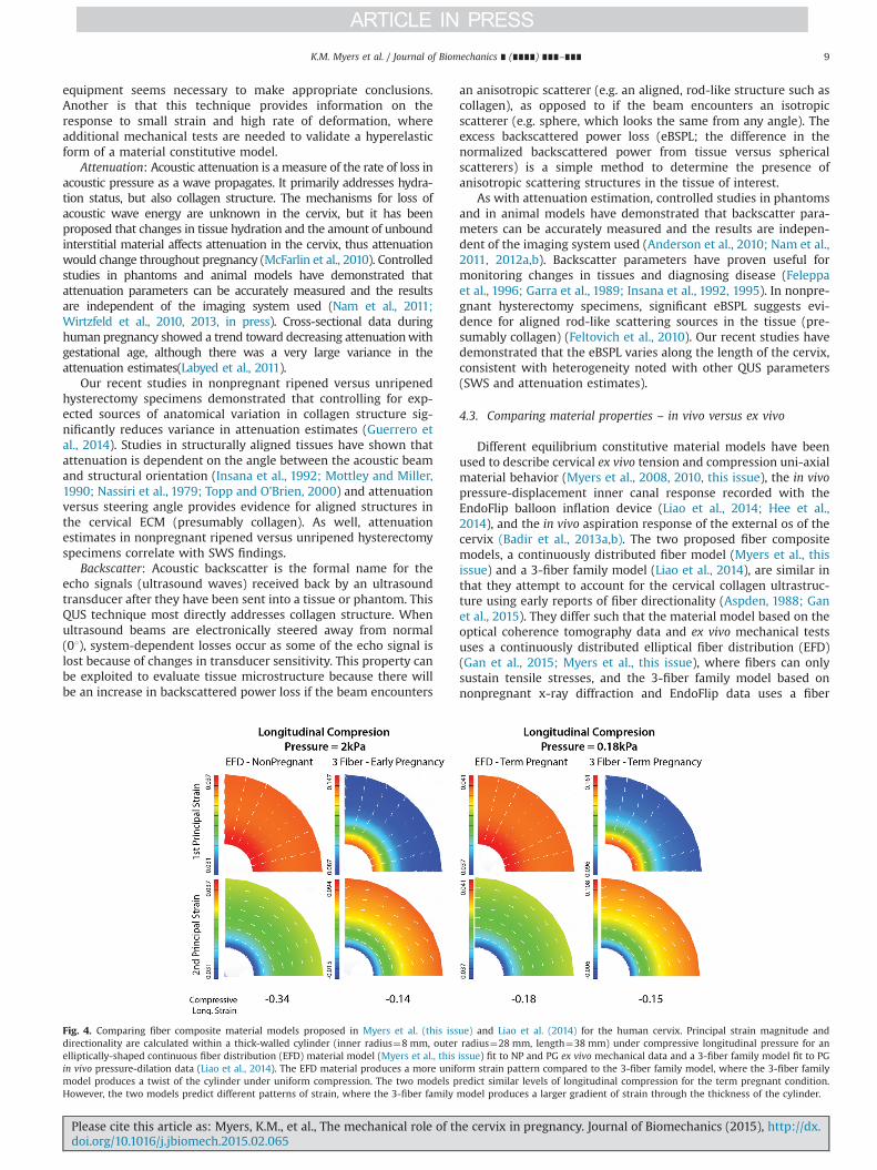
Different equilibrium constitutive material models have beenused to describe cervical ex vivo tension and compression uni-axialmaterial behavior (Myers et al., 2008, 2010, this issue), the in vivopressure-displacement inner canal response recorded with theEndoFlip balloon inflation device (Liao et al., 2014; Hee et al.,2014), and the in vivo aspiration response of the external os of thecervix (Badir et al., 2013a,b). The two proposed fiber compositemodels, a continuously distributed fiber model (Myers et al., thisissue) and a 3-fiber family model (Liao et al., 2014), are similar inthat they attempt to account for the cervical collagen ultrastruc-ture using early reports of fiber directionality (Aspden, 1988; Ganet al., 2015). They differ such that the material model based on theoptical coherence tomography data and ex vivo mechanical testsuses a continuously distributed elliptical fiber distribution (EFD)(Gan et al., 2015; Myers et al., this issue), where fibers can onlysustain tensile stresses, and the 3-fiber family model based onnonpregnant x-ray diffraction and EndoFlip data uses a fiber
Fig. 4. Comparing fiber composite material models proposed in Myers et al. (this issue) and Liao et al. (2014) for the human cervix. Principal strain magnitude anddirectionality are calculated within a thick-walled cylinder (inner radius¼8 mm, outer radius¼28 mm, length¼38 mm) under compressive longitudinal pressure for anelliptically-shaped continuous fiber distribution (EFD) material model (Myers et al., this issue) fit to NP and PG ex vivo mechanical data and a 3-fiber family model fit to PGin vivo pressure-dilation data (Liao et al., 2014). The EFD material produces a more uniform strain pattern compared to the 3-fiber family model, where the 3-fiber familymodel produces a twist of the cylinder under uniform compression. The two models predict similar levels of longitudinal compression for the term pregnant condition.However, the two models predict different patterns of strain, where the 3-fiber family model produces a larger gradient of strain through the thickness of the cylinder.
K.M. Myers et al. / Journal of Biomechanics ∎ (∎∎∎∎) ∎∎∎–∎∎∎ 9
Please cite this article as: Myers, K.M., et al., The mechanical role of the cervix in pregnancy. Journal of Biomechanics (2015), http://dx.doi.org/10.1016/j.jbiomech.2015.02.065i
network with no fiber dispersion and the fiber strain energydensity can sustain both tension and compression (Aspden,1988; Hee et al., 2014; Liao et al., 2014). The third model, fit tothe aspiration data, is a neo-Hookean material, where the tissue isassumed to be incompressible (Badir et al., 2013a). This modelingstrategy is suitable because one set of mechanical testing data isconsidered and the collagen ultrastructure is mostly unknown inthe pregnancy.
To compare material properties reported in these studies, theprincipal strain response of a thick-walled cylinder under long-itudinal compression (Fig. 4) was calculated using FEA (methodol-ogy reported in Myers et al., this issue). Cervical material modelsand corresponding parameters for the EFD fiber, 3-fiber family,and the neo-Hookean models are reported in Myers et al. (thisissue), Liao et al. (2014) and Badir et al. (2013a), respectively.Under a uniform compression of the cylinder, the 3-fiber familymodel produces a twist about the inner canal because of thehelical fibers circumferentially ringing around the inner canal(Aspden, 1988; Liao et al., 2014), whereas the EFD and neo-Hookean materials do not produce this twist. Both fiber compositemodels, with preferential circumferential fibers, reduce theamount of circumferential strain. However, because the 3-fiberfamily material model does not have fiber reinforcement in theradial direction, strains in this direction are larger than an EFDprediction with similar fiber moduli. Additionally, there is a largegradient in the radial and circumferential strain for the 3-fiberfamily model because the 3-fiber families are fully aligned with nodispersion. The EFD produces a small strain gradient from theinner to the outer radius, and as expected the isotropic neo-Hookean model produces the same amount of radial and circum-ferential strains (0.083, 0.17, and 0.32 for NP, 1st trimester, 2ndtrimester tissue, respectively, under 2 kPa of compressive load and0.05 for 3rd trimester tissue under 0.18 kPa).
These predicted deformation patterns need to be validatedwith bulk tissue mechanical tests. However, overlapping findingsfrom these studies confirm that the term pregnant cervix is ordersof magnitude softer than the nonpregnant cervix. Additionally,comparing the nonpregnant ex vivo EFD results with the in vivoEndoFlip and aspirator data, early softening is present in the 1sttrimester of pregnancy. In terms of longitudinal strain, the resultsfrom all three models predicted similar values, with 0.18, 0.15, and0.08 strain throughout the cylinder for the EFD, 3-fiber family, andneo-Hookean models, respectively.
5. In summary: integrating strategies
Complementary methods to characterize cervical remodelingduring pregnancy can help derive a fully descriptive materialconstitutive model for tissue behavior under mechanical loading.Mechanical characterization methodologies described here havelimitations and advantages. In terms of capturing overall bulktissue properties during cervical remodeling, in vivo QUS methodsand mechanical aspiration offer noninvasive techniques to char-acterize cervical properties during pregnancy. These tools arecritical to understand cervical tissue changes in early pregnancyand to develop mechanical biomarkers for diagnosis. The majortechnical advantage of in vivo methods is the ability to conductlongitudinal studies in pregnancy to relate cervical remodeling toclinical outcomes. Small studies of biomechanical and QUS para-meters in human ex vivo and in vivo tissue support a model ofcervical remodeling that is consistent with animal studies, speci-fically, that early softening is primarily due to loss of maturecollagen crosslinking with preservation of general collagen align-ment (microstructure) while late ripening is associated with
marked increase in tissue hydration within a vulnerable ECM(Mahendroo, 2012; Yoshida et al., 2014a).
That said, this is a challenging problem and much more informa-tion about both ex vivo and in vivo human tissue is necessary forcomprehensive understanding of the complex process of cervicalremodeling. In terms of constitutive model development, QUS andmechanical aspiration data are limited by complex boundary condi-tions, contact, testing locations, directionality, strain levels, and strainrates. In vivo mechanical aspiration is currently limited to interroga-tion of the external os. Conversely, while QUS methods can inter-rogate the entire structure, they involve small strain and high rate ofdeformation, which are difficult to interpret on soft anisotropicmaterials, which in turns makes fitting a 3-D material constitutivemodel to intrinsic tissue material properties a challenge. Ex vivomechanical testing offers insight into the large deformation tissuematerial behavior and allows for predictive material modeling, butex vivo mechanical tests are limited by possible tissue damage,possible tissue degradation, tissue swelling, ill definitions of thetissue micro and ultrastructure, and the inability to access pregnanttissue. These drawbacks are apparent in the wide range of tensilemoduli reported in the literature (Table 1).
Extensive cross-validation of ex vivo and in vivo methodologiesis needed to identify meaningful and comparative cervical tissuematerial properties. This requires overcoming the limitationsoutlined here, which can certainly be facilitated through a multi-disciplinary approach, encompassing medical imaging, quant-itative ultrasound, fresh tissue imaging, in vivo mechanical inter-rogation, advanced material modeling, biochemical analysis, andcorrelation with animal models of normal and abnormal cervicalremodeling. We hope this approach will allow reframing of clini-cal definitions of cervical remodeling and deformation basedon intrinsic material property changes, and rigorous definiti-ons of mechanical loading conditions and strain with respectto the reference configuration of the cervix. Through combiningthese individual approaches in a collaborative engineering-clinicalframework, we hope to begin to develop a biomechanical rubricthat will aid in understanding the mechanical function of thecervix, and its role in spontaneous preterm birth.
Conflict of interest statement
None declared.
Acknowledgments
K.M., J.V., and R.W. acknowledge the support of the NationalScience Foundation BRIGE1125670 award, the Office of the Provostat Columbia University, and the Columbia University Medical CenterIrving Institute for Clinical and Translational Research, which issupported by the National Center for Advancing Translational Sciences,National Institutes of Health through Grant no. UL1 TR000040. M.B.and E.M. gratefully acknowledge financial support by Swiss Natio-nal Science Foundation, Grant no. 32003B_156450/1. H.F. and T.J.H.gratefully acknowledge the support of NIH R21HD063031, NIH R21HD061896, NIH R01HD072077 from the Eunice Kennedy ShriverNational Institute of Child Health and Human Development, andIntermountain Research & Medical Foundation. The content is solelythe responsibility of the authors and does not necessarily representthe official views of the National Science Foundation, National Inst-itutes of Health, nor the Swiss National Science Foundation.
K.M. Myers et al. / Journal of Biomechanics ∎ (∎∎∎∎) ∎∎∎–∎∎∎10
Please cite this article as: Myers, K.M., et al., The mechanical role of the cervix in pregnancy. Journal of Biomechanics (2015), http://dx.doi.org/10.1016/j.jbiomech.2015.02.065i

References
Akgul, Y., Holt, R., Mummert, M., Word, A., Mahendroo, M., 2012. Dynamic changesin cervical glycosaminoglycan composition during normal pregnancy andpreterm birth. Endocrinology 153, 3493–3503.
Akgul, Y., Word, R.A., Ensign, L.M., Yamaguchi, Y., Lydon, J., Hanes, J., Mahendroo, M.,2014. Hyaluronan in cervical epithelia protects against infection-mediatedpreterm birth. J. Clin. Invest. 124, 5481–5489.
Akins, M., Luby-Phelps, K., Mahendroo, M., 2010. Second harmonic generationimaging as a potential tool for staging pregnancy and predicting preterm birth.J. Biomed. Opt. 15, 026020.
Akins, M.L., Luby-Phelps, K., Bank, R., Mahendroo, M., 2011. Cervical softeningduring pregnancy: regulated changes in collagen cross-linking and compositionof matricellular proteins in the mouse. Biol. Reprod. 84, 1053–1062.
American College of Obstetricians and Gynecologists, 2012. ACOG (2012a) practicebulletin no. 130: Prediction and prevention of preterm birth. American Collegeof Obstetricians and Gynecologists. Obstet. Gynecol. 120, 964–973.
Anderson, J.J., Herd, M.T., King, M.R., Haak, A., Hafez, Z.T., Song, J., Oelze, M.L.,Madsen, E.L., Zagzebski, J.A., O'Brien, W.D., Hall, T.J., 2010. Interlaboratorycomparison of backscatter coefficient estimates for tissue-mimicking phan-toms. Ultrason. Imaging 32, 48–64.
Aspden, R., 1988. Collagen organization in the cervix and its relation to mechanicalfunction. Coll. Relat. Res. 8, 103–112.
Badir, S., Bajka, M., Mazza, E., 2013a. A novel procedure for the mechanicalcharacterization of the uterine cervix during pregnancy. J. Mech. Behav.Biomed. Mater. 27, 143–153.
Badir, S., Mazza, E., Zimmermann, R., Bajka, M., 2013b. Cervical softening occursearly in pregnancy: characterization of cervical stiffness in 100 healthy womenusing the aspiration technique. Prenat. Diagn. 33, 737–741.
Barbone, P.E., Bamber, J.C., 2002. Quantitative elasticity imaging: what can andcannot be inferred from strain images. Phys. Med. Biol. 47, 2147–2164.
Barros, F.C., Papageorghiou, A.T., Victora, C.G., Noble, J.A., Pang, R., Iams, J., Ismail,L.C., Goldenberg, R.L., Lambert, A., Kramer, M.S., et al., 2015. The distribution ofclinical phenotypes of preterm birth syndrome: implications for prevention.JAMA Pediatr. 169(3), 2015 Mar 1, 220-9.
Bauer, M., Mazza, E., Jabareen, M., Sultan, L., Bajka, M., Lang, U., Zimmermann, R.,Holzapfel, G.A., 2009. Assessment of the in vivo biomechanical properties of thehuman uterine cervix in pregnancy using the aspiration test. Eur. J. Obstet.Gynecol. Reprod. Biol. 144, S77–S81.
Bauer, M., Mazza, E., Nava, A., Zeck, W., Eder, M., Bajka, M., Cacho, F., Lang, U.,Holzapfel, G.A., 2007. In vivo characterization of the mechanics of humanuterine cervices. Ann. N. Y. Acad. Sci. 1101, 186–202.
Bishop, E., 1964. Pelvic scoring for elective induction. Obstet. Gynecol. 24, 266.Carlson, L.C., Feltovich, H., Palmeri, M.L., 2014a. Estimation of shear wave speed in
the human uterine cervix. Ultrasound Obstet. Gynecol. 43, 452–458.Carlson, L.C., Romero, S.T., Palmeri, M.L., Muñoz del Rio, A., Esplin, S.M., Rotemberg,
V.M., Hall, T.J., Feltovich, H., 2014. Changes in shear wave speed pre and postinduction of labor: a feasibility study. Ultrasound Obstet. Gynecol. http://dx.doi.org/10.1002/uog.14663.
Conde-Agudelo, A., Romero, R., Nicolaides, K., Chaiworapongsa, T., O’Brien, J.M.,Cetingoz, E., Da Fonseca, E., Creasy, G., P., Soma-Pillay, S., Fusey, C., Cam, Z.,Alfirevic, Hassan, S.S., 2013. Reports of major impact. Am. J. Obstet. Gynecol.208, 42.e1–42.e18.
Conrad, J.T., Tokarz, R.D., Williford, J.F., 1980. Dilatation of the Uterine Cervix:Connective Tissue Biology and Clinical Management. Raven Press, New York.
Conrad, J.T., Ueland, K., 1976. Reduction of the stretch modulus of human cervicaltissue by prostaglandin E2. Am. J. Obstet. Gynecol. 126, 218–223.
Conrad, J.T., Ueland, K., 1979. The stretch modulus of human cervical tissue inspontaneous, oxytocin-induced, and prostaglandin E2-induced labor. Am.J. Obstet. Gynecol. 133, 11–14.
Danforth, D., 1983. The morphology of the human cervix. Clin. Obstet. Gynecol. 26,7–13.
Danforth, D.N., 1947. The fibrous nature of the human cervix, and its relation to theisthmic segment in gravid and nongravid uteri. Am. J. Obstet. Gynecol. 53,541–560.
Danforth, D.N., Buckingham, J.C., Roddick, J.W., 1960. Connective tissue changesincident to cervical effacement. Am. J. Obstet. Gynecol. 80, 939–945.
Danforth, D.N., Veis, A., Breen, M., Weinstein, H.G., Buckingham, J.C., Manalo, P.,1974. The effect of pregnancy and labor on the human cervix: changes incollagen glycoproteins and glycosaminoglycans. Am. J. Obstet. Gynecol. 120,641–651.
Danielson, K.G., Baribault, H., Holmes, D.F., Graham, H., Kadler, K.E., Iozzo, R.V.,1997. Targeted disruption of decorin leads to abnormal collagen fibril mor-phology and skin fragility. J. Cell Biol. 136, 729–743.
Elovitz, M., Mrinalini, C., 2004. Animal models of preterm birth. Trends Endocrinol.Metab. 15, 479–487.
Feleppa, E., Kalisz, A., Sokil-Melgar, J., Lizzi, F., Liu, T., Rosado, A., Shao, M., Fair, W.,Wang, Y., Cookson, M., et al., 1996. Typing of prostate tissue by ultrasonicspectrum analysis. IEEE Trans. Ultrason. Ferroelectr. Freq. Control 43, 609–619.
Feltovich, H., Hall, T., Berghella, V., 2012. Beyond cervical length: emergingtechnologies for assessing the pregnant cervix. Am. J. Obstet. Gynecol., 1–43.
Feltovich, H., Hall, T.J., 2013. Quantitative imaging of the cervix: setting the bar.Ultrasound Obstet. Gynecol. 41, 121–128.
Feltovich, H., Nam, K., Hall, T.J., 2010. Quantitative ultrasound assessment of cervicalmicrostructure. Ultrason. Imaging 32, 131–142.
Fernandez, M., House, M., Jambawalikar, S., Vink, J., Wapner, R., Myers, K.Investigating the mechanical function of the cervix during pregnancy usingfinite element models derived from high resolution 3D MRI. Comput. MethodsBiomech. Biomed. Eng., in prees.
Fernandez, M., Vink, J., Yoshida, K., Wapner, R., Myers, K., 2013. Direct measurementof the permeability of human cervical tissue. J. Biomech. Eng. 135, 021024.
Fruscalzo, A., Londero, A.P., Fröhlich, C., Meyer-Wittkopf, M., Schmitz, R., 2014.Quantitative elastography of the cervix for predicting labor induction success.Ultraschall Med. http://dx.doi.org/10.1055/s-0033-1355572.
Gan, Y., Yao, W., Myers, K.M., Vink, J.Y., Wapner, R.J.,Hendon, C.P., Analyzing three-dimensional ultrastructure of human cervical tissue using optical coherencetomography, Biomed. Opt. Express 6, 2015, 1090-1108.
Garra, B.S., Insana, M.F., Shawker, T.H., Wagner, R.F., Bradford, M., Russell, M., 1989.Quantitative ultrasonic detection and classification of diffuse liver disease.Comparison with human observer performance. Invest. Radiol. 24, 196–203.
Goya, M., Pratcorona, L., Merced, C., Rodó, C., Valle, L., Romero, A., Juan, M.,Rodríguez, A., Muñoz, B., Santacruz, B., Bello-Muñoz, J.C., Llurba, E., Higueras,T., Cabero, L., Carreras, E., Pesario Cervical para Evitar Prematuridad PECEP TrialGroup, 2012. Cervical pessary in pregnant womenwith a short cervix PECEP: anopen-label randomised controlled trial. The Lancet 379, 1800–1806.
Gracie, S., Pennell, C., Ekman-Ordeberg, G., Lye, S., McManaman, J., Williams, S.,Palmer, L., Kelley, M., Menon, R., Gravett, M., Group, t.P.O.R., 2011. An integratedsystems biology approach to the study of preterm birth using “-omic”technology – a guideline for research. BMC Pregnancy Childbirth 11, 71.
Gravett, M.G., Rubens, C.E., Nunes, T.M., GAPPS Review Group, 2010. Global reporton preterm birth and stillbirth (2 of 7): discovery science. BMC PregnancyChildbirth 10 Suppl. 1, S2.
Grobman, W.A., Thom, E.A., Spong, C.Y., Iams, J.D., Saade, G.R., Mercer, B.M., Tita, A.T.N., Rouse, D.J., Sorokin, Y., Wapner, R.J., Leveno, K.J., Blackwell, S., Esplin, M.S.,Tolosa, J.E., Thorp, J.M., Caritis, S.N., Van Dorsten, J.P., Eunice Kennedy ShriverNational Institute of Child Health and Human Development Maternal-FetalMedicine Units (MFMU) Network, 2012. 17 alpha-hydroxyprogesterone capro-ate to prevent prematurity in nulliparas with cervical length less than 30 mm.Am. J. Obstet. Gynecol. 207, 390.e1–390.e8.
Guerrero, Q.W., Carlson, L.C., Feltovich, H., Hall, T., 2014. Quantitative ultrasoundbackscatter parameters in the human cervix. In: 2014 IEEE InternationalUltrasonics Symposium (IUS). IEEE, Chicago, IL. pp. 224–227.
Hall, C.S., Scott, M.J., Lanza, G.M., Miller, J.G., Wickline, S.A., 2000. The extracellularmatrix is an important source of ultrasound backscatter from myocardium.J. Acoust. Soc. Am. 107, 612–619.
Hee, L., Liao, D., Sandager, P., Gregersen, H., Uldbjerg, N., 2014. Cervical stiffnessevaluated in vivo by endoflip in pregnant women. PLoS ONE 9, e91121.
Hee, L., Sandager, P., Petersen, O., Uldbjerg, N., 2013. Quantitative sonoelastographyof the uterine cervix by interposition of a synthetic reference material. ActaObstet. Gynecol. Scand. 92, 1244–1249.
Hernandez-Andrade, E., Romero, R., Korzeniewski, S.J., Ahn, H., Aurioles-Garibay, A.,Garcia, M., Schwartz, A.G., Yeo, L., Chaiworapongsa, T., Hassan, S.S., 2014.Cervical strain determined by ultrasound elastography and its association withspontaneous preterm delivery. J. .Perinat. Med. 42, 159–169.
Hollenstein, M., Bugnard, G., Joos, R., Kropf, S., Villiger, P., Mazza, E., 2013. Towardslaparoscopic tissue aspiration. Med. Image Anal. 17, 1037–1045.
Holt, R., Timmons, B., Akgul, Y., Akins, M., Mahendroo, M., 2011. The molecularmechanisms of cervical ripening differ between term and preterm birth.Endocrinology 152, 1036–1046.
House, M., Bhadelia, R., Myers, K., Socrate, S., 2009. Magnetic resonance imaging ofthree-dimensional cervical anatomy in the second and third trimester. Eur.J. Obstet. Gynecol. Reprod. Biol. 144 (Suppl. 1), S65–S69.
House, M., Feltovich, H., Hall, T., Stack, T., Patels, A., Socrate, S., 2012. Three-dimensional, extended field-of-view ultrasound method for estimating largestrain mechanical properties of the cervix during pregnancy. Ultrason. Imaging34, 1–14.
House, M., Kaplan, D., Socrate, S., 2009b. Relationships between mechanicalproperties and extracellular matrix constituents of the cervical stroma duringpregnancy. Semin. Perinatol. 33, 300–307.
House, M., McCabe, R., Socrate, S., 2013. Using imaging-based, three-dimensionalmodels of the cervix and uterus for studies of cervical changes duringpregnancy. Clin. Anat. (New York, N.Y.) 26, 97–104.
House, M., OCallaghan, M., Bahrami, S., Chelmow, D., Kini, J., Wu, D., Patz, S.,Bhadelia, R., 2005. Magnetic resonance imaging of the cervix during pregnancy:effect of gestational age and prior vaginal birth. Am. J. Obstet. Gynecol. 193,1554–1560.
House, M., Socrate, S., 2006. The cervix as a biomechanical structure. UltrasoundObstet. Gynecol. 28, 745–749.
Hricak, H., Chang, Y.C., Cann, C.E., Parer, J.T., 1990. Cervical incompetence:preliminary evaluation with MR imaging. Radiology 174, 821–826.
Iams, J.D., Berghella, V., 2010. Care for women with prior preterm birth. Am.J. Obstet. Gynecol. 203, 89–100.
Iams, J.D., Goldenberg, R.L., Meis, P.J., Mercer, B.M., Moawad, A., Das, A., Thom, E.,McNellis, D., Copper, R.L., Johnson, F., Roberts, J.M., 1996. The length of thecervix and the risk of spontaneous premature delivery. National Institute ofChild Health and Human Development Maternal Fetal Medicine Unit Network.N. Engl. J. Med. 334, 567–572.
Insana, M.F., Wood, J.G., Hall, T.J., 1992. Identifying acoustic scattering sources innormal renal parenchyma in vivo by varying arterial and ureteral pressures.Ultrasound Med. Biol. 18, 587–599.
K.M. Myers et al. / Journal of Biomechanics ∎ (∎∎∎∎) ∎∎∎–∎∎∎ 11
Please cite this article as: Myers, K.M., et al., The mechanical role of the cervix in pregnancy. Journal of Biomechanics (2015), http://dx.doi.org/10.1016/j.jbiomech.2015.02.065i

Insana, M.F., Wood, J.G., Hall, T.J., Cox, G.G., Harrison, L.A., 1995. Effects ofendothelin-1 on renal microvasculature measured using quantitative ultra-sound. Ultrasound Med. Biol. 21, 1143–1151.
Labyed, Y., Bigelow, T.A., Mcfarlin, B.L., 2011. Estimate of the attenuation coefficientusing a clinical array transducer for the detection of cervical ripening in humanpregnancy. Ultrasonics 51, 34–39.
Leppert, P., 1992. Cervical softening, effacement, and dilatation: a complexbiochemical cascade. J. Matern. Fetal Neonatal. Med. 1, 213–223.
Leppert, P., 1995. Anatomy and physiology of cervical ripening. Clin. Obstet.Gynecol. 38, 267–279.
Leppert, P., 1998. The biochemistry and physiology of the uterine cervix duringgestation and parturition. Prenat. Neonat. Med. 3, 103–105.
Liao, D., Hee, L., Sandager, P., Uldbjerg, N., Gregersen, H., 2014. Identification ofbiomechanical properties in vivo in human uterine cervix. J. Mech. Behav.Biomed. Mater. 39, 27–37.
Mahendroo, M., 2012. Cervical remodeling in term and preterm birth: insights froman animal model. Reproduction 143, 429–438.
Mahmoud, H., Wagoner Johnson, A., E.K., Chien, M.J., Poellmann, B., McFarlin, 2013.System-level biomechanical approach for the evaluation of term and pretermpregnancy maintenance. J. Biomech. Eng. 135, 021009.
Maldjian, C., Adam, R., Pelosi, M., 1999. MRI appearance of cervical incompetence ina pregnant patient. Magn. Reson. Imaging 17, 1399–1402.
Martin, J., Hamilton, B., Ventura, S., Osterman, M., Mathews, T., 2013. Births: finaldata for 2011. Natl. Vital Stat. Rep. 62, 1–90.
Marturano, J., Xylas, J., Sridharan, G., Georgakoudi, I., Kuo, C., 2014. Acta biomater-ialia. Acta Biomater. 10, 1370–1379.
Maurer, M., Badir, S., Bajka, M., Abitabile, P., Zimmermann, R., Mazza, E., Challengingthe in-vivo assessment of biomechanical properties of the uterine cervix: Acritical analysis of ultrasound based quasi-static procedures, J. Biomech, thisissue, http://dx.doi.org/10.1016/j.jbiomech.2015.02.038.
Mazza, E., Nava, A., Bauer, M., Winter, R., Bajka, M., Holzapfel, G.A., 2006.Mechanical properties of the human uterine cervix: an in vivo study. Med.Image Anal. 10, 125–136.
Mazza, E., Nava, A., Hahnloser, D., Jochum, W., Bajka, M., 2007. The mechanicalresponse of human liver and its relation to histology: an in vivo study. Med.Image Anal. 11, 663–672.
Mazza, E., Parra-Saavedra, M., Bajka, M., Gratacos, E., Nicolaides, K., Deprest, J., 2013.In vivo assessment of the biomechanical properties of the uterine cervix inpregnancy. Prenat. Diagn., 33–41.
McFarlin, B.L., Bigelow, T.A., Laybed, Y., O'Brien, W.D., Oelze, M.L., Abramowicz, J.S.,2010. Ultrasonic attenuation estimation of the pregnant cervix: a preliminaryreport. Ultrasound Obstet. Gynecol. 36, 218–225.
Molina, F.S., Gómez, L.F., Florido, J., Padilla, M.C., Nicolaides, K.H., 2012. Quantifica-tion of cervical elastography: a reproducibility study. Ultrasound Obst. Gynecol.39, 685–689.
Mottley, J.G., Miller, J.G., 1990. Anisotropy of the ultrasonic attenuation in softtissues: measurements in vitro. J. Acoust. Soc. Am. 88, 1203–1210.
Myers, K., Ateshian, G., 2014. Interstitial growth and remodeling of biologicaltissues: tissue composition as state variables. J. Mech. Behav. Biomed. Mater. 29,544–556.
Myers, K., Hendon, C., Gan, Y., Yao, W., Vink, J., Wapner, R., 2015. A continuous fiberdistribution material model for human cervical tissue. J. Biomech., this issue,http://dx.doi.org/10.1016/j.jbiomech.2015.02.060.
Myers, K., Paskaleva, A., House, M., Socrate, S., 2008. Mechanical and biochemicalproperties of human cervical tissue. Acta Biomater. 4, 104–116.
Myers, K., Socrate, S., Paskaleva, A., House, M., 2010. A study of the anisotropy andtension/compression behavior of human cervical tissue. J. Biomech. Eng. 132,021003.
Myers, K., Socrate, S., Tzeranis, D., House, M., 2009. Changes in the biochemicalconstituents and morphologic appearance of the human cervical stroma duringpregnancy. Eur. J. Obstet. Gynecol. Reprod. Biol. 144 (Suppl. 1), S82–S89.
Nam, K., Rosado-Mendez, I.M., Wirtzfeld, L.A., Ghoshal, G., Pawlicki, A.D., Madsen, E.L.,Lavarello, R.J., Oelze, M.L., Zagzebski, J.A., O'Brien, W.D., Hall, T.J., 2012a. Comparisonof ultrasound attenuation and backscatter estimates in layered tissue-mimickingphantoms among three clinical scanners. Ultrason. Imag 34, 209–221.
Nam, K., Rosado-Mendez, I.M., Wirtzfeld, L.A., Kumar, V., Madsen, E.L., Ghoshal, G.,Pawlicki, A.D., Oelze, M.L., Lavarello, R.J., Bigelow, T.A., Zagzebski, J.A., O'Brien,W.D., Hall, T.J., 2012b. Cross-imaging system comparison of backscatter coeffi-cient estimates from a tissue-mimicking material. J. Acoust. Soc. Am. 132,1319–1324.
Nam, K., Rosado-Mendez, I.M., Wirtzfeld, L.A., Pawlicki, A.D., Kumar, V., Madsen, E.L.,Ghoshal, G., Lavarello, R.J., Oelze, M.L., Bigelow, T.A., Zagzebski, J.A., O'Brien, W.D.,Hall, T.J., 2011. Ultrasonic attenuation and backscatter coefficient estimates ofrodent-tumor-mimicking structures: comparison of results among clinical scan-ners. Ultrason. Imaging 33, 233–250.
Nassiri, D.K., Nicholas, D., Hill, C.R., 1979. Attenuation of ultrasound in skeletalmuscle. Ultrasonics 17, 230–232.
Norman, J.E., Shennan, A.H., 2013. Prevention of preterm birth—why can't we doany better? The Lancet 381, 184–185.
Osmers, R., Rath, W., Pflanz, M.A., Kuhn, W., Stuhlsatz, H.W., Szeverényi, M., 1993.Glycosaminoglycans in cervical connective tissue during pregnancy and par-turition. Obstet. Gynecol. 81, 88–92.
Oxlund, B.S., Ørtoft, G., Brüel, A., Danielsen, C., Oxlund, H., Uldbjerg, N., 2010a.Cervical collagen and biomechanical strength in non-pregnant women with ahistory of cervical insufficiency. Reprod. Biol. Endocrinol. 8, 92.
Oxlund, B.S., Ørtoft, G., Brüel, A., Danielsen, C.C., Bor, P., Oxlund, H., Uldbjerg, N.,2010b. Collagen concentration and biomechanical properties of samples fromthe lower uterine cervix in relation to age and parity in non-pregnant women.Reprod. Biol. Endocrinol. 8, 82.
Palmeri, M., Feltovich, H., Homyk, A., Carlson, L., Hall, T., 2013. Evaluating thefeasibility of acoustic radiation force impulse shear wave elasticity imaging ofthe uterine cervix with an intracavity array: a simulation study. In: IEEETransactions on Ultrasonics, Ferroelectrics and Frequency Control, pp. 2053–2064.
Parra-Saavedra, M., Gómez, L., Barrero, A., Parra, G., Vergara, F., Navarro, E., 2011.Prediction of preterm birth using the cervical consistency index. UltrasoundObstet. Gynecol. 38, 44–51.
Paskaleva, A., 2007. Biomechanics of Cervical Function in Pregnancy – Case ofCervical Insufficiency (Ph.D. thesis). MIT.
Petersen, L.K., Oxlund, H., Uldbjerg, N., Forman, A., 1991. In vitro analysis ofmuscular contractile ability and passive biomechanical properties of uterinecervical samples from nonpregnant women. Obstet. Gynecol. 77, 772–776.
Ramanah, R., Berger, M.B., Chen, L., Riethmuller, D., DeLancey, J.O.L., 2012. See it in3D! Researchers examined structural links between the cardinal and uterosa-cral ligaments. Am. J. Obstet. Gynecol. 207, 437.e1–437.e7.
Read, C.P., Word, R.A., Ruscheinsky, M.A., Timmons, B.C., Mahendroo, M.S., 2007.Cervical remodeling during pregnancy and parturition: molecular character-ization of the softening phase in mice. Reproduction 134, 327–340.
Rechberger, T., Uldbjerg, N., Oxlund, H., 1988. Connective tissue changes in thecervix during normal pregnancy and pregnancy complicated by cervicalincompetence. Obstet. Gynecol. 71, 563–567.
Schoen, C.N., Tabbah, S., Iams, J.D., Caughey, A.B., Berghella, V., 2014. Why theunited states preterm birth rate is declining. Am. J. Obstet. Gynecol., in press.http://dx.doi.org/10.1016/j.ajog.2014.12.011.
Shimizu, T., Endo, M., Yosizawa, Z., 1980. Glycoconjugates (glycosaminoglycans andglycoproteins) and glycogen in the human cervix uteri. Tohoku J. Exp. Med. 131,289–299.
Society for Maternal-Fetal Medicine Publications Committee, 2012. Progesteroneand preterm birth prevention: translating clinical trials data into clinicalpractice. Am. J. Obstet. Gynecol. 206, 376–386.
Solomon, C., Iams, J., 2014. Prevention of preterm parturition. N. Engl. J. Med. 370,254–261.
Swiatkowska-Freund, M., Preis, K., 2011. Elastography of the uterine cervix:implications for success of induction of labor. Ultrasound Obstet. Gynecol. 38,52–56.
Timmons, B., Akins, M., Mahendroo, M., 2010a. Cervical remodeling duringpregnancy and parturition. Trends Endocrinol. Metab. 21, 353–361.
Timmons, B., Akins, M., Mahendroo, M., 2010b. Cervical remodeling duringpregnancy and parturition. Trends Endocrinol. Metab. 21, 353–361.
To, M.S., Skentou, C., Liao, A.W., Cacho, A., Nicolaides, K.H., 2001. Cervical length andfunneling at 23 weeks of gestation in the prediction of spontaneous earlypreterm delivery. Ultrasound Obstet. Gynecol. 18, 200–203.
Topp, K.A., O'Brien, W.D., 2000. Anisotropy of ultrasonic propagation and scatteringproperties in fresh rat skeletal muscle in vitro. J. Acoust. Soc. Am. 107,1027–1033.
Uldbjerg, N., Ekman, G., Malmstrom, A., Olsson, K., Ulmsten, U., 1983. Ripening ofthe human uterine cervix related to changes in collagen glycosaminoglycansand collagenolytic activity. Am. J. Obstet. Gynecol. 147, 662–666.
Weiss, S., Jaermann, T., Schmid, P., Staempfli, P., Boesiger, P., Niederer, P., Caduff, R.,Bajka, M., 2006. Three-dimensional fiber architecture of the nonpregnanthuman uterus determined ex vivo using magnetic resonance diffusion tensorimaging. Anat. Rec. Part A 288, 84–90.
Wirtzfeld, L.A., Ghoshal, G., Hafez, Z.T., Nam, K., Labyed, Y., Anderson, J.J., Herd, M.T.,Haak, A., He, Z., Miller, R.J., Sarwate, S., Simpson, D.G., Zagzebski, J.A., Bigelow,T.A., Oelze, M.L., Hall, T.J., O'Brien, W.D., 2010. Cross-imaging platform compar-ison of ultrasonic backscatter coefficient measurements of live rat tumors.J. Ultrasound Med. 29, 1117–1123.
Wirtzfeld, L.A., Ghoshal, G., Rosado-Mendez, I.M., Nam, K., Park, Y., Pawlicki, A.D.,Miller, R., Simpson, D.G., Zagzebski, J.A., Oelze, M.L., Hall, T.J., OBrien, W.Quantitative ultrasound comparison of MAT and 4T1 mammary 1 tumors inmice and rats across multiple imaging systems. J. Ultrasound Med., in press.
Wirtzfeld, L.A., Nam, K., Labyed, Y., Ghoshal, G., Haak, A., Sen-Gupta, E., He, Z., Hirtz,N.R., Miller, R.J., Sarwate, S., Simpson, D.G., Zagzebski, J.A., Bigelow, T.A., Oelze,M.L., Hall, T.J., O'Brien, W.D., 2013. Techniques and evaluation from a cross-platform imaging comparison of quantitative ultrasound parameters in anin vivo rodent fibroadenoma model. IEEE Trans. Ultrason. Ferroelectr. Freq.Control 60, 1386–1400.
Word, R.A., Li, X., Hnat, M., Carrick, K., 2007. Dynamics of cervical remodelingduring pregnancy and parturition: mechanisms and current concepts. Semin.Reprod. Med. 25, 69–79.
World Health Organization, 2014. World Health Organization Fact Sheet No. 363,November 17th, 2014. http://www.who.int/mediacentre/factsheets/fs363/en/(accessed 12.03.14).
Yao, W., Yoshida, K., Fernandez, M., Vink, J., Wapner, R., Ananth, C., Oyen, M., Myers,K., 2014. Measuring the compressive viscoelastic mechanical properties ofhuman cervical tissue using indentation. J. Mech. Behav. Biomed. Mater. 34,18–26.
Yoshida, K., Jiang, H., Kim, M., Vink, J., Cremers, S., Paik, D., Wapner, R., Mahendroo,M., Myers, K., 2014a. Quantitative evaluation of collagen crosslinks andcorresponding tensile mechanical properties in mouse cervical tissue duringnormal pregnancy. PLoS ONE 9, e112391.
K.M. Myers et al. / Journal of Biomechanics ∎ (∎∎∎∎) ∎∎∎–∎∎∎12
Please cite this article as: Myers, K.M., et al., The mechanical role of the cervix in pregnancy. Journal of Biomechanics (2015), http://dx.doi.org/10.1016/j.jbiomech.2015.02.065i

Yoshida, K., Reeves, C., Vink, J., Kitajewski, J., Wapner, R., Jiang, H., Cremers, S.,Myers, K., 2014b. Cervical collagen network remodeling in normal pregnancyand disrupted parturition in Antxr2 deficient mice. J. Biomech. Eng. 136,021017.
Zilianti, M., Azuaga, A., Calderon, F., Pagés, G., Mendoza, G., 1995. Monitoring theeffacement of the uterine cervix by transperineal sonography: a new perspec-tive. J. Ultrasound Med. 14, 719–724.
Zork, N.M., Myers, K.M., Yoshida, K., Cremers, S., Jiang, H., Ananth, C.V., Wapner, R.J.,Kitajewski, J., Vink, J., 2015. A systematic evaluation of collagen cross-links inthe human cervix. Am. J. Obstet. Gynecol. 212, 2015, 321.e1-321.e8.
K.M. Myers et al. / Journal of Biomechanics ∎ (∎∎∎∎) ∎∎∎–∎∎∎ 13
Please cite this article as: Myers, K.M., et al., The mechanical role of the cervix in pregnancy. Journal of Biomechanics (2015), http://dx.doi.org/10.1016/j.jbiomech.2015.02.065i