the marine fauna of new zealand: porifera: lithistid...
TRANSCRIPT
ISSN 1174–0043; 121
The Marine Fauna of New Zealand:
Porifera: Lithistid Demospongiae (Rock Sponges)Michelle Kelly NIWA Biodiversity Memoir 121

Cover photo: Lithistid sponges on the surface of a boulder from hydrothermally active rungapapa Knoll in the Bay of plenty. the blue nodules and chalice-like sponges are juvenile Reidispongia coerulea (phymatellidae), and the cream knobs are Macandrewia spinifoliata (Macandrewiidae). the boulder was retrieved using a NIWA epibenthic sled towed at 160 m depth from rv Kaharoa in 2000. photo: NIWA

NATIONAL INSTITUTE OFWATER AND ATMOSPHERIC RESEARCH (NIWA)
The Marine Fauna of New Zealand:
Porifera: Lithistid Demospongiae (Rock Sponges)
Michelle Kelly
National Centre for Aquatic Biodiversity & BiosecurityNational Institute of Water and Atmospheric Research (NIWA)
Private Bag 109-695, NewmarketAuckland, New Zealand
NIWA Biodiversity Memoir 121
2007

Cataloguing in Publication
KELLY, M.The marine fauna of New Zealand: Porifera: Lithistid Demospongiae (rock sponges) / by Michelle Kelly—Wellington: NIWA (National Institute of Water and Atmospheric Research), 2007(NIWA Biodiversity Memoir, ISSN 1174-0043)
ISBN 978–0–478–23277–6
Series Editor: Dennis P. Gordon
Copy edited and typeset by: Geoff Gregory, Word Therapy, Paraparaumu
Printed and bound by: Graphic Press & Packaging Ltd, Levin
Received for publication—30 June 2006
NIWA Copyright 2007

3
CONTENTS PageABSTRACT ................................................................................................................................................................. 5INTRODUCTION ...................................................................................................................................................... 6THE NEW ZEALAND LITHISTID SPONGE FAUNA ................… .................................................................. 7MATERIALS AND METHODS..............................................................................……………………………… 8 Sample collection .....................................................................................................................……………… 8 Spicule dimensions .................................................................……………………………………………… 8 Registration of type and general materials ..........................................................................……………… 9 Area of study ...........................................................................……………………………………………… 9 Terminology .............................................................................……………………………………………… 9 Abbreviations of institutions ...................................................................................……………………… 10CHECKLIST OF SPECIES ...................................................................................................................................... 11SYSTEMATICS ........................................................................................................................................................ 12Lithistid DEMOSPONGIAE (formerly order Lithistida Schmidt, 1870) ......................................................... 12 Family Theonellidae ..................................................................................................................................... 13 Genus Discodermia .....................................................................……………………………………… 13 Family PhymaTellidae ......................................................................……………………………………… 15 Genus Neoaulaxinia ...................................................................……………………………………… 15 Genus Neosiphonia ....................................................................……………………………………… 22 Genus Reidispongia ....................................................................……………………………………… 27 Family CorallisTidae ...................................................................................................................................... 29 Genus Neoschrammeniella .....................................................................……………………………… 30 Genus Herengeria ......................................................................……………………………………… 34 Genus Awhiowhio n. gen. ……………………………………… ......................................................... 39 Family neoPelTidae ....................................................................................................................................... 45 Genus Homophymia ...................................................................……………………………………… 45 Genus Callipelta ..........................................................................……………………………………… 47 Genus Neopelta ...........................................................................……………………………………… 50 Genus Lepidothenea ....................................................................……………………………………… 52 Family maCandrewiidae ...................................................................……………………………………… 53 Genus Macandrewia ..................................................................……………………………………… 53 Family Pleromidae .......................................................................................................................................... 54 Genus Pleroma ...........................................................................……………………………………… 56 Family isoraPhiniidae ........................................................................………………………………………61 Genus Costifer ............................................................................……………………………………… 61 Family sCleriTodermidae ..................................................................……………………………………… 64 Genus Microscleroderma ..........................................................................……………………………...65 Genus Scleritoderma ................................................................................................................................ 67 Genus Aciculites .........................................................................………………………………………69 Family azoriCidae .......................................................................................................................................... 76 Genus Leiodermatium ............................................................................……………………………… 77DISCUSSION ........................................................................................................................................................... 88ACKNOWLEDGMENTS ....................................................................................................................................... 90REFERENCES .......................................................................................................................................................... 90APPENDIX 1. Key to diagnostic characters ....................………………………………………………………94APPENDIX 2. Key to diagnostic characters ....................………………………………………………………95APPENDIX 3. List of stations ............................................………………………………………………………96TAXONOMIC INDEX ...................................................................……………………………………………… 99
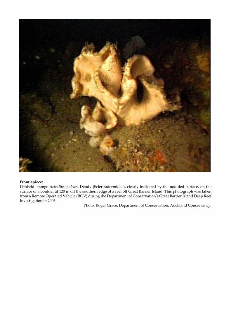
Frontispiece: Lithistid sponge Aciculites pulchra Dendy (Scleritodermidae), clearly indicated by the noduled surface, on the surface of a boulder at 120 m off the southern edge of a reef off Great Barrier Island. This photograph was taken from a Remote Operated Vehicle (ROV) during the Department of Conservation’s Great Barrier Island Deep Reef Investigation in 2003.
Photo: Roger Grace, Department of Conservation, Auckland Conservancy.

5
NATIONAL INSTITUTE OFWATER AND ATMOSPHERIC RESEARCH (NIWA)
The Marine Fauna of New Zealand:
Porifera: Lithistid Demospongiae (Rock Sponges)
Michelle Kelly
National Centre for Aquatic Biodiversity & BiosecurityNational Institute of Water and Atmospheric Research (NIWA)
Private Bag 109-695, NewmarketAuckland, New Zealand
ABSTRACT
The lithistid Demospongiae fauna of New Zealand has been inventoried from existing and new collections, and is reviewed here and revised where necessary. Most of the 282 specimens examined were recorded from the largest collection of sponges in New Zealand, in the NIWA Invertebrate Collection, Wellington. Significant collections were also examined from the Museum of New Zealand Te Papa Tongarewa. The lithistid Demospongiae (formerly order Lithistida Schmidt, 1870) is represented in the New Zealand region by nine families, 18 genera (one of which is new to science), and 30 species (12 of which are new to science): Theonellidae (1 genus, 1 species), Phymatellidae (3 genera, 6 species), Corallistidae (3 genera, 7 species), Neopeltidae (4 genera, 4 species), Macandrewiidae (1 genus, 1 species), Pleromidae (1 genus, 3 species), Isoraphiniidae (1 genus, 1 spe-cies), Scleritodermidae (3 genera, 5 species), and Azoricidae (1 genus, 2 species). This work records the first lithistid species, Neoschrammeniella antarctica n. sp., known from polar regions, and provides the first record of the genus Leiodermatium further south than the Philippines. Two additional species of Leiodermatium described here are found only in the west-central Pacific and Micronesian deep waters, but are included for the sake of a complete review of the genus in the Pacific. New species of the previously monospecific phymatellid genera Neoaulaxinia and Neosiphonia are described, and a new corallistid genus, Awhiowhio, is recognised from New Zealand waters. All specimens were dredged from between 80 and 1700 m, but were commonest between 200 and 800 m. With the exception of one specimen from the eastern edge of the Challenger Plateau on New Zealand’s west coast, and a new species from the Ross Sea, Antarctica, all were found north of the northern edge of the Chatham Rise and in New Zealand’s northernmost waters. Known and new species are redescribed from representative New Zealand material and, in some cases, the characters used to define genera and species are redefined and clarified. In particular, ornamentation of the desma skeleton and morphology of the augmenting microscleres are emphasised for distinction at the species level.
Keywords: Porifera, lithistid Demospongiae, Lithistida, Theonellidae, Phymatellidae, Corallistidae, Neopeltidae, Macan-drewiidae, Pleromidae, Isoraphiniidae, Scleritodermidae, Azoricidae, Desmanthidae, polyphyletic group, systematics, sponges, taxonomy, new species, New Zealand

6
Lithistid demosponges are a polyphyletic group com-prising 13 extant families; 36 genera are included in the most recent classification (Pisera & Lévi 2002a) but five genera remain of uncertain status. They differ from other demosponges in that the dominant structural spi-cules (desmas) are articulate, forming in most species a solid, rigid, heavily siliceous skeleton. These desmas are highly diverse morphologically; the overall architecture of the desma, the ornamentation of the desma surface, and the pattern of articulation with adjacent spicules, are diagnostically important (Schrammen 1910, see Kelly 2000a; Pisera & Lévi 2002a).
Lithistid sponges were traditionally placed in the single order Lithistida Schmidt, 1870 owing to the com-mon possession of desmas, even though these display considerable morphological diversity. For some time, lithistids have been recognised as polyphyletic, with several points of origin within the Demospongiae (de Laubenfels 1936; Reid 1963, 1970; Kelly-Borges & Pom-poni 1994; Pisera & Lévi 2002a). An indication of the polyphyletic nature of lithistid sponges is revealed in the wide range of microscleres and ectosomal megascleres, and desma axial geometries, which include tetraxial, monaxial, polyaxial, and anaxial forms.
Familial and ordinal affiliations of lithistids with each other and with non-desma-bearing sponges are uncommon but can be found in the megasclere and microsclere components of the skeleton (see Pisera & Lévi 2002a). The presence of triaene megascleres and asterose microscleres in some lithistid genera clearly indicates affinity with demosponge Astrophorida, and this has been supported by recent DNA studies (Kelly-Borges & Pomponi 1994; Chombard et al. 1998; McIn-erney et al. 1999). The exotylostyles of Gastrophanella are considered to demonstrate affinity with the non-lithistid demosponge order Hadromerida (Van Soest & Stentoft 1988).
A greater difficulty arises when taxa do not have accessory spicules, or the spicules that they have bear only superficial resemblance to those in non-lithistid demosponges. Species of Aciculites, for example, contain only acanthose anisostrongyles as accessory spicules, and Leiodermatium and Vetulina possess only desmas and diactines, and desmas, respectively. In all three taxa, molecular evidence was required to indicate their phylogenetic relationships (Kelly-Borges & Pomponi 1994).
The lithistid sponges are of interest to biomedical science because of the great variety of pharmaceutically relevant biological activities of their chemical extracts (see Pomponi 2001; Bewley et al. 1998; Munro et al. 1999;
INTRODUCTION
Mayer & Hamann 2004; Piel et al. 2004). A recent survey of the literature revealed some 40 papers reporting bio-logically active compounds and their synthesis from the Southwest Pacific genera Aciculites, Callipelta, Discoder-mia, Microscleroderma, Neosiphonia, Pleroma, Reidispongia, and Scleritoderma. Of these genera, the first example of double bioalkylation of the sterol side chain at position 26 was reported from New Zealand’s Aciculites pulchra (Crist et al. 1983). Furthermore, Pleroma menoui Lévi & Lévi, Reidispongia coerulea Lévi & Lévi, and Neosiphonia superstes Sollas from New Caledonia were the subject of considerable attention for their production of bromind-oles (Guella et al. 1989) and antiviral (Laille et al. 1998), antifungal (D’Auria et al. 1995), and cytotoxic macrolides (D’Auria et al. 1996; Zampella et al. 1997; Carbonelli et al. 1999; Bassarello et al. 2000).
Lithistid demosponges are of particular interest to paleontology because many extant genera and species are relict from sponge faunas that were more abundant before the present. Lithistid demosponges are known from the Ordovician, Silurian, Devonian, Permian, Late Jurassic, Late Cretaceous, and Eocene (Pisera 2002, 2006; Reid 2004). The Paleozoic fauna was dominated by Orchocladina (extinct in the Permian) and sphaero-cladinid sponges (solely represented in the Recent fauna by Vetulina stalactites from the tropical Atlantic). The Mesozoic (260–60 Ma) lithistid fauna was dominated by Tetracladina, Megamorina, and Rhizomorina, groups that are still relatively common in Tertiary and Recent lithistid faunas (Pisera 2002, 2006). These sponges fos-silise well because of their rigid silica skeletons; 13 suborders, 34 families, and more than 200 fossil genera have been recently revised (Pisera 2002, 2006), and many more nominal genera are known.
Lithistid sponges are known from almost all temper-ate and tropical oceans but are generally restricted to depths greater than about 80 m in South Pacific locations including New Zealand and the Norfolk Ridge, and 150 m in the tropical Western Atlantic region (Pomponi et al. 2001). Apart from the proliferation of the genera Ac-iculites, Neoschrammeniella, and Pleroma in the Southwest Pacific, Corallistes and Discodermia in the western tropical Atlantic, and Theonella in the Indo-Pacific, most genera are known only from fragments or single specimens that have been rarely, if ever, recollected (e.g. Lyidium [= Pleroma] torquilla Schmidt, 1870 from Cuba). Lithistid sponges are rarely collected from below about 1700 m depth.
Prior to the present work, two major regional fau-nas were known worldwide: the continental shelf and slope fauna of the tropical western Atlantic (Schmidt

7
THE NEW ZEALAND LITHISTID SPONGE FAUNA
1870, 1880; van Soest & Stentoft 1988; Kelly-Borges et al. 1994; Kelly-Borges & Pomponi 1994; Lehnert & van Soest 1996; Pisera 1999; Pomponi et al. 2001) and the seamount fauna of the Southwest Pacific including the seamounts of the New Caledonian Norfolk Ridge (Lévi & Lévi 1983, 1988; Lévi 1991; Schlacher-Hoenlinger et al.
Prior to 1991, when research on the lithistid fauna of New Zealand commenced, only two Recent lithistid species had been described, although several more were known (P.R. Bergquist pers. comm.). Arthur Dendy described the first lithistid sponges from New Zealand―Aciculites pulchra Dendy, 1924 and Lepi-dothenea incrustans (Dendy, 1924) from the vicinity of North Cape and the Three Kings Islands, respectively. Bergquist (1968) redescribed this material but recorded no further species. No further species were added by Dawson (1993) in his comprehensive index to the New Zealand Porifera.
From 1991 onwards, many of the lithistid species described by Lévi and Lévi (1983, 1988) from the south New Caledonian slope and seamounts were progres-sively discovered in the New Zealand region (Kelly et al. 1999; Kelly 2000b; Kelly 2001a,b; Pomponi et al. 2001; Kelly 2003; Kelly et al. 2003; Kelly & Buckeridge 2003; Kelly 2004; Kelly & Tubbs 2006; Kelly et al. in press), along with several undescribed species. A voyage to the seamount region south of New Caledonia in Au-gust 1999, courtesy of IRD, Nouméa, assisted greatly in the author’s understanding of the faunal relation-ships between the two countries and of lithistid sponge ecology in general.
Homophymia stipitata Kelly, 2000a, discovered in New Zealand waters, was only the second known spe-cies of the genus, which was known previously only from a single species from Madagascar and Réunion. Also recently recognised in national collections was Pleroma aotea Kelly, 2003a, the third species to be described in a genus principally known from New Caledonian waters.
Prior to the work of Dendy (1924), Hinde and Holmes (1892) described a species-rich assemblage based on siliceous spicules from marine diatomaceous sediments now known to be from the early Runangan (Late Eocene) horizon within the Oamaru Diatomite Member of the basaltic Waireka Volcanics (c. 35 Ma) at Oamaru in North Otago (Suggate et al. 1978; Edwards 1991). Amongst the many non-lithistids represented were microfossil spicules of what were considered to be a species of Lyidium [= Pleroma], and species of Corallistes, Discodermia, and Theonella were illustrated. Hinde and Holmes (1892) also illustrated desmas of
2005). In both locations, lithistid sponges dominate the sponge fauna (Lévi 1991; Reed & Pomponi 1997; Richer de Forges et al. 2000) between 150 and 1800 m but the structure and taxonomic composition of the communi-ties differ considerably (Pomponi et al. 2003).
tetracladine lithistids (which are comparable to those of the Recent family Phymatellidae) and described a new species of Vetulina, V. oamaruensis, based upon the sphaerocladine desmas. These desmas strongly resemble those found in a species of Crambe from Spirits Bay, Northland and their conspecificity cannot be discounted (Kelly et al. 2003).
Whole-body fossils of lithistid sponges are also known from the mouth of the Kakanui River in the Oamaru district (Kelly et al. 2003). These fossils occur in a volcaniclastic Ototara Limestone bed of lower Whaingaroan age just above the top of the Mineral Breccia Member of the Deborah Volcanics (31.6 Ma) (Dickey 1968; Daesch et al. 1970). The body fossils were found to be very similar morphologically to the living pleromid sponge Pleroma aotea Kelly when compared morphometrically to known extant sponges from New Zealand and New Caledonia. Kelly et al. (2003) consid-ered them to be conspecific with living sponges from deepwater seamounts and banks off northeastern New Zealand, citing several additional examples from the substantial record of lithistid sponges in the Oamaru Diatomite.
Lithistid microfossil spicules and partial body fossils are also known from the Tutuiri Greensand (Teurian–basal Waipawan) in the Chatham Islands (Buckeridge in Campbell et al. 1993; Buckeridge & Kelly 2002; Kelly & Buckeridge 2005). The spicules are trapped within the skeletons of fossil hexactinel-lid sponges. These are remarkable for their diversity, and the combination of lithistid, astrophorid, and hexactinellid taxa indicates a paleoenvironment very similar to that found today at depths of 500–800 m on the Chatham Rise (Buckeridge & Kelly 2005) and in the tropical Atlantic (cf. Pomponi et al. 2001).
Reasonably well-preserved sponge body fossils resembling lithistid Corallistidae, Isoraphiniidae, Pleromidae, Phymatellidae, and Scleritodermidae, have also been found in the Red Bluff Tuff (Teur-ian–Waipawan) on Chatham Island (Buckeridge in Campbell et al. 1993; Buckeridge & Kelly 2006), provid-ing an interesting record for this group that straddles the Mesozoic–Cenozoic boundary (Maastrichtian to early Ypresian) (Kelly et al. 2006).
8
SAMPLE COLLECTION
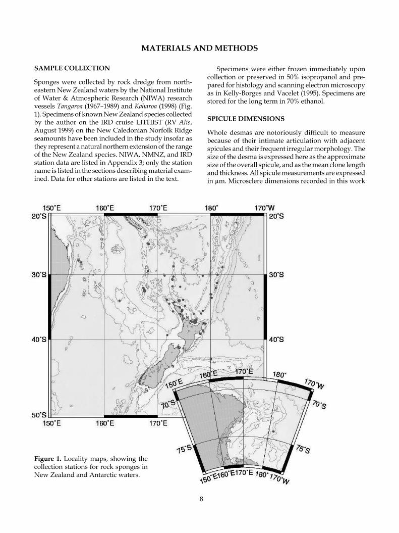
Sponges were collected by rock dredge from north-eastern New Zealand waters by the National Institute of Water & Atmospheric Research (NIWA) research vessels Tangaroa (1967–1989) and Kaharoa (1998) (Fig. 1). Specimens of known New Zealand species collected by the author on the IRD cruise LITHIST (RV Alis, August 1999) on the New Caledonian Norfolk Ridge seamounts have been included in the study insofar as they represent a natural northern extension of the range of the New Zealand species. NIWA, NMNZ, and IRD station data are listed in Appendix 3; only the station name is listed in the sections describing material exam-ined. Data for other stations are listed in the text.
Specimens were either frozen immediately upon collection or preserved in 50% isopropanol and pre-pared for histology and scanning electron microscopy as in Kelly-Borges and Vacelet (1995). Specimens are stored for the long term in 70% ethanol.
SPICULE DIMENSIONS
Whole desmas are notoriously difficult to measure because of their intimate articulation with adjacent spicules and their frequent irregular morphology. The size of the desma is expressed here as the approximate size of the overall spicule, and as the mean clone length and thickness. All spicule measurements are expressed in µm. Microsclere dimensions recorded in this work
MATERIALS AND METHODS
Figure 1. Locality maps, showing the collection stations for rock sponges in New Zealand and Antarctic waters.

9
are generally given as mean length (range of length measurements) and mean width (range of width meas-urements) using the measurements of 10–15 spicules where possible. In the case of the diactinal megascleres, it was very difficult to measure whole spicules as they were typically fragmented. Dimensions are listed in tabular format in the description of each species. The colour of the sponges in life and in ethanol is given by name and also code according to the Reinhold Colour Atlas (Kornerup & Wanscher 1961).
REGISTRATION OF TYPE AND GENERAL MATERIALS
Primary and secondary type materials of new species, and additional material, are deposited in the NIWA Invertebrate Collection at the National Institute of Water & Atmospheric Research (NIWA; formerly New Zealand Oceanographic Institute/ NIWA), Greta Point, Wellington. Previously published type and gen-eral registration numbers for the species Homophymia stipitata and Pleroma aotea are retained in this work, but their new registration numbers (NIWA----) are given in parentheses for reference. Some type and additional material collected by the Coral Reef Research Founda-tion was previously registered at the Natural History Museum, London (BMNH) and the practice is con-tinued here if the material is from non-New Zealand locations. Some material was previously registered in the Museum of New Zealand Te Papa Tongarewa (for-merly National Museum of New Zealand, NMNZ) and retains the prefix NMNZ Por. ---. The prefix IRDR---- is for material sourced with permission from Institut de Recherche pour le Développment (IRD) Noumea, col-lections. Registration numbers are cited in the text.
AREA OF STUDY
Sponge specimens in this monograph are from col-lections made by NIWA (formerly as New Zealand Oceanographic Institute of the DSIR) and the National Museum of New Zealand Te Papa Tongarewa. The col-lection area (Fig. 1) extends from 24° to 74° S and 155° E to 178° W, covering essentially the Lord Howe Rise, Dampier Ridge, South Fiji Basin, parts of the Norfolk Ridge, the Southwest Pacific Basin, Chatham Rise and Subantarctic Slope in the south, and the Ross Sea, Antarctica (Appendix 3). The region includes Norfolk Island, Lord Howe Island, and the Kermadec Islands in the north. Depths range from 80 to nearly 1680 m. In addition to these New Zealand collections, specimens collected by the author from the New Caledonian Norfolk Ridge seamounts, the Lord Howe Seamount Chain, and Palau, Micronesia, were included in the monograph to complete the present revision.
TERMINOLOGY
Specialist terminology for lithistid sponges follows Kelly (2000a) and Pisera and Lévi (2002a). General terminology for sponges is available in Boury-Esnault and Rützler (1997), but some terms are included here for convenience.acantho—prefix meaning roughened or microspined,
e.g. acanthoxea, acanthorhabdamphiaster—microsclere with equal numbers of rays
projecting from both ends of an elongate centrum, equidistant from the centre; the prefix defines the shape of the ray, whether fine and pointed (oxy-) or rounded and robust (strongylo-)
anaxial desma—devoid of a crepis (see sphaeroclone)clad(ome)—see triaene clone—ray-like arm of a desma that is partly (crepis
extends a short way along the clone from the proxi-mal end) or entirely anaxial (devoid of a crepis); the number of clones is determined by the desma geom-etry (modified from Pisera & Lévi 2002a)
crepis—the inceptional body or axial filament of the desma that is visible as a short thread-like canal in monocrepid desmas, or cruciform canals in tetracrepid desmas (modified from Pisera & Lévi 2002a)
desma—articulate choanosomal megasclere with a variety of geometries, often with complex inter-connected morphology, often highly ornamented; found in lithistid sponges (modified from Pisera & Lévi 2002a)
dichotriaene—an ectosomal spicule of triaenose sym-metry (see triaene) with a regular cladome of three branches (protoclads) that can divide further to form usually two clads (deuteroclads); the clads can be smooth, tuberculate, or spinose and are tan-gential to the sponge surface; the rhad can be short or long and is always perpendicular to the clads or cladome, penetrating the sponge surface; axial canals extend the whole length of the rhabd and clads (modified from Pisera & Lévi 2002a)
dicranoclone—see monocrepid desma; arch-shaped and bearing well-developed fungiform tubercles, can be bi-, tri-, or sometimes tetrapodial (four-footed); zygomes are terminal on clones and articulate with the upper tubercles of clones from adjacent desmas, e.g. in Corallistes and Herengeria; (Corallistidae) (modified from Pisera & Lévi 2002a)
discotriaene—ectosomal megasclere with tangential cladome forming a flat or slightly concave oval disc, margins can be even or incised; rhabd is short, crepis is tetraxial, e.g. in Discodermia (Theonellidae) (modi-fied from Pisera & Lévi 2002a)
ectosome—refers to the region just below and at the surface of the sponge; frequently referred to in terms of the architecture of this region as it is of-ten diagnostic and quite different from the under

10
lying choanosome (see Boury-Esnault & Rützler 1997)
heloclone—see monocrepid desma; elongate, oxea-like, smooth with notch-shaped zygomes, e.g. in Costifer (Isoraphiniidae)
megaclone—see monocrepid desma; arched and smooth with cup- or saddle-shaped zygomes, e.g. in Pleroma (Pleromidae)
megasclere—see spicule microsclere—see spicule monaxial desma—see monocrepid desmamonocrepid desma—monaxial desma; the monocrepidial
nature is revealed by the crepis which is a short straight canal in the middle of the clone (modified from Pisera & Lévi 2002a) (see megaclone, heloclone, dicranoclone, rhizoclone)
phyllotriaene—ectosomal spicule with a single ray called a rhabd, usually perpendicular to the sponge surface and penetrating it, and three others are more or less flat and tangential to the sponge surface, termed the cladome; the cladome is branched in an irregular manner with the clads resembling feathers; the crepis is tetraxial and very short in the radiat-ing branches of the cladome (modified from Pisera & Lévi 2002a)
pseudodiscotriaene—ectosomal megasclere resem-bling a discotriaene and analogous to it, but is monocrepid with the crepis located in the rhabd or cladome (e.g. Neopeltidae) (modified from Pisera & Lévi 2002a)
pseudophyllotriaene—ectosomal spicule closely re-sembling a phyllotriaene and analogous to it, but monocrepid (crepis may be located in the rhabd or in the cladome) (modified from Pisera & Lévi 2002a)
pseudospheraster—microscleres with slightly acentri-cally projecting massive spiny rays and a swollen centrum, that resemble spherasters, but are most probably modified amphiasters
pseudotetraclone—monocrepid desma; complex shape with elongate clones resembling a tetraclone or rhizoclone; the term megarhizoclone has been used to describe a variety of non-related desmas that superficially resemble rhizoclones; Pisera and Lévi (2002a) recommended that this term be abandoned and favoured the use of pseudotetraclone instead of megarhizoclone (see Kelly 2000)
rhab(dome)—see triaene rhizoclone—monocrepid desma; usually with numerous
spines and or tubercles that serve as lateral zygomes (modified from Pisera & Lévi 2002a)
sphaeroclone—anaxial desma; in which several ray-like arms extend from a globular centre that may be spinose (modified from Pisera & Lévi 2002a)
spicule—opaline-silica or calcium carbonate com-ponent of the poriferan skeleton; often of elabo-rate morphology and design; three general size categories include the megasclere―the primary
structural skeletal component, the mesosclere― a medium-sized spicule found in Homosclero-phorida, and microsclere―the smallest size of spicule (see Boury-Esnault & Rützler 1997)
streptaster—microsclere with rays projecting irregu-larly along an elongate centrum, the number of rays ranging from numerous to only a few
tetraclone—tetracrepid desma; may be smooth or tu-berculate; very regular, often with four clones, but often secondarily modified
tetracrepid desma—tetraxial desma, tetraclone; the tetracrepidial nature is revealed by the crepis which is cruciform; zygomes mostly terminal on clones (modified from Pisera & Lévi 2002a)
tetraxial desma—see tetracrepid desmatriaene—general term for a tetractinal megasclere with
four rays (clads) emanating from a single point in three axial planes (cladome), having one unequal ray (rhabd, rhabdome) that is commonly much longer than the other three rays (see Boury-Esnault & Rützler 1997)
tuberculate—warty; tubercles have a rounded apex and straight or restricted sides, forming a fungiform (mushroom-shaped) tubercle
zygome—the articulating clasp formed between the tips of clones of adjacent desmas (modified from Pisera & Lévi 2002a)
ABBREVIATIONS OF INSTITUTIONS
AUT Auckland University of Technology.CRRF Coral Reef Research Foundation, based in
the Republic of Palau, Micronesia.IRD Institut de Recherche pour le Développe-
ment (formerly ORSTOM).MNHN Muséum National d’Histoire Naturelle,
Paris.NHM The Natural History Museum (formerly
British Museum (Natural History)), London.NIWA National Institute of Water and Atmos-
pheric Research (including the former New Zealand Oceanographic Institute, NIWA), Greta Point, Wellington.
NMNZ Museum of New Zealand Te Papa Ton-garewa (formerly National Museum of New Zealand), Wellington.
0CDN Sample numbers for material collected by the Coral Reef Research Foundation for United States National Cancer Institute shallow-water collection programme. A complete collection of all 0CDN sponge specimens is located at the Smithsonian Institution (U.S. National Museum), and with the author.
USNM U.S. National Museum (Smithsonian Insti-tution).
11
Class DEMOSPONGIAEFamily AZORICIDAE
Leiodermatium colini n. sp.*Leiodermatium dampieri n. sp.Leiodermatium intermedia (Sollas, 1888)*Leiodermatium linea n. sp.
Family CORALLISTIDAE
Awhiowhio osheai n. gen. n. sp.Awhiowhio sepulchrum n. gen. n. sp.Awhiowhio unda n. gen. n. sp.Herengeria auriculata Lévi & LéviHerengeria vasiformis Schlacher-Hoenlinger et al.Neoschrammeniella antarctica n. sp.Neoschrammeniella fulvodesmus (Lévi & Lévi)
Family ISORAPHINIIDAE
Costifer wilsoni Lévi
Family MACANDREWIIDAE
Macandrewia spinifoliata Lévi & Lévi
Family NEOPELTIDAE
Callipelta punctata Lévi & LéviHomophymia stipitata KellyLepidothenea incrustans (Dendy) Incertae sedisNeopelta pulvinus n. sp.
Family PLEROMIDAE
Pleroma aotea KellyPleroma menoui Lévi & LéviPleroma turbinatum Sollas
Family PHYMATELLIDAE
Neoaulaxinia clavata (Lévi & Lévi)Neoaulaxinia persicum n. sp.Neoaulaxinia zingiberadix n. sp.Neosiphonia motukawanui n. sp.Neosiphonia superstes SollasReidispongia coerulea Lévi & Lévi
Family Scleritodermidae
Aciculites manawatawhi n. sp.Aciculites pulchra DendyAciculites sulcus n. sp.Microscleroderma novaezelandiae n. sp.Scleritoderma flabelliformis Sollas
Family THEONELLIDAE
Discodermia proliferans Lévi & Lévi
* The two asterisked species of Leiodermatium described in this monograph are found only in the west-central Pacific and Micronesian deep waters, but are included in here for the sake of a complete review of the genus in the Pacific.
CHECKLIST OF SPECIES

12
The lithistid Demospongiae have recently been revised by Pisera (2002) and Pisera and Lévi (2002a-o) and are considered by them to comprise 13 extant families with 26 valid genera. The systematics scheme that is used in this volume follows these recent revisions. The reader is referred to the Systema Porifera―a major publication for full family and genus-level synonymies.
Lithistid taxa are extremely difficult to identify without practice. To assist field and laboratory iden-tification, a summary section termed Key Diagnostic Characters is given immediately after the Remarks following the formal description of each species. The characters listed are those that can be used rapidly in succession in the field and laboratory to determine ge-nus and species in relation to morphologically similar other species. Keys to the diagnostic field and micro-scopic characters of New Zealand lithistid sponges is given in Appendices 1 and 2.
Lithistid Demospongiae(formerly order Lithistida Schmidt, 1870)
Polyphyletic group of encrusting, pedunculate, fungi-form, auricular (ear-shaped), fan-shaped, cup-shaped, or massive-amorphous demosponges characterised by the presence of articulated desma megascleres of diverse morphology that render most such sponges rigid and rock-like. Choanosomal desmas are morpho-logically diverse and comprise tetraxial (tetraclone), monaxial (rhizoclone, megaclone, dicranoclone, helo-clone, or various complex branched forms), polyax-ial, or anaxial (sphaeroclone) geometry. Additional choanosomal megascleres may include long hair-like oxeas. Ectosomal spicules may include dichotriaenes, phyllotriaenes, pseudophyllotriaenes, discotriaenes, pseudodiscotriaenes, and various roughened oxea-like spicules. Microscleres include a combination of spirasters, streptasters, amphiasters, sigmaspires, roughened oxeas or raphides (after Pisera & Lévi 2002a).
Remarks: The informal group lithistid Demospongiae has recently been revised by Pisera (2002) and Pisera and Lévi (2002a-o) and is considered by them to contain 13 extant families with 26 valid genera (five of uncer-tain affinity). The classification scheme that is used in this volume, and the sequence of families and genera, follows these recent revisions. For a full discussion of each family and genus see Pisera (2002) and Pisera and Lévi (2002a-o).
Family THEONELLIDAE Lendenfeld, 1903
Theonellidae Lendenfeld, 1903: 125 (in part); Lendenfeld 1907: 343 (part); Wilson 1925: 447; Lévi 1991: 79; Pisera & Lévi 2002e: 327.
Discoderminae Schrammen, 1910: 97.Discodermiidae: Schrammen 1924: 37, 48; de Laubenfels
1955: E58 (part).
Polymorphic; choanosomal spicules are tetraclone desmas that, in several genera, may be non-articulated with neighbouring desmas, being tetralophose in general shape; ectosomal spicules are phyllotriaenes, discotriaenes, or variations between these forms; cho-anosomal diactinal megascleres often have a strongy-lote morphology, some with tylote (hammer-like) proximal and blunt stylote distal ends, or these may be long thin oxeas; microscleres include acanthorhabds, acanthostrongyles, microxeas, streptasters, or pseu-dospherasters (modified from Pisera & Lévi 2002e).
Discodermia du Bocage, 1870
Discodermia du Bocage, 1870: 1; Zittel 1878: 37, 87; Sollas 1888: 292 (part); Pisera & Lévi 2002e.
Theonellidae with ectosomal discotriaenes; regular tetraclone desmas with smooth or tuberculate clones and zygoses; choanosomal megascleres long oxeote or stylote; microscleres are large curved acanthoxeas and smaller acanthorhabds.
Remarks: Hinde and Holmes (1892) illustrated several microfossil discotriaenes from the Oamaru Diatomite (c. 35 Ma) at Oamaru, North Otago, indicating that the genus Discodermia (or Theonella as per Hinde & Holmes, 1892, pl. 14, figs 4, 5, 8–11) was present in the southern New Zealand location that is now Oamaru, during the late Eocene. One microfossil was attributed to Discodermia sinuosa Carter, 1881, originally described from the Gulf of Manaar and the southeast coast of Sri Lanka (Hinde & Holmes 1892, pl. 14, fig. 12). The dis-cotriaene of this species also has indented margins, but the similarity of the illustration to the phyllotriaenes of Macandrewia spinifoliata Lévi & Lévi cannot be over-looked. Conspecificity with an extant species from the Western Indian Ocean is less likely.
TyPe sPeCies: Dactylocalyx polydiscus Bowerbank, 1869.
SYSTEMATICS

13
Discodermia proliferans Lévi & Lévi, 1983 (Fig. 2)
Discodermia proliferans: Lévi & Lévi, 1983: 121, pl. 6 (fig. 4), pl. 7 (figs 1, 2, 4–9); Schlacher-Hoenlinger et al. 2005: 681, figs 4A, 17, 31.
maTerial examined: niwa s ta t ions : S572 (NIWA18212) ; X768 (NIWA18213). ird stations: LITHIST Cruise (August 1999) DW5 (NIWA18480), DW7 [NIWA18481, NIWA18482 (IRDR1829)].
disTribuTion: Passe de la Havannah (holotype), Introu-vable, Stylaster, and Éponge Seamounts, south New Caledonian slope of Norfolk Ridge; west of Three Kings Ridge, northern New Zealand; Hikurangi Plateau, east of East Cape, New Zealand.
habiTaT: Sponges were dredged from hard surfaces on the tops of seamounts and continental margins; the substratum of sponges from NIWA Stn X768 was grey mudstone. Depth range 175–936 m.
desCriPTion: Morphology arborescent, often a curved arched column with lateral tuberculate projections, frequently joined to neighbouring sponges (Fig. 2A).
Dimensions 40–65 mm long, column 5–10 mm thick, 20 mm thick at widest point including projections.
Texture stony, granular.Surface smooth with subdermal aquiferous canals
visible.Colour cinnamon brown in life (6D6), ivory in
ethanol (4B3).Choanosomal skeleton is composed of a dense articu-
lation of robust tetraclone desmas interspersed with dense masses of large acanthorhabds (Fig. 2B,C).
Ectosomal skeleton is composed of discotriaenes, acanthoxeas, and acanthorhabds (Fig. 2D). Occasional diactinal spicules traverse the deeper choanosome
perpendicular to the surface but do not pierce the surface.
Megascleres (Table 1) robust tetraclone desmas (Fig. 2B–D) with thick short smooth tubercles covering the entire spicule, zygoses are very tight and intricate. Discotriaenes (Fig. 2C–E) with the plane of the disc uneven and the edges frequently faintly crenulate, or deeply incised owing to obstruction from the rhabds of adjacent discotriaenes, rhabds short and conical. Diac-tinal megascleres with strongylote proximal and distal termini, with spicule curved and abruptly slimming at the ends and curveing inwards, uncommon.
Microscleres (Table 1) large acanthoxeas, faintly centrotylote, and occasionally sharply angled around midsection of spicule (Fig. 2C). Acanthorhabds with strongylote ends, occasionally centrotylote (Fig. 2F).
remarks: All specimens examined conform to the holo-type of Discodermia proliferans from New Caledonia (Lévi & Lévi 1983) and the species is typical of the genus with distinct discotriaenes and two forms of acanthose microsclere. However, although some specimens from Éponge Seamount DW7 (NIWA18481–2) had the same morphology, they had abundant diactinal spicules and differred in the possession of smaller thinner acanthox-eas and acanthorhabds, and the desmas were smaller and ridged rather than distinctly tuberculate.
Tuberculate ornamentation on the clones of Atlan-tic and Pacific species of Discodermia spp. is rare. The only other species of Discodermia known to the author that has annular rings or partial ridges and slight nodules on the clones is an undescribed species from Jamaica (Kelly & Pomponi unpubl.). NIWA18481 and NIWA18482 from Éponge Seamount (DW7) are very close to Discodermia gorgonoides Burton, 1928 described from the Andaman Islands in the Indian Ocean, except that the latter species lacks acanthoxeas. The holotype of D. gorgonoides was only a fragment of what was thought to be a branching sponge with oxeas and notched discotriaenes, but these are only about 180 µm wide. The nodules on D. gorgonoides are not as pronounced as on typical specimens of D. proliferans.
Table 1. Spicule dimensions (µm) of Discodermia proliferans Lévi & Lévi, 1983. Desmas Discotriaenes Diactines Acanthoxeas Acanthorhabds (Clone L/Thickness) (Max disc ø) (L) (L/Thickness) (L/Thickness)
New ZealandNIWA18212 300 (250–400)/ 243 (190–243) 400–1500 104 (95–112)/3–4 13 (10–15)/3–4 100 (80–150)NIWA18213 340 (250–400)/100 540 (375–725) 400–1500 90 (90–100)/5–6 8 (7–10)/3–4
New CaledoniaNIWA18480 300(200-400)/100 420 (240–620) 530 86 (70–110)/2–3 10 (7–13)/2–3Lévi & Lévi (1983) 300/50 200–300, 250–400 — 60–80/3–5 8–11S.-H. et al. (2005) 500–600/80–90 492–673 — 84–117/6–7 10–14/5–6

14
Figure 2. Discodermia proliferans Lévi & Lévi, 1983.
15
Branching morphology in species of Discodermia is not uncommon although most species are cup-shaped or hemispherical with massive or nodular morphology. Discodermia vermicularis Döderlein, 1884 from Japan has digitate outgrowths arising from an encrusting base and D. ramifera Topsent, 1892 from the Azores is digi-tate. One of the commonest species of Discodermia in the central western tropical Atlantic is digitate (Kelly-Borgest et al. 1994) and a further (undescribed) tiny branching species is known from the Gulf of Mexico (Kelly & Pomponi unpubl.).
Specimens from New Zealand and New Caledonia (Lévi & Lévi 1983; Schlacher-Hoenlinger et al. 2005) present a broad range of spicule dimensions, particu-larly the desmas, discotriaenes, and acanthoxeas, but are considered to be conspecific despite this.
key diagnosTiC CharaCTers:• Sponge forms a small nodulose branch or thick ir-
regular nodulose lump.• Surface of sponge covered with disc-like triaenes.• Desmas are heavily tuberculate.• One form of small microrhabd microsclere.
Family PHYMATELLIDAE Schrammen, 1910
Phymatellinae Schrammen, 1910: 72. Phymatellidae: Schrammen 1924; Lévi 1991: 79; Pisera &
Lévi 2002k: 366.
Clavate, multiclavate (tuberose or corm-like), spheri-cal with a restricted base, and cup-shaped to foliose sponges; desmas tetraclones with large, smooth, tuber-culate or semi-spinose clones; in some genera clones are branched and ramified; ectosomal megascleres are relatively short-shafted dichotriaenes and long thin oxeas; ectosomal microscleres are acanthose strongy-loamphiasters with robust rays, some with streptaster or spiraster modifications, choanosomal microscleres are acanthose amphiasters that merge into streptasters with slender oxeote rays, often in two size categories.
remarks: The Phymatellidae contains three valid extant genera differentiated primarily by their habit (Pisera & Lévi 2002k), described as being clavate or apple-shaped in Neoaulaxinia Pisera & Lévi, spherical with a stem in Neosiphonia Sollas, and cup-shaped or lamellate in Reidispongia Lévi & Lévi. New specimens of Neoaulaxinia clavata (Lévi & Lévi), Neosiphonia su-perstes Sollas, and Reidispongia coerulea Lévi & Lévi have enabled clarification of the skeletal differences between the genera, focusing on the degree of and nature of ornamentation of the tetraclone desmas and the definition of the microscleres. The recognition of
two new species of Neoaulaxinia, and one of Neosiphona has extended the list of diagnostic characters that en-able easier differentiation at the generic and species level within the family.
It is interesting to note that microfossil tetraclones and ectosomal triaenes indistinguishable from those of extant phymatellid genera were found in the Tutuiri Greensand (Teurian–basal Waipawan) in the Chatham Islands (Buckeridge & Kelly 2005, fig. 4C), and illus-trated from the Oamaru Diatomite at Oamaru in North Otago (Hinde & Holmes 1892; pl. 13, figs 28–30), con-firming a more southern distribution for Phymatellidae during the Paleogene.
Neoaulaxinia Pisera & Lévi, 2002
Spherical Phymatellidae with a restricted base, elongated to various degrees, forming clavate (club-shaped), multiclavate, tuberose, or apple-shaped spe-cies; subdermal aquiferous canals visible beneath the ectosome converge on a single membranous oscule in a shallow apical depression; desmas are simple tetra-clones with smooth clones that vary in the degree of ornamentation (single to multiple fungiform tubercles, to spires of tubercles). Desmas branch at the terminus of the clones. Zygosis is highly developed in mature spicules, forming an enlarged complex tangle; zygosis is positioned terminally and laterally with saddle for-mations; ectosomal dichotriaenes with finely tapered frequently irregularly curved clades and rhabd; three clearly differentiated microscleres, a robust strongy-loamphiaster restricted to an ectosomal crust, a large streptaster with only a few conical rays and a smaller streptaster with numerous fine rays, restricted to the choanosome.
TyPe sPeCies: Aulaxinia clavata Lévi & Lévi, 1988.
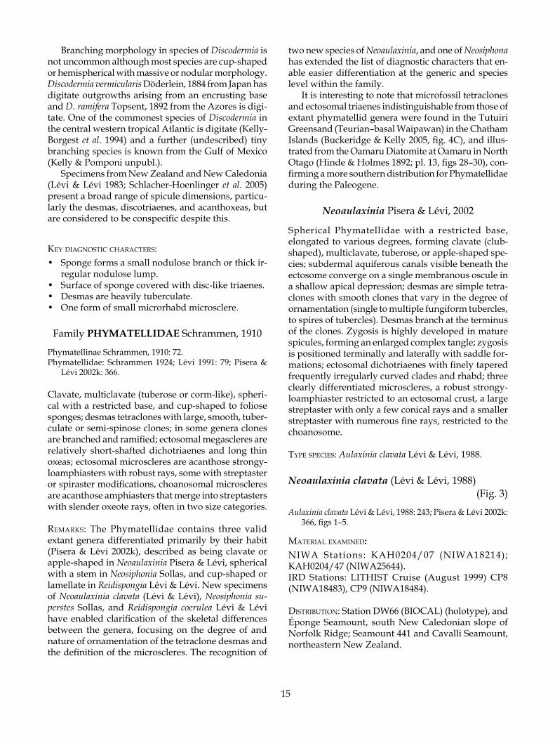
Neoaulaxinia clavata (Lévi & Lévi, 1988) (Fig. 3)
Aulaxinia clavata Lévi & Lévi, 1988: 243; Pisera & Lévi 2002k: 366, figs 1–5.
maTerial examined: niwa Stations: KAH0204/07 (NIWA18214); KAH0204/47 (NIWA25644).IRD stations: LITHIST Cruise (August 1999) CP8 (NIWA18483), CP9 (NIWA18484).
disTribuTion: Station DW66 (BIOCAL) (holotype), and Éponge Seamount, south New Caledonian slope of Norfolk Ridge; Seamount 441 and Cavalli Seamount, northeastern New Zealand.
16
Figure 3. Neoaulaxinia clavata (Lévi & Lévi, 1988).
17
habiTaT: Dredged from hard coral substratum on the tops of seamounts. Depth range 505–880 m.
desCriPTion:Morphology club-shaped, slightly thicker in the upper third, tapering to a rounded apex (Fig. 3A), or multicla-vate in older specimens, with a rounded stem, apple-shaped apical bulb, and lateral club-shaped protuber-ances (Fig. 3B). A single small membranous oscule is visible on the apex of each specimen (or protuberance), less than 1 mm diameter; apical cavity visible only in some specimens, and then only 1–2 mm deep.
Dimensions of single clubs 60–70 mm long, 15–20 mm thick at broadest; multiclavate specimen 160 mm high and 80 mm wide.
Texture firm, slightly compressible.Surface smooth, glistening, with longitudinal sub-
dermal aquiferous canals clearly visible.Colour in life and in ethanol ivory (4B3).Choanosomal skeleton relatively cavernous with aquif-
erous canals permeating the skeleton, and composed of weakly articulated dispersed desmas, between which are abundant microscleres of the largest category.
Ectosomal skeleton encrusted with robust microscle-res, interspersed with the cladomes of the dichotri-aenes. Large robust oxeas arise in tracts from within the deeper choanosome, but do not pierce the surface.
Megascleres (Table 2) tetraclone desmas with smooth even clones that branch just before they terminate, each branch dividing into multiple tubercular fingers (Fig. 3C); tubercles are typically single and slightly fungi-form (Fig. 3D), zygosis with adjacent spicules is weak, and can be positioned laterally. Dichotriaenes (Fig. 3E) have finely pointed clads and are relatively thin overall. The individual deuteroclads are curved and separated by approximately 45°. In a New Zealand specimen, modification of the cladome to strongylote ends was relatively common (Fig. 3F). Diactinal spicules are relatively thick, finely pointed oxeas (Fig. 3C,E).
Microscleres (ectosomal) are strongyloamphiasters (Table 2, Ms 1, Fig. 3G), choanosomal microscleres are
streptasters in two distinct size categories with fine oxeote rays (Table 2, Ms 2.1, 2.2, Fig. 3F,G). NIWA18483 has spirasters with 2–4 whorls.
remarks: Only a single specimen has been recorded from New Zealand waters; the description and illustra-tions provided are based on this and specimens col-lected from New Caledonian seamounts. The morphol-ogy and skeletal details of the New Zealand specimen are very close to the New Caledonian holotype except that the cladome of the dichotriaenes is frequently malformed with rounded strongylote clades (Fig. 3F) and the cladome is occasionally monotrianeose, with unbranched clades. Excellent illustrations of the details of this species in New Caledonia were provided by Lévi and Lévi (1988) and Pisera and Lévi (2002k).
A large multiclavate specimen was recovered from Éponge Seamount on the New Caledonian end of the Norfolk Ridge that is in every aspect of the skeletal and spicule morphology and dimensions, identical to described and new material (Fig. 3B). The size of this specimen indicates that it might be older than the sin-gle club-shaped specimens typically recovered in the past, and by this we have an indication of the mature morphology.
key diagnosTiC CharaCTers:
• Sponge typically forms an elongate club with a single oscule at the apex.
• Desmas branch terminally and laterally; zygomes are lightly tuberculate.
• There may be spirasters amongst the micro- scleres.
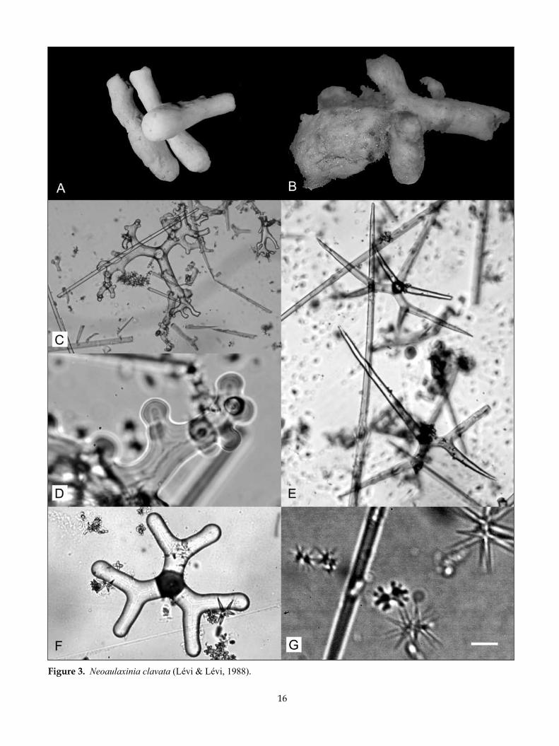
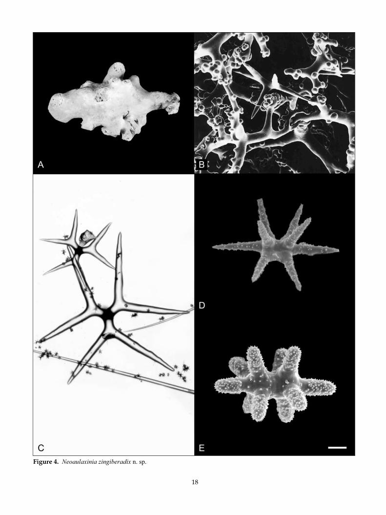
Neoaulaxinia zingiberadix n. sp. (Fig. 4)
maTerial examined: Type material: Holotype BMNH 1995.3.30.3 from NIWA Stn J953.
Table 2. Spicule dimensions (µm) of Neoaulaxinia clavata (Lévi & Lévi, 1988). Desma (Clone L/T) Cladome (Max ø) Diactines Ms 1 (L) Ms 2.1 (L) Spirasters Desma (Overall size) Rhabd (L) (L) Ms 2.2 (L) (L)
New ZealandNIWA18214 375–550/50–60 430 (325–625) — 14 (10–20) 36 (30–40) — 575–750 320 (250–375) 20 (15–25)
New CaledoniaNIWA18483 200–400/40–60 478 (375–575) 350-4000 14 (10–18) 42 (38–48) 15 (8–18) 575–900 overall 423 (325–475) 15 (13–18) Lévi & Lévi (1988) 300–600/100 250–600 700–4000 13–15 20–40 — 600–1000 overall 200–480
18
Figure 4. Neoaulaxinia zingiberadix n. sp.

19
TyPe loCaliTy: Western continental slope, Northland, New Zealand.
disTribuTion: Western continental slope, Northland, New Zealand.habiTaT: Dredged from hard coral substratum on the tops of seamounts. Depth range 260–270 m.
desCriPTion:Morphology tuberose or corm-like with multiple pro-tuberances of irregular size and shape emerging from the mass of the sponge; the point of attachment is a short stem (Fig. 4A); multiple membranous oscules are visible on apex of each protuberance. No apical cavity around oscules.
Dimensions 50 mm high, 30 mm thick.Texture firm, almost stony.Surface smooth.Colour in life unknown, dried, cream (4A2).Choanosomal skeleton relatively cavernous and is
composed of relatively firmly articulated desmas (Fig. 4B).
Ectosomal skeleton encrusted with microscleres. Ox-eas arise in tracts from within the deeper choanosome, but do not pierce the surface.
Megascleres (Table 3) tetraclone desmas with smooth even clones that branch just before they terminate, each branch dividing into multiple fingers; tubercles are fungiform and frequently multiple (Fig. 4B). Zygosis is strong and situated laterally and terminally. Dicho-triaenes have a short rhabd (Fig. 4B,C). The individual deuteroclads are relatively straight and separated by a greater than 45° angle, so that the individual clads lie close together (Fig. 4C). Diactinal spicules are rela-tively thick centrally, but taper abruptly to strongylote ends.
Microscleres (choanosomal) are amphiasters with streptaster modifications in two close size categories, differentiated primarily on the number of rays on the spicule; the smaller category has more rays than the larger category (Table 3, Ms 2.1, 2.2) (Fig. 4D). Ectos-omal microscleres are strongyloamphiasters with a few robust rays (Table 3, Ms 1, Fig. 4E).
eTymology: Named for the morphology of the sponge that resembles the root (Latin, radix) of the ginger plant (Latin, zingiber).
remarks: Neoaulaxinia zingiberadix is only the second species described in this genus, known only from the Southwest Pacific. The primary character that differentiates this new species of Neoaulaxinia is the morphology of the sponge. Although only a single specimen has been recovered, it is sufficiently distinct from multiclavate specimens of N. clavata to warrant the establishment of a new species.
Neoaulaxinia zingiberadix is much less regularly clavate than N. clavata and the protuberances vary greatly in size and shape (Fig. 4A). Spicule morphology and dimensions also differ considerably; the strongy-loamphiasters are larger in the new species than in N. clavata, but the larger category of streptasters in N. zingiberadix is generally smaller than N. clavata. The dichotriaenes differ dramatically, in the smaller length of the rhabd in N. zingiberadix and in the general shape and orientation of the deuteroclads. In N. zingiberadix the individual deuteroclads are relatively straight and separated by a greater than 45° angle, so that the clads lie close together (Fig. 4C). In N. clavata the individual deuteroclads are curved and emerge with an approxi-mately 45° angle, so that the clads are well separated. As for N. clavata, the desmas are sparsely tuberculate and the tubercles are usually fungiform, but in N. zin-giberadix the tubercles are often double or triple. The articulation of these desmas is very intricate and the sponge is a lot harder as a result. The morphology of the terminal zygosis in the new species is that of sliding clasped fingers (zygosis is lateral as well as terminal) and the lateral zygoses are saddle shaped.
key diagnosTiC CharaCTers:
• Sponge forms a tuberose or corm-like mass. • Desma clones branch terminally and laterally and
are lightly tuberculate, tubercles are frequently double.
• Dichotriaenes have a short rhab and clads emerge from the protorhabd well separated.
Table 3. Spicule dimensions (µm) of Neoaulaxinia zingiberadix n. sp. Desma (Clone L/T) Cladome (Max ø) Diactines Ms 1 (L) Ms 2.1 (L) Desma (Overall size) Rhabd (L) (L) Ms 2.2 (L) New ZealandBMNH 1995.3.30.3 300–500/50–70 490 (250–775) 775+ 19 (18–23) 29 (23–35)(holotype) 500–875 158 (100–300) 18 (15–20)
20
Figure 5. Neoaulaxinia persicum n. sp.
21
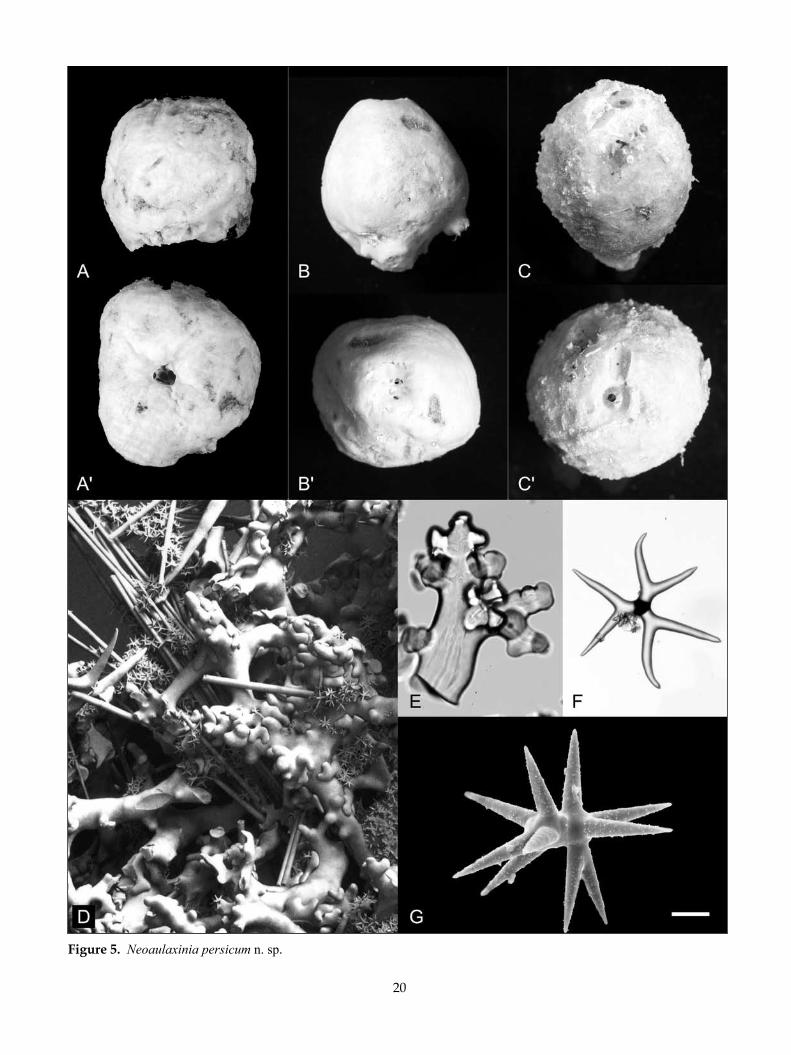
Neoaulaxinia persicum n. sp. (Fig. 5)
maTerial examined: Type material: Holotype NIWA18215 from NIWA Stn X140; paratypes NIWA18221 from NIWA Stn KAH0204/29; NIWA18222 from NIWA Stn KAH0204/30; NIWA18216 from NIWA Stn KAH0204/32; NIWA18217, NIWA18218, NIWA 18219 and NIWA18220 from NIWA Stn KAH0204/40.
NIWA Stations: E731 (NIWA25613); KAH0204/02 (NIWA18225) ; KAH0204/07 (NIWA18226) ; KAH0204/29 (NIWA18227, NIWA18228, NIWA18229, NIWA18230); KAH0204/32 (NIWA18231, NIWA18232, NIWA18233) ; KAH0204/38 (NIWA18234) ; KAH0204/40 (NIWA18235, NIWA18236); KAH0204/44 (NIWA18237) ; KAH0204/47 (NIWA18238) ; RAPUHIA2-15 (25647); T226 (NIWA25650); TAN0104/001 (NIWA18240) ; TAN0104/002 (NIWA1824, NIWA18247, NIWA18248); TAN0104/048 (NIWA18239); TAN0104/148 (NIWA18242); TAN0104/194 (NIWA18243) ; TAN0104/288 (NIWA18244); TAN0104/289 (NIWA18245); TAN0104/394 (NIWA18246); TAN0107/232; X201 (NIWA18223); Z9025 (NIWA18224).
TyPe loCaliTy: Bay of Plenty, North Island, New Zea-land.
disTribuTion: Seamount east of Three Kings Ridge; West Cavalli, Cavalli, South Cavalli Seamounts, and Seamount 441, northeastern New Zealand; Knights Terrace, East of Poor Knights Islands; Rumble V Seamount, West of White Island, and Ngatoro Ridge, Bay of Plenty; North of Raoul Island, Kermadec Ridge; Diabolical, Graveyard, Morgue, and Scroll Seamounts, Chatham Rise.
habiTaT: Attached to small pieces of scleractinean coral rubble and rock surfaces on the sides of seamounts. Many of the specimens examined appear to have set-tled on hard coral, eventually engulfing them as they
grew. Depth range 503–1680 m.
desCriPTion:Morphology spherical (Fig. 5A) to egg-shaped (Fig. 5B,C); the base is a thin skirt, or encompassing branches of scleractinian corals. A single membranous oscule is situated at the apex of the sponge surrounded by deep subdermal aquiferous canals that converge on the oscule (Fig. 5A). A fringe of very long diactines is present on the base of some specimens.
Dimensions 50 (20–65) mm high, 50 (20–80) mm wide, and 40 (20–60) mm thick.
Texture stony overall, but crumbly on the surface, ectosomal region easily sloughed off.
Surface smooth, slightly rough, unabraided sections have short robust conules dispersed about 5–10 mm apart, raised from projecting diactines (Fig. 5C).
Colour in ethanol pale orange (5A3), honey yellow (5D6) to topaz (5C5).
Choanosomal skeleton composed of robust smooth tetraclones tightly zygosed with adjacent desmas both terminally and laterally. Long diactines emerge from the centre of the sponge in tracts, piercing the surface in low conules. Large microscleres are abundant and regularly distributed within the choanosome.
Ectosomal skeleton an encrustation of small micro-scleres.
Megascleres (Table 4) tetraclone desmas with very thick smooth clones that rarely branch, and if they do it is towards the terminus of the clone (Fig. 5D). The clones are ornamented with single or double pairs of tubercles or spires of numerous tubercles. Spires oc-cur around the axis of the clone (which is generally quite smooth) and are generally in the most mature spicules (Fig. 5E). Zygosis is terminal although zygoses frequently adhere laterally with a saddle-shaped for-mation when against adjacent clones (Fig. 5D). The zygoses in older portions of the skeleton are often so intricate and over-developed as to be enlarged many times thicker than the clones. Dichotriaenes with one set of clads shorter than the other two, and one of these
Table 4. Spicule dimensions (µm) of Neoaulaxinia persicum n. sp. Desma (Clone L/T) Cladome (Max ø) Diactines Ms 1 (L) Ms 2.1 (L) Desma (Overall size) Rhabd (L) (L) Ms 2.2 (L) New ZealandNIWA18215 250–500/60–100 627 (350–875) 750+ 17 (15–20) 50 (43–65)(holotype) 675–750 496 (375–550) 26 (18–35)
NIWA18216 375–450/100–120 694 (525–900) 800+ 19 (18–23) 46 (33–55)(paratype) 750–750 388 (375–400) 21 (18–32)
NIWA18221 250–500/80–100 600 (475–700) 1250+ 17 (15–20) 46 (37–50) (paratype) 500–875 — 24 (20–25)
22
shorter clads is usually shorter than the other giving the cladome a lopsided appearance (Fig. 5F). The re-maining two sets of clads curve inwards, sometimes acutely, to resemble calipers. The tips of the clads are often blunt and strongylote. The rhad is often curved with a strongylote end. Diactines (Fig. 5D) are very thick with oxeote or frequently strongylote ends and are up to several mm long. They are bundled in tracts that emerge within the choanosome. Mature spicules are over 3 mm long.
Microscleres (ectosomal) are small strongyloam-phiasters with very robust rays (Table 4, Ms 1), cho-anosomal microscleres are large streptasters (Fig. 5G) with a reduced number of rays (Table 4, Ms 2.1), and smaller streptasters with numerous sharp rays (Table 4, Ms 2.2).
eTymology: persicum (L.) = peach.
remarks: Neoaulaxinia persicum is clearly differentiated morphologically from N. clavata and N. zingiberadix. The new species is egg-shaped with a restricted base, contrasting sharply with the elongate club-shaped N. clavata and tuberose N. zingiberadix. The tracts of ox-eas that emerge in a conule at regular intervals on the surface of N. persicum are also highly characteristic of sponges that have been recovered with little damage.
At the microscopic level there are several clear differences in the size of the various categories of mi-croscleres and the morphology of the key diagnostic megascleres. In particular, the ornamentation of the tetraclone desmas with multituberculate spires is unique in the genus and the clones are much thicker than in both other species. Zygosis is highly developed in mature spicules, with an enlarged complex tangle of terminal clasps and lateral saddle formations. The dichotriaenes in N. persicum have a much wider and more irregular cladome and a longer rhabdome than in the other two species. The choanosomal microscleres are the largest and most robust of species in the genus and all microscleres are only very slightly acanthose, much less so than all other Phymatellidae described thus far.
N. persicum is an easily recognised species that is so far known only from the north and east of New Zealand from the Three Kings Rise and the Chatham Rise.
key diagnosTiC CharaCTers:
• Sponge is spherical to egg-shaped with an apical oscule and subdermal canals
• Desmas with very thick smooth clones, with spires of tubercles, zygosis is very intricate and robust
• Microscleres are very large and only slightly acan-those
• The immature sponge could be mistaken for Ne-osiponia motukawanui n. sp.
Neosiphonia Sollas, 1888
Club-shaped to spherical body with a restricted base of attachment, a bundle of vertical canals arises in the central axis of the sponge emerging within a cavity on the sponge apex, the cavity is covered with a delicate parchment-like ectosomal roof; desmas are branched and ramified tetraclones with sculpted sinuous or tu-berculate projections on the clones. Zygosis is intricate in inner parts of the skeleton, weak in the outer (grow-ing) regions of the sponge; dichotriaenes are generally short-shafted (100–200 µm on average), the cladome is wide with clads of irregular length; microscleres are acanthose in two clearly differentiated groups, a robust strongyloamphiaster restricted to an ectosomal crust, and a choanosomal streptaster with conical rays.
TyPe sPeCies: Neosiphonia superstes Sollas, 1888.
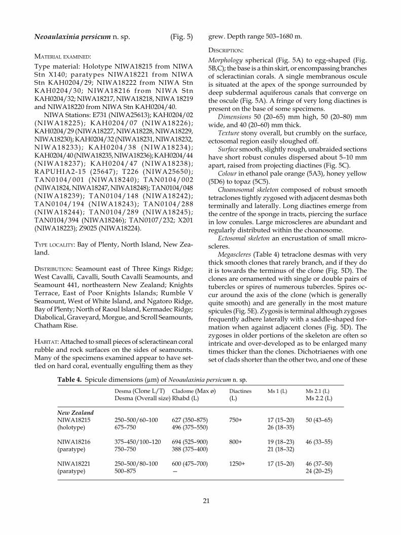
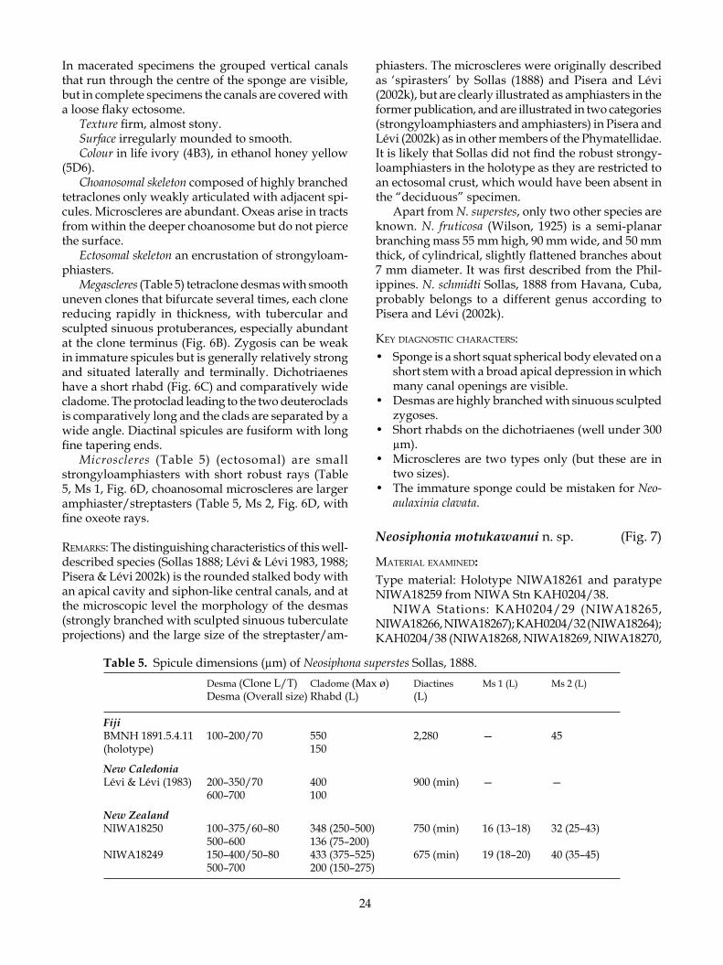
Neosiphonia superstes Sollas, 1888 (Fig. 6)
Neosiphonia superstes Sollas, 1888: 299, pl. 31, figs 7–12; Lévi & Lévi 1983: 119, fig. 10; Lévi & Lévi 1988: 47, pl. 3, fig. 3; Pisera & Lévi 2002k: 366, figs 6–9.
maTerial examined: NIWA Stations: E731 (NIWA25614); KAH0204/07 (NIWA18258) ; KAH0204/32 (NIWA18257) ; KAH0204/47 (NIWA18256) ; TAN0107/124 (NIWA18255) ; TAN0205/20 (NIWA25643) ; TAN0205/32 (NIWA18249, NIWA18250, NIWA18251); TAN0413/120 (NIWA25051).
disTribuTion: Challenger Station 173, off Matuka, Fiji Islands (holotype); BIOCAL Stn DW66, south New Caledonian slope of Norfolk Ridge; East Kermadec Ridge Slope, Volcano E, Kermadec Ridge; Rumble III Seamount and Mahina Knoll, Bay of Plenty; Seamount 441, West Cavalli, and Cavalli Seamounts, northeastern New Zealand.
habiTaT: Attached to hard dead coral or rocky substra-tum. Depth range 430–999 m.
desCriPTion:Morphology short, squat, spherical body elevated on a short restricted base of attachment with a moderately deep apical cavity (Fig. 6A).
Dimensions typically 35 (20–50) mm high and 35 (15–60) mm at the widest part of the body. The base ranges from 15–20 mm wide. The apical cavity indents 30–35% of the apex and ranges in depth from 5–10 mm.
23
Figure 6. Neosiphona superstes Sollas, 1888.
24
phiasters. The microscleres were originally described as ‘spirasters’ by Sollas (1888) and Pisera and Lévi (2002k), but are clearly illustrated as amphiasters in the former publication, and are illustrated in two categories (strongyloamphiasters and amphiasters) in Pisera and Lévi (2002k) as in other members of the Phymatellidae. It is likely that Sollas did not find the robust strongy-loamphiasters in the holotype as they are restricted to an ectosomal crust, which would have been absent in the “deciduous” specimen.
Apart from N. superstes, only two other species are known. N. fruticosa (Wilson, 1925) is a semi-planar branching mass 55 mm high, 90 mm wide, and 50 mm thick, of cylindrical, slightly flattened branches about 7 mm diameter. It was first described from the Phil-ippines. N. schmidti Sollas, 1888 from Havana, Cuba, probably belongs to a different genus according to Pisera and Lévi (2002k).
key diagnosTiC CharaCTers:• Sponge is a short squat spherical body elevated on a
short stem with a broad apical depression in which many canal openings are visible.
• Desmas are highly branched with sinuous sculpted zygoses.
• Short rhabds on the dichotriaenes (well under 300 µm).
• Microscleres are two types only (but these are in two sizes).
• The immature sponge could be mistaken for Neo-aulaxinia clavata.
Neosiphonia motukawanui n. sp. (Fig. 7)
maTerial examined: Type material: Holotype NIWA18261 and paratype NIWA18259 from NIWA Stn KAH0204/38.
NIWA Stations: KAH0204/29 (NIWA18265, NIWA18266, NIWA18267); KAH0204/32 (NIWA18264); KAH0204/38 (NIWA18268, NIWA18269, NIWA18270,
In macerated specimens the grouped vertical canals that run through the centre of the sponge are visible, but in complete specimens the canals are covered with a loose flaky ectosome.
Texture firm, almost stony.Surface irregularly mounded to smooth.Colour in life ivory (4B3), in ethanol honey yellow
(5D6).Choanosomal skeleton composed of highly branched
tetraclones only weakly articulated with adjacent spi-cules. Microscleres are abundant. Oxeas arise in tracts from within the deeper choanosome but do not pierce the surface.
Ectosomal skeleton an encrustation of strongyloam-phiasters.
Megascleres (Table 5) tetraclone desmas with smooth uneven clones that bifurcate several times, each clone reducing rapidly in thickness, with tubercular and sculpted sinuous protuberances, especially abundant at the clone terminus (Fig. 6B). Zygosis can be weak in immature spicules but is generally relatively strong and situated laterally and terminally. Dichotriaenes have a short rhabd (Fig. 6C) and comparatively wide cladome. The protoclad leading to the two deuteroclads is comparatively long and the clads are separated by a wide angle. Diactinal spicules are fusiform with long fine tapering ends.
Microscleres (Table 5) (ectosomal) are small strongyloamphiasters with short robust rays (Table 5, Ms 1, Fig. 6D, choanosomal microscleres are larger amphiaster/streptasters (Table 5, Ms 2, Fig. 6D, with fine oxeote rays.
remarks: The distinguishing characteristics of this well-described species (Sollas 1888; Lévi & Lévi 1983, 1988; Pisera & Lévi 2002k) is the rounded stalked body with an apical cavity and siphon-like central canals, and at the microscopic level the morphology of the desmas (strongly branched with sculpted sinuous tuberculate projections) and the large size of the streptaster/am-
Table 5. Spicule dimensions (µm) of Neosiphona superstes Sollas, 1888. Desma (Clone L/T) Cladome (Max ø) Diactines Ms 1 (L) Ms 2 (L) Desma (Overall size) Rhabd (L) (L) FijiBMNH 1891.5.4.11 100–200/70 550 2,280 — 45 (holotype) 150
New CaledoniaLévi & Lévi (1983) 200–350/70 400 900 (min) — —
600–700 100
New ZealandNIWA18250 100–375/60–80 348 (250–500) 750 (min) 16 (13–18) 32 (25–43) 500–600 136 (75–200)NIWA18249 150–400/50–80 433 (375–525) 675 (min) 19 (18–20) 40 (35–45)
500–700 200 (150–275)

25
Figure 7. Neosiphona motukawanui n. sp.

26
NIWA18271, NIWA18272); KAH0204/47 (NIWA18260); KAH0204/52 (NIWA18262, NIWA18263, NIWA18273, NIWA18274, NIWA18275).TyPe loCaliTy: West Cavalli Seamount, Northland.
disTribuTion: Cavalli Seamount Region, northeastern New Zealand.
habiTaT: Sponges were dredged from hard rocky sub-strata or were attached to small pieces of coral. Depth range 780–910 m.
desCriPTion:Morphology a block-shaped mass with a restricted base of attachment, the apex of which is a flattened parch-ment-textured roof, under which numerous aquiferous canals open from within the sponge (Fig. 7A,B). The upper third of some specimens may be expanded to form a shallow cup with thick rolled edges (Fig. 7B). The base of these sponges is very narrow and root-like (Fig. 7A, B). NIWA18268 has a thin beard of diatines about 40 mm long extending from around the base of the sponge into the substratum, probably acting as an anchoring device. Immature sponges are elongate to spherical with a shallow broad parchment-like apex, gradually thinning to a narrow base of attachment. The sponges are generally taller than they are wide.
Dimensions of immature specimens in the collection are 25 mm high, 15 mm at the widest point, and 5 mm at the base. Most specimens are approximately 50 mm high, 35 mm wide (maximum), and 15 mm wide at the base. Mature specimens are 60–80 mm high, 40–60 mm at the widest point, and have a base of 20 mm.
Texture internally incompressible, externally flakey, crumbly, easily abraided, especially at the apex.
Surface typically smooth, flakey or crumbly. The surface of many specimens is tufted with long diactinal spicules that form conules on the surface, but these are often abraided away in damaged specimens.
Colour apricot yellow (5B6) in ethanol.Choanosomal skeleton dominated by desmas with
thick tuberculate clones and large intricate zygoses. Oxeote microscleres are abundant.
Ectosomal skeleton a crust of robust strongyloam-phiasters. Oxeas arise in tracts from within the deeper choanosome, piercing the surface, and in some loca-tions forming a distinct beard.
Megascleres (Table 6) tetraclone desmas with very thick short clones (Fig. 7C), tuberculation variable and more prominent on the ends of the clones. Single, mul-tiple and spires of tubercles are common. Clones divide at least once, and often twice. Zygoses are very strong, intricate, and enlarged. Dichotriaenes (Fig. 7D) with very wide cladome; clads are thick, irregular and often of uneven length, rhabd short and often strongylote. Dichotriaenes of NIWA18261 are frequently trichotri-aenose and the clads are occasionally strongylote, as is the rhabd. Diactines are very long, straight, evenly thick and strongylote.
Microscleres (ectosomal) are robust strongyloam-phiasters with a few blunt conical rays (Table 6 Ms 1; Fig. 7E), choanosomal microscleres are larger amphiaster/streptasters with fine rays (Table 6 Ms 2; Fig. 7E). In addition to the standard microscleres, specimen NIWA18264 has rhabds with short projec-tions postioned very irregularly along the length of the spicule which is 17–20 µm long. These were not seen in other specimens, and are most probably malformed strongyloamphiasters.
eTymology: Named for the largest island in the Cavalli Island group, Motukawanui, encompassed by the Cav-alli Seamount region that is the general type locality of this new species. In the literal sense, motu means ‘island’, kawa is the New Zealand pepper tree used extensively in ceremonial protocol, and nui means ‘big’ (motukawanui, New Zealand Māori).
remarks: Neosiphonia motukawanui is only the fourth known species of this genus, and one of these, N. schmidti Sollas, 1888 from Havana, Cuba, is thought to belong to a different genus (Pisera & Lévi 2002k). Neosiphonia is characterised primarily by a massive globular body that narrows to a restricted columnar base of attachment, the apex of which consists of a cov-ered shallow depression or atrium into which vertical aquiferous canals open.
Table 6. Spicule dimensions (µm) of Neosiphonia motukawanui n. sp. Desma (Clone L/T) Cladome (Max ø) Diactines Ms 1 (L) Ms 2 (L) Desma (Overall size) Rhabd (L) (L) New ZealandNIWA18261 125–375/70–80 523 (450–675) all broken 18 (18–20) 30 (23–35)(holotype) 460–600 138 (75–250)
NIWA18259 250–400/90–100 619 (425–900) 600+ 21 (18–28) 33 (20–43)(paratype) 500–800 142 (75–250)
NIWA18264 250–500/70–100 590 (350–725) all broken 18 (18–20) 31 (25–38) 500–750 100

27
Neosiphonia motukawanui differs from the genus type N. superstes Sollas in the shape of the sponge; the body region of N. superstes is more globular with a columnar ‘stalk’ (see Fig. 6A), whereas the body of the former species tapers to a thin attenuating stalk (see Fig. 7A). The apical depression atop N. motukawanui is extremely prominent, covering the entire flattened apex of the sponge, while in N. superstes the atrium is smaller and deeper. Long tufted diactines in N. motukawanui are also a prominent feature in well-preserved material. The dichotriaenes have a wider, larger cladome than in N. superstes and the strongyloamphiasters are much thicker and slightly bigger. The desmas of N. motuka-wanui are more robust and tuberculate than the thinner, less ornamented desmas of N. superstes.
Younger specimens of N. motukawanui may be mistaken in the field for Neoaulaxinia persicum, but the latter are spherical without an atrium over the apical cavity, and the former are always club-shaped with low blunt conules. The nature of the ornamentation of the desmas, the size and shape of the microscleres, and the greater rhabd length of the dichotriaenes in N. persicum, are always indicative, however.
key diagnosTiC CharaCTers:• Sponge is block-, club-, or shallow cup-shaped and
the flattened top of the sponge is a delicate ‘parch-ment’ over a shallow cavity; the bottom of the cavity has numerous aquiferous canals opening into it.
• Desmas are very thick, branched several times, and are tuberculate, with single or double spires.
• The robust microscleres are very thick with few conical blunt rays.
• Could be confused with Neoaulaxinia persicum.
Reidispongia Lévi & Lévi, 1988
Cup-shaped with thin smooth walls, elongate chalice-shaped as a young sponge, foliose as a mature sponge; tetraclone desmas are highly ramified with frequently smooth branched clones and tuberculate termini. Zy-gosis is weak or non-existant on the outer (younger) portions of the lamellae, otherwise it is intricate and very strong in the older skeleton; microscleres are two types of amphiaster in two closely related size categories, one a strongyloamphiaster, and the other an amphiaster with fine rays.
TyPe sPeCies: Reidispongia coerulea Lévi & Lévi, 1988.
Reidispongia coerulea Lévi & Lévi, 1988 (Fig. 8)
Reidispongia coerulea Lévi & Lévi, 1988: 245; Pisera & Lévi 2002k: 371, figs 11–14; Schlacher-Hoenlinger et al. 2005: 688, figs 4D, 20, 34.
maTerial examined: NIWA Stations: B314 (NIWA18277); E731 (NIWA25612); KAH0011/30 (NIWA18278, NIWA18279); KAH0011/41 (NIWA18280) ; KAH9907/49 (NIWA18281) ; TAN0413/099 (NIWA25056) ; TAN0413/112 (NIWA25055); TAN0413/117 (NIWA25058); TAN0413/118 (NIWA25057); X861 (NIWA25622).IRD Stat ions : LITHIST DW7 (NIWA18486, NIWA18487).
disTribuTion: BIOCAL Stn DW 66 (holotype) and Éponge Seamount, south New Caledonian slope of Norfolk Ridge; Eastern edge of the Challenger Plateau; west of White Island and Rungapapa Knoll, Bay of Plenty; South Kermadec Ridge.
habiTaT: Growing on hard volcanic rock; the cover photo of this memoir shows the surface of a boulder from a volcanic seamount, clearly indicating the earliest stages of settlement and growth of this species. Settle-ment and coalescence of adjacent individuals indicates that older sponge masses may be made up of several individuals. Depth range 141–1132 m.
desCriPTion:Morphology of mature sponges a folded, tubular mass (Fig. 8A). The smallest individuals are rounded tu-berculate nodules with a slightly restricted base and a deep cavity on the apex. As these sponges mature, the walls extend upwards and outwards, the sponge then resembling a low flat, or high narrow chalice (Fig. 8B; see also cover photo). Portions of large sponges can appear to be lamellar or tubular when broken.
Dimensions of juveniles 5 mm high, 4 mm wide at apex, 4 mm diameter at base; typical immature chalice-shaped sponges 15–22 mm high, 20 mm wide at apex, 9 mm wide at base; the holotype is 60–80 mm high, 90–100 mm wide at the apex, but the sponge grows much larger, up to 150 mm high and 300 mm wide; 3–5 mm thick.
Texture stony to touch, but the lamella is easily broken and crushed.
Surface very smooth, slightly glistening owing to faintly projecting diactines, oscules not visible.
Colour characteristically vivid blue (22B8) in life, this colour able to be retained in ethanol, becoming sapphire blue (23D7), but several sponges are cream (4A3) or putty-coloured (4B2) in preservative.
Choanosomal skeleton very regular and packed with small tetraclone desmas between which are abundant amphiasters. Dichotriaenes are regularly spaced along the ectosome with rhabds clearly visible perpendicular to the ectosome surface. Slightly sinuous diactines are dispersed sparsely throughout the outer choanosome (Fig. 8C). These frequently pierce the ectosome.

28
Figure 8. Reidispongia coerulea Lévi & Lévi, 1988.

29
Ectosomal skeleton a crust of strongyloamphiasters.Megascleres (Table 7) desmas (Fig. 8C,D) are smooth,
highly branched and ramified, branching up to 4 times, and the termini are tuberculate. The centre of the lamel-la zygosis is very intricate and forms a rigid enlarged articulation mass (Fig. 8D); in the outer (younger) sections of the lamella, zygosis is weaker (Fig. 8C) or non-existant. Dichotriaenes have long straight deute-roclads and a relatively short spured rhabd (Fig. 8E). Diactines (Fig. 8C) are thickened centrally, thinner and flexuous on the ends that are strongylote.
Microscleres (ectosomal) are robust strongyloam-phiasters (Table 7 Ms 1); choanosomal microscleres are larger fine-rayed amphiasters (Table 7, Ms 2; Fig. 8F).
remarks: This sponge has been well described by Lévi and Lévi (1988) and Pisera and Lévi (2002k). The New Zealand specimens are almost identifical in all aspects of morphology and spicule dimensions. The descrip-tion given here adds to knowledge of the settlement patterns and morphology of the most juvenile forms.
key diagnosTiC CharaCTers:• Folded thin-walled vivid blue mass or tiny blue
chalices or nubbins.• Two clearly differentiated types of amphiaster
microscleres of a similar size.
Family CORALLISTIDAE Sollas, 1888Corallistidae Sollas, 1888: 301 (partim); Topsent 1894: 290
(partim); Schrammen 1910: 64 (partim).
Massive ear-, vase-, plate-, or cup-shaped lithistid Dem-ospongiae; choanosomal desmas are dicranoclones, ectosomal megascleres are dichotriaenes, microscleres are spirasters typically of two types; streptasters, am-phiasters, microxeas, microstyles, and microstrongyles (from Pisera & Lévi 2002c).
remarks: Redefinition of the Corallistidae by Pisera and Lévi (2002c) and description of new species of Coral-listes, Herengeria, and Neoschrammeniella by Schlacher-Hoenlinger et al. (2005) have facilitated the recognition of corallistid genera in New Zealand waters. Species of Corallistidae occur in deep-water worldwide, but Herengeria and Neoschrammeniella are so far known only from the Southwest Pacific. Most species are poorly known and badly in need of revision (Pisera & Lévi 2002c).
Neoschrammeniella Pisera & Lévi (formerly Iouea Lévi & Lévi, 1988) differs from Corallistes Schmidt in
the possession of two types of spirasters (with short blunt and long oxeote rays), a very large streptaster in some species, and smooth anisoxeas; Corallistes also has two types of spiraster, both with oxeote rays (short and long rays), but lacks the anisoxeas. Research here has resulted in the transfer of C. fulvodesmus Lévi & Lévi, 1983 to Neoschrammeniella as it possesses anisoxeas and a very large streptaster, amongst other characters. C. multituberculatus Lévi & Lévi, 1983 is probably also a species of Neoschrammeniella as it has two types of spiraster, while C. undulatus Lévi & Lévi, 1983 has only one and remains in Corallistes. Only two species of Corallistes are presently recognised in the Southwest Pacific — C. undulatus Lévi & Lévi, 1983 and C. australis Schlacher-Hoenlinger, Pisera & Hooper, 2005.
Species of Herengeria are separated from Neoschram-meniella in the possession of acanthose centrotylote oxeas and spiral or rod shaped micropolyrhabd mi-croscleres in addition to normal spirasters, and in the manner in which the desmas zygose. Awhiowhio n. gen. is similar to Herengeria, but lacks the ectosomal acanthose microxeas and choanosomal spirasters, and the desmas are less ornamented with smooth nodular projections rather than multituberculate fungiform
Table 7. Spicule dimensions (µm) of Reidispongia coerulea Lévi & Lévi, 1988. Desma (Clone L/T) Cladome (Max ø) Diactines Ms 1 (L) Ms 2 (L) Desma (Overall size) Rhabd (L) (L)
New ZealandNIWA 18277 200–300/20–30 300 (220–400) 446 (250–770)+ 8 (8–11) 15 (13–18) 300–500 200 (160–270)
New CaledoniaLévi & Lévi (1988) 200/20 300 500–1000 9–10 15–18 300–475 125–200/18–20
S.-H. et al. (2005) —/20–30 190–300 500–1000 10–13 16–21 380-420 —
30
projections. This new genus is endemic to New Zea-land waters.
The separation of species with nodulose dichotri-aene cladomes from Corallistes to Neophrissospongia, suggested some years ago by Lévi (1991) and supported by Kelly (2000), further differentiates the family. This genus is represented in the southwest Pacific by a single species, Neophrissospongia microstylifer (Lévi & Lévi, 1983) (Pisera & Lévi 2002c), which has not been found in New Zealand waters.
It is interesting to note that microfossil ectos-omal triaenes indistinguishable from those of extant Corallistidae were found in the Tutuiri Greensand (Teurian–basal Waipawan) in the Chatham Islands (Buckeridge & Kelly 2005), and illustrated from the Oamaru Diatomite at Oamaru in North Otago (Hinde & Holmes 1892, pl. 13, figs 7–8, 11–12), suggesting a more southern distribution for Corallistidae during the Paleogene.
Neoschrammeniella Pisera & Lévi, 2002
Iouea Lévi & Lévi, 1988.
Shallow cup-shaped or deep vase-shaped Corallistidae with a smooth surface; desmas are heavily tuberculate dicranoclones. Ectosomal spicules are smooth dichotri-aenes. Medium-length anisoxeas with attenuated tips may be present in the ectosome in several species and large diactines may form a fringe along the sponge mar-gin. Microscleres are two types of acanthose spirasters, one with robust rays and one with thin pointed rays, and a large amphiaster/streptaster with smooth thin pointed rays in several species (emended from Pisera & Lévi 2002).
TyPe sPeCies: Iouea moreti Lévi & Lévi, 1988.
Neoschrammeniella fulvodesmus (Lévi & Lévi, 1983) (Fig. 9)
Corallistes fulvodesmus Lévi & Lévi, 1983: 110, pl. 5, fig. 2,5, pl. 13, 2, fig. 4.
maTerial examined: NIWA Stations: I92 (NIWA18289); KAH0204/29 (NIWA18295) ; KAH0204/32 (NIWA18293) ; K A H 0 2 0 4 / 4 0 ( N I W A 1 8 2 9 2 ) ; K A H 0 2 0 4 / 4 4 (NIWA18294); S571 (NIWA18291); S572 (NIWA18284, NIWA18285) ; TAN0413/070 (NIWA25049) ; T A N 0 4 1 3 / 1 1 1 ( N I W A 1 8 2 9 6 , N I W A 1 8 2 9 7 , NIWA25050); TAN0413/173 (NIWA18298); U606 (NIWA18287); X128 (NIWA18283); X140 (NIWA18290); Z9025 (NIWA18288); Z2098 (NIWA18286); Z9751 (NIWA18515).
IRD Stations: LITHIST DW7 (NIWA18488).
disTribuTion: Éponge Seamount, and south New Caledonian slope of Norfolk Ridge (holotype); South Norfolk Basin; Norfolk Ridge south of Norfolk Island; north, west and east of Three Kings Ridge, northern New Zealand; West and South Cavalli Seamounts, northeastern New Zealand; Rungapapa, Tuatoru and Mahina Knolls, Bay of Plenty, New Zealand.
habiTaT: Sponges were dredged from hard rocky sub-strata; specimens frequently had coral incorporated into their bases. Depth range 180–1680 m.
desCriPTion:Morphology a shallow cup-shape with flared margin and narrow base of attachment, or an erect concave plate attached along a restricted curved base (Fig. 9A–C). Immature specimens form a nodule on the substratum with a single apical channel that gradu-ally widens and expands as the sponge matures into either a cup or a plate. Several small specimens had hermitcrabs living in their narrow aperture.
Dimensions of cups are 40–90 × 30–90 mm wide and 25–50 mm high with a margin 4–8 mm thick. The base of attachement is circular or elliptical and varies with the size of the sponge. Small sponges typically have a base of c. 10 mm; larger sponges have a base c. 40 mm wide. Plate sponges are 25–120 mm wide and 25–60 mm high with a base of attachment 10–40 mm wide.
Texture very rigid, stony.Surface smooth, granular to the touch, regular.Colour cream (4A3) or putty-coloured (4B2) in etha-
nol preservative.Choanosomal skeleton dense, with very heavily tu-
berculated dicranoclones (Fig. 9D).Ectosomal skeleton a dense layer of robust micro-
scleres that obscure the cladomes of the dichotriaenes. Large streptasters are abundant in the choanosome with smaller thin-rayed spirasters (Fig. 9D).
Megascleres (Table 8) — desmas are highly tu-berculate dicranoclones with fungiform tubercles across the primary arched clone (Fig. 9E–F). There are approximately 4 clones arising off each arch. Di-chotriaenes are robust with short conical clads (Fig. 9F), and characterised by bifurcating tips, a character first noted by Lévi and Lévi (1983). The diactines are extremely long. Attenuated oxeas are present in ecto-somal preparations.
It is worth noting that in specimen NIWA18298 from Rungapapa Knoll, Bay of Plenty, over 50% of the dichotraienes are severely malformed, with only two of the three clads present, with a rhabd-like structure continuing through the cladome, to odd spurs emerg-ing from the centre of rhabds. The tips of cladomes

31
Figure 9. Neoschrammeniella fulvodesmus (Lévi & Lévi, 1983).
32
are frequently bifurcate, but in this specimen they are often trifurcate and multiply branched.
Microscleres (Table 8) (ectosomal) are robust spirasters (Fig. 9H); choanosomal microscleres are spirasters with numerous thin rays, and very large streptasters with thin rays (Fig. 9G).
remarks: New Zealand specimens examined here are clearly conspecific with Corallistes fulvodesmus Lévi & Lévi, 1983, which is now included in Neoschrammeniella. Corallistid genera are largely differentiated on the morphology of the microscleres and the presence of various diactines in the ectosome. Neoschrammeniella norfolkii Schlacher-Hoenlinger, Pisera & Hooper, 2005, N. antarctica n. sp. (described below), and New Zealand and New Caledonian specimens of N. fulvodesmus are characterised by the presence of extremely large streptaster/ amphiasters in the choanosome in addition to two forms of spiraster, which, in combination with anisoxeas, define the genus. This cluster of characters notwithstanding, the large streptaster/amphiasters are present in all species except the actual type of the genus, i.e. N. moreti (Lévi & Lévi, 1988), and N. castrum Schlacher-Hoenlinger, Pisera & H, 2005. Two courses of action are available to accommodate these species with an additional huge streptaster category. Either a new genus can be erected for these sponges with an additional microsclere category, or the diagnosis of Neoschrammeniella can be emended. Although there may seem to be some justification for the former action (N. moreti is columnar with an axial canal compared to the cup- or plate-shaped morphology of N. fulvodesmus), N. antarctica forms a tall narrow cup that approaches the morphology of the genus type. Moreover, there is very little difference in the desma morphologies of
N. moreti and N. fulvodesmus (both have very strongly tuberculate desmas with terminal articulation), and thus the decision only to expand the diagnosis is upheld.
key diagnosTiC CharaCTers:• Relatively thin-walled regular cup or plate with
smooth granular surface. • Diagnostic spiraster microscleres of two types
(thick-rayed robust spirasters and very large thin-rayed spirasters).
• May be mistaken in the field for Herengeria vasiformis but the concave surface is much less furry; the microscleres of H. vasiformis are also unique, and diagnostic of that species.
• May be confused in the field with Awhiowhio osheai n. sp., but this sponge has raised oscules on the concave face, unique polymicrorhabds, and lacks the large streptaster category.
Neoschrammeniella antarctica n. sp. (Fig. 10)
maTerial examined: Type material: Holotype NIWA18299 and paratype NIWA18300 from NIWA Stn A0527.
TyPe loCaliTy: Ross Sea, Antarctica.
disTribuTion: Ross Sea, Antarctica.
habiTaT: Both specimens were dredged from hard substrata; the smaller of the two bears remains of the substratum, which appears to be igneous rock. Depth range 352 m.
Table 8. Spicule dimensions (µm) of Neoschrammeniella fulvodesmus (Lévi & Lévi, 1983).
New Zealand New Caledonia NIWA18285 NIWA18288 Lévi & Lévi (1983)
Desma Clone L/T 200/50–120 100–200/60–100 —/100–150
Overall size 400–600 500–-600 600
Cladome (Max ø) 500 (400–625) 348 (250–400) 80–200
Rhabd (L) 500 (400–625) 475 (375–625) 600
Diactine (long) > 1 mm > 1 mm 1200–1300
Anisoxea — 396 (240–600) —
Spiraster (robust) 17 (15–20) 19 (15–25) 10–15
Spiraster (thin) 21 (15–25) 22 (20–25) not measured but figured
Streptasters 61 (52–67) 58 (50–62) 35–45
Ray length 28 25–30 20–25

33
Figure 10. Neoschrammeniella antarctica n. sp.

34
desCriPTion:Morphology of holotype is a tall, thick-walled, regular conical cup with a deep aperture and square margin at the apex (Fig. 10A). The cup diameter reduces to a regular narrow base of attachment that is circular in profile. The smaller immature paratype has a similar morphology but the sponge has rounder sides and the upper margin is rounded. There is no aperture, just a shallow depression with a soft parchment-like surface.
Dimensions of holotype: 50 × 60 mm wide at apex, tapering to 20 mm wide at base, 65 mm high, wall thickness 16 mm, aperture 30 long × 15 mm wide. Paratype 34 × 30 mm wide at apex, tapering to 12 mm wide at base, 33 mm high, central cavity 31 × 13 mm wide, 2 mm deep.
Texture stony.Surface smooth, slightly pitted, granular, except
for the concave surface of the aperture that is furry with projecting diactines, and the inner margin that is fringed in places with the same spicules.
Choanosomal skeleton extremely dense with large heavily tuberculate dicranoclones and abundant mi-croscleres. The choanosome is traversed by extremely long thick diactines.
Ectosomal skeleton crowded with robust spirasters and dichotriaenes.
Megascleres (Table 9) — desmas are large dicrano-clones with clones that are heavily ornamented on all surfaces with bumpy fungiform tubercles (Fig. 10 B, C). Zygosis is predominantly terminal, with clasp-like structures. Dichotriaenes are large and robust with short conical clads, the tips of which are frequently bifurcate (Fig. 10 B, C). The diactines are extremely long and thick. Oxeas with attenuated tips are present in ectosomal preparations.
Microscleres (Table 9) (ectosomal) are robust spirasters that form a crust (Fig. 10E); choanosomal microscleres are small spirasters with numerous thin rays and large streptasters with thin smooth rays (Fig. 10D, E).
eTymology: Named for its Antarctic distribution.
remarks: Neoschrammeniella antarctica is very similar to N. fulvodesmus from northern New Zealand and the Norfolk Ridge and to N. norfolkii from the Norfolk Ridge, but differs in gross morphology, in the subtle detail of the morphology and ornamentation of the desmas, and in the size of the accessory megascleres. Specimens of N. antarctica are tall thick-walled vases with a deep cavity and square lip, whereas both N. fulvodesmus and N. norfolkii appear more cup-shaped.
The desmas in all three species are quite similar in overall size but those of N. fulvodesmus and antarctica
are much larger overall. The desmas of N. fulvodesmus have very large nodular fungiform tubercles that are commoner on the primary arched clone, as opposed to being spread regularly over all clones in N. norfolkii. The desmas of N. antarctica have large pedestals of tubercles as well, principally on the arched side of the primary clone.
While the dichotriaenes in all three species are quite similar in cladome shape, the rhab is longer than in the two other species and the clads are frequently trifur-cate. All categories of microscleres are quite similar in length with the exception of the thin-rayed spirasters in N. norfolkii (22–26 um), which are larger than in the other two species. The large category of streptaster is quite a lot smaller in N. fulvodesmus, however. The anisoxeas of N. antarctica are smaller than those in N. fulvodesmus; these spicules are not mentioned in the description of N. norfolkii.
N. antarctica is the first record of a lithistid sponge from a polar region and the only lithistid sponge record south of the Challenger Plateau and the Chatham Rise of New Zealand.
key diagnosTiC CharaCTers:• Very thick-walled conical cup.• From Antarctica.
Herengeria Lévi & Lévi, 1988
Massive or cup-shaped. Massive species have a lateral inhalant sieve-pore area and long tufts of diactines; cup-shaped species have no localised inhalant struc-ture, but the upper concave part of the cup is bristly. Choanosomal megascleres are smooth dicranoclones with multituberculate fungiform nodules and spires;
Table 9. Spicule dimensions (µm) of Neoschrammeniella antarctica n. sp.
New Zealand NIWA 18299 (holotype)
Desma Clone L/T 350/70
Overall size 350–750
Cladome (Max ø) 386 (250–500)
Rhabd (L) 612 (425–725)
Diactine (long) > 2 mm
Anisoxea 203 (150–350)
Spiraster (robust) 14 (8–18)
Spiraster (thin) 17 (15–20)
Streptasters 74 (57–90)
Ray length 25–38
35
zygoses are intricate, terminal or predominantly lateral via saddle-shaped formations, clones forming rein-forced ‘bridges’ around ‘trabeculae’ or channels in the network. Ectosomal spicules are smooth dichotriaenes; choanosomal microscleres are abundant acanthose centrotylote microxeas and long-rayed streptaster/am-phiasters. Ectosomal microscleres include short-rayed spirasters, amphiasters, and highly irregular micropo-lyrhabds. Large oxeas protrude from the upper surface (modified from Pisera & Lévi 2002c).
TyPe sPeCies: Herengeria auriculata Lévi & Lévi, 1988.
remarks: The diagnosis has been modified from that given by Pisera and Lévi (2002c) to include a new form of choanosomal amphiaster microsclere found in all specimens of Herengeria auriculata.
Herengeria auriculata Lévi & Lévi, 1988 (Fig. 11A–G)
Herengeria auriculata Lévi & Lévi, 1988: 250, pl. 5, fig. 5; Schlacher-Hoenlinger, Pisera & Hooper 2005: 665, figs 2G, 11, 25.
maTerial examined: NIWA Stations: U591 (NIWA18301); Z2098 (NIWA18302).IRD Stations: LITHIST DW7 (NIWA18516).
disTribuTion: BIOCAL Stn DW 66 (holotype), Éponge and Introuvable Seamounts, south New Caledonian slope of Norfolk Ridge; Tui Seamount and north of Three Kings Ridge, northern New Zealand.
habiTaT: Free-living with no indication of an attach-ment. Depth range 486–920 m.
desCriPTion:Morphology globular, egg-shaped, with a truncate apex. One side of the sponge is a flattened demarcated
‘auricle’ that is softer and more porous than the sur-rounding sponge material, representing a localised concentration of inhalant ostia. Tufts of very long spicules emerge from the apex and sides of the sponge (Fig. 11A, B). A short oscular chimney may emerge from base of sponge.
Dimensions 15–25 mm high, 8–20 mm wide, 6–15 mm thick, excluding spicule tufts that emerge from 2–7 mm beyond the sponge surface. The holotype was described as 15 mm high and 10 mm wide (Lévi & Lévi 1988).
Texture stony; inhalant auricle is soft, crumbly. Surface relatively smooth except for the tufts of
spicules.Colour apricot-yellow (5B6) in life and in ethanol,
with a reddish tinge. Choanosomal skeleton composed of very large di-
cranoclone desmas that form a cavernous structure (Fig. 11C), in between which are dispersed numerous microxeas, sharp-rayed spirasters and, less commonly, long-rayed amphiasters. Enormous tracts of oxeas are visible emerging from the choanosome, penetrating and emerging beyond the sponge surface.
Ectosomal skeleton a crust of microxeas and spiraled microscleres.
Megascleres (Table 10) dicranoclone desmas (Fig. 11 C,D), slightly curved, tripod-shaped, bearing abundant fungiform projections, the upper part of each being nodulose (Fig. 11D). Zygoses are terminal and lateral along the clones between the fungiform processes. Di-chotriaenes of typical form with a regular long conical rhabd and regular smooth cladome. Diactines, very long, centrally thickened oxeas that project in bundles from the choanosome to form tufts on the surface.
Microscleres (ectosomal) acanthose microxeas, either acutely bent in the middle or straight and centrotylote (Fig. 11E,G); ectosomal spirasters, thick curved spirals (Table 10 Ms 1; Fig. 11E); choanosomal spirasters with numerous sharp slightly curved rays (Table 10 Ms 2; Fig. 11F); choanosomal amphiasters with long curved rays (Table 10 Ms 3; Fig. 11F).
Table 10. Spicule dimensions (µm) of Herengeria auriculata Lévi & Lévi, 1988. Clone (thickness) Cladome Diactines Microxeas Ms 1 (L) Ms 2 (L) Ms 3 (L) Overall size (Max ø) (L) (L) Ray (L) Ray (L) of arch Rhabd (L)
New CaledoniaLévi & Lévi (1988) 50–75 160–680 < 10 mm 70–100 23–33 20–22 –(holotype) 450–800 300–475
S.-H. et al. (2005) 50–75 245–500 10 mm 70–100 32–40 19–25 – 450–800 300–475
New ZealandNIWA18301 80–100 454 (375–625) < 10 mm 85 (75–95) 30 (25–35) 23 (18–25) 23 (20–25) 375–1000 366 (270–430) 5–6 10–13

36
Figure 11. Herengeria auriculata Lévi & Lévi, 1988.

37
remarks: The New Zealand specimens conform well to the holotype description. All New Zealand and new New Caledonian specimens contain an addi-tional choanosomal microsclere — an amphiaster with long, curved rays — which has not been described previously. The genus diagnosis has been emended accordingly.
key diagnosTiC CharaCTers:• Small stony nubbin sponges with tufts of long spi-
cules emerging from the top• With an ear-shaped expansion that contains local-
ised inhalant pores
Herengeria vasiformis Schlacher-Hoenlinger, Pisera & Hooper, 2005 (Fig. 12)
Herengeria vasiformis Schlacher-Hoenlinger, Pisera & Hooper, 2005: 668, figs 2F, 10, 24.
maTerial examined: NIWA Stations: E636 (NIWA18309); KAH0011/30 (NIWA18312); I372 (NIWA25781); KAH0011/40 (NIWA18313) ; KAH0011/41 (NIWA18314) ; K A H 0 0 1 1 / 4 4 ( N I W A 1 8 3 1 0 ) ; K A H 9 9 0 7 / 4 8 (NIWA18316, NIWA18317); KAH9907/50 (NIWA18319, NIWA18320, NIWA18321, NIWA18322, NIWA18323); S572 (NIWA18308); TAN0205/32 (NIWA18315); TAN0413/074 (NIWA18327) ; TAN0413/077 (NIWA25047); TAN0413/085 (NIWA18326); TAN0413/112 (NIWA18328 , NIWA25046) ; TAN0413/117 (NIWA18324) ; TAN0413/118 (NIWA18325); U594 (NIWA18307); U606 (NIWA18303, NIWA18304); X207 (NIWA18305); Z9748 [NIWA18817 (0CDN6853-J)].IRD Stations: LITHIST DW7 (NIWA18517).
disTribuTion: New Caledonian slope of Norfolk Rige; east Kermadec Ridge slope; South Norfolk Basin; east of Hikurangi Plateau; West Three Kings Ridge; Doubtless Bay, North Cape; Tuatoru and Rungapapa Knolls, and west of White Island, Bay of Plenty, New Zealand.
habiTaT: Dredged from hard rocky (volcanic) substrata. Depth range 154–1100 m.
desCriPTion:Morphology a shallow regular spherical cup, the sides shorter than wide (Fig. 12B), occasionally vase-shaped (Fig. 12A,D); immature specimens short and dome- shaped with central cavity (Fig. 12C); margins of cup smooth, wavy; sections of cup margin may be indented; base flanged.
Dimensions 86 (45–120) x 79 (45–115) mm wide, 42 (20–80) mm high, wall thickness 6–10 mm (outer margin) to 12–25 mm at the base.
Texture stony.Surface — internal surface furry with projecting
diactinal spicules (Fig. 12B), surface textured, slightly rough; convex outer surface rough and slightly ir-regular.
Colour in life ivory (4B3), in preservative teak brown (6F5).
Choanosomal skeleton dense with aquiferous canals visible under the ectosome, but the desma skeleton is largely obscured by extremely abundant acanthose mi-croxeas, particularly on the outer ostial face. Microxeas are absent from the oscular face. Desmas form thick heavy arches within the deep choanosome and a less dense reticulation in the ectosome.
Ectosomal skeleton a dense crust of micropolyrhabd microscleres (Fig. 12F).
Megascleres (Table 11) — desmas very thick, with 2–4 clones, heavily ornamented with fungiform nodu-lose tubercles (Fig. 12E). Zygosis is lateral and terminal, building a very dense reticulation in the mature inner sections of the sponge; desmas have a granular brown core, immature desmas are sparsely tuberculate with relatively smooth thin clones; dichotriaenes (Fig. 12F), regular, long rhabd and short thick clads, rhabd often with a spur; oxeote diactines, smooth, very thick cen-trally, with whip-like extremities.
Microscleres — micropolyrhabds (Table 11 Ms 1; Fig. 12G), acanthose, extremely irregular, ranging from rod-shaped to nodulose, to spiraster-shaped with thick blunt nodules. Most spicules are non-uniform, with nodules projecting randomly along one or both sides; streptasters (Table 11 Ms 2), extremely fine and irregular, with very long thin occasional rays, rare; microxeas, slender, curved, or slightly angled around central tylote expansion; acanthose with short sharp spines that curve inwards towards middle of spicule (Fig. 12E).
remarks: The New Zealand specimens agree well with the holotype recently described from seamounts off southern New Caledonia on the Norfolk Ridge (Schlacher-Hoenlinger et al. 2005), except that the streptaster microscleres (Table 11, Ms 2) in the New Zealand specimens are extremely rare, and the sponges are larger with a thinner wall. The micropolyrhabds are unique in Corallistidae, and unusual in the lithis-tid sponges. They are comparable to similar micro-scleres in several new species of Neoschrammeniella (Schlacher-Hoenlinger et al. 2005) but this genus lacks the centrotylote microxea — an abundant microsclere within Herengeria. Herengeria vasiformis is very different
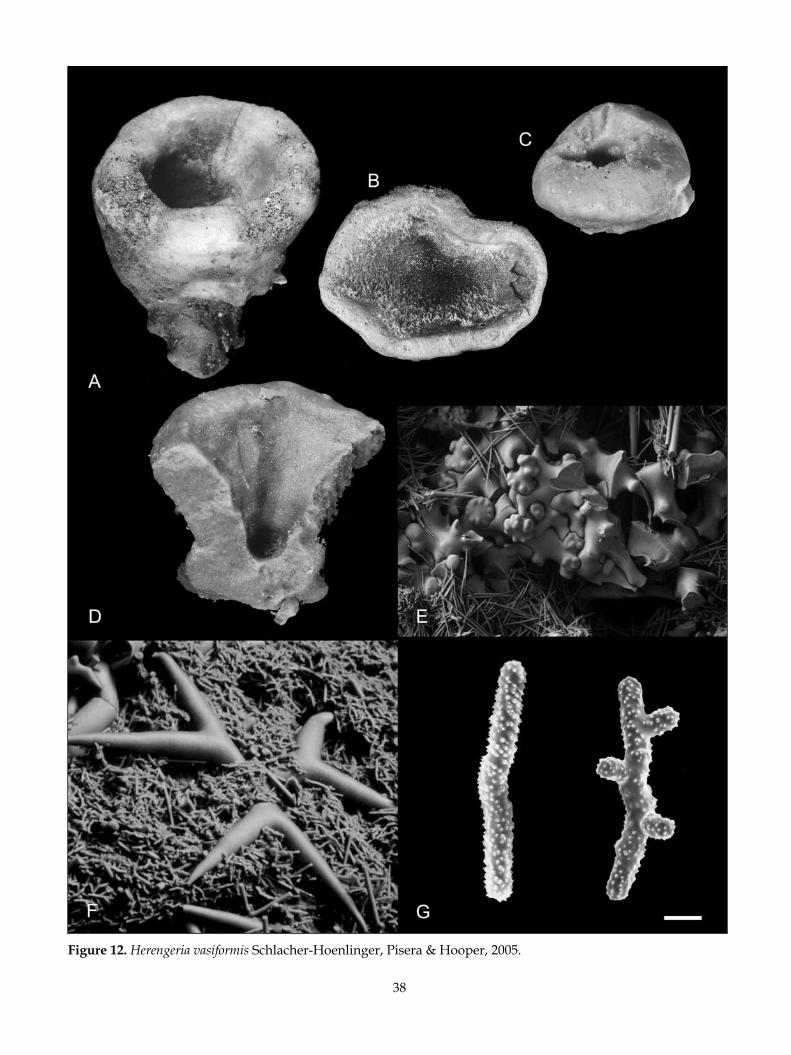
38
Figure 12. Herengeria vasiformis Schlacher-Hoenlinger, Pisera & Hooper, 2005.
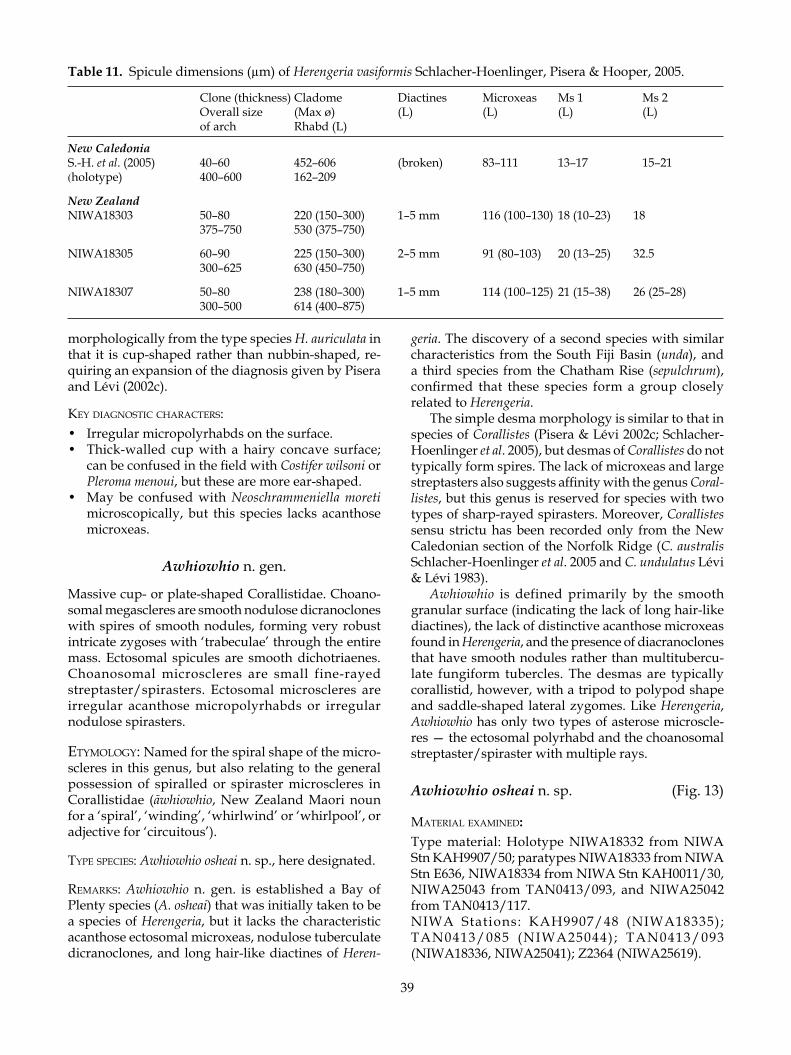
39
morphologically from the type species H. auriculata in that it is cup-shaped rather than nubbin-shaped, re-quiring an expansion of the diagnosis given by Pisera and Lévi (2002c).
key diagnosTiC CharaCTers:• Irregular micropolyrhabds on the surface.• Thick-walled cup with a hairy concave surface;
can be confused in the field with Costifer wilsoni or Pleroma menoui, but these are more ear-shaped.
• May be confused with Neoschrammeniella moreti microscopically, but this species lacks acanthose microxeas.
Awhiowhio n. gen.
Massive cup- or plate-shaped Corallistidae. Choano-somal megascleres are smooth nodulose dicranoclones with spires of smooth nodules, forming very robust intricate zygoses with ‘trabeculae’ through the entire mass. Ectosomal spicules are smooth dichotriaenes. Choanosomal microscleres are small fine-rayed streptaster/spirasters. Ectosomal microscleres are irregular acanthose micropolyrhabds or irregular nodulose spirasters.
eTymology: Named for the spiral shape of the micro-scleres in this genus, but also relating to the general possession of spiralled or spiraster microscleres in Corallistidae (āwhiowhio, New Zealand Maori noun for a ‘spiral’, ‘winding’, ‘whirlwind’ or ‘whirlpool’, or adjective for ‘circuitous’).
TyPe sPeCies: Awhiowhio osheai n. sp., here designated.
remarks: Awhiowhio n. gen. is established a Bay of Plenty species (A. osheai) that was initially taken to be a species of Herengeria, but it lacks the characteristic acanthose ectosomal microxeas, nodulose tuberculate dicranoclones, and long hair-like diactines of Heren-
geria. The discovery of a second species with similar characteristics from the South Fiji Basin (unda), and a third species from the Chatham Rise (sepulchrum), confirmed that these species form a group closely related to Herengeria.
The simple desma morphology is similar to that in species of Corallistes (Pisera & Lévi 2002c; Schlacher-Hoenlinger et al. 2005), but desmas of Corallistes do not typically form spires. The lack of microxeas and large streptasters also suggests affinity with the genus Coral-listes, but this genus is reserved for species with two types of sharp-rayed spirasters. Moreover, Corallistes sensu strictu has been recorded only from the New Caledonian section of the Norfolk Ridge (C. australis Schlacher-Hoenlinger et al. 2005 and C. undulatus Lévi & Lévi 1983).
Awhiowhio is defined primarily by the smooth granular surface (indicating the lack of long hair-like diactines), the lack of distinctive acanthose microxeas found in Herengeria, and the presence of diacranoclones that have smooth nodules rather than multitubercu-late fungiform tubercles. The desmas are typically corallistid, however, with a tripod to polypod shape and saddle-shaped lateral zygomes. Like Herengeria, Awhiowhio has only two types of asterose microscle-res — the ectosomal polyrhabd and the choanosomal streptaster/spiraster with multiple rays.
Awhiowhio osheai n. sp. (Fig. 13)
maTerial examined: Type material: Holotype NIWA18332 from NIWA Stn KAH9907/50; paratypes NIWA18333 from NIWA Stn E636, NIWA18334 from NIWA Stn KAH0011/30, NIWA25043 from TAN0413/093, and NIWA25042 from TAN0413/117.NIWA Stations: KAH9907/48 (NIWA18335); TAN0413/085 (NIWA25044) ; TAN0413/093 (NIWA18336, NIWA25041); Z2364 (NIWA25619).
Table 11. Spicule dimensions (µm) of Herengeria vasiformis Schlacher-Hoenlinger, Pisera & Hooper, 2005.
Clone (thickness) Cladome Diactines Microxeas Ms 1 Ms 2 Overall size (Max ø) (L) (L) (L) (L) of arch Rhabd (L)
New CaledoniaS.-H. et al. (2005) 40–60 452–606 (broken) 83–111 13–17 15–21(holotype) 400–600 162–209
New ZealandNIWA18303 50–80 220 (150–300) 1–5 mm 116 (100–130) 18 (10–23) 18
375–750 530 (375–750)
NIWA18305 60–90 225 (150–300) 2–5 mm 91 (80–103) 20 (13–25) 32.5 300–625 630 (450–750)
NIWA18307 50–80 238 (180–300) 1–5 mm 114 (100–125) 21 (15–38) 26 (25–28) 300–500 614 (400–875)
40
Figure 13. Awhiowhio osheai n. sp.

41
TyPe loCaliTy: Bay of Plenty, New Zealand.
disTribuTion: West of White Island, Rungapapa and Tuatoru Knolls, Bay of Plenty, New Zealand.
habiTaT: Encrusting on basalt rocks. Depth range 159–318 m.
desCriPTion:Morphology conical to cup-shaped with thick straight walls (Fig. 13A,C). NIWA18333 is a vertical plate with a spherical outline (Fig. 13B); the base is broad and elliptical, margins of the plate are smooth.
Dimensions 33–70 mm wide, 22–53 mm high, 8–12 mm thick, base 18–46 mm long, 13–26 mm wide.
Texture stony.Surface — outer surface of cup smooth to undulating
with a granular texture; oscules, c. 0.1 mm diameter, scattered regularly over the concave face in cup-shaped specimens, flush with the surface in some specimens and raised in others; ostia not visible.
Colour dull yellow (3B3) in life and in ethanol.Choanosomal skeleton a dense reticulation of desmas
and massive intricate zygoses that are pierced by cir-cular channels (Fig. 13D).
Ectosomal skeleton a dense regular crust of micropo-lyrhabds over the dichotriaene cladomes.
Megascleres (Table 12) desmas are relatively slender with 2-4 clones, heavily nodulose with single or mul-tiple nodular spires (Fig. 13D). Zygoses are massive, highly intricate and spread laterally along the clones in mature sections of the sponge skeleton, forming a dense aggregation with trabeculae. Desmas have a granular brown core, immature desmas are sparsely tuberculate with relatively smooth thin clones; dichotriaenes (Fig. 13D–F) have a relatively long rhabd and short robust clads, with frequent trichotriaene modifications.
Microscleres (Table 12) relatively regular rod-shaped acanthose micropolyrhabds (Fig. 13G), with bulbous ends and a central expansion, straight or curved or in a few cases, abruptly angled centrally. Streptasters (Table 12) with extremely fine long rays are common in the choanosome (Fig. 13G).
eTymology: Named for Dr Steve O’Shea, formerly of NIWA Wellington, for his considerable knowledge of
New Zealand deep-sea faunas and his passion for the discovery of new lithistid sponges and other inverte-brate groups.
remarks: Awhiowhio osheai differs from the two known species of Herengeria in the lack of acanthose microxeas that are abundant on the ostial face of H. vasiformis and H. auriculata, the lack of hair-like diactines, and the simpler ornamentation of the desmas. The ectosomal micropolyrhabds are also simpler, with no branches and only small irregular protrusions. In A. osheai the oscules are clearly visible and raised on the concave surface of the cup. A. osheai is so far known only from seamounts in the Bay of Plenty, New Zealand.
key diagnosTiC CharaCTers:• Relatively regular bean- or rod-shaped micropoly-
rhabds on the surface.• No surface microxeas and diactines.• Thick straight-walled cup with a smooth dimpled
concave surface; can easily be confused in the field with Aciculites pulchra but the oscules on the latter are more pronounced and flatter and the sponge is a curvaceous ear rather than straight-walled.
Awhiowhio unda n. sp. (Fig. 14)
maTerial examined: Type material: Holotype NIWA18337 from NIWA Stn K826.
TyPe loCaliTy: Southern South Fiji Basin.
disTribuTion: Southern South Fiji Basin.
habiTaT: Attached to a piece of dead coral. Depth range 390–490 m.
desCriPTion:Morphology comprising a highly foliose thick-walled lamella attached along the base, the lamella with mul-tiple infoldings and undulations, the margin slightly expanded and flared (Fig. 14A).
Dimensions 64 mm long, 45 mm wide, 35 mm high, lamella 7–10 mm thick.
Texture stony.
Table 12. Spicule dimensions (µm) of Awhiowhio osheai n. sp.
Clone (thickness) Cladome Micropolyrhabd (L) Streptaster (L) Overall size (Max ø) (Thickness) of arch Rhabd (L) NIWA18332 50–60 206 (150–250) 27 (20–38) 30 (25–38)(holotype) c. 200–500 461 (325–700) 4–5
NIWA18333 40–50 183 (100–240) 24 (15–30) 30 (23–38)(paratype) c. 200–500 490 (260–675) 2–5

42
Figure 14. Awhiowhio unda n. sp.
43
Surface smooth, granular.Colour coffee brown (5F6) in preservative.Choanosomal skeleton comparatively delicate, formed
by small, relatively slender tripod-shaped dicrano-clones. Small streptasters are abundant in the choano-some amongst the desmas (Fig. 14B).
Ectosomal skeleton a shallow layer of robust micro-scleres. Dichotriaene cladomes line the ectosome (Fig. 14B).
Megascleres (Table 13) — desmas small, frequently tripod-shaped and relatively slender, the clones are smooth with smooth nodules (Fig. 14C). The nodules on the apex of tripod desmas are spires with bumpy tubercles (Fig. 14B). Zygosis is mostly terminal and relatively weak as articulation is via saddle-shaped clasps to adjacent clones. Dichotriaenes are small and the cladomes are short and robust (Fig. 14B, D).
Microscleres (Table 13) — choanosomal microscleres are streptasters verging on spirasters with sharply pointed rays (Fig. 14E). Ectosomal microscleres are irregular thick micropolyrhabds that may have an ir-regular spiraster-shape (Fig. 14D,E).
eTymology: Named for the wave-like morphology of the sponge walls and the microscleres (unda, L.)
remarks: Awhiowhio unda is similar to A. osheai in that it has small, relatively smooth desmas with nodulose clones, but the streptasters and micropolyrhabds are more spiraster-like. The species has a unique wavy morphology.
key diagnosTiC CharaCTers:• Frilled wave-like sponge wall.• Small non-tuberculate desmas compared to other
species.
Awhiowhio sepulchrum n. sp. (Fig. 15)
maTerial examined: Type material: Holotype NIWA18338 from NIWA Stn TAN0104/002.
TyPe loCaliTy: Graveyard Seamount, Chatham Rise.
disTribuTion: Known only from the Graveyard seamount complex, a series of small knolls and hills on the Chatham Rise.
habiTaT: Silty hard substratum. Depth range 757–875 m.
desCriPTion:Morphology comprising a large erect plate with a broad undulation in the plane of the upper margin, the mar-gin indented by what appear to be large shallow pits c. 5–7 mm wide. Base of sponge thick, flanged (Fig. 15A).
Dimensions c. 300 mm wide, c. 210 mm high, base 130 wide, 40 mm wide; thickness of plate at base 44 mm, at middle of plate 28 mm, margin 12 mm.
Texture brittle, stony, the margin slightly flexible.Surface macerated, the underlying skeleton smooth,
indicating its possible condition in life.Colour dirty white in macerated condition.Choanosomal skeleton of heavily zygosed desmas
that form ‘bridges’ (Fig. 15C,D). Only a single triaene was seen (Fig. 15E).
Megascleres (Table 14) — large desmas that form large rounded arches or ‘bridges’ joined laterally via saddle-shaped zygomes (Fig. 15C–E). Nodular spires arise from the arched side of the primary clone. Desmas are large and thick, with a granular centre. Clones are smooth with occasional nodules that are ‘sculpted’ around the termini. Smaller, more intricately branched desmas (Fig. 15 B) were observed in preparations of the abraided surface of the sponge, where short clones arise on all sides of the primary linear clone. Dichotriaenes with robust straight conical rhabd and clads.
Microscleres absent owing to macerated condition of sponge.
eTymology: Named for the type locality, the Graveyard Seamount on the Chatham Rise (sepulchrum, Latin, grave).
Table 13. Spicule dimensions (µm) of Awhiowhio unda n. sp.
Clone (thickness) Cladome (Max ø) Microrhabds (L) Streptaster (L) Overall size of arch Rhabd (L) (Thickness)
NIWA18337 20–50 195 (150–240) 18 (15–23) 25 (20–33)(holotype) c. 200–300 296 (150–420) 5–8
Table 14. Spicule dimensions (µm) of Awhiowhio sepulchrum n. sp.
Clone (thickness) Cladome (Max ø) Overall size of arch Rhabd (L) Overall size of smaller ramose desmas
NIWA18338 70–150 375–900(holotype) c. 900–1200 450–750 c. (600–800) – (300–400)

44
Figure 15. Awhiowhio sepulchrum n. sp.
45
remarks: The specimen described above is tentatively assigned to Awhiowhio n. gen. because the absence of diagnostic microscleres precludes full identifica-tion. The specimen was dead before collection, with nothing remaining of the surface and mesohyl or all microscleres and non-desma megascleres, with the exception of a single dichotriaene, having sloughed off the desma skeleton.
The assignment to Awhiowhio is based on the mor-phology of the desmas. These are relatively smooth dicranoclones with a few nodulose spires on the outer edge of the arched tripod-shaped primary clone. The basic shape and ornamentation of the desma, with nodules rather than multituberculate fungiform pro-jections, separates the sponge from Herengeria, but the strong lateral bridge-forming zygoses strongly resem-bles that of both Awhiowhio and Herengeria, linking it to Corallistidae.
Despite the lack of diagnostic microscleres and ac-cessory megascleres, the assignment of a species name to NIWA18338 is justified by the possession of several unique features. NIWA18338 is the only known rep-resentative of the family Corallistidae in the Chatham Rise region. With the exception of Neoschrammeniella antarctica from the Ross Sea, Corallistidae are common in waters only as far south as Raukumara Plain off East Cape. The species is also readily distinguished from other Corallistidae by the greater size and simpler ornamentation of the dicranoclone desmas. A more detailed evaluation of the genus assignment is not possible without living specimens in which megasclere and microsclere complements are intact.
key diagnosTiC CharaCTers:• Sponge forms a very large flat plate.• Very large arched simple dicranoclones with a
dense brown granular core.
Family NEOPELTIDAE Sollas, 1888Neopeltidae Sollas, 1888: 344; Topsent 1894: 290; Schrammen
1903: 20; Kelly 2000: 281 (partim). Daedalopeltidae de Laubenfels, 1936: 184.
Thickly encrusting, planar, pedunculate, fungiform lithistid demosponges with monaxial pseudodiscotri-aenes, pseudophyllotriaenes, and/or siliceous discs; desmas are monocrepid with a complex branching shape resembling tetraclones (pseudotetraclones); may be accompanied by strongyles; microscleres are amphiasters and/or acanthose microxeas.
Remarks: The discovery of a second species of Homo-phymia from continental-shelf banks off northeastern New Zealand led Kelly (2000) to redefine the genus and to evaluate the integrity of the Corallistidae as a
whole. In doing so, it became obvious that Callipelta, Daedalopelta, and Homophymia differ considerably from typical corallistid genera like Corallistes, Herengeria, and Neoschrammeniella in having pseudotriaenes, smooth monocrepid pseudotetraclones, and amphiasters as microscleres. Callipelta, Daedalopelta and Homophymia were accordingly transferred to the Neopeltidae.
Pisera and Lévi (2002g) upheld this decision in their recent review of the Neopeltidae but they disagreed with the hypothesis of Kelly (2000) that Neopeltidae and Corallistidae are closely linked in the suborder Di-cranocladina, a subgroup of lithistid sponges that have monaxial desmas. Recent rDNA data analyses (Kelly & McCormack unpubl.) indicate that the Neopeltidae and Corallistidae are indeed linked and that this group is quite separate from tetracladine, rhizomorine, and megamorine taxa.
Homophymia Vacelet & Vasseur, 1971
Fan-shaped or clavate Neopeltidae with monocrepid pseudotetraclones, smooth monocrepid pseudophyl-lotriaenes as ectosomal megascleres, and a single form of amphiaster microsclere.
TyPe sPeCies: Homophymia lamellosa Vacelet & Vasseur, 1971.
Homophymia stipitata Kelly, 2000 (Fig. 16A–F)
Homophymia stipitata Kelly, 2000: 9, figs 5–8.
maTerial examined: NIWA Stations: I96 [NIWA holotype H-705 (NIWA7503), NIWA paratypes P-1152 (NIWA4775) and NIWA P-1155 (NIWA4778)]; J953 [NIWA paratype P-1153 (NIWA4776, BMNH 1995.3.30.2)]; J954 [NIWA paratype P-1156 (NIWA4779, BMNH 1995.3.30.5), NIWA25611]; P8 (NIWA25645); P10 [NIWA paratype P-1154 (NIWA4777)]; Z9747 [BMNH 2005.9.23.1 (frag-ment of 0CDN6731-Z)]. NMNZ Stations: NMNZ Por. 615.
disTribuTion: Wanganella Bank (holotype); West Nor-folk Ridge; west of Pandora Bank; western continental slope of Northland, New Zealand.
habiTaT: Collected from coralline and shell rubble banks on the northeastern continental shelf of North-land. The sponge is typically attached to hard substrata but may be partially buried. This species is part of a biodiverse invertebrate assemblage in which hydroids, soft corals, bryozoans, and ascidians are typical. Other lithistid sponges include Aciculites pulchra, Discodermia proliferans, and several species of Neoschrammeniella and Pleroma (Kelly 2000). Depth range 192–757 m.

46
Figure 16. Homophymia stipitata Kelly, 2000.

47
desCriPTion (after Kelly 2000):Morphology parsnip-shaped (stipitate) with an irregular globular body that bulges in places (Fig. 16A); apex is depressed, forming a translucent oscular sieve through which exhalant canals are clearly visible (Fig. 16B).
Dimensions of body 40–50 mm diameter, about one third of total length of 150–220 mm, elevated upon and tapering to a relatively long fine stalk ranging from 20 mm diameter at the junction of the body, to 5 mm diameter at the base of the stalk.
Texture barely compressible, stalk is rigid but rela-tively flexible along entire length.
Surface smooth but slightly undulating and irregu-lar, granular to the touch. Exhalant canals are visible on surface of body and stalk beneath the translucent ectosome (Fig. 16B).
Colour in life ivory (4B3), in preservative light golden brown (5B7), apical depression dark grayish blue (20E4).
Choanosomal skeleton composed of huge smooth monocrepid pseudotetraclones (Fig. 16C) and tracts of large oxeas that radiate through the stalk towards the apical surface. Amphiasters are abundant throughout the choanosome and the ectosomal region.
Ectosomal skeleton packed with pseudophyllotri-aenes, the sinuous ‘fingers’ of the ‘cladome’ partially zygose with neighbouring ‘cladomes’ and desmas below the surface. The ‘rhabds’ of these pseudophyl-lotriaenes are perpendicular to the surface.
Megascleres (Table 15) — desmas of the choanosome are huge smooth ramose monocrepid pseudotetra-clones (Fig. 16C). Ectosomal megascleres are pseu-dophyllotraienes (Fig. 16D,E) in which the ‘rhabd’ is irregular in shape and thickness, is sinuous, and the cladome is polyfurcate with sinuous branches. The spicules are laminated and granular in appearance. Diactines are strongyloxeas, curved, syringe-like, in two size classes.
Microscleres (Table 15 Ms 1) — amphiasters (Fig. 16F) with curved spines on the distal two thirds of oxeote rays.
remarks: Homophymia stipitata was the second species to be described, the genus being previously known only from Madagascar. Schlacher-Hoenlinger et al. (2005) recently described a third species of Homophy-mia from Kaimon Maru Seamount in New Caledonian
waters; it is spherical with an apical depression and a short stalk. A fourth species has since been collected from the New Caledonian Norfolk Ridge seamounts (Kelly unpubl.), the morphology of which is similar to that of H. stipitata but with a smaller, penicillate body and only a slight expansion of the apical region. The typical form of the type species, H. lamellosa, is easily distinguishable from the above three species in that it is a folded lamellate fan and is apparently restricted to the Western Indian Ocean.
It is noteworthy that very similar ‘phyllotriaenes’ to those of H. stipitata were figured from the Oamaru Diatomite (c. 35 Ma) (Hinde & Holmes 1892, pl. 14, fig. 1), and described from the Tutuiri Greensand of Chatham Island (c. 63–50 Ma). Superficially, the Tu-tuiri, Oamaru, and Recent spicules appear to be very similar, and were indeed considered to be congeneric by Kelly and Buckeridge (2005). Closer examination of the Tutuiri specimens, however, indicates that while they are indeed identical to the Oamaru material, they differ from those of Recent specimens in the possession of a triaenose inception canal, which is clearly illus-trated in the Hinde and Holmes (1892) figure.
key diagnosTiC CharaCTers:
• Parsnip-shaped with a grey-blue apical depres-sion.
• Surface covered with pseudotriaenes with an extremely sinuous cladome and short irregular rhabd.
• Desmas are very big and resemble tetraclones but the epirhabd is only a short bar.
• One type of amphiaster.
Callipelta Sollas, 1888
Pillow-shaped, spherical, clavate, and multiclavate Neopeltidae, with relatively small irregular mono-crepid desmas that resemble tetraclones; ectosomal megascleres are moncrepid pseudodiscotriaenes to pseudophyllotriaenes with strongly incised denticulate margins on the ‘clads’, the upper surface of which may be tuberculate; microscleres are amphiasters.
TyPe sPeCies: Callipelta ornata Sollas, 1888.
Table 15. Spicule dimensions (µm) of Homophymia stipitata Kelly, 2000.
Desma (Clone L/T) Cladome (Max ø) Diactines 1 (L) Ms 1 (L) Desma (Overall size) Rhabd (L) Diactines 2 (L) Epirhabd (L)
NIWA7503 147–588/20–59 627 (465–711) 1055 (809–1372) 15 (12–19)(holotype) 400–682 254 (186–323) 472 (308–617) 193 (147–225)

48
Callipelta punctata Lévi & Lévi, 1983 (Fig. 17)
Callipelta punctata Lévi & Lévi, 1983: 118, pl. 6, fig. 1, pl. 8, figs 1–3, 9.
maTerial examined: NIWA Stations: KAH0011/30 (NIWA18339); TAN0413/117 (NIWA24045).
disTribuTion: New Caledonia (holotype); Rungapapa Knoll, Bay of Plenty.
habiTaT: Numerous small specimens encrusting speci-mens of Erylus nigra Bergquist, 1968 (Porifera, Astro-phorida, Geodiidae) and volcanic basalt substrata, with numerous other invertebrates including immature Reidispongia coerulea. Depth range 159–360 m.
desCriPTion:Morphology semi-spherical to columnar, with a single small oscule on the slightly depressed apex of the sponge (Fig. 17A). NIWA24045 from TAN0413/117 is a tiny slightly raised encrustation.
Dimensions 3–7 mm diameter, 5–10 mm high, oscule 0.5 mm diameter. The New Caledonian material was reported as 15–18 mm diameter and 10 mm high.
Texture stony, crumbly.Surface microscopically rough and punctate, slightly
hispid with projecting diactines; occasional aquiferous canals converging on the oscules incising the surface or appear below it.
Colour in life cream (4A3) and in ethanol golden brown (5D7).
Choanosomal skeleton composed of monocrepid desmas and short diactines.
Ectosomal skeleton lined with the scale-like cladome of the pseudophyllotriaenes.
Megascleres (Table 16) are monocrepid pseudotetra-clone desmas with a short epirhabd in which a short slender bar crepis (30 µm) resides (Fig. 17C). Two thick clones arise from each end of the epirhabd; these have a granular core, the terminus of each clone tuberculate or spinose, the zygosis between spicules relatively
rigid (Fig.17C); ectosomal megascleres are pseudo-phyllotriaenes with a short rhabd that is often slightly obliquely set (Fig. 17B). The centre of each rhabd has a crepis about 30 µm long as in the desmas. The cladome is not precisely perpendicular to the rhabd, and often appears slightly concave in profile. The pseudoclads may be composed of a pseudoprotoclad, which may split to form two branches, or may remain single. The end of the clad may branch again; the whole is very delicate and lacy in appearance. The upper surface of the cladome is denticulate (Fig. 17B). Diactines are relatively short fusiform oxeas.
Microscleres (Table 16) amphiasters with long fine acanthose rays (Fig. 17D).
remarks: These sponges are most similar to Callipelta punctata Lévi & Lévi, 1983 from New Caledonia in terms of spicule morphology and dimensions. In par-ticular, the upper surfaces of the pseudophyllotriaenes have tuberculate projections, a character that appears to be unique to C. punctata and species of Macandrewia (Macandrewiidae).
The New Zealand sponges are more columnar than those described from New Caledonia, but the punctate surface is characteristic. In gross morphology they are perhaps more similar to Callipelta sollasi Lévi & Lévi, 1989 from about 100 m depth off Luzon and Mindoro Islands in the Philippines, but this latter species has pseudophyllotriaenes with a smooth cladome surface, which is also much wider (300–350 µm) than those of C. punctata.
The only other described species, C. cavernicola (Vacelet & Vasseur, 1965), C. thoosa Lévi, 1965, and C. mixta Vacelet, Vasseur & Lévi, 1976 are quite different morphologically and in terms of spiculation, and all three species are known only from submarine tunnels and grottos on the coast of Madagascar. Callipelta mixta is probably a species of Neopelta as it contains discotri-aene-like megascleres and acanthose microxea.
Despite extensive collection, this species has so far been found only at a single New Zealand location, the Rumbles Seamount in the Bay of Plenty.
Table 16. Spicule dimensions (µm) of Callipelta punctata Lévi & Lévi, 1988.
Desma (Clone L/T) Pseudophyllotriane Diactines (L) Amphiaster (L) Desma (Overall size) cladome (Max ø) Rhabd (L)
New Caledonia(holotype) 60–80/20–30 200–300 350–500 20 ― 75–150 New ZealandNIWA18339 80–130/30 224 (150–280) 400–600 16 (15–18) 180–230 100 (50–140)

49
Figure 17. Callipelta punctata Lévi & Lévi, 1983.
50
key diagnosTiC CharaCTers:• Tiny columnar or encrusting sponges (< 20 mm
diameter) with a tiny apical oscule and a rough punctuate surface.
• Desmas and pseudophyllotriaenes are granular in the centre.
• Desmas have a single fine bar crepis (monocre-pidial) in the epirhabd.
• Can be mistaken for Macandrewia spinifoliata in the field; the presence of amphiasters and the lack of ectosomal smooth strongylote oxeas (present in Macandrewia spinifoliata) in Callipelta punctata, are indicative.
Neopelta Schmidt, 1880
Pillow-shaped and columnar Neopeltidae with small complex tuberculate monocrepid desmas that resem-ble tetraclones; ectosomal megascleres are moncrepid circular, oval to elongate, sculpted and angular and ridged discs; some are pseudodiscotriaenes with an oblique pseudorhabd projection that contains the monaxial crepis; other megascleres may include slen-der curved oxea-like diactines that occur singly or in bundles; microscleres are amphiasters; acanthose microxea occur in some species.
TyPe sPeCies: Neopelta perfecta Schmidt, 1880.
remarks: Neopelta plinthosellina Lévi & Lévi, 1988 from New Caledonia differs from the type species N. perfecta in that it lacks acanthose microxea and has elongate and highly sculpted discs. The diagnosis above has been broadened to accommodate the peculiar sculpted discs of N. plinthosellina, and the lack of microxeas and choanosomal oxeas and tuberculate regular palmate desmas in both southwestern Pacific species.
Neopelta pulvinus n. sp. (Fig. 18)
maTerial examined: Type material: Holotype NIWA18340 from NIWA Stn TAN0205/75 spicule slide labelled ‘NIWA18340’.
TyPe loCaliTy: Volcano L, Kermadec Ridge, northeast-ern New Zealand.
disTribuTion: Volcano L, Kermadec Ridge, northeastern New Zealand.
habiTaT: Encrusting hard basalt substrate. Depth range 154–240 m.
desCriPTion:Morphology pillow- or cushion-shaped, edges thinning to the substratum.
Dimensions 10 mm long, 7 mm wide, 4 mm thick.Texture stony, brittle.Surface smooth.Colour in ethanol honey yellow (5D6).Choanosomal skeleton composed of small robust
monocrepidial desmas that form very intricate zygoses. Amphiasters are abundant in the choanosome.
Ectosomal skeleton partially lost, but in patches pseu-dodiscotriaenes form a scaled surface.
Megascleres (Table 17) — monocrepid pseudotetra-clone desmas (Fig. 18B) with a short thick epirhabd with four short thick clones that extend into very tuber-culate termini. The clones are less tuberculate towards the smooth epirhabd. On the sponge surface, clones are palmate and similar to moose antlers in shape. The zygoses are very complex and highly robust. Ectos-omal megascleres (Table 17) are pseudodiscotriaenes with a flat to slightly concave disc, which are circular, elongate, or multilobate in overall shape, with circular indentations on the edge of the disc, usually one per spicule. The upper surface of the discs is ornamented (Fig. 18A), some with swirls of small evenly spaced tubercles, some with what appear to be knobbed ridges leading to the centre of the disc, and some which lack either embellishment.
Microscleres (Table 17) are amphiasters with long fine rays.
eTymology: Named for the pillow- or cushion-shaped gross morphology (pulvinus, L.).
remarks: This single specimen of Neopelta pulvinus was initially identified as a species of Callipelta (Ne-opeltidae) because it has ectosomal monocrepidial pseudodiscotriaenes, amphiasters as microscleres, and lacks the characteristic acanthose microxea of the type species (Neopelta perfecta Schmidt, 1880) and N. imperfecta Schmidt, 1880, both from the Caribbean.
Table 17. Spicule dimensions (µm) of Neopelta pulvinus n. sp.
Desma (Clone L/T) Pseudodiscotriaene disc (Max ø) Amphiaster (L) Desma (Overall size) Pseudorhabd (L) Desma crepis (L) Epirhabd (L)
New ZealandNIWA18340 100–250/50–70 227 (88–300) by 140 (63–220) 19 (15–25)(holotype) 300–500 20
30 18

51
Figure 18. Neopelta pulvinus n. sp.

52
The ectosomal discotriaenes are, however, quite differ-ent from those of most species of Callipelta in that they are discate rather than phyllamentous. The discotriaene pseudorhabd in this new species is also unlike those of the pseudophyllotriaenes of Callipelta species, which are almost always well defined and perpendicular to the cladome. In N. pulvinus the rhabds are solid, short, frequently with two flanges that adhere to the underside of the disc, and the pseudorhabd is often positioned obliquely or perpendicular to the under-side of the disc. This unusual rhabd morphology has been described for both species of Caribbean Neopelta (Sollas 1888), albeit rarely, but not for the only other known species, New Caledonian N. plinthosellina Lévi & Lévi, 1988.
Neopelta plinthosellina, like N. pulvinus n. sp., lacks acanthose microxeas and both species have very similar desmas; some are typically pseudotetraclones with smooth or tuberculate clones and heavily tuberculate zygoses, but some are plamate and spreading as il-lustrated by Lévi and Lévi (1988, pl. 6). The desmas of the southwestern Pacific species also differ from those of the Caribbean species, which are complex with smooth rays and are frequently branched (see Pisera & Lévi 2002g). It is thus with some confidence that the Kermadec Ridge specimen is assigned to the same group as N. plinthosellina from New Caledonia, but little confidence is placed in the assignment of both species to the Caribbean genus Neopelta. Despite the obvious differences between the Caribbean and southwestern Pacific species of Neopelta, additional specimens of both species, particularly of N. pulvinus, are required for full characterisation of these differences, and separation from Neopelta as a new genus.
key diagnosTiC CharaCTers:• Tiny encrusting cushion sponge.• Surface has scale-like discs with tiny projecting
tubercles on the upper surface and usually one or more circular indentations.
• Desmas are frequently palmate.
INCERTAE SEDIS
Lepidothenea de Laubenfels, 1936
Sponge forms a very thin encrustation, without des-mas; skeleton comprises an upper and lower adherent of overlapping scale-like discs or occasionally discotri-aenes, between which is a thin choanosome. Microscle-res are microrhabds. (Modified from Dendy 1924).
TyPe sPeCies: Lepidospongia incrustans Dendy, 1924.
remarks: As pointed out by Bergquist (1968), De Laubenfels (1936) proposed the new name Lepidothenea
for Lepidospongia Dendy, 1924, which was preoccupied. He transferred Lepidothenea incrustans to the Kaliap-sidae de Laubenfels (= Phymaraphiniidae in part) owing to the possession of what he perceived to be ‘phyllotriaenes’ instead of dichotriaenes (de Laubenfels 1936). Bergquist (1968) did not re-examine this mate-rial but retained it in the original family Theonellidae, no doubt convinced that the report of what appeared to be rhabds on some discs, i.e. ‘discotriaenes’, abun-dant ectosomal microrhabds, and vestigial style-like megascleres, indicated broad affinity with species in the genus Discodermia. Pisera (pers. comm. 2004) has suggests that while the sponge does indeed resemble a juvenile Discodermia, albeit without desmas, it also resembles members of the genus Neopelta. It is his opin-ion that Lepidothenea is probably a younger synonym of Neopelta.
Lepidothenea incrustans (Dendy, 1924) (Fig. 19)
Lepidospongia incrustans Dendy, 1924: 317, pl. 13, figs 1–3.Lepidothenea incrustans: de Laubenfels 1936: 176; Bergquist
1968: 64.
maTerial examined: None.
disTribuTion: Holotype Terra Nova Station 90, approxi-mately 20 km southwest of the summit of Great King Island (34º24’S, 172º06’E) (Harmer & Lillie 1914).habiTaT: Dendy (1924) described the habitat of ‘Terra Nova’ Station 90 as being remarkable in that it yielded thirty-eight species of sponges. The depth was about 183 m and the substratum was rocky. The sponge formed a thin crust on a small block of basalt with numerous other encrusting organisms.
desCriPTion:Morphology a thinly spreading encrustation. The specimen was described as partially rubbed off the substratum.
Dimensions 10 5 10 mm, the thickness was not given by Dendy (1924).
Texture crisp, delicate.Surface formed of overlapping scales, interrupted by
fairly numerous delicate minute conical apertures (Dendy 1924), presumably oscules.
Colour in life described as ‘beautifully’ silvery. The sandwiched choanosome was dark brown and dry.
Skeleton comprised of an upper and lower layer of over-lapping discate scales, the latter adherent to the underlying substratum, separated by an equally thin choanosome. Some thin vestigial megascleres were found to be associated with the rhabds of some discotriaenes.
Megascleres almost exclusively irregular oval to circu-lar discs, perfectly smooth and structureless, the margins occasionally indented, without any trace of a shaft or axial
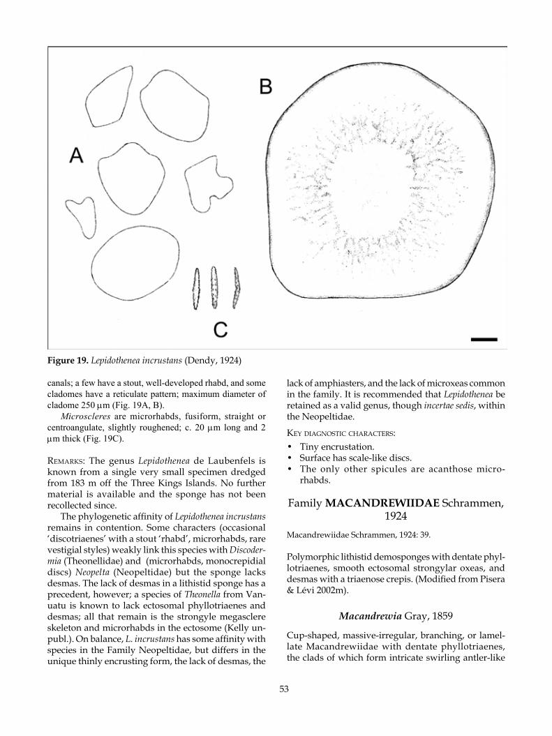
53
canals; a few have a stout, well-developed rhabd, and some cladomes have a reticulate pattern; maximum diameter of cladome 250 µm (Fig. 19A, B).
Microscleres are microrhabds, fusiform, straight or centroangulate, slightly roughened; c. 20 µm long and 2 µm thick (Fig. 19C).
remarks: The genus Lepidothenea de Laubenfels is known from a single very small specimen dredged from 183 m off the Three Kings Islands. No further material is available and the sponge has not been recollected since.
The phylogenetic affinity of Lepidothenea incrustans remains in contention. Some characters (occasional ‘discotriaenes’ with a stout ‘rhabd’, microrhabds, rare vestigial styles) weakly link this species with Discoder-mia (Theonellidae) and (microrhabds, monocrepidial discs) Neopelta (Neopeltidae) but the sponge lacks desmas. The lack of desmas in a lithistid sponge has a precedent, however; a species of Theonella from Van-uatu is known to lack ectosomal phyllotriaenes and desmas; all that remain is the strongyle megasclere skeleton and microrhabds in the ectosome (Kelly un-publ.). On balance, L. incrustans has some affinity with species in the Family Neopeltidae, but differs in the unique thinly encrusting form, the lack of desmas, the
lack of amphiasters, and the lack of microxeas common in the family. It is recommended that Lepidothenea be retained as a valid genus, though incertae sedis, within the Neopeltidae.
key diagnosTiC CharaCTers:• Tiny encrustation.• Surface has scale-like discs.• The only other spicules are acanthose micro-
rhabds.
Family MACANDREWIIDAE Schrammen, 1924
Macandrewiidae Schrammen, 1924: 39.
Polymorphic lithistid demosponges with dentate phyl-lotriaenes, smooth ectosomal strongylar oxeas, and desmas with a triaenose crepis. (Modified from Pisera & Lévi 2002m).
Macandrewia Gray, 1859
Cup-shaped, massive-irregular, branching, or lamel-late Macandrewiidae with dentate phyllotriaenes, the clads of which form intricate swirling antler-like
Figure 19. Lepidothenea incrustans (Dendy, 1924)

54
patterns, desmas with a triaenose crepis resembling tetraclones overall; the ectosome is encrusted with smooth strongylar curved oxeas. Microscleres may include roughened centrotylote microxeas.
TyPe sPeCies: Macandrewia azorica Gray, 1859.
Macandrewia spinifoliata Lévi & Lévi, 1983 (Fig. 20)
Macandrewia spinifoliata Lévi & Lévi, 1983: 116, fig. 8, pl. 6, fig. 3, pl. 8, figs 4–6.
maTerial examined: NIWA Stations: KAH9907/48 (spicule slide labelled ‘MKB 1829’); I372 (NIWA25780).
disTribuTion: New Caledonia (holotype); Doubtless Bay, North Cape; west of White Island, Bay of Plenty, New Zealand.
habiTaT: Encrusting basalt rock. Depth range 211–395 m.
desCriPTion:Morphology nubbin-shaped; Lévi and Lévi (1983) re-corded numerous specimens that were digitate and ramified with knobby ends.
Dimensions 13 mm wide, 4 mm high.Texture fragile, crumbles easily.Surface smooth.Colour in ethanol ivory (4B3).Choanosomal skeleton composed of small pseu-
dotetraclone desmas that form a close mesh and in-tricate zygoses.
Ectosomal skeleton lined with the phyllotriaene cladomes, encrusted with strongylote oxeas and rough microrhabds.
Megascleres (Table 18) comprising small robust desmas (Fig. 20A) with a triaenose crepis (one actine is longer than the other three) that resemble tetraclones (Fig. 20B). The clones are smooth around the epirhabd but can be tuberculate towards the zygoses, which are highly intricate. Phyllotriaenes (Fig. 20C) with straight conical rhabd, cladome irregularly circular to polyclad,
with differential development of some clads, the edges crenulate and indented.
Microscleres (Table 18) smooth curved spicules with strongylote ends, the ends (Fig. 20D).
remarks: The details of spiculation conform to the holotype description except that the desmas are triae-neose, not monocrepidial rhizoclones as described by Lévi and Lévi (1983). While Pisera and Lévi (2002m) refuted the hypothesis put forward by Kelly (2000) that this genus has affinities with the Neopeltidae, it is interesting to note that the phyllotriaenes of Macan-drewia spinifoliata are very similar in structure and size to the desmas, as in the neopeltid genera Homophymia and Callipelta. The contention that the phyllotriaenes of Macandrewia are pseudophyllotriaenes is supported by the fact that the short axial filaments of the crepis do not extend in the plane of the cladome to give rise to monoclads or deuteroclads as in the triaenes in sponges with tetraclone desmas. The phylogenetic affinities of this monogeneric family remain elusive.
A single specimen of Macandrewia spinifoliata was recognised during close inspection of the minute spe-cies that grow on basalt boulders dredged from the Bay of Plenty seamounts. These boulders have proved to be a rich source of new species of lithistid sponges.
key diagnosTiC CharaCTers:• Tiny encrustation (on basalt rock on Bay of Plenty
seamounts).• Surface has feather-like triaenes and curved stron-
gylote oxeas.• Microscleres in the shape of asters are absent.
Family PLEROMIDAE Sollas, 1888Megamorina Zittel, 1878: 99 (partim).Pleromidae Sollas, 1888: 312; Topsent 1894: 290; Wilson 1925:
461; de Laubenfels 1955: E50.Pleromatidae: Lendenfeld, 1903: 140.Pléromides: Moret, 1926: 118.
Massive, conical, plate- to ear-shaped or globose lithis-tid demosponges; surface usually granular or hispid with projecting megascleres, texture stony. Desmas smooth or nodulose monocrepid megaclones with
Table 18. Spicule dimensions (µm) of Macandrewia spinifoliata Lévi & Lévi, 1983.
Desma (Clone L/T) Phyllotriaene disc (Max ø) Diactines (L) Microxea (L) Desma (Overall size) Rhabd (L)
New CaledoniaLévi & Lévi, 1983 ― 200–300 400 60–70(holotype) 150–250 100–125
New Zealand ‘MKB 1829’ 120–150/30–34 200 (150–250) x 272 (160–450) 378 (250–425) 80 (63–108) 200–250 121 (90–150)
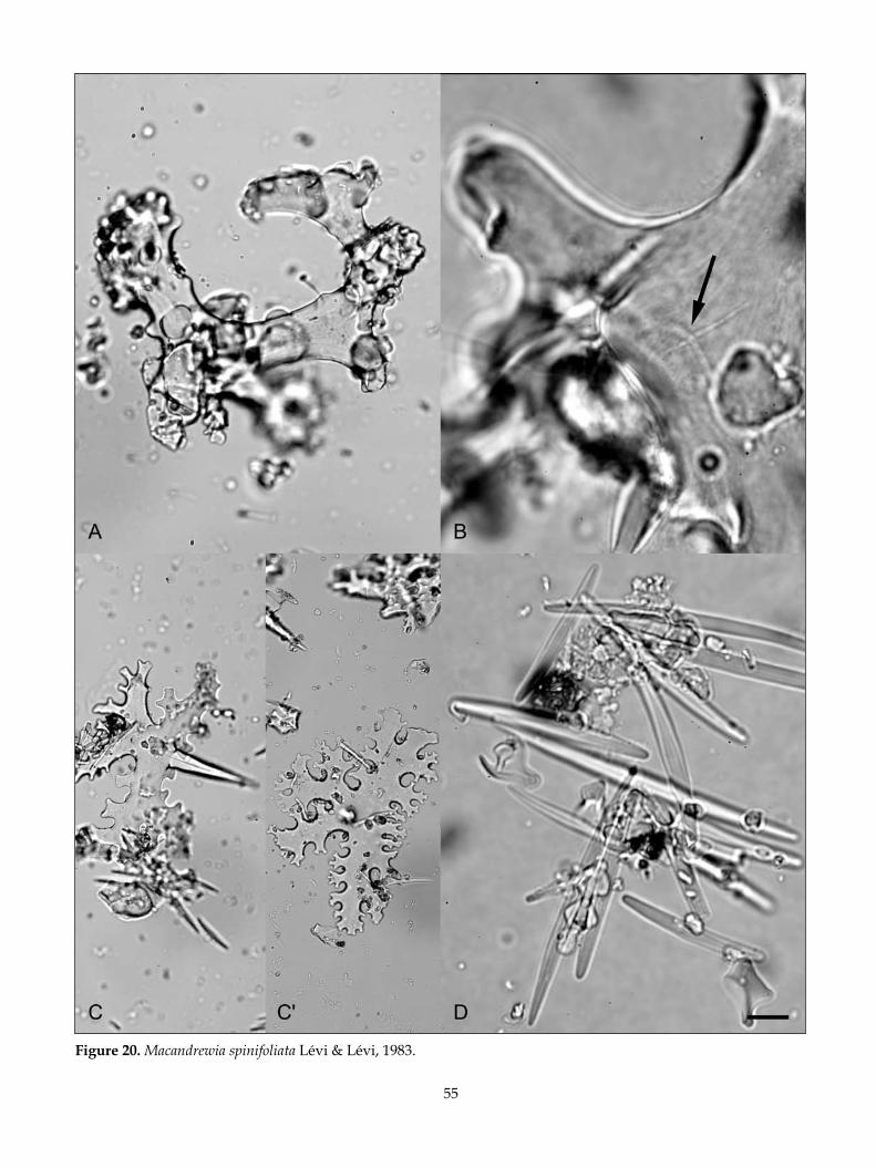
55
Figure 20. Macandrewia spinifoliata Lévi & Lévi, 1983.

56
saddle- or cup-shaped zygomes; ectosomal spicules are dichotriaenes and/or anatriaene-like megascleres; microscleres are spirasters, amphiasters, streptasters in various sizes, and finely spined microxeas and/or lightly acanthose style-like monactines. (From Kelly 2003).
remarks: Two valid extant genera, Pleroma and Ana-derma, are recognised in Pleromidae, with four species known from seamounts and banks in the Southwest Pacific including New Zealand and New Caledonia (Pisera & Lévi 2002d; Kelly 2003; Kelly et al. 2003).
Pleroma Sollas, 1888
Lyidium Schmidt, 1870: 84.Pleroma Sollas, 1888: 312; Lendenfeld 1903: 140; de Laubenfels
1955: E50; Pisera & Lévi 2002d: 324, figs 5–7.
Massive irregular encrustations, thick folded plates, or globular with a flattened apex; the surface granular and hispid in places, texture stony. Desmas are smooth or fully nodulose monocrepidial megaclones with cup-shaped, saddle-shaped, or irregular zygomes with indented margins, megascleres are large oxeas and dichotriaenes, microscleres are roughened microxeas and one to two types of microspined spiraster, amphi-aster, and streptaster. (From Kelly 2003).
Remarks: Hinde and Holmes (1892) illustrated several megamorine microfossil desmas from the Oamaru Diatomite (c. 35 million years ago), at Oamaru in North Otago. These are remarkably similar to desmas of Pleroma turbinatum (Hinde & Holmes 1892, pl. 13, fig. 25) and P. menoui (Ibid., pl. 13, figs 26, 27).
TyPe sPeCies: Pleroma turbinatum Sollas, 1888.
Pleroma turbinatum Sollas, 1888 (Figs 21, 22)
Pleroma turbinatum Sollas, 1888: 312, pl. 33, figs 5–7; Lenden-
feld 1903: 140; de Laubenfels 1955: E50, fig. 31.3; Lévi & Lévi 1983: 105, fig. 2, pl. 2, figs 5–8; Pisera & Lévi 2002d: 324–326, figs 5–7; Kelly 2003: 116, figs 2A,B, 3.
maTerial examined: NIWA Stations: I92 (BMNH 2002.3.4.1, NIWA25061); KAH9907/50 (NIWA18344); X140 (NIWA18341, NIWA18342) ; Z2092 (NIWA18343) ; Z9400 (NIWA25620).IRD Stations: LITHIST CP9 (NIWA18519, NIWA18520), DW7 (NIWA18521).
disTribuTion: Makutu coast, Fiji Islands (holotype); Havannah, south coast of New Caledonia, and Éponge Seamount, south New Caledonian slope of Norfolk Ridge; Lord Howe Seamount Chain; Norfolk Ridge south of Norfolk Island; Challenger Plateau, northern slopes; west of White Island, Bay of Plenty.
habiTaT: The sponges were attached to hard rocky substrata on the flanks of seamounts. Depth range 230–860 m.
desCriPTion (modified from Kelly 2003):Morphology a curved ear-like semicircular plate, at-tached to the substratum along the base of the sponge (Fig. 21A), or with a restricted base of attachment (Fig. 21B). The edges of the plate are rounded in profile.
Dimensions of body up to 240 mm high and wide, 10–30 mm thick.
Texture stony.Surface velvety on the upper concave surface,
granular on the lower convex surface, oscules are not visible (see Fig. 21A).
Colour in life beige, in preservative cream to beige.
Choanosomal skeleton composed of megaclones with saddle-shaped zygomes. Abundant microxeas fill the interstices between the desmas (Fig. 22A).
Ectosomal skeleton an encrustation of microscleres and dichotriaenes.
Table 19. Spicule dimensions (µm) of Pleroma turbinatum Sollas, 1888 (summarised from Kelly 2003).
Desma (Clone L/T) Cladome (Max ø) Diactines (L) MO (L) Ms1 Ms2 Desma (Overall size) Rhabd (L)
Fiji Sollas (1888) ―/50–90 448 1350 180–210 24–30 ―(holotype) 500–800 960
New CaledoniaLévi & Lévi 200–300/70–90 360–590 1500–1700 130–150 12 ―(1983) 500–550 1000–2000
New Zealand Kelly (2003) 100–800/60–100 375–750 1000–2000 130–165 10–20 12 375–625 650–875

57
Figure 21. A, B, Pleroma turbinatum Sollas, 1888; C, P. menoui Lévi & Lévi, 1983; D, P. aotea Kelly, 2003.
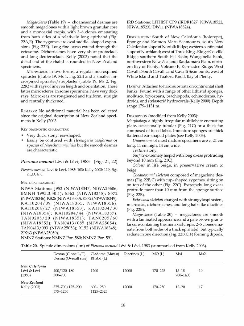
58
Megascleres (Table 19) — choanosomal desmas are smooth megaclones with a light brown granular core and a monoaxial crepis, with 3–6 clones emanating from both sides of a relatively long epirhabd (Fig. 22A,E). The zygomes are oval saddle- shaped expan-sions (Fig. 22E). Long fine oxeas extend through the ectosome. Dichotriaenes have very short protoclads and long deuteroclads. Kelly (2003) noted that the distal end of the rhabd is rounded in New Zealand specimens.
Microscleres in two forms, a regular microspined spiraster ((Table 19, Ms 1; Fig. 22I) and a smaller mi-crospined spiraster/streptaster (Table 19, Ms 2; Fig. 22K) with rays of uneven length and orientation. These latter microscleres, in some specimens, have very thick rays. Microxeas are roughened and fusiform, straight and centrally thickened.
remarks: No additional material has been collected since the original description of New Zealand speci-mens in Kelly (2003
key diagnosTiC CharaCTers:• Very thick, stony, ear-shaped.• Easily be confused with Herengeria vasiformis or
species of Neoschrammeniella but the smooth desmas are characteristic.
Pleroma menoui Lévi & Lévi, 1983 (Figs 21, 22)
Pleroma menoui Lévi & Lévi, 1983: 103; Kelly 2003: 119, figs 2C,D, 4, 6.
maTerial examined: NIWA Stations: J953 (NIWA18347, NIWA25606, BMNH 1995.3.30.1); S562 (NIWA18345); S572 (NIWA18346); K826 (NIWA18350); K872 (NIWA18349); K A H 0 2 0 4 / 0 9 ( N I W A 1 8 3 5 5 , N I W A 1 8 3 5 6 ) ; K A H 0 2 0 4 / 2 7 ( N I W A 1 8 3 5 3 ) ; K A H 0 2 0 4 / 3 0 (NIWA18354) ; KAH0204/44 (NIWA18357) ; T A N 0 2 0 5 / 2 0 ( N I W A 1 8 3 5 1 ) ; T A N 0 2 0 5 / 6 0 (NIWA18352); TAN0413/085 (NIWA25054); TAN0413/093 (NIWA25053); X152 (NIWA18348); Z9263 (NIWA25059).NMNZ Stations: NMNZ Por. 580; NMNZ Por. 591.
IRD Stations: LITHIST CP9 (IRDR1827; NIWA18522, NIWA18523); DW11 (NIWA18524).
disTribuTion: South of New Caledonia (holotype), Éponge and Kaimon Maru Seamounts, south New Caledonian slope of Norfolk Ridge; western continental slope of Northland; west of Three Kings Ridge; Colville Ridge; southern South Fiji Basin; Wanganella Bank, northwestern New Zealand; Raukumara Plain, north-ern Bay of Plenty; Volcano E, Kermadec Ridge; West Cavalli, South Cavalli, and Cavalli Seamounts; west of White Island and Tuatoru Knoll, Bay of Plenty.
habiTaT: Attached to hard substrata on continental shelf banks. Found with a range of other lithistid sponges, molluscs, bryozoans, brachiopods, echinoderms, hy-droids, and stylasterid hydrocorals (Kelly 2000). Depth range 179–1131 m.
desCriPTion (modified from Kelly 2003):Morphology a highly irregular multilobate encrusting plate, occasionally tubular (Fig. 21C) or a thick fan composed of fused lobes. Immature sponges are thick flattened ear-shaped plates (see Kelly 2003).
Dimensions of most mature specimens are c. 21 cm long, 11 cm high, 14 cm wide.
Texture stony.Surface extremely hispid with long oxeas protruding
beyond 10 mm (Fig. 21C).Colour in life beige, in preservative cream to
beige.Choanosomal skeleton composed of megaclone des-
mas (Fig. 22B,C) with cup- shaped zygomes, sitting on on top of the other (Fig. 22C). Extremely long oxeas protrude more than 10 mm from the sponge surface (Fig. 22B).
Ectosomal skeleton charged with strongylospirasters, microxeas, dichotriaenes, and long hair-like diactines (Fig. 22B).
Megascleres (Table 20) — megaclones are smooth with a laminated appearance and a pale brown granu-lar core containing the monaxial crepis; 2–5 clones ema-nate from both sides of a thick epirhabd, but typically radiate in one direction (Fig. 22B,C,F) forming dipods,
Table 20. Spicule dimensions (µm) of Pleroma menoui Lévi & Lévi, 1983 (summarised from Kelly 2003).
Desma (Clone L/T) Cladome (Max ø) Diactines (L) MO (L) Ms1 Ms2 Desma (Overall size) Rhabd (L)
New CaledoniaLévi & Lévi 400/120–180 1200 12000 170–225 15–18 10(1983) 300–700 700–1400
New Zealand Kelly (2003) 375–700/125–200 600–1250 12000 170–250 12–20 17 575–1250 1125–2325

59
Figure 22. A, E, I, K, Pleroma turbinatum Sollas, 1888; B, C, F, H, L, P. menoui Lévi & Lévi, 1983; D, G, J, P. aotea Kelly, 2003.

60
tripods, and quadripods (see also Lévi & Lévi 1983, pl. 2, figs 1–2). The epirhabd is slightly thicker than the clones. The zygomes are spherical and cup-shaped. Diactines are long and fine. Dichotriaenes are large, slender and curved, the deuteroclads and protoclads are quite long and fine (Fig. 22B).
Microscleres in two forms, a regular strongylos-piraster derived from a spiraster with rays encased in additional silica (Table 20 Ms 1; Fig. 22H), and a smaller streptaster (Table 20 Ms 2; Fig. 22L). Roughened micro-xeas are thick and slightly angulate centrally.
remarks: Specimens of Pleroma menoui Lévi & Lévi are identical in New Zealand and New Caledonian specimens; the desmas are very large, arched, smooth and thick, with cup-shaped zygomes, and microscleres include characteristic strongylospirasters.
Since the revision and discussion by Kelly (2003), the species range has been extended to the Kermadec Ridge and Cavalli Seamounts of northeastern New Zealand.
key diagnosTiC CharaCTers:• Stalked or convoluted plate sponge with thick walls,
stony and very hairy.• Desmas with distinctive smooth clones in the form
of a tripod, dipod, or quadripod, ends of clones have sucker-like zygomes.
• Microscleres include acanthose bean-like spicules.
Pleroma aotea Kelly, 2003 (Figs 21, 22)
Pleroma aotea Kelly, 2003: 123, figs 2E–I, 5, 6.
maTerial examined: NIWA Stations: AUT 137; I97 [NIWA holotype H-807 (NIWA7613)], [NIWA paratype P-1276 (NIWA5097, BMNH 2002.3.4.2)]; KAH0204/08 (NIWA18358); KAH0204/44 (NIWA18359, NIWA18360, NIWA18361); KAH9907/50 (NIWA18362); S568 [NIWA paratype P-1278 (NIWA5101)]; TAN0413/016 (NIWA25052); U591 [NIWA paratype P-1277 (NIWA5098)]; Z9262 (NIWA25607).
disTribuTion: Challenger Plateau, northern slope; Wanganella Bank, West Norfolk Ridge (holotype); Tui Seamount, Three Kings Ridge; western flank of Three Kings Ridge; Cavalli and South Cavalli Seamounts;
west of White Island, and Whakatane Seamount, Bay of Plenty.
habiTaT: Attached to hard substrata on seamounts. Depth range 230–1538 m.
desCriPTion (modified from Kelly 2003):Morphology of immature sponges comprisinge spheri-cal knobs with a restricted base of attachment. As the sponge matures it flattens and the apical surface becomes slightly concave with one or more oscular depressions. Immature sponges usually have a single apical oscule and slightly depressed vestibule beneath this oscule, whereas the largest have numerous oscules spread across the surface (Fig. 21D).
Dimensions of mature specimens can reach up to 80 cm diameter by 35 mm high, but the majority of specimens in the collection average 40 mm diameter by 30 mm high.
Texture stony.Surface smooth and granular.Colour in life cream to beige (4A3). Choanosomal skeleton composed of tripod-like nodu-
lose megaclones (Fig. 22D). Centrotylote microxeas are abundant in the outer choanosome and ectosome along with 2 forms of microscleres. Large oxeas may protrude a short distant from the surface.
Ectosomal skeleton with abundant centrotylote mi-croxeas and two forms of microscleres line the surface. Dichotriaenes are also abundant in the ectosome.
Megascleres (Table 21) — megaclones are irregular with 2–4 clones emanating from a short, slightly bul-bous epirhabd (Fig. 22D, G) containing a monaxial crepis. The surface of the desma has relatively well-defined low mounds or nodules. The clones emanating from the epirhabd are often shorter and thinner than the epirhabd and are frequently dichotomous. The zygomes are enormous with an oval profile and they are occasionally dichotomous with irregular indented margins (Fig. 22G). Dichotriaenes are robust with a broad cladome, the protoclads are thick and straight, and the deuteroclads are sturdy and often split at the extreme end (Fig. 22D). Large oxea are common and protrude a short distance from the surface.
Microscleres are amphiaster/streptasters in two size categories (Table 21, Ms 1, 2; Fig. 22J) with very fine sharply pointed rays. Roughened microxeas are thick, straight, and cigar-shaped (see Fig. 22D) and often centrotylote.
Table 21. Spicule dimensions (µm) of Pleroma aotea Kelly, 2003 (summarised from Kelly 2003).
Desma (Clone L/T) Cladome (Max ø) Diactines (L) MO (L) Ms1 Ms2 Desma (Overall size) Rhabd (L)
New Zealand Kelly (2003) 375 (275–500)/125–225902 (725–1125) 2000–3000 130–160 20–22 10 703 (475–950) – 1086 (825–1325) 1096 (700–1375)

61
remarks: Pleroma aotea is relatively easy to recognise in the field as it is mushroom- shaped and very stony. The desmas are very characteristic of the species and differ considerably in both size and shape from those of both P. turbinatum and P. menoui.
Kelly et al. (2003) reported the discovery of numer-ous fossil sponges from the Late Eocene–Early Oli-gocene Ototara Limestone at Kakanui, North Otago, that were considered to be indistinguishable from P. aotea at the gross morphological level. Because the sponges are now solid calcite, a full confirmation of this species identity is not possible. However, desmas very similar to those of P. aotea were also reported from the Tutuiri Greensand (Late Paleocene) of Chatham Island (Kelly & Buckeridge 2005), confirming a southern dis-tribution in New Zealand during this time.
key diagnosTiC CharaCTers:• Small mushroom-like sponges with a restricted base
and slightly concave bumpy surface.• Desmas are highly characteristic with the nodulose
surface and saddle-shaped zygomes.
Family ISORAPHINIIDAE Schrammen, 1910
Helomorinidae Schrammen, 1910: 128.Isoraphiniidae Schrammen, 1924a: 38; de Laubenfels 1955b:
E51.
Lithistid demosponges with smooth, linear oxea-like sinuous heloclones bearing lateral notch-like zygoses, plagiotriaenes, and/or dichotriaenes, and accessory diactines. Microscleres are acanthose microxeas, am-phiaster/strepasters, and micropolyrhabds (modified from Pisera & Lévi 2002l).
REMARKS: The Isoraphiniidae is based on two species of the Recent genus Costifer —the type species C. vasi-formis Wilson, 1925 from the Philippines and C. wilsoni Lévi 1993 from New Caledonia (Pisera & Lévi 2002l). The family is represented by numerous Mesozoic genera. Several new specimens of C. wilsoni from New Zealand and one from New Caledonia, described here, add to knowledge of this previously rare genus.
Costifer Wilson, 1925
Costifer Wilson, 1925: 461.
Vase-shaped, lamellar, or auricular lithistid demos-ponges; surface hispid, texture incompressible, slightly flexible. Choanosomal desmas are heloclones in two categories — those of the deep choanosome have ir-regular expansions and restrictions along the spicule, the ends are bulbous strongylote or attenuated, and
they have lateral notch-like zygoses; the desmas of the outer choanosome are like sinuous oxeas. Other megascleres include long sinuous projecting diactines, plagiotriaenes, and/or dichotriaenes. Choanosomal mi-croscleres are microxeas, amphiasters, or streptasters; ectosomal microscleres are micropolyrhabds.
Type species: Costifer vasiformis Wilson, 1925.
Costifer wilsoni Lévi, 1993 (Figs 23, 24)
Costifer wilsoni Lévi, 1993: 20, fig. 5B, pl. 2, fig. 2, pl. 9, figs 1–2.
maTerial examined: NIWA Stations: KAH0204/08 (NIWA18364, NIWA18365, NIWA18366); S572 (NIWA18363); Z9262 (NIWA25617, NIWA25618).NMNZ Stations: NMNZ Por. 597, NMNZ Por. 712.IRD Stations: LITHIST CP9 (NIWA18525).
disTribuTion: South New Caledonian slope (holotype), Stn CP105, and Éponge Seamount, south New Caledo-nian slope of the Norfolk Ridge; Challenger Plateau, northern slope; Wanganella Bank, West Norfolk Ridge; West of Three Kings Ridge; Cavalli Seamount, north-eastern New Zealand.
habiTaT: Attached to hard rocky substrata in seamount environments. IRD Stn CP9 was extremely diverse with sea fans and numerous Herengeria vasiformis, Neo-aulaxinia spp., Geodia spp., and Pleroma menoui (Kelly unpubl.). Depth range 206–904 m.
desCriPTion:Morphology a thick, heavy ear-shaped or semicircular lamella attached to the substratum by a short stem 30–40 mm wide and c. 10 mm high that projects from the back of the sponge base (Fig. 23A); margin of la-mella curved.
Dimensions 400 mm wide, 160 mm high, 40 mm thick (IRD Stn CP9). New Zealand specimens 70–200 mm wide, 40–130 mm high, 20–25 mm thick.
Texture tough but slightly flexible, not stony.Surface generally smooth but furry or granular to
the touch.Colour in life peach-coloured (6A6), in preservative
dark mustard-brown (5E8). Choanosomal skeleton composed of large thick helo-
clones that are abundant throughout the choanosome, arranged randomly and sometimes radially at the surface (Fig. 23B). Heavily notched desmas appear to be commoner in the sponge interior, while the ox-eote desmas are commoner on the exterior projecting through the ectosome. Microxeas are abundant and pack the choanosome.
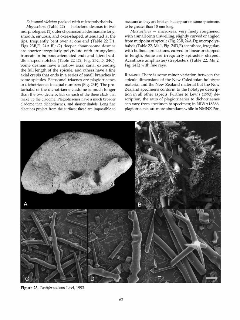
62
Ectosomal skeleton packed with micropolyrhabds. Megascleres (Table 22) — heloclone desmas in two
morphologies: (1) outer choanosomal desmas are long, smooth, sinuous, and oxea-shaped, attenuated at the tips, frequently bent over at one end (Table 22 D1, Figs 23B,E, 24A,B); (2) deeper choanosome desmas are shorter irregularly polytylote with strongylote, truncate or bulbous attenuated ends and lateral sad-dle-shaped notches (Table 22 D2; Fig. 23C,D, 24C). Some desmas have a hollow axial canal extending the full length of the spicule, and others have a fine axial crepis that ends in a series of small branches in some spicules. Ectosomal triaenes are plagiotriaenes or dichotriaenes in equal numbers (Fig. 23E). The pro-torhabd of the dichotriaene cladome is much longer than the two deuteroclads on each of the three clads that make up the cladome. Plagiotriaenes have a much broader cladome than dichotriaenes, and shorter rhabds. Long fine diactines project from the surface; these are impossible to
measure as they are broken, but appear on some specimens to be greater than 10 mm long.
Microscleres — microxeas, very finely roughened with a small central swelling, slightly curved or angled from midpoint of spicule (Fig. 23B, 24A,D); micropolyr-habds (Table 22, Ms 1, Fig. 24D,E) acanthose, irregular, with bulbous projections, curved or linear or stepped in length. Some are irregularly spiraster- shaped. Acanthose amphiaster/streptasters (Table 22, Ms 2, Fig. 24E) with fine rays.
remarks: There is some minor variation between the spicule dimensions of the New Caledonian holotype material and the New Zealand material but the New Zealand specimens conform to the holotype descrip-tion in all other aspects. Further to Lévi’s (1993) de-scription, the ratio of plagiotriaenes to dichotriaenes can vary from specimen to specimen; in NIWA18366, plagiotriaenes are more abundant, while in NMNZ Por.
Figure 23. Costifer wilsoni Lévi, 1993.
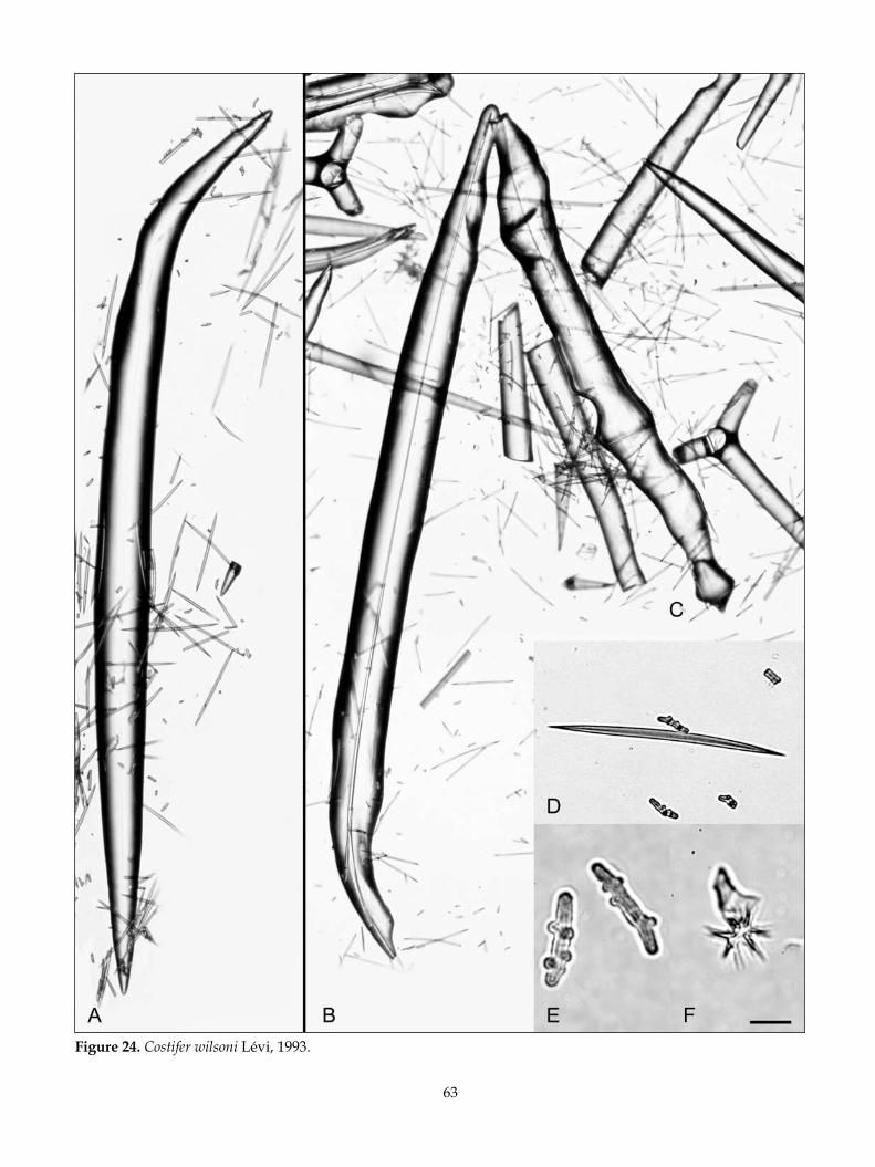
63
Figure 24. Costifer wilsoni Lévi, 1993.

64
597 dichotriaenes dominate. Furthermore, the desmas are in two recognisable categories — the desmas of the outer choanosome are predominantly long, smooth, sinuous, and oxea-shaped, and frequently bent over at one end, while the deeper choanosomal desmas are generally shorter and more irregular with uneven expansions and contractions along the axis and with strongylote, truncate or bulbous ends. Along the axis of these latter spicules are conspicuous lateral saddle-shaped notches indicating the proximity of adjacent spicules. Desmas may have a hollow axial canal extend-ing the full length of the spicule, or a fine axial crepis. Either form of desma may have a hollow or thread-like axial morphology.
Pisera and Lévi (2002l) suggested that C. wilsoni is very similar to the genus holotype Costifer vasiformis Wilson, 1925 and that it differs only in the possession of dichotriaenes and in the absence of lateral notches in the heloclones. Examination of this new material has afforded a clearer differentiation (see Table 22) between the two species; the microxeas, heloclones and plagi-otriaenes of C. vasiformis are considerably smaller than those of C. wilsoni, and the morphology of the former is vase-shaped with a rough irregular surface. This contrasts considerably with C. wilsoni, which is smooth and ear-shaped. Note that lateral notches are certainly present in New Caledonian material (NIWA18525); contrary to what Pisera and Lévi (2002l) suggested, these desmas with notches are perhaps commoner in the deeper choanosome.
Microfossil heloclones indistinguishable from those of extant Cosfiter wilsoni were found in the Tutuiri Greensand (Teurian–basal Waipawan) in the Chatham Islands (Buckeridge & Kelly 2005), confirming a more southern distribution for this genus during the Paleo-gene.
key diagnosTiC CharaCTers:• Large, heavy, ear-shaped; may be mistaken for
Pleroma turbinatum but the former is flexible while the latter is stony.
• Presence of heloclones in choanosome.
Family SCLERITODERMIDAE Sollas, 1888Scleritodermidae Sollas, 1888: 346; Wilson 1925: 463; de
Laubenfels 1936: 165.
Massive-amorphous, encrusting, flabellate, ear-, cup- or vase-shaped. Desmas are thorny or tuberculate rhizo-clones; ectosomal spicules when present are irregular curved smooth or acanthose spicules with strongylote ends, some are polyrhabdose, or larger acanthose tylos-trongyles; microscleres when present are sigmaspires. (Modified from Pisera and Lévi 2002b).
remarks: A partial rhizoclone desma skeleton, very similar to those in extant species of Scleritodermidae, was found in the Tutuiri Greensand (Teurian–basal Waipawan) in the Chatham Islands (Buckeridge & Kelly 2005), confirming a more southern distribution for this family during the Palaeogene.
Table 22. Comparison of the spicule dimensions (µm) of Costifer wilsoni Lévi, 1993 with the type species C. vasi-formis Wilson, 1925 (P cladome = plagiotriaene dimensions, D cladome = dichotriaene dimensions).
D 1 (L) D 2 (L) D Cladome (Max ø) Microxeas Ms 1 Ms 2 (W) (W) Rhabd (L) (L) (L) (L) P Cladome (Max ø) Rhabd (L)
New CaledoniaLévi (1993) 1500–4000 ― 600–1000 130–220 30 12(holotype) 40–120 ― 800–1000
New ZealandNMNZ Por. 597 2488 (2000–3000) 1907 (1550–2550) 1468 (1250–1650) 211 (175–232) 24 15 100 (70–150) 152 (100–200) 813 (650–1000) 1750 1125NIWA18366 2438 (1650–2700) 2469 (2375–2600) 1375–1625 183 (100–220) 29 15 200–225 100–125 2375 1937 (1500–2250) 1775 (1625–1700)
PhilippinesC. vasiformis 1880–2370 ― 525–830 90–165 18–25 15(Wilson, 1925) 220–280 850–1310
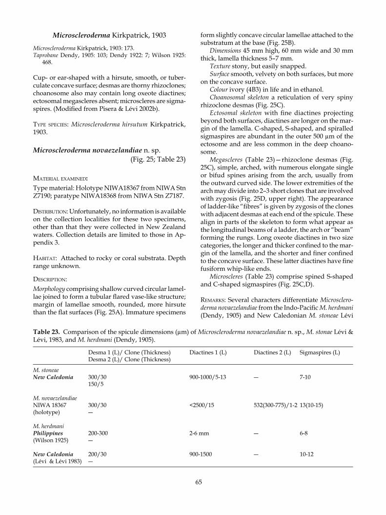
65
Microscleroderma Kirkpatrick, 1903
Microscleroderma Kirkpatrick, 1903: 173.Taprobane Dendy, 1905: 103; Dendy 1922: 7; Wilson 1925:
468.
Cup- or ear-shaped with a hirsute, smooth, or tuber-culate concave surface; desmas are thorny rhizoclones; choanosome also may contain long oxeote diactines; ectosomal megascleres absent; microscleres are sigma-spires. (Modified from Pisera & Lévi 2002b).
TyPe sPeCies: Microscleroderma hirsutum Kirkpatrick, 1903.
Microscleroderma novaezelandiae n. sp. (Fig. 25; Table 23)
maTerial examined: Type material: Holotype NIWA18367 from NIWA Stn Z7190; paratype NIWA18368 from NIWA Stn Z7187.
disTribuTion: Unfortunately, no information is available on the collection localities for these two specimens, other than that they were collected in New Zealand waters. Collection details are limited to those in Ap-pendix 3.
habiTaT: Attached to rocky or coral substrata. Depth range unknown.
desCriPTion:Morphology comprising shallow curved circular lamel-lae joined to form a tubular flared vase-like structure; margin of lamellae smooth, rounded, more hirsute than the flat surfaces (Fig. 25A). Immature specimens
form slightly concave circular lamellae attached to the substratum at the base (Fig. 25B).
Dimensions 45 mm high, 60 mm wide and 30 mm thick, lamella thickness 5–7 mm.
Texture stony, but easily snapped.Surface smooth, velvety on both surfaces, but more
on the concave surface.Colour ivory (4B3) in life and in ethanol.Choanosomal skeleton a reticulation of very spiny
rhizoclone desmas (Fig. 25C). Ectosomal skeleton with fine diactines projecting
beyond both surfaces, diactines are longer on the mar-gin of the lamella. C-shaped, S-shaped, and spiralled sigmaspires are abundant in the outer 500 µm of the ectosome and are less common in the deep choano-some.
Megascleres (Table 23)—rhizoclone desmas (Fig. 25C), simple, arched, with numerous elongate single or bifud spines arising from the arch, usually from the outward curved side. The lower extremities of the arch may divide into 2–3 short clones that are involved with zygosis (Fig. 25D, upper right). The appearance of ladder-like “fibres” is given by zygosis of the clones with adjacent desmas at each end of the spicule. These align in parts of the skeleton to form what appear as the longitudinal beams of a ladder, the arch or “beam” forming the rungs. Long oxeote diactines in two size categories, the longer and thicker confined to the mar-gin of the lamella, and the shorter and finer confined to the concave surface. These latter diactines have fine fusiform whip-like ends.
Microscleres (Table 23) comprise spined S-shaped and C-shaped sigmaspires (Fig. 25C,D).
remarks: Several characters differentiate Microsclero-derma novaezelandiae from the Indo-Pacific M. herdmani (Dendy, 1905) and New Caledonian M. stoneae Lévi
Table 23. Comparison of the spicule dimensions (µm) of Microscleroderma novaezelandiae n. sp., M. stonae Lévi & Lévi, 1983, and M. herdmani (Dendy, 1905).
Desma 1 (L)/ Clone (Thickness) Diactines 1 (L) Diactines 2 (L) Sigmaspires (L) Desma 2 (L)/ Clone (Thickness)
M. stoneaeNew Caledonia 300/30 900-1000/5-13 ― 7-10 150/5
M. novaezelandiaeNIWA 18367 300/30 <2500/15 532(300-775)/1-2 13(10-15)(holotype) ― M. herdmaniPhilippines 200-300 2-6 mm ― 6-8(Wilson 1925) ―
New Caledonia 200/30 900-1500 ― 10-12(Lévi & Lévi 1983) ―

66
Figure 25. Microscleroderma novaezelandiae n. sp.
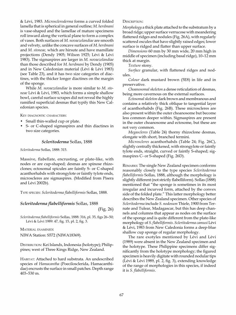
67
& Lévi, 1983. Microscleroderma forms a curved folded lamella that is spherical in general outline; M. herdmani is vase-shaped and the lamellae of mature specimens roll inward along the vertical plane to form a complex of vases. Both surfaces of M. novaezelandiae are smooth and velvety, unlike the concave surfaces of M. herdmani and M. stoneae, which are hirsute and have mamillate projections (Dendy 1905; Wilson 1925; Lévi & Lévi 1983). The sigmaspires are larger in M. novaezelandiae than those described for M. herdmani by Dendy (1905) and in New Caledonian material (Lévi & Lévi 1983) (see Table 23), and it has two size categories of diac-tines, with the thicker longer diactines on the margin of the sponge.
While M. novaezelandiae is more similar to M. sto-neae Lévi & Lévi, 1983, which forms a simple shallow bowl, careful surface scrapes did not reveal the highly ramified superficial desmas that typify this New Cal-edonian species.
key diagnosTiC CharaCTers:• Small thin-walled cup or plate.• S- or C-shaped sigmaspires and thin diactines in
two size categories.
Scleritoderma Sollas, 1888
Scleritoderma Sollas, 1888: 315.
Massive, flabellate, encrusting, or plate-like, with nodes or are cup-shaped; desmas are spinose rhizo-clones; ectosomal spicules are faintly S- or C-shaped acanthorhabds with strongylote or faintly tylote ends, microscleres are sigmaspires. (Modified from Pisera and Lévi 2002b).
TyPe sPeCies: Scleritoderma flabelliformis Sollas, 1888.
Scleritoderma flabelliformis Sollas, 1888 (Fig. 26)
Scleritoderma flabelliformis Sollas, 1888: 316, pl. 35, figs 26–50; Lévi & Lévi 1989: 47, fig. 15, pl. 2, fig. 3.
maTerial examined: NIWA Station: S572 (NIWA18369).
disTribuTion: Kei Islands, Indonesia (holotype); Philip-pines; west of Three Kings Ridge, New Zealand.
habiTaT: Attached to hard substrata. An undescribed species of Hamacantha (Poecilosclerida, Hamacanthi-dae) encrusts the surface in small patches. Depth range 403–530 m.
desCriPTion:Morphology a thick plate attached to the substratum by a broad ridge; upper surface verrucose with meandering flattened ridges and nodules (Fig. 26A), with regularly scattered oscules that have slightly raised edges; lower surface is ridged and flatter than upper surface.
Dimensions 60 mm by 30 mm wide, 20 mm high in middle of specimen (including basal ridge), 10–12 mm thick at margin.
Texture stony.Surface granular, with flattened ridges and nod-
ules.Colour dark mustard brown (5E8) in life and in
preservative.Choanosomal skeleton a dense reticulation of desmas,
being more cavernous on the external surfaces.Ectosomal skeleton dark brown and membranous and
contains a relatively thick oblique to tangential layer of acanthorhabds (Fig. 26B). These microscleres are also present within the outer choanosome but become less common deeper within. Sigmaspires are present in the outer choanosome and ectosome, but these are not very common.
Megascleres (Table 24) thorny rhizoclone desmas, elongate with short, branched termini.
Microscleres acanthorhabds (Table 24; Fig. 26C), slightly centrally thickened, with strongylote or faintly tylote ends, straight, curved or faintly S-shaped; sig-maspires C- or S-shaped (Fig. 26D).
remarks: The single New Zealand specimen conforms reasonably closely to the type species Scleritoderma flabelliformis Sollas, 1888, although the morphology is slightly different (not strictly flabelliform). Sollas (1888) mentioned that “the sponge is sometimes in its most irregular and incurved form, attached by the convex side of the folded plate.” This latter morphology better describes the New Zealand specimen. Other species of Scleritoderma include S. nodosum Thiele, 1900 from Ter-nate and Tulear, Madagascar, but this has deep chan-nels and columns that appear as nodes on the surface of the sponge and is quite different from the plate-like morphology of S. flabelliformis. Scleritoderma camusi Lévi & Lévi, 1983 from New Caledonia forms a deep-blue shallow cup sponge of regular morphology.
The rare exotyles mentioned by Lévi and Lévi (1989) were absent in the New Zealand specimen and the holotype. These Philippine specimens differ sig-nificantly from the holotype morphology; the figured specimen is heavily digitate with rounded nodular tips (Lévi & Lévi 1989, pl. 2, fig. 3), extending knowledge of the range of morphologies in this species, if indeed it is S. flabelliformis.
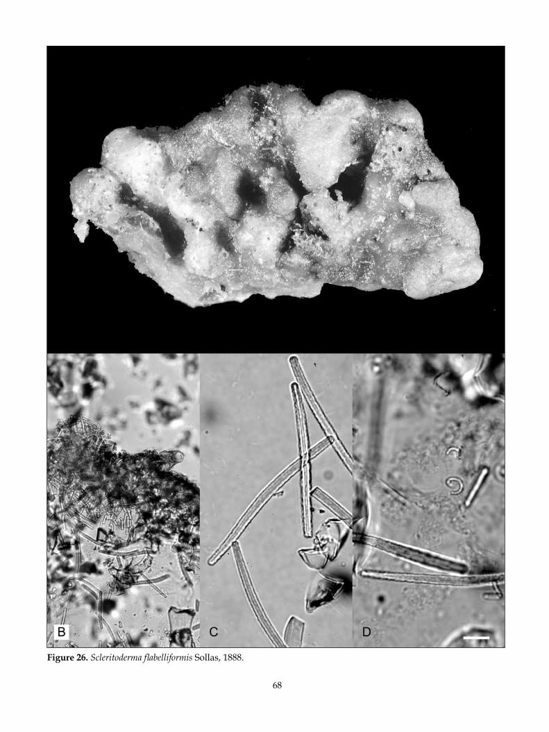
68
Figure 26. Scleritoderma flabelliformis Sollas, 1888.

69
Table 24. Spicule dimensions (µm) of Scleritoderma flabelliformis Sollas, 1888.
Desmas (L) Acanthorhabd (L) Exotyles (L) Sigmaspires (L) Clone (Thickness) (Thickness)
Indonesia(Sollas 1888) 240–500 80–90 ― 10(holotype) 39 13
Philippines(Lévi & Lévi 1989) ― 60–95 160 (rare) 8–10 5–7 2
New ZealandNIWA18369 200–400 75 (60–90) ― 9–10 30–45 5–7
The rare occurrence of S. flabelliformis in New Zea-land probably indicates the southernmost limit of this species, which is commoner in the Indo-Pacific.
key diagnosTiC CharaCTers:• Massive plate with a verrucose (corrugated and
nodular) upper surface.• Presence of curved, weakly S- or C-shaped acan-
thorhabds with faintly bulbous (tylote) ends, and sigmaspires.
Aciculites Schmidt, 1879
Aciculites Schmidt, 1879: 29.
Cushion-shaped, with raised or tubular oscules, encrusting, or massive-columnar with blind digits; choanosomal desmas are rhizoclones with spinose zygomes; ectosomal megascleres are curved strongyles to tylostrongyles with one or both ends acanthose, arranged tangentially or paratangentially in the ecto-some; microscleres absent. (Modified from Pisera and Lévi 2002b).
TyPe sPeCies: Aciculites higginsi Schmidt, 1879.
Aciculites pulchra Dendy, 1924 (Fig. 27, 28)
Aciculites pulchra Dendy, 1924: 315, pl. 6, fig.1–1a; Bergquist 1968: 63, fig. 30, pl. 10a.
maTerial examined: NIWA Sta t ions : E636 (NIWA18377) ; F993 (NIWA18372); I14 (NIWA18370); J954 (NIWA18375, NIWA25609) ; KAH0011/30 (NIWA18378) ; K A H 0 0 1 1 / 4 0 ( N I W A 1 8 3 7 9 , N I W A 1 8 3 8 0 ) ; KAH0011/41 (NIWA18381); KAH9301 (NIWA25610); KAH9907/48 (NIWA18382, NIWA18383, NIWA18384); P8 (NIWA25608, NIWA25646); RAPUHIA2-15 (NIWA25779); TAN0413/068 (NIWA25040); TAN0413/092 (NIWA18389) ; TAN0413/099
(NIWA18390); TAN0413/109 (NIWA18386); TAN0413/111 (NIWA18391) ; TAN0413/118 (NIWA18387 , NIWA18388) ; TAN0413/170 (NIWA18385); TAN0413/173 (NIWA18392); TAN0413/174 (NIWA18393) ; TAN9915/3B (NIWA25528); V473 (NIWA18373); X128 (NIWA18374); Z8487 (NIWA18371); Z9041 [NIWA18376, NIWA25033]; Z9742 [NIWA18823 (0CDN6692-J)]; Z9747 (NIWA25060 (fragment of 0CDN6730-Y]); Z9890 (NIWA25616); Z9892 (NIWA25615). NMNZ Stations: NMNZ Por. 308; NMNZ Por. 562; NMNZ Por. 563; NMNZ Por. 564; NMNZ Por. 570; NMNZ Por. 571; NMNZ Por. 573; NMNZ Por. 620.
disTribuTion: West Norfolk Ridge; southern Three Kings Ridge; western continental slope, Northland; west of Pandora Bank; North Cape; Louisville Seamount Chain; Kermadec Ridge; Cavalli Seamount; Ngunguru Bay, Northland; outer Bay of Islands; off Tutukaka; Rungapapa, Tumokemoke, Mahina, and Tuatoru Knolls, west of White Island, and Mayor Island, Bay of Plenty; east of Gisborne; south of East Cape; Mahia Peninsula.
habiTaT: CRRF specimens were dredged from ‘coarse sand and rubble’, and ‘rocky reef substrate’. The base of the sponge encrusts hard substrata. Depth range 80–1050 m.
desCriPTion:Morphology vase-shaped with foliose lamellae (Fig. 27A,B), or ear-shaped with a flat lamella (Fig. 27C, D).
Dimensions 50–200 mm wide, 10–15 mm thick, up to 30 mm at the base of some specimens.
Texture stony.Surfaces sharply differentiated; the exhalent surface,
usually the concave surface, is smooth and crowded with numerous regularly dispersed low hemispherical oscular projections c. 0.5 mm high; the other (inhalent) surface is smooth, granular, and irregular growth rings may be visible.
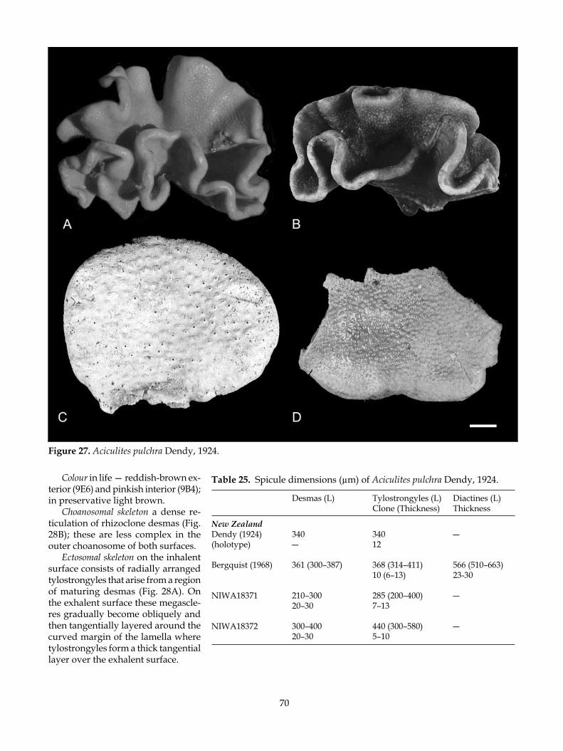
70
Figure 27. Aciculites pulchra Dendy, 1924.
Colour in life — reddish-brown ex-terior (9E6) and pinkish interior (9B4); in preservative light brown.
Choanosomal skeleton a dense re-ticulation of rhizoclone desmas (Fig. 28B); these are less complex in the outer choanosome of both surfaces.
Ectosomal skeleton on the inhalent surface consists of radially arranged tylostrongyles that arise from a region of maturing desmas (Fig. 28A). On the exhalent surface these megascle-res gradually become obliquely and then tangentially layered around the curved margin of the lamella where tylostrongyles form a thick tangential layer over the exhalent surface.
Table 25. Spicule dimensions (µm) of Aciculites pulchra Dendy, 1924.
Desmas (L) Tylostrongyles (L) Diactines (L) Clone (Thickness) Thickness
New ZealandDendy (1924) 340 340 ―(holotype) ― 12
Bergquist (1968) 361 (300–387) 368 (314–411) 566 (510–663) 10 (6–13) 23-30
NIWA18371 210–300 285 (200–400) ― 20–30 7–13 NIWA18372 300–400 440 (300–580) ― 20–30 5–10

71
Figure 28. Aciculites pulchra Dendy, 1924.
Megascleres — thorny rhizoclone desmas (Table 25; Fig. 28A–C) with long crepi, terminating in 1- to 3-spined cladi, spines single or bifurcate; zygosis is lateral and terminal and may involve the spinose pro-jections (Fig. 28C); tylostrongyles (Fig. 28D,E) (‘rhabdi’ of Dendy 1924: 316), curved or straight with slightly tylote acanthose proximal head and smooth strongy-lote distal terminus. Most megascleres are strongylote with one end acanthose.
Microscleres absent.
remarks: Bergquist (1968) found large stout ‘oxeas’ in addition to the typical tylostrongyles in specimens off
Mahia Peninsula. These oxeas were described as being radially disposed and arranged singly or in groups of up to four just below the oscular surface. Occasional spicules fitting this description were found in a new spicule preparation of Bergquist’s specimen. However, large ‘true’ oxea were not described from Dendy’s holo-type and have not been found in any of the specimens examined in this study.
There is considerable variation in the dimensions and morphology of the tylostrongyles; in Bergquist’s Mahia specimen, the tylostrongyles are markedly ty-lote, almost bulbous at the proximal end, and faintly tylote hammer-like at the distal end. In some specimens

72
oxeote diactines have been found but they are rare and probably derived from the tylostrongyle megasclere; they are syringe-like with a thick hollow inception channel, and one or both ends are slightly sinuous and elongate-fusiform. Some also have faintly tylote ends. These “diactines” are no longer than the tylostrongyles. Some specimens have tylostrongyles that taper distally to an oxeote point and it may be these that were seen in Bergquist’s specimen.
The spiculation of this species is thus quite variable. However, the vase-like morphology with raised oscules, coupled with the presence of a range of tylostrongyles (dimensions and morphology) is diagnostic.
Aciculites pulchra Dendy is almost identical morpho-logically to the Indo-Pacific A. ciliata Wilson, 1925 with reddish-brown colouration, a vase-like foliose or ear-shaped lamella, and raised oscular projections on the exhalant surface. Examination of numerous specimens from the Philippines and Papua New Guinea (courtesy of CRRF) reveal that the tylostrongyles in A. ciliata are much longer (up to 540 µm long) and are irregularly bulbous at one end, however. The spicules also taper to a fine blunt distal point. This species also possesses much longer fine tylostrongyles that resemble oxeas, up to 850 µm long; these are radially arranged along the inhalant surface. The outer choanosome in A. ciliata Wilson is highly cavernous with elongate immature desma.
key diagnosTiC CharaCTers:• Vase- or ear-shaped lamella with differentiated
surfaces.• Reddish-brown colouration.• Presence of tylostrongyles with one end rough-
ened.• Could be mistaken for Neoschrammeniella fulvodes-
mus but that species does not have raised oscules.
Aciculites manawatawhi n. sp. (Fig. 29)
maTerial examined: Type material: Holotype BMNH 2005.9.15.1 from NIWA Station Z9747 [fragment of 0CDN6740-L]; para-type NIWA18394 from NIWA Stn J953.NIWA Station: P14 (NIWA25623). NMNZ Station: NMNZ Por. 583.
TyPe loCaliTy: West of Pandora Bank, northwestern New Zealand.
disTribuTion: West Norfolk Ridge; west of Pandora Bank, and Middlesex Bank, northwest of Three Kings Islands; western continental slope, northeastern New Zealand.
habiTaT: Coarse sandy rubble. Depth range 198–319 m.
desCriPTion:Morphology tree-like with one or more thick, sometimes hollow columns fused along the stem or above where the digits emerge; stem circular in profile or flanged if more than one stem is present (Fig. 29A). Some speci-mens present a more columnar shape (Fig. 29B). The stem flares and gives rise to numerous branches that may be hollow at the base, each diverging to form one or more smaller branches. These terminate in a rounded or pointed blind tip (Fig. 29C). The overall impression is of an irregular squat tree with an irregularly truncate top that is compressed in one plane. Oscules are not apparent, although there are soft areas at the tips of the sponge that may function as exhalant areas.
Dimensions of the holotype are 100 mm wide at the apex, 50 mm thick, 70 mm tall, and 50 mm wide at the base. NMNZ Por. 583 is 85 mm wide at the apex, 60 mm thick, 110 mm tall, and 30 mm wide at the base. Sec-ondary branches are c. 15 mm thick, smaller branches c. 5–10 mm thick.
Texture of the stem and lower branches is stony, tips are fragile and soft.
Surface granular, rough, encrusted by numerous epizoites including other sponges, bryozoans, and tubeworms (Fig. 29A).
Colour in life dark grayish-blue (20E4), in preser-vative light golden brown (5B7). Tips of branches are paler than the digits of the apex.
Choanosomal skeleton composed of thorny rhizoclone desmas that are less complex and dense in the outer choanosome.
Ectosomal skeleton composed of tylostrongyles ar-ranged obliquely to paratangentially over most of the body surface and radially at the growing tips of the sponge.
Megascleres (Table 26) rhizoclone desmas are elongate and arched with short clads at the ends of the desma (Fig. 29D). Ornamented with short thick bifurcate spines; overall a thick, robust spicule. Zygosis appears to be predominantly terminal. Tylostrongyles or ‘oxytyles’ with pronounced acanthose bulbous heads and strongylote or tylote acanthose distal ends in some spicules the proximal end is bent sharply as in a rhabdostyle (Fig. 29E).
Microscleres absent.
eTymology: Named for the largest island in the Three Kings group, Manawa Tawhi (Great Island), the type locality. Manawa tawhi (New Zealand Māori) means ‘heart food’ (manawa, heart, breath, emotions), but it may mean ‘the heart beckons’ (tawhi, food, beckons) (Weno Iti & Guy Penny pers. comm.).

73
Figure 29. Aciculites manawatawhi n. sp.

74
remarks: Aciculites manawatawhi is unique in the genus in terms of gross morphology and is easily differenti-ated from other Indo- and Southwest Pacific species, which are ear- or cup-shaped (A. pulchra Dendy, 1924 from New Zealand, A. ciliata Wilson, 1925 from New Caledonia, and A. tulearensis Vacelet & Vasseur, 1965 from Madagascar) or spherical papillate masses (A. orientalis Dendy, 1905 from Sri Lanka and A. papillata Lévi & Lévi, 1983 and A. oxytylota Lévi & Lévi, 1983 from New Caledonia). Aciculites spinosa Vacelet & Vas-seur, 1971, also from Madagascar, forms tiny cushions and is found in deep reef tunnels and caves.
The oxytyles of A. manawatawhi are also quite differ-ent from those in other species with their bulbous acan-those heads, but similar to those of A. oxytylota Levi & Levi, 1983 from New Caledonia. Like A. manawatawhi, A. oxytylota also has tubular surface features, but these arise from a cushion base while A. manawatawhi is columnar with blind digits.
Levi and Levi (1983) compared A. oxytylota to the genus Siphonidium (Siphonidiidae), specifically with S. capitatum Sollas, 1888 from Indonesia, as it also has tubular oscules, but this species is columnar, and more similar, in fact, to A. manawatawhi. However, in addi-tion to the similarly shaped ‘oxytylotes’ (Sollas 1888), S. capitatum has distinctive planar ectosomal desmas, and the oxytyles are exotylote in arrangement — the spicule lies perpendicular to the sponge surface with the tylote head facing outwards and the oxeote end pointing into the interior (Sollas 1888). Aciculites manawatawhi and A. oxytylota have an irregular but distinct layer of oxytyles that are strewn tangentially and paratangentially in the surface. This layer is not as thick or distinct as in A. pulchra, for example, but it is definitely present and the arrangement is certainly not exotylote.
Aciculites manawatawhi and A. oxytylota can also be compared the siphonidiid Gastrophanella, but this genus also has exotyles [the acanthose subtylostyles
and tylostrongyles protrude ‘head-first’ through the surface, an architecture that is well described by Pisera and Levi (2002f) and Muricy and Minervino (2000)], which do not form a tangential layer, and the species typically forms club-shaped columns with a deep broad cavity. Pisera and Levi (2002f) noted that Gastrophanella also has long smooth styles and long oxeas, absent from A. manawatawhi and A. oxytylota. The diagnosis of Aciculites has been expanded to include species with oxytylotes in a paratangential layer.
It is clear that the dimensions of tylostrongyles and desmas cannot be used as a species-level differentia-tor in Aciculites as the lengths of these spicules in the different species are very similar, ranging from about 200–500µm, and the full range often present in some species. (A notable exception is A. spinosa, which has quite small tylostrongyles ranging from 125 to 275 µm.) The shape of the tylostrongyle appears to be more diagnostic; A. oxytylota and A. manawatawhi have ox-ytyles with pronounced bulbous heads, but the latter species has tylote bulbs on both ends of many spicules. The proximal end is also frequently sharply bent in the shape of a rhabdostyle. This new species is also notable for the large overall size of these megascleres.
While some authors recognise oxea-like spicules in some species, others appear to lack a second megasclere category. Perhaps a more careful study of all species will reveal anatomically differentiated categories.
key diagnosTiC ChareCTers:• Tree-like morphology with small finger-like branch-
es of uneven length, surface covered in epizoites.• Deep bluish-gray on the main body, paler branch
tips.• Long tylostrongyles with pronounced bulbous
heads.
Aciculites sulcus n. sp. (Fig. 30)
maTerial examined: Type material: Holotype NIWA18395 from NIWA Stn KAH9907/48; paratype NIWA18821 from NIWA Stn Z9748. NIWA Stations: I372 (NIWA25648); RAPUHIA2-15 (NIWA25778).
TyPe loCaliTy: North Cape and Doubtless Bay; west of White Island, Bay of Plenty.
disTribuTion: Bay of Plenty.
habiTaT: Encrusting rocky substrata. Depth range 211–250 m.
Table 26. Spicule dimensions (µm) of Aciculites mana-watawhi n. sp.
Desmas (L) Oxytyles (L) Clone Thickness (Thickness)
New ZealandBMNH 2005.9.15.1 200–400 380 (300–500)(holotype) 30–40 8–10
NIWA18394 300–400 345 (280–430) 50 8–12 51
NMNZ Por. 583 250–300 390 (340–500) 20–40 8–10

75
Figure 30. Aciculites sulcus n. sp.
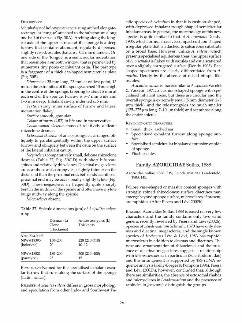
76
desCriPTion:Morphology of holotype an encrusting arched elongate-rectangular ‘tongue’ attached to the substratum along one half of the base (Fig. 30A). Arching along the long-est axis of the upper surface of the sponge is a deep furrow that contains abundant, regularly dispersed, slightly raised, oscules that are c. 0.5 mm diameter. On one side of the ‘tongue’ is a semicircular indentation that resembles a smooth window that is permeated by numerous tiny pores or inhalant ostia. The paratype is a fragment of a thick ear-haped semicircular plate (Fig. 30B).
Dimensions 35 mm long, 23 mm at widest point, 15 mm at the extremities of the sponge, arched 13 mm high in the centre of the sponge, tapering to about 5 mm at each end of the sponge. Furrow is 3–7 mm wide and 1–5 mm deep. Inhalant cavity indented c. 5 mm.
Texture stony, inner surface of furrow and lateral indentation flakey.
Surface smooth, granular.Colour of putty (4B2) in life and in preservative.Choanosomal skeleton mass of relatively delicate
rhizoclone desmas.Ectosomal skeleton of anisostrongyles, arranged ob-
liquely to paratangentially within the upper surface furrow and obliquely between the ostia on the surface of the lateral inhalant cavity.
Megascleres comparatively small, delicate rhizoclone desmas (Table 27; Fig. 30C,D) with short bifurcate spines and relatively thin clones. Diactinal megascleres are acanthose anisostrongyles, slightly thinner on the distal end than the proximal end, both ends acanthose, proximal end may be occasionally slightly tylote (Fig. 30D). These megascleres are frequently quite sharply bent in the middle of the spicule and often have a tylote bulge midway along the spicule.
Microscleres absent.
Table 27. Spicule dimensions (µm) of Aciculites sulcus n. sp. Desmas (L) Anisostrongyles (L) Clone Thickness (Thickness)
New ZealandNIWA18395 150–200 228 (210–310)(holotype) 20 10–12
NIWA18821 180–200 300 (210–400)(paratype) 25 15
eTymology: Named for the specialised exhalant oscu-lar furrow that runs along the surface of the sponge (Latin, sulcus).
remarks: Aciculites sulcus differs in gross morphology and spiculation from other Indo- and Southwest Pa-
cific species of Aciculites in that it is cushion-shaped, with depressed inhalant trough-shaped semicircular inhalant areas. In general, the morphology of this new species is quite similar to that of A. orientalis Dendy, 1905, which forms a massive, compact cushion-shaped irregular plate that is attached to calcareous substrata on a broad base. However, unlike A. sulcus, which presents specialised aquiferous areas, the upper surface of A. orientalis is flakey with oscules and ostia scattered over a slightly corrugated surface (Dendy 1905). Ear-shaped specimens are clearly differentiated from A. pulchra Dendy by the absence of raised pimple-like oscules.
Aciculites sulcus is more similar to A. spinosa Vacelet & Vasseur, 1971, a cushion-shaped sponge with spe-cialised inhalant areas, but these are circular and the overall sponge is extremely small (5 mm diameter, 2–3 mm thick), and the tylostrongyles are much smaller (125–275 µm long, 7–10 µm thick) and acanthose along the entire spicule.
key diagnosTiC CharaCTers:• Small, thick, arched ear.• Specialised exhalant furrow along sponge sur-
face.• Specialised semicircular inhalant depression on side
of sponge.• Flush oscules.
Family AZORICIDAE Sollas, 1888Azoricidae Sollas, 1888: 319; Leiodermatidae Lendenfeld,
1903: 145.
Foliose vase-shaped or massive conical sponges with strongly spined rhizoclones; surface diactines may emerge beyond sponge surface; microscleres, if present, are raphides. (After Pisera and Lévi 2002h).
remarks: Azoricidae Sollas, 1888 is based on very few characters and the family contains only two valid genera, recently reviewed by Pisera and Lévi (2002h). Species of Leiodermatium Schmidt, 1870 have only des-mas and diactinal megascleres, and the single known species of Jereicopsis Lévi & Lévi, 1983 has raphide microscleres in addition to desmas and diactines. The type and ornamentation of rhizoclones and the pres-ence of diactinal megascleres suggests a relationship with Microscleroderma in particular (Scleritodermidae) and this arrangement is supported by 18S rDNA se-quence analysis (Kelly-Borges & Pomponi 1994). Pisera and Lévi (2002h), however, concluded that, although there are similarities, the absence of ectosomal rhabds and microscleres in Leiodermatium and the presence of raphides in Jereicopsis distinguish the groups.

77
Leiodermatium Schmidt, 1870
Leiodermatium Schmidt, 1870: 22; Sollas 1888: 352; Wilson 1925: 465; Pisera & Lévi 2002h: 353.
Azorica Carter, 1873: 439.Heterophymia Pomel, 1872: 143.
Plate-like, ear-shaped, foliose, or vase-shaped with smooth faintly hispid surface; oscules may be visible. Desmas are strongly spined rhizoclones; there are no special ectosomal spicules and no microscleres. Acces-sory megascleres are whip-like diactines. (Modified from Pisera and Lévi 2002h).
TyPe sPeCies: Leiodermatium lynceus Schmidt, 1870.
remarks: The genus Leiodermatium is best known from the type species Leiodermatium lynceus Schmidt, 1870 (Atlantic) and L. pfeifferae (Carter, 1873) and variants (Atlantic, Southeast Asia), with additional data be-ing provided by much Pacific material (Sollas 1888; Wilson 1925).
With relatively few characters available to differen-tiate Leiodermatium at the species level, the discovery of two new Southwest Pacific taxa — L. linea n. sp. from New Zealand and L. dampieri n. sp. from the Lord Howe Seamount Chain — has provided an opportunity to examine here the genus as a whole. This revision has re-sulted in the recognition of Southeast Asian specimens of Leiodermatium pfeifferae [Sollas’s (1888) morphotype ‘Azorica pfeifferae intermedia’ and Leiodermatium pfeifferae sensu Wilson (1925)] as L. intermedia n. comb., and in the recognition of a new taxon from Micronesia and the west central pacific, L. colini n. sp.
Leiodermatium intermedia (Sollas, 1888) (Fig. 31)
Azorica pfeifferae intermedia Sollas, 1888: 319, pl. 36, figs 1–29.
Leiodermatium pfeifferae: Wilson 1925: 465, pl. 43, figs 4, 8, pl. 51, figs 4–7; Lendenfeld 1903: 148.
maTerial examined: Type material: Holotype USNM21321 of Leiodermatium pfeifferae (Carter, 1873) and L. pfeifferae f. striata Wilson, 1925; tangential and vertical sections; paratype BMNH 2005.9.15.2, west side of Uchelbeluu Reef, Mutremdiu, Koror, Palau, Micronesia (07º16.27’ N, 134º31.37’ E), collected by Patrick L. Colin, Coral Reef Research Foundation (Manned submersible Deep Worker 2000), 21 March 2001, 240 m [fragment of 0CDN7878-W].
TyPe loCaliTy: Ambon Island, Moluccas, Indonesia, 27–46 m (Sollas 1888).
disTribuTion: Patinti Strait between Halmahera and Bacan Islands, Moluccas, Indonesia; Banda Sea; Palau, Micronesia.
habiTaT: Sand and coral boulder slopes. Depth range 27–46 m (Sollas 1888); 375 m (Wilson 1925); 240 m (Palau).
desCriPTion:Morphology a convoluted mass of thin flaring folded lamellae that may form tubes, with rounded incised margins (Wilson 1925; Sollas, 1888) (Fig. 31A,B). The ex-ternal surface of the Palau specimen BMNH 2005.9.15.2 [0CDN 7878-W] has patches of tiny (< 0.5 mm diameter) ostial pits and the inner surface is completely covered in regularly and closely distributed oscules (< 0.5 mm diameter). Cup margin in all specimens is thick and rounded, sometimes indented or incised.
Dimensions of Patinti Strait specimens 50–90 mm high, 70–130 mm wide, cup wall 0.5–2.5 mm thick (Wilson 1925); Ambon specimen 77 mm high, 110 mm wide, lamellae 3.2–4.0 mm thick (Sollas 1888); Palau specimen forms a mass c. 500 mm diameter, wall 2.5 mm thick.
Texture stony, dense.Surface smooth and granular with low irregular
horizontal ridges, internal osculiferous surface slightly furry with brishes of projecting diactines.
Colour tan (Palau); topaz (5C5) to putty-like (4B2).Skeleton of thorny rhizoclone desmas dominate the
choanosome and fine diactinal spicules project from the ectosome. Diactines are longer on the margin.
Megascleres (Table 28) rhizoclone desmas (Fig. 31C,D) with a straight or curved axis, clones with dis-
Table 28. Spicule dimensions (µm) of Leiodermatium intermedia (Sollas, 1888).
Desmas (L x W) Diactines (L) Clone Thickness (Thickness)
Moluccas, IndonesiaL. pfeifferae sensu Wilson 1925USNM 21321 340 (1) 350–420(holotype) 35 (2) ? 6
Azorica pfeifferae 160–350 (1) ? intermedia Sollas, 1888 20–25 (2) 750 8
Palau, MicronesiaBMNH 2005.9.15.2 340–500(linear (1) 240–500 forms)(paratype) 35 (2) >1000 1–8

78
Figure 31. Leiodermatium intermedia (Sollas, 1888)

79
crete single or bifurcate spines, the tips of which are often bifid, sinuous, or dendritic. On the outer surface of the sponge the rhizoclones are smaller and more intricate, slightly flattened and with numerous short blunt spines. Diactines (Fig. 31D) in two size categories, the smallest are fine and hair-like, confined to osculif-erous face of sponge, whereas thicker longer spicules are confined to the margin of the sponge.
Microscleres absent.
remarks: Leiodermatium pfeifferae Carter, 1873 was first described from Portugal in the northern Atlantic Ocean, from 684 m. Carter (1876) considered L. pfeifferae to be very similar to the type species L. lynceus Schmidt, 1870, also from Portugal, but upheld the separation of his new species because of the presence of oscules on the outside of the lamellae rather than on the inside, as in L. pfeifferae and most other planar sponges. Moreover, Carter (1876) considered the differences in the distri-bution of the fine diactines (abundant and restricted to the margin of L. pfeifferae as a fringe, as opposed to “isolated diactines around the margin of L. lynceus”) to be significant and thus diagnostic for the two species (Table 29). A recent illustration of L. lynceus by Pisera and Lévi (2002h) shows blunt denticulate spines on the desmas whereas Sollas (1888) described the desmas as terminating in bifurcate spines with bifid rounded ends. The ornamentation of these termini is clearly an important diagnostic indicator of species differences (as in other lithistid genera) and careful comparison of the ornamentation and morphology of the desmas using SEM is required to confirm or otherwise the integrity of L. pfeifferae and L. lynceus.
Sollas (1888) assigned to L. pfeifferae a range of specimens collected by H.M.S. Challenger (1873–1876) from various Atlantic localities including Bermuda (795 and 1965 m), Cape Verde Islands (183–234 m), and Brazil (no depth given), but his description of the species (Sollas 1888: 320) was based on a specimen col-lected off Ambon Island in the Moluccas Archipelago, Indonesia (Table 29). This was the only specimen found to be in the living state upon collection, and Sollas’s description was based upon this specimen. All of the Atlantic specimens were dead and denuded skeletons when collected.
Sollas (1888, p. 322) recognised differences among the Atlantic specimens and between those and the Indonesian specimen. Within the Atlantic complex he recognised at least three morphological ‘groups’ (Table 29) that included (1) ‘Azorica pfeifferae’ — sponges with comparatively thick walls, large marginate oscules, and slender smooth desmas (the holotype specimen of the type species and a specimen from the Cape Verde Islands); (2) ‘Azorica pfeifferae tenui-laminaris’
— sponges with comparatively thin walls, small closely spaced oscules, and slender smooth desmas (speci-mens from Bahia); (3) ‘Azorica pfeifferae tenui-laminaris osculis disjunctis’ — sponges with comparatively thin walls, small remotely distributed oscules confined to the sponge margin (specimen from Bermuda); and this was differentiated quite clearly from (4) ‘Azorica pfeifferae intermedia’ — sponges with relatively thick walls, small closely spaced oscules, and thick, highly tuberculate desmas (specimen from Ambon Island, Indonesia). While revision of these Atlantic speci-mens is beyond the scope of this work, it appears that the major differences described are within the range of characters expected from sponges collected from widespread locations. Whether or not the species name pfeifferae can be applied to these specimens or not will be largely dependent on the ornamentation of the desmas as other details such as the distribution of the diactines are lost.
Wilson (1925) also attributed several specimens from the 375 m in Patinti Strait, Moluccas, to L. pfeifferae, and these are very similar to the Challenger specimen from nearby Ambon Island.
A specimen collected more recently from Palau (240 m), by the Coral Reef Research Foundation (Table 29), is very similar to Wilson’s and Sollas’s Indonesian material.
All of the above Southeast Asian sponges are united in the possession of a relatively thin-walled foliose habit, diactines on all surfaces including the sponge margin, and large richly tuberculate desmas with thick clones. They are clearly separate morphologically and biogeographically from Atlantic L. pfeifferae, and are segregated here as L. intermedia by raising Sollas’s (1888) “variety” to full species. Interestingly, this spe-cies is the only known source of two new selectively cytotoxic macrocyclic lactones, and these are the first secondary metabolites reported in the literature to date (Sandler et al. 2006).
key diagnosTiC CharaCTers:• Convoluted mass of thin, flaring, folded lamellae
that may form tubes.• Oscules and ostia inconspicuous.• Sponge surface slightly furry.• Rhizoclones are relatively smooth with numerous
discrete bifurcate, bifid, dendritic spines.
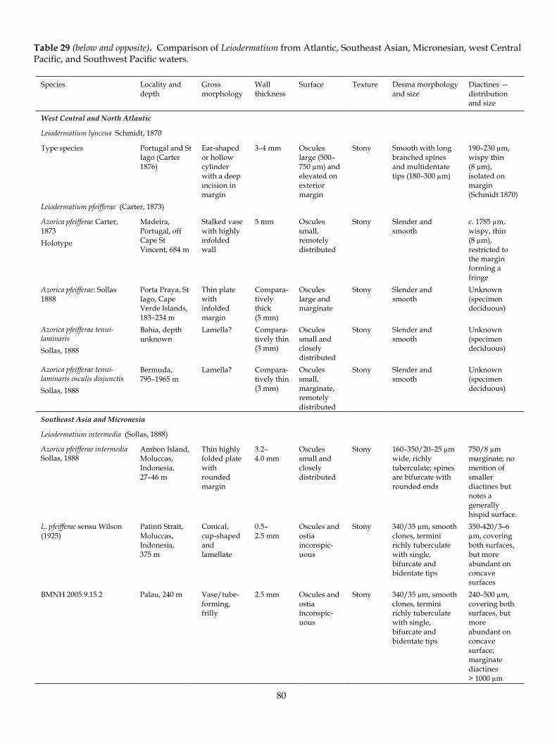
80
Table 29 (below and opposite). Comparison of Leiodermatium from Atlantic, Southeast Asian, Micronesian, west Central Pacific, and Southwest Pacific waters.
Species Locality anddepth
Gross morphology
Wallthickness
Surface Texture Desma morphology and size
Diactines — distribution and size
West Central and North Atlantic
Leiodermatium lynceus Schmidt, 1870
Type species Portugal and St Iago (Carter 1876)
Ear-shaped or hollow cylinderwith a deep incision in margin
3–4 mm Oscules large (500–750 µm) and elevated on exteriormargin
Stony Smooth with long branched spines and multidentate tips (180–300 µm)
190–230 µm, wispy thin (8 µm), isolated on margin (Schmidt 1870)
Leiodermatium pfeifferae (Carter, 1873)
Azorica pfeifferae Carter, 1873Holotype
Madeira,Portugal, off Cape St Vincent, 684 m
Stalked vase with highly infoldedwall
5 mm Oscules small,remotely distributed
Stony Slender and smooth
c. 1785 µm, wispy, thin (8 µm), restricted to the margin forming a fringe
Azorica pfeifferae: Sollas 1888
Porta Praya, St Iago, Cape Verde Islands, 183–234 m
Thin plate withinfoldedmargin
Compara-tivelythick (5 mm)
Oscules large and marginate
Stony Slender and smooth
Unknown (specimen deciduous)
Azorica pfeifferae tenui-laminarisSollas, 1888
Bahia, depth unknown
Lamella? Compara-tively thin (3 mm)
Oscules small and closelydistributed
Stony Slender and smooth
Unknown (specimen deciduous)
Azorica pfeifferae tenui-laminaris osculis disjunctisSollas, 1888
Bermuda, 795–1965 m
Lamella? Compara-tively thin (3 mm)
Oscules small,marginate, remotely distributed
Stony Slender and smooth
Unknown (specimen deciduous)
Southeast Asia and Micronesia
Leiodermatium intermedia (Sollas, 1888)
Azorica pfeifferae intermediaSollas, 1888
Ambon Island, Moluccas,Indonesia, 27–46 m
Thin highly folded plate withrounded margin
3.2– 4.0 mm
Oscules small and closelydistributed
Stony 160–350/20–25 µm wide, richly tuberculate; spines are bifurcate with rounded ends
750/8 µm marginate; no mention of smallerdiactines but notes a generallyhispid surface.
L. pfeifferae sensu Wilson (1925)
Patinti Strait, Moluccas,Indonesia, 375 m
Conical, cup-shaped and lamellate
0.5– 2.5 mm
Oscules and ostia inconspic-uous
Stony 340/35 µm, smooth clones, termini richly tuberculate with single, bifurcate and bidentate tips
350-420/3–6 µm, covering both surfaces, but more abundant on concavesurfaces
BMNH 2005.9.15.2 Palau, 240 m Vase/tube-forming, frilly
2.5 mm Oscules and ostia inconspic-uous
Stony 340/35 µm, smooth clones, termini richly tuberculate with single, bifurcate and bidentate tips
240–500 µm, covering both surfaces, but more abundant on concavesurface; marginate diactines > 1000 µm

81
Table 29. (continued)
Leiodermatium colini n. sp. (Fig. 32)
maTerial examined: Type material: Holotype BMNH 2005.3.14.1, cave off Blue Hole Caverns, West Barrier Reef, Palau, Micronesia, 07º08.10’ N, 134º13.90’ E, 22 May 2000, 28 m [fragment of 0CDN7069-S]; paratype BMNH 2005.3.14.2, cave off Blue Hole Caverns, West Barrier Reef, Palau, Micronesia, 07º08.10’ N, 134º13.90’ E, 26
June 1997, 31 m [fragment of 0CDN5026-W]; paratype BMNH 2005.3.14.3, Robert’s Reef, Kimbe Bay, West New Britain, Papua New Guinea, 05º25.66’ S, 150º22.65’ E, 17 November 1992, 18 m [fragment of 0CDN0779-G]; paratype BMNH 2005.3.14.4, Nomundo Reef, Kimbe Bay, West New Britain, Papua New Guinea, 05º27.64 S, 150º06.28’ E, 5 July 2003, 46 m [fragment of 0CDN9062-K]. All specimens collected by P.L. Colin, Coral Reef Research Foundation, by SCUBA.
Species Locality anddepth
Gross morphology
Wallthickness
Surface Texture Desma morphology and size
Diactines — distribution and size
Micronesia and West Central Pacific
Leiodermatium colini n. sp.
BMNH 2005.3.14.2 BMNH 2005.3.14.1
BMNH 2005.3.14.3 BMNH 2005.3.14.4
Found in holes in reef near Blue Corner, 28–31 m, West Barrier Reef, Palau, Micronesia
Robert’s Reef, Kimbe Bay, West New Britain, Papua New Guinea, underoverhang, 18–46 m
Ear- or shallow vase-shaped withoccasionalundulations in lamella; surface and margin shaggy
As above
8–10 mm
6–8 mm
Oscules and ostia inconspic-uous, the latter in meandering grooves
Stony 100–300/50–100 µm, small desmas with thick clones and short irregular solitary spines
Very long diactines (> 2000 µm) extending in tufts from the margin, smaller (250–500 µm) finer diactines cover osculiferous surface only
Southwest Pacific and New Zealand
Leiodermatium linea n. sp.
Bay of Plenty, New Zealand, 232–302 m
Frilled cup (no peduncle)
3–4 mm Oscules and ostia inconspic-uous, aligned in parallel or circularchannels
Stony,slightly flexiblein plane of lamella
Small desmas(120–200)/20 µm; discrete long single or bifurcate spines with bifid or trifid tips
Fine hair-like diactines (250–300 µm)
Leiodermatium dampieri n. sp.
DampierRidge, Lord HoweSeamountChain, 420 m
Chalice- to cup- shaped with a short peduncle
2.5–4 mm Oscules and ostia inconspic-uous; ostial pits on some parts of the outer surface
Stony 250–350/20–30 µm, smooth clones, termini richly tuberculate with single, bifurcate and bidentate tips
300–450 µm, covering both surfaces, but more abundant on concavesurface; marginate diactines 675 µm

82
Figure 32. Leiodermatium colini n. sp.
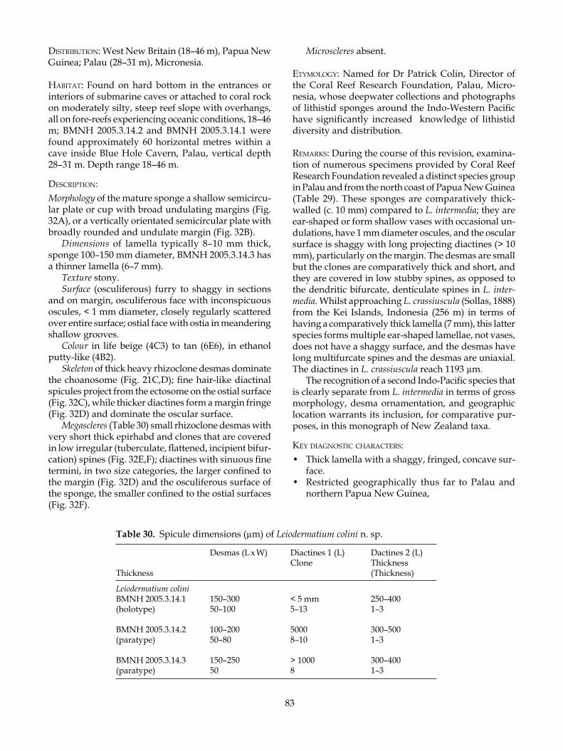
83
disTribuTion: West New Britain (18–46 m), Papua New Guinea; Palau (28–31 m), Micronesia.
habiTaT: Found on hard bottom in the entrances or interiors of submarine caves or attached to coral rock on moderately silty, steep reef slope with overhangs, all on fore-reefs experiencing oceanic conditions, 18–46 m; BMNH 2005.3.14.2 and BMNH 2005.3.14.1 were found approximately 60 horizontal metres within a cave inside Blue Hole Cavern, Palau, vertical depth 28–31 m. Depth range 18–46 m.
desCriPTion:Morphology of the mature sponge a shallow semicircu-lar plate or cup with broad undulating margins (Fig. 32A), or a vertically orientated semicircular plate with broadly rounded and undulate margin (Fig. 32B).
Dimensions of lamella typically 8–10 mm thick, sponge 100–150 mm diameter, BMNH 2005.3.14.3 has a thinner lamella (6–7 mm).
Texture stony.Surface (osculiferous) furry to shaggy in sections
and on margin, osculiferous face with inconspicuous oscules, < 1 mm diameter, closely regularly scattered over entire surface; ostial face with ostia in meandering shallow grooves.
Colour in life beige (4C3) to tan (6E6), in ethanol putty-like (4B2).
Skeleton of thick heavy rhizoclone desmas dominate the choanosome (Fig. 21C,D); fine hair-like diactinal spicules project from the ectosome on the ostial surface (Fig. 32C), while thicker diactines form a margin fringe (Fig. 32D) and dominate the oscular surface.
Megascleres (Table 30) small rhizoclone desmas with very short thick epirhabd and clones that are covered in low irregular (tuberculate, flattened, incipient bifur-cation) spines (Fig. 32E,F); diactines with sinuous fine termini, in two size categories, the larger confined to the margin (Fig. 32D) and the osculiferous surface of the sponge, the smaller confined to the ostial surfaces (Fig. 32F).
Microscleres absent.
eTymology: Named for Dr Patrick Colin, Director of the Coral Reef Research Foundation, Palau, Micro-nesia, whose deepwater collections and photographs of lithistid sponges around the Indo-Western Pacific have significantly increased knowledge of lithistid diversity and distribution.
remarks: During the course of this revision, examina-tion of numerous specimens provided by Coral Reef Research Foundation revealed a distinct species group in Palau and from the north coast of Papua New Guinea (Table 29). These sponges are comparatively thick-walled (c. 10 mm) compared to L. intermedia; they are ear-shaped or form shallow vases with occasional un-dulations, have 1 mm diameter oscules, and the oscular surface is shaggy with long projecting diactines (> 10 mm), particularly on the margin. The desmas are small but the clones are comparatively thick and short, and they are covered in low stubby spines, as opposed to the dendritic bifurcate, denticulate spines in L. inter-media. Whilst approaching L. crassiuscula (Sollas, 1888) from the Kei Islands, Indonesia (256 m) in terms of having a comparatively thick lamella (7 mm), this latter species forms multiple ear-shaped lamellae, not vases, does not have a shaggy surface, and the desmas have long multifurcate spines and the desmas are uniaxial. The diactines in L. crassiuscula reach 1193 µm.
The recognition of a second Indo-Pacific species that is clearly separate from L. intermedia in terms of gross morphology, desma ornamentation, and geographic location warrants its inclusion, for comparative pur-poses, in this monograph of New Zealand taxa.
key diagnosTiC CharaCTers:• Thick lamella with a shaggy, fringed, concave sur-
face.• Restricted geographically thus far to Palau and
northern Papua New Guinea,
Table 30. Spicule dimensions (µm) of Leiodermatium colini n. sp.
Desmas (L x W) Diactines 1 (L) Dactines 2 (L) Clone Thickness Thickness (Thickness)
Leiodermatium colini BMNH 2005.3.14.1 150–300 < 5 mm 250–400(holotype) 50–100 5–13 1–3
BMNH 2005.3.14.2 100–200 5000 300–500(paratype) 50–80 8–10 1–3
BMNH 2005.3.14.3 150–250 > 1000 300–400(paratype) 50 8 1–3

84
Leiodermatium dampieri n. sp. (Fig. 33)
maTerial examined: Type material: Holotype NIWA18400 from NIWA Stn P925; paratype NIWA18401, NIWA18402, NIWA18403.NIWA Stations: P925 (NIWA18404: 37 specimens from the same collection, all dried and mostly deciduous).
TyPe loCaliTy: Dampier Ridge, Lord Howe Seamount Chain.
disTribuTion: Dampier Ridge, Lord Howe Seamount Chain.habiTaT: Attached to rocky substrata. Depth range 420 m.
desCriPTion:Morphology of chalice-shaped or flared plates with a short peduncle by which the sponge attaches to the substratum (Fig. 33A). As the sponge grows in diam-eter, the walls appear to flatten into an undulating plate and younger specimens are more distinctly chalice-shaped. The base of the peduncle is often flanged and flared at the point of attachment. The cup margin in all specimens is thick and rounded, sometimes indented or incised. Oscules are inconspicuous but visible under magnification and confined to the concave surface, excluding the sponge margin (Fig. 33B).
Dimensions — width 10–30 mm wide, overall height 6–20 mm, peduncle height 1–6 mm, peduncle width 8–15 mm; wall thickness 2.5–4 mm. It is not known if these are juvenile sponges and grow much larger as adults.
Texture stony, pumice-like (deciduous and dead upon collection).
Surface smooth and granular, slightly undulating, convex surface occasionally knobbed or ridged.
Colour putty-like (4B2) in the dry state.Skeleton of thorny rhizoclone desmas that dominate
the choanosome; fine diactinal spicules project from the ectosome.
Megascleres (Table 31) — rhizoclone desmas (Fig. 33C,D) with a straight or curved axis, clones smooth with occasional rounded to oval prominences, often emerging in an angle to the clone, some tubercles bifur-cate at the extreme tip forming tiny spines. Diactines in two size categories — the smallest are fine and hair-like, confined to osculiferous face of sponge; thicker longer spicules are confined to the margin of the sponge.
Microscleres absent.
eTymology: Named for the type locality.
remarks: The collection of small cup sponges from the Dampier Ridge in the Lord Howe Seamount Chain is interesting as it presents a full range of morphologies from the smallest chalice-shaped individuals to cups with flattened undulating walls, yet it is not known whether these are adults or juveniles. The large number of specimens collected from one location, in one or two size classes, suggests that they may be immature sponges.
Superficially, these specimens resemble the Asian-Pacific species L. intermedia and L. colini described in this volume, but careful inspection of the desmas reveals that they differ in ornamentation; the desmas of L. intermedia are large, arched and thorny with long bifurcate spines, those of L. colini are quite thick and coarse with low flat non-bifurcate spines, while those of L. dampieri are more delicate and complex in zygosis, with rounded protuberances that bifurcate at the very tip. The diactines in L. intermedia and L. colini are also extremely long compared to those of L. dampieri.
Until further material of L. dampieri is collected from the remote Dampier Ridge or other seamounts in the region, it remains to be seen whether these specimens include fully mature individuals; the majority are quite small and appear to be juvenile.
key diagnosTiC CharaCTers:• Chalice- to plate-shaped tiny sponges.• Rhizoclones are relatively smooth with smooth
rounded protuberances that may bifurcate at the tips.
• May be mistaken for Reidispongia coerulea in the ju-venile and mature form, but the desma morphology is completely different and this species lacks unique megascleres in the surface of the sponge.
Table 31. Spicule dimensions (µm) of Leiodermatium dampieri n. sp.
Desmas (L x W) Diactines (L) Clone Thickness (Thickness)
Leiodermatium dampieri NIWA18400 150–350 (1) 450(holotype) 10–20 (2) 600+
NIWA18402 200–325 none visible 10–30
NIWA18403 250–425 (1) 300–450 20–30 (2) 675 1–8

85
Figure 33. Leiodermatium dampieri n. sp.

86
Leiodermatium linea n. sp. (Fig. 34)
maTerial examined: Type material: Holotype NIWA18820 and paratype NIWA18399 from NIWA Stn KAH0011/30; paratype NIWA25048 from TAN0413/118.
TyPe loCaliTy: Rungapapa Knoll, Bay of Plenty.
disTribuTion: Bay of Plenty.
habiTaT: Encrusting basalt substrata. Depth range 159–165 m.
desCriPTion:Morphology (holotype) is a frilled shallow cup with a slightly expanded rim (Fig. 34A); undulations in the wall may anastomose to form a tube. The paratype is a thin basal plate 15 mm diameter and 1 mm thick, from which arises a 3 mm thick undulating wall (Fig. 34B). Oscules and ostia are inconspicuous. Under magnifica-tion, oscules and ostial pits are c. 0.1 mm diameter. On the concave surface, the oscules are aligned in clearly visible shallow aquiferous channels 0.1 mm wide that meander from the base of the lamella, stopping short of the margin. On the convex side, ostial pits are con-nected in oval interconnecting aquiferous channels. Margin slightly thicker than lamella.
Dimensions 55 mm wide and long, 22 mm high (holotype); walls 3–4 mm thick.
Texture stony.Surface smooth, slight growth rings visible, par-
ticularly under the margin, some slight bulges, but otherwise very smooth; slightly furry to the touch with hair-like projecting diactines. Parallel and circular aquiferous channels visible on concave and convex surfaces.
Colour in life cream (4A3), putty-like (4B2) in etha-nol.
Skeleton (choanosome) packed with thorny rhizo-clone desmas; hair-like diactinal spicules are arranged radially in the ectosome, protruding slightly from the edge of the sponge.
Megascleres (Table 32) — rhizoclone desmas (Fig. 34C) with an arched or straight longitudinal axis; 3–6 clones arise from either side of a straight or curved axis and are covered in well separated discrete single or multiple spines; the apex of these spines may be bifid or trifid but not dendritic (Fig. 34 D,E). Desmas are very fine and comparatively small. Diactines are extremely fine and raphide-like.
Microscleres absent.
Table 32. Spicule dimensions (µm) of Leiodermatium linea n. sp. Desmas (L x W) Diactines (L) Clone Thickness (Thickness)
Leiodermatium linea NIWA18820 170 (120–200) x 250–300 130 (100–150)(holotype) 20 1
NIWA18399 158 (120–200) x 250 100 (80–130(paratype) 10–20
eTymology: Named for the aquiferous channels that form visible lines on the lamellae of the sponge, run-ning from the base to the margin (Latin, linea).
remarks: The discovery of Leiodermatium in New Zea-land waters is interesting in that it is the southernmost record of the genus, which has not been reported from New Caledonia despite numerous collections on the northern seamounts of the Norfolk Ridge. Although L. linea is similar in gross morphology to L. intermedia n. sp., subtle differences separate these species (see Table 29). Leiodermatium linea has characteristic sur-face aquiferous channels that run parallel from the base of the lamella to the margin, and are just visible with the unaided eye. On the ostial surface, these lines anastomose to form ovals. The desmas and diactines of this species are the smallest yet described, and the ornamentation of the desmas is unique, having short conical bifid or trifid spines.
key diagnosTiC CharaCTers:• Thin-walled cup that may be easily mistaken for
Reidispongia coerulea, although the latter is blue.
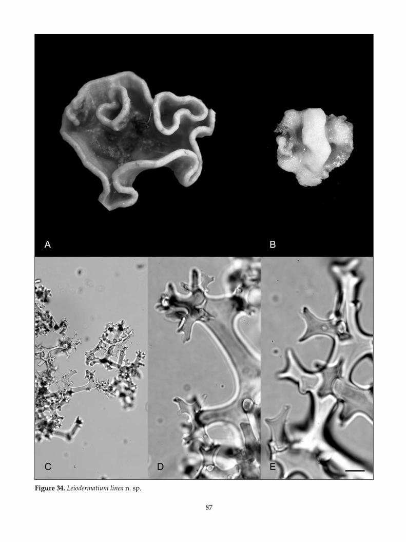
87
Figure 34. Leiodermatium linea n. sp.
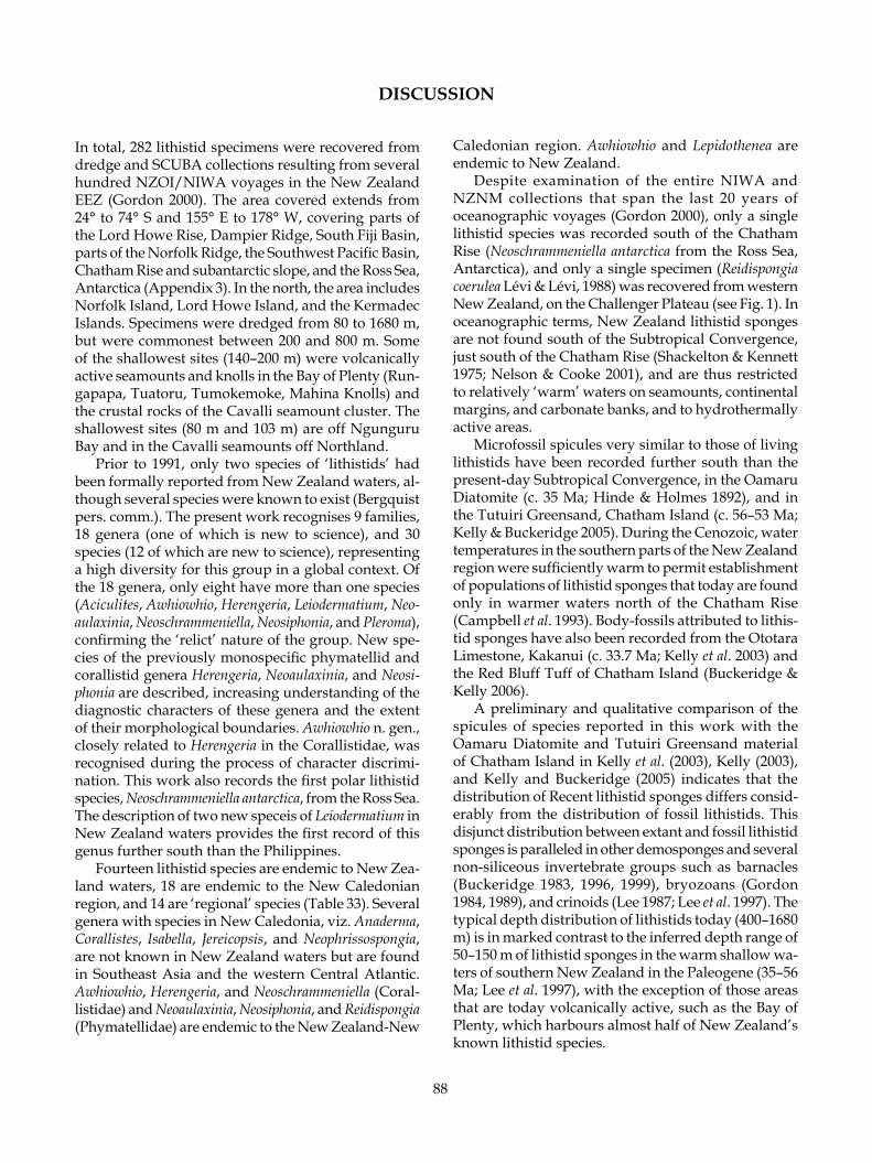
88
In total, 282 lithistid specimens were recovered from dredge and SCUBA collections resulting from several hundred NZOI/NIWA voyages in the New Zealand EEZ (Gordon 2000). The area covered extends from 24° to 74° S and 155° E to 178° W, covering parts of the Lord Howe Rise, Dampier Ridge, South Fiji Basin, parts of the Norfolk Ridge, the Southwest Pacific Basin, Chatham Rise and subantarctic slope, and the Ross Sea, Antarctica (Appendix 3). In the north, the area includes Norfolk Island, Lord Howe Island, and the Kermadec Islands. Specimens were dredged from 80 to 1680 m, but were commonest between 200 and 800 m. Some of the shallowest sites (140–200 m) were volcanically active seamounts and knolls in the Bay of Plenty (Run-gapapa, Tuatoru, Tumokemoke, Mahina Knolls) and the crustal rocks of the Cavalli seamount cluster. The shallowest sites (80 m and 103 m) are off Ngunguru Bay and in the Cavalli seamounts off Northland.
Prior to 1991, only two species of ‘lithistids’ had been formally reported from New Zealand waters, al-though several species were known to exist (Bergquist pers. comm.). The present work recognises 9 families, 18 genera (one of which is new to science), and 30 species (12 of which are new to science), representing a high diversity for this group in a global context. Of the 18 genera, only eight have more than one species (Aciculites, Awhiowhio, Herengeria, Leiodermatium, Neo-aulaxinia, Neoschrammeniella, Neosiphonia, and Pleroma), confirming the ‘relict’ nature of the group. New spe-cies of the previously monospecific phymatellid and corallistid genera Herengeria, Neoaulaxinia, and Neosi-phonia are described, increasing understanding of the diagnostic characters of these genera and the extent of their morphological boundaries. Awhiowhio n. gen., closely related to Herengeria in the Corallistidae, was recognised during the process of character discrimi-nation. This work also records the first polar lithistid species, Neoschrammeniella antarctica, from the Ross Sea. The description of two new speceis of Leiodermatium in New Zealand waters provides the first record of this genus further south than the Philippines.
Fourteen lithistid species are endemic to New Zea-land waters, 18 are endemic to the New Caledonian region, and 14 are ‘regional’ species (Table 33). Several genera with species in New Caledonia, viz. Anaderma, Corallistes, Isabella, Jereicopsis, and Neophrissospongia, are not known in New Zealand waters but are found in Southeast Asia and the western Central Atlantic. Awhiowhio, Herengeria, and Neoschrammeniella (Coral-listidae) and Neoaulaxinia, Neosiphonia, and Reidispongia (Phymatellidae) are endemic to the New Zealand-New
Caledonian region. Awhiowhio and Lepidothenea are endemic to New Zealand.
Despite examination of the entire NIWA and NZNM collections that span the last 20 years of oceanographic voyages (Gordon 2000), only a single lithistid species was recorded south of the Chatham Rise (Neoschrammeniella antarctica from the Ross Sea, Antarctica), and only a single specimen (Reidispongia coerulea Lévi & Lévi, 1988) was recovered from western New Zealand, on the Challenger Plateau (see Fig. 1). In oceanographic terms, New Zealand lithistid sponges are not found south of the Subtropical Convergence, just south of the Chatham Rise (Shackelton & Kennett 1975; Nelson & Cooke 2001), and are thus restricted to relatively ‘warm’ waters on seamounts, continental margins, and carbonate banks, and to hydrothermally active areas.
Microfossil spicules very similar to those of living lithistids have been recorded further south than the present-day Subtropical Convergence, in the Oamaru Diatomite (c. 35 Ma; Hinde & Holmes 1892), and in the Tutuiri Greensand, Chatham Island (c. 56–53 Ma; Kelly & Buckeridge 2005). During the Cenozoic, water temperatures in the southern parts of the New Zealand region were sufficiently warm to permit establishment of populations of lithistid sponges that today are found only in warmer waters north of the Chatham Rise (Campbell et al. 1993). Body-fossils attributed to lithis-tid sponges have also been recorded from the Ototara Limestone, Kakanui (c. 33.7 Ma; Kelly et al. 2003) and the Red Bluff Tuff of Chatham Island (Buckeridge & Kelly 2006).
A preliminary and qualitative comparison of the spicules of species reported in this work with the Oamaru Diatomite and Tutuiri Greensand material of Chatham Island in Kelly et al. (2003), Kelly (2003), and Kelly and Buckeridge (2005) indicates that the distribution of Recent lithistid sponges differs consid-erably from the distribution of fossil lithistids. This disjunct distribution between extant and fossil lithistid sponges is paralleled in other demosponges and several non-siliceous invertebrate groups such as barnacles (Buckeridge 1983, 1996, 1999), bryozoans (Gordon 1984, 1989), and crinoids (Lee 1987; Lee et al. 1997). The typical depth distribution of lithistids today (400–1680 m) is in marked contrast to the inferred depth range of 50–150 m of lithistid sponges in the warm shallow wa-ters of southern New Zealand in the Paleogene (35–56 Ma; Lee et al. 1997), with the exception of those areas that are today volcanically active, such as the Bay of Plenty, which harbours almost half of New Zealand’s known lithistid species.
DISCUSSION

89
Table 33. Checklist of lithistid species described from the New Zealand--New Caledonian region. Note that this list excludes Neoschrammeniella antarctica from the Ross Sea, Antarctica (SEA, Southeast Asia; IO, Indian Ocean).
Species NZ NC OtherAciculites orientalis Dendy, 1905 IOAciculites oxytylota Lévi & Lévi, 1983 Aciculites papillata Lévi & Lévi, 1983 Aciculites pulchra Dendy, 1924 Aciculites sulcus n. sp. Aciculites manawatawhiAnaderma rancureli Lévi & Lévi, 1983 Callipelta punctata Lévi & Lévi, 1983 Corallistes australis Schlacher-Hoenlinger, Pisera & Hooper, 2005 Corallistes multituberculatus Lévi & Lévi, 1983 Corallistes undulatus Lévi & Lévi, 1983 Costifer wilsoni Lévi, 1993 Discodermia proliferans Lévi & Lévi, 1983 Awhiowhio osheai n. sp. Awhiowhio sepulchrum n. sp. Awhiowhio unda n. sp. Herengeria auriculata Lévi & Lévi, 1988 Herengeria vasiformis Schlacher-Hoenlinger, Pisera & Hooper, 2005 Homophymia pollubrum Schlacher-Hoenlinger, Pisera & Hooper, 2005 Homophymia stipitata Kelly, 2000 Isabella mirabilis Schlacher-Hoenlinger, Pisera & Hooper, 2005 Jereicopsis graphidophora Lévi & Lévi, 1983 Leiodermatium dampieri n. sp. Leiodermatium linea n. sp. Lepidothenea incrustans (Dendy, 1924) Macandrewia spinifoliata Lévi & Lévi, 1983 Microscleroderma herdmani (Dendy, 1905) IOMicroscleroderma novaezealandiae n. sp. Microscleroderma stoneae Lévi & Lévi, 1983 Neoaulaxinia clavata Lévi & Lévi, 1988 Neoaulaxinia persicum n. sp. Neoaulaxinia zingiberadix n. sp. Neopelta plinthosellina Lévi & Lévi, 1988 Neopelta pulvinus n. sp. Neophrissospongia microstylifer Lévi & Lévi, 1983 Neoschrammeniella castrum Schlacher-Hoenlinger, Pisera & Hooper, 2005 Neoschrammeniella fulvodesmus Lévi & Lévi, 1983 Neoschrammeniella moreti (Lévi & Lévi, 1988) Neoschrammeniella norfolkii Schlacher-Hoenlinger, Pisera & Hooper, 2005 Neosiphonia superstes Sollas, 1888 Neosiphonia motukawanui n. sp. Pleroma aotea Kelly, 2003 Pleroma menoui Lévi & Lévi, 1983 Pleroma turbinatum Sollas, 1888 Reidispongia coerulea Lévi & Lévi, 1988 Scleritoderma camusi Lévi & Lévi, 1983 Scleritoderma flabelliformis Sollas, 1888 SEATOTAL 29 32 3

90
ACKNOWLEDGMENTS
The author thanks Dr Andrzej Pisera, Institute of Paleo-biology, Polish Academy of Sciences, Warszawa, Po-land, for his review and comments on the manuscript; these have added considerable value to this work. Dr Steve O’Shea, formerly of NIWA Wellington, and Bruce Marshall, Museum of New Zealand/Te Papa Ton-garewa, assisted in sourcing material from the NIWA and NMNZ collections, respectively. The Coral Reef Research Foundation contributed much new material from their research cruise on NIWA’s RV Kaharoa in 1999, around northeastern New Zealand. These and Palau collections were supported by the U.S. National Cancer Institute shallow-water marine collections pro-gramme. This monograph is thus a contribution of the Coral Reef Research Foundation.
Dame Professor Patricia Bergquist (retired, Auck-land University), provided photographic material for several species. Helena Armiger and Lincoln Tubbs of NIWA Auckland completed all photographic il-lustrations, and Michael Ellwood of NIWA Hamilton prepared the locality map. Drs Alex Ball and Chris Jones, Electron Microscopy Unit, Natural History Museum, London were extremely helpful in providing access and training for the author and Klaus Borges, UNITEC, Auckland, who carried out the majority of the scanning microscopy featured in this work. Thanks to Mark Hamann and Jennifer Allman, Pharmacology School, University of Mississippi, for their assistance
with information on biological activity of lithistid sponges.
Research and fieldwork was funded by a New Zealand Lotteries Grants Board Repatriation Fellow-ship (1997–1999), and a Royal Society of New Zealand Marsden Fund Grant (2000–2002). This research has also been supported by New Zealand Foundation for Research Science & Technology Contracts C01X0028 (Seamounts) and C01X0219 (Biodiversity) to NIWA. This research was also facilitated by the Biological and Biotechnological Research Council (BBSRC), UK, and British Airways Conservation Travel Awards, while the author was employed at the Natural History Mu-seum, London (1992–1996). Ongoing support from the National Institute of Water & Atmospheric Research (NIWA) is gratefully acknowledged.
Comparative research on the south New Caledo-nian slope, which assisted this study immensely, was carried out onboard the French IRD (Institut de Re-cherche pour le Développement) RV Alis, and I thank Captain Raymond Proner and his crew for their help during the voyage. This collaborative work was carried out with financial help of the French Foreign Affair Ministry through the IRD research teams. I particularly acknowledge the assistance of Dr Bertrand Richer de Forges for his help in the field and Drs Cecile Debitus and Dominique Laurent from the Chemical Product Study Program (SMIB).
REFERENCES
BASSARELLO, C.; NAPOLITANO, A.; BIFULCO, G.; BRUNO, I.; CASAPULLO, A.; RANDAZZO, A.; ZAM-PELLA A.; RICCIO, R. 2000: Bioactive macrolides and cyclopeptides from the South Pacific sponges. Revista Latinoamericana de Química 28: 59–62.
BERGQUIST, P.R. 1968: The Marine Fauna of New Zealand: Porifera, Demospongiae, part 1 (Tetractinomorpha and Lithistida). New Zealand Oceanographic Institute Memoir 37: 1–98.
BEWLEY, C.A.; FAULKNER, D.; ANGEW, J. 1998: Lithistid sponges — star performers or hosts to the stars [re-view]. Angewanta Chemi International [English Edition] 37: 2163–2178.
BOURY-ESNAULT, N.; RÜTZLER, K. (Eds) 1997: Thesaurus of sponge morphology. Smithsonian Contributions to Zool-ogy 596: 1–55.
BUCKERIDGE, J.S. 1983: The fossil barnacles (Cirripedia: Thoracica) of New Zealand and Australia. New Zealand Geological Survey Paleontological Bulletin 50: 1–151.
BUCKERIDGE, J.S. 1993: Cirripedia and Porifera. In: Camp-bell, H.J.; et al. Cretaceous-Cainozoic geology and bios-
tratigraphy of the Chatham Islands. New Zealand Institute of Geological and Nuclear Sciences Monograph 2: 1–240.
BUCKERIDGE, J.S. 1996: Phylogeny and biogeography of the primitive Sessilia and a consideration of a Tethyan origin for the group. Crustacean Issues 10: 225–267.
BUCKERIDGE, J.S. 1999: Post Cretaceous biotic recovery: a case study on Crustacea : Cirripedia) from the Chatham Islands, New Zealand. Records of the Canterbury Museum 13: 43–51.
BUCKERIDGE, J.S.; KELLY, M. 2006: Abstract. On the pal-aeobiology of two distinctive sponge faunas from the Chatham Islands, New Zealand. 7th International Sponge Symposium: Biodiversity, Innovation, Sustainability (7–13 May 2006, Armação de Búzios, Rio de Janiero, Brazil). Museu Nacional Série Livros [Published Abstracts] 16: 181.
BUCKERIDGE, J.; KELLY, M. 2002: Abstract. Early Cainozoic hexactinellid sponges (Phylum Porifera) from the Tutuiri Greensand, Chatham Island, New Zealand. Proceedings of the Geological Society of New Zealand Annual Conference, Whangarei, 2–5 December 2002: 10.

91
CAMPBELL, H.J.; ANDREWS, P.B.; BEU, A.G.; MAXWELL, P.A.; EDWARDS, A.R.; LAIRD, M.G.; HORNIBROOK, N.de B.; MILDENHALL, D.C.; WATTERS, W.A.; BUCK-ERIDGE, J.S.; LEE, D.E.; STRONG, C.P.; WILSON, G.J.; HAYWARD, B.W. 1993: Cretaceous–Cainozoic geology and biostratigraphy of the Chatham Islands. New Zealand Institute of Geological and Nuclear Sciences Monograph 2: 1–240.
CARBONELLI, S.; ZAMPELLA, A.; RANDAZZO, A.; DEBI-TUS, C.; GOMEZ-PALOMA, L. 1999: Sphinxolides E–G and reidispongiolide C: four new cytotoxic macrolides from the new caledonian lithistida sponges N. superstes and R. coerulea. Tetrahedron 55: 14665–14674.
CARTER, H.J. 1876: Descriptions and figures of deep-sea sponges and their spicules from the Atlantic Ocean, dredged up on board H.M.S. ‘Porcupine’, chiefly in 1869 (concluded). Annals and Magazine of Natural History, ser. 4, 18: 226–240, 307–324, 388–410, 458–479, pls 12–16.
CHOMBARD, C.; BOURY-ESNAULT, N.; TILLIER, S. 1998: Reassessment of homology of morphological characters in Tetractinellid sponges based on molecular data. Sys-tematic Biology 47: 351–366.
CRIST, B.V.; LI, X.; BERGQUIST, P.R.; DJERASSI, C. 1983: Sterols of marine invertebrates. 44. Isolation, structure elucidation, partial synthesis, and determination of ab-solute configuration of pulchrasterol. The first example of double bioalkylation of the sterol side chain at position 26. Journal of Organic Chemistry 48: 4472–4479.
DASCH, E.J.; EVANS, A.L.; ESSENE, E. 1970: Radiometric and petrologic data from eclogites and megacrysts of the Kakanui Mineral Breccia, New Zealand. Annual Meeting, Geological Society of America, Abstracts with Programs 2 (7): 532–533.
D’AURIA, M.V.; GOMEZ-PALOMA, L.; MINALE, L.; ZAM-PELLA, A.; DEBITUS, C.; PEREZ, J. 1995: Neosiphoni-amolide A, a novel cyclodepsipeptide, with antifungal activity from the marine sponge Neosiphonia superstes. Journal of natural Products 58: 121–123.
D’AURIA, M. V.; ZAMPELLA, A.; GOMEZ-PALOMA, L.; MINALE, L.; DEBITUS, C.; ROUSSAKIS, C.; LE BERT, V. 1996: Callipeltins B and C; bioactive peptides from a marine Lithistida sponge Callipelta sp. Tetrahedron 52: 9589–9596.
DAWSON, E.W. 1993: The marine fauna of New Zealand: Index to the fauna: 2. Porifera. New Zealand Oceanographic Institute Memoir 100: 1–98.
DENDY, A.O. 1905: Report on the sponges collected by Professor Herdman, at Ceylon, in 1902. In: Herdman, W.A. (ed.), Report to the Government of Ceylon on the Pearl Oyster Fisheries of the Gulf of Manaar 3 (Suppl. 18): 57–246, 16 pls.
DENDY, A.O. 1922: Report on the Sigmatotetraxonida col-lected by the H.M.S. ‘Sealark’ in the Indian Ocean. In: Reports of the Percy Sladen Trust Expedition to the Indian Ocean in 1905, Volume 7. Transactions of the Linnean Society of London, ser. 2, 18: 1–164, 18 pls.
DENDY, A.O. 1924: Porifera. Part I. Non-Antarctic Sponges. Natural History Report. British Antarctic (“Terra Nova”) Expedition, 1910, Zoology 6(3): 269–392, 15 pls.
DICKEY, J.S. 1968: Observations on the Kakanui Mineral Formation near Kakanui, New Zealand. New Zealand Journal of Geology and Geophysics 11: 1159–1162.
EDWARDS, A.R. 1991: The Oamaru Diatomite. New Zealand Geological Survey Paleontological Bulletin 64: 1–260.
GORDON, D.P. 1984: The marine fauna of New Zealand: Bryozoa: Gymnolaemata from the Kermadec Ridge. New Zealand Oceanographic Institute Memoir 91: 1–198.
GORDON, D.P. 2000: The Pacific Ocean and global OBIS: a New Zealand perspective. Oceanography 13(3): 41–47.
GUELLA, G.; MANCINI, I.; DUHET, D.; RICHER de FORG-ES, B.; PIETRA, F. 1989: Ethyl 6 bromo-3-indolcarboxylate and 3-hydroxyacetal-6-bromoindole, novel bromoindoles from the sponge Pleroma menoui of the Coral Sea. Zoolo-gische Naturforsch, C Bioscience 44: 914–916.
HARMER, S.F.; LILLIE, D.G.; 1914: List of collecting stations “Terra Nova” Expedition. Terra Nova Reports, British Museum (Natural History) 2: 1–12.
HINDE, G.J.; HOLMES, W.M. 1892: On the sponge remains in the Lower Tertiary strata near Oamaru, Otago, New Zealand. Journal of the Linnean Society (London), Zoology 24: 177–262.
KELLY, M. 2000a: Description of a new lithistid sponge from northeastern New Zealand, and consideration of the phylogenetic affinities of families Corallistidae and Neopeltidae. Zoosystema 22: 265–283.
KELLY, M. 2000b: Seamount refuges: sponges show the way. Water & Atmosphere 8(3): 7.
KELLY, M. 2001a: Rock sponges — “living fossils”. Marsden Fund Update 16: 5.
KELLY, M. 2001b: Rock sponges on seamounts: relicts from the past. Biodiversity Update 4: 3.
KELLY, M. 2003: Revision of the sponge genus Pleroma Sol-las (‘Lithistida’: Megamorina: Pleromidae) from New Zealand and New Caledonia, and description of a new species. New Zealand Journal of Marine and Freshwater Research 37: 113–127.
KELLY, M.; BELL, L.J.; BUCKERIDGE, J.S. 1999: Treasures from the deep: “Living fossil” sponge finds in northern New Zealand are a key to the evolution of the demos-ponges. Water & Atmosphere 7: 11–12.
KELLY, M.; BUCKERIDGE, J.S. 2005: An early Paleogene sponge fauna, Chatham Island, New Zealand. New Zealand Journal of Marine and Freshwater Research 39: 899–914.
KELLY, M.; EDWARDS, A.R.; WILKINSON, M.R.; ALVAR-EZ, B.; COOK, S.deC.; BERGQUIST, P.R.; BUCKERIDGE, J.S.; CAMPBELL, H.; REISWIG, H.M.; VALENTINE, C. In press: Phylum Porifera—sponges. In: Gordon, D.P. (ed.), The New Zealand Inventory of Biodiversity Volume 1. King-dom Animalia: Radiata, Lophotrochozoa, and Deuterostomia. Canterbury University Press, Christchurch.
92
KELLY, M. 2004: News: The oceans’ own ‘tuatara’ discovered. Water & Atmosphere 11(4): 5.
KELLY, M.; LEE, D.; KELLY, S.: BUCKERIDGE, J.S. 2003: A Recent sponge, Pleroma aotea Kelly (‘order’ Lithistida: family Pleromidae), in the Late Eocene Ototara Limestone of Otago, New Zealand. New Zealand Journal of Marine and Freshwater Research 37: 129–148.
KELLY, M.; TUBBS, L.2006: The lithistid Demospongiae of New Zealand. 7th International Sponge Symposium: Biodiversity, Innovation, Sustainability (7–13 May 2006, Armação de Búzios, Rio de Janiero, Brazil). Museu Na-cional Série Livros 16 [Published abstracts]: 288.
KELLY-BORGES, M. ROBINSON, E.V.; GUNASEKERA, S.; GUNASEKERA, M.; GULAVITA, N.; POMPONI, S.A. 1994: Species differentiation in the marine sponge genus Discodermia (Demospongiae, Lithistida): The utility of secondary metabolites as species-specific markers. Bio-chemical Systematics and Ecology 22: 353–365.
KELLY-BORGES, M.; POMPONI, S.A. 1994: Phylogeny and classification of lithistid sponges (Porifera: Demospong-iae): a preliminary assessment using ribosomal DNA sequence comparisons. Molecular Marine Biology and Biotechnology 3: 87–103.
KELLY-BORGES, M.; VACELET, J. 1995: A revision of Diacar-nus Burton and Negombata de Laubenfels (Demospongiae: Latrunculiidae) with descriptions of new species from the West Central Pacific and the Red Sea. Memoirs of the Queensland Museum 38: 477–503.
KORNERUP, A.; WANSCHER, J.H. 1961: Reinhold Color Atlas. Reinhold Publishing Corporation, New York. 224 p.
LAILLE, M.; GERALD, F.; DEBITUS, C. 1998: In vitro antiviral activity on dengue virus of marine natural products. Cel-lular and Molecular Life Sciences 54: 167–170.
LAUBENFELS, M.W. de 1936: A discussion of the sponge fauna of the Dry Tortugas in particular and the West Indies in general, with material for the revision of the families and orders of the Porifera. Publications of the Carnegie Institution, No. 467, Papers from the Tortugas Laboratory 30: 1–224.
LAUBENFELS, M.W. de 1955: Porifera. Pp. 21–112 in: Moore, R.C. (ed.), Treatise on Invertebrate Paleontology, Part E. Ar-chaeocyatha and Porifera, Geological Society of America and University of Kansas Press, Boulder & Lawrence.
LEE, D. 1987: Cenozoic and Recent inarticulate brachiopods of New Zealand: Discinisca, Pelagodiscus and Neocrania. Journal of the Royal Society of New Zealand 17: 49–72.
LEE, D.E.; SCHOLZ, J.; GORDON, D.P. 1997: Paleoecology of a Late Eocene mobile rockground biota from north Otago. Palaios 5: 568–581.
LEHNERT, H.; SOEST, R.W.M. van 1996: North Jamaican deep fore-reef sponges. Beaufortia 46: 53–81.
LÉVI, C. 1991: Lithistid sponges from the Norfolk Rise: Recent and Mesozoic genera. Pp. 72–82 in: Reitner, J.; Keupp, H. (eds), Fossil and Recent Sponges. Springer-Verlag, Berlin.
LÉVI, C. 1993: Porifera Demospongiae: Spongiaires bathyaux de Nouvelle-Calédonie, récoltés par le ‘Jean Charcot’. In:
Crosnier, A. (ed.), Résultats des campagnes MusorsTom 11. Mémoires du Muséum nationale d’Histoire naturelle, sér. A, Zoologie, 158: 9–87.
LÉVI, C.; LÉVI, P. 1983: Éponges tetractinellides et lithistides bathyales de Nouvelle Calédonie. Bulletin du Muséum national d’Histoire naturelle, sér. 4, 5(A,1): 101–168.
LÉVI, C.; LÉVI, P. 1988: Nouveaux Spongiaires — éponges lithistides bathyaux à affinitiés crétacées de la Nouvelle-Calédonie. Bulletin du Muséum national d’Histoire naturelle, sér. 4, 10(A,2): 241–263.
LÉVI, C.; LÉVI, P. 1989: Spongiaires (MusorsTom 1 & 2). In: Forest, J. (ed.) Résultats des Campagnes MusorsTom, 4. Mémoires du Muséum nationale d’Histoire naturelle, sér. A, 143: 25–103
MAYER, A.M.S.; HAMANN, M.T. 2004: Marine pharmacol-ogy in 2000: marine compounds with antibacterial, anti-coagulant, antifungal, anti-inflammatory, antimalarial, antiplatelet, antituberculosis, and antiviral activities; affecting the cardiovascular, immune and nervous Re-systems and other miscellaneous mechanisims of action. Marine Biotechnology 6: 37–52.
MCINERNEY, J.O.; ADAMS, C.L.; KELLY, M. 1999: Phylo-genetic resolution potential of 18S and 28S rRNA genes within the lithistid Astrophorida. Memoirs of the Queens-land Museum 44: 343–352.
MORET, L. 1926: Contribution à l’étude des Spongiaires silicieux de Crétacé supérieur français. Mémoires de la Société Géologique de France 5: 1–267.
MUNRO, M.H.G.; BLUNT, J.W.; DUMDEI, E.J.; HICKFORD, S.J.H.; LILL, R.E.; LI, S.; BATTERSHILL, C.N.; DUCK-WORTH, A.R. 1999: The discovery and development of marine compounds with pharmaceutical potential. Journal of Biotechnology 70: 15–25.
NELSON, C.S.; COOKE, P.J. 2001: History of oceanic front development in the New Zealand sector of the Southern Ocean during the Cenozoic — a synthesis. New Zealand Journal of Geology and Geophysics 44: 535–553.
PIEL, J.; HUI, D.; WEN, G.; BUTZKE, D.; PLATZER, M.; FUSETANI, N.; MATSUNAGA, S. 2004: Antitumor polyketide biosynthesis by an uncultivated bacterial sym-biont of the marine sponge Theonella swinhoei. Proceedings of the National Academy of Sciences 101: 16222-16227.
PISERA, A. 1999: Lithistid sponge Setidium obtectum Schmidt, 1879, rediscovered. Memoirs of the Queensland Museum 44: 473–477.
PISERA, A. 2002: Fossil lithistids: an overview. Pp. 388–402 in: Hooper, J.N.A.; Soest, R.W.M. van (eds), Systema Porifera: Guide to the Classification of Sponges. Kluwer Academic/Plenum Publishers, New York.
PISERA, A. 2006: Palaeontology of sponges — a review. Canadian Journal of Zoology 84: 242–261.
PISERA, A.; LÉVI, C. 2002a: lithistid Demospongiae. Pp. 299–301 in: Hooper, J.N.A.; Soest, R.W.M. van (eds), Sys-tema Porifera: Guide to the Classification of Sponges. Kluwer Academic/Plenum Publishers, New York.

93
PISERA, A.; LÉVI, C. 2002b: Family Scleritodermidae Sollas, 1888. Ibid: 302–311.
PISERA, A.; LÉVI, C. 2002c: Family Corallistidae Sollas, 1888. Ibid: 312–320.
PISERA, A.; LÉVI, C. 2002d: Family Pleromidae Sollas, 1888. Ibid: 321–326.
PISERA, A.; LÉVI, C. 2002e: Family Theonellidae Lendenfeld, 1888. Ibid: 327–337.
PISERA, A.; LÉVI, C. 2002f: Family Siphonidiidae Lenden-feld, 1888. Ibid: 338–343.
PISERA, A.; LÉVI, C. 2002g: Family Neopeltidae Sollas, 1888. Ibid: 344–351.
PISERA, A.; LÉVI, C. 2002h: Family Azoricidae Sollas, 1888. Ibid: 352–355.
PISERA, A.; LÉVI, C. 2002i: Family Desmanthidae Topsent, 1888. Ibid: 356–362.
PISERA, A.; LÉVI, C. 2002j: Family Vetulinidae Lendenfeld, 1888. Ibid: 363–365.
PISERA, A.; LÉVI, C. 2002k: Family Phymatellidae Schram-men, 1888. Ibid: 366–373.
PISERA, A.; LÉVI, C. 2002l: Family Isoraphiniidae Schram-men, 1888. Ibid: 374–376.
PISERA, A.; LÉVI, C. 2002m: Family Macandrewiidae Schrammen, 1888. Ibid: 377–379.
PISERA, A.; LÉVI, C. 2002n: Family Phymaraphinidae Schrammen, 1888. Ibid: 380–383.
PISERA, A.; LÉVI, C. 2002o: Lithistids incertae sedis. Ibid: 384–387.
POMPONI, S.A. 2001: The oceans and human health: the discovery and development of marine-derived drugs. Oceanography 14: 78–87.
POMPONI, S.A.; KELLY, M.; REED, J.; WRIGHT, A. 2001: Di-versity and bathymetric distribution of lithistid sponges in the tropical western Atlantic region. Bulletin of the Biological Society of Washington 10: 344–353.
REED, J.K.; POMPONI, S.A. 1997: Biodiversity and distribu-tion of deep and shallow water sponges in the Bahamas. Proceedings of the Eighth International Coral Reef Symposium 2: 1387–1392.
REID, R.E.H. 1963: A classification of the Demospongia. Neues Jahrbuch für Geologie und Paläontologie. Monatshefte 4: 196–207.
REID, R.E.H. 1970: Tetraxons and demosponge phylogeny. Pp. 63–89 in: Fry, W.G. (ed.), The Biology of the Porifera. [Symposia of the Zoological Society of London 25.] Aca-demic Press, London.
REID, R.E.H. 2004: Mesozoic and Cenozoic lithisitd sponges: Dicranocladina, Pseudorhizomorina, Didymorina, Helomorina, Megamorina, Megarhizomorina, Sphaero-cladina and order and suborder uncertain. Pp. 239–274 in: Kaessler, R.E. (ed.), Treatise on Invertebrate Palaeontol-ogy. Part E (revised). Porifera. Vol 3. Geological Society of America & University of Kansas, Boulder & Lawrence.
RICHER de FORGES, B.; KOSLOW, J.-A.; POORE, G.C.B. 2000: Diversity and endemism of the benthic seamount macrofauna in the southwest Pacific. Nature 405: 944–947.
SANDLER, J.S.; COLIN, P.L.; KELLY, M.; FENICAL, W. 2006: Cytotoxic macrolides from a new species of deep-water marine sponge Leidermatium. Journal of Organic Chemistry 71: 7245–7251.
SCHLACHER-HOENLINGER, M.A.; PISERA, A.; HOOPER, J.N.A. 2005: Deep-sea “lithistid” assemblages from the Norfolk Ridge (New Caledonia), with description of seven new species and a new genus (Porifera, Demos-pongiae). Zoosystema 27: 649–698.
SCHMIDT, O. 1870: Gundzüge einer Spongien Fauna des atlan-tischen Gebietes. Wilhelm Engelman, Leipzig. 88 p.
SCHMIDT, O. 1880: Die Spongien des Meerbusens von Mexico und des Caraibischen Meeres. Gustav Fischer, Jena. 90 p.
SCHRAMMEN, A. 1910: Die Kieselspongien der oberen Kreide von Nordwestdeutschland. Tetraxonia, Monaxo-nia und Silicea. Grundzüge einer Spongien fauna des At-lantischen Gebietes. Palaeontographica, Suppl. 5(1): 1–84.
SHACKLETON, N.; KENNETT, J. 1975: Paleotemperature history of the Cenozoic and the initiation of Antarctic glaciation: oxygen and carbon isotope analyses in DSDP Sites 277, 279 and 281. In: Kennett, J.; Houtz, R. (eds). Initial Reports of the Deep Sea Drilling Program, Washington 29: 743–755.
SOEST, R.W.M. van; STENTOFT, N. 1988: Barbados deep-water sponges. Studies on the Fauna of Curaçao and other Caribbean Islands 70: 1–175.
SOLLAS, W.J. 1888: Report on the Tetractinellida collected by H.M.S. Challenger during the years 1873–1876. Reports on the Scientific Results of the Voyage of the H.M.S. Challenger, Zoology 25(63): 1–458.
SUGGATE, R.P.; STEVENS, G.R.; TE PUNGA, M.T. (Eds) 1978: The Geology of New Zealand.Government Printer, Wellington. 2 vols, 820 p.
VACELET J.; VASSEUR, P. 1965: Spongiaires des grottes et surplombs des récifs de Tuléar (Madagascar). Recueil des travaux de la Station marine d’Endoume 4: 71–123.
VACELET, J., VASSEUR, P. 1971: Éponges des Récifs coral-liens de Tuléar (Madagascar). Tethys, Suppl. 1: 56–126.
WILSON, H.V. 1925. Siliceous and horny sponges collected by the U.S. Fisheries Steamer ‘Albatross’ during the Philippine Expedition, 1907–1910. In: Contributions to the biology of the Philippine Archipelago and adjacent regions. Bulletin of the United States National Museum 100: 273–532.
ZAMPELLA, A.; D’AURIA M.V.; MINALE, L.; DEBITUS, C. 1997: Callipeltosides B and C, two novel cytotoxic glycoside macrolides from a marine lithistida sponge Callipelta sp. Tetrahedron 53: 3243–3248.
ZITTEL, K.A. von 1878: Studien über fossile Spongien. Zweite Abtheilung: Lithistidae. Abhandlungen der K. Bayer Akad-emie der Wissenschaft 13: 65–154.

94
APPENDIx 1
Key to diagnostic characters for massive, spherical, club-shaped and small encrusting lithistid sponges in New Zealand waters
This key differentiates massive, spherical, and club-shaped sponges, or small encrusting or cushion-shaped sponges on their gross morphology, a character that can be used reasonably reliably in the field. Where the morphology of species is similar, diagnostic microscopic characters (mic) are given for a final confirmation. For very closely related species in the genera Neoaulaxinia and Neosiphonia, a full microscopic examination of the spicules will be required.
Small stony nubbin sponges with tufts of long spicules emerging from the top (Fig. 11A,B) Herengeria auriculata
Small nodulose branches or irregular tuberose mass ......................................................................................................1
1. Stony texture with surface discotriaenes (mic) (Fig. 2A,D) ................................................ Discodermia proliferans
Comparatively soft with short-rhabd dichotriaenes (mic) (Fig. 4A,B) ........................... Neoaulaxinia zingiberadix
Club-shaped with apical region expanding from a narrow base ..................................................................................2
2. Parsnip-shaped with a grey-blue apical oscular depression (Fig. 16A,B) .............................Homophymia stipitata
Thin elongate club with a single oscule at the apex (Fig. 3A,B) ...............................................Neoaulaxinia clavata
Tree-like with small finger-like branches of uneven length, surface covered in epizoites (Fig. 29A–C) ............. ...................................................................................................................................................... Aciculites manawatawhi
Small mushroom-shaped bulbous body with an apical depression and short stalk ..................................................3
3. Small stony squat spherical body with a restricted base and slightly concave bumpy surface (Fig. 21D,E) ...... ....................................................................................................................................................................... Pleroma aotea
Squat body elevated on a short stem, deep roofed apical depression, comparatively soft and crumbly (Fig. 6A) ........................................................................................................................... Neosiphonia superstes
Egg-shaped, to spherical block-like masses, surface flakey, granular or conulose in places .................................... 4
4. Spherical to egg-shaped with a single apical oscule, short dichotriaene rhabd (mic) (Fig. 5A–C) ....................... ......................................................................................................................................................... Neoaulaxinia persicum
Typically block-shaped with apical cavity and ‘parchment’ roof, long dichotriaene rhabd (mic.) (Fig. 7A,B) . ................................................................................................................................................... Neosiphonia motukawanui
Tiny nodules and cushions .................................................................................................................................................. 5
5. Columnar sponges (<20 mm diamater) with apical oscule, with surface pseudophyllotriaenes (mic) (Fig. 17A) ............................................................................................................................................. Callipelta punctata
Cushion-like encrustation with surface scale-like discs (mic) (Fig. 18A) .....................................Neopelta pulvinus
Thin flat silvery encrustation............................................................................................................................................... 6
6. Surface has feather-like triaenes and curved strongylote oxeas (mic) (Fig. 20 C,D) ...... Macandrewia spinifoliata
Surface has scale-like discs and acanthose microrhabds (mic) (Fig. 19) ............................Lepidothenea incrustans
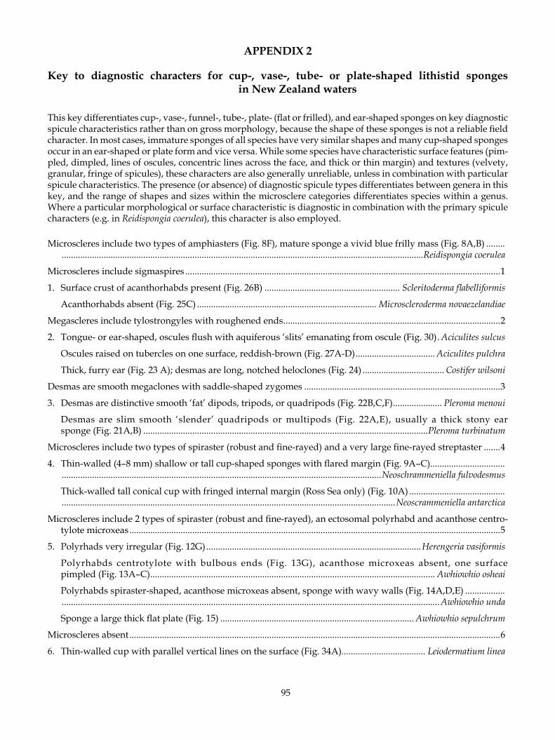
95
APPENDIx 2
Key to diagnostic characters for cup-, vase-, tube- or plate-shaped lithistid sponges in New Zealand waters
This key differentiates cup-, vase-, funnel-, tube-, plate- (flat or frilled), and ear-shaped sponges on key diagnostic spicule characteristics rather than on gross morphology, because the shape of these sponges is not a reliable field character. In most cases, immature sponges of all species have very similar shapes and many cup-shaped sponges occur in an ear-shaped or plate form and vice versa. While some species have characteristic surface features (pim-pled, dimpled, lines of oscules, concentric lines across the face, and thick or thin margin) and textures (velvety, granular, fringe of spicules), these characters are also generally unreliable, unless in combination with particular spicule characteristics. The presence (or absence) of diagnostic spicule types differentiates between genera in this key, and the range of shapes and sizes within the microsclere categories differentiates species within a genus. Where a particular morphological or surface characteristic is diagnostic in combination with the primary spicule characters (e.g. in Reidispongia coerulea), this character is also employed.
Microscleres include two types of amphiasters (Fig. 8F), mature sponge a vivid blue frilly mass (Fig. 8A,B) ........ ...........................................................................................................................................................Reidispongia coerulea
Microscleres include sigmaspires .......................................................................................................................................1
1. Surface crust of acanthorhabds present (Fig. 26B) .......................................................... Scleritoderma flabelliformis
Acanthorhabds absent (Fig. 25C) ............................................................................. Microscleroderma novaezelandiae
Megascleres include tylostrongyles with roughened ends .............................................................................................2
2. Tongue- or ear-shaped, oscules flush with aquiferous ‘slits’ emanating from oscule (Fig. 30) . Aciculites sulcus
Oscules raised on tubercles on one surface, reddish-brown (Fig. 27A-D) .................................. Aciculites pulchra
Thick, furry ear (Fig. 23 A); desmas are long, notched heloclones (Fig. 24) ................................... Costifer wilsoni
Desmas are smooth megaclones with saddle-shaped zygomes ....................................................................................3
3. Desmas are distinctive smooth ‘fat’ dipods, tripods, or quadripods (Fig. 22B,C,F) ..................... Pleroma menoui
Desmas are slim smooth ‘slender’ quadripods or multipods (Fig. 22A,E), usually a thick stony ear sponge (Fig. 21A,B) ..........................................................................................................................Pleroma turbinatum
Microscleres include two types of spiraster (robust and fine-rayed) and a very large fine-rayed streptaster .......4
4. Thin-walled (4–8 mm) shallow or tall cup-shaped sponges with flared margin (Fig. 9A–C) ................................ .........................................................................................................................................Neoschrammeniella fulvodesmus
Thick-walled tall conical cup with fringed internal margin (Ross Sea only) (Fig. 10A) ......................................... ...............................................................................................................................................Neoscrammeniella antarctica
Microscleres include 2 types of spiraster (robust and fine-rayed), an ectosomal polyrhabd and acanthose centro-tylote microxeas ...............................................................................................................................................................5
5. Polyrhads very irregular (Fig. 12G) ............................................................................................Herengeria vasiformis
Polyrhabds centrotylote with bulbous ends (Fig. 13G), acanthose microxeas absent, one surface pimpled (Fig. 13A–C) .......................................................................................................................... Awhiowhio osheai
Polyrhabds spiraster-shaped, acanthose microxeas absent, sponge with wavy walls (Fig. 14A,D,E) ................. ..................................................................................................................................................................Awhiowhio unda
Sponge a large thick flat plate (Fig. 15) ................................................................................... Awhiowhio sepulchrum
Microscleres absent ...............................................................................................................................................................6
6. Thin-walled cup with parallel vertical lines on the surface (Fig. 34A) .................................... Leiodermatium linea

96
APPENDIx 3
STATION DATA
NIWA/NZOI (New Zealand Oceanographic Institute) StationsNIWA Stn Geographic location Latitude Longitude Depth (m) Date
A527 Ross Sea, Antarctica 74°10.00’ S 178°17.0’ W 352 02.07.1960 B314 Eastern edge of Challenger Plateau 39°22’ S 171°50.0’ E 236 25.10.1960E636 Bay of Plenty 37°28.49’ S 177°13.0’ E 190 10.10.1966 E731 Bay of Plenty 37°23.50’ S 177°12.0’ E 602–503 25.03.1967F993 South Three Kings Ridge 34°24’ S 173°10.3’ E 249–212 15.10.1968I14 Cavalli Seamount, Northland 35°35.9’ S 174°39.7’ E 103 04.05.1975I92 Norfolk Ridge, south of Norfolk Island 29°24.8’ S 168°13.2’ E 578–570 23.07.1975I96 Wanganella Bank, West Norfolk Ridge 32°10.8’ S 167°21.2’ E 356 05.02.1975I97 Wanganella Bank, West Norfolk Ridge 32°22.9’ S 167°28.2’ E 544–540 25.07.1975I372 Doubtless Bay, North Cape 34°32.20’ S 173°29.50’ E 215–211 23.11.1977J953 (I807) Western continental slope, Northland 34°39.6’ S 172°13.1’ E 270–260 18.06.1981J954 (I808) Western continental slope, Northland 34°38.8’ S 172°13.5’ E 204–192 18.06.1981K826 Southern South Fiji Basin 28°48.0’ S 177°48.0’ W 490–390 25.07.1974K872 Colville Ridge, southeastern South Fiji Basin 31°20.4’ S 178°49.2’ W 280–235 02.08.1974KAH0011/30 Rungapapa Knoll, Bay of Plenty 37º32.98’ S 176°58.84’ E 165–159 04.11.2000KAH0011/40 Rungapapa Knoll, Bay of Plenty 37°32.99’ S 176°58.25’ E 280–155 05.11.2000KAH0011/41 Rungapapa Knoll, Bay of Plenty 37°33’ S 176°58.23’ E 260–154 05.11.2000KAH0011/44 Rungapapa Knoll, Bay of Plenty 37°33.14’ S 176°58.09’ E 348–181 05.11.2000 KAH0204/02 Knights Terrace, east of Poor Knights Islands 34°57.62’ S 175°10.79’ E 581 13.04.2002KAH0204/07 Cavalli Seamount, Northland 34°7.16’ S 174°9.15’ E 800 14.04.2002KAH0204/08 Cavalli Seamount, Northland 34°6.91’ S 174°8.70’ E 640–610 14.04.2002KAH0204/09 Cavalli Seamount, Northland 34°6.68’ S 174°8.29’ E 600–562 14.04.2002KAH0204/27 Cavalli Seamount, Northland 33°09.39’ S 179°57.2’ E 667–613 16.04.2002KAH0204/29 West Cavalli Seamount, Northland 34°9.79’ S 173°57.75’ E 790–782 17.04.2002KAH0204/30 West Cavalli Seamount, Northland 34°8.79’ S 173°57.79’ E 825–800 17.04.2002KAH0204/32 West Cavalli Seamount, Northland 34°9.72’ S 173°57.71’ E 810–780 17.04.2002KAH0204/38 West Cavalli Seamount, Northland 34°09.52’ S 173°57.78’ E 800–780 18.04.2002KAH0204/40 West Cavalli Seamount, Northland 34°09.85’ S 173°57.84’ E 820–805 18.04.2002KAH0204/44 South Cavalli Seamount, Northland 34°15.94’ S 174°06.19’ E 850–840 18.04.2002KAH0204/47 Seamount 441, Cavalli Seamount region 34°02.55’ S 174°49.02’ E 880–792 19.04.2002KAH0204/52 Seamount 441, Cavalli Seamount region 34°03.30’ S 174°48.49’ E 910–820 19.04.2002KAH9301 Bay of Plenty to Wairarapa shelf 36°57.24’ S 176°15.19’ E 31.12.1992 to 41°01.62’ S 176°24.87’ E 27.01.1993KAH9907/48 West of White Island, Bay of Plenty 37°28.15’ S 177°06.71’ E 250 05.06.1999KAH9907/49 West of White Island, Bay of Plenty 37°28.21’ S 177°07.03’ E 320–220 05.06.1999 KAH9907/50 West of White Island, Bay of Plenty 37°28.14’ S 177°06.97’ E 318–230 05.06.1999P8 West Norfolk Ridge 32°40.8’ S 167°26.80’ E 757–660 25.01.1977P10 West Norfolk Ridge 32°40.00’ S 167°28.40’ E 378–352 25.01.1977P14 West Norfolk Ridge 31°47.20’ S 167°51.6’ E 319–316 25.01.1977 P925 Dampier Ridge, Lord Howe Seamount Chain 27°59.60’ S 155°37.50’ E 420 11.12.1979RAPUHIA2-15 [NIWA Stns U545–U624, cruise to study manganese crusts; depths 406–3354 m] Challenger Plateau to southern Norfolk Ridge 40°16.5’ S 165°59.0’ E 29.01.1988 to 30°20.0’ S 172°60.0’ E 14.02.1988S562 Western continental slope, Northland 35°49.20’ S 172°54.00’ E 600–505 05.08.1983S568 Western flank of Three Kings Ridge 30°10.00’ S 171°20.20’ E 900–650 13.08.1983S571 Three Kings Ridge 30°47.30’ S 172°45.20’ E 509–480 15.08.1983S572 West of Three Kings Ridge 30°45.50’ S 172°47.70’ E 530–403 15.08.1983T226 North of Raoul Island, Kermadec Ridge 28°33.00’ S 177°49.99’ W 800–930 22.03.1982TAN0104/001 Graveyard Seamount, Chatham Rise 42°45.60’ S 179°59.26’ W 979–770 15.04.2001TAN0104/002 Graveyard Seamount, Chatham Rise 42°45.93’ S 179°59.34’ W 875–757 15.04.2001TAN0104/048 Diabolical Seamount, Chatham Rise 42°47.17’ S 179°59.12’ W 993–900 16.04.2001TAN0104/148 Morgue Seamount, Chatham Rise 42°42.84’ S 179°57.51’ W 980–893 18.04.2001TAN0104/194 Scroll Seamount, Chatham Rise 42°47.27’ S 179°59.81’ W 1042–880 18.04.2001TAN0104/288 Graveyard Seamount, Chatham Rise 42°45.64’ S 179°59.27’ W 972–890 19.04.2001

97
NIWA/NZOI (New Zealand Oceanographic Institute) StationsNIWA Stn Geographic location Latitude Longitude Depth (m) Date
TAN0104/289 Graveyard Seamount, Chatham Rise 42°45.89’ S 179°59.16’ W 800–757 19.04.2001TAN0104/394 Graveyard Seamount, Chatham Rise 42°45.68’ S 179°59.33’ W 920–771 21.04.2001TAN0107/124 Rumble III Seamount, Bay of Plenty 35°44.36’ S 178°30.25’ E 620–435 20.05.2001TAN0107/232 Rumble V Seamount, Bay of Plenty 36°08.73’ S 178°11.98’ E 750–570 24.05.2001TAN0205/20 Volcano E, Kermadec Ridge 33°44.18’ S 179°49.90’ W 619–490 14.04.2002TAN0205/32 East slope of Kermadec Ridge 33°10.25’ S 179°58.21’ W 999–643 16.04.2002TAN0205/60 Cavalli Seamount, Northland 30°11.87’ S 178°28.75’ W 1131–1073 21.04.2002 TAN0205/75 Volcano L, Kermadec Ridge 30°01.37’ S 178°42.88’ W 240–154 23.04.2002TAN0413/016 Whakatane Seamount, Bay of Plenty 36°47.50’ S 177°25.52’ E 1538–1503 08.11.2004TAN0413/068 Tuatoru Knoll, Bay of Plenty 37°28.28’ S 177°12.30’ E 295–200 11.11.2004TAN0413/070 Tuatoru Knoll, Bay of Plenty 37°28.12’ S 177°12.40’ E 330–218 11.11.2004TAN0413/074 Tuatoru Knoll, Bay of Plenty 37°28.19’ S 177°13.20’ E 200–175 12.11.2004TAN0413/077 Tuatoru Knoll, Bay of Plenty 37°28.26’ S 177°12.89’ E 180–177 12.11.2004TAN0413/085 Tuatoru Knoll, Bay of Plenty 37°28.25’ S 177°13.35’ E 216–207 12.11.2004TAN0413/092 Tuatoru Knoll, Bay of Plenty 37°29.14’ S 177°12.92’ E 400–225 12.11.2004TAN0413/093 Tuatoru Knoll, Bay of Plenty 37°28.58’ S 177°12.94’ E 180–179 13.11.2004TAN0413/099 Rungapapa Knoll, Bay of Plenty 37°32.80’ S 176°58.85’ E 151–141 13.11.2004TAN0413/109 Rungapapa Knoll, Bay of Plenty 37°32.93’ S 176°59.27’ E 142–136 13.11.2004TAN0413/111 Rungapapa Knoll, Bay of Plenty 37°32.79’ S 177°00.22’ E 337–256 13.11.2004 TAN0413/112 Rungapapa Knoll, Bay of Plenty 37°32.11’ S 176°58.26’ E 217–169 13.11.2004 TAN0413/117 Rungapapa Knoll, Bay of Plenty 37°32.16’ S 176°58.25’ E 188–182 13.11.2004TAN0413/118 Rungapapa Knoll, Bay of Plenty 37°33.22’ S 176°58.13’ E 190–154 13.11.2004TAN0413/120 Mahina Knoll, Bay of Plenty 37°21.14’ S 177°04.70’ E 520 14.11.2004TAN0413/170 Tumokemoke Knoll, Bay of Plenty 37°27.69’ S 176°54.23’ E 376–295 16.11.2004TAN0413/173 Mahina Knoll, Bay of Plenty 37°21.04’ S 177°05.96’ E 434–272 16.11.2004TAN0413/174 Mahina Knoll, Bay of Plenty 37°20.09’ S 177°04.58’ E 502–430 16.11.2004TAN9915/3B Ngunguru Bay, off Taiharuru Head, Northland 35°41.80’ S 174°38.90’ E 80 13.12.1999U591 Tui Seamount, Three Kings Ridge 30°50.60’ S 172°48.30’ E 486 07.02.1988U594 West of Three Kings Ridge 30°20.10’ S 172°59.60’ E 406 07.02.1988U606 South Norfolk Basin 31°54.70’ S 172°47.40’ E 1100–1085 10.02.1988V473 South of East Cape 38°50.92’ S 178°36.75’ E 800–365 05.06.1994X128 Bay of Plenty 37°24.40’ S 176°59.40’ E 405 25.11.1989X140 Bay of Plenty 37°11.40’ S 176°58.30’ E 860–745 27.11.1989X152 Raukumara Plain, off East Cape 36°09.70’ S 176°48.40’ E 940–820 28.11.1989X201 Ngatoro Ridge, off East Cape 37°08.12’ S 177°17.82’ E 1050–700 06.12.1989X207 East of Hikurangi Plateau 37°32.77’ S 176°57.70’ E 380 06.12.1989 X681 South Kermadec Ridge 35°24.91’ S 178°’39.23 E 1132–1125 13.02.1996X768 Hikurangi Plateau 37°41.30’ S 179°00.75’ E 936 05.06.1999Z2092 Lord Howe Seamount Chain 27°41.20’ S 155°08.80’ E 521–412 26.08.1967Z2098 North of Three Kings Ridge 28°39.50’ S 173°01.00’ E 920 04.09.1967Z2364 Bay of Plenty 37°25.00’ S 176°25.99’ E 277 00.00.1971Z7187 New Zealand waters [89-BIP-1] 1989Z7190 New Zealand waters [90-PUP] 1990Z8487 Louisville Seamount Chain 37°40.00’ S 169°06.00’ W 1050–700 00.06.1996Z9025 East of Three Kings Ridge 31°58.82’ S 174°15.87’ E 1680–677 00.02.1998Z9041 Kermadec Ridge 32°10.90’ S 179°04.20’ W 260–128 05.04.1998Z9262 Challenger Plateau, northern slope 37°29.04’ S 167°41.09’ E 904 10.09.1998 Z9263 Lord Howe Rise 34°09.20’ S 162°51.90’ E 791 18.09.1998Z9400 Challenger Plateau, northern slope 37°18.06’ S 167°22.01’ E 755 30.09.1998Z9742 ~3 NM east of North Cape 34°24.82’ S 173°08.00’ E 210–133 19.04.1999Z9748 Northwest of Volkner Rocks, Bay of Plenty 37°28.01’ S 177°07.00’ E 232 30.04.1999Z9747 ~17 NM west of Pandora Bank, Northland 34°38.91’ S 172°13.20’ E 208–198 21.04.1999 Z9751 Three Kings Islands 34°10.09’ S 172°12.04’ E 180 16.04.1999Z9890 Western continental slope 35°00.49’ S 172°31.82’ E 167–169 30.10.1999Z9892 Western continental slope 35°02.10’ S 172°29.25’ E 194-174 23.10.1999
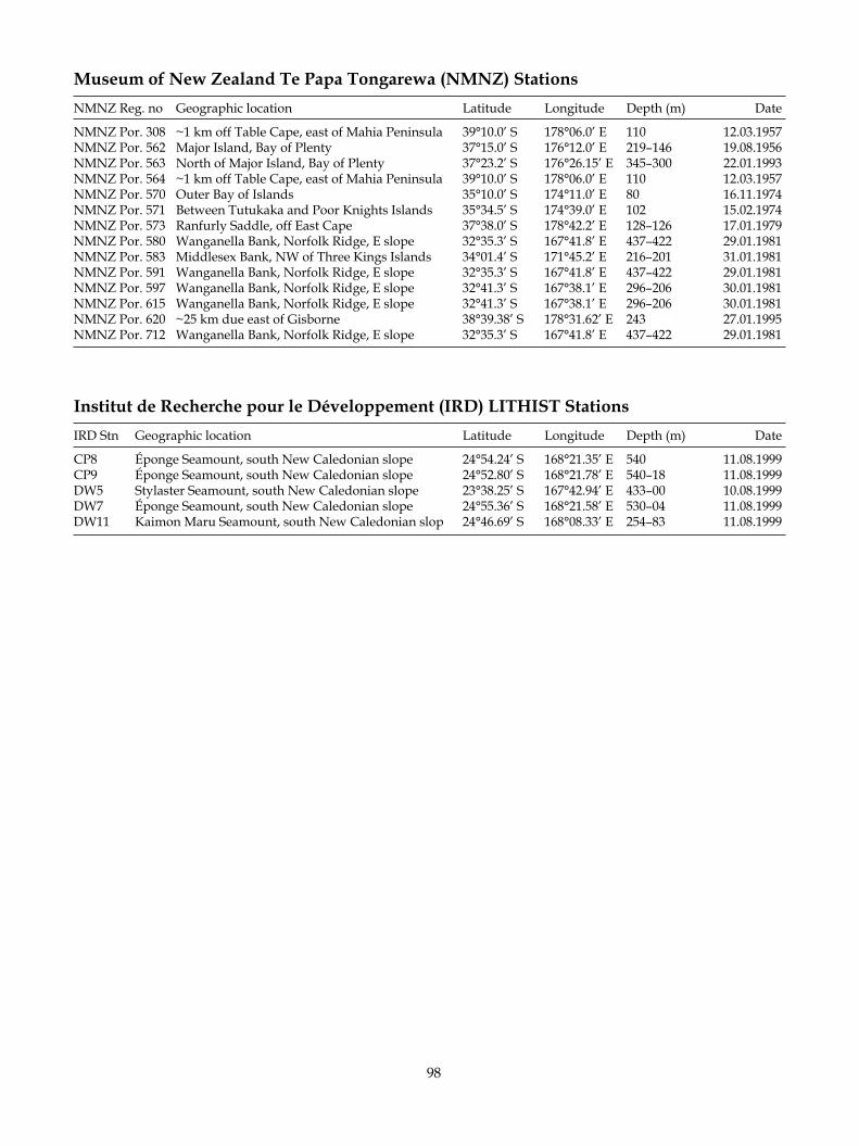
98
Museum of New Zealand Te Papa Tongarewa (NMNZ) Stations NMNZ Reg. no Geographic location Latitude Longitude Depth (m) Date
NMNZ Por. 308 ~1 km off Table Cape, east of Mahia Peninsula 39°10.0’ S 178°06.0’ E 110 12.03.1957NMNZ Por. 562 Major Island, Bay of Plenty 37°15.0’ S 176°12.0’ E 219–146 19.08.1956NMNZ Por. 563 North of Major Island, Bay of Plenty 37°23.2’ S 176°26.15’ E 345–300 22.01.1993NMNZ Por. 564 ~1 km off Table Cape, east of Mahia Peninsula 39°10.0’ S 178°06.0’ E 110 12.03.1957NMNZ Por. 570 Outer Bay of Islands 35°10.0’ S 174°11.0’ E 80 16.11.1974NMNZ Por. 571 Between Tutukaka and Poor Knights Islands 35°34.5’ S 174°39.0’ E 102 15.02.1974NMNZ Por. 573 Ranfurly Saddle, off East Cape 37°38.0’ S 178°42.2’ E 128–126 17.01.1979NMNZ Por. 580 Wanganella Bank, Norfolk Ridge, E slope 32°35.3’ S 167°41.8’ E 437–422 29.01.1981NMNZ Por. 583 Middlesex Bank, NW of Three Kings Islands 34°01.4’ S 171°45.2’ E 216–201 31.01.1981NMNZ Por. 591 Wanganella Bank, Norfolk Ridge, E slope 32°35.3’ S 167°41.8’ E 437–422 29.01.1981 NMNZ Por. 597 Wanganella Bank, Norfolk Ridge, E slope 32°41.3’ S 167°38.1’ E 296–206 30.01.1981NMNZ Por. 615 Wanganella Bank, Norfolk Ridge, E slope 32°41.3’ S 167°38.1’ E 296–206 30.01.1981 NMNZ Por. 620 ~25 km due east of Gisborne 38°39.38’ S 178°31.62’ E 243 27.01.1995 NMNZ Por. 712 Wanganella Bank, Norfolk Ridge, E slope 32°35.3’ S 167°41.8’ E 437–422 29.01.1981
Institut de Recherche pour le Développement (IRD) LITHIST StationsIRD Stn Geographic location Latitude Longitude Depth (m) Date
CP8 Éponge Seamount, south New Caledonian slope 24°54.24’ S 168°21.35’ E 540 11.08.1999CP9 Éponge Seamount, south New Caledonian slope 24°52.80’ S 168°21.78’ E 540–18 11.08.1999DW5 Stylaster Seamount, south New Caledonian slope 23°38.25’ S 167°42.94’ E 433–00 10.08.1999DW7 Éponge Seamount, south New Caledonian slope 24°55.36’ S 168°21.58’ E 530–04 11.08.1999DW11 Kaimon Maru Seamount, south New Caledonian slop 24°46.69’ S 168°08.33’ E 254–83 11.08.1999
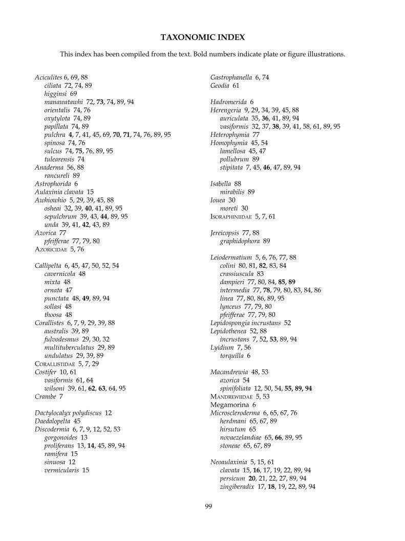
99
TAxONOMIC INDEx
This index has been compiled from the text. Bold numbers indicate plate or figure illustrations.
Aciculites 6, 69, 88 ciliata 72, 74, 89 higginsi 69 manawatawhi 72, 73, 74, 89, 94 orientalis 74, 76 oxytylota 74, 89 papillata 74, 89 pulchra 4, 7, 41, 45, 69, 70, 71, 74, 76, 89, 95 spinosa 74, 76 sulcus 74, 75, 76, 89, 95 tulearensis 74Anaderma 56, 88 rancureli 89Astrophorida 6Aulaxinia clavata 15Awhiowhio 5, 29, 39, 45, 88 osheai 32, 39, 40, 41, 89, 95 sepulchrum 39, 43, 44, 89, 95 unda 39, 41, 42, 43, 89Azorica 77 pfeifferae 77, 79, 80azoriCidae 5, 76
Callipelta 6, 45, 47, 50, 52, 54 cavernicola 48 mixta 48 ornata 47 punctata 48, 49, 89, 94 sollasi 48 thoosa 48Corallistes 6, 7, 9, 29, 39, 88 australis 39, 89 fulvodesmus 29, 30, 32 multituberculatus 29, 89 undulatus 29, 39, 89CorallisTidae 5, 7, 29Costifer 10, 61 vasiformis 61, 64 wilsoni 39, 61, 62, 63, 64, 95Crambe 7
Dactylocalyx polydiscus 12Daedalopelta 45Discodermia 6, 7, 9, 12, 52, 53 gorgonoides 13 proliferans 13, 14, 45, 89, 94 ramifera 15 sinuosa 12 vermicularis 15
Gastrophanella 6, 74Geodia 61
Hadromerida 6Herengeria 9, 29, 34, 39, 45, 88 auriculata 35, 36, 41, 89, 94 vasiformis 32, 37, 38, 39, 41, 58, 61, 89, 95Heterophymia 77Homophymia 45, 54 lamellosa 45, 47 pollubrum 89 stipitata 7, 45, 46, 47, 89, 94
Isabella 88 mirabilis 89Iouea 30 moreti 30isoraPhiniidae 5, 7, 61
Jereicopsis 77, 88 graphidophora 89
Leiodermatium 5, 6, 76, 77, 88 colini 80, 81, 82, 83, 84 crassiuscula 83 dampieri 77, 80, 84, 85, 89 intermedia 77, 78, 79, 80, 83, 84, 86 linea 77, 80, 86, 89, 95 lynceus 77, 79, 80 pfeifferae 77, 79, 80Lepidospongia incrustans 52Lepidothenea 52, 88 incrustans 7, 52, 53, 89, 94Lyidium 7, 56 torquilla 6
Macandrewia 48, 53 azorica 54 spinifoliata 12, 50, 54, 55, 89, 94mandrewiidae 5, 53Megamorina 6Microscleroderma 6, 65, 67, 76 herdmani 65, 67, 89 hirsutum 65 novaezelandiae 65, 66, 89, 95 stoneae 65, 67, 89
Neoaulaxinia 5, 15, 61 clavata 15, 16, 17, 19, 22, 89, 94 persicum 20, 21, 22, 27, 89, 94 zingiberadix 17, 18, 19, 22, 89, 94
100
Neopelta 50, 52, 53 imperfecta 50 perfecta 50 plinthosellina 50, 52, 89 pulvinus 50, 51, 89, 94neoPelTidae 5, 45, 53Neophrissospongia 30, 88 microstylifer 30, 89Neoschrammeniella 6, 29, 30, 37, 45, 58, 88 antarctica 5, 32, 33, 34, 88, 95 castrum 32, 89 fulvodesmus 30, 31, 32, 34, 72, 89, 95 moreti 32, 39, 89 norfolkii 32, 34, 89Neosiphonia 5, 6, 22, 88 fruticosa 24 motukawanui 22, 24, 25, 26, 27, 45, 89 schmidti 24, 26 superstes 6, 22, 23, 24, 27, 89, 94
PhymaTellidae 5, 7, 15Pleroma 6, 7, 10, 45, 56, 88 aotea 7, 57, 59, 60, 61, 89 menoui 6, 39, 57, 58, 59, 61, 89, 95 torquilla 6 turbinatum 56, 57, 59, 61, 64, 89, 95
Pleromidae 5, 7, 54
Reidispongia 6, 27, 88 coerulea 6, 27, 28, 29, 48, 84, 89, 95Rhizomorina 6
Scleritoderma 6, 67 camusi 67, 89 flabelliformis 67, 68, 69, 89, 95 nodosum 67sCleriTodermidae 5, 7, 64Siphonidium 74 capitatum 74
Tetracladina 6Theonella 6, 7Theonellidae 5, 12
Vetulina 6, 7 oamaruensis 7 stalactites 6