the making of a photosynthetic animalmary e. rumpho1,*, karen n. pelletreau1, ahmed moustafa2 and...
TRANSCRIPT
303
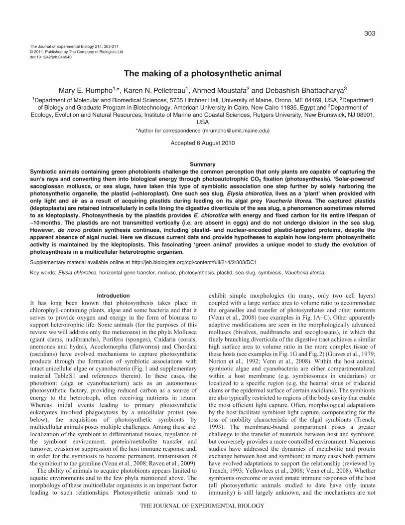
IntroductionIt has long been known that photosynthesis takes place inchlorophyll-containing plants, algae and some bacteria and that itserves to provide oxygen and energy in the form of biomass tosupport heterotrophic life. Some animals (for the purposes of thisreview we will address only the metazoans) in the phyla Mollusca(giant clams, nudibranchs), Porifera (sponges), Cnidaria (corals,anemones and hydra), Acoelomorpha (flatworms) and Chordata(ascidians) have evolved mechanisms to capture photosyntheticproducts through the formation of symbiotic associations withintact unicellular algae or cyanobacteria (Fig.1 and supplementarymaterial TableS1 and references therein). In these cases, thephotobiont (alga or cyanobacterium) acts as an autonomousphotosynthetic factory, providing reduced carbon as a source ofenergy to the heterotroph, often receiving nutrients in return.Whereas initial events leading to primary photosyntheticeukaryotes involved phagocytosis by a unicellular protist (seebelow), the acquisition of photosynthetic symbionts bymulticellular animals poses multiple challenges. Among these are:localization of the symbiont to differentiated tissues, regulation ofthe symbiont environment, protein/metabolite transfer andturnover, evasion or suppression of the host immune response and,in order for the symbiosis to become permanent, transmission ofthe symbiont to the germline (Venn et al., 2008; Raven et al., 2009).
The ability of animals to acquire photobionts appears limited toaquatic environments and to the few phyla mentioned above. Themorphology of these multicellular organisms is an important factorleading to such relationships. Photosynthetic animals tend to
exhibit simple morphologies (in many, only two cell layers)coupled with a large surface area to volume ratio to accommodatethe organelles and transfer of photosynthates and other nutrients(Venn et al., 2008) (see examples in Fig.1A–C). Other apparentlyadaptive modifications are seen in the morphologically advancedmolluscs (bivalves, nudibranchs and sacoglossans), in which thefinely branching diverticula of the digestive tract achieves a similarhigh surface area to volume ratio in the more complex tissue ofthese hosts (see examples in Fig.1G and Fig.2) (Graves et al., 1979;Norton et al., 1992; Venn et al., 2008). Within the host animal,symbiotic algae and cyanobacteria are either compartmentalizedwithin a host membrane (e.g. symbiosomes in cnidarians) orlocalized to a specific region (e.g. the heamal sinus of tridacnidclams or the epidermal surface of certain ascidians). The symbiontsare also typically restricted to regions of the body cavity that enablethe most efficient light capture. Often, morphological adaptationsby the host facilitate symbiont light capture, compensating for theloss of mobility characteristic of the algal symbionts (Trench,1993). The membrane-bound compartment poses a greaterchallenge to the transfer of materials between host and symbiont,but conversely provides a more controlled environment. Numerousstudies have addressed the dynamics of metabolite and proteinexchange between host and symbiont; in many cases both partnershave evolved adaptations to support the relationship (reviewed byTrench, 1993; Yellowlees et al., 2008; Venn et al., 2008). Whethersymbionts overcome or avoid innate immune responses of the host(all photosynthetic animals studied to date have only innateimmunity) is still largely unknown, and the mechanisms are not
The Journal of Experimental Biology 214, 303-311© 2011. Published by The Company of Biologists Ltddoi:10.1242/jeb.046540
The making of a photosynthetic animal
Mary E. Rumpho1,*, Karen N. Pelletreau1, Ahmed Moustafa2 and Debashish Bhattacharya3
1Department of Molecular and Biomedical Sciences, 5735 Hitchner Hall, University of Maine, Orono, ME 04469, USA, 2Departmentof Biology and Graduate Program in Biotechnology, American University in Cairo, New Cairo 11835, Egypt and 3Department of
Ecology, Evolution and Natural Resources, Institute of Marine and Coastal Sciences, Rutgers University, New Brunswick, NJ 08901,USA
*Author for correspondence ([email protected])
Accepted 6 August 2010
SummarySymbiotic animals containing green photobionts challenge the common perception that only plants are capable of capturing thesun’s rays and converting them into biological energy through photoautotrophic CO2 fixation (photosynthesis). ‘Solar-powered’sacoglossan molluscs, or sea slugs, have taken this type of symbiotic association one step further by solely harboring thephotosynthetic organelle, the plastid (chloroplast). One such sea slug, Elysia chlorotica, lives as a ‘plant’ when provided withonly light and air as a result of acquiring plastids during feeding on its algal prey Vaucheria litorea. The captured plastids(kleptoplasts) are retained intracellularly in cells lining the digestive diverticula of the sea slug, a phenomenon sometimes referredto as kleptoplasty. Photosynthesis by the plastids provides E. chlorotica with energy and fixed carbon for its entire lifespan of~10months. The plastids are not transmitted vertically (i.e. are absent in eggs) and do not undergo division in the sea slug.However, de novo protein synthesis continues, including plastid- and nuclear-encoded plastid-targeted proteins, despite theapparent absence of algal nuclei. Here we discuss current data and provide hypotheses to explain how long-term photosyntheticactivity is maintained by the kleptoplasts. This fascinating ‘green animal’ provides a unique model to study the evolution ofphotosynthesis in a multicellular heterotrophic organism.
Supplementary material available online at http://jeb.biologists.org/cgi/content/full/214/2/303/DC1
Key words: Elysia chlorotica, horizontal gene transfer, mollusc, photosynthesis, plastid, sea slug, symbiosis, Vaucheria litorea.
THE JOURNAL OF EXPERIMENTAL BIOLOGY
304
understood. However, recent transcriptomic work studyingcoral–algal symbioses has shown that gene expression of the host,when infected by the appropriate symbiont, shows little or nochange over time (Voolstra et al., 2009). By contrast, when the hostis exposed to foreign symbionts (those incapable of establishing asymbiosis) significant changes in the host transcriptome result andthe immune response is launched. This difference implies thatappropriate symbionts evade detection by the host and fail to elicitrecognition, rejection and immune responses such as apoptosis andproteolysis (Voolstra et al., 2009). Further studies of the immuneresponse in other photosynthetic animals are needed to determinewhether similar mechanisms are employed across all of theseorganisms. Transmission of photosynthetic symbionts is largelyhorizontal, with each generation acquiring its photosyntheticpartner anew from the surrounding environment. Transmission to
the germline remains a final barrier towards a more permanentphotosynthetic animal.
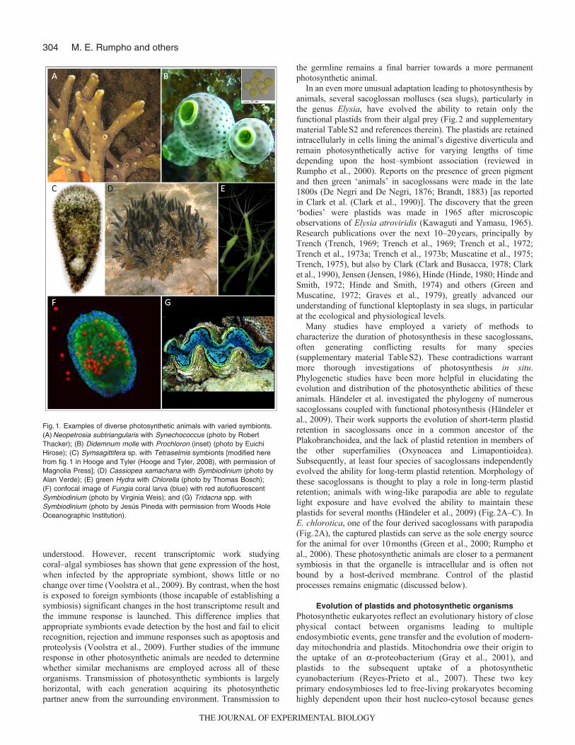
In an even more unusual adaptation leading to photosynthesis byanimals, several sacoglossan molluscs (sea slugs), particularly inthe genus Elysia, have evolved the ability to retain only thefunctional plastids from their algal prey (Fig.2 and supplementarymaterial TableS2 and references therein). The plastids are retainedintracellularly in cells lining the animal’s digestive diverticula andremain photosynthetically active for varying lengths of timedepending upon the host–symbiont association (reviewed inRumpho et al., 2000). Reports on the presence of green pigmentand then green ‘animals’ in sacoglossans were made in the late1800s (De Negri and De Negri, 1876; Brandt, 1883) [as reportedin Clark et al. (Clark et al., 1990)]. The discovery that the green‘bodies’ were plastids was made in 1965 after microscopicobservations of Elysia atroviridis (Kawaguti and Yamasu, 1965).Research publications over the next 10–20years, principally byTrench (Trench, 1969; Trench et al., 1969; Trench et al., 1972;Trench et al., 1973a; Trench et al., 1973b; Muscatine et al., 1975;Trench, 1975), but also by Clark (Clark and Busacca, 1978; Clarket al., 1990), Jensen (Jensen, 1986), Hinde (Hinde, 1980; Hinde andSmith, 1972; Hinde and Smith, 1974) and others (Green andMuscatine, 1972; Graves et al., 1979), greatly advanced ourunderstanding of functional kleptoplasty in sea slugs, in particularat the ecological and physiological levels.
Many studies have employed a variety of methods tocharacterize the duration of photosynthesis in these sacoglossans,often generating conflicting results for many species(supplementary material TableS2). These contradictions warrantmore thorough investigations of photosynthesis in situ.Phylogenetic studies have been more helpful in elucidating theevolution and distribution of the photosynthetic abilities of theseanimals. Händeler et al. investigated the phylogeny of numeroussacoglossans coupled with functional photosynthesis (Händeler etal., 2009). Their work supports the evolution of short-term plastidretention in sacoglossans once in a common ancestor of thePlakobranchoidea, and the lack of plastid retention in members ofthe other superfamilies (Oxynoacea and Limapontioidea).Subsequently, at least four species of sacoglossans independentlyevolved the ability for long-term plastid retention. Morphology ofthese sacoglossans is thought to play a role in long-term plastidretention; animals with wing-like parapodia are able to regulatelight exposure and have evolved the ability to maintain theseplastids for several months (Händeler et al., 2009) (Fig.2A–C). InE. chlorotica, one of the four derived sacoglossans with parapodia(Fig.2A), the captured plastids can serve as the sole energy sourcefor the animal for over 10months (Green et al., 2000; Rumpho etal., 2006). These photosynthetic animals are closer to a permanentsymbiosis in that the organelle is intracellular and is often notbound by a host-derived membrane. Control of the plastidprocesses remains enigmatic (discussed below).
Evolution of plastids and photosynthetic organismsPhotosynthetic eukaryotes reflect an evolutionary history of closephysical contact between organisms leading to multipleendosymbiotic events, gene transfer and the evolution of modern-day mitochondria and plastids. Mitochondria owe their origin tothe uptake of an -proteobacterium (Gray et al., 2001), andplastids to the subsequent uptake of a photosyntheticcyanobacterium (Reyes-Prieto et al., 2007). These two keyprimary endosymbioses led to free-living prokaryotes becominghighly dependent upon their host nucleo-cytosol because genes
M. E. Rumpho and others
Fig.1. Examples of diverse photosynthetic animals with varied symbionts.(A)Neopetrosia subtriangularis with Synechococcus (photo by RobertThacker); (B) Didemnum molle with Prochloron (inset) (photo by EuichiHirose); (C) Symsagittifera sp. with Tetraselmis symbionts [modified herefrom fig.1 in Hooge and Tyler (Hooge and Tyler, 2008), with permission ofMagnolia Press]; (D) Cassiopea xamachana with Symbiodinium (photo byAlan Verde); (E) green Hydra with Chlorella (photo by Thomas Bosch);(F) confocal image of Fungia coral larva (blue) with red autofluorescentSymbiodinium (photo by Virginia Weis); and (G) Tridacna spp. withSymbiodinium (photo by Jesús Pineda with permission from Woods HoleOceanographic Institution).
THE JOURNAL OF EXPERIMENTAL BIOLOGY

305Mollusc photosynthesis
were lost or transferred to the nucleus in the first examples ofmassive intracellular gene transfer. The ancestral photosyntheticorganism gave rise to three primary lineages, the glaucophytes,the rhodophytes (red algae) and the Chloroplastida orViridiplantae (green algae and land plants) (Bhattacharya andMedlin, 1995; Falkowski et al., 2004; Adl et al., 2005) (seeevolutionary scheme in Fig.3). In turn, a diverse group ofsecondary or ‘complex’ algae evolved following the engulfmentof a single-celled green or red alga (or both) by a heterotrophic,eukaryotic host (McFadden, 2001). Genome modification againoccurred as a result of gene transfer and loss from the symbiontplastid, but also as a result of the ‘merger’ of the two nuclei (hostand endosymbiont) (reviewed in Lane and Archibald, 2008). Theevolution of photosynthetic organisms did not stop here, however.Serial secondary endosymbiosis, the replacement of the originalprimary plastid by a different primary plastid, and tertiaryendosymbiosis, the engulfment of a secondarily derived plastidby a heterotrophic or autotrophic eukaryotic host (Bhattacharyaand Nosenko, 2008), gave rise to additional algal diversificationand genome chimerism (Keeling, 2004; Yoon et al., 2005; Laneand Archibald, 2008; Sanchez-Puerta and Delwiche, 2008). Inorganisms that evolved through serial endosymbiosis, previouslytransferred plastid genes would have been present in the hostnuclear genome, but could have also been replaced by plastid ornuclear genes of the new symbiont. In the case of tertiaryendosymbiosis, genes required to support functions of the mostrecently acquired plastid may have been present in the host froma previous endosymbiotic partner, or they may also have beentransferred from the newest endosymbiont’s nuclear and plastid
genomes to the host’s nuclear genome (Lane and Archibald,2008). Finally, recent data suggest that stramenopiles and otherchromalveolates share a cryptic green algal endosymbiont thatpredates the canonical red algal capture that has also contributedsignificantly (100s of genes) to their nuclear genome (Moustafaet al., 2009). Therefore, the genomes of many chromalveolatetaxa (minimally) contain genes of red and green algal originderived from eukaryotic endosymbioses (Moustafa et al., 2009).
Evolution of kleptoplasty and photosynthesis in ElysiaTo the extent that it has been studied, the sea slug Elysia chloroticaGould 1870 is specific for its algal prey, feeding on and acquiringplastids with any success from only two Vaucheria species (V.litorea and V. compacta) (West, 1979; West et al., 1984). Not onlyis the association specific, but it is also obligate; the sea slug willnot complete metamorphosis and develop into an adult in theabsence of its algal prey and plastid uptake. Vaucheria is a memberof the stramenopiles that currently contain red algal secondary-derived plastids. Biochemically, the alga is characterized bychlorophylls a and c, vaucheriaxanthin accessory pigments, lipidcarbon reserves (Anderson, 2004; Lee, 2008) and high mannitollevels (M.E.R., unpublished data). Its coenocytic (single-celled,multi-nucleate) filaments are largely vacuolated and surrounded bya thin cell wall. As a result of secondary plastid evolution, theseorganelles are surrounded by four membranes in V. litorea(Rumpho et al., 2001): the inner and outer envelopes, the periplastidmembrane (a remnant of the plasma membrane of the engulfedalga) and the outermost plastid endoplasmic reticulum membrane(Gibbs, 1993; Bhattacharya et al., 2004). Interestingly, the outer
Fig.2. Examples of photosyntheticsacoglossans with varied times of chloroplastretention. (A)Elysia chlorotica [reprinted withpermission (Rumpho et al., 2008)], (B) Elysiacrispata, (C) Plakobranchus ocellatus, (D)Costasiella ocellifera, (E) Thuridilla gracilis, (F)Costasiella kurishimae, (G) Alderia modesta,(H) Lobiger viridis and (I) Oxynoe antillarum.Photos in panels B, D, E, G and I wereprovided with permission by Patrick Krug;photos in panels C, F and H were providedwith permission by Heike Wägele.
THE JOURNAL OF EXPERIMENTAL BIOLOGY

306
two membranes are not readily observed in the sea slug (Rumphoet al., 2001) and this has potential implications for protein targeting,but will not be discussed here.
Because E. chlorotica obtains its plastids from a eukaryotecontaining plastids of secondary origin, the evolution of plastidretention (kleptoplasty) and photosynthesis in the sea slug can beexplained in a parallel fashion to that of other tertiary-evolvedphotosynthetic organisms encompassing both endosymbiosis andpotentially horizontal gene transfer (HGT; defined as the non-sexual exchange of genetic material) as follows. (1) Theheterotrophic eukaryotic host and symbiont establish close,prolonged physical contact; i.e. the host sea slug (E. chlorotica)grazes on mats of the algal prey (V. litorea), sucking out the cellularcontents (including intact and broken plastids, mitochondria andnuclei), which then slowly traverse the digestive gut. (2) Thesymbiont is taken up by the host; i.e. algal plastids in the digestivegut are phagocytosed by the digestive epithelial cells of the hostsea slug. (3) Genome modification occurs; i.e. gene transfer mayhave occurred through HGT from the kleptoplasts in the epithelialcells and from broken algal nuclei releasing DNA in the gut lumento the host nucleus. (4) New metabolic properties evolve as a resultof the kleptoplastic association; i.e. an animal (E. chlorotica) is ableto sustain itself solely by photoautotrophic CO2 fixation, as a plant.
There are two major differences to consider in this comparison.First, the entire V. litorea algal cell, including intact nuclei, is notintracellularly taken up into the host sea slug. Second, thekleptoplastic association is not transmitted vertically. In the firstcase, the absence of the algal nucleus in the sea slug cell hassignificant implications for providing nuclear-encoded plastidproteins on a long-term basis. In the second case, absence ofheritability is most likely due to a soma–germline barrier that couldprevent the movement of plastids into the invertebrate germlineand, hence, into the progeny. Whether the sea slug is ‘on the path’to the evolution of permanent photosynthesis is unknown, in largepart because of the lack of genome data.
Laboratory culturing of kleptoplastic sea slugsTo date, most of the studies on E. chlorotica have been carried outon adult kleptoplastic animals collected from the ocean. The ‘true’age of the sea slugs, the extent of feeding, the history of thepopulation and sea slug behavior (e.g. mating) are unknown inthese collections. Furthermore, because the association is alreadyestablished we cannot explore the early steps in development,uptake and retention of the plastids or the initiation ofphotosynthesis. Virtually nothing is known at the biochemical ormolecular level about any of these processes. Finally, we also donot know what contributes to the great disparity in longevity of thedifferent kleptoplastic associations or, in particular, theirphotosynthetic activity.
The successful establishment of a laboratory culture system hasprovided us the necessary controls to fully investigate sea slugdevelopment and establishment of kleptoplasty and photosynthesis.We optimized an artificial saltwater (ASW) culture system usingaposymbiotic eggs produced by E. chlorotica populations fromMartha’s Vineyard Island, MA, USA, and near Halifax, NovaScotia (see life cycle in Fig.4). Successful planktotrophicdevelopment was recorded for all developing larvae that were feda unicellular algal diet of Isochrysis galbana. Metamorphosis oflarvae to the juvenile stage requires the presence of V. litoreafilaments. Immediately following metamorphosis, the juvenilesbegin feeding on the filamentous alga, engulfing plastids andturning green. A transient nature to the plastid symbioticassociation is observed in recently metamorphosed juvenile seaslugs if removed from the presence of V. litorea too soon (less than~6days); this also results in cessation of their morphologicaldevelopment. Plastid uptake until the establishment of irreversiblekleptoplasty appears to be required for full adult development andsurvival, although one report of ‘albino ghost’ E. chlorotica wasdocumented in 1986 (Gibson et al., 1986). Establishment of thekleptoplastic association involves specific recognition processesthat comprise at least two steps: (1) planktonic larvae require V.litorea filaments to be present for settlement and metamorphosis tothe juvenile stage, and (2) adult development requires uptake andretention of V. litorea plastids by cells lining the digestivediverticula.
Semi-autonomy of plastids and the need for the nucleusEndosymbiosis and the associated gene transfer that followedrendered extant plastid genomes greatly reduced in size(37–224kb), encoding between 61 and 273 proteins(http://www.ncbi.nlm.nih.gov/genomes/GenomesGroup.cgi?taxid2759&optplastid) compared with the cyanobacterial progenitors[1.6–9.0Mb in free-living taxa, encoding 1717 to 7672 proteins(Meeks et al., 2001; Rocap et al., 2003)]. These semi-autonomousorganelles encode a small percentage of the predicted 1000 to 5000
M. E. Rumpho and others
Fig.3. Evolutionary scheme for primary, secondary and tertiary plastids.The secondary endosymbiotic origin of plastids is illustrated in Vaucherialitorea from the red algal lineage. The subsequent acquisition of V. litoreaplastids by the sea slug Elysia chlorotica in a tertiary endosymbiotic eventimparts photosynthetic activity to this heterotroph.
THE JOURNAL OF EXPERIMENTAL BIOLOGY

307Mollusc photosynthesis
proteins required to sustain the full metabolic capacity of the plastid(Martin et al., 2002; Richly and Leister, 2004; Bock and Timmis,2008). This is also true for the 115-kb V. litorea plastid genome,which we recently sequenced, demonstrating that it contains only139 protein-encoding genes (Rumpho et al., 2008). If we considersolely photosynthesis, the V. litorea plastid genome does notencode all of the components for any of the four multi-subunitcomplexes of the photosynthetic electron transport chain[photosystems I and II (PSI and PSII, respectively), the cytochromeb6/f complex and ATP synthase] or the reductive pentose phosphatepathway (RPPP, or the Calvin–Benson cycle) (reviewed inRaghavendra, 1998; Nelson and Yocum, 2006). Some of theessential missing genes in the thylakoid-localized electron transportchain include: the PSI and PSII light-harvesting complexpigment/protein genes (vcp in V. litorea), the PSII Mn-stabilizingprotein of the oxygen evolution complex (MSP, encoded by psbO),the Reiske Fe-S protein of the cytochrome b6/f complex and atpC,which encodes the critical redox-regulated subunit of ATPsynthase (see Fig.5 schematic).
Only one enzyme of the RPPP is plastid encoded, the essentialcarboxylating enzyme ribulose-1,5-bisphosphate carboxylase/oxygenase (RuBisCO) (Fig.5). Unlike plants and green algae, boththe large (rbcL) and small (rbcS) subunits are plastid-encoded inV. litorea (Rumpho et al., 2008). The other ten enzymes of the cycleare nuclear-encoded, and all but phosphoribulokinase (prk) andsedoheptulose-1,7-bisphosphatase (sbp) are also encoded by thenuclear genome of animals for glycolysis and/or the oxidative
pentose phosphate pathway [see Rumpho et al. (Rumpho et al.,2008) for a complete listing of plastid-encoded genes in V. litorea].As a result, it is possible that the animal could provide substituteproteins for the majority of the nuclear-encoded RPPP enzymes ifthey were properly targeted to the foreign plastids. The remainingtwo RPPP enzymes, PRK and SBP, as well as the nuclear-encodedelectron transport proteins discussed above, are excellent targets forthe study of HGT.
Plastids are highly evolved to absorb the intense energy ofsunlight and fuel photosynthetic carbon reduction and, as a result,plastid proteins are subject to constant photo-oxidative damage byreactive oxygen species (Aro et al., 1993; Aro et al., 2005). Tomaintain homeostasis and uninterrupted function of the plastid,these damaged proteins must be removed [presumably by proteases(Adam and Clarke, 2002)] and/or repaired (frequently involving
Fig.4. Life cycle of Elysia chlorotica. After 4days, veliger larvae hatch fromegg ribbons and live planktonically for 3weeks until competent formetamorphosis. Upon detection of the algal prey Vaucheria litorea, matureveligers settle out of the water onto the algal filaments and metamorphoseinto juvenile sea slugs. Feeding occurs immediately and plastids areobserved inside the animal within 24h of settlement and metamorphosis.After continual feeding of 5 to 7days, the association becomes permanentand the plastids are stable within the animal. Additional feeding leads togrowth of the juvenile to the adult stage and further incorporation ofplastids into the animal tissues. Adults live for ~10months in the wild,senescing often after mating in the spring.
Fig.5. Schematic of the light and dark reactions of photosynthesis showingplastid- vs nuclear-encoded genes. (A)Adult, kleptoplastic Elysia chlorotica.(B)Transmission electron micrograph showing numerous algal plastidswithin a cell lining the digestive diverticuli of the sea slug. (C,D)Schematicof the two photosynthetic processes overlaid on a plastid illustrating theessential proteins required in each pathway. Nuclear-encoded plastidproteins are shaded blue for both the electron transfer chain (C) and theCalvin–Bensen cycle (D). In the latter, RuBisCO is shaded green toindicate a plastid-encoded protein. Two of the enzymes,phosphoribulokinase and sedoheptulose-1,7-bisphosphatase, are shadeddark blue to indicate that, although they are nuclear-encoded like the light-blue-shaded enzymes, these enzymes are unique to phototrophs and arenot typically found in an animal, whereas the light-blue-shaded enzymes allhave homologs in animal metabolism.
THE JOURNAL OF EXPERIMENTAL BIOLOGY

308
chaperones) (Sakamoto, 2006); the entire process requiresextensive nucleo-cytosolic communication as well as de novosynthesis of plastid- and nuclear-encoded plastid proteins (Biehl etal., 2005). The consequences of severing the plastid–algal nucleo-cytosolic communication network in the sea slug cells areunknown, but one would expect it minimally to result inuncoordinated plastid activity and, more likely, to the rapid demiseof the organelles. What is so remarkable in this case is that theoriginal acquired plastids sustain the starved animals for their entirelifespan (at least 10months in nature or in the laboratory) throughphotoautotrophy, despite the absence of any detectable algalnucleo-cytosol in the sea slug (reviewed in Rumpho et al., 2006);this is both impressive and intriguing. We have been working underthe hypothesis that the sea slug provides at least some of theessential nuclear-encoded plastid-proteins as a result of HGT fromthe algal nucleus to the sea slug as discussed above. Morespecifically, we propose that as the algal cell contents pass throughthe sea slug gut, DNA (and possibly RNA) released from brokennuclei may have been taken up directly or transferred by a viralvector. The foreign DNA then become part of the animal nuclearDNA, transferring genetic information from the algal nucleus to thesea slug and helping to impart new traits to the animal over thecourse of evolutionary time. It is also likely that transfer of plastidDNA has occurred or is still occurring as a result of rupture of someof the plastids in the sea slug cells.
Horizontal gene transferWith the increased access to affordable high-throughputsequencing technology, evidence supporting HGT has greatlyincreased, including in eukaryotes (see reviews by Bock, 2010;Keeling, 2009). Along with this has emerged a greater acceptanceof HGT as a mechanism for the more rapid evolution of metabolicnovelty in prokaryotes and eukaryotes (Bock, 2010; Keeling, 2009;Klasson et al., 2009; Marchetti et al., 2009; Moran and Jarvik,2010). A common theme among examples of HGT is a closephysical association between host and symbiont, whether this is aresult of parasitism, mutualism, phagotrophy or some otherphenomenon that brings the genetic materials of the organisms inclose proximity. HGT was first reported in prokaryotes, where it isabundant and of particular interest owing to the implications forhuman health as a result of increased bacterial resistance andvirulence (Akiba et al., 1960; de la Cruz and Davies, 2000; Kooninet al., 2001; Friesen et al., 2006) and the relatively ‘easy’acquisition of novel metabolic properties (reviewed by Sanders,2006). Viruses can be a source as well as a vector for HGT. Thiswas shown early on for cyanophage photosynthesis genes (Mannet al., 2003) and phosphate metabolism genes (Sullivan et al., 2005;Heinemann and Kurenbach, 2009). This was demonstrated evenmore broadly, covering many more physiological functions andmetabolic pathways and organisms, by the microbial metagenomicanalyses of the Global Ocean Sampling Expedition led by J. CraigVenter (Williamson et al., 2008).
Many of the examples of HGT between prokaryotes andunicellular (Andersson, 2005; Loftus et al., 2005; Nosenko andBhattacharya, 2007) or multicellular eukaryotes (Nikoh et al., 2008;Starcevic et al., 2008) are associated with parasitism orphagotrophy (reviewed by Bock, 2010). The classic example ofHGT from a prokaryote to a multicellular eukaryote is the transferof DNA via the Agrobacterium Ti plasmid to plants (reviewed byGelvin, 2009). However, more recently, HGT has been elegantlydemonstrated between the Wolbachia bacterium and a number ofinvertebrates (beetles, fruit flies, mosquitoes and nematodes), and
biological functionality has been shown as well in some cases(Kondo et al., 2002; Nikoh et al., 2008; Wasmuth et al., 2008;Klasson et al., 2009; Woolfit et al., 2009). A notable example ofHGT, discovered through a recent whole-genomic sequencingeffort, is the discovery of >500 bacterial genes in the diatomPhaeodactylum tricornutum (Bowler et al., 2008) and >250 inThalassiosira pseudonana (Armbrust et al., 2004). In addition, theacquisition of a prokaryote ferritin gene by some pinnate diatomsappears to have given them an advantage in sequestering iron fromthe ocean (Marchetti et al., 2009). Thus, the incidence of HGT fromprokaryotes to eukaryotes may be more prolific than once thought.
Evidence supporting the exchange of genetic material betweeneukaryotes is still quite rare, but new examples are being identifiedwith high-throughput genomic sequencing (reviewed by Bock,2010; Keeling, 2009). Most recently, Moran and Jarvik identifiedfungal genes in the genome of the pea aphid (Moran and Jarvik,2010); these foreign eukaryote genes were overlooked at firstbecause the focus was only on prokaryote gene transfer. HGT ofthe fungal genes yielded a new animal function, the biosynthesisof carotenoids, believed to be utilized by the aphids in mimicry toavoid predators. In another genomics study, Richards et al.identified five fungal genes in the genomes of four land plants, abryophyte and a lycophyte (Richards et al., 2009). In turn, fourplant genes were found to have been transferred to fungi. Genetransfer between two different fungi has resulted in a new, highlyvirulent wheat pathogen (Friesen et al., 2006). Large-scale transferbetween a number of algal species and the phagocyticchlorarachniophyte alga Bigelowiella natans has also been detected(Archibald et al., 2003).
Exchange of mitochondrial DNA is fairly rampant between hostand parasitic or epiphytic plants (reviewed in Richardson andPalmer, 2007), and entire mitochondria (Carlsson et al., 2007) andDNA have been shown to move locally through a graft junction(Stegemann and Bock, 2009). Mitochondrion to mitochondriongene transfer is now recognized as a dominant mode of HGT inplants because of the larger and more plastic mitochondrialgenomes in these taxa (Richardson and Palmer, 2007). Thesmaller, compact animal or metazoan mitochondrion genome isgenerally believed to be a poorer target for foreign gene insertion.However, some basal metazoans do exhibit greater variation inmitochondrial genome size and gene content (Lavrov, 2007). Thisincludes multiple examples of HGT of group I intron sequences(normally not found in animals) into the mitochondrial genome ofa sponge (Rot et al., 2006), a sea anemone (Beagley et al., 1996)and a coral (van Oppen et al., 2002). Non-mitochondrial HGTinvolving metazoans is best characterized in the bdelloid rotiferAdineta vaga (e.g. Gladyshev and Meselson, 2008). Two thingsstand out in this case: the diversity of sources of transferred genes,which included bacteria, fungi and plants, and the commoninsertion of the foreign genes near the telomere ends of thebdelloid chromosomes.
HGT between V. litorea and E. chlorotica was first alluded tofollowing biochemical studies; protein radiolabeling andimmunoprecipitation demonstrated de novo synthesis of an algalnuclear-encoded plastid light harvesting protein (VCP) in E.chlorotica in the presence of a plastid translation inhibitor (Pierceet al., 1996). A second line of evidence came from experimentsdemonstrating de novo synthesis of the PRK protein in sea slugsstarved for several months of their algal prey (Rumpho et al., 2009),and quantitative real-time PCR measurements of changes in prktranscript levels in response to light induction in starved sea slugs(Soule, 2009). Amplification of algal nuclear genes encoding
M. E. Rumpho and others
THE JOURNAL OF EXPERIMENTAL BIOLOGY

309Mollusc photosynthesis
plastid proteins in Elysia provided a third line of evidence. Thefocus again was on the family of light harvesting genes (vcp)(Pierce et al., 2007), psbO (Rumpho et al., 2008), prk (Rumpho etal., 2009) and, more recently, chlorophyll biosynthesis genes(Pierce et al., 2009; Schwartz et al., 2010). In these cases, the geneswere PCR-amplified from E. chlorotica adult DNA and usuallyaposymbiotic sea slug egg or veliger DNA. Not surprisingly, noneof these genes was found when we sequenced and analyzed themitochondrial DNA (mtDNA) of E. chlorotica (Rumpho et al.,2008). It is more likely that large-scale gene insertion would bemore readily accommodated in sea slug nuclear DNA than inmtDNA. The conclusion that the source of these genes was V.litorea was based on the fact that the DNA sequences from thepredator and prey genomes were identical to near identical. It islogical to look for HGT targeted at V. litorea genes based on thephysical biological association of the host and prey. However, wedo not believe that V. litorea is the only source of HGT in E.chlorotica.
Claims of HGT, in particular between members of unrelatedtaxa, require considerable caution. One only has to look at theproblems with the early reports of massive HGT in the draft humangenome (Lander et al., 2001) and the later dismissal of these claimswith additional sequence data and analysis (Roelofs et al., 2001;Salzburg et al., 2001). Contamination is always a concern withHGT (Willerslev et al., 2002), in particular when using PCR todetect targeted genes. As discussed above, PCR has been theprimary experimental approach used in the Elysia system to date.Potential contamination from animal contents, including algalremnants and nuclei, is a concern. All of the experiments in theauthors’ laboratory were carried out on sea slugs that had not beenin contact with any algal prey for several months. PCR reactionswere also carried out on aposymbiotic sea slug egg DNA toexclude the possibility of algal nuclei remaining in the gut of thesea slug and contaminating the DNA or RNA preparations.Because plastids are not inherited in E. chlorotica, eggs are asource of animal DNA and RNA that is theoretically free of algalcontamination (Green et al., 2000). As further PCR controls,primers complementary to the V. litorea internal transcribed spacerregion (ITS1) were used as a positive algal nuclear control (Greenet al., 2000), negative control templates (pufferfish andDictyostelium DNA) were tested, and identical PCR reactionswere carried out on sea slugs collected from two sites on multipleoccasions over a 3-year period (Rumpho et al., 2008; Rumpho etal., 2009). Despite all of these safeguards, one still has to considerthe possibility of contaminants when doing any PCR and also thepossibility of a ‘stray’ algal nucleus or another ecto- orendosymbiont making its way into the sea slug and avoidingdetection by microscopic and molecular approaches. This furtheremphasizes the need for high-throughput genome studies toprovide direct, definitive evidence for HGT by showingintegration into sea slug chromosomal DNA, as elegantly shownfor the fungal genes in the pea aphid chromosomes (Moran andJarvik, 2010).
Preliminary results of transcriptome analyses in kleptoplasticE. chlorotica
We have initiated gene discovery in the sea slug usingtranscriptomic methods to test the HGT hypothesis and to betterunderstand how the kleptoplastic association is formed andsustained. The size of the E. chlorotica genome was estimated incollaboration with Gregory (University of Guelph, Canada) to beca. 2.4Gb. In our approach, RNA was isolated and normalized from
adult sea slugs; i.e. that were actively photosynthesizing. Using175Mb of data derived from Roche ‘454’ pyrosequencing, wegenerated a unigene set of 13,941 cDNAs using the Roche GSAssembler V2.3 Software (454 Life Sciences, Branford, CT, USA).The contigs encompassed ca. 10.3Mb of sequence data with anaverage size of 739bp. These data were used in a blastx analysisagainst a comprehensive genome database (see Moustafa et al.,2009) to identify the top hits and, subsequently, their taxonomicaffiliation and reading frame. The predicted reading frame was usedto translate the ‘454’ expressed sequence tags when significant (e-value <1E–10) hits were found, and the encoded proteins were usedfor phylogenomic analyses, returning 4219 trees (Moustafa andBhattacharya, 2008; Moustafa et al., 2008a; Moustafa andBhattacharya, 2008b). Analyses of these trees underlined the factthat most expressed genes in the adult sea slug are of metazoanderivation and failed to turn up a single robust example of a foreignexpressed gene in the adult sea slug partial transcriptome. Similarresults were recently reported (Wägele et al., 2010) for two otherkleptoplastic sea slugs, E. timida and Plakobranchus ocellatus,based on partial transcriptome analysis. We did find 32 unigenesthat are Vaucheria plastid DNA, but they appear to be products ofartifactual plastid RNA priming that nonetheless provides clearevidence of plastid photosynthetic activity in the adult starved seaslug.
The surprising absence of algal-derived nuclear-encoded genesinvolved in photosynthesis remains to be explained and awaits amore exhaustive analysis of the sea slug transcriptome using theIllumina platform (currently underway in the laboratories of M.E.R.and D.B.). However, even with these preliminary transcriptomedata, it is apparent that the majority of the greater than 1000nuclear-encoded plastid-proteins supporting plastid function are nothighly expressed in the adult sea slug, and therefore may not bepresent in the animal nuclear genome. These surprising data led toan alternative hypothesis to the prevailing HGT model, that long-term, functional kleptoplasty in E. chlorotica may result from acombination of yet-to-be characterized physical and molecularmechanisms; more specifically, unusual plastid stability (Green etal., 2005) combined with some degree of HGT, and long-termmaintenance of cryptic algal products (DNA, RNA and proteins)that persist months after feeding on prey has ended. It should bestressed that additional sea slug transcriptome and genome data areneeded to assess these intriguing ideas to explain maintenance ofphotosynthesis in the sea slug.
ConclusionsSymbiosis has contributed greatly to generating biological diversityand novel functions, including photosynthesis in animals. Theendosymbiotic association of algal plastids in the digestive cells ofthe sea slug E. chlorotica provides a rare opportunity to examinehow genetic and biochemical components from two distantlyrelated taxa have evolved to form a functional and productivephotosynthetic union. Using genomic, biochemical, molecular andcellular approaches we are in the process of unraveling not onlyhow this kleptoplastic association forms, but also how plastids andphotosynthesis are sustained for months, and the potential for theevolution of permanent photosynthesis in an animal.
AcknowledgementsThis research was supported by the National Science Foundation (grant IOS-0726178 to M.E.R.) and the National Institutes of Health (grant R01ES013679 toD.B.). This is Maine Agricultural and Forest Experiment Station PublicationNumber 3137, Hatch Project no. ME08361-08MRF (NC 1168). Deposited in PMCfor release after 12 months.
THE JOURNAL OF EXPERIMENTAL BIOLOGY
310
ReferencesAdam, Z. and Clarke, A. K. (2002). Cutting edge of chloroplast proteolysis. Trends
Plant Sci. 7, 451-456.Adl, S. M., Simpson, A. G. B., Farmer, M. A., Andersen, R. A., Anderson, O. R.,
Barta, J. R., Bowser, S. S., Brugerolle, G., Fensome, R. A., Fredericq, S. et al.(2005). The new higher level classification of eukaryotes with emphasis on thetaxonomy of protists. J. Eukaryot. Microbiol. 52, 399-451.
Akiba, T., Koyama, K., Ishiki, Y., Kimura, S. and Fukushima, T. (1960). On themechanism of the development of multiple-drug-resistant clones of Shigella. Jpn. J.Microbiol. 4, 219-227.
Andersen, R. A. (2004). Biology and systematics of heterokont and haptophyte algae.Am. J. Bot. 91, 1508-1522.
Andersson, J. O. (2005). Lateral gene transfer in eukaryotes. Cell. Mol. Life Sci. 62,1182-1197.
Archibald, J. M., Rogers, M. B., Toop, M., Ishida, K. and Keeling, P. J. (2003).Lateral gene transfer and the evolution of plastid-targeted proteins in the secondaryplastid-containing alga Bigelowiella natans. Proc. Natl. Acad. Sci. USA 100, 7678-7683.
Armbrust, E. V., Berges, J. A., Bowler, C., Green, B. R., Martinez, D., Putnam, N.H., Zhou, S. G., Allen, A. E., Apt, K. E., Bechner, M. et al. (2004). The genome ofthe diatom Thalassiosira pseudonana: ecology, evolution, and metabolism. Science306, 79-86.
Aro, E., Virgin, I. and Andersson, B. (1993). Photoinhibition of photosystem II.Inactivation, protein damage and turnover. Biochim. Biophys. Acta 1143, 113-134.
Aro, E. M., Suorsa, M., Rokka, A., Allahverdiyeva, Y., Paakkarinen, V., Saleem, A.,Battchikova, N. and Rintamaki, E. (2005). Dynamics of photosystem II: aproteomic approach to thylakoid protein complexes. J. Exp. Bot. 56, 347-356.
Beagley, C. T., Okada, N. A. and Wolstenholme, D. R. (1996). Two mitochondrialgroup I introns in a metazoan, the sea anemone Metridium senile: one introncontains genes for subunits 1 and 3 of NADH dehydrogenase. Proc. Natl. Acad. Sci.USA 93, 5619-5623.
Bhattacharya, D. and Medlin, L. (1995). The phylogeny of plastids: A review basedon comparisons of small subunit ribosomal RNA coding regions. J. Phycol. 31, 489-498.
Bhattacharya, D. and Nosenko, T. (2008). Endosymbiotic and horizontal genetransfer in chromalveolates. J. Phycol. 44, 7-10.
Bhattacharya, D., Yoon, H. W. and Hackett, J. D. (2004). Photosynthetic eukaryotesunite: endosymbiosis connects the dots. BioEssays 26, 50-60.
Biehl, A., Richly, E., Noutsos, C., Salamini, F. and Leister, D. (2005). Analysis of101 nuclear transcriptomes reveals 23 distinct regulons and their relationship tometabolism, chromosomal gene distribution and co-ordination of nuclear and plastidgene expression. Gene 344, 33-41.
Bock, R. (2010). The give-and-take of DNA: horizontal gene transfer in plants. TrendsPlant Sci. 15, 11-22.
Bock, R. and Timmis, J. N. (2008). Reconstructing evolution: gene transfer fromplastids to the nucleus. BioEssays 30, 556-566.
Bowler, C., Allen, A. E., Badger, J. H., Grimwood, J., Jabbari, K., Kuo, A.,Maheswari, U., Martens, C., Maumus, F., Otillar, R. P. et al. (2008). ThePhaeodactylum genome reveals the evolutionary history of diatom genomes. Nature456, 239-244.
Brandt, K. (1883). Über die morphologische und physiologische bedeutung deschlorophylls bei tieren. Mit. Zool. Stn. Neapel. 4, 191-302.
Carlsson, J., Lagercrantz, U., Sundstrorm, J., Teixeira, R., Wellmer, F.,Meyerowitz, E. M. and Glimelius, K. (2007). Microarray analysis reveals alteredexpression of a large number of nuclear genes in developing cytoplasmic malesterile Brassica napus flowers. Plant J. 49, 452-462.
Clark, K. B. and Busacca, M. (1978). Feeding specificity and chloroplast retention in4 tropical ascoglossa, with a discussion of the extent of chloroplast symbiosis andthe evolution of the order. J. Moll. Stud. 44, 272-282.
Clark, K. B., Jensen, K. R. and Stirts, H. M. (1990). Survey for functional kleptoplastyamong West Atlantic Ascoglossa (Sacoglossa) (Mollusca, Opisthobranchia). Veliger33, 339-345.
de la Cruz, F. and Davies, J. (2000). Horizontal gene transfer and the origin ofspecies: lessons from bacteria. Trends Microbiol. 8, 128-133.
De Negri, A. and De Negri, G. (1876). Farbstoff aus Elysia viridis. Ber. Deut. Chem.Gesellsch. 9, 84.
Erwin, P. M. and Thacker, R. W. (2008). Phototrophic nutrition and symbiont diversityof two Caribbean sponge–cyanobacteria symbioses. Mar. Ecol. Prog. Ser. 362, 139-147.
Falkowski, P. G., Katz, M. E., Knoll, A. H., Quigg, A., Raven, J. A., Schofield, O.and Taylor, F. J. R. (2004). The evolution of modern eukaryotic phytoplankton.Science 305, 354-360.
Friesen, T. L., Stukenbrock, E. H., Liu, Z. H., Meinhardt, S., Ling, H., Faris, J. D.,Rasmussen, J. B., Solomon, P. S., McDonald, B. A. and Oliver, R. P. (2006).Emergence of a new disease as a result of interspecific virulence gene transfer. Nat.Genet. 38, 953-956.
Gelvin, S. B. (2009). Agrobacterium in the genomics age. Plant Physiol. 150, 1665-1676.
Gibbs, S. P. (1993). The evolution of algal chloroplasts. In Origins of Plastids (ed. R.A. Lewin), pp. 107-121. New York: Chapman and Hall.
Gibson, G. D., Toews, D. P. and Bleakney, J. S. (1986). Oxygen production andconsumption in the sacoglossan (ascoglossan) Elysia chlorotica Gould. Veliger 28,397-400.
Gladyshev, E. and Meselson, M. (2008). Extreme resistance of bdelloid rotifers toionizing radiation. Proc. Natl. Acad. Sci. USA 105, 5139-5144.
Graves, D. A., Gibson, M. A. and Bleakney, J. S. (1979). Digestive diverticula ofAlderia modesta and Elysia chlorotica (Opisthobranchia, Sacoglossa). Veliger 21,415-422.
Gray, M., Burger, G. and Lang, B. F. (2001). The origin and early evolution ofmitochondria. Genome Biol. Rev. 2, 1018.1-1018.5.
Green, B. J., Li, W.-Y., Manhart, J. R., Fox, T. C., Summer, E. J., Kennedy, R. A.,Pierce, S. K. and Rumpho, M. E. (2000). Mollusc–algal chloroplast endosymbiosis:photosynthesis, thylakoid protein maintenance, and chloroplast gene expressioncontinue for many months in the absence of the algal nucleus. Plant Physiol. 124,331-342.
Green, B. J., Fox, T. C., Manhart, J. R. and Rumpho, M. E. (2005). Stability ofisolated chromophytic algal chloroplasts that participate in a unique molluscan/algalendosymbiosis. Symbiosis 40, 31-40.
Greene, R. W. and Muscatine, L. (1972). Symbiosis in sacoglossan opisthobranchs –photosynthetic products of animal–chloroplast associations. Mar. Biol. 14, 253-259.
Händeler, K., Grzymbowski, Y. P., Krug, P. K. and Wagele, H. (2009). Functionalchloroplasts in metazoan cells – a unique evolutionary strategy in animal life. Front.Zool. 6, 1-18.
Heinemann, J. A. and Kurenbach, B. (2009). Horizontal transfer of genes betweenmicroorganisms. In Encyclopedia of Microbiology, 3rd edn (ed. M. Schaechter). NewYork: Academic Press.
Hinde, R. (1980). Chloroplasts “symbiosis” in sacoglossan molluscs. InEndocytobiology: Endosymbiosis and Cell Biology, a Synthesis of Recent Research(ed. W. Schwemmler and H. E. A. Schenk), pp. 729-736. Berlin: Walter de Gruyter.
Hinde, R. and Smith, D. C. (1972). Persistence of functional chloroplast in Elysiaviridis (Opisthobranchia, Sacoglossa). Nat. New Biol. 239, 30-31.
Hinde, R. and Smith, D. C. (1974). “Chloroplast symbiosis” and extent to which itoccurs in Sacoglossa (Gastropoda, Mollusca). Biol. J. Linn. Soc. 6, 349-356.
Hooge, M. D. and Tyler, S. (2008). Acoela (Acoelomorpha) from Bocas del Toro,Panama. Zootaxa 1719, 1-40.
Jensen, K. R. (1986). Observations on copulation in two species of Elysia from Florida(Opisthobranchi, Ascoglossa). Ophelia 25, 25-32.
Kawaguti, S. and Yamasu, T. (1965). Electron microscopy on the symbiosis betweenan elysioid gastropod and chloroplasts from a green alga. Biol. J. Okayama Univ. II,57-64.
Keeling, P. J. (2004). Diversity and evolutionary history of plastids and their hosts.Am. J. Bot. 91, 1481-1493.
Keeling, P. J. (2009). Functional and ecological impacts of horizontal gene transfer ineukaryotes. Curr. Opin. Genet. Dev. 19, 613-619.
Klasson, L., Kambris, Z., Cook, P. E., Walker, T. and Sinkins, S. P. (2009).Horizontal gene transfer between Wolbachia and the mosquito Aedes aegypti. BMCGenomics 10, 33.
Kondo, N., Nikoh, N., Ijichi, N., Shimada, M. and Fukatsu, T. (2002). Genomefragment of Wolbachia endosymbiont transferred to X chromosome of host insect.Proc. Natl. Acad. Sci. USA 99, 14280-14285.
Koonin, E. V., Makarova, K. S. and Aravind, L. (2001). Horizontal gene transfer inprokaryotes: quantification and classification. Annu. Rev. Microbiol. 55, 709-742.
Lander, E. S., Linton, L. M., Birren, B., Nusbaum, C., Zody, M. C., Baldwin, J.,Devon, K., Dewar, K., Doyle, M., FitzHugh, W., et al. (2001). Initial sequencingand analysis of the human genome. Nature 409, 860-921.
Lane, C. E. and Archibald, J. M. (2008). The eukaryotic tree of life: endosymbiosistakes its TOL. Trends Ecol. Evol. 23, 268-275.
Lavrov, D. V. (2007). Key transitions in animal evolution: a mitochondrial DNAperspective. Integr. Comp. Biol. 47, 734-743.
Lee, R. E. (2008). Heterokontophyta, Xanthophyceae. In Phycology (ed. R. E. Lee),pp. 413-423. Cambridge: Cambridge University Press.
Loftus, B., Anderson, I., Davies, R., Alsmark, U. C. M., Samuelson, J., Amedeo,P., Roncaglia, P., Berriman, M., Hirt, R. P., Mann, B. J. et al. (2005). The genomeof the protist parasite Entamoeba histolytica. Nature 433, 865-868.
Mann, N. H., Cook, A., Millard, A., Bailey, S. and Clokie, M. (2003). Marineecosystems: bacterial photosynthesis genes in a virus. Nature 424, 741.
Marchetti, A., Parker, M. S., Moccia, L. P., Lin, E. O., Arrieta, A. L., Ribalet, F.,Murphy, M. E., Maldonado, M. T. and Armbrust, E. V. (2009). Ferritin is used foriron storage in bloom-forming marine pennate diatoms. Nature 457, 467-470.
Martin, W., Rujan, T., Richly, E., Hansen, A., Cornelsen, S., Lins, T., Leister, D.,Stoebe, B., Hasegawa, M. and Penny, D. (2002). Evolutionary analysis ofArabidopsis, cyanobacterial, and chloroplast genomes reveals plastid phylogeny andthousands of cyanobacterial genes in the nucleus. Proc. Natl. Acad. Sci. USA 99,12246-12251.
McFadden, G. I. (2001). Primary and secondary endosymbiosis and the origin ofplastids. J. Phycol. 37, 951-959.
Meeks, J. C., Elhai, J., Thiel, T., Potts, M., Larimer, F., Lamerdin, J., Predki, P.and Atlas, R. (2001). An overview of the genome of Nostoc punctiforme, amulticellular, symbiotic cyanobacterium. Photosyn. Res. 70, 85-106.
Moran, N. A. and Jarvik, T. (2010). Lateral transfer of genes from fungi underliescarotenoids production in aphids. Science 328, 624-627.
Moustafa, A. and Bhattacharya, D. (2008). PhyloSort: a user-friendly phylogeneticsorting tool and its application to estimating the cyanobacterial contribution to thenuclear genome of Chlamydomonas. BMC Evol. Biol. 8, 6.
Moustafa, A., Chan, C., Danforth, M., Zear, D., Ahmed, H., Jadhav, N., Savage, T.and Bhattacharya, D. (2008a). A phylogenomic approach for studying plastidendosymbiosis. Gen. Inform. Intern. Conf. Genome Inform. 21, 165-176.
Moustafa, A., Reyes-Prieto, A. and Bhattacharya, D. (2008b). Chlamydiae hascontributed at least 55 genes to Plantae with predominantly plastid functions. PLoSONE 3, e2205.
Moustafa, A., Beszteri, B., Maier, U. G., Bowler, C., Valentin, K. and Bhattacharya,D. (2009). Genomic footprints of a cryptic plastid endosymbiosis in diatoms. Science324, 1724-1726.
Muscatine, L., Pool, R. R. and Trech, R. K. (1975). Symbiosis of algae andinvertebrates – aspects of symbiont surface and host-symbiont interface. Trans. Am.Microsc. Soc. 94, 450-469.
Nelson, N. and Yocum, C. F. (2006). Structure and function of photosystems I and II.Annu. Rev. Plant Biol. 57, 521-565.
Nikoh, N., Tanaka, K., Shibata, F., Kondo, N., Hizume, M., Shimada, M. andFukatsu, T. (2008). Wolbachia genome integrated in an insect chromosome:
M. E. Rumpho and others
THE JOURNAL OF EXPERIMENTAL BIOLOGY
311Mollusc photosynthesis
Evolution and fate of laterally transferred endosymbiont genes. Gen. Res. 18, 272-280.
Norton, J. H., Shepherd, M. A., Long, H. M. and Fitt, W. K. (1992). Zooxanthellaltubular system in the giant clam. Biol. Bull. 183, 503-506.
Nosenko, T. and Bhattacharya, D. (2007). Horizontal gene transfer inchromalveolates. BMC Evol. Biol. 7, 173.
Pierce, S. K., Biron, R. W. and Rumpho, M. E. (1996). Endosymbiotic chloroplasts inmolluscan cells contain proteins synthesized after plastid capture. J. Exp. Biol. 199,2323-2330.
Pierce, S. K., Curtis, N. E., Hanten, J. J., Boerner, S. L. and Schwartz, J. A.(2007). Transfer, integration and expression of functional nuclear genes betweenmulticellular species. Symbiosis 43, 57-64.
Pierce, S. K., Curtis, N. E. and Schwartz, J. A. (2009). Chlorophyll a synthesis by ananimal using transferred algal nuclear genes. Symbiosis 49, 121-131.
Raghavendra, A. S. (1998). Photosynthesis. A Comprehensive Treatise. Cambridge:Cambridge University Press.
Raven, J. A., Beardall, J., Flynn, K. and Maberly, S. (2009). Phagotrophy in theorigins of photosynthesis in eukaryotes and as a complementary mode of nutrition inphototrophs: relation to Darwin’s insectivorous plants. J. Exp. Bot. 60, 3975-3987.
Reyes-Prieto, A., Weber, A. P. M. and Bhattacharya, D. (2007). The origin andestablishment of the plastid in algae and plants. Annu. Rev. Genet. 41, 147-168.
Richards, T. A., Soanes, D. M., Foster, P. G., Leonard, G., Thornton, C. R. andTalbot, N. J. (2009). Phylogenomic analysis demonstrates a pattern of rare andancient horizontal gene transfer between plants and fungi. Plant Cell 21, 1897-1911.
Richardson, A. O. and Palmer, J. D. (2007). Horizontal gene transfer in plants. J.Exp. Bot. 58, 1-9.
Richly, E. and Leister, D. (2004). An improved prediction of chloroplast proteinsreveals diversities and commonalities in the chloroplast proteomes of Arabidopsisand rice. Gene 329, 11-16.
Rocap, G., Larimer, F. W., Lamerdin, J., Malfatti, S., Chain, P., Ahlgren, N. A.,Arellano, A., Coleman, M., Hauser, L. and Hess, W. R. (2003). Genomedivergence in two Prochlorococcus ecotypes reflects oceanic niche differentiation.Nature 424, 1042-1047.
Roelofs, J. and Van Haastert, P. J. M. (2001). Genomics: genes lost during evolution.Nature 411, 1013-1014.
Rot, C., Goldfarb, I., Ilan, M. and Huchon, D. (2006). Putative cross-kingdomhorizontal gene transfer in sponge (Porifera) mitochondria. BMC Evol. Biol. 6, 71.
Rumpho, M. E., Summer, E. J. and Manhart, J. R. (2000). Solar-powered sea slugs.Mollusc/algal chloroplast symbiosis. Plant Physiol. 123, 29-38.
Rumpho, M. E., Summer, E. J., Green, B. J., Fox, T. C. and Manhart, J. R. (2001).Mollusc/algal chloroplast symbiosis: how can isolated chloroplasts continue tofunction for months in the cytosol of a sea slug in the absence of an algal nucleus?Zoology 104, 303-312.
Rumpho, M. E., Dastoor, F. P., Manhart, J. R. and Lee, J. (2006). The Kleptoplast.In Advances in Photosynthesis and Respiration – the Structure and Function ofPlastids, Vol. 23 (ed. R. R. Wise and J. K. Hoober), pp. 451-473. New York:Springer.
Rumpho, M. E., Worful, J. M., Lee, J., Kannan, K., Tyler, M. S., Bhattacharya, D.,Moustafa, A. and Manhart, J. R. (2008). Horizontal gene transfer of the algalnuclear gene psbO to the photosynthetic sea slug Elysia chlorotica. Proc. Natl. Acad.Sci. USA 105, 17867-17871.
Rumpho, M. E., Pochareddy, S., Worful, J. M., Summer, E. J., Bhattacharya, D.,Pelletreau, K. N., Tyler, M. S., Lee, J., Manhart, J. R. and Soule, K. M. (2009).Molecular characterization of the Calvin cycle enzyme phosphoribulokinase in thestramenopile alga Vaucheria litorea and the plastid hosting mollusc Elysia chlorotica.Mol. Plant 2, 1384-1396.
Sakamoto, W. (2006). Protein degradation machineries in plastids. Annu. Rev. PlantBiol. 57, 599-621.
Salzberg, S. L., White, O., Peterson, J. and Eisen, J. A. (2001). Microbial genes inthe human genome: lateral transfer or gene loss? Science 292, 1903-1906.
Sánchez-Puerta, M. V. and Delwiche, C. F. (2008). A hypothesis for plastid evolutionin chromalveolates. J. Phycol. 44, 1097-1107.
Sanders, I. R. (2006). Rapid disease emergence through horizontal gene transferbetween eukaryotes. Trends Ecol. Evol. 21, 656-658.
Schwartz, J. A., Curtis, N. E. and Pierce, S. K. (2010). Using algal transcriptomesequences to identify transferred genes in the sea slug, Elysia chlorotica. Evol. Biol.37, 29-37.
Soule, K. M. (2009). Light-regulated photosynthetic gene expression and enzymeactivity in the heterokont alga Vaucheria litorea and its symbiotic partner thesacoglossan mollusc Elysia chlorotica. MSc thesis. University of Maine.
Starcevic, A., Akthar, S., Dunlap, W. C., Shick, J. M., Hranueli, D., Cullum, J. andLong, P. F. (2008). Enzymes of the shikimic acid pathway encoded in the genomeof a basal metazoan, Nematostella vectensis, have microbial origins. Proc. Natl.Acad. Sci. USA 105, 2533-2537.
Stegemann, S. and Bock, R. (2009). Exchange of genetic material between cells inplant tissue grafts. Science 324, 649-651.
Sullivan, M. B., Coleman, M. L., Weigele, P., Rohwer, F. and Chisholm, S. W.(2005). Three Prochlorococcus cyanophage genomes: signature features andecological interpretations. PLoS Biol. 3, e144.
Trench, R. K. (1969). Chloroplasts as functional endosymbionts in the molluscTridachnia crispata (Bërgh), (Opisthobranchia, Sacoglossa). Nature 222, 1071-1072.
Trench, R. K. (1975). Of ‘leaves that crawl’: functional chloroplasts in animal cells. InSymposia of the Society for Experimental Biology (ed. D. H. Jennings), pp. 229-265.London: Cambridge University Press.
Trench, R. K. (1993). Microalgal-invertebrate symbiosis: a review. Endocyt. Cell Res.9, 135-175.
Trench, R. K., Greene, R. W. and Bystrom, B. G. (1969). Chloroplasts as functionalorganelles in animal tissues. J. Cell Biol. 42, 404-417.
Trench, R. K., Trench, M. E. and Muscatine, L. (1972). Symbiotic chloroplasts – theirphotosynthetic products and contribution to mucus synthesis in two marine slugs.Biol. Bull. 142, 335-349.
Trench, R. K., Boyle, E. J. and Smith, D. C. (1973a). The association betweenchloroplasts of Codium fragile and the mollusc Elysia viridis. I. Characteristics ofisolated Codium chloroplasts. Proc. R. Soc. Lond. B Biol. Sci. 184, 51-61.
Trench, R. K., Boyle, E. J. and Smith, D. C. (1973b). Association betweenchloroplasts of Codium fragile and the mollusc Elysia viridis. II. Chloroplastultrastructure and photosynthetic carbon fixation in Elysia viridis. Proc. R. Soc. Lond.184, 63-81.
van Oppen, M. J. H., Catmull, J., McDonald, B. J., Hislop, N. R., Hagerman, P. J.and Miller, D. J. (2002). The mitochondrial genome of Acropora tenuis (Cnidaria:Scleractinia) contains a large group I intron and a candidate control region. J. Mol.Evol. 55, 1-13.
Venn, A. A., Loram, J. E. and Dougalas, A. E. (2008). Photosynthetic symbiosis inanimals. J. Exp. Bot. 59, 1069-1080.
Voolstra, C. R., Schwarz, J. A., Schnetzer, J., Sunagawa, S., Desalvo, M. K.,Szmant, A. M., Coffroth, M. A. and Medina, M. (2009). The host transcriptomeremains unaltered during the establishment of coral–algal symbioses. Mol. Ecol. 1,1823-1833.
Wägele, H., Deusch, O., Händeler, K., Martin, R., Schmitt, V., Christa, G., Pinzger,B., Gould, S. B., Dagan, T., Klussmann-Kolb, A. and Martin, W. (2010).Transcriptomic evidence that longevity of acquired plastids in the photosyntheticslugs Elysia timida and Plakobrachus ocellatus does not entail lateral transfer ofalgal nuclear genes. Mol. Biol. Evol. doi:10.1093/molbev/msq239.
Wasmuth, J., Schmid, R., Hedley, A. and Blaxter, M. (2008). On the extent andorigins of genetic novelty in the phylum nematoda. PLoS Negl. Trop. Dis. 2, e258.
West, H. H. (1979). Chloroplast symbiosis and development of the ascoglossanopistobranch Elysia chlorotica. PhD thesis, Northeastern University, Boston, MA.
West, H. H., Harrigan, J. and Pierce, S. K. (1984). Hybridization of two populations ofa marine opistobranch with different development patterns. Veliger 26, 199-206.
Willerslev, E., Mourier, T., Hansen, A. J., Christensen, B., Barnes, I. and Salzberg,S. L. (2002). Contamination in the draft of the human genome masquerades aslateral gene transfer. DNA Seq. 13, 75-76.
Williamson, S. J., Rusch, D. B., Yooseph, S., Halpern, A. L., Heidelberg, K. B.,Glass, J. I., Andrews-Pfannkoch, C., Fadrosh, D., Miller, C. S., Sutton, G. et al.(2008). The sorcerer II global ocean sampling expedition: metagenomiccharacterization of viruses within aquatic microbial samples. PLoS ONE 3, e1456.
Woolfit, M., Iturbe-Ormaetxe, I., McGraw, E. A. and O’Neill, S. L. (2009). An ancienthorizontal gene transfer between mosquito and the endosymbiotic bacteriumWolbachia pipientis. Mol. Biol. Evol. 26, 367-374.
Yellowlees, D., Rees, T. and Leggat, W. (2008). Metabolic interactions between algalsymbionts and invertebrate hosts. Plant Cell Environ. 31, 679-694.
Yoon, H. S., Hackett, J. D., Van Dolah, F. M., Nosenko, T., Lidie, K. L. andBhattacharya, D. (2005). Tertiary endosymbiosis driven genome evolution indinoflagellate algae. Mol. Biol. Evol. 22, 1299-1308.
THE JOURNAL OF EXPERIMENTAL BIOLOGY