the localization and metabolism of glutamate, aspartate and gaba in the rat retina
TRANSCRIPT

:;G~¢ro~o~i~Py Vol. 1, pp. 151-165. Pergamon Press. Ltd. 1980. Printed in Great Britain.
THE LOCALIZATION AND METABOLISM OF GLUTAMATE, ASPARTATE AND GABA IN THE RAT RETINA
M. J. Voaden, B. Morjaria and A. C. I. Oraedu, Dept. of Visual Science, Institute of Ophthalmology,
Univ. of London, London, England
ABSTRACT
Radiolabelled glucose and glutamine have been used to study the metabolism of the neuroactive amino acids glutamate, aspartate and GABA in the rat retina. Endoge- nous amino acids have been measured by double label dansylation. Significant in- creases in the concentrations of all three amino acids have been found on light- adaptation.
l__~n vitro, glutamine is metabolized alongside glucose and, at 600 ~M (the concentra. tion in the rat vitreous), is a major precursor of the neuroactive amino acids. GABA is formed principally in the amacrine and inner plexiform layers of the tiss- ue, whereas, in photoreceptor cells, glutamate and aspartate are heavily labelled. Evidence has been obtained for decreased turnover of all three amino acids on lig- ht stimulation. The data are consistent with~ a role for glutamate and/or asparta- te as photoreceptor neurotransmitters. Glucose utilization is reduced in the lig- ht-stimulated retina.
KEYWORDS
Rat retina; photoreceptors; glucose and glutamine metabolism; light stimulation; aspartate; glutamate; GABA.
INTRODUCTION
Species differences in the homeostasis of ~-aminobutyric acid (GABA) in retinas are now well-recognized and were seen initially when uptake sites were compared by autoradiography - GABA entering predominantly, if not solely, into the glial cells of MSller in rat and primate retinas, and into various neurones in such species as frog, pigeon, chicken and goldfish (for reviews see Lam, 1975; Neal, 1976; Voaden, 1976; 1979). The differences can also be detected metabolically (Voaden, Lake and Nathwani, 1977). In both groups, however, a growing body of evidence attests to possible role(s) for GABA in retinal neurotransmission, associated with the func- tioning of h~gher order neurones (Voaden, 1979).
Glutamate and aspartate are less well studied as regards the inner retinal layers, but these amino acids have, for a long time, been favoured candidates to be photo-
151
1 5 2 >[ . , J . \ , ' u a d e n , 1}. b l o r j a r i a a ~ d A . C . . t )~:~md<~
receptor neurotransmitters (Neal, 1976; Voaden, 1979).
LOCALIZATION
All three amino acids exist in the retina at about the same levels as L~ Ebe brain (Table I). In the few species investigated (monkey, rat, frog and rabbit), GABA has been found to be most concentrated in the amacrine, inner plexiform and gang- lion cell layers of the tissue (Fig. i; Voaden, 1978; 1979), whereas glutamate and aspartate are present at higher levels in photoreceptor cells. However, distribu- tion of these latter is more species variable~ In the monkey the highest concent- ration of both is in the ganglion cell layer (Berger and colleagues, ]977), where- as in the rat it has been found more distally (Kennedy, Neal and Lolley, iq77: Mor jaria and Voaden, 1979a~.
TAURINE
,,ol
tO0-
. . . . . . . . . . . . . . . . ! I _ _ i ~ i . . . . . . . . . . . . . . . . . . ]
J GABA
{
II " - - - 7 40~ --| 1
F--!
2o i I
o- I . . . . . . . ~ . . . . I
GLYCINE
i
4o,
oi OS IS ON OP H A IP G
GLUTAMATE
6o!
1 4O
t 40~
~1
L ~
ASPARTATE
GLUTAMINE
OS IS O~ OPH ,~ IP O
Fig. i. The distribution of neuroactive amino acids in the
rat retina. The values have been obtained from Graham (1974) ( .... ); Kennedy, Neal and Lolley (1977) ~ ......... ); and Morjaria and Voaden (1979a) ( ). Recalculations have been based oi~ 12% protein and 83% water contents for the retina. The ret- inal layers are : OS - photoreceptor outer segments; OP - outer plexiform layer; H - horizontal cell rich portion of the inner nuclear layer; A - amacrine cell rich portion of the inner nuclear layer; IP - inner plexiform layer: G - ganglion cell and nerve fibre layer. Reproduced from Voaden (1979).
Metabolism of Glutamate, Asparate and Gaba
TABLE i The Concentrations of Glutamate~ Aspartate and GABA in Lisht- and Dark-Adapted Retinas
153
Species Glutamate Aspartate GABA D L D L D L
Rat i. 4.7 5.9* 2.2 2.9* 2.5 2.9* 2. 4.4 4.7 1.5 1.4 1.9 1.9
Mouse 3. 3.7 4.6* 1.2 1.4 1.4 1•7"
Chicken 2. 4.2 4.6 0.4 0.4 3.4 3.7 4. 3.1 3.6 0.5 0.5 2.6 2.5
Frog 2. 4.0 3.9 0.8 0.7 2.3 2.7* 5. 2.8 2.7 1.5 2.6*
Goldfish 2. 1.7 1.7 0.3 0.3 1.4 2.0* 6. 3.8 3.9 1.7 3.2*
Marine fish 7. 2.8 3.1 0.3 0.2 i.i IIi.2 • 4*
Brain 7.8 -12.5 1.5 - 2.7 0.8 - 2.3
D - dark-adapted; L - light-stimulated or adapted. Results are expressed as ~mol / g wet wt. Recalculations have been based on a retinal water content of 85%, and protein of 10%. *p < 0.05 Brain values are from Mcllwain and Bachelard (1971), and retina from : I. Table 2; 2. Starr (1973); 3. Cohen, McDan- iel and Orr (1973); 4. Pasantes-Morales and colleagues (1973); 5. Graham, Baxter and Lolley (1970); 6. Lam (1972); 7. Van Gelder and Drujan (1978).
Effects of Lisht
Differences between light- and dark-adapted retinas have been sought in a range of species (cf. Table i). The endogenous GABA level is increased in light-stimulated frog, goldfish, marine fish (flickering illumination only), mouse and, in our hands, rat retinas (see also Table 2), but is unaltered in the chicken. In contra- st, in studies on Whole retinas, changes in glutamate and/or aspartate have only been observed in the rodents. However, a rise in glutamate concentration has been detected in light-stimulated photoreceptor cells of the frog {Graham, Baxter and Lolley, 1970).
In the rat, the differences in all three amino acids are enhanced further when the animals are exposed to strong fluorescent light for 18 hr (Table 2; Oraedu, Voaden and Marshall, 1979). At the stage these retinas were analysed they appear normal, but the photoreceptor cells do go on to develop typical 'light-damage' lesions, even when the animals are returned to a normal environment. Nevertheless, increas- es have also been seen following 6 and 12 hr exposure periods, and these do not lead to overt symptoms (Oraedu, Marshall and Voaden, unpublished)• In addition, we have never observed lesions in the inner retinal layers. The changes, although perhaps extreme are probably physiological, therefore• The concentration of GABA
I54 M. J. Voade[~, B. }lurjaria and A. C. [. t)F~{{!(]ll
decreases on continued exposure to the light (Oraedu, Voaden and Marshall, 1979).
TABLE 2 Aspartate~ Glutamate and GABA in the Albino Rat
Retina
Amino Dark-Adapted Light-Adapted Fluorescent light + Acid 48 hr (n=15) (n=60) 18 hr (n=,18)
Aspartate 3.3~O. 2 4.3~O. i*** 5.9±0.2~':** Glutamate 7. i+-O.3 8. 940.2*** II. I±0.6*** GABA 3.7±0.2 4.3±0. i* 5.3±0.3***
*p <O.O5; ***p<O.OOl. Amino acids were estimated by double label dansylation (Mo- rjaria and Voaden, 1979a). Results are expressed as nmol/ 3 mm dia. disc retina~SEM (approximate wet wt. 1.5 mg). +Data taken from Oraedu, Voaden and Marshall (1979). A photographic light box was placed on top of the cage and temperature was maintained below 25°C with a fan. 7~o animals were exposed at one time.
The above data supports the observation of changes in amino acid levels in normal light- as compared with dark-adaptation. It is, therefore, anomalous that Starr (1973) did not observe them in his study on rats. However, Cohen, McDaniel and Orr (1973) noted erratic changes in endogenous amino acid levels in mice, and both Starr (1973) and Neal (1976) have commented on the inconsistency of GABA uptake in light- as compared with dark-adapted rat retinas - increases occasionally being observed in the light-adapted tissue. Seasonal variation has been postulated by both authors as a possible explanation; differences appearing in the winter. The results from dark-adapted rats, shown in Table 2, were obtained in the summer months. Alternatively the extent of dark-adaptation may be relevant as the prese- nt results were obtained after 48 hr, whereas Starr (1973) and Cohen, McDaniel and Orr (1973) dark-adapted animals for 18-24 hr and 90 min respectively.
To localize further the reactive pools of amino acids, retinas of rats exposed for 18 hr to the light source have been bisected at approximately the outer plexiform layer (cf. Morjaria and Voaden, 1979a), and the two sections analysed. The results (Table 3) showed significant increases in aspartate, glutamate and GABA in the photoreceptor cells, and of glutamate and GABA in the inner retinal layers.
Glutamate a~d aspartate as photoreceptor neurotransmitters. A considerable body of evidence supports the conclusion that photoreceptor cells release their neuro- transmitters in the dark and that this release is curtailed on light-stimulation (eg. Neal, 1976; Voaden, 1979). Equally it has been postulated many times that glutamate and/or aspartate might be the neurotransmitter(s). Most recently neuro- physiological studies on goldfish (Kleinschmidt and Yazulla, 1978) and skate reti- nas (Wu and Dowling, 1979) have supported this contention - the latter providing strong evidence for aspartate being the likely compound in this species.
The present observations of raised glutamate and aspartate in rat photoreceptor cells on light stimulation are consistent with a curtailment of release, and, the- refore, with the suggestion that one or other might be the neurotransmitter. In
Metabolism of Glutamate, Asparate and Gaba
TABLE 3 Endosenous Amino Acids in the Retinas of Rats Ex- posed for 18 hr to Fluorescent Lisht.
155
Light-Adapted Fluorescent Light+ control (n=8) 18 hr (n=8)
I00 ~m Aspartate 1.7±0.3 3.3~0.5 * Photoreceptor Glutamate 3.3~0.5 4.8±0.5 *
Section GABA 0.4~0.06 O.7~0.06 **
Inner Aspartate 2.9~0.3 3.310.4 Retinal Glutamate 5.7±0.5 6.9±0.7 * Layers GABA 2.8~0.3 5.1±0.4-**
*p<0.05; **p<O.Ol; ***p<O.O01. Results are expressed as the mean~SEM and represent nmol / 3mm dia. disc of retina (estimations were done on 4 mm dia. discs and the data recalculated, cf. Table 2). Animals were exposed as described in Table 2 and then bi- sected using the technique of Arden and Ernst (1971). Amino acids were estimated by double label dansylation. +Data taken from Oraedu, Voaden and Marshall (1979).
addition, in uptake studies with extracellular concentrations of 3H-glutamate or 3H-aspartate of less than 5 ~M, and thus with high-affinity uptake predominating (cf. Neal and White, 1978; White and Neal, 1976), a significant amount of label can be detected, by autoradiography, in rat photoreceptor cells (Marshall and Voaden, unpublished) - adding further support to the above premise.
GABA in photoreceptor cells. A small concentration of GABA has consistently been observed in the photoreceptor cell layer of the rat and other species (Fig. I; Table I; Voaden, 1978) - Morjaria and Voaden (1979a; cf. Fig.l) finding 7% of the total retinal GABA in an 80 ~m section of this layer (Fig. 2). As well as photo- receptors, this fraction would also contain about 15% of the MUller cell cytoplasm of the tissue (Rasmussen, 1972). If all of the GABA was associated with this, and the amino acid had an even distribution through these ceils, they would then con- tain approximately 45% of the total in the tissue. The GABA profile, shown in Fig. i argues against this as MUller cell cytoplasm reaches its peak contribution at the inner limiting membrane (Rasmussen, 1972). It is, therefore, probable that some GABA is associated with photoreceptor cells themselves (cf. Voaden, 1978). About 5% of the glutamate decarboxylase activity of the rat retina has also been found in this layer (Graham, 1974; Morjaria and Voaden, 1979a) and histochemistry has provided evidence for the presence, in photoreceptor inner limbs, of GABA-~- oxoglutarate transaminase (Hyde and Robinson, 1974; rat) and succinic semialdehyde dehydrogenase (Moore and Gruberg, 1974; salamander). Thus the GABA bypath may be present. It must be noted, however, that little or no glutamate decarboxylase or GABA-~-oxoglutarate transaminase activity has been found by Sarthy and Lam (1979) in isolated turtle photoreceptor cells and, with glutamate as precursor, no GABA synthesis detected ~Sarthy and Lam, 1978). Species differences may exist.
METABOLISM
An increase in the concentration of a compound could result from increased synthe-

iSh >!. ,I. Voaden, i~. Murjaria and A. ~]. J. Ora~,cJtL
sis or decreased release. To investigate this radioactive precursors are needed, and in the following the effects of light stimulation on the metabolism of 14C-glu- cose and 14C-glutamine by rat retinas have been studied. Glucose is considered the main substrate of the retina, providing a source not only of energy but also oi carbon atoms for the synthesis of other molecules (Lolley, ]969). Glutamine, which is a ma)or metabolic product of neuroactive amino acids taken up by MUller cells FRiepe and Norenburg, 1977; Starr, 1975a; Voaden, Lake and Nathwani, 1977; White and Neal, ]976), is also readily available as a substrate. It is present in vitreous at 200-600 ~M (Coull and Cutler, 1978; Morjaria and Voaden, 1979a) and autoradiography suggests that it can enter most, if not all cells of tile rat reti- na (Voadeo and colleagues, 1978).
Glucose and glutamine contribute equally in terms of carbon atoms, to the pools of glutamate and aspartate in the dark-adapted retina, whereas glutamine may be a better precursor of GABA (Table 4; Morjaria and Voaden, 1979a). The presence of glutamine has no effect on the entry of carbon atoms from glucose (Morjaria and Voadei~, 19/9a).
T__ABLE 4 The Relative Incorporation of Label from 14C-Glu- cose and 14C-Glutamine into Amino Acids in the Isolated, Dark-Adapted Rat Retina.
Relative Specifi~ Activity*
14C-glucose 14C-glutamine +glutamine +glucose
80 •m [Aspar tate 9.7 8. I
Photoreceptor JGlutamate 23.7 20.3 Section IGABA 5.0 7.8
Inner IAspartate 7.2 6.6 Retinal JGlutamate 16.6 16.6 Layers |GABA 4.5 8.4
*Specific activity of the amino acid relative to that of the precursor at the beginning of the incubation. For 14C- glucose this was 11.77 X 10⁻³ dpm / nmol and for 14C-gluta- mine 55.5 X 10⁻³ dpm / nmol. Glucose was present in both media at 5.5 mM and glutamine at 600 ~M. Retinas were in- cubated for 30 min at 37aC with the labelled precursors and then 3 mm diao discs of the tissue sectioned as descri- bed in Fig. 2. The labelled products were then isolated (Morjaria and Voaden, 1979a). The concentration of endo- genous amino acids was estimated by double label dansyla- tion in retinas incubated as above, but with non-radioac- tive substrates (cf. Table 7).

Metabolism of Glutamate, Asparate and Gaba 157
Fig. 2. An 80~m section of rat photoreceptor cells (out- er and inner segments plus nuclei) obtained by the techni-
que of Arden and Ernst (1971). A 3.0 mm dla. disc of retina was placed, photorec~ptors downwards, into a 3.5 mm dia. well, 80pm deep, drilled in a block of aluminlum. The block was frozen and the protru- ding tissue sliced away. The above section was recovered from the well, and processed for light microscopy.

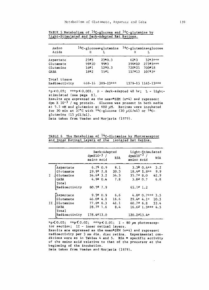
Metabolism of Glutamate, Asparate and Gaba 159
TABLE 5 Metabolism of 14C-$1ucose and 14C-$1utamine by Light-Stimulated and Dark-Adapted Rat Retinas.
Amino 14C-glucose+glutamine 14C-glutamine+glucose Acids D L D L
Aspartate 25~3 23~O.3 62~3 32~3,** Glutamate 99"10 99±3 394~iO 273*5*** Glutamine 14~i 12~O.3 720~21 7OO~18 GABA 18±2 15±1 157~13 107±3 *
Total tissue Radioactivity 669-16 389-23*** 1379-15 1145-13"**
*p~O.O5; ***p<O.OOl. D - dark-adapted 48 hr; L - light- stimulated (see page 8).. Results are expressed as the mean~SEM (n=4) and represent dpm X 10 -3 / mg protein. Glucose was present in both media at 5.5 mM and glutamine at 600 ~M. Retinas were incubated for 30 min at 37°C with 14C-glucose (30 ~Ci/ml) or 14C- glutamine (15 ~Ci/ml). Data taken from Voaden and Morjaria (1979).
TABLE 6 The Metabolism of 14C-Glutamine by Photoreceptor and Inner Retinal Layers of the Isolated Rat Retina.
Dark-Adapted Light-Stimulated
dpmXiO-3 / RSA dpmXiO-3 / RSA amino acid amino acid
Aspartate 6.7 ~ 0.9 8.1 3.3 @ 0.4** 3.2 IGlutamate 29.9 ~ 2.8 20.3 18.4 ± 1.8"* 9.9
I ~Glutamine 34.6 ~ 3.2 34.3 35.7 x 8.0 42.9 IGABA 4.9 ~ 0.4 7.8 3.8 ~ 0.7 6.8 ITotal LRadioactivity 80.5 ~ 7.9 65.1 ~ 1.2
Aspartate 9.5 ± 0.9 6.6 4.8 • 0.7*** 3.5 IGlutamate 46.0 ~ 4.1 16.6 29.4 = 4.1" 10.3
II ~Glutamine 77.6 t 6.3 41.1 60.7 ~ 6.8 33.4
I~ BA 28.7 ~ 1.6 8.4 16.6 r 1.3"** 4.5 otal
LRadioactivity 178.4~13.O 120.2x13.4 *
*p<O.O5; **p<O.O2; ***p<O.Ol; I - 80 ~m photorecep- tor section; II - inner retinal layers. Results are expressed as the mean~SEM (n=4) and represent radioactivity per 3 mm dia. disc retina. Experimental con- ditions were as in Tables 4 and 5. RSA = specific activity of the amino acid relative to that of the precursor at the beginning of the incubation. Data taken from Voaden and Morjaria (1979).

160 M. ,J. Voaden , B. M o r j a r i a a~d A. C. ,J. Oracd~
Effects of Lisht
Approximately 95% of the photoreceptor cells of the rat retina are rods (Cicerone, Green and Fisher, 1979)o Although they are highly insensitive to red light, they
are saturated by very low levels of shorter wavelength illumination ( ~max rhodops- in 500 nm). For the present experiments, therefore, daylight was reduced to
0.15 ~watts / sq. metre. In terms of human vision this corresponds to the low mesopic range. Eight to ten week-old female albino Wistar rats were used through- out the study. They were dark-adapted for 48 hr before use and were then killed and the retinas dissected out under a dim red light. Incubations were done either in darkness or in the reduced daylight and the relevant lighting maintained until the tissue was frozen (for bisection) or added to a protein precipitant.
As shown in Table 5, when the metabolism of 14C-glucose was followed under these conditions and the retinas processed after only a 2 min wash to avoid loss of solu- ble compounds, there was a 40% reduction in total tissue radioactivity on light st- imulation, but no change in the entry of label into the amino acids. In contrast, when 14C-glutamine was metabolised, light caused a significant decrease in the labelling of aspartate, glutamate and GABA. There was also an 18% reduction in the total tissue radioactivity, accounted for by the reduced label in the amino acids.
In previous in vivo studies with the glucose analogue 2-deoxy-glucose, Morjaria
and Voaden (1979b) have also obtained results which imply a decrease in the rate of glucose consumption by light- as compared with dark-adapted rat retinas (the former being maintained in normal room lighting). The decrease (38%) occurred in the photoreceptor cell layer.
To localize further the changes in amino acid metabolism, retinas, incubated with the radioactive precursors, were sliced to a depth of 80 >m from the photoreceptor side (cf. Fig. 2). Table 6 shows that with 14C-glutamine as precursor, l~ht re- duced the entry of the label by 51% and 39% respectively into aspartate and gluta-
TABLE 7 Endogenous Amino Acids in Isolated Rat Retinas In-
cubated with both Glucose and Glutamine as Substrates.
80 >m Photoreceptor Inner Retinal Section Layers
D L D L
Aspartate 1.5 i 0.2 1.8 t 0.2 2.6 ± 0.4 2.5 i 0.3 Glutamate 2.7 ~ 0.2 3.4 ~ O.4 5.O Z 0.6 5.1 ± O.8 Glutamine 1.8 ± 0.2 1.5 r 0.3 3.4 ~ O.4 3.3 t 0.4 GABA I.i ± O.i I.O ± O.i 6.2 i I.O 6.7 ~ 0.8
D - dark-adapted 48 hr; L - light-stimulated (see page 8). Results are expressed as the mean ~ SEM (n~ 7) and represe- nt nmol / 3 mm dia. disc retina. Retinas were incubated at 37=C for 30 min with 5.5 mM glucose and 600 ~M glutamine. Discs of tissue were then sectioned as described in Fig. 2. Endogenous amino acids were measured by double-label dansy- lation (Morjaria and Voaden, 1979a).

Metabolism of Glutamate, Asparate and Gaba 161
mate in the photoreceptor layer, and into these amino acids and GABA in the inner retinal layers. When the endogenous amino acids were analysed in retinas treated similarly but incubated with unlabelled substrates, there were no significant dif- ferences between the light- and dark-adapted tissue (Table 7), although trends to- wards the changes seen between freshly-isolated light- and dark-adapted retinas (Table 2) were apparent. These results imply that changes occur in amino acid turnover on light-stimulation, followed later by adjustment in the tissue levels of the endogenous amino acids.
Increased tissue levels combined with decreased entry of label would in turn imply decreased turnover of glutamate and aspartate in photoreceptor cells and of gluta- mate, aspartate and GABA in the inner retinal layers of the rat retina on light stimulation. The changes in photoreceptor metabolism are consistent, therefore, with a role for glutamate and/or aspartate in photoreceptor transmission (cf. page 4). As the inner retinal sections would have contained photoreceptor terminals, it is not possible to say definitely whether or not the changes observed here are also associated with photoreceptor function. However, results from a more detail- ed profile of the entry of label from 3H-glutamine into these amino acids suggests not (Morjaria and Voaden, 1979a). The profile also suggests that the principal location of GABA formation from glutamine is in the amacrine/inner plexiform lay- ers of the tissue. This is consistent with (i) the position of labelled cells seen after a pulse and chase study with 3H-glutamine, in which GABA was the princi- pal end product (Voaden and colleagues, 1978), (2) the location of endogenous pools of GABA in the rat retina (Fig. I) and (3) the tentative localization of glutamate decarboxylase to amacrine synaptic terminals (Wood, McLaughlin and Vaughn, 1976;
TABLE 8 The Metabolism of 14C-Glucose by Photoreceptor and Inner Retinal Layers of the Isolated Rat Retina.
Dark-Adapted Light-Stimulated dpmXiO -3 / RSA dpmXIO -3 / RSA amino acid amino acid
I Aspartate 1.7~0.2 9.7 1.3±O.2 5.9 Glutamate 7.4~I.0 23.7 6.8~1.4 17.3
I Glutamine 0.6'0.I 2.9 0.7±0.1 4.0 GABA 0.710.2 5.0 O.8~0.i 7.0
]Total LRadioactivity 19.3~1.9 16.4±2.6
Aspartate 2.2~0.3 7.2 1.9~O.2 6.3 [Glutamate 9.8±1.6 16.6 8.6~1.2 14.2
II ~ Glutamine 2.0±0.3 5.0 1.6±0.2 4.0 ]GABA 3.3±0.7 4.5 2.7~0.4 3.4 [Total LRadioactivity 38.3t6.5 35.1~2.9
I - 80 >m photoreceptor section; II - inner retinal layers. Results are expressed as the mean~SEM (n=4) and represent radioactivity per 3 mm dia. disc retina. Experimental con- ditions were as in Tables 4 and 5. RSA = specific activity of the amino acid relative to that of the precursor at the beginning of the incubation. Data taken from Voaden and Morjaria (1979).

162 M.J. Voaden, g. Morjaria and A. C. J. Oraed~
cf. also Graham, 1974). It thus seems probable that changes in GABA turnover are occurring here. Although the present results suggest that this is decreased turn- over, the lowered specific activities might also result from the release of newly- formed, more highly-labelled pools that have not equilibrated with the general ti- ssue stores. The preferential release of neuroactive amino acids derived from glu- tamine as compared with those from glucose has been found in studies on rat corti- cal synaptosomes (Bradford, Ward and Thomas, 1978), pigeon optic tectum (Reubi, Van den Berg and Cuenod, 1978) and rabbit hippocampus (Hamberger and colleagues, 1979). As yet, no effects of light stimulation on endogenous GABA release from the rat retina have been found (Coull and Cutler, 1978; Kennedy and Neal, 1978; Starr, 1975b), although Bauer and Ehinger (1977) reported an increased output of preloaded labelled GABA from the rabbit retina. Species differences undoubtedly ~ist in the response of retinal GABA systems to light as Starr (1975b), using
C-glucose as the radiolabelled precursor and working with the frog retina in vivo, found increased entry of label into GABA, combined with raised endogenous levels, suggesting increased turnover; less label entered or remained in glutamate In the same study no effects were seen in the rat, perhaps because 14C-glucose was used as the precursor (cf. Table 5).
Little is known of the function(s) of GABA in the rat retina. It is not possible, therefore, to link the changes observed here with a physiological role. However, Graham (1974) has provided evidence suggesting that this amino acid may be involv- ed with light-adaptation over the scotopic visual range.
Retinas incubated with 14C-glucose were also bisected : the results are shown in Table 8. Again, as with the data in Table 5, entry of label into the amino acid pool is not significantly altered by light-stimulation. However, because of slig- ht changes in labelling of the photoreceptor layer, combined with subtle altera- tions in the endogenous amino acids there (Table 7), the specific activities of glutamate and aspartate are decreased - about half as much as when 14C-glutamine was the precursor. Complex changes may occur in glucose metabolism on light-stim- ulation of photoreceptors (eg. Cohen and Noell, 1965). Consequently, it cannot be concluded that glucose and glutamine are here labelling separate pools of the amino acids. However, the data for the 'inner retinal sections' (Tables 6 and 8) do suggest the existence of separate pools. The profiles of GABA production from 14C-glucose and 3H-glutamine through the rat retina are very similar, but differe- nces exist in those for glutamate and aspartate (Morjaria and Voaden, 1979a).
In contrast to the data shown in Table 5, there was no evidence for a decrease in total tissue radioactivity, on light stimulation, in retinas incubated with 14C- glucose and then processed for slicing. The non-amino acid, 'light-sensitive', glucose derivatives may, therefore, have been washed from the tissue.
Less ATP may be needed in the light-stimulated photoreceptor because of cessation of the sodium current that flows between the inner and outer limb of the dark- adapted cell, and the consequent decrease in Na+/K + ATPase activity in the inner limb. It has also been proposed that the tricarboxylic acid cycle fTCAC) will be inhibited by a build-up of 6-phosphogluconate, occurring because of stimulation of the pentose phosphate pathway (Cohen and Noell, 1965). It is significant, there- fore, that oxygen uptake is depressed in photoreceptors subjected to steady illu- mination (Hanawa and Kuge, 1961; Sickel, 1972), and possible that the 'light-sen- sitive' derivatives observed here (Table 5) are TCAC intermediates.
Under similar conditions succinoxidase activity in photoreceptor inner limbs may increase (Epstein and O'Connor, 1966). It is, therefore, of interest that the GABA bypath may be present there (cf. page 5), forming a route by which carbon

Metabolism of Glutamate, Asparate and Gaba
atoms could enter the TCAC at the level of succinate. However, in the present studies no significant changes were observed in GABA turnover in photoreceptor cells on light stimulation (Tables 6 and 8).
The present results emphasize the potential importance of glutamine in retinal metabolism and show changes in the homeostasis of glutamate, aspartate and GABA in the rat retina in response to light. In particular, the data obtained is con- sistent with a role for glutamate and/or aspartate as photoreceptor neurotrans- mitters.
163
REFERENCES
Arden, G. B. and Ernst, W (1971). A method of determining photoreceptor ion con- tents. J. Physiol., 216, 5p-7p.
Bauer, B. and Ehinger, B. (1977). Light-induced release of radioactivity from rabbit retina preloaded with 3H-GABA. Experientia, 33, 470-471.
Berger, S. J., McDaniel, M. L., Carter, J. G. and Lowry, O. H. (1977). Distribu- tion of four potential transmitter amino acids in monkey retina. J. Neurochem., 28, 159-163.
Bradford, H. F., Ward, H. K. and Thomas, A. J. (1978). Glutamine - a major subs- trate for nerve endings. J. Neurochem., 30, 1453-1459.
Cicerone, C. M., Green, D. G. and Fisher, L. J. (1979). Cone inputs to ganglion cells in hereditary retinal degeneration. Science, 203, 1113-1114.
Cohen, A. I., McDaniel, M. and Orr, H. (1973). Absolute levels of some free amino acids in normal and biologically fractionated retinas. Invest. Ophthalmol., 12, 686-693.
Cohen, L. H. and Noell, W. K. (1965). Relationships between visual function and metabolism. In C. N. Graymore (Ed.), Biochemistry of the Retina. Academic Press, London and New York. pp. 36-49.
Coull, B. M. and Cutler, R. W. P. (1978). Light-evoked release of endogenous gly- cine into perfused vitreous of the intact rat eye. Invest. Ophthalmol. Visual Sci., 17, 682-684.
Durham, D. G., Dickinson, J. C. and Hamilton, P. B. (1971). Ion-exchange chroma- tography of free amino acids in human intraocular fluids. Clin. Chem., 17, 285-289.
Epstein, M. H. and O'Connor, J. S. (1966). Enzyme changes in isolated retinal layers in light and darkness. J. Neurochem., 13, 907-911.
Graham, L. T. Jr. (1974). Comparative aspects of neurotransmitters in the retina. In H. Davson and L. T. Graham, Jr. (Eds.), The Eye, Vol. 6, Comparative Phy- siology. Academic Press, New York and London. pp. 283-342.
Graham, L. T., Baxter, C. F. and Lolley, R. N. (1970). In vivo influence of light or darkness on the GABA system in the retina of the frog (Rana pipiens). Brain Research, 20, 379-388.
Hamberger, A. C., Chiang, G. H., Nylen, E. S., Scheff, S. W. and Cotman, C. W. (1979). Glutamate as a CNS transmitter I. Evaluation of glucose and glutamine as precursors for the synthesis of preferentially released glutamate. Brain Research, 168, 513-530.
Hanawa, I. and Kuge, K. (1961). The effect of light intensity upon the oxygen consumption of the isolated outer segment rods. Jap. J. Physiol., ii, 38-43.
Hyde, J. C. and Robinson, N. (1974). Localisation of sites of GABA catabolism in the rat retina. Nature, 248, 432-433.
Kennedy, A. J. and Neal, M. J. (1978). The effect of light and potassium depola- rization on the release of endogenous amino acids from the isolated rat retina Exp. Eye Res., 26, 71-75.
Kennedy, A. J., Neal, M. J. and Lolley, R. N. (1977). The distribution of amino acids within the rat retina. J. Neurochem., 29, 157-159.

164 M.J. Voaden, B. Morjaria and A. C. ~. Or~e~dti
Kleinschmidt, J. and Yazulla, S. (1978). Effects of glutamate agonists and anta- gonists on goldfish horizontal cells. Arvo Abstracts : Suppl. to Invest. O~hthalmol. Visual Sci., 17, pp. 284.
Lam, D. M. K. (1972). The biosynthesis and content of gamma-aminobutyric acid in the goldfish retina. J. Cell Biol., 54, 225-231.
Lam, D. M. K. (1975). Synaptic chemistry of identified cells in the vertebrate retina. Cold Spr. Harb. S}mnp. quant. Biol., 40, 571-579.
Lolley, R. N. (1969). Metabolic and anatomical specialization within the retina. In A. Lajtha (Ed.), Handbook of Neurochemistry. Plenum Press, New York and London. pp. 473-504.
Lowry, O. H., Rosebrough, N. J., Farr, A. L. and Randall, R. J. ~1951). Protein measurement with the folin phenol reagent. J. Biol. Chem., 193, 265-275.
McIlwain, H. and Bachelard, H. S. (1971). Biochemistry and the Central Nervous System, 4th Edn. Churchill Livingstone, Edinburgh.
Moore, C. L. and Gruberg, E. R. (1974). The distribution of succinic semialdehyde dehydrogenase in the brain and retina of the tiger salamander (Ambystoma tig- rinum). Brain Research, 67, 467-478.
Morjaria, B. and Voaden, M. J. (1979a). The formation of glutamate, aspartate and GABA in the rat retina : glucose and glutamine as precursors. J. Neurochem., 33, 541-551.
Morjaria, B. and Voaden, M. J. (1979b). The uptake of ~H]-2-deoxy glucose by light- and dark-adapted rat retinas in vivo. J. Neurochem., 32, 1881-1883.
Neal, M. J. (1976). Amino acid transmitter substances in the vertebrate retina. Gen. Pharmac., 7, 321-332.
Neal, M. J. and White, R. D. (1978). Discrimination between descriptive models of L-glutamate uptake by the retina using non-linear regression analysis. J. Physiol., 277, 387-394.
Oraedu, A. C. I., Voaden, M. J. and Marshall, J. (1979). Photochemical damage in the rat retina : morphological changes and endogenous amino acids. J. Neuro- chem., submitted for publication.
Pasantes-Morales, H., Klethi, J., Ledig, M. and Mandel, P. (1973). Influence of light and dark on the free amino acid pattern of the developing chick retina. Brain Research, 57, 59-65.
Rasmussen, K.-E. (1972). A morphometric study of the MUller cell cytoplasm in the rat retina. J. Ultrastructure Research, 39, 413-429.
Reubi, J. C., Van den Berg, C. and Cuenod, M. (1978). Glutamine as precursor for the GABA and glutamate transmitter pools. Neuroscience Letters, iO, 171-174.
Riepe, R. E. and Norenburg, M. D. (1977). M~ller cell localization of glutamine synthetase in rat retina. Nature, 268, 654-655.
Sarthy, P. V. and Lam, D. M. K. (1978). Biochemical studies of isolated glial (MUller) cells from the turtle retina. J. Cell Biol., 78, 675-684.
Sarthy, P. V. and Lam, D. M. K. (1979). Endogenous levels of neurotransmitter candidates in photoreceptor cells of the turtle retina. J. Neurochem., 32, 455-461.
Sickel, W. (1972). Retinal metabolism in light and dark. In }I. G. F. Fuortes (Ed.), Handbook of Sensory Physiology, Vol. VII/2 Physiology of Photoreceptor Organs. Springer, Berlin. pp. 667-727.
Starr, M. S. (1973). Effects of dark adaptation on the GABA system in retina. Brain Research, 59, 331-338.
Starr, M. S. (1975a). A comparative study of the utilization of glucose, acetate, glutamine and GABA as precursors of amino acids by retinae of the rat, frog, rabbit and pigeon. Biochem. Pharmacol., 24, 1193-1197.
Starr, M. S. (1975b). The effect of light stimulation on the synthesis and relea- se of GABA in rat and frog retinae. Brain Research, iOO, 343-353.
Van Gelder, N. M. and Drujan, B. D. (1978). Interrelated changes of amino acids in the retina and optic tectum of a marine fish with alterations of illumina- ting conditions. Brain Research, 159, 137-148.

Metabolism of Glutamate, Asparate and Gaba 165
Voaden, M. J. (1976). $-aminobutyric acid and glycine as retinal neurotransmitte- rs. In S. L. Bonting (Ed.), Transmitters in the Visual Process. Pergamon Press, Oxford. pp. 107-125.
Voaden, M. J. (1978). The localization and metabolism of neuroactive amino acids in the retina. In F. Fonnum (Ed.), Amino Acids as Neurotransmitters. Plenum Press, New York and London. pp. 257-274.
Voaden, M. J. (1979). The chemical specificity of neurones in the retina. Prosress in Brain Research, In Press.
Voaden, M. J., Lake, N., Marshall, J. and Morjaria, B. (1978). The utilization of glutamine by the retina : an autoradiographic and metabolic study. J. Neurochem., 31, 1069-1076.
Voaden,'M. J., Lake, N. and Nathwani, B. (1977). A comparison of ~-aminobutyric acid metabolism in neurones versus glial cells using intact isolated retinas. J. Neurochem., 28, 457-459.
Voaden, M. J. and Morjaria, B. (1979). The synthesis of neuroactive amino acids from radioactive glucose and glutamine in the rat retina : Effects of light- stimulation. J. Neurochem., submitted for publication.
White, R. D. and Neal, M. J. (1976). The uptake of L-glutamate by the retina. Brain Research~ iii, 79-93.
Wood, J. G., McLaughlin, B. J. and Vaughn, J. E. (1976). Immunocytochemical loca- lization of GAD in electron microscopic preparations of rodent CNS. In E. Roberts, T. Chase and D. B. Tower (Eds.), GABA in Nervous System Function. Raven Press, New York. pp. 133-148.
Wu, S. M. and Dowling, J. E. (1978). L-Aspartate : Evidence for a role in cone photoreceptor synaptic transmission in the carp retina. Proc. Nat. Acad. Sci., 75, 5205-5209.