the lapidus procedure as salvage after failed … · the lapidus procedure as salvage after failed...
TRANSCRIPT
The PDF of the article you requested follows this cover page.
This is an enhanced PDF from The Journal of Bone and Joint Surgery
2004;86:30-36. J Bone Joint Surg Am.J. Chris Coetzee, Scott G. Resig, Michael Kuskowski and Khaled J. Saleh
Hallux Valgus. Surgical TechniqueThe Lapidus Procedure as Salvage After Failed Surgical Treatment of
This information is current as of February 20, 2009
Reprints and Permissions
Permissions] link. and click on the [Reprints andjbjs.orgarticle, or locate the article citation on
to use material from thisorder reprints or request permissionClick here to
Publisher Information
www.jbjs.org20 Pickering Street, Needham, MA 02492-3157The Journal of Bone and Joint Surgery

COPYRIGHT © 2004 BY THE JOURNAL OF BONE AND JOINT SURGERY, INCORPORATED
The Lapidus Procedure as Salvage After Failed Surgical Treatment of Hallux ValgusSurgical Technique By J. Chris Coetzee, MD, FRCSC, Scott G. Resig, MD, Michael Kuskowski, PhD, and Khaled J. Saleh, MD, MSc, FRCSC
Investigation performed at the Department of Orthopaedic Surgery, University of Minnesota School of Medicine, Minneapolis, Minnesota
The original scientific article in which the surgical technique was presented was published in JBJS Vol. 85-A, pp. 60-65, Jan. 2003
INTRODUCTIONPaul Lapidus was an orthopaedic surgeon who worked at the Hospital for the Ruptured and Crippled in New York when he wrote his first arti-cle on the surgical correction of the metatarsus primus varus in patients with hallux valgus deformities1. The premise of his approach was that the metatarsus primus varus was a result of instability at the first tar-
ABSTRACT
BACKGROUND:Recurrent hallux valgus is a rela-tively common, yet challenging, condition for both the patient and the surgeon. The literature on the treatment of recurrent hallux val-gus is sparse. The purpose of this study was to evaluate pro-spectively the functional outcome and patient satisfaction follow-ing the Lapidus procedure for the treatment of recurrent hallux val-gus deformity.
METHODS:Twenty-four patients with a total of twenty-six symptomatic recur-rences of hallux valgus after pre-vious procedures for treatment of the deformity were included in the study. Exclusion criteria in-cluded prior fusion procedures on the foot or ankle, a previous Keller or Mayo procedure, insulin-dependent diabetes, peripheral vascular disease, or peripheral neuropathy. A visual analog pain scale and the American Ortho-paedic Foot and Ankle Society
continued
Figs. 1-A through 1-D
Schematic illustrations of
the Lapidus procedure.
Figs. 1-A and 1-B Antero-
posterior (Fig. 1-A) and
medial (Fig. 1-B) views of
the foot, showing meta-
tarsus primus varus and
hallux valgus deformities.
FIG. 1-A

TH E JOUR N AL OF BON E & JOINT SURGER Y · SU R G I C A L TE CH N I QU E S MA RCH 2004 · VO LU M E 86-A · SUPPLEMENT NU M B E R 1 · JB JS .OR G
sometatarsal joint. Correcting the alignment at the base of the deformity yielded the most pow-erful correction. The Lapidus procedure consists of correction of the metatarsus primus varus at the first tarsometatarsal joint through fusion of the joint (Figs. 1-A through 1-D)2.
SURGICAL TECHNIQUEThe patient is placed supine with a tourniquet around the thigh. A 6-cm incision is made over the dorsum of the foot in line with the extensor hallucis longus tendon. The interval between the extensor hallucis longus and the extensor hal-lucis brevis is used to expose the first and second tarsometa-tarsal joints. A large vein cross-ing the line of the dissection is almost uniformly present at the distal end of the incision. The vein is cauterized and is an in-dication that the dissection is
distal enough (Fig. 2).The dorsalis pedis artery
and the deep peroneal nerve are usually encountered in the inter-val between the extensor hallucis longus and the extensor hallucis brevis. Careful dissection is mandatory to prevent damage to these structures. The dorsalis pedis enters the plantar aspect of the foot through the first web space, about 1 to 1.5 cm distal to the first tarsometatarsal joint. One should be aware of this fact when débriding the interval be-tween the bases of the first and second metatarsals.
The entire first tarsometa-tarsal joint must be exposed (Fig. 3). The medial aspect of the sec-ond metatarsal is exposed and denuded of soft tissue, and the cortex is also perforated to aug-ment the subsequent fusion.
The articular cartilage is removed from the opposing sur-faces of the first tarsometatarsal
ABSTRACT | continued
Hallux Metatarsophalangeal-Interphalangeal Scale were administered preoperatively, at six months postoperatively, and yearly thereafter. Weight-bearing radiographs were also made preoperatively; at six weeks, three months, six months, and one year postop-eratively; and yearly thereafter. Patient satisfaction was as-sessed at the latest follow-up evaluation.
RESULTS:At twenty-four months, the mean score according to the American Orthopaedic Foot and Ankle Society (AOFAS) Hallux Metatarsophalangeal-Interphalangeal Scale had in-creased from 47.6 to 87.9 points, the mean score accord-ing to the visual analog pain scale had improved from 6.2 to 1.4, the mean hallux valgus angle had improved from 37.1° to 17.1°, and the mean inter-metatarsal angle had improved from 18° to 8.6°. The patients were very satisfied after 77% of the twenty-six procedures, satis-fied after 4%, and somewhat satisfied after 19%; no patient was dissatisfied. There were no cases of hallux varus. Com-plications included three non-unions, all of which occurred in smokers, and two superficial wound infections.
CONCLUSIONS:In appropriately selected pa-tients, the Lapidus procedure is a reliable and effective operation after failed surgical treatment of hallux valgus.
FIG. 1-B
TH E JOUR N AL OF BON E & JOINT SURGER Y · SU R G I C A L TE CH N I QU E S MA RCH 2004 · VO LU M E 86-A · SUPPLEMENT NU M B E R 1 · JB JS .OR G
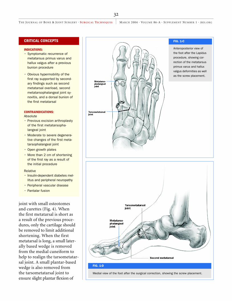
joint with small osteotomes and curettes (Fig. 4). When the first metatarsal is short as a result of the previous proce-dures, only the cartilage should be removed to limit additional shortening. When the first metatarsal is long, a small later-ally based wedge is removed from the medial cuneiform to help to realign the tarsometatar-sal joint. A small plantar-based wedge is also removed from the tarsometatarsal joint to ensure slight plantar flexion of
CRITICAL CONCEPTS
INDICATIONS: • Symptomatic recurrence of
metatarsus primus varus and hallux valgus after a previous bunion procedure
• Obvious hypermobility of the first ray supported by second-ary findings such as second metatarsal overload, second metatarsophalangeal joint sy-novitis, and a dorsal bunion of the first metatarsal
CONTRAINDICATIONS: Absolute• Previous excision arthroplasty
of the first metatarsopha-langeal joint
• Moderate to severe degenera-tive changes of the first meta-tarsophalangeal joint
• Open growth plates
• More than 2 cm of shortening of the first ray as a result of the initial procedure
Relative• Insulin-dependent diabetes mel-
litus and peripheral neuropathy
• Peripheral vascular disease
• Pantalar fusion
Anteroposterior view of
the foot after the Lapidus
procedure, showing cor-
rection of the metatarsus
primus varus and hallux
valgus deformities as well
as the screw placement.
FIG. 1-C
FIG. 1-D
Medial view of the foot after the surgical correction, showing the screw placement.

TH E JOUR N AL OF BON E & JOINT SURGER Y · SU R G I C A L TE CH N I QU E S MA RCH 2004 · VO LU M E 86-A · SUPPLEMENT NU M B E R 1 · JB JS .OR G
CRITICAL CONCEPTS | continued
PITFALLS: • The dorsalis pedis artery and
the deep peroneal nerve are usually encountered in the in-terval between the extensor hallucis longus and the exten-sor hallucis brevis. Careful dissection is mandatory to prevent damage to these structures (Fig. 2).
• One should be cognizant of the tendency to fuse the first tar-sometatarsal joint in dorsiflex-ion, which can lead to lesser metatarsal overload. The dor-soplantar extent of this joint is about 30 mm. It can be diffi-cult to denude the plantar one-half or one-third of the joint without adequate exposure. A mini lamina spreader is very helpful to ensure complete vi-sualization of the joint.
• When the interval between the first and second metatar-sal bases has not been ade-quately débrided, it might be difficult to completely reduce the intermetatarsal angle.
• The medial cuneiform is only 1.5 cm wide. Screw placement is therefore very important. It is most reliable to insert the first screw from the middle of the medial cuneiform into the first metatarsal. The second screw is then inserted from the base of the first metatarsal into the base of the second metatarsal.
• During the insertion of the intermetatarsal screw, one should keep the first and sec-ond metatarsals in the same plane, avoiding excessive plan-tar flexion or dorsiflexion of the first ray.
continued
FIG. 2
The surgical approach to the first tarsometatarsal joint. The interval between
the extensor hallucis longus and brevis is visible, as is the proximity of the neu-
rovascular bundle in the interval. (The forceps is placed on the artery.) The first
tarsometatarsal joint is exposed.
FIG. 3
The dorsoplantar extent of the first tarsometatarsal joint is about 30 mm.
TH E JOUR N AL OF BON E & JOINT SURGER Y · SU R G I C A L TE CH N I QU E S MA RCH 2004 · VO LU M E 86-A · SUPPLEMENT NU M B E R 1 · JB JS .OR G
the metatarsal (Fig. 1-B).Next, the adductor hallucis
tendon is released through a 2-cm incision in the first web space. The lateral aspect of the first metatar-sophalangeal joint capsule is incised longitudinally to allow the sesamoids to reduce. A me-dial incision is then made over the first metatarsophalangeal
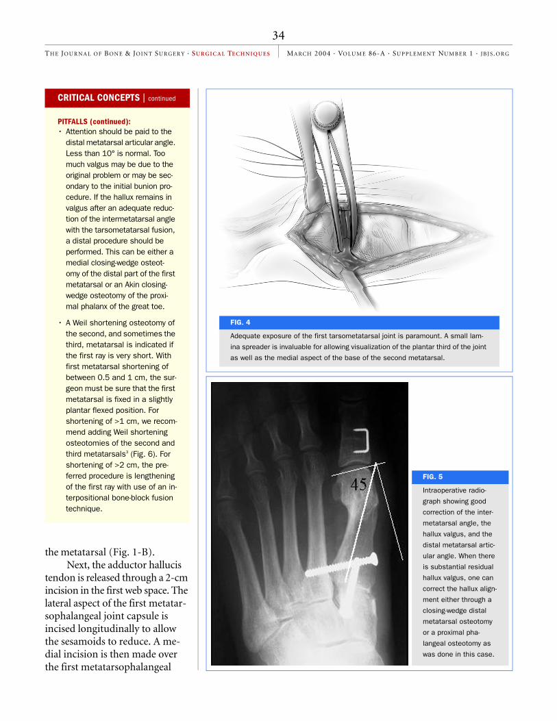
FIG. 4
Adequate exposure of the first tarsometatarsal joint is paramount. A small lam-
ina spreader is invaluable for allowing visualization of the plantar third of the joint
as well as the medial aspect of the base of the second metatarsal.
FIG. 5
Intraoperative radio-
graph showing good
correction of the inter-
metatarsal angle, the
hallux valgus, and the
distal metatarsal artic-
ular angle. When there
is substantial residual
hallux valgus, one can
correct the hallux align-
ment either through a
closing-wedge distal
metatarsal osteotomy
or a proximal pha-
langeal osteotomy as
was done in this case.
CRITICAL CONCEPTS | continued
PITFALLS (continued): • Attention should be paid to the
distal metatarsal articular angle. Less than 10° is normal. Too much valgus may be due to the original problem or may be sec-ondary to the initial bunion pro-cedure. If the hallux remains in valgus after an adequate reduc-tion of the intermetatarsal angle with the tarsometatarsal fusion, a distal procedure should be performed. This can be either a medial closing-wedge osteot-omy of the distal part of the first metatarsal or an Akin closing-wedge osteotomy of the proxi-mal phalanx of the great toe.
• A Weil shortening osteotomy of the second, and sometimes the third, metatarsal is indicated if the first ray is very short. With first metatarsal shortening of between 0.5 and 1 cm, the sur-geon must be sure that the first metatarsal is fixed in a slightly plantar flexed position. For shortening of >1 cm, we recom-mend adding Weil shortening osteotomies of the second and third metatarsals3 (Fig. 6). For shortening of >2 cm, the pre-ferred procedure is lengthening of the first ray with use of an in-terpositional bone-block fusion technique.

TH E JOUR N AL OF BON E & JOINT SURGER Y · SU R G I C A L TE CH N I QU E S MA RCH 2004 · VO LU M E 86-A · SUPPLEMENT NU M B E R 1 · JB JS .OR G
joint, the capsule is incised longitudinally, and any residual bunion prominence is removed.
The first metatarsal is then reduced parallel to the second by closing the intermetatarsal gap. It is very important at this time to confirm that the first metatarsal is slightly plantar flexed and is rotated correctly. One 3.5-mm cortical screw is then in-serted from the medial cuneiform into the first metatarsal under compression. A second screw is inserted from the medial aspect of the base of the first metatarsal into the base of the second metatar-sal to close the intermetatarsal gap securely. With the intermetatarsal gap reduced, the medial aspect of the capsule of the first metatarsophalangeal joint is plicated. It should not be necessary to overtighten the capsule in order to maintain the alignment of the hallux (Fig. 5). Local bone graft is packed into any osseous defects at the bases of the metatarsals. The tourniquet is deflated, and the wounds are closed in layers.
Postoperatively, the foot is immobilized in a slipper cast (fiberglass great-toe spica) for two weeks. At two weeks, the sutures are removed and a second slipper cast is applied; this cast is worn for an additional four to six weeks. The patient
should remain non-weight-bearing for six weeks. If the six-week radiographs demonstrate satisfac-tory progression of the fusion, the cast is removed
FIG. 6
The Weil osteotomy is a
long oblique osteotomy of
the second and/or third
metatarsal to shorten
them to the level of the
first. The head is trans-
lated proximally and is
secured to the shaft with
a mini-fragment screw.
CRITICAL CONCEPTS | continued
AUTHOR UPDATE: Only a few minor changes have been made in the proce-dure since our original article was published.
It is almost never necessary to use a saw when perform-ing the preparation of the first tarsometatarsal joint. Careful use of small osteotomes will allow an adequate preparation without producing excessive shortening of the first ray.
We now use 4-mm self-tapping cortical screws (Synthes USA, Paoli, Pennsylvania). These screws have the same head as ordinary small-fragment screws, but an in-crease in the shaft diameter has reduced the potential for screw breakage and nonunion. (Our nonunion rate over the past year has been reduced to 2%.)
The patients are allowed to begin “heel-touch” weight-bearing on Day 1. They can put 30 lb (13.6 kg) of weight on the heel when standing and walking. This does not ad-versely affect healing, and patient compliance is much easier than with complete non-weight-bearing.

TH E JOUR N AL OF BON E & JOINT SURGER Y · SU R G I C A L TE CH N I QU E S MA RCH 2004 · VO LU M E 86-A · SUPPLEMENT NU M B E R 1 · JB JS .OR G
and weight-bearing and physical therapy are started. Patients are advised not to return to any vigorous physical activity for at least three months, although they may begin swimming and bicycling at eight weeks.
J. Chris Coetzee, MD, FRCSCScott G. Resig, MDKhaled J. Saleh, MD, MSc, FRCSCDepartment of Orthopaedic Surgery, University of Minnesota School of Medi-cine, 420 Delaware Street S.E., Box 492, Minneapolis, MN 55455. E-mail address for J.C. Coetzee: [email protected]
Michael Kuskowski, PhDMinneapolis Veterans Affairs Medical Center, 1 Veterans Drive, Minneapolis, MN 55417
The authors did not receive grants or outside funding in support of their research or preparation of this manuscript. They did not receive payments or other benefits or a commitment or agreement to provide such benefits from a commercial entity. No commercial entity paid or directed, or agreed to pay or direct, any benefits to any research fund, foundation, educational institution, or other charitable or nonprofit organization with which the authors are affili-ated or associated.
The line drawings in this article are the work of Jennifer Fairman ([email protected]).
REFERENCES1. Lapidus PW. Operative correction of the metatarsus varus primus
in hallux valgus. Surg Gynecol Obstet. 1934;58:183-91.2. Lapidus PW. The author’s bunion operation from 1931 to 1959.
Clin Orthop. 1960;16:119-35.3. Barouk LS. [Weil’s metatarsal osteotomy in the treatment of
metatarsalgia]. Orthopade. 1996;25:338-44. German.