the laboratory mouse (handbook of experimental animals) - usp
TRANSCRIPT

GeneralMice are the most widely used animals for a range ofexperiments including medical, chemical, pharmaco-logical, toxicological, biological, and genetic. Theadministration of test substances, such as chemical ele-ments, compounds, drugs, antibodies, cells or otheragents, to mice is one of the major methods for evalu-ating their biological activity.
The route of administration is largely dependent onthe property of the test substance and the objective ofthe experiment. All administration should be performedwith knowledge of the chemical and physical characteris-tics of the substance. All routes have both demerit andmerit, such as the absorption, bioavailability and metab-olism of the substance. Consideration should be paid tothe pH, viscosity, concentration, sterility, pyrogenicity,toxicity as well as the existence of hazardous substances.A knowledge of available methods and techniques ofadministration as well as knowledge of the depositionand fate of the administered substance will help the scien-tist/investigator to select the most appropriate route forher/his purpose. This route must be selected before thestart of any experiment (Nebendahl, 2000).
Proper restraint is the most important techniquewhen mice were treated as this decreases stress and
increases successful treatment. Personnel using experi-mental animals should be well trained in handling andrestraint, should obtain authentication for responsibleuse of experimental animals and attain a scientificallyhigh standard (ETS 123, 1986; Nebendahl, 2000).Further experience will lead to repeatable and reliableresults (see Chapter 31 on Handling and Restraint).
During administration mice should be protectedfrom pain, suffering, distress or lasting harm or at leastpain and distress shall be kept to a minimum (ETS123, 1986). Some injections (such as footpad injec-tion) are strongly discouraged and if required must bejustified on a case by case basis (CCAC, 2002).
Principles ofadministrationHandling and restraintGood handling and restraint is the most importanttechnique for correct administration. Proper restrain-ing leads to successful administration and varies withthe routes of administration. Disposable gloves must beworn as manual restraint is frequently used for injections.
527
PRO
CED
URES
RO
UTES
OF
AD
MIN
ISTRA
TION
32C H A P T E R
Routes of Administration
Shinya ShimizuNational Institute of Animal Health,Tsukuba, Japan
The Laboratory MouseCopyright 2004 ElsevierISBN 0-1233-6425-6 All rights of production in any form reserved

There are two styles of manual restraint, one uses bothhands and the other is single handed. (Chapter 31 onHandling and Restraint is helpful; Donovan andBrown, 1991; Suckow et al., 2000).
Double handed manual restraint
The mouse is lifted by the base of the tail and placed onthe cage lid or other solid surface with one hand and thenits tail is pulled gently back (Figure 32.1a). It is quicklyand firmly picked up by the scruff of the neck behind theears with the thumb and index finger of the other hand(Figure 32.1b). The tail is transferred from the first handto between the palm and little or ring finger of the otherhand, then fixed (Figure 32.1c). The mouse is restrained(Figure 32.1d).
Single handed restraint
The tail is picked up using thumb and fore finger of thechosen hand (Figure 32.2a), then the mouse is placedon the cage lid or other solid surface (Figure 32.2b).The tail is immediately grasped by the palm and middlefinger, ring finger and/or little finger, and the thumband forefinger released (Figure 32.2c). The fold of skinfrom the scruff of neck down the back is immediatelygripped using the thumb and forefinger (Figure 32.2dand e). The mouse is then restrained (Figure 32.2f ).
To prevent kicking by the hind legs, the tail isfixed using the palm and forefinger and then the lefthind leg is held firmly between the ring and little fin-ger (where the mouse is restrained by the left hand)(Figure 32.3).
528
PRO
CED
URE
SR
OU
TES
OF
AD
MIN
ISTR
ATI
ON
(a)
(c) (d)
(b)
Figure 32.1 Manual restraint of a mouse using both hands. (a) The mouse is placed on the cage lid with the preferredhand.The tail is pulled gently back by the hand. (b) The mouse is quickly and firmly picked up by the scruff of the neckbehind the ears with thumb and index finger of other hand. (c) The tail is transferred from the preferred hand to betweenpalm and little or ring finger of the other hand, then held firmly. (d) The mouse is restrained.

(CCAC, 2002). In some cases a local anesthetic may beapplied first to prevent pain.
Preparation, solubility andsafety of solutionsTest substances, solutions and equipment should beprepared aseptically and free from pyrogens, especiallyfor parenteral injections. Solutions can be sterilised byfiltration (0.22 m). Living organisms or cells must befree from contaminants when administered. The toxic-ity of the substance, the volume and the way of admin-istration should be considered to prevent tissue damageand to give precise dosage.
The following solvents or vehicles have been foundsuitable in most instances and do not greatly affectdrug action because of their own inherent properties:water, water with 0.85% sodium chloride, water withup to 50% polyethylene glycol, water with not over10% Tween 80, water with up to 0.25% methylcellu-lose or carboxymethylcellulose, corn oil; vegetable oil;
529
PRO
CED
URES
RO
UTES
OF
AD
MIN
ISTRA
TION
(a) (b) (c)
(d) (e) (f )
Figure 32.2 Single-handed restraint of the mouse. (a) The tail is picked up using thumb and forefinger of the preferredhand. (b) The mouse is placed on the cage lid or other solid surface pulling gently back by the hand. (c) The tail isimmediately grasped by the palm and middle finger, ring finger and/or little finger and then, the tail held between thumband forefinger is released. (d) and (e) The fold of skin from the scruff of the neck down the back is immediately grippedusing the thumb and forefinger. (f ) The mouse is restrained.
Site of administrationAmong several possibilities for the administration ofsubstances to mice, the most common routes aresubcutaneous, intraperitoneal or intravenous injection.The intramuscular administration is not recommended,as the muscle of the mouse is too small. Some sites, suchas footpad injection of Freund’s complete adjuvant,intrasplenic injection and intra lymph node injectionare unacceptable nowadays (CCAC, 2002), and shouldbe restricted to cases where it is absolutely necessary.
Preparation of the siteThe area for administration is clipped (Figure 32.4) orcleaned with warm water if necessary before cleaningthe skin with alcohol- or disinfectant-moistened cotton.Where aseptic skin is necessary; the fur must be clippedfollowed by a three-stage surgical preparation: surgicalsoap, alcoholic rinse and surgical preparation solution.The skin is dried immediately before administration

peanut oil (oral and intramuscular route only). A lowpercentage of the lower alcohols, glycols, and acetonecan also be used, provided the volume administered iskept small (Woodard, 1965). Phosphate buffered saline(PBS) or various culture media are also suitable vehicles(Nebendahl, 2000). Lipid-soluble substances can onlybe dissolved in oil but this delays absorption. Oil solu-ble drugs have been successfully given intravenously in15% oil–water emulsions using lecithin as an emulsi-fier (Woodard, 1965).
Unless experience has indicated otherwise, solutionsor suspensions should be prepared as near to the timeof use as possible because some substances will deterio-rate in solution within a few hours (Woodard, 1965).When administering drugs, the solvent should ideallybe the same as the one in which the drug is normallyformulated (Nebendahl, 2000).
Although distilled water can be used under certainconditions, saline is preferable because water ad injec-tionem injected subcutaneously causes pain and intra-venous injection produces hemolysis. Oil and viscousfluids cannot be injected intravenously (Nebendahl,2000). If suspended material is to be used for intra-venous injection, the particles should be removed byfiltration to prevent embolism (Woodard, 1965).
The temperature of fluids must be raised at least toroom temperature or better still up to body tempera-ture before use, because the injection of cold fluids ispainful (Baumans et al., 1993).
Concentration of substancesThe concentration can vary over a fairly wide rangewithout greatly influencing the end result of the experi-ment. Lower concentrations are clearly desirable(Waynforth and Flecknell, 1992). Factors limiting theuse of aqueous solutions for parenteral administrationare probably related to their osmotic pressure. Lowconcentrations can be corrected by the addition ofsodium chloride but ought not to be so high as tomaterially exceed the osmotic pressure of 0.15 Msodium chloride (Woodard, 1965). Highly concen-trated solutions can be administered intravenouslyprovided the rate of injection is kept slow and precau-tions are taken to avoid getting the solution outsidethe vein.
530
PRO
CED
URE
SR
OU
TES
OF
AD
MIN
ISTR
ATI
ON
Figure 32.3 Manual restraint of a mouse to prevent kicksby hind leg.The tail is held using the palm and forefingerand then the left hind leg is fixed between the ring andlittle finger (when the mouse is restrained by the left hand).
Figure 32.4 Clipping of hair on the back. Hair on the back is clipped by a cordless electric clipper.

pH of the injected solutionFor most routes of administration, providing the solu-tions are not highly buffered, a pH range of 4.5–8.0 issatisfactory. For oral administration a pH as low as 3 canbe tolerated, but alkaline solutions are very poorly toler-ated. A rather wide range of pH is indicated for intra-venous administration, because of the buffering effect ofblood and dilution by blood flow, following use of theintramuscular and then subcutaneous routes. When lowor high pH solutions are intravenously injected, the rateof injection is kept slow and again precautions taken toavoid getting solution outside the vein (Woodard, 1965).
Volume and frequency ofadministrationThe injection volume is limited by any toxicity of thesubstance and by the size of the mouse. It should be keptas small as possible. Excess volumes of solution can star-tle the animal. The frequency of administration shouldbe limited to a minimum, to avoid unnecessary stress. Ifsolutions are administrated intravenously, hemodynamicchanges and pulmonary oedema may occur while veryrapid injections can produce cardiovascular failure andbe lethal (Nebendahl, 2000). Maximum volumes areshown in Table 32.1. (Flecknell 1987; Reeves et al.,1991; Wolfensohn and Lloyd, 1994). For immuniza-tion, the maximum is still lower, because of the mixingwith adjuvant. Maximum volumes for injection of anti-gen with or without adjuvant per route are indicated inthe section on Immunization of mice in this chapter.
The rate of absorption anddistribution of administratedsubstancesThe blood flow to the site of administration, the natureof the substance and its concentration influence the
rate of absorption (Wolfensohn and Lloyd, 1994;Nebendahl, 2000). The time-course of the effect of thesubstance is an important factor in determining thedosage and is influenced by the rate of absorption(Waynforth and Flecknell, 1992). Normally, injectedsubstances must be absorbed from the site of adminis-tration into the blood. Therefore, the rate of absorptionwill be determined by the size of the absorbing surface,the blood flow and the solubility of the substance inthe tissue fluid. The rate of absorption is also influ-enced by lipid solubility, physicochemical properties,degree of ionization and molecular size of the substance(Nebendahl, 2000). Compounds, which are highly sol-uble in the body fluids, will be absorbed quickly.Substances that are ionized and are not lipid solublecan only be absorbed if a specific carrier exists. In gen-eral, the rate of absorption is arranged in the order iv �
ip � im � sc � po (Wolfensohn and Lloyd, 1994).
Needles and syringesUsually, 26–27-G, 1/2- to 5/8-in. (12.5–15.6-mm)needles are satisfactory for injection. The smallest gaugeshould be selected as a fine needle prevents leakage offluids and will help to minimize discomfort to the ani-mal (Nebendahl, 2000). 1–2 ml syringes size is enoughfor most injections. When a small volume (less than1.0 ml) is administered, an insulin syringe plus needle isconvenient (Figures 32.5 and 32.6; 27–30-G, 5/16- to1/2-in. (8.0–12.5-mm)) these syringes can be obtainedfrom the companies (Terumo, Tokyo, Japan; BectonDickinson, Franklin Lakes, USA). Intradermal needlesare practical for intracerebral injections (Figure 32.7;Top, Tokyo, Japan). Plastic syringes cannot be used withsolvents such as acetone.
The withdrawal of hazardous substances from bot-tles requires great care. An alcohol-moistened cottonpledget can be kept at the point where the needle entersthe stopper in order to minimize the inadvertent for-mation of aerosols (Silverman, 1987). Because of therisk of embolism, air bubbles in fluid and syringe and
531
PRO
CED
URES
RO
UTES
OF
AD
MIN
ISTRA
TION
TABLE 32.1: Guidelines for maximal administration volumes (in milliliters) and needle size
Oral Subcutaneous Intraperitoneal Intravenous Intradermal Intramuscular Intracerebral Intranasal
0.2 2–3 (scruff ) 2–3 0.2 0.05 0.05 �0.03 �0.02
0.2 (inguinal)
�22 G �25 G �23 G �25 G �26 G 25–27 G �27 G
Source: Flecknell, 1987; Reeves et al., 1991; Wolfensohn and Lloyd, 1994.

needle must be purged. Air bubbles can be purged bygently tapping the side of the syringe and slowlyexpelling the air into absorbent tissue to prevent anydispersion of the contents until fluid appears at the tipof the needle. The needle size will vary with the viscos-ity of the substance being used, the greater the viscosity,and the bigger the needle (Reeves et al., 1991). If bloodor body fluid flow back into the needle, it must beremoved and a fresh attempt be made.
EnteraladministrationEnteral administration has the advantages that it is pos-sible to give quite large amounts of non-sterile sub-stances or solution and that a pH as low as 3 can beadministrated by this route. On the other hand, alkalinesolutions are very poorly tolerated by mouth (Woodard,1965). When using the oral route it should be under-stood that substances can be destroyed by the gastricjuices and that the food content of the stomach influ-ences both rate and order of the gastric emptying. Therate of absorption is markedly influenced by its time ofresidence in the stomach and is also is directly relatedto the rate at which substances are passed from thestomach into the intestine (Levine, 1970). Enzymes ofthe host and microflora of the digestive tract can alsometabolize the substance. On the other hand someinsoluble substances become solubilized as the result ofenzymatic activity during their passage through thestomach and intestine making absorption possible(Nebendahl, 2000). The two major methods for enteraladministration are mixing the substance with food orwater or direct administration using gavage. Rectaladministration is also possible (Woodard, 1965).
Oral administration (per os, p.o.)The simplest method for administration is to give thesubstance with food or drinking water. However, this isnot practicable with those that are unpalatable, insoluble
532
PRO
CED
URE
SR
OU
TES
OF
AD
MIN
ISTR
ATI
ON
(a)
(b)
(c)
(d)
(e)
Figure 32.5 Insulin syringes. (a) 29 G � 1/2 in., 0.5 ml,Terumo; (b) 27 G � 1/2 in., 1.0 ml,Terumo;(c) 29 G � 1/2 in., 0.3 ml, Becton Dickinson; (d) 29 G � 1/2in.,0.5 ml, Becton Dickinson; (e) 29 G � 1/2 in., 1.0 ml,Becton Dickinson.
(a)
(b)
(c)
(d)
Figure 32.6 Needles for insulin syringes. (a) 30 G � 3/8 in.,0.3 ml, Becton Dickinson; (b) 29 G � 1/2 in., 0.5 ml, BectonDickinson; (c) 29 G � 1/2 in., 0.5 ml,Terumo; (d) 27 G � 1/2in., 1.0 ml,Terumo.
(a)
(b)
Figure 32.7 Intradermal needle. (a) 26 G � 1/2 in. needle,Terumo, Japan; (b) 1/2 in. intradermal needle,Top, Japan (tip is 27 G, base is 22 G).

or chemically unstable in drinking water or whenthey irritate the mucosa of the gastrointestinal tract(Nebendahl, 2000). The daily food and water intake ofmouse should be known before the experiment, to calcu-late the quantity of substance to be mixed (see Part 5 onAnimal Husbandry and Production).
Because food and water wastage happens all thetime, it is difficult to determine the precise amount offood and water intake and therefore the precise intake ofthe substance. The only way this can be done is by keep-ing mice in metabolic cages and recording the wastage.
Intragastric administrationDirect administration by oral gavage is preferred tomixing substances with food or drinking water becausethe intake of the substances is precisely measured. Aball tip needle is used to prevent damaging the oesophagusand from passing through the glottal opening intothe trachea (Figure 32.8). A 22 G ball tip needle is suit-able for administration to adult mice and can beobtained from Popper and Sons, Inc. (New Hyde Park,USA). The conscious mouse is manually restrainedfirmly by gripping a fold of skin from the scruff of neckdown the back (Figure 32.9a), immobilization of thehead is essential for this procedure (Figure 32.9b).When the neck is extended the position is vertical.A straight line is formed between the mouth and thecardiac sphincter through the oesophageal orifice(Figure 32.9b). The needle is passed gently through themouth and pharynx into the oesophagus (Figure 32.9c).
The mouse usually swallows as the feeding needleapproaches the pharynx, these swallowing movementscan help so that the probe slips through theoesophageal opening. The substance is then adminis-tered slowly. If any obstruction is felt, if the mousecoughs, chokes or begins to struggle vigorously afterthe gavage begins, or if fluid is seen coming outthrough the nose, these may indicate that the needlehas entered the lungs. Any of these signs would necessi-tate immediate withdrawal of the needle, and themouse must be observed very carefully. If there is anysign that fluid has got into the lungs, the mouse shouldbe euthanized. As soon as administration is finished,the needle must be withdrawn (Cunliffe-Beamer and
533
PRO
CED
URES
RO
UTES
OF
AD
MIN
ISTRA
TION
(a)
(b)
(c)
Figure 32.8 Syringes with a gavage needle. (a) 1.0 mlsyringe with a 22 G � 1.0 in. feeding needle; (b) 1.0 mlsyringe with a 20 G � 11⁄2 in. feeding needle; (c) 1.0 mlsyringe with a 20 G � 11⁄2 in. disposable feeding needle.
Extend the mouse’s head(a) (b) (c)
Figure 32.9 Procedure for intragastric administration using a ball tip needle. (a) First extend the neck; (b) A straight lineis formed between the mouth and stomach; (c) Intragastric injection using 1.0 ml syringe with 22 G � 1.0 in. feedingneedle is made.
Les, 1987; Suckow et al., 2000). A volume of �2 ml isrecommended.
ParenteraladministrationAdministration of substances other than via the ali-mentary canal to the body includes injection, infusion,topical application and inhalation, and implantation ofan osmotic pump or a controlled-release drug deliverypellet. Small amounts of solution are injected, and largevolumes are infused. In both cases the skin must bepenetrated by a needle.
Subcutaneous, intraperitoneal and intravenousadministration are the most common and major routesto inject substance solution or suspension in to themouse. The rate of absorption is dependent on theroute of administration. The substance will immedi-ately disperse following intravenous injection; thereforethe most rapid absorption is achieved by this route.The large surface area of the abdominal cavity and itsabundant blood supply also facilitate rapid absorption,absorption from this route is usually one-half to one-quarter as rapid as that from the intravenous route(Woodard, 1965).
Subcutaneousadministration (s.c.)Subcutaneous administrations are easy. As they arerarely painful (Wolfensohn and Lloyd, 1994) a conscious
mouse can usually be used. The rate of absorption islower than from intraperitoneal or intramuscular injec-tions (Simmons and Brick, 1970).
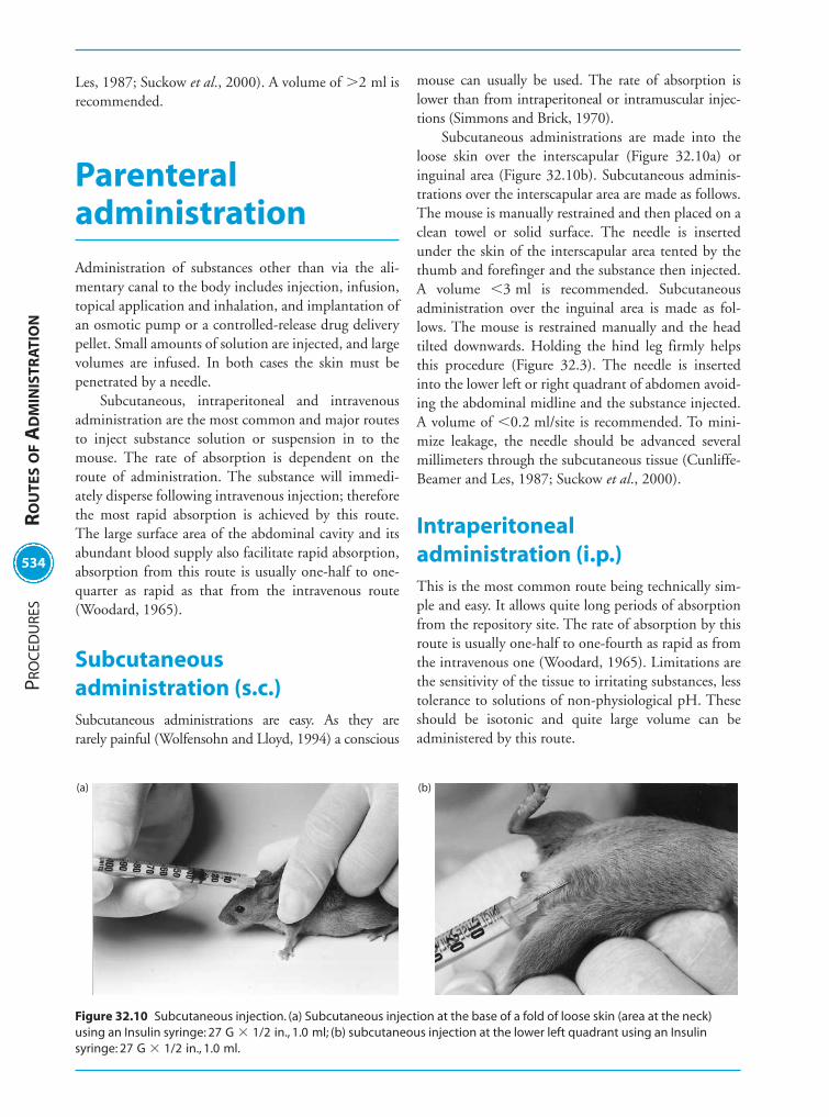
Subcutaneous administrations are made into theloose skin over the interscapular (Figure 32.10a) oringuinal area (Figure 32.10b). Subcutaneous adminis-trations over the interscapular area are made as follows.The mouse is manually restrained and then placed on aclean towel or solid surface. The needle is insertedunder the skin of the interscapular area tented by thethumb and forefinger and the substance then injected.A volume �3 ml is recommended. Subcutaneousadministration over the inguinal area is made as fol-lows. The mouse is restrained manually and the headtilted downwards. Holding the hind leg firmly helpsthis procedure (Figure 32.3). The needle is insertedinto the lower left or right quadrant of abdomen avoid-ing the abdominal midline and the substance injected.A volume of �0.2 ml/site is recommended. To mini-mize leakage, the needle should be advanced severalmillimeters through the subcutaneous tissue (Cunliffe-Beamer and Les, 1987; Suckow et al., 2000).
Intraperitoneal administration (i.p.)This is the most common route being technically sim-ple and easy. It allows quite long periods of absorptionfrom the repository site. The rate of absorption by thisroute is usually one-half to one-fourth as rapid as fromthe intravenous one (Woodard, 1965). Limitations arethe sensitivity of the tissue to irritating substances, lesstolerance to solutions of non-physiological pH. Theseshould be isotonic and quite large volume can beadministered by this route.
534
PRO
CED
URE
SR
OU
TES
OF
AD
MIN
ISTR
ATI
ON
(a) (b)
Figure 32.10 Subcutaneous injection. (a) Subcutaneous injection at the base of a fold of loose skin (area at the neck)using an Insulin syringe: 27 G � 1/2 in., 1.0 ml; (b) subcutaneous injection at the lower left quadrant using an Insulinsyringe: 27 G � 1/2 in., 1.0 ml.

The conscious mouse is manually restrained(Simmons and Brick, 1970) and is held in a supineposition with its posterior end slightly elevated or thehead can be tilted lower than the body (Figure 32.11).The needle and syringe should be kept almost parallelto its vertebral column in order to avoid accidentalpenetration of the viscera (Eldridge et al., 1982). Theneedle is pushed in at an approximately 10� anglebetween the needle and the abdominal surface in thelower quadrant of the abdomen (Simmons and Brick,1970). To avoid leakage from the puncture point, theneedle is run through subcutaneous tissue in a cranialdirection for 2–3 mm and then inserted through theabdominal wall (Cunliffe-Beamer and Les, 1987). Therecommended volume is �3.0 ml.
Intravenous administration (i.v.)Intravenous injection has advantages over other routes.Solutions at a high concentration, high or low pH orirritating can be administered intravenously providedthat the rate of injection is kept slow and precautions aretaken to avoid getting the solution outside the vein.Compounds that are poorly absorbed by the digestivetract may be given intravenously but intravenous admin-istrations require technical expertise and skill. Thesyringe plus needle or the catheter must first be filledwith the solution to remove air bubbles. Administrationsare usually made into the lateral tail veins not into thedorsal tail vein (Figure 32.12a), as it is not straight.
The lateral veins are readily visualized, but havequite small diameters. If anaesthesia is not used, arestraining device is usually necessary (see Chapter 31
on Handling and Restraint; Reeves et al., 1991;Suckow et al., 2000; Weiss et al., 2000).
The mouse is either placed in the restrainer or anes-thetized and the tail is then warmed with a lamp orwarm towel, or immersed in warm water (40–45�C) inorder to dilate the vessels (Flecknell, 1987). The tail isswabbed with 70% alcohol on a gauze sponge or swab.Insert the needle parallel to the tail vein penetrating2–4 mm into the lumen while keeping the bevel of theneedle face upwards (Figure 32.12b). The solution isthen injected slowly and no resistance should be felt ifthe solution is properly administered (Figure 32.12c).The injected solution temporarily replaces the blood butthen should be washed away by the blood stream. If thisdoes not happen the position of the needle is certainlynot in the vein but in the surrounding tissue so it mustbe moved in the surrounding tissue in such a way that itthen enters the vein or a new try must be made. Whenthe intravenous administration is finished or the cannulais pulled out, the injection site must be pressed firmlywith a swab or fingers to prevent backflow of the admin-istered solution and/or blood (Nebendahl, 2000;Suckow et al., 2000). If the same vein must be used sev-eral times the first administration should be made as dis-tal as possible in relation to the heart and subsequentadministrations should be placed progressively moreproximally. Because venipuncture and the administra-tion of substances can damage and/or block the vein, thedistal part of vein may no longer be used (Nebendahl,2000). The recommended volume is �0.2 ml.
The ophthalmic plexus route is also used for intra-venous administration (Pinkerton and Webber, 1964).The technique resembles the blood collection by retro-orbital sinus puncture (see Chapter 33 on Collection ofBody Fluids). The mouse is anesthetized, and thenmanually restrained on a solid surface being held gentlybut firmly by the nape of the neck. By pressing downwith the thumb and forefinger in the occipital area andpulling back the skin, the point of the needle can bedirected toward the back of the orbit at a 20–40� angle.The needle is inserted medially through the conjunc-tiva on the inner side of the ocular cavity. If entry isblocked by bone, the needle is withdrawn slightly(Figure 32.13a). Fluid is injected slowly loosening theskin slightly (Figure 32.13b). Also this route is usefulfor rescuing mice showing anaphylaxis by administra-tion of an isotonic solution.
Other routes for intravenous administration viathe external jugular vein (Kassel and Levitan, 1953), thedorsal metatarsal vein (Nobunaga et al., 1966) and thesublingual vein (Waynforth and Parkin, 1969) havebeen reported.
535
PRO
CED
URES
RO
UTES
OF
AD
MIN
ISTRA
TION
Figure 32.11 Intraperitoneal injection to lower leftquadrant using an Insulin syringe: 27 G � 1/2 in., 1.0 ml.

Intramuscular administration (i.m.)This should usually be avoided, as mouse muscles aresmall. If necessary, it may be given into the thigh musclewith injection volumes �0.05 ml. The tip of needle
should be directed away from the femur and sciaticnerve (Figure 32.14). The mouse is anesthetized or ismanually restrained by another person. The needle tipis inserted through the skin and into the muscle.Aspirate briefly with the syringe before injection. Ifblood or body fluid reverses, stop the procedure. The
536
PRO
CED
URE
SR
OU
TES
OF
AD
MIN
ISTR
ATI
ON
Turn 90° for injection
Left lateraltail vein
Dorsal vein
Right lateraltail vein
Skin
(a) (b)
(c)
Lateral tail vein
Needle
Coccygealvertebra
Ventral artery
Tendon bundles
Figure 32.12 (a) Transverse section view of the mouse tail; (b) sagittal view of the mouse tail (the tail is turned 90�);(c) intravenous injection into the lateral tail vein of an anesthetized mouse using an Insulin syringe: 27 G � 1/2 in., 1.0 ml.
Retroorvitalvenous plexus
Skull bone(osseous orbita)
ChoroidSclera
RetinaOptic nerve
Eyelash
Eyelid
CorneaLens
Needle and syringe
Vitreousbody
(b)(a)
Figure 32.13 (a) Sagittal view of the mouse eyeball and retro-orbital injection; (b) intravenous injection into the retro-orbital sinus of an anesthetized mouse using an Insulin syringe: 27 G � 1/2 in., 1.0 ml.

needle must be moved or a fresh attempt must bemade. Good technique and restraint are necessary andintramuscular administration should only be per-formed by well-trained personnel (Woodard, 1965;Cunliffe-Beamer and Les, 1987; Donovan and Brown,1991; Nebendahl, 2000).
Intradermal administrationThis route is not recommended in general and shouldbe restricted to cases of absolute necessity (Saloga et al.,1993; CCAC, 2002). It is very difficult in the mousedue to the very thin skin. Using a fine needle (29 G orsmaller) is recommended. The mouse is anesthetized,the fur clipped or hair removed from an area on theback, ventral abdomen, or hind footpad, which iswiped with 70% ethanol on a gauze sponge or swab.The skin is held tautly with thumb and index fingerand the needle inserted, bevel up and at a shallowangle, just under the superficial layer of epidermis. Thevolume should be �0.05 ml per site. Resistance shouldbe felt both as the needle is advanced and as the com-pound is injected. A hard bleb will be seen upon suc-cessful intradermal injection of even a small quantity offluid (Figure 32.15; Suckow et al., 2000). If multiplesites are injected, adequate separation is necessary toprevent coalescing of lesions.
Intracerebral administrationThis is made as follows (Prier, 1966; Liu et al., 1970).The mouse is anesthetized and then restrained manu-ally on a solid surface. The site of injection is approxi-mately half way between the eye and ear and just offthe midline (Figure 32.16a). The recommended maxi-mum volume per suckling mouse is 0.01 ml and that
for weanling or older mice is up to 0.03 ml. The needledirectly pierces the cranium (Figure 32.16b). An intra-dermal needle (Figure 32.7) is convenient in order toprevent the needle from extending too deeply into thebrain.
Intrathoracic administrationIntrathoracic injection is restricted to special experi-ments. It can be made in mice with a slightly bent orcurved needle, which should be inserted between theribs at approximately the midpoint of the rib cage.Caution must be taken to insert it at an angle, thus pre-venting injection directly into lung tissue. The speed ofabsorption is similar to the intraperitoneal route(Simmons and Brick, 1970).
Intranasal administration (i.n.)These are usually performed with the mouse lightlyanesthetized. The mouse is manually restrained and thetail anchored between the small finger and the palm(Simmons and Brick, 1970). The mouse is held in asupine position with the head elevated. The end of themicropipette is placed at or in the external nares, andthen the solution is poured in slowly (Figure 32.17;Prier, 1966; Shen et al., 2000, 2001). The volumeshould be �0.02 ml, excess volume or rapid injectionwill induce suffocation and death.
Topical applicationIt is not often realized that the skin is the largest organof the body and survival depends on its patency perhapsmore than for most other organs. An animal or man cansurvive with only about one-seventh of its liver or
537
PRO
CED
URES
RO
UTES
OF
AD
MIN
ISTRA
TION
Figure 32.14 Intramuscular injection into the leg muscle. Figure 32.15 Intradermal injection into the back skin.

one-fourth of its kidney functioning. In contrast, thedestruction of more than 50% of the skin usually resultsin death (Woodard, 1965). The skin is also a convenientsite for the administration of drugs. Numerous factors,such as the physicochemical properties of the substance,the attributes of the vehicle and the permeability of theskin, can affect the degree of percutaneous absorption.(Wester and Maibach, 1986; Franklin et al., 1989). Theability of a substance to be absorbed through the skinand enter the systemic circulation is determined by its
ability to partition into both lipid and water phases(Nebendahl, 2000).
The usual site is the skin covering the back or theabdomen. After clipping the hair for topical adminis-tration (Figure 32.4), the hairless area should becleaned from any fat and grease and other debris. Thesubstance should be dissolved in a volatile solvent ormixed in a suitable cream before application and thenapplied with a dropper or smeared onto the skin with aswab (Nebendahl, 2000). Some precautions are usuallynecessary to prevent the animal from licking or scratch-ing the application sites (Woodard, 1965).
InhalationThis route is used for experiments on asthma, air pollu-tion or respiration (Haddad el-B et al., 2002;Hopfenspirger and Agrawal, 2002). The inhalation routeincidentally is the nearest akin to an intravenous injectionbecause of the relatively large area presented for absorp-tion by a membrane that is separated from the blood byonly one or two cell layers. Consequently, absorptionof gases and aerosols that reach the alveoli is virtuallycomplete. The greatest problems surrounding the use ofthe inhalation route are the generation of a suitableaerosol of the test substance, if it is not sufficiently
538
PRO
CED
URE
SR
OU
TES
OF
AD
MIN
ISTR
ATI
ON
(a) Ear line Eye line (b)
Figure 32.16 Intracerebral injection. (a) Injection site of head for intracerebral injection; (b) intracerebral injection intoan anesthetized mouse using an intradermal needle.
Figure 32.17 Intranasal injection into an anesthetizedmouse using a pipette (Gilson P-20).

volatile, a constant and suitable air level of the materialunder study and the determination of the dosage given.Both small and large particle sizes are not adequate, it isgenerally believed that particle size of 0.5–2.0 m indiameter are optimum (Woodard, 1965). Equipment isavailable to purchase from Omuron, Kyoto, Japan; BuxcoElectronics, Inc., Sharon, USA among others.
Other routesOther routes of administration have been reportedsuch as intra-arterial administration using the femoralartery (Simmons and Brick, 1970) or the carotid artery(Sugano and Nomura, 1963), intrathymic injection(Donovan and Brown, 1991), or intraspinal injection(Habel and Li, 1951).
Dosing and treatment of new born mice providesspecial problems because of their size or that theirmother is apt to reject or cannibalize neonates that havebeen handled. Subcutaneous injections can be madeover the neck and shoulders using a less than30 G � 5/16 needle. Up to 0.1 ml (depending on theage of the infant mice) may be administered orallyusing a piece of plastic tubing inserted over a needle(Ujiie and Kobari, 1970; Cunliffe-Beamer and Les,1987). The direct injection in to the stomach of infantmice can be made through the abdominal wall (Deanet al., 1972). Intravenous injection of infant mice hasalso been reported (Anderson et al., 1959; Barnes et al.,1963; Cunliffe-Beamer and Les 1987).
Implantable pump, controlled-release drug delivery pelletand cannulasThe delivery of substances at a slow, steady rate over aperiod of days, weeks or months without the need forexternal connection or frequent animal handling canbe supplied by using an osmotic pump or controlled-release drug delivery pellets.
Osmotic pumps, ALZET pumps (Figure 32.18),can be used for systemic administration whenimplanted subcutaneously or intraperitoneally or canbe attached to a catheter for intravenous, intracerebralor intra-arterial infusion. The pumps have been used totarget delivery to a wide variety of sites including thespinal cord, spleen, liver, organ or tissue transplantsand wound healing sites. ALZET pumps are suppliedby DURECT Corporation (Cupertino, USA).
Controlled-release drug delivery pellets effectivelyand continuously release the active product in the ani-mal. The pellets are intended for, although not limitedto, simple subcutaneous implantation in laboratoryanimals. The pellets are available from InnovativeResearch of America (Sarasota, USA) and SouthernBioSystems, Inc. (Birmingham, USA).
Implantable cannulas permit continuous accessto the venous or arterial system for either intravenoussubstance administration or blood withdrawal. Usingstrict aseptic techniques, the cannula is inserted intoa vein or artery (the femoral vessels, jugular vein, and
539
PRO
CED
URES
RO
UTES
OF
AD
MIN
ISTRA
TION
Deliveredsubstances
Aqueousenvironment
Delivery portal
Removal cap
Flow moderator
Impermiablereservoir wall
Semipermiablemembrane
Reservoir
Osmotic agent
Flange
(a) (b)
Figure 32.18 Cross section of an Alzet®-osmotic pump, demonstrating (a) the design and (b) the working method.

carotid artery are common sites) and secured in place.The other end of the cannula is attached to a small portthat is secured in a subcutaneous location, most oftenover the shoulders (Suckow et al., 2000). See Desjardins(1986) for more information on implantable cannulas.
ImmunizationMice are not used for the production of polyclonal anti-bodies because the small amounts produced. On theother hand, mice are a good source of antibody produc-ing lymphoid cells or the production of hybridomas(Kohler and Milstein, 1975). In general, immunizationconsists of two stages; primary and booster. The primaryantigen is usually injected with adjuvant. Boosters areinjected once or more with/without adjuvant dependingon the immunogen. The footpad, intrasplenic (Nilsonand Larsson, 1992) or intra-lymph (Goudie et al., 1966)node injection is not recommended in general. Ifrequired the scientist/investigators should provide scien-tific justification to ethical committees for such proto-cols (such as the need to use extremely unique andirreplaceable antigens, or extremely small quantities ofantigen). The injection of immunogens at the base of thetail or in the popliteal area substitute for footpad injec-tions with much less distress to the animal becauseimmunogens injected into the footpad are processed bythe popliteal node. (Leenars et al., 1999; CCAC, 2002).The intraperitoneal route for injection of Freund’s com-plete adjuvant (FCA) is permitted in small rodents only.FCA should be administered only once, and be limitedto minimal volumes of up to 0.1 ml. In the mouse, up to0.1 ml with adjuvant may be administered in the neckregion subcutaneously. The injection of oil-based or vis-cous gel adjuvant should not be given by the intramus-cular route (CCAC, 1991). The intravenous routeshould not be used for oil-based adjuvants, viscous geladjuvants or large particle antigens due to the risk of pul-monary embolism (Herbert, 1978). Though FCA is the strongest adjuvant, use of other adjuvants can be
recommended. Mice must be closely monitored imme-diately following injection for any anaphylactic reac-tions, both after the primary and any booster injections(CCAC, 2002). The recommended route and volumesare shown in Table 32.2. Mice showing anaphylaxis willrecover by the administration of the isotonic solution byOpthalmic plexus route (see this chapter on intravenousadministration).
AcknowledgmentWe are grateful to Mr. Andoh Y. for his skilfulphotography.
ReferencesAnderson, N.F., Delorme, E.J., Woodruff, M.F.A. and
Simpton, D.C. (1959). Nature, 1952–1953.Barnes, D.W.H., Ford, C.E. and Harris, J.E. (1963).
Transplantation 1, 574.Baumans, V., ten Berg, R.G.H., Bertens, A.P.M.G.,
Hackbarth, H.J. and Timmermann, A. (1993). In Principlesof Laboratory Animal Science, (eds L.F.M. van Zutphen,V. Baumans and A.C. Beynen), p. 389. Elsevier, Amsterdam.
CCAC (The Canadian Council on Animal Care) (1991).CCAC Guidelines on Acceptable Immunological Procedures.CCAC, Ottawa. http://www.ccac.ca/english/gui_pol/policies/IMMUNO.HTM
CCAC (The Canadian Council on Animal Care) (2002).Guidelines on Antibody Production, pp. 1–40. CCAC,Ottawa.
Cunliffe-Beamer, T.L. and Les, E.P. (1987). In The UFAWHandbook on the Care and Management of LaboratoryAnimals, 6th edn (ed. T. B. Poole), pp. 275–308.Longman Scientific and Technical, Essex.
Dean, A.G., Ching, Y.C., Williams, R.G. and Harden, L.B.(1972). J. Infect. Dis. 125, 407–411.
540
PRO
CED
URE
SR
OU
TES
OF
AD
MIN
ISTR
ATI
ON
TABLE 32.2: Maximum volume (in milliliters) of administration of antigen with or without adjuvant per route
Subcutaneous Intramuscular Intraperitoneal Intravenous Intradermal
with adjuvant 0.1 not recommended 0.2 no not recommended
without adjuvant 0.5 0.05 1.0 0.2 0.05
ID route should be restricted to the cases where it is absolutely necessary (CCAC, 2002).
Source: van Zutphen et al., 1993; Iwarsson et al., 1994.

Desjardins, C. (1986). In Research Surgery and Care of SmallLaboratory Animals Part A. Patient Care, Vascular Access,and Telemetry (eds W.I. Gay and J.E. Heavner), p. 143–194. Academic Press, Orlando.
Donovan, J. and Brown, P. (1991). In Current Protocol inImmunology (eds Coligan et al.) pp. 1.4.1–1.4.4. JohnWiley & Sons, New York.
Eldridge, S.F., McDonald, K.E., Renne, R.A. and Lewis, T.R.(1982). Lab. Anim. 11, 50–54.
ETS 123 (1986). European Convention For The Protection ofVertebrate Animals used for Experimental and OtherScientific Purposes. Council of Europe, Strasbourg.
Flecknell, P.A. (1987). In Laboratory Animals: AnIntroduction for New Experimenters (ed. Tuffery), pp.225–260. John Wiley & Sons, Chichester, England.
Franklin, C.A., Somers, D.A. and Chu, I. (1989). J. Am. Col.Toxicol. 8, 815–827.
Goudie, R.B., Home, C.H. and Wilkinson, P.C. (1966).Lancet 7475, 1224–1226.
Habel, K. and Li, C. P. (1951). Proc. Soc. Exp. Biol. Med. 76,357–361.
Haddad, el-B., Underwood, S.L., Dabrowski, D., Birrell, M.A.,McCluskie, K., Battram, C.H., Pecoraro, M., Foster,M.L. and Belvisi, M.G. (2002). J. Immunol. 168,3004–3016.
Herbert, W.J. (1978). In Handbook of ExperimentalImmunology, 3rd edn (ed. D.M. Weir), pp. A3, 1–3, 15.Blackwell Scientific Publications, Oxford.
Hopfenspirger, M.T. and Agrawal, D.K. (2002). J. Immunol.168, 2516–2522.
Iwarsson, K., Lindberg, L. and Waller, T. (1994). In Handbookof Laboratory Animal Science, Vol. 1 (eds P. Svensen andJ. Hau), pp. 229–272. CRC Press, Boca Raton.
Kassel, R. and Levitan, S. (1953). Science 118, 563–564.Köhler, G. and Milstein, C. (1975). Nature 256,
495–497.Leenars, M.P.P.A., Hendriksen, C.F.M., De Leeuw, W.A.,
Carat, F., Delahaut, P., Fischer, R., Halder, M., Hanly, W.C.,Hartinger, J., Hau, J., Lindblad, E.B., Nicklas, W.,Outschoorn, I.M. and Stewart-Tull, E.S. (1999). AlternativesLab. Anim. 27, 79–102, http://altweb.jhsph.edu/publications/ECVAM/ecvam35.htm
Levine, R.R. (1970). Am. J. Dig. Dis. 15, 171–188.Liu, C., Voth, D.W., Rodina, P., Shauf, L.R. and
Gonzalez, G. (1970). J. Infect. Dis. 122, 53–63.Nebendahl, K. (2000). In The Laboratory Rat (ed.
G. Krinke), pp. 463–483. Academic Press, London.Nilson, B.O. and Larsson, A. (1992). Res. Immunol. 143,
553–557.Nobunaga, T., Nakamura, K. and Imamichi, T. (1966). Lab.
Anim. Care 16, 40–49.Prier, J. E. (1966). In Basic Medical Virology (ed. J. E. Prier),
pp. 38–77. The Williams & Wilkins Company, Baltimore.Pinkerton, W. and Webber, M. (1964). Proc. Soc. Exp. Biol.
Med. 116, 959–961.
Reeves, J.P., Reeves, P.A. and Chin, L.T. (1991). In CurrentProtocol in Immunology (eds. Coligan et al.), pp.1.6.1–1.6.9. John Wiley & Sons, New York.
Saloga, J., Renz, H., Lack, G., Bradley, K.L., Greenstein, J.L.,Larsen, G. and Gelfand, E.W. (1993). J. Clin. Invest. 91,133–140.
Shen, X., Lagergard, T., Yang, Y., Lindblad, M., Fredriksson,M. and Holmgren, J. (2000). Infect. Immun. 68,5749–5755.
Shen, X., Lagergard, T., Yang, Y., Lindblad, M., Fredriksson, M.and Holmgren, J. (2001). Infect. Immun. 69,297–306.
Simmons, M.L. and Brick, J.O. (1970). In The LaboratoryMouse (ed. A. Hollaender), pp. 127–129. Prentice-HallInc., Englewood Cliffs.
Silverman, J. (1987). In Laboratory Hamsters (eds G.L. VanHoosier and C.W. McPherson), pp. 72–75. AcademicPress, Orland.
Suckow, M.A., Danneman, P. and Brayton, C. (2000). InThe Laboratory Mouse (ed. Suckow), pp. 120–125. CRCPress, Boca Raton.
Sugano, S. and Nomura, S. (1963). Bull. Exp. Anim. (Jap.Eng. Abstract) 12, 1–5.
Ujiie, A. and Kobari, K. (1970). J. Infect. Dis. 121,s50–s55.
van Zutphen, L.F.M., Baumans, V. and Beynen, A.C.(1993). In Principles of Laboratory Animal Science (edsL.F.M. van Zutphen, V. Baumans and A.C. Beynen),p. 389. Amsterdam, Elsevier.
Waynforth, H. B. and Parkin, R. (1969). Lab. Anim. 3,35–37.
Waynforth, H. B. and Flecknell, P. A. (1992). InExperimental and Surgical Technique in the Rat (eds H. B.Waynforth and P. A. Flecknell), pp. 1–67. AcademicPress, London.
Weiss, J., Taylor, G. R., Zimmermann, F. and Nebendahl, K.(2000). In The Laboratory Rat (ed. G. Krinke),pp. 485–510. Academic Press, London.
Wester, R.C. and Maibach, H. I. (1986). In Progress in DrugMetabolism, Vol. 9 (eds J.W. Bridges and L.F. Chasseaud),pp. 95–109. Taylor and Francis, London.
Wolfensohn, S. and Lloyd, M. (1994). In Handbook ofLaboratory Animals Management and Welfare (edsS. Wolfensohn and M. Lloyd), pp. 143–173. OxfordUniversity Press, Oxford.
Woodard, G. (1965). In Methods of Animal Experimentation, Vol.1 (ed. W.J. Gay), pp. 343–359. Academic Press, New York.
Additional informationBecton Dickinson, Franklin Lakes, USA: http://www.bd.comBuxco Electronics, Inc., Sharon, USA: http://www.buxco.com/index.html
541
PRO
CED
URES
RO
UTES
OF
AD
MIN
ISTRA
TION