the karyotype and ultrastructural characteristics of spontaneous preimplantation mouse parthenotes
TRANSCRIPT

Gamete Research 9:451-467 (1984)
The Karyotype and Ultrastructural Characteristics of Spontaneous Preimplantation Mouse Parthenotes Everett Anderson, Peter C. Hoppe, and Gloria S. Lee
Department of Anatomy and Laboratory of Human Reproduction and Reproductive Biology, Harvard Medical School, Boston, Massachusetts (E.A., G.S. L.) and The Jackson Laboratory, Bar Harbor, Maine (P. C. H.)
Karyotypic and light and electron microscopical analyses were made of spontaneous preimplantation mouse parthenotes from the LT/Sv inbred strain. It was found that the activated oocyte and developing embryos were diploid. We believe that diploidization is achieved by the oogonium undergoing a premeiotic mitosis without cytokinesis followed by two meiotic divisions, thus producing diploid parthenotes.
The developmental events with respect to membrane specialization, such as junctional complexes, were similar to those observed in fertilized embryos.
A unique feature of the developing parthenote was the failure of the mitochondria to change during the morula stage. The mitochondria retained a few irregularly oriented cristae rather than many transversely oriented ones observed in morulae developing from fertilized eggs. The significance of this observation is discussed.
Key words: karyotype, parthenote, meiosis, mitochondria
INTRODUCTION
According to Beatty [1967], “Parthenogenesis is the production of an embryo from a female gamete without any genetic contributions from the male gamete and with or without eventual development into an adult. ” The first indication that parthen- ogenesis may be a natural phenomenon was the observation of deGreef [cf Wilson, 19281 in the starfish Asterias glacialis. Since then parthenogenesis has been noted in other invertebrates and vertebrates. In mammals, as in other animals, many chemical and physical agents have been used to parthenogenetically activate eggs, including salt and sugar solutions; hyaluronidase; anesthetics; pricking; mechanical shock; electricity; heat and cold shock; Ca’ + ions; and calcium ionophore, A23 187 [Balak- ier and Tarkowski, 1976; Cuthbertson et al, 1981; Chang, 1954; Guylas, 1976; Kaufman, 1975, 1976; Komar, 1973; Pincus, 1936; Pincus and Shapiro, 1940; Steinhardt et al, 1974; Uehara and Yanagimachi, 1977; also see Surane and Kaufman,
Received June 24, 1983; accepted January 17, 1984.
Address reprint requests to Everett Anderson, Dept. of Anatomy and Laboratory of Human Reproduction and Reproductive Biology, Harvard Medical School, 45 Shattuck St., Boston, MA 02115.
0 1984 Alan R. Liss, Inc.

452 Anderson, Hoppe, and Lee
19771. The rationale for identifying the various artificial inducing parthenogenetic agents was to discover common denominators between the mechanisms employed by those physical and chemical agents that induce activation and those utilized by sperm.
It is well known that in the guinea pig, hamster, and ferret, oocytes undergo spontaneous parthenogenetic activation [Chang, 1957; Longo, 1974; Pincus, 19361. Moreover, investigators correctly identified within the ovary cleavage stages of embryos that developed parthenogenetically [Austin, 1949; Dempsey, 19391. Further- more, ovarian teratomas are known to be produced by the parthenogenetic activation and subsequent development of the ovarian oocyte in mouse and man [Stevens and Varnum, 1974; Linder et al, 19751. In 1974, Stevens and Varnum found in the LT/Sv inbred strain that approximately 50% of these mice have ovarian teratomas by the time they are 90 days old. These teratomas are the results of parthenogenetic devel- opment of ovarian oocytes. Furthermore, when the LT/Sv mice ovulate, about 10% of the ovulated eggs undergo spontaneous parthenogenetic development. The parthen- otes of this strain develop to the blastocyst stage and implant in the uterus, and are reabsorbed within a few days.
The description of parthenogenesis in the LT strain produced much excitement among geneticists and embryologists, for in addition to the possibility of having a natural model to analyze the initial events of oocyte activation without the intervention of the sperm, investigators could also use the parthenote to study maternal gene function during early development. We have designed experiments dealing with oocyte activation and gene action. However, before any of the aforementioned studies can be made, it is necessary to analyze the karyotype and the ultrastructural features of the preimplantation parthenote. Moreover, we believe that the process by which the LT parthenotes achieve diploidy is unlike other mammalian material so as to warrant presentation of the observations contained in this communication.
MATERIALS AND METHODS To study ovarian oocytes after the resumption of meiosis I, 40 females were
injected intraperitoneally (IP) with 4 IU of pregnant mare’s serum (PMS) (Organon) in 0.2 ml saline at 4:OO PM, and 48 hours later with 4 IU human chorionic gonadotro- pin (HCG) (Sigma) in 0.2 ml saline. The animals were killed at 9, 10, 10’15, 11, 11 %, l l % , 1134, 12, 12%, or 13 hours after HCG injection. The ovaries were fixed in Kale’s solution, embedded in paraffin, sectioned at 6 pm and stained with haemotoxy- lin and eosin [Humason, 19621.
Collection of Activated Oocytes and Embryos
Adult LT females were superovulated as previously described. Nonactivated oocytes and activated eggs and embryos were studied under both in vivo and in vitro conditions at various times after ovulation. For the in vitro methodology see Hoppe and Pitts [ 19731.
For light and electron microscopy, all embryos (nonactivated and activated oocytes; two-four-eight-cell embryonic stages; early and mid-morulae, and early and late blastocysts) from both in vivo and in vitro conditions were fixed in a 3% glutaraldehyde-0.5 % paraformaldehyde mixture contained in a 0.1-M cacodylate buffer (pH 7.4). After fixation for 40 minutes at room temperature, the materials were postfixed in 1% cacodylate buffered osmium (pH 7.4) for 1 hour. Some

Spontaneous Preimplantation Mouse Parthenotes 453
specimens were impregnated with lanthanum. For lanthanum impregnation of em- bryos, the same fixation procedure was followed except that both the fixative and buffer wash solutions (buffered to pH 7.4) contained 2% lanthanum nitrate and the tissues were incubated in 0.03 N NaOH for about 1 hour prior to osmication [Albertini and Anderson, 19741. Further rapid processing of embryos was accomplished as follows: The specimens were processed in a Syracuse-type watchglass (Thomas Scientific App. No. 9790-N12) and covered with a 13-cm-diameter7 round perforated screen (Perforated Products, Brookline, MA) that fits into the watchglass concavity. The 75 pm holes in the screen (Mesh No. 203LPI with 32.5% open area) allowed fluids to be changed easily above the screen and exchange to occur with the solution below containing the embryos. In order to insure thorough mixing of the layers at the beginning of dehydration, several changes of 30% ethanol were made initially. Embryos were processed in the previously stated manner until 1:l propylene oxide and Epon was achieved.
For all preparations for electron microscopy, about 15-20 samples were made with each containing apprpximately 2-3 specimens. Thin sections (many of the activated oocytes were serially sectioned) were cut from each block with either a Porter-Blum MT- 1 or 2-ultramicrotome, stained with uranyl acetate, followed by a lead stain [Sato, 19681, and examined with a Philips 300 electron microscope. Semi- thin sections were also made and stained with 0.1 % toluidine blue containing 1 % sodium borate.
Karyotypic Analysis
Slides were cleaned by soaking in HCL: methanol (1:l) for 3 to 5 days, rinsed several times in tap water, followed by five rinses in distilled water. Slides were stored at 5°C in ethyl ether: methanol (1:l). Prior to use, the slides were dried by wiping the solvents off with Kimwipes.
Approximately 1 hour before collection of embryos at various developmental stages, 0.1 ml of an aqueous 0.5 % colchicine solution was injected IP into the embryo donors. Occasionally, parthenogenetic embryos from noninjected females were cul- tured for 2 to 3 hours in culture medium containing 2 pg/ml demecolcine (Sigma, St. Louis). Embryos were placed in 1 ml of a 1 .O % sodium citrate solution in a depression slide for 10 to 15 minutes at room temperature before being fixed by the air-dry method of Tarkowski [1966]. After the slides were thoroughly dried, the chromo- somes were stained with either Giemsa or by the chromosomal G-banding method of Davisson et al [1981]. Slides of the embryos were scanned and metaphases were photographed to facilitate karyotyping . Pronuclear Volume
In an effort to learn something about the pronuclear volume of spontaneous and experimentally activated mouse eggs, the following was done: Oocytes from LT/Sv or C57BL/6J (B6) inbred mice were parthenogenetically activated by incubation in 8% ethanol in culture medium for 5 minutes, washed in fresh medium, and cultured [Cuthbertson et al, 19811. We activated the eggs at 9:OO AM, so the pronuclei development was similar to spontaneous LT parthenotes. Pronuclear volume was measured in those eggs with a second polar body and a single haploid pronucleus. At around 3:OO PM, the eggs from the three groups were photographed (ie, spontaneously activated LT, ethanol-activated LT, and ethanol-activated B6). We used B6 since we

454 Anderson, Hoppe, and Lee
could be assured of no spontaneous activation. Negatives were projected and the pronuclear volume was measured by use of an Optomax IV Image Analyzer (Opto- max, Inc., NH).
OBSERVATIONS
Parthenotes cultured in vitro underwent the same morphogenetic changes and at comparable times as those observed after growth in vivo. The temporal development of parthenotes in culture was very similar to our observation from culturing in vivo and in vitro fertilized oocytes [Anderson et al, 19751.
Karyotype Analysis
Seventy-four metaphases were obtained from 36 LT parthenote embryos in developmental stages ranging from two cells to 28 cells (Table 1 and Fig. 1). Those cases where fewer than 40 chromosomes were counted can probably be attributed to the loss of individual chromosomes during spreading. Metaphases were observed to be haploid, and we conclude that the LT parthenote embryo is diploid. These results agree with the previously reported finding of Moustafa [ 19781. The possible mecha- nism by which the activated LT oocyte derives its diploidy is discussed below.
Pronuclear Volume
As shown in Table 2, the pronucleus of spontaneously activated eggs is double the volume of ethanol-activated LT or B6 haploid eggs, demonstrating that sponta- neous LT oocytes must be diploid after meiosis.
Activated lntraovarian Oocytes
For those animals injected with 4 IU of PMS followed by HCG, paraffin sections were examined for the presence of chromatin in the first poIar body. Approximately 3,150 ovarian sections were examined from the group fixed 12 hours post HCG. In these sections, we examined 37 oocytes and each had first polar bodies with chromatin. Another 43 oocytes had chromosomes aligned on the metaphase plate, and in some oocytes chromosomes were condensed.
It is interesting to note that animals that had been killed 12% hours following the HCG injection had ovaries with many two- four-, and eight-cell parthenogenetic embryos.
Activated LT Oocytes
The second polar body was associated with the main oocyte via a bridge that contained numerous microtubules and some mitochondria (Fig. 2; also see 2-PB in inset). The microtubules were imbedded in a dense granular matrix of the mid-body (MB), the nature of which is unknown. The plasmalemma in the region between the
TABLE 1. Karyotypes of LT Parthenote Embryos
No. chromosomes No. embryos No. karyotypes 37 38 39 40
36 74 4 8 12 50

Fig. 1. Karyotype of LT spontaneous parthenogenetic embryo.

456 Anderson, Hoppe, and Lee
TABLE 2. Pronuclear Volume of Spontaneous Parthenogenetic LT Eggs and Experimentally Activated Parthenogenetic LT and B6 Mouse Eggs
Pronuclei Experimental group No. eggs Avg. radius (p) Avg. vol. (p3) Range
Spontaneous LT 12 11.72 6,740.9 4,601.8-8,58 1.2 Exp. Activated LT 19 9.24 3,302.8 2,2 18.6-5,696.4 Exp. Activated B6 10 8.94 2,997.0 2,187.94,532.4
protrusion and the main ooplasmic mass is thrown into many slender projections (Fig. 2, SP). Eventually, the second polar body becomes separated from the oocyte and appears as a diminutive cell with many of the organelles that appeared in the oocyte.
At the time of activation, the contents of some cortical granules are released. We say “some” are released since in serial sections we noticed many fewer cortical granules when compared with nonactivated oocytes [see Guylas, 1974; Solter et al, 19741.
The plasma membrane of the activated LT one-cell egg is similar to that of the oocyte prior to formation of polar bodies, ie, its surface is adorned with many microvilli. At the two-cell stage, the blastomeres are rather spherical (Fig. 3, inset), still adorned with many slender microvilli except where the two appose each other, in which case they interdigitate with each other (Fig. 3, MV). In the four-eight-cell stage embryos, the blastomeres remain rather spherical with large intercellular spaces between them and many microvilli on their surface. The presence of microvilli during the development from the oocyte to the four-eight-cell stage may play a role in increasing the physiologically active surface area of blastomeres.
During the eight-cell stage, there is a change in the shape of the blastomeres from spherical to somewhat wedge-shape and the embryo becomes compacted [Du- cibella et al, 19751. In lanthanum-impregnated specimens, the tracer appears to stop at the apical surfaces. This is the first junctional membrane specialization, namely the tight junction (Fig. 5 , TJ). This junction has been interpreted as being the first sign for the future segregation of the inner cell mass and presumptive trophoectoderm [Ducibella et al, 19751.
Figure 4 reveals another membrane specialization between the surface cells in late morulae. This figure was made from-lanthanum impregnated material. This junction, the gap junction, is subjacent to the tight junction (Fig. 5 , TJ). Cells comprising the trophoectoderm are also held together by desmosomes (Fig. 6D).
Organelles Mitochondria. In the nonactivated, activated, one- and 2-cell stages, the mito-
chondrial matrix is somewhat dense and contains some peripherally located cristae. All mitochondria of the four-eight-cell embryo, morula, and blastocyst are rather pleomorphic and contain vacuole-like inclusions (Fig. 7M). It is interesting to point out that in a previous investigation of mice and rabbits, the majority of mitochondria of morulue and blustocysts obtained from fertilized eggs contain transverse cristae
Fig. 2. An electron micrograph through the midbody (MB) (also see inset). Note the second polar body (2-PB inset), mitochondria (M), microtubules (MT), and slender cytoplasmic projections (SP). X5,206; inset: toluidine blue stained, X500.

Spontaneous Preimplantation Mouse Parthenotes 457
458 Anderson, Hoppe, and Lee
with no distinct vacuolelike inclusions. As shown in Figures 8 and 9 (M), the majority of the mitochondria of the morulae and blastocysts of LT parthenotes possess rather “feeble” cristae. In all of the many observations made, mitochondria of the morulae and blastocysts possess the same cristae configuration.
Endoplasmic reticulum. Scattered in the cytoplasm are large numbers of cisternae of endoplasmic reticulum, many of which are associated with mitochondria. In some instances, the cisternae have associated with them a few ribosomal particles.
In some of the profiles of the endoplasmic reticulum are some dense spherical structures (Fig. 7, SV). In other profiles, there are some dense plaques on the cisternae (Fig. 7, DP). The plaques appear to be the initial stage in the formation of the dense spherical structures. Presumably these dense plaques become internalized by inpocketing of the membrane. It is difficult to identify precisely these intracisternal units. Several investigators have identified such units [Calarco, 1975; Bicyzsko et al, 1973, 19741 as intracisternal A particles (IAP). The IAP particles are included in the taxonomy of oncogenic RNA virus [Dalton et al, 19661. The dense units in the present study appear similar to the IAPs observed by other investigators.
Other organelles and nonorganelles were similar to those described previously. During the development of the embryo, it was noted that large accumulations of glycogen appeared in the cytoplasm of not only blastomeres, but also of cells of the inner cell mass and trophoblast (Fig. 5, GP).
DISCUSSION
The in vivo and in vitro analyses of the LT preimplantation parthenotes have revealed that the spontaneously activated oocyte undergoes morphogenetic activities similar to the naturally activated (ie, fertilized) mammalian oocyte. Our chromosomal analyses indicated that the spontaneously activated oocyte and the subsequent devel- oping embryo are diploid. Previous reports using enzyme analyses of parthenogenet- ically derived teratomas concluded that diploidy of LT parthenotes arise from germ cells that have completed the first meiotic division but do not complete the second meiotic division [Eppig et al, 1977; Eicher, 1978; Eppig and Eicher, 19831. However, our morphological observations demonstrate that two meiotic divisions occur in LT parthenogenetic oocytes and both polar bodies are extruded and contain chromatin. The second polar body persists and can be observed in preimplantation stage parthen- ogenetic embryos similar to the second polar body of fertilized embryos. Pronuclear volume of spontaneously activated LT eggs is twice that of haploid parthenogenetic eggs and demonstrates that LT parthenotes are diploid after activation and completion of meiosis. We tentatively propose that diploid LT parthenotes develop from oogonia in which a premeiotic mitosis without cytokinesis occurs [Mittwoch, 19781 with a diploid parthenogenetic egg being produced after the completion of meiosis. Exam- ples of the occurrence of a premeiotic endoreduplication in the reproduction of parthenogenetic lizards has been discussed [Uzzell, 1970; Hardy and Cole, 198lJ. It is interesting to note that in mammals, under normal conditions, ie, at fertilization, there is no 2N uninucleated zygote since there is no fusion of the male pronucleus.
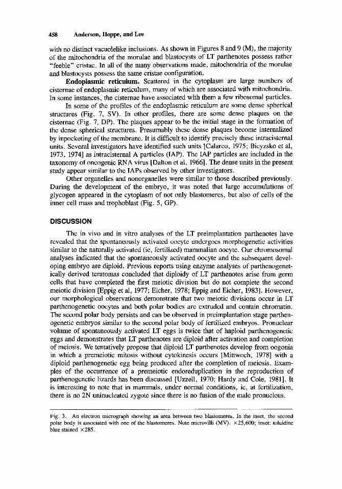
Fig. 3. An electron micrograph showing an area between two blastomeres. In the inset, the second polar body is associated with one of the blastomeres. Note microvilli (MV). X25,600; inset: toluidine blue stained X285.

Spontaneous Preimplantation Mouse Parthenotes 459

460 Anderson, Hoppe, and Lee
Other possibilities for achieving diploidization have been offered for partheno- genetically activated embryos. Some investigators envision that metaphase II is accomplished without the production of a second polar body. Therefore, the oocyte possesses two pronuclei, one of which is formed by the chromosomes that were destined to form the second polar body [Kaufman, 1973; Graham and Deussen, 19741. On the other hand, some evidence has been presented where a single pronu- cleus develops from metaphase II. In other words, the female pronucleus and that of the second polar body are never separated [Chang, 1954; Graham and Deussen, 19741. Balakeir and Tarkowski [ 19761 produced parthenogenetic embryos by heat shock, which was followed by suppression of the second polar body with cytochalasin B, and the resulting parthenotes were diploid. It should be pointed out that our studies show that both polar bodies are present.
During this study, no unique features were noted during the development of the parthenote other than the observation that the majority of the mitochondrial population of the morulae and blastocysts did not contain an abundance of transversely oriented cristae. Van Blerkom and Runner [1976] activated the oocytes in noninbred color stocks of mice using electricity and hyaluronidase. They found that their parthenotes (morulae and blastocysts) contained vacuolated mitochondria but were somewhat more elongated with transversely oriented cristae than earlier stages. The organization of the mitochondria of the morulae and blastocysts of the LT strain was unlike that of equivalent embryonic stages of in vivo and in vitro fertilized eggs [Anderson et al, 1970, 1975; Stern et al, 19761. It should be pointed out that under the aforementioned conditions, mitochondria commence their transformation at the morula stage, ie, from a population with few irregularly oriented cristae to one with many transversely organized cristae. In fact, it has been shown that the morula is an important stage during mammalian embryogenesis since it is at this stage that not only are new biochemical events inaugurated, but the first signs of detectable regionalization are realized. That is to say, the outer layers of cells become the presumptive trophoblast and the inner cells the presumptive inner cell mass [Ducibella et al, 1975, also see Brinster, 19731.
The second genetic system of the cell, namely mitochondria, have self-perpetu- ating genetic functions; they possess their own complement of DNA that regulates, in part, the synthesis of mitochondrial protein [also see Birky, 19831. There is also good evidence from studies utilizing sea urchins and amphibia that the number of mito- chondria increases during oogenesis and that a large amount of cytoplasmic DNA is in the mitochondria [Piko et al, 1967; Chase and Dawid, 19721. The changes in the internal morphology of mitochondria during mammalian embryogenesis, as indicated above, could be due to either a morphological change within individual mitochondria or by the replacement of one generation of mitochondria with another. In connection
Fig. 4. A small portion of a lanthanum impregnated late morula showing a gap junction (GJ). X60,OOO.
Fig. 5 . A small portion of a lanthanum-impregnated blastocyst showing a tight junction (TJ). Note glycogen particles (GP). x 120,OOO.
Fig. 6. A section between two trophoblast cells of the blastocyst showing a desmosome (D). X70,OOO.

Spontaneous Preimplantation Mouse Parthenotes 461

462 Anderson, Hoppe, and Lee
with this, Cascio and Wassarman [1981] studied the synthesis of mitochondrial proteins during oogenesis and early embryogenesis in the mouse. These authors concluded that “ . . . the mitochondrial transformations accompanying oocyte growth and early cleavage in the mouse are under nucleocytoplasmic control but are not attributable to the turning on and off of mitochondrial gene expression. ”
It is well known that investigators have been unable to produce viable pups from either spontaneously activated LT oocytes or experimentally activated mouse oocytes. The outstanding question is “why.” Could the lack of the production of viable pups be due to the lack of a new generation of mitochondria during the critical stage of development, ie, the morula? Dawid and Blackler [1972] concluded that in Xenopus leavis, mitochondria synthesized by the egg are sufficient to supply the needs of the embryo for about 2 days of development [also see Chase and Dawid, 19721. Nothing is known for mammals. Moreover, we have no evidence to answer the proposed question as to why viable pups are not produced from spontaneously activated LT oocytes. However, our observations prompt us to speculate about the paucity of mitochondria with many transversely oriented cristae in parthenogenetic morulae and blastcysts. Since a few mitochondria show more cristae than others, it is possible that cells whose mitochondria contain many more cristae are capable of further differentiation if they are found in an appropriate environment. If this be the case, it may explain why in a teratoma one finds some highly differentiated tissues but not an organized embryo [Stevens and Varnum, 19741. Moreover, what we have speculated above may explain the results obtained by Stevens et al [1977]. These authors made the interesting observation that parthenogenetic cells are capable of differentiating normally in combination with normal cells.
Much evidence exists that the biosynthesis of functional mitochondria demands the cooperation of two genetic systems, that of the nucleus and that of the mitochon- drion. We usually consider the normal diploid condition as being sufficient to produce a viable organism. It should be pointed out that the parthenote’s diploidy was not achieved by sperm-oocyte genomic association. Therefore, the proper gene interac- tion may not be available for the de novo synthesis of a new population of mitochon- dria or the transformation of the old ones. The aforementioned statement brings up the significance of the contents of the sperm for ongoing development [also see Surani and Barton, 19831. Very little is known concerning the significance of the various components of the sperm, other than its nucleus, to the support of normal embryonic differentiation. In a study by Anderson and Perotti [ 19751 dealing with the fate of the sperm mitochondrion during fertilization in seas urchins, they noted that frequently the sperm mitochondrion was in contact with many egg mitochondria [also see Anderson, 19681. Anderson and Perotti believed that “ . . . this transient contact may be all that is needed for the transference of key substances between membranes of the contiguous organelles. ”
Some investigators have argued that one of the reasons why the implanted parthenote dies derives from the fact that its genes are from one parent, and therefore express lethal recessive genes. That this may not be the case gains support from the investigations of Hoppe and Illmensee [ 19821. These investigators produced offspring after transplantation of nuclei from inner-cell mass cells of LT parthenogenic blasto- cysts into fertilized enucleated mouse eggs. It is obvious that further study of the oocytes and the developing parthenote at both molecular and physiological levels is necessary for a better understanding of sperm-oocyte interactions leading to viable development.

Fig. 7. A section through an activated one cell embryo. Note dense plaques (DP), dense spherical structures (SV), and mitochondria (M). x80,OOO.

464 Anderson, Hoppe, and Lee
Figs. 8 and 9. of the trophoblast. Note mitochondria (M). Figure 8, X48,000; Figure 9, ~ 8 0 , 0 0 0 .
Figure 8 is a section through a compacted morula and Figure 9 is a section through cells

Spontaneous Preirnplantation Mouse Parthenotes 465
ACKNOWLEDGMENTS
This investigation was supported by grants from NIH USPHS (HD06645, Center Grant; HD14574 to E.A. and HD10381 to P.C.H.). The authors would like to thank Dr. Muriel T. Davisson and Ellen A. Akeson for preparation of karyotypes and Mrs. Jeanne Baumgartner for her patience and skill in preparing this manuscript.
Mice used in this study were maintained in accordance with the guidelines of the Committee on Animals of the Harvard Medical School and the Jackson Labora- tory, and those prepared by the Committee on Care and Use of Laboratory Animals of the Institute of Laboratory Animal Resources, National Research Council (DHEW publication No. NIH78-23, revised 1978). The Jackson Laboratory is fully accredited by the American Association for the Accreditation of Laboratory Animal Care.
REFERENCES
Albertini D, Anderson A (1974): The appearance and structure of the intercellular connections during the ontogeny of the rabbit ovarian follicle with particular reference to gap junctions. J Cell Biol
Anderson E, Condon W, Sharp D (1970): A study of oogenesis and early embryogenesis in the rabbit, Oryctalugus cuniculus, with special reference to the structural changes of mitochondria. J
Anderson E, Hoppe PC, Whitten WK, Lee GS (1975): I n vitro fertilization and early embryogenesis: A
Anderson W (1968): Structure and fate of the paternal mitochondrion during early embryogenesis of
Anderson W, Perotti ME (1975): An ultracytochemical study of the respiratory potency, integrity, and
Austin CR (1949): The fragmentation of eggs following induced ovulation in immature rats. J Endocrinol
Balakier H, Tarkowski AK (1976): Diploid parthenogenetic mouse embryos produced by heat-shock and cytochalasin B. J Embryo1 Exp Morphol 35:25-39.
Beatty RA (1967): Parthenogenesis in vertebrates. In Metz CB, Monroy A (eds): “Fertilization,” Vol 1. New York: Academic Press, p 413.
Biczysko W, Pienkowski M, Solter D, Korpowski H (1973): Virus particles in early mouse embryos. J Natl Cancer Inst 51: 104-1050.
Biczysko W, Solter D, Graham C, Koprowski H (1974): Synthesis of endogenous type A virus particles in parthenogenetically stimulated mouse eggs. J Natl Cancer Inst 52:483-489.
Birky CW Jr (1983): Relaxed cellular controls and organelle heredity. Science 222:468-475. Brinster RL (1973): Parental glucose phosphate isomerase activity in three-day mouse embryos. Biochem
Calarco PG (1975): Intracisternal A particle formation and inhibition in preimplantation mouse embryos.
Cascio SM, Wassarman P (1981): Program of early development in the mammaI: Synthesis of mitochon-
Chang MC (1954): Development of parthenogenetic rabbit blastocysts induced by low temperature
Chang MC (1957): Natural occurrence and artificial induction of parthenogenetic cleavage of ferret ova.
Chase JW, Dawid IB (1972): Biogenesis of mitochondria during Xenopus leavis development. Dev Biol
Cuthbertson KSR, Whittingham DG, Cobbold PH (1981): Free CAZ+ increases in exponential phases
631234-250.
Movhol 130167-92.
cytological analysis. J Ultrastruct Res 50:231-252.
paracentrotus lividus. J Ultrastruct Res 24:311-321.
fate of the sea urchin sperm mitochondria during early embryogenesis. J Cell Biol66:367-376.
6: 104-110.
Genet 9:187-191.
Biol Reprod 12:448-454.
drial proteins during oogenesis and early embryogenesis in the mouse. Dev Biol 83: 166-172.
storage of unfertilized ova. J Exp Zoo1 125: 127-150.
Anat Rec 128: 187-200.
27 :504-5 18.
during mouse oocyte activation. Nature 294:754-757.

466 Anderson, Hoppe, and Lee
Dalton kl, deHarven E, Dmochowski L, Feldman D, Haguenau F, Harris WW, Howatson AF, Moore D, Pitelka D, Smith K, Uzman B, Zeigel R (1966): Suggestions for the classification of oncogenic RNA viruses. J Natl Cancer Inst 37:395-397.
Davisson MT, Poorman PA, Roderick TH, Moses MJ (1981): A pericentric inversion in the mouse. Cytogenet Cell Genet 30:70-76.
Dawid IB, Blackler AW (1972): Maternal and cytoplasmic inheritance of mitochondria1 DNA in Xenopus. Dev Biol29: 152-161.
Dempsey EW (1939): Maturation and cleavage figures in ovarian ova. Anat Rec 75:223-236. Ducibella T, Albertini DF, Anderson E, Biggers JD (1975): The preimplantation mammalian embryo:
Characterization of intercellular junctions and their appearance during development. Dev Biol
Eicher EM (1978): Murine ovarian teratomas and parthenotes as cytogenetic tools. Cytogenet Cell Genet
Eppig JT, Eicher EM (1983): Application of the ovarian teratoma mapping method in the mouse.
Eppig JJ, Kozak LP, Eicher EM, Stevens LC (1977): Ovarian teratomas in mice are derived from
Graham CF, Deussen ZA (1974): In vitro activation of mouse eggs. J Embryol Exp Morphol 31:497-
Gulyas BJ (1974): Cortical granules in artificially activated (parthenogenetic) rabbit eggs. Am J Anat
Guylas BJ (1976): Ultrastructural observations on rabbit, hamster and mouse eggs following electrical
Hardy LM, Cole CJ (1981): Parthenogenetic reproduction in lizards: Histological evidence. J Morphol
Hoppe PC, Illmensee K (1982): Full-term development after transplantation of parthenogenetic embry-
Hoppe PC, Pitts S (1973): Fertilization in vitro and development of mouse ova. Biol Reprod 8:420-426. Humason GL (1962): “Animal Tissue Techniques.” San Francisco: WH Freeman and Co. Kaufman MH (1975): Parthenogenetic activation of mouse oocytes following avertin anaesthesia. J
Kaufman MH (1973): Timing of the first cleavage division of haploid eggs, and the duration if its
Kaufman MH, Barton SC, Surani MAH (1976): Normal postimplantation development of mouse
Komar A (1973): Parthenogenetic development of mouse eggs activated by heat-shock. J Reprod Fertil
Linder D, McCaw BK, Hecht F (1975): Parthenogenic origin of benign ovarian teratomas. N Eng J Med
Longo FJ (1974): An ultrastructural analysis of spontaneous activation of hamster eggs aged in vitro.
Mittwoch U (1978): Parthenogenesis. J Med Genet 15:165-181. Moustafa LA (1978): Parthenogenic mammalian cells cloning in mouse. Genetics 88:S70-S71. Piko L, Tyler A, Vinograd J (1967): Amount, location, priming capacity, circularity and other properties
of cytoplasmic DNA in sea urchin eggs. Biol Bull 132:68-90. Pincus G (1936): “The Eggs of Mammals.” New York: MacMillan. Pincus G, Shapiro H (1940): Further studies on the parthenogenetic activation of rabbit eggs. Proc Natl
Sat0 T (1968): A modified method for lead staining of thin sections. J Electron Microsc 17:158-159. Solter D, Biczysko W, Graham C, Pienkowski M, Koprowski H (1974): Ultrastructure of early
Steinhardt RA, Epel D, Carrol EJ, Yanagimachi R (1974): Is calcium ionophore a universal activator
Stern S, Biggers JD, Anderson E (1976): Mitochondria and early development of the mouse. J Exp Zool
Stevens LC, Varnum DS (1974): The development of teratomas from parthenogenetically activated
45:231-250.
20:232-239.
Genetics 103 : 797-8 12.
oocytes that have completed the first meiotic division. Nature 269517-518.
512.
140:577-582.
stimulation in vitro. Am J Anat 147:203-218.
170x215-237.
onic nuclei into fertilized mouse eggs. Proc Natl Acad Sci 79: 1912-1916.
Embryol Exp Morphol33:941-946.
component stages. J Cell Sci 13:553-566.
parthenogenetic embryos to the forelimb bud stage. Nature 265:53-55.
35:433-443.
292: 63-66.
Anat Rec 179:27-56.
Acad Sci 26: 163-165.
development of mouse parthenogenomes. J Exp Zool 188: 1-24.
for unfertilized eggs? Nature 252:41-43.
176: 179-192.
ovarian mouse eggs. Dev Biol37:369-380.

Spontaneous Preirnplantation Mouse Parthenotes 467
Stevens LC, Varnum DS, Eicher EM (1977): Viable chemaeras produced from normal and parthenoge- netic mouse embryos. Nature 269:515-517.
Surani MAH, Kaufman MH (1977): Influence of extracellular Caz’ and Mg2+ ions on the second meiotic division of mouse oocytes: Relevance to obtaining haploid and diploid parthenogenetic embryos. Dev Biol59:86-90.
Surani MAH, Barton SC (1983): Development of gynogentic eggs in the mouse: Implications for parthenogenetic embryos. Science 222: 1034-1036.
Tarkowski AK (1966): An air-drying method for chromosome preparation from mouse eggs. Cytogenet- ics 5:394-400.
Uehara T, Yanagimachi R (1977): Activation of hamster eggs by pricking. J Exp Zool 199:269-274. Uzzell T (1970): Meiotic mechanisms of naturally occurring unisexual vertebrates. Am Naturalist
Van Blerkom J, Runner MN (1976): The fine structure development of preimplantation mouse parthen-
Wilson EB (1928): “The Cell in Development and Heredity.” New York: MacMillan.
104:433-445.
otes. J Exp Zool 196:113-124.