the journal of the lebanese dental association · the journal of the lebanese dental association...
TRANSCRIPT

The Journal of the Lebanese Dental AssociationOfficial organ of the Lebanese Dental AssociationFormer titles: Lebanese Dental Magazine/Lebanese Dental Journal/Revue Dentaire Libanaise
The Journal of the Lebanese Dental Association -JLDA- is a multidisciplinary peer-reviewedjournal published biannually (June and December) by the Lebanese Dental Association. JLDA hasan ultimate aim of introducing and improving research in contemporary aspects of dental andcraniofacial basic and clinical sciences. The JLDA publishes manuscripts on all aspects of dentalmedicine and surgery, including surgical dentistry, restorative and prosthetic dentistry, geriatricand pediatric dentistry, periodontology and implant dentistry, endodontics, esthetic and cosmeticdentistry, adhesive dentistry, orthodontics and dentofacial orthopedics, oral biology, oral andmaxillofacial surgery, oral diagnosis/pathology/medicine, dental research, oral and maxillofacialradiology and imaging, public health and community dentistry, special care/needs dentistry,forensic odontology, and social dentistry. Dentistry related fields are broadly defined and mayinclude, for instance, facial growth/embryology, dental and orofacial genetics, orofacial anti-agingand esthetic medicine, dental and maxillofacial tissues engineering, medically compromisedpatients treated in dental practice, temporomandibular disorders and orofacial pains, sleepmedicine in relation to dental practice, clinical trials of drugs relevant to dental/craniofacialpractice, dental/oral histopathology, immunopathology and microbiology, dental anesthesiology,dental ergonomics, computer-assisted dentistry, aeronautic and veterinary dentistry.
Statements and opinions in the papers and communications herein are those of the author(s) andnot necessarily of the editors and the editorial advisory board. The editors disclaim anyresponsibility or liability for such material and do not guarantee, warrant, or endorse any productadvertised in their publication, nor do they guarantee any claim made by the manufacturer of suchproduct or service.
All rights are reserved and no part of this publication (JLDA) may be reproduced or stored ina retrieved form or transmitted in any other form without prior permission of the copyright owner(Lebanese Dental Association, Beirut, Lebanon).
Address: Lebanese Dental Association, Victoria Tower, 2nd floor,Corniche du Fleuve, Beirut, Lebanon
Tel/Fax: 00961-1-611222 / 00961-1-611555E-mail: [email protected] Website: www.LDA.org.lb
Layout and Printing: Al Matni Printing PressBeirut, LebanonTel: 00961-1-283631E-mail: [email protected]
JLDA June 2015:JLDA 9/25/15 10:55 AM Page 1
The Journal of the Lebanese Dental AssociationOfficial organ of the Lebanese Dental AssociationFormer titles: Lebanese Dental Magazine / Lebanese Dental Journal
The Journal of the Lebanese Dental Association -JLDA- is an open access multidisciplinary journal, published biannualy (June and December) by the Lebanese Dental Association. JLDA has an ultimate aim of introducing and improving research in contemporary aspects of dental and craniofacial basic and clinical science. The JLDA publishes manuscripts on all aspects of dental medicine and surgery, including surgical dentistry, restorative and prosthetic dentistry, geriatric and pediatric dentistry, periodontology, implant dentistry, endodontics, esthetic and cosmetic dentistry, adhesive dentistry, orthodontics and dentofacial orthopedics, oral biology, oral and maxillofacial surgery, oral and maxillofacial diagnosis/pathology/medicine, dental research, oral and maxillofacial radiology and imaging, public health and community dentistry, special care dentistry, forensic dentistry, social dentistry, neuromuscular dentistry, preventive dentistry, and public health dentistry. Dentistry related fields are broadly defined, as well, and may include, cranio-facial anatomy and physiology, facial growth/embryology, dental and orofacial genetics, orofacial anti-aging and esthetic medicine, dental and maxillofacial tissues engineering, medically compromised patients treated in dental practice, temporomandibular disorders and orofacial pains, sleep medicine in relation to dental practice, clinical trials of drugs relevant to dental practice, dental/oral histopathology, immunopathology and microbiology, dental anesthesiology, dental ergonomics, computer-assisted dentistry, aeronautic and veterinary dentistry.The JLDA is a peer-reviewed scientific publication. Submitted manuscripts are reviewed by independant clinicians and scientists who are fully conversant with current dental literature, and who maintain the highest standards of competence, objectivity, and integrity.The JLDA welcomes original research papers, review papers, case reports, technical notes, case series, expert opinions, technical notes, clinical comments, pictorial essays, and conference proceedings. Redundant and duplicate publications will not be considered for review.Statements and opinions in the papers and communications herein are those of the author(s) and not necessarily of the editors and the editorial advisors. Editors disclaim any responsibility or liability for such material and do not guarantee, warrant, or endorse any product advertised in their publication, nor do they guarantee any claim made by the manufacturer of such product or service.All rights are reserved and no part of this publication (JLDA) may be reproduced or stored in a retrieved form or transmitted in any other form without prior permission of the copyright owner (Lebanese Dental Association, Beirut, Lebanon).
Publisher: The Lebanese Dental Association - LDA, Beirut, LebanonEditor-in-Chief: Ziad E.F. Noujeim
Address: Lebanese Dental Association, Victoria Tower, 2nd floor, Corniche du Fleuve, Beirut, Lebanon Tel/Fax: 00961-1-611222 / 00961-1-611555 E-mails: [email protected] and [email protected] / Mobile: 00961-3-483983Website: www.LDA.org.lb
Online Manager: Fadi S. Kayyali
Layout and Printing: Matni Easy Printing Beirut, Lebanon +961 1 489900 / +961 3 302136 [email protected]

2
Editor-in-SubchiefMichel Goldberg, Chir. Dent., Dr. Odont. Sc., Dr. Natural Sc., PU,Emeritus Professor, Saints-Pères Biomedical College, INSERM/Unité 747-Equipe 5, Paris Descartes University, Paris, [email protected]
Elie M. Ferneini, B.Sc., MS Health Sciences, DMD,MD, MHS, MBA/HCM, FACS, FACDAssistant Clinical Professor, Oral and Maxillofacial Surgery Division, Department of Craniofacial Sciences, University of ConnecticutSchool of Dental Medicine, Farmington, Connecticut,Clinical Instructor, Department of Surgery, Division of GeneralSurgery, University of Connecticut School of Medicine, Farmington,Connecticut, [email protected]
Hani F. Ounsi, Dr. Chir. Dent., DES Endo.,M.Sc. Dental Mat., DEA Oral Biol., FICD, Ph.D.,Lecturer, Department of Endodontics, Lebanese University,School of Dentistry, Beirut,Visiting Professor, Department of Endodontics and Restorative Dentistry,Tuscan School of Dental Medicine, University of Siena, [email protected]
Tony Daher, Dr. Chir. Dent., CES Fixed Prostho.,CES Remov. Prostho., PG Cert. Prostho., MS (Education), FACP, FICP, FICD, MAO,Diplomate, American Board of Prosthodontics,Clinical Associate Professor, Department of Restorative Dentistry,Former Director of Advanced Prosthodontics Program,Loma Linda University, School of Dentistry, Loma Linda, California, [email protected]
Tara Renton, BDS, M. Dent. Sc., FRACDS,FDSRCS (Engl.), Ph.D., ILTM, FHEA,Consultant Oral Surgeon, Professor and Head, Department of OralSurgery, King’s College London -KCL- Dental Institute, London, UK,[email protected]
Zeina A.K. Majzoub, Dr. Chir. Dent., DMD, Dott. Odont.,CAGS, M.Sc.D,Former Professor of Periodontology and Research, University ofPadova, Institute of Clinical Dentistry, Padova, Italy,Professor of Periodontology and Research and FormerChairperson, Department of Research, Lebanese University,School of Dentistry, Beirut,[email protected]
Karine Feghali, BDS, DESS Perio., Ph.D.,Associate Professor, Department of Periodontics,Research Scientist, Oral Ecology Research Group (GREB),Laval University, Faculty of Dental Medicine, Quebec City, [email protected]
Editor-in-ChiefZiad E.F. Noujeim, Dr. Chir. Dent., CES Oral Biol., CES Surg. Dent., Dipl. Oral Pathol., DU Cell Therapy, DIU Anti-Aging/Esthetic Medicine, DU Oral Dermatology, FICD, FACOMS, FIAOMS, Diplomate, European Board of Oral Surgery,Senior Lecturer and Postgraduate Tutor, Departments of Oral and Maxillofacial Surgery, Oral Pathology and Diagnosis, Basic Science,Director, Oral Pathology and Diagnosis Postgraduate Program,Former Director, Oral Surgery Postgraduate Program,Lebanese University, School of Dentistry, Beirut, Atending Oral and Jaw Surgeon, Baabda University Hospital, Baabda, LebanonFormer Scientific Chairperson, Lebanese Dental Association, [email protected]
Ziad Salameh, Dr. Chir. Dent., CES Prostho., M.Sc.,Ph.D., HDR, FICD,Chairperson, Department of Research,Associate Professor, Departments of Prosthodontics andResearch, Lebanese University, School of Dentistry, Beirut,Adjunct Associate Professor, Center for Craniofacial Regeneration,University of Pittsburgh, Pittsburgh, [email protected]@drziadsalameh.com
Charles Sfeir, Dr. Chir. Dent., CES Perio., MS, Ph.D.,Associate Dean for Research,Associate Professor and Chairperson, Department of Periodontics and Preventive Dentistry, University of Pittsburgh, School of Dental Medicine, Pittsburgh, [email protected]
Assem Soueidan, Dr. Chir. Dent., CES Perio.,DU Perio., DU Oral Rehab./Implant., DU OralDermatology, DEA, Dr. Univ., HDR, PU, PH,Professor and Chairperson, Department of Periodontology,Nantes University, Faculty of Dental Surgery, Nantes, [email protected]
Sami Mouwakdié, Dr. Chir. Dent., DU Perio.,DU Implant., DEA Génie Biologique, MBA,Assistant Professor, Department of Periodontology,Lebanese University, School of Dentistry, Beirut,[email protected]
Fadl Khaled, BDS, CES Endo., DES Endo.,Adjunct Clinical Assistant Professor, Department of RestorativeSciences, Beirut Arab University, Faculty of Dentistry,Chief of Clinical Services, Department of Endodontics,Lebanese University, School of Dentistry, [email protected]
Senior Associate Editors
Journal of the Lebanese Dental AssociationVolume 49 - Nº 2 - December 2014

3
Associate EditorsMarcel Noujeim, BDS, DESS Oral Biol., DESS OralRadiol., MS (Dent. Diagnostic Sc.),Diplomate, American Board of Oral/MaxillofacialRadiology,Associate Professor, Department of Comprehensive Dentistry,Division of Oral and Maxillofacial RadiologyDirector of Oral and Maxillofacial Radiology Postgraduate Program,University of Texas Health Science Center at San Antonio -UTHSCSA, [email protected]
Radhouane Dallel, Dr. Chir. Dent., Dr. Univ., HDR, PU, PH,Senior Scientist and Director, Neurobiology of Trigeminal Pain/Migraine Laboratory, NEURO-DOL, INSERM/UdA, U1107,Professor, Clermont 1 University, Faculty of Dental Surgery,Clermont-Ferrand, [email protected]@u-clermont1.fr
Pascale Habre Hallage, Dr. Chir. Dent., CES Oral Biol.,CES Fixed Prostho., DUICP, MSBM, DEA Neurosc.,Dr. Biomed. Sc.,Assistant Professor and Director of Postgraduate Program,Department of Fixed Prosthodontics and Occlusion,Saint-Joseph University, Faculty of Dental Medicine,Beirut, [email protected]@usj.edu.lb
Roula Abiad, BDS, MS Endo., Dr. Dent. Sc.,Assistant Dean, Assistant Professor of Endodontics,Director, Division of Endodontics,Beirut Arab University, Faculty of Dentistry, Beirut, [email protected]@gmail.com
Maroun F. Dagher, Dr. Chir. Dent., CAGS Perio.,M.Sc.D. Oral Biol., MS Research, MAAP, MAO, MIADR,Diplomate, American Board of Periodontology,Senior Lecturer and Clinical Tutor, Department of Periodontology,Saint-Joseph University, Faculty of Dental Medicine,Beirut, [email protected]@aol.com
Jaime S. Brahim, DDS, MS, FACOMS, FAAOMS,Diplomate, American Board of Oral and MaxillofacialSurgery, American Board of Oral Medicine,Clinical Professor, Department of Oral and Maxillofacial Surgery,Director, Clinical Research Unit, University of Maryland Schoolof Dentistry, Baltimore, Maryland,Former Senior Oral and Maxillofacial Surgeon, National Institutesof Health-NIH, Bethesda, Maryland, [email protected]
Mary Ann Jabra-Rizk, BS, Ph.DAssociate Professor, Department of Oncology and DiagnosticSciences, University of Maryland School of Dentistry, Baltimore,Maryland, [email protected]
Rima Abdallah, BDS, CAGS, D.Sc. Oral Biol.,Diplomate, American Board of Periodontology,Adjunct Clinical Assistant Professor, Periodontology andImplantology Divisions, Oral Surgical Sciences Department,Beirut Arab University, Faculty of Dentistry, Beirut, [email protected]
Carla Zogheib Moubarak, Dr. Chir. Dent., DES Endo.,MS Oral Biomat. Res., Dr. Univ.,Assistant Professor, Department of Endodontics,Saint-Joseph University, Faculty of Dental Medicine,Beirut, [email protected]@gmail.com
Chimène Chalala, BDS, DESS Ortho., ResidencyOrtho. AUB,Clinical Associate, Division of Orthodontics and DentofacialOrthopedics, American University of Beirut Medical Center,Associate Chief of Clinical Services, Department of Orthodontics,Lebanese University, School of Dentistry, Beirut,[email protected]
Zoubeida Yahfoufi Al Hage, DDS, Dr. Dentistry, FITI,Practice limited to Periodontology and Implant Dentistry,Beirut, [email protected]
Journal of the Lebanese Dental AssociationVolume 49 - Nº 2 - December 2014

4
Editorial AdvisorsEssam Osman, BDS, MSD, Ph.D.Vice-President for Medical Sciences, Beirut Arab University,Dean, Faculty of Dentistry,Chairperson, Departments of Restorative Sciences and OralSurgical Sciences, and Director, Division of Dental Biomaterials,Professor of Dental Materials, Faculty of Dentistry, [email protected]@bau.edu.lb
Antoine F. Saadé, DCD, CES Oral Biol., CES Ortho., DU Ortho., CECSMO, Chief of Clinical Services and Former Chairperson,Department of Orthodontics, Lebanese University, School of Dentistry, Beirut, Lebanon,[email protected]
Nadim Z. Baba, DMD, MSD, FICD, FACP, Diplomate, American Board of Prosthodontics,Professor, Department of Restorative Dentistry, Director, Hugh Love Center for Research and Education in Technology, Loma Linda University, School of Dentistry, Loma Linda, California, USAAssociate Editor, Journal of Dental Traumatology, Director, Pacific American College of Prosthodontists,[email protected]
Maria E. Saadeh, BDS, MS Human Morphology,Residency Ortho. AUB,Clinical Associate, Division of Orthodontics and DentofacialOrthopedics, American University of Beirut Medical Center,Clinical Instructor, Department of Orthodontics,Lebanese University, School of Dentistry, Beirut,[email protected]@aub.edu.lb
Leila Chahine, DMD, FAADSM,Diplomate, American Board of Dental Sleep Medicine,Dental Staff, Danbury Hospital, Western Connecticut HealthNetwork, Connecticut, [email protected]
Biostatistics and Epidemiology ConsultantsM Fouad Ziadé, Ph.D Biostat., C. Stat., FRSS, MASA,MIEAAssociate Professor, Lebanese University, Faculty of PublicHealth, Beirut/Tripoli, [email protected] / [email protected]
Nada E. El-Osta, DCD, DES Prostho., MS (Biol. Med.Sc.), DIU Biostat., DU Forensic ScienceConsultant in Biostatitics / Epidemiology,Saint-Joseph University, Faculties of Medicine and DentalMedicine, Beirut, [email protected] / [email protected]
Antoine Berbéri, BDS, MS, DU Perio,CES Odont. Chir., Dr. Univ., HDR, FICD,Diplomate, European Board of Oral Surgery,Professor and Former Chairperson,Department of Oral and Maxillofacial Surgery,Lebanese University, School of Dentistry, Beirut,Research Scientist, Doctoral School of Sciences and Technology,Lebanese University, Beirut,[email protected]
Joseph Bouserhal, Dr. Chir. Dent., Specialist Ortho., DURCO, Ling. Ortho. Dip., Dr. Sc. Dent., MWFO, MAAO,Professor and Former Chairperson, Department of Orthodontics, Saint-Joseph University Faculty of Dental Medicine, Beirut, Adjunct Clinical Professor of Orthodontics, Boston University Henry M. Goldman School of Dental Medicine, Boston, USAPast President, Lebanese Orthodontic Society, Beirut, Lebanon,Instructor, Tweed Foundation for Orthodontic Research, USA,Visiting Professor in Orthodontics, Beirut Arab University, Universities of Paris V and VII (France), Aristotle University of Thessaloniki (Greece), and Paul Sabatier University of Toulouse (France)
Georges Tawil, Dr. Chir. Dent., DDS, CES Odont.Chir., CES Perio., Dr.Sc.Odont., FICD, FACDFormer Professor and Chairperson, Department of Periodontology, Saint-Joseph University, Faculty of Dental Medicine, Beirut, Lebanon,Editorial Consultant, International Journal of Oral andMaxillofacial Implants, Clinical Oral Implant [email protected]
Ghassan Yared, DCD, DSO, FRCD (Can.), MRCDSOFormer Associate Professor, Department of Endodontics, andformer Director of Endodontics undergraduate program,University of Toronto, Faculty of Dentistry, Toronto, [email protected]
Dina Debaybo, Dr. Chir. Dent., CAGS, M.Sc.D.,Diplomate, American Board of Pediatric Dentistry,Clinical Associate Professor of Pediatric Dentistry,European University College, Dubai, [email protected]
Nabil Tabbara, DMD, FAAFO, FAACPAdjunct Clinical Professor, University of Western Ontario, SchulichSchool of Medicine and Dentistry, London, Ontario, [email protected]
Sukumaran Anil, BDS, MDS, Ph.D., FICD, FPFAProfessor and Consultant, Division of Periodontics, King SaudUniversity, College of Dentistry, Riyadh, KSA,[email protected]
Hani Adbul Salam, B.Sc., BDS, M.Sc., Ph.DAdjunct Professor and Director of Continuing Dental Educationfor the Middle East and North Africa, McGill University, Facultyof Dentistry, Montreal, [email protected]
Editors EmeritiPhilippe E. Aramouni, DCD, DEA, CAGS Prostho., M.Sc.D., FICDProfessor Nadim Z. Baba, DMD, MSD, FICD, FACP, Diplomate, American Board of ProsthodonticsMichel Salameh, DCD, CES Pediat. Dent., MIADP, MIADH, MSFOPAssistant Professor Georges Aoun, BDS, DES Oral Biol., DES Prostho., DSOAssociate Professor Antoine Cassia, DCD, CES Oral Biol. Bucc., CES Surg. Dent., DU Maxillofacial Prostho., DSOProfessor Levon Naltchayan, DCD, CES Prostho.Pierre Souaid, DCD
Journal of the Lebanese Dental AssociationVolume 49 - Nº 2 - December 2014

5Journal of the Lebanese Dental AssociationVolume 49 - Nº 2 - December 2014
EditorialShifting Paradigms in Oral Surgery Map: Facts, Fictions, and Fallacies.
Ziad E.F. Noujeim
The effect of irrigation with EDTA on calcium-based root canal sealers: A SEM-EDS and XRD study.Randa Harik, Ziad Salameh, Roland Habchi, Josette Camilleri
Potential contribution of dental pulp stem cells to pulp and dentin repair in response to tooth injury: A preliminary study.
Yassine Harichane, Sasha Dimitrova-Nakov, Odile Kellermann, Michel Goldberg
Fluoride in dentistry: use, dosage, and possible hazards.Mounir Doumit, Mohammad Omar Machmouchi, Hicham Diab
Full-mouth rehabilitation of a severely worn dentition: A case report.Farah Antar, Pascale Habre Hallage, Elie Zebouni
Molar-Incisor-Hypomineralisation (MIH) in Lebanon: A preliminary clinical observation.Mary Antoun, Paul Nahas, Assaad Nasr, Hicham Mansour
Pediatric dental management of patients with von Willebrand disease: reporting the cases of two brothers.Nada R. Chedid, Jean-Claude Abou Chedid, Zeina Akil
Internal root resorptions: current state of understanding. Valérie Batrouni, Chadi Torbay, Jad Azzi, Hassan El-Husseini
Endodontic non-surgical retreatment: how to remove a silver point and a post? A case report.Cynthia Antoine Kamel, Carla Zogheib Moubarak
The forgotten Bichat’s Buccal Fat Pad: A practical note for surgical dentists and oral surgeons.Maissa Aboul Hosn, Ziad E.F. Noujeim
Usefulness of Cone Beam Computed Tomography (CBCT) in the removal of a separated endodontic file in an aberrant root canal morphology: an unusual case report.
Maysoun Ahmad
JLDA Guide for contributors and authors
Volume 49 - N0 2 - December 2014
6
12
24
32
37
42
49
58
64
69
75
79

6
Shifting Paradigms in Oral Surgery Map: Facts, Fictions, and Fallacies.
"As goes your technology and your skills, so goes your practice"Robert E. Marx and Diane Stern
The term "surgery" is derived from greek "chir" (hand) and "ergos" (work): according to this etymology,surgery is a hands-on clinical work,but in daily clinical practice, this word's meaning is restricted to practical therapeutic acts,such as those involving soft tissues incisions, flap raisings, osteotomies, reconstructions, repairing, and dressing living tissues.
Contemporary oral surgery encompasses maxillary sinus membrane lifts,onlay and inlay bone grafts,placement of dental osseointegrated implants,dento-alveolar surgery (including simple exodontia, surgical extraction of impacted teeth and teeth-like structures, surgical endodontics, and minor orthodontic surgery), incision and drainage of cellulitis, surgical management of (odontogenic and non-odontogenic) jaw cysts and tumors,management of pathological conditions and lesions of oral mucosa, surgical removal of intra-oral salivary calculi, management of oral lacerations and oro-antral communications. But despite these different specific titles, limits of oral surgery are not yet well defined and may reach "maxillofacial" surgery,a term that implies a greater scope of surgical interest,like TMJ surgery,orthognathic surgery,maxillo-facial trauma,and surgery of oro-facial malignant tumors.
General dentists are ethically required to undertake, only, surgical treatments of teeth, teeth-like structures,and soft tissues surrounding teeth, and the UK General Dental Council defined "Surgical Dentistry" ("Odontologie Chirurgicale" in France) as "those surgical procedures within the mouth, which would normally be accomplished for a cooperative patient under local anaesthesia, with or without sedation, in a tolerably short operating time".
The last three decades witnessed a significant progress in the specialty of oral surgery, and specifically in the fields of surgical implantology, LASER-assisted oral surgery, and surgical oro-facial pathology. Oral Surgery is rapidly evolving, and oral surgeons, worldwide, are hardly coping with revolutionary changes. Emergence of new technologies is permitting the improvement of our surgical knowledge,and new insights on oral surgical and pathological discoveries are providing us with broader and deeper view of teeth, osseointegrated implants, maxillary sinus, salivary glands, and jawbones.
CURRICULUM VITAE
December 2012
ZIAD E. F. NOUJEIMOral Surgeon / Dental Educator
Oral Surgery, Oral Medicine,
Dental Implants, Facial Anti-Aging & Esthetic Medicine
Diplomate, European Board of Oral Surgery
Fellow, International College of Dentists
Fellow, American College of Oral and MaxillofacialSurgeons
Ziad E.F.Noujeim,Editor-in-Chief, JLDA
Journal of the Lebanese Dental AssociationVolume 49 - Nº 2 - December 2014

7Journal of the Lebanese Dental Association
Prophylactic extraction of third molars is still regarded as a public health hazard (JW Friedman, 2007): third molar surgery is a multi-billion dollar industry and it is driven by misinformation and myths that continue to be promulgated by the dental profession (JW Friedman, 1983). Indeed, much controversy surrounds the indications of removal, especially around the so-called "orthodontic indication". Retrospective studies indicate a lack of correlation between mandibular third molars and post-retention incisor crowding and there is still no evidence to incriminate third molars (particularly mandibular ones) as being the only, or even the major,etiological factor in the post-treatment changes in incisor alignment (SE Bishara, 1999). Early this year (April 2014), a Saudi-American study came to an interesting conclusion on the role of third molars in the development of anterior teeth crowding: Indeed, KH Zawawi (of King Abdul Aziz University, Jeddah, KSA) and M Melis (of Tufts University, Boston, USA) conducted a systematic review, and in assessing the quality of the 12 selected studies, they discovered a high risk of bias in most of reviewed articles, and many of the so-called "controlled" studies included in their study, obtained "inadequate" and "unclear" scoring during the quality assessment procedure. The only study that scored"adequate"in all the items,reported no association between presence of third molars and anterior teeth crowding. And in the light of evidence addressed in the Zawawi and Melis review, extraction of third molars to prevent anterior teeth crowding or post-orthodontic relapse is not supported until proven otherwise by further well designed studies. In this regard, the National Institutes of Health-NIH-Consensus Development Conference (held on November 1979) recommended that impacted third molars be removed when there is evidence of pericoronitis, follicular enlargement, resorption of adjacent teeth, or irreversible pathological changes around the third molar (cyst,tumor,etc...): at that time,the 200 participants couldn't reach a consensus on the removal of asymptomatic impacted third molars with no evidence of pathology (WC Guralnick and D Laskin, in J Oral Surg, 1980).
As of the early 90s, delineation of risks and benefits of third molar removal was "the" topic of discussion in Asia, Europe, and North America, and, during this period, most oral surgeons agreed that prophylactic removal of wisdom teeth was unjustified (Mercier and Precious, 1992). In 1993, a British study (Brickley, Sheperd, and Mancini, in Brit Dent J) compared clinical treatment decisions in the UK, with US/NIH 1979 Consensus indications for mandibular third molar removal: this audit demonstrated that 20-30% of mandibular third molars were removed without apparent NIH Consensus indication for surgery.
In the USA, 10 million wisdom teeth, classified as impactions, are removed every year from mostly healthy young individuals (ADA survey, 1999): these teeth are extracted from approximately 5 million people, each year, at an annual cost of over 3 billion US dollars. In addition, more than 11,000 people suffer permanent anesthesia of lip, tongue, or cheek, as a consequence of nerve injury during third molar surgery.
Nowadays, a primary objective must be to rigorously define the indications for third molar removal and,then,to adhere to them,and common sense, worldwide, suggests that an operation should only be performed when it results in health gain: which of us would have his/her appendix removed, only because it MAY cause trouble in the future?!!! The place of ANY prophylactic surgical operation remains highly questionable,treatment decisions have important cost implications, as well, and unnecessary surgeries should be strictly avoided.In the USA, removal of pathology-free third molars is considered indefensible by the Courts in many areas.
In March 2000, The UK National Institute for Clinical Excellence -NICE- published an appraisal guidance on wisdom teeth removal, a guidance that would help dentists and oral surgeons to decide why and when wisdom teeth should be removed? As a result of NICE evaluation and recommendations, it was advocated that practice of prophylactic removal of pathology-free impacted third molars should be discontinued in the UK. Also, it was

8 Volume 49 - Nº 2 - December 2014
recommended that first episode of pericoronitis should not be an indication for surgery,unless very severe, and that second or subsequent episodes were appropriate indications for third molar removal.
In 2007, the American Association of Oral and Maxillofacial Surgeons -AAOMS- issued statements concerning the management of impacted third molar teeth. According to the AAOMS, "if there is insufficient anatomical space to accomodate normal eruption... removal of such teeth at an early age is a valid and scientifically sound treatment rationale based on medical necessity".
Combination of minor oral surgery with orthodontics can help lessening time needed for orthodontic correction.In this respect, Corticotomy-assisted orthodontics (or corticotomy -facilitated orthodontics) can accelerate orthodontic tooth movement (OTM), but long-terms effects of this technique remain unclear and questionable. Indeed, Periodontally Accelerated Osteogenic Orthodontics -PAOO-, also known as Wilckodontics® (Wilcko WM and Wilcko MT, of Erie, PA, USA, 1995, 2001, 2003, 2008, 2009), is mainly indicated in patients willing to benefit from orthodontic therapy but cannot afford the time (Ferguson D., 2001, 2002). The Wilcko brothers, both dental professionals, worked together in order to modify two methods of growing bones (Distraction Osteogenesis -DO- and Regional Accelerated Phenomenon -RAP-), and with limited trauma to the para-apical surgical site, they succeeded to prove that decortication (that cuts into alveolar bone and decorticates it, provocating osteopenia) can help orthodontic braces moving teeth very quickly because of softer bone that is obviously less resistant to the forces of the braces.Following subapical corticotomies, dental pulps may sustain reactive inflammation,vascular degeneration, atrophy,and sometimes, necrosis. However, earlier reports claim that rapid movement of teeth after corticotomy does not cause any damage to vascular supply of the pulp (Duker J.,1975). Brackets are placed one week before surgery and alveolar augmentation with particulate bone graft(layered over decorticated alveolar bone) follows selective decortication surgery (MT Wilcko et al., 2009 - and KG Murphy, 2009). It is claimed that PAOO makes orthodontic treatment three times faster than conventional treatment (many cases are completed in 4 to 8 months instead of 2 or 3 years, but some cases may take longer).
The near future will probably witness the Er:YAG LASER (erbium-doped yttrium aluminium garnet LASER) bone ablation taking over from surgical drill osteotomy in oral surgical practice: indeed, Scanning Electron Microscope -SEM- observations revealed that Er:YAG LASER have well-defined edges,and melting and carbonization produced by CO2 LASER couldn't be observed on sites irradiated by Er:YAG LASER. Also, Fourier Transform InfraRed -FTIR- spectroscopy proved that chemical composition of bony surfaces after Er:YAG LASER ablation was almost the same as that following bur drilling,all these proving that Er:YAG LASER ablation will probably become an alternative for traditional bur ablation in oral and periodontal osseous surgeries, in particular for mandibular ramus onlay blocks harvesting, apicoectomy, cysts and benign jaw tumors surgery, and irradiation of biphosphonate-associated jaw osteonecrosis.
Doxycycline fluorescence-guided Er:YAG LASER, combined with Nd:YAG/diode LASER (neodymium-doped yttrium aluminium garnet) are currently used to biostimulate Biphosphonate-Related Osteonecrosis of the Jaw (BRONJ). Many patients with malignancies are treated with biphosphonates-BPs (such as alendronate, risedronate, pamidronate, and zoledronic acid): these patients often sustain severe BRONJ, especially those who receive a BP by IV route; in such conditions, Italian authors (from Milano-Bicocca University, in Monza, Italy) lately reported regression of BRONJ lesions from stage 3 to stage 1, after 3 cycles of ablation with Er:YAG LASER guided by doxycycline fluorescence in vital bone, under UV light, and 23 cycles of biostimulation using Nd:YAG /diode LASER (Porcaro G et al., 2014).

9Journal of the Lebanese Dental Association
Currently, Er:YAG LASER is considered as a possible future alternative to conventional methods of bone ablation in oral surgery,but the routine application of this kind of LASERs is still limited because of technical drawbacks,such as missing depth control and difficulty of a safe guidance of the LASER beam.
Piezosurgery® has lately reemerged as well, as an attractive and advantegeous osteotomy technique for delicate structures of the oral region, and application of ultrasound is obviously superior to other conventional mechanical instruments because of the extremely precise and virtually arbitrary cut geometries,easy handling,efficient bone ablation,and minimal accidental damage to adjacent oral soft tissues (such as inferior alveolar nerve -IAN-, mental, and infra-orbital nerves), the risk of accidental damage to neighbouring nerves being relatively high with the use of dental drills, air drills, or saws. Ultrasonic bone cutting in oral surgery remains a contemporary and minimally invasive technique that permits micrometric selective cutting and a clear surgical site due to the cavitation effect created by the cooling solution and the oscillating tip (Stübinger S et al., 2008). Piezosurgery (or the use of piezoelectric devices) is now used in dental implant surgery for the "piezo harvesting " of bone grafts, in third molar surgery for bone removal, and for harvesting of tooth, as a candidate for tooth transplantation. It is obvious that intra-operative and post-operative morbidities are reduced with the use of piezosurgery.
Italian periodontists and oral surgeons (Mavrigi L, Scarano A, Mortellaro C) are currently conducting clinical studies on IAN mobilization using ultrasonic surgery with crestal approach technique, followed by immediate dental implant insertion: results are somehow encouraging, after post-operative neurosensory disturbance evaluation by means of neurosurgery function test over a 3-year period: patients are gradually returning to normal sensation, after a short period of neurosensory disturbance of lower lip.
On a different battlefield, Dental Pulp Stem Cells (DPSCs) are now cryopreserved and stored for years,and still retain their multipotency and bone-producing capacity: these mesenchymal, multipotent, highly specialized cells are easy to collect from extracted wisdom teeth or buds, with very low morbidity,and they also interact with bone biomaterials and substitutes,which makes them an ideal cell population for jaw reconstruction. Indeed, allogenic banking of of DPSCs is now implemented after extraction of wisdom teeth, especially in children and young adults. Biomedical research in cell-based therapy and regenrative medicine and dentistry has brought very promising results for the use of DPSCs: these stem cells (obtained from extracted wisdom teeth) should be transformed into advanced therapy medicinal products (ATMPs) for therapeutic application: for that puropse,they should be stored in accordance with European Good Manufacturing Practice (EGMP) conditions. Allogenic biobanking, nowadays popular in western culture, is an important step for a sustainable progress in cell-based therapy research and clinical translation. In western countries,more than 75% of teenagers and young adults have their wisdom teeth extracted, mostly for "orthodontic reasons", and storage and collection of DPSCs from third molars has became popular: It is interesting to know that pulp of one wisdom tooth may contain between 200,000 and 300,000 DPSCs (Couble ML et al., 2000). Wisdom teeth should be extracted in aseptic conditions and transferred to the cell bank in a sterile transport tube: following this first step, teeth should be cracked, opened, and pulps mechanically disrupted. And to overcome rejection, DPSCs should be stored only after HLA* isotyping is achieved. HLA compatibility is an efficient method that minimizes the risk of rejection, and DPSCs-based ATMPs could potentially serve as tools for unsolved medical problems: the range of possible medical applications include salivary gland cells (Lombaert et al., 2008 - Yamamura Y et al., 2013), bone repair and regeneration (Graziano A et al., 2008 - D'Aquino et al., 2009), damaged hearts (Gandia C et al., 2008), damaged liver (Ikeda E et al., 2008), and dystrophic muscles (Kerkis I et al., 2008). Also, DPSCs were able to improve neural regeneration in vivo, after spinal cord injury (Young F et al., 2013 - Taghipour Z et al., 2012). DPSC-based therapy already entered in a very advanced stage of development: IV injection and local injection of DPSCs into salivary glands is considered to be a novel immunotherapeutic tool for Sögren's syndrome.
* HLA (Human Leukocyte Antigen) is a gene encoding the Major Histocompatibility Complex -MHC- in humans
10 Volume 49 - Nº 2 - December 2014
Also, new therapeutic modalities are now developed using DPSCs by direct injection into irradiated salivary glands, in order to reactivate salivary gland cells and,consequently, reduce radiation-induced salivary hypofunction during head and neck cancers management (Collart-Dutilleul PY et al., 2014 - Yamamura Y et al., 2013 - Nanduri LS et al., 2013 - Sumita Y et al., 2011).
Oral Surgery continues to grow and develop with new technologies and horizons being embraced, but patient's assessment and diagnosis still form the cornerstone of this specialty. Consequently, developing the Decision-Making process remains a challenge for oral surgeons and surgical dentists. Indeed, Clinical Decision-Making --CDM-- is a complex cognitive process that involves consideration of surgical patient's complaints and preferences, availability of evidence-based data, and practitioner's case-specific clinical judgement. Inter-clinician variability and disparity in decision making is very well known in dentistry and medicine as well (T.Kvist et al., 2004 --- LK McCaul et al., 2001 --- GR Persson et al., 2003 --- AA Rawski et al., 2003 --- J Cosyn et al., 2007), and in oral surgery, treatment recommendations, options, and decisions can widely vary among practicing dentists: indeed,they are often based more on personal expertise than on, objective,rigorous, evidence-based, analysis of treatment alternatives, risks, prognosis, and benefits, and if treatment guidelines are now clear for impacted wisdom teeth management, they remain uncertain and debatable for aggressive and relapsing jaw cysts and odontogenic tumors where documented long-term treatment success is not yet available. As a result of that, treatment planning process in oral surgery remains a controversy and warrants further interest and research. As a matter of fact, regional differences in training,education,and dental school treatment philosophy ("school's effect") may influence CDM process (S Aryanpour et al., 2000 --- BR Bigras et al., 2008). A better understanding of inter-clinician variability in CDM will definitely help oral health community in improving consistency and implementation of oral surgical treatment recommendations and options.
One of the most promising future trends in oral "surgery" is the "medical" (nonsurgical) treatment of aggressive tumors and lesions of the jaws: indeed,and since 2003, R Marx and D Stern claimed a 65% rate of complete resolution of jaw central giant cell lesions -CGCLs- after intralesional corticosteroid injections (in the remaining 35% cases,lesions either recurred in a more aggressive form, or failed to positevely respond). Dexamethasone and triamcinolone are currently the most popular intra-lesional steroids used, a weekly injection being recommended: and such strategy is now common practice not only in CGCLs, but also in solitary jawbone lesions of Langerhans Cell Histiocytosis -LCH-, a rare, proliferative disease of the macrophage/dendritic cell lineage.
CGCLs, considered as troublesome pathologies, are also currently medically managed by Calcitonin, a polypeptide hormone produced,in humans, primarily by parafollicular, C cells of thyroid gland. Calcitonin is known to counteract PTH (ParaThyroid Hormone), inhibit osteoclast activity, and increase calcium influx in bones. On this matter, salmon calcitonin (already used in postmenopausal osteoporosis,hypercalcemia, Paget's disease, and bone metastases), which is considered to be more active than human calcitonin, is nowadays an important tool in the medical treatment of jaw tumors and lesions. Intra-nasal spray of salmon calcitonin is FDA approved, and aggressive and recurrent giant cell lesions are now managed by calcitonin, mainly because CGCLs express calcitonin receptors: consequently, scientists assumed that giant cells of CGCLs are directly inhibited, in their functions, by calcitonin: bioavailability of calcitonin is 70% in subcutaneous injections (Miacalcin®) and 3% to 25% in nasal spray (Miacalcin®, Fortical®).
Last, but not least, many clinicians and clinical investigators fully believe in radical treatment of ameloblastoma,an odontogenic tumor well known for its noteworthy aggressiveness and capacity of high recurrence after conservative treatment. For these reasons, the so-called "en bloc" resection is often implemented to treat ameloblastomas,this

11
procedure including a resection of at least 1-2 cm of normal sound jawbone beyond tumor's margins: such radical surgical procedure is unacceptable in children with growing jaws where segmental resection often leads to jaw deformity and dysfunction,which in turn,may hamper physical growth and mental well-being of the child/adolescent, and, at the very least,conservative treatment of an ameloblastoma (if indicated) will gain time until jaw's growth is finally complete (DG Gardner and RL Corio,1983). And considering that the majority of ameloblastomas in children are unicystic, and that these tumors have a relatively low rate of recurrence (DG Gardner,1984), they can be managed by decompression and/or enucleation with Carnoy's solution chemoablation, which are conservative forms of surgical treatment (RA Voorsmit et al., 1981 --- AA Olaitan and EO Adekeye, 1996 --- VJ Paikkatt et al., 2007--- J Hong et al., 2007 --- V Chacko and KS Kuriakose, 2011 --- WA Abdallah, 2011 --- PJW Stoelinga, 2012 --- BP RajeshKumar et al., 2013 --- V Ebenezer and B Ramalingam, 2014 --- SR Naidu et al., 2014 --- SP Xavier et al., 2014).
The development of modern oral surgery is a wave-like, forward spiral: this development is amazingly linked to other bio-medical disciplines, such as biophysics, pharmacology, histology, molecular biology, biomedical engineering, cell-based therapy, gross anatomy, biochemistry, immunology, anatomic pathology, cytopathology, genetics, and 3D imaging. Before 1949, diseases now in the realm of oral and jaw surgery, were scattered within the fields of otolaryngology, internal medicine, general surgery, plastic surgery, and head and neck surgery.
Lebanese and periodontal oral surgeons made important contributions for the rise of the oral surgical specialty, in Lebanon and abroad (Europe, China, Japan, North America, and Latin America...). They treated complex oral and jaw diseases, carried out delicate surgeries, and were pioneers in promoting new surgical techniques, worldwide. Lebanon is currently one of the leading Middle-Eastern and Near-Eastern countries in Implant Dentistry and Oral LASER Science research, with the Lebanese and Saint-Joseph Universities Dental Schools at the top of cutting-edge technologies. But many oral surgical issues are still to be resolved and there is also an urgent need to improve the level of prevention, especially in the fields of dental and jaw pathologies. Lebanon and the Arab world still have large gaps, compared to very advanced countries throughout the world. Hard and sustainable efforts are still needed in order to fill these gaps.
Treatment of oral surgical diseases in the 21st century is an age of regenerative dentistry, cell-based therapy, and molecular biology, and the treatment style is inescapably changing into a comprehensive, sequential multidisciplinary treatment, based on a team approach, in order to better ensure our patient's welfare, comfort, and quality of life.
Ziad E.F. Noujeim, Dr. Chir. Dent., CES Oral Biol., CES Odont. Chir., Dipl. Oral Med., DU Cell Therapy, DIU Anti-Aging/Esthetic Medicine, DU Oral Dermatology, FICD, FACOMS, FIAOMS, Diplomate of the European Board of Oral Surgery, Senior Lecturer and Postgraduate Tutor, Departments of Oral and Maxillofacial Surgery, Oral Pathology and Diagnosis, Basic Science,Director, Oral Pathology and Diagnosis Postgraduate Program,Former Director, Oral Surgery Postgraduate Program, Lebanese University, School of Dentistry, Beirut, Attending Oral and Jaw Surgeon, Baabda University Hospital, Baabda, LebanonScientific Chairperson, Lebanese Society of Oral Surgery,Former Scientific Chairperson, Lebanese Dental AssociationFormer Clinical Fellow, Massachusetts General Hospital / Harvard School of Dental Medicine, Boston, USAFormer Chief of Clinical Services, Department of Oral Surgery, Saint-Joseph University Faculty of Dental Medicine, Beirut, Lebanon
Journal of the Lebanese Dental AssociationVolume 49 - Nº 2 - December 2014

12
INTRODUCTIONInstrumentation of root canals leaves a smear layer
on dentinal walls. Root canal irrigants are used during shaping and cleaning procedures to disinfect canal space
and remove smear layer. Maintaining or keeping the smear layer remains controversial, however, this layer may protect bacteria within dentinal tubules and hinder the penetration of root canal sealers into these tubules. It has been suggested that mechanical interlocking of the sealer plug inside dentinal tubules following smear layer removal may improve dislocation resistance of root filling materials. However, chemical irrigants can alter dentin surface composition and, therefore, affect its interaction with root canal filling materials1.
Many authors2,32,33,34,35,36 proposed the irrigation of canals with a solution of ethylenediaminetetraacetic acid (EDTA), which is capable of chelating calcium ions off dentin. The chelating effect of EDTA continues to exist as long as there are available calcium ions until all of EDTA molecules are utilized3.
The effect of irrigation with EDTA on calcium-based root canal sealers: a SEM-EDS and XRD study.Randa Harik1, BDS, DES Endo., Ziad Salameh2, DCD, DES Prostho., M.Sc., Ph.D., HDR, FICD,Roland Habchi3, Ph.D. (Exp. Physics/Material Sc.),Josette Camilleri4, B.Ch.D., M.Phil. (Dent. Surg.), Ph.D., FADM, FIMMM, FAAE, FBES, FESE
Introduction: Calcium silicate-based root canal sealers interact with dentine through reaction of calcium hydroxide with dentinal fluids. Ethylenediaminetetraacetic acid (EDTA), a calcium chelator, is used as the final irrigant for smear layer removal. EDTA presence in root canal may potentially affect the interaction of calcium silicate–based sealers with dentine. The aim of this study was to investigate the effect of EDTA final rinse during root canal therapy on the chemical composition and interaction of tricalcium silicate-based sealers with dentine.
Methodology: Tricalcium silicate-based root canal sealers MTA Fillapex, EndoSequence BC sealer, BiorootTM RCS, and a calcium hydroxide-based sealer, Apexit Plus, were investigated. AH Plus was used as control. Roots standardized to 14 mm were debrided and shaped with ProTaper instruments. Final rinse with EDTA, then water, was followed by filling the root canals with the different sealers. After immersion in physiological solution for 28 days, the roots were split and the sealers were characterized by scanning electron microscopy (SEM), energy dispersive X-ray spectroscopy (EDS), and X-ray diffraction analysis (XRD).
Results: A final rinse with water resulted in the uptake of phosphate from the storage solution through dentinal tubules leading to calcium phosphate phase formation in Bioroot RCS and Apexit Plus. The calcium phosphate phase formation could not be verified for EndoSequence BC sealer as this phase was already present in the material. Rinsing with EDTA led to depletion of calcium ions and elimination of the calcium phosphate phase in EndoSequence BC and Apexit Plus sealer. Conversely, calcium phosphate phase formation was demonstrated with MTA Fillapex after rinsing with EDTA.
Conclusions: The use of EDTA final irrigation during root canal therapy affects the interaction of tricalcium silicate-based sealers used to obturate the root canal with dentine. The changes observed were dependent on the chemical composition of the sealer.
1. Clinical Instructor, Department of Endodontics, Lebanese University School of Dentistry, Beirut, Lebanon;2. Professor of Prosthodontics and Research and Chairperson, Department of Research, Lebanese University School of Dentistry, Beirut,Adjunct Associate Professor, Center for Craniofacial Regeneration, University of Pittsburgh, Pittsburgh, USASenior Associate Editor, JLDA3. Professor, Lebanese University Faculty of Science II, Pierre Gemayel Campus, Fanar, and Research Platform for Nanosciences and Nanotechnologies, Beirut, Lebanon4. Senior Scientist and Associate Professor, Department of Restorative Dentistry, Faculty of Dental Surgery, University of Malta Medical School, Malta
Dental Research
Journal of the Lebanese Dental AssociationVolume 49 - Nº 2 - December 2014

13Journal of the Lebanese Dental Association
An irrigation regimen based on the alternating use of NaOCl and EDTA is commonly used, considering that hypochlorite would eliminate the organic part of the smear layer in addition to the pulpal tissue, and the EDTA would eliminate the inorganic part due to its ability to chelate calcium ions4.
Mineral Trioxide Aggregate (MTA) is a biomaterial that has been investigated for endodontic applications since the early 1990s5. First, it was suggested to treat root perforations and in root-end fillings6,7. Later, it started being used in conservative pulpal treatments, repair of root resorptions, and apexification procedures8,9. MTA is widely accepted for its biocompatibility and excellent sealing capacity10,11. However, despite favourable characteristics, MTA has physical properties that hinder its use for root canal filling5. The need for a biocompatible material that induces the formation of mineralized tissue, and has suitable flow rate and manipulation, led to the development of MTA-based root canal sealers or tricalcium silicate based sealers12.
Materials based on tricalcium silicate leach calcium in solution. The calcium ion leaching is implicated in the material bioactivity13,14 and also in the formation of tag like structures within dentine15. The use of calcium chelators as root canal irrigants may potentially affect the calcium releasing ability of calcium silicate-based sealers. The aim of the study was to investigate the properties of tricalcium silicate-based sealers used to obturate root canals where EDTA was used as an irrigant. The effect of irrigation with EDTA on calcium-based root canal sealers was also assessed and the results were compared to that of AH Plus, an epoxy-bis-phenol resin chosen as a reference.
MATERIALS AND METHODSThe materials used in this study included:1- MTA Fillapex* (Angelus, Londrina, Brazil)2- EndoSequence® BC Sealer (Brasseler, Savannah,
GA, USA) 3- BiorootTM RCS (Septodont, Saint-Maur-des-Fossés,
France);4- Apexit Plus (Ivoclar, Schaan, Lichtenstein);5- AH Plus® (Dentsply International, Addlestone,
UK).The effect of EDTA irrigation on the sealer
composition was assessed by scanning electron
microscopy (SEM) in secondary electron mode, X-ray energy dispersive spectroscopy (EDS) and X-ray Diffraction (XRD) analysis.
Tooth preparationSingle rooted teeth extracted for orthodontic and
periodontal reasons were used in this study. Specimens were cleaned of soft tissue and calculus using an ultra-sonic device. Teeth were then decoronated standardizing the root length to 15 mm. Root canals were instrumented with ProTaper up to size F2, 1 mm shorter than the standardized root length (14 mm), irrigated with 2 mL of 5% NaOCl between the changes of the rotary files using a 30 gauge NaviTip® (Ultradent Products) attached to the plastic syringe and introduced up to 3 mm shorter to the apex. Teeth were divided in two groups, and in one group a final rinse was performed with 2 mL of 17% EDTA, which remained for 5 min inside the root canals, followed by distilled water (2 mL) to remove any traces of chemical solutions. In the control group, no EDTA was used as a final rinse. Root canals were dried with paper points. Sealers were mixed according to manufacturer’s instructions or injected in the case of premixed injectable forms. Sealers were placed inside the root canals using a lentulo. Canals were sealed coronally and apically with a flowable composite.
The roots were stored in Hank’s balanced salt solution (HBSS; H6648, Sigma Aldrich, St. Louis, MO, USA) for 28 days at 37°C. HBSS simulates tissue fluid at 37°C to partially reconstruct in vivo conditions14. The specimens were removed from the solution, and were allowed to dry for 24 h in a desiccator. Longitudinal grooves were cut on the root surface as deeply as possible without affecting the sealer-dentine interface and with the help of pliers, roots were cracked open to expose root canal sealers.
Scanning electron microscopy (SEM) and energy dispersive X-ray spectroscopy (EDS) analysis
The roots’ surfaces were coated with gold for electrical conductivity and microstructural assessment was performed under scanning electron microscope -SEM- (Seron Technologies Inc. Gyeonggi-do, South Korea) in secondary electron mode and compared to the control group. Energy dispersive X-ray spectroscopy (EDS) was performed in different locations in order to establish the elemental analysis. Furthermore,
Harik R et al.

14 Volume 49 - Nº 2 - December 2014
Fig. 1. Part 1. Secondary electron micrographs of sealers with and without the use of EDTA for irrigation
Harik R et al.

15Journal of the Lebanese Dental Association
semi-quantitative analysis to establish the calcium ion quantity was carried out.
X-ray Diffraction (XRD) analysisSurface analysis of the sealers exposed to EDTA
and controls was performed using a Rigaku Ultima IV (Rigaku, Tokyo, Japan) with a CuKα-source set in grazing incidence asymmetric Bragg (GIAB) mode with an incidence angle of 3°. The diffractometer was operated at 40 mA and 45 kV from 10 to 60°2θ range, with a sampling width 0.05°, and a scan speed 1°/min. The diffractometer slit system includes divergent slits at 1 mm, divergent height slits of 10mm, a scintillator slit of 8mm, and a receiver slit of 13mm. Phase
identification was accomplished using a search-match software indexing the peaks against Power Diffraction Files (PDF) data provided by ICDD (International Centre for Diffraction Data, Newtown Square, PA, USA).
RESULTSScanning electron microscopy and energy dispersive X-ray spectroscopy (SEM-EDS)
The scanning electron micrographs and EDS* analysis of the materials exposed to EDTA and without EDTA irrigation in the canals are shown in figure 1 (Parts 1 and 2). EDS analysis is shown in figure 2 (Parts 1 and 2). All the materials exhibited a rough surface
Fig. 1. Part 2. Secondary electron micrographs of sealers with and without the use of EDTA for irrigation
Harik R et al.
* EDS is sometimes called EDX or EDXS.

16 Volume 49 - Nº 2 - December 2014
Fig. 2. Part 1. Energy dispersive X-ray spectroscopic analysis of test sealers with and without exposure to EDTA
Harik R et al.

17Journal of the Lebanese Dental Association
microstructure with a globular deposit clearly observed on Bioroot RCS and Apexit Plus sealers. AH Plus surface was smoother when exposed to EDTA.
EDS analysis for MTA Fillapex showed a drop in calcium peak in relation to silicon and similar bismuth content after exposure to EDTA. The EndoSequence BC sealer also exhibited a reduction in calcium peak intensity but the silicon peak was more severely affected when EDTA was present in the root canal. The Bioroot RCS had a reduction in calcium phosphate ratio with EDTA exposure. Both Bioroot RCS and Apexit Plus exhibited peaks for calcium and phosphorus. An EDTA final rinse did not affect AH Plus.
The semi-quantitative analysis is shown in Table 1. MTA Fillapex, Bioroot RCS and Apexit Plus exhibited a reduction in the calcium ion content on contact with
EDTA. EndoSequence BC sealer and AH Plus were unaffected.
X-ray Diffraction (XRD) analysisThe results for XRD surface analysis of the sealers
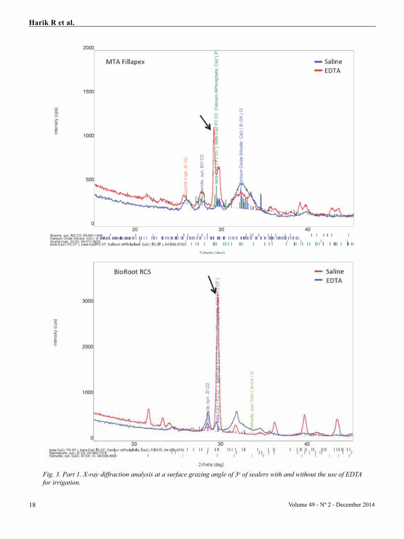
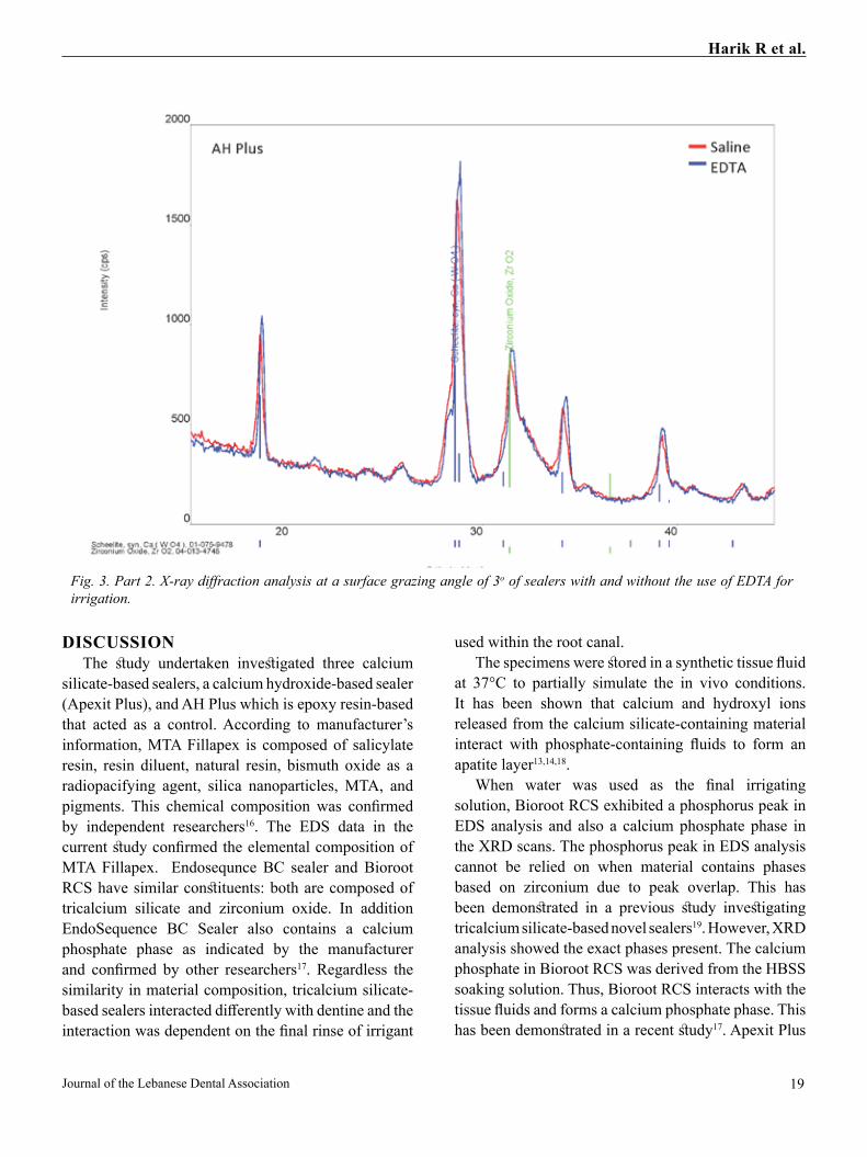
are shown in Figure 3 (Parts 1 and 2). The MTA Fillapex exposed to EDTA exhibited a peak for calcium phosphate (marked with arrow), which was not visible in the MTA Fillapex placed in a tooth where water was used as final irrigant. Conversely, EndoSequence BC sealer, Bioroot RCS, and Apexit Plus had a calcium phosphate peak when water was used as a final irrigant. This peak was not present with EDTA irrigation. AH Plus was not affected by the irrigating solutions.
Fig. 2. Part 2. Energy dispersive X-ray spectroscopic analysis of test sealers with and without exposure to EDTA
Harik R et al.
18 Volume 49 - Nº 2 - December 2014
Fig. 3. Part 1. X-ray diffraction analysis at a surface grazing angle of 3o of sealers with and without the use of EDTA for irrigation.
Harik R et al.
19Journal of the Lebanese Dental Association
DISCUSSIONThe study undertaken investigated three calcium
silicate-based sealers, a calcium hydroxide-based sealer (Apexit Plus), and AH Plus which is epoxy resin-based that acted as a control. According to manufacturer’s information, MTA Fillapex is composed of salicylate resin, resin diluent, natural resin, bismuth oxide as a radiopacifying agent, silica nanoparticles, MTA, and pigments. This chemical composition was confirmed by independent researchers16. The EDS data in the current study confirmed the elemental composition of MTA Fillapex. Endosequnce BC sealer and Bioroot RCS have similar constituents: both are composed of tricalcium silicate and zirconium oxide. In addition EndoSequence BC Sealer also contains a calcium phosphate phase as indicated by the manufacturer and confirmed by other researchers17. Regardless the similarity in material composition, tricalcium silicate-based sealers interacted differently with dentine and the interaction was dependent on the final rinse of irrigant
used within the root canal. The specimens were stored in a synthetic tissue fluid
at 37°C to partially simulate the in vivo conditions. It has been shown that calcium and hydroxyl ions released from the calcium silicate-containing material interact with phosphate-containing fluids to form an apatite layer13,14,18.
When water was used as the final irrigating solution, Bioroot RCS exhibited a phosphorus peak in EDS analysis and also a calcium phosphate phase in the XRD scans. The phosphorus peak in EDS analysis cannot be relied on when material contains phases based on zirconium due to peak overlap. This has been demonstrated in a previous study investigating tricalcium silicate-based novel sealers19. However, XRD analysis showed the exact phases present. The calcium phosphate in Bioroot RCS was derived from the HBSS soaking solution. Thus, Bioroot RCS interacts with the tissue fluids and forms a calcium phosphate phase. This has been demonstrated in a recent study17. Apexit Plus
Fig. 3. Part 2. X-ray diffraction analysis at a surface grazing angle of 3o of sealers with and without the use of EDTA for irrigation.
Harik R et al.

20 Volume 49 - Nº 2 - December 2014
behaved in a similar fashion. This sealer is composed of calcium hydroxide, which interacts with tissue fluids to form calcium phosphate. In EndoSequence, this interaction cannot be proved since the material contains calcium phosphate, thus it is difficult to know if this phase originates from the interaction with tissue fluids or is a material constituent. Addition of calcium phosphate to tricalcium silicate-based materials, such as Bioaggregate, has been shown to reduce the long term production of calcium hydroxide, thus this could potentially affect the material bioactivity20. Conversely, MTA Fillapex did not exhibit interaction with HBSS when the final irrigant was water, but when EDTA was used as the final irrigating solution.
Formation of an interfacial layer develops a chemical bond between calcium silicate-based materials and dentinal walls14. Calcium silicate-based root canal sealers have been found to show bioactivity while being in contact with phosphate ions21,22: this
might be attributed to diffusion of phosphate ions from phosphate-containing storage media into the canal22. It has been shown that during a 7-day storage period in a phosphate-containing fluid, roots filled with a calcium silicate-based sealer and gutta-percha will develop amorphous calcium phosphate precursors along the apical third of root canal walls. With increased storage period, apatite-like crystalline clusters will be observed up to the middle third of the canal walls. With more extended storage in phosphate-containing fluid, there would be more phosphate ions available for the interaction with the calcium and hydroxyl ions released by the calcium silicate-based sealer, which could result in higher bond strength of calcium silicate-containing sealers in the upper levels of the roots23.
The alkaline pH of root canal sealers could neutralize lactic acid from osteoclasts and prevent dissolution of mineralized components of teeth; therefore, root canal sealers, especially tricalcium silicate-based sealers,
Table 1. Mean of weight percentage of calcium ions within the sealers with and without exposure to EDTA.
Harik R et al.

21Journal of the Lebanese Dental Association
can contribute to hard tissue formation by activating alkaline phosphatase and thus phosphorous uptake16. To fully appreciate the characteristics associated with the use of tricalcium silicate cement, one must understand the hydration reactions involved in the setting of the material24. The tricalcium silicate in the powder hydrates to produce a calcium silicate hydrate gel and calcium hydroxide25,26. The calcium hydroxide reacts with the phosphate ions to precipitate hydroxyapatite and water13,14. The water continues to react with the tricalcium silicate to precipitate additional gel-like calcium silicate hydrate. The water supplied through this reaction is an important factor in controlling the hydration rate and the setting time.
Dentin is composed of approximately 20 percent water (by volume), and it is this water that initiates the setting of the material and ultimately results in the formation of hydroxyapatite. Hydraulic cements-like tricalcium silicate cement and Portland cement need water to set and develop their properties. EndoSequence BC sealer is a premixed sealer, which is designed to harden only when exposed to a moist environment, such as that produced by dentinal tubules27,28. A recent study investigated the setting of EndoSequence BC sealer and other calcium silicate-based sealers using a dentine pressure set up that mimics in vivo conditions. Under these conditions, EndoSequence BC sealer hydrated and full setting was achieved17.
The smear layer contains moisture and might act as a coupling agent, thereby improving the adaptation of hydrophilic materials to the root canal wall. The removal of smear layer might have a negative effect on hydrophilic root canal sealers29.
Very little information is available on the effect of EDTA on tricalcium silicate-based materials. However, several studies have examined the effects of endodontic irrigants on physicochemical properties of MTA30,31. Lee and co-workers30 showed the adverse effect of EDTA on hydration and microhardness of MTA. The residual EDTA in the root canal system may chelate calcium ions released from MTA during hydration, and thereby, interfere with the precipitation of hydrated products.
Clearly, the presence of EDTA caused a drop in calcium ions due to calcium ions chelation as observed in the EDS studies and the stoichiometry*, however it
might have increased the permeability of roots, allowing the penetration of phosphate ions from the physiologic solution in which the roots were immersed for 28 days. In EndoSequence BC sealer, Bioroot RCS and Apexit Plus, the calcium phosphate phase was absent when EDTA was used as a final irrigant. Thus, formation of calcium phosphate was impaired. Conversely, in MTA Fillapex, a calcium phosphate phase was obvious after irrigation with EDTA. The chelation effect of EDTA increased the permeability of the material, making it more reactive and allowing uptake of phosphate phases form the environment. MTA Fillapex was the only tricalcium silicate-based material with a resin matrix. AH Plus was unaffected. The use of EDTA according to the data found in this study showed a strong impact on the hydration of calcium silicate sealers.
CONCLUSIONThe use of EDTA final irrigation during root
canal therapy affects the interaction of tricalcium silicate-based sealers used to obturate the root canal with dentine. The observed changes depended on the chemical composition of the sealer.
AcknowledgementsEngineer James C. Camilleri of the Department
of Metallurgy and Materials Engineering, Faculty of Engineering (DMME Laboratory), University of Malta for his technical expertise. Nicholas Gautier and Gilles Richard of Septodont, for Bioroot RCS. Dr Ralph Rawls for the EndoSequence BC sealer. ERDF (Malta) for the financing of the testing equipment throughout the project: “Developing an Interdisciplinary Material Testing and Rapid Prototyping R&D Facility” (Ref. no. 012).
* Stoichiometry = the calculations with chemical formulas and equations.
Harik R et al.

22 Volume 49 - Nº 2 - December 2014
Conflict of Interest Disclosure StatementWe affirm that we have no financial affiliation
(employment, honoraria, direct payment, stock ownership, retainers, consultantships, patent licensing arrangements or honoraria) or involvement with any commercial organization or corporation, with any direct financial or economic interest in the subject or materials discussed in this manuscript, and such arrangements didn't exist during the past three years. Any other potential conflict of interest is disclosed.
R. Harik, Z. Salameh, R. Habchi, and J. Camilleri
REFERENCES1. Shokouhinejad N, Hoseini A, Gorjestani H, Shamshiri AR. Effect of Different Irrigation Protocols for Smear Layer Removal on Bond Strength of a New Bioceramic Sealer. Iran Endod J. 2013;8:10-13.2. Ram Z. Chelation in root canal therapy. Oral Surg Oral Pathol Oral Med 1980;49(1):64-74.3. Saquy P., Campos G.M., Sousa Neto M.D., Guimaraes L.F., Pecora J.D. Evaluation of chelating action of EDTA in association with dakin’s solution. Braz Dent J 1994;5:65-70.4. Soares J.A., Auxiliadora Roque de Carvalho M., Santos S.M.C., Mendonça R.M.C., Ribeiro-Sobrinho A.P., Brito-Júnior M., Prazeres Magalhães P., Santos M.H., Farias L.M. Effectiveness of Chemomechanical Preparation with Alternating Use of Sodium Hypochlorite and EDTA in Eliminating Intracanal Enterococcus faecalis Biofilm. J Endod 2010;36:894-898.5. Roberts HW, Toth JM, Berzins DW, Charton DG. Mineral trioxide aggregate material use in endodontic treatment: a review of the literature. Dent Mater 2008;24:149–64.6. Lee ES, Monsef M, Torabinejad M. Sealing ability of a mineral trioxide aggregate for repair of lateral root perforations. J Endod 1993;19:541–4.7. Torabinejad M, Watson TF, Pitt Ford TR. Sealing ability of a mineral trioxide aggregate when used as a root end filling material. J Endod 1993;19:591–5.8. Menezes R, Bramante CM, Letra A, Carvalho VG, Garcia RB. Histologic evaluation of pulpotomies in dog using two types of mineral trioxide aggregate and regular and white Portland cements as wound dressings. Oral Surg, Oral Med, Oral Pathol, Oral Radiol, Endod 2004;98:376–9.9. Jacobovitz M, Lima RKP. The use of calcium hydroxide and mineral trioxide aggregate on apexification of a replanted tooth: a case report. Dent Traumatol 2009;25:e32–6.10. Scarparo RK, Haddad H, Acasigua GA, Fossati ACM, Fachin EVF, Grecca FS. Mineral trioxide aggregate-based sealer: analysis of tissue reactions to a new endodontic material. J Endod 2010;36:1174–8.11. Torabinejad M, Chivian N. Clinical applications of mineral trioxide aggregate. J Endod 1999;25:197–205.
12. Morgental, R. D., Vier-Pelisser, F. V., Oliveira, S. D., Antunes, F. C., Cogo, D. M. and Kopper, P. M. P. Antibacterial activity of two MTA-based root canal sealers. Int Endod J 2011;44:1128–3313. Tay FR, Pashley DH, Rueggeberg FA, Loushine RJ, Weller RN. Calcium phosphate phase transformation produced by the interaction of the portland cement component of white mineral trioxide aggregate with a phosphate-containing fluid. J Endod. 2007;33:1347-51. 14. Sarkar NK, Caicedo R, Ritwik P, Moiseyeva R, Kawashima I. Physicochemical basis of the biologic properties of mineral trioxide aggregate. J Endod. 2005;31:97–100.15. Reyes-Carmona JF, Felippe MS, Felippe WT. Biomineralization ability and interaction of mineral trioxide aggregate and white portland cement with dentin in a phosphate-containing fluid. J Endod. 2009;35:731-6. 16. Zhou H.M., Shen Y., Zheng W., Li L., Zheng Y.F., Haapasalo M. Physical Properties of 5 Root Canal Sealers. J Endod 2013;39:1281-6.17. Xuereb M, Vella P, Damidot D, Sammut C, Camilleri J. In situ assessment of setting of Endosequence BC sealer and MTA Fillapex using a dentine pressure model. J Endo. Epub 2014 Oct 30.18. Gandolfi MG, Van Landuyt K, Taddei P, Modena E, van Meerbeek B, Prati C. Environmental scanning electron microscopy connected with energy dispersive x-ray analysis and Raman techniques to study ProRoot mineral trioxide aggregate and calcium silicate cements in wet conditions and in real time. J Endod. 2010;36:851–7. 19. Viapiana R, Guerreiro-Tanomaru J, Tanomaru-Filho M, Camilleri J. Interface of dentine to root canal sealers. J Dent 2014 March;42:336-50.20. Camilleri J, Sorrentino F, Damidot D. Characterization of un-hydrated and hydrated Bioaggregate™ and MTA Angelus™. Clin Oral Investig.Epub 2014 Aug 1.21. Huffman B.P., Mai S., Pinna L., Weller R.N.,Primus C.M., Gutmann J.L.,Pashley D.H.,Tay F.R. Dislocation resistance of ProRoot Endo Sealer, a calcium silicate-based root canal sealer from radicular dentine. Int Endod J 2009; 42:34-46.22. Weller RN, Tay KC, Garrett LV, Mai S, Primus CM, Gutmann JL, et al. Microscopic appearance and apical seal of root canals filled with gutta-percha and ProRoot Endo Sealer after immersion in a phosphate-containing fluid. Int Endod J. 2008;41:977–86. 23. Shokouhinejad N., Hoseini A., Gorjestani H., Raoof M., Assadian H., Shamshiri A.R. Effect of phosphate-buffered saline on push-out bond strength of a new bioceramic sealer to root canal dentin. Dent Res J (Isfahan). 2012;9:595–599.24. Koch K, Brave D, Nasseh AA. Bioceramic technology: closing the endo-restorative circle, Part I. Dent Today 2010;9:100-5.25. Camilleri J. Characterization of hydration products of mineral trioxide aggregate. Int Endod J 2008;41:408–17.26. Camilleri J. Characterization and hydration kinetics of tricalcium silicate cement for use as a dental biomaterial. Dent Mater 2011;27:836-4427. Koch K, Brave D. Bioceramic technology-the game changer in Endodontics. Endodontic Practice 2009;2:13–7.
Harik R et al.

23
28. Jefferies SR. Bioactive and biomimetic restorative materials: a comprehensive review. Part I. J Esthet Restor Dent. 2014 Jan-Feb;26(1):14-26. 29. Shokouhinejad N., Gorjestani H., Nasseh A.A., Hoseini A., Mohammadi M., Shamshiri A.R. Push-out bond strength of gutta-percha with a new bioceramic sealer in the presence or absence of smear layer. Aus Endod J 2013;39:102-6.30. Lee YL, Lin FH, Wang WH, Ritchie HH, Lan WH, Lin CP. Effects of EDTA on the hydration mechanism of mineral trioxide aggregate. J Dent Res. 2007;86:534–8.31. Yan P, Peng B, Fan B, Fan M, Bian Z. The effects of sodium hypochlorite (5.25%), Chlorhexidine (2%), and Glyde File Prep on the bond strength of MTA-dentin. J Endod. 2006;32:58–60. 32. Stewart GG. Chelation and flotation in endodontic practice: an update. J Am Dent Assoc 1986 Oct;113(4):618-622.33. Medina Cardenas ME, Calvo Pérez V, Sánchez Planells U. Rev Dent Chile 1989 Apr;80(1):4-10 (article in Spanish).34. Zehnder M, Schmidlin P, Sener B, Waltimo T. Chelation in root canal therapy reconsidered. J Endo 2005 Nov;31(11):817-820.35. Mohammadi Z et al. EDTA in endodontics. Europ J Dent 2013 Sep;7(Suppl 1). PMCID:PMC4054072.36. Nygaard-Ostby B. Chelation in root canal therapy: EDTA for cleansing and widening of root canals. Odont Tidskr. 1957;65:3-11.
Correspond with:Randa [email protected]
Harik R et al.
Journal of the Lebanese Dental AssociationVolume 49 - Nº 2 - December 2014
24
INTRODUCTION During dentinogenesis, odontoblasts (which derive
from neural crest) become post-mitotic before the onset of dentin formation and are highly polarized after terminal differentiation. After the last division, one daughter cell close to the basement membrane (BM) becomes a functional odontoblast, whereas the other will form the sub-odontoblastic Hoehl’s cell
layer1. In adult teeth, odontoblasts and Hoehl's cells form a superficial layer at the periphery of the pulp (outer border lining dental pulp). In mild pathological conditions such as moderate carious lesions, the odontoblasts are stimulated and elaborate a reactionary dentin. If the odontoblasts are injured, Hoehl’s cells may be reactivated and differentiate into odontoblasts2
forming orthodentine. More severe carious lesions lead to the irreversible alteration of odontoblasts/Hoehl’s cells. In such case, pulp cells endowed with the capacity to acquire an osteo/odontoblast phenotype, synthesize extracellular matrix components contributing to the formation of a mineralized reparative osteodentin.
Potential contribution of dental pulp stem cells to pulp and dentin repair in response to tooth injury: A preliminary study.Yassine Harichane1, DCD, DSO, Sasha Dimitrova-Nakov2, DCD, DSO, Odile Kellermann3, Dr. Natural Sc., Michel Goldberg4, Chir. Dent., Dr. Odont. Sc., Dr. Natural Sc., PU
AbstractWe investigated the capacity of dental pulp stem cells to induce the formation of a reparative dentin-like structure
upon implantation within the pulp, after a surgical exposure. For this purpose, we established cell lines displaying stem cells properties from molar dental pulp of mouse embryos. Among these cells, « A4 » cell line maintains a stable mesoblastic phenotype and never differentiates spontaneously under long-term standard culture conditions. This clone was implanted in the rat first maxillary molar (a model used in our laboratory), to evaluate whether these pulp-derived precursor cells are able to synthesize, in vivo, a dentin-like structure.
When the surgery alone was performed, after an early inflammatory process, a heterogeneous matrix, characterized by large gaps and pulp remnants, gradually filled the mesial part of pulp chamber in 30 days. This non-mineralized permeable structure is unable to resist a bacterial re-infection. When clone A4 was implanted into a rat molar pulp lesion, after one month, a dense mineralized dentin-like structure was formed in the implantation site, filling homogeneously the mesial part of pulp chamber.
Agarose beads were implanted alone but also were used as cell carrier. Preliminary experiments using A4 progenitors carried out by alginate beads suggest 1) that dental pulp stem cells induce the formation of robust reparative dentin and therefore constitute useful tool in pulp therapies, and 2) that alginate is a suitable carrier for cell implantation. Despite surgical trauma and stem cells implantation, reparative processes do not affect the structure and vitality of residual pulp in central and distal parts of pulp chamber. Future prospects will be focused to determine whether implanted progenitor cells are directly involved in the formation of reparative dentin or whether they induce recruitment and differentiation of host progenitor cells.
INSERM UMR-S 747-Equipe 5, Cellules Souches, Signalisation et Prions, and Université Paris Descartes, Paris, France 1. IFRO's grantee2. STEM's grantee3. Professor and Scientist4. Emeritus Professor and Senior Scientist
Dental Research
Journal of the Lebanese Dental AssociationVolume 49 - Nº 2 - December 2014

25Journal of the Lebanese Dental Association
Gronthos and co-workers3,4, and Miura and associates5 have shown that cells displaying stem cells properties are present in the human dental pulp: they were named Dental Pulp Stem Cells (DPSCs), or SHED, when these Stem cells were obtained from Human Exfoliated Deciduous Teeth.
Transplanted into ectopic sites (subcutaneous and/or sublingual) in immunocompromised mice, transplants of heterogenous colonies of human DPSCs generate a pulp-like tissue lined by odontoblast-like cells, surrounded by a dentinal structure.The differentiation potential of these colonies upon implantation within the pulp has never been investigated. Besides, when DPSC sub-populations are induced to differentiate in vitro, they give rise not only to osteoblasts and odontoblast-like cells, secreting an extracellular matrix (which undergoes mineralization), but also to chondrocytes, adipocytes, and even neuronal cells6. DPSCs share many characteristics with stromal stem cells derived from red bone marrow (BMSSC)7. They express cell surface molecules (CD90, CD117, Sca-1) used to characterize mesenchymal stem cells (MSC)8. It is somehow difficult to know if DPSCs represent one or distinct subpopulation(s) of stem cells depending on their localization within the pulp. Indeed, no specific marker(s) allow(s) to identify, localize, and thus, select these cells within the pulp. Despite an extensive knowledge on tooth formation and development, molecular mechanisms underlying the recruitment and differentiation of these progenitors in response to tooth injury still need to be clarified. The nature of dentin produced, in vivo, by pulpal stem cells raises another set of questions with respect to its clinical relevance. Orthodentin bearing the structural characteristics of a true tubular dentin is produced in some cases, but generally, the formation of a atubular mineralized tissue is observed. The so-called osteodentin displays lacunae interconnected by a network of thin canaliculi, thus ressembling to bone. Our aim was to exploit a precursor cell line that we derived from the pulp to analyse the impact of the cells to promote dentin formation upon implantation. Thus, we explored, for the first time, the potential of pulp-derived “stem” cells
to improve dental repair upon injury.
Experimental cell modelIn order to characterize the dental pulp progenitors
and to study the molecular and cellular mechanisms governing their differentiation, our laboratory has obtained clonal cell lines from tooth germs of day 18 mouse embryos transgenic for an adenovirus-SV40 recombinant plasmid (pK4)9. Among the 50 clones obtained from molar dental pulp, the clone named A4 represents an homogenous population of precursor cells that display in vitro properties of multipotent mesoblastic cells10.
Moreover, our in situ approach was essential since the definition of a “progenitor” implicates to outline its in vivo potential into a repair/regeneration context.
Experimental animal modelImplantation into the mouse incisor
In a first in vivo pilot study11, A4 cells were implanted in the mouse mandibular incisor and this led to the formation of a mineralized osteodentin within dental pulp. Since mouse incisor is a continuously growing tooth, it was necessary to choose a model closer to human tooth physiology.
Implantation into the rat molarThe small size of the mouse molars makes
difficult the experimental procedures implicating cells implantation. In contrast, rat molar provides an easier experimental approach involving the preparation of tooth lesions. The protocol was initially described by Ohshima and co-workers12 and modified by Decup and his team13. Therefore, we used the first maxillary molar of rat, a tooth of limited growth, to explore the feasibility of a cell-based therapy following a surgical pulp injury.
We performed a pulp exposure with or without cells implantation in the first maxillary molar of 9-week old Sprague-Dawley rats*, following institutionally approved protocols for animal experimentation * Sprague-Dawley rat is an outbred multipurpose breed of albino rat extensively used in biomedical research. Its main advantage is its ease of handling and calmness.
Harichane Y et al.

26 Volume 49 - Nº 2 - December 2014
A
C
E
B
D
F
Fig. 1. Sham** pulp exposure: Perforation of the rat molar pulpal chamber causes a pulp tissue fibrosis. (A)(B): 2 days after surgery, pulp shows a discrete inflammation limited to mesial and central pulp chambers. The residual pulp displays a normal histological aspect (C)(D): 14 days after surgery, mesial pulp tissue is reorganized and a few mineralization spots are detectable. The isthmus between mesial and central pulp is widely open (E)(F): 28 days after surgery, mesial pulp chamber is filled by a heterogeneous fibrous matrix, with several cell inclusions and empty spaces. The mesial is separated from the central pulp by a thickening of the reactionary dentin. The residual pulp is normal without major histological changes. AC: access cavity, C: central, D: dentin, DB: dentin barrier, Di: distal, FM: fibrous matrix, M: mesial, ND: neodentin, P: pulp.
** In research, a "Sham-operated group" is a control group in laboratory experiments involving surgical procedures.
Harichane Y et al.

27Journal of the Lebanese Dental Association
research. Animals were anesthetized by intra-peritoneal injection of 20% ketamine (Imalgene; Alcyon) and 5% xylazine (Rompun; Alcyon) solution. The mesial gingival papilla was removed by electrosurgery to expose cemento-enamel junction. A cavity was drilled on mesial aspect of the tooth using a tungsten dental bur (ISO 006; Dentsply) with a low-speed hand-piece. Pulp perforation was accomplished by pressure with the tip of a steel probe. This method avoids rolling the pulp around the dental burr.
A4 cells were cultured in a complete medium (DMEM)*** with 5% of fetal calf serum. About 105
cells were collected in a tube and centrifuged to form a cell pellet subsequently implanted into the pulp of 14 rat’s molars.
To protect the pulp from bacterial contamination, after the surgery, the cavity was filled with glass-ionomer cement (Fuji IX; GC). Animals were anesthetized and euthanized at different periods of time (2, 14, and 30 days after surgery; except for the cell/alginate group, the rats being killed at day 30 after implantation). Block sections (including the hemimaxillaries) were dissected and fixed in a 4% para-formaldehyde solution overnight at +4°C. Samples were demineralized in EDTA**** (4.13% solution) for 5 months. Dehydrated in graded alcoholic solutions, the hemi-maxillaries were included in paraffin (Paraplast plus; Kendall). 7mm thick sections were collected on glass slides and observed histologically after Masson’s trichrome staining.
• Surgery without cells implantation: Sham groupA surgical pulp exposure was performed in 14 rat
molars, without cells or alginate bead implantation. Two days after the surgery, a slight inflammatory reaction was observed in the mesial and central parts of the pulp (Figures 1A, 1B). The distal part of a pulp appeared normal, without any inflammation. After 2 weeks, reorganization of the pulp tissue was observed together with the onset of a diffuse fibrosis and some small mineralization area in the mesial pulp chamber
(Figures 1C, 1D). One month after the surgery, the mesial pulp chamber became fibrotic (Figure 1E). A dentin barrier was formed between the mesial and central parts of the pulp chambers (Figure 1F).
• Surgery followed by cells implantation: the “experimental cell” group
When we implanted pellets of “A4” cells just after surgery, at day 2, inflammatory cells were recruited near the implantation site (Figures 2A, 2B). After 2 weeks, the mesial part of the pulp chamber started to be gradually filled by osteodentin (Figures 2C, 2D). After one month, a fully mineralized osteodentin massively filled the mesial pulp chamber (Figure 2E). Moreover, a dentin barrier was formed in the isthmus separating the mesial part from the central area of the pulp chambers (Figure 2F). In central and distal parts of the crown, pulp was fully preserved to ensure tooth vitality and well protected by a reparative dentin plug formed at the site of injury.
• Surgery followed by implantation of alginate beads loaded with or without A4 cells
In order to improve the implantation protocol, i.e. precisely visualize the implantation site and decrease the number of introduced cells within the pulp, we used, as a carrier, alginate beads. Alginate, a linear co-polymer of anionic polysaccharide, was selected as a suitable and potentially non toxic biomaterial since its pH is close to neutral during and after gelation. Moreover, this irreversible hydrocolloid is biocompatible and biodegradable14. On a trial basis, alginate beads alone or loaded with cells were implanted in the molars.
After one month, the group implanted with alginate beads alone showed reactionary dentin formation which was comparable to the Sham group (Figures 3A, 3B). This indicates that implantation of alginate beads does not cause, by itself, any inflammatory process and does not interfere with the slight formation of reactionary dentin occurring spontaneously in the rat molar, in response to pulp exposure and implantation.
**** EDTA = Ethylene Diamine Tetra-Acetic*** DMEM is a modification of Basal Medium Eagle -BME- that contains a 4-fold higher concentration of amino acids and vitamins, as well as additional supplementary components. The original DMEM formula contains 1000 mg/L of glucose.
Harichane Y et al.
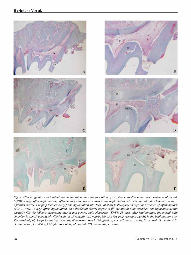
28 Volume 49 - Nº 2 - December 2014
A
C
E
B
D
F
Fig. 2. After progenitor cell implantation in the rat molar pulp, formation of an osteodentin-like mineralized matrix is observed. (A)(B): 2 days after implantation, inflammatory cells are recruited in the implantation site. The mesial pulp chamber contains a fibrous matrix. The pulp located away from implantation site does not show histological changes or presence of inflammatory cells. (C)(D): 14 days after implantation, an osteodentin matrix begins to fill the mesial pulp chamber. The reparative dentin partially fills the isthmus separating mesial and central pulp chambers. (E)(F): 28 days after implantation, the mesial pulp chamber is almost completely filled with an osteodentin-like matrix. No or a few pulp remnants persist in the implantation site. The residual pulp keeps its vitality, structure, dimensions, and histological aspect. AC: access cavity, C: central, D: dentin, DB: dentin barrier, Di: distal, FM: fibrous matrix, M: mesial, ND: neodentin, P: pulp.
Harichane Y et al.

29Journal of the Lebanese Dental Association
Conversely, one month after the implantation of an alginate bead loaded with 3.103 A4 cells, we observed the formation of reparative osteodentin filling the mesial part of pulp chamber and a dentin plug protecting the pulp located in the central and distal parts of the crown pulp chambers (Figures 4A, 4B). Thus, these preliminary data, using alginate beads as a cell carrier for implantation of pulpal precursor cells, suggest that this biomaterial is suitable for cell implantation and allows to obtain an efficient pulp repair.
Implantation process and pulp vitalityAfter the surgery with or without cell implantation,
pulp was well preserved. No chronic inflammation was detectable at 30 days. This experimental protocol did not introduce any bias in the interpretation of the
results since no iatrogenic pathology was detectable. The inflammatory response, normally observed upon injury, is not enhanced by the introduction of mouse derived pulpal cells in a rat molar: thus, these data provide the first evidence that implantation of pulpal precursor cells within pulp promotes the formation of a robust dentin barrier separating residual pulp from reparative area.
CONCLUSION AND PERSPECTIVES18
Stem cells appear as tools to get a better understanding of cellular mechanisms of pulp repair. They display innovating potentials in dental therapies. We proved here, and for the first time, that a dental pulp cell line has the capacity to promote the repair of a dentin lesion. Our results indicate that direct implantation of
Fig. 3. Dentin repair after the implantation of alginate beads loaded with A4 cells. (A)(B): 28 days after A4 cells implantation using an alginate bead as a carrier, a dense and homogenous mineralized tissue fills mesial pulp horn, root canal entry, and restricts the width of isthmus between mesial and central pulp. In the middle of the implantation site, a fibrous zone persists, with many cell inclusions and remnants of alginate beads. The residual pulp keeps a normal aspect. AC: access cavity, C: central, D: dentin, DB: dentin barrier, Di: distal, M: mesial, ND: neodentin, P: pulp, REAC: reactionary dentin.
Fig. 4. Formation of a homogenous fibrous matrix after the implantation of an alginate bead alone. 28 days after the implantation of the alginate beads alone, implantation site is completely filled by a fibrous matrix that is not mineralized and contains many cell inclusions. A dentin barrier is observed and it separates mesial pulp chamber from the rest of the pulp. AC: access cavity, C: central, D: dentin, DB: dentin barrier, Di: distal, FM: fibrous matrix, M: mesial, ND: neodentin, P: pulp.
A B
Harichane Y et al.

30 Volume 49 - Nº 2 - December 2014
mouse progenitor cells in the dental pulp of a rat molar, a tooth with limited growth, leads to the formation of reparative osteodentin. From the present experimental approach, we may deduce that:
1- When mouse stem cells are implanted in rat teeth, they are apparently well tolerated. Therefore, these experiments validate the possibility for inter-species implantation.
2- After one month, the implanted stem cells, into a dental pulp, form a reparative osteodentin at implantation site, obstructing totally the lesion. These results also show the absence of inflammation or necrosis in implantation site, and good preservation of pulp vitality, despite surgical trauma.
3- Finally, the preliminary study presented here, using an alginate bead as a cell carrier, suggests that this biomaterial is suitable for cell implantation. This approach provides the possibility to control the number of implanted cells necessary to obtain an efficient pulp repair.
It is important to determine whether precursor cells re-introduced in a pulpal “natural”environment differentiate into osteo-odontogenic cells capable to elaborate an extra-cellular mineralized matrix2 or whether the implanted cells, by secreting growth factors, recruit resident pulp stem cells towards osteo-odontogenic differentiation and, indirectly, promote formation of dentinal bridge. Thus, the direct or indirect implication of implanted stem cells in the dentin formation process is still an open question3.
In conclusion, this study proved that pulpal stem cells have the capacity to heal a dentin lesion by promoting a repair after implantation in the pulp. Our pre-clinical experimental approach paves the way to the development of cell-based therapies for pulp injury10,16,17. The long-term goal is to provide new strategies for dental surgeons to restore the functionality of an injured tooth by using pulp stem cells.
Acknowledgements:This work was supported by a grant to Research
Scientist Yassine Harichane, awarded by the “Institut Français pour la Recherche Odontologique” (IFRO). Research Scientist Sasha Dimitrova-Nakov was supported by a grant awarded by STEM-Pôle Ile de France.
REFERENCES1. Hoehl E. Beitrag zur histologie der pulpa und des dentins. Arch Anat Physiol. 1896; 32: 31-54. 2. Lesot H, Kubler M, Meyer J, Smith A, Cassidy N, Ruch JV. Experimental induction of odontoblast differentiation and stimulation during reparative process. Cells and Materials. 1993; 4: 199-218. 3. Gronthos S, Mankani M, Brahim J, Robey PG, Shi S. Postnatal human dental pulp stem cells (DPSCs) in vitro and in vivo. Proc. Natl. Acad. Sci. U.S.A. 2000; 97(25):13625-13630. 4. Gronthos S, Brahim J, Li W, Fisher LW, Cherman N, Boyde A, et al. Stem cell properties of human dental pulp stem cells. J. Dent. Res. 2002. 81(8):531-535. 5. Miura M, Gronthos S, Zhao M, Lu B, Fisher LW, Robey PG, et al. SHED: stem cells from human exfoliated deciduous teeth. Proc. Natl. Acad. Sci. U.S.A. 2003; 100 (10): 5807-5812. 6. Arthur A, Rychkov G, Shi S, Koblar SA, Gronthos S. Adult human dental pulp stem cells differentiate toward functionally active neurons under appropriate environmental cues. Stem Cells. 2008 Jul;26(7):1787-95.7. Shi S, Robey PG, Gronthos S. Comparison of human dental pulp and bone marrow stromal stem cells by cDNA microarray analysis. Bone 2001; 29(6):532-539. 8. Balic A, Aguila HL, Caimano MJ, Francone VP, Mina M. Characterization of stem and progenitor cells in the dental pulp of erupted and unerupted murine molars. Bone 2010 Jun;46(6):1639-51.9. Priam F, Ronco V, Locker M, Bourd K, Bonnefoix M, Duchêne T, et al. New cellular models for tracking the odontoblast phenotype. Arch. Oral Biol. 2005; 50(2):271-277. 10. Lacerda-Pinheiro S, Dimitrova-Nakov S, Harichane Y, Souyri M, Petit-Cocault L, Legrès L, Marchadier A, Baudry A, Ribes S, Goldberg M, Kellermann O, Poliard A. Concomitant multipotent and unipotent dental pulp progenitors and their respective contribution to mineralised tissue formation. European Cells and Materials 2012;23:371 - 386.11. Lacerda-Pinheiro S, Marchadier A, Donãs P, Septier D, Benhamou L, Kellermann O, Goldberg M, Poliard A. An In vivo Model for Short-Term Evaluation of the Implantation Effects of Biomolecules or Stem Cells in the Dental Pulp. Open Dent J. 2008; 2: 67-72. 12. Ohshima H. Ultrastructural changes in odontoblasts and pulp capillaries following cavity preparation in rat molars. Arch. Histol. Cytol. 1990;53(4):423-438. IFRO = French Institute of Dental Research
Harichane Y et al.

31
13. Decup F, Six N, Palmier B, Buch D, Lasfargues JJ, Salih E, et al. Bone sialoprotein-induced reparative dentinogenesis in the pulp of rat’s molar. Clin Oral Investig. 2000; 4(2): 110-119. 14. Klöck G, Pfeffermann A, Ryser C, Gröhn P, Kuttler B, Hahn HJ, et al. Biocompatibility of mannuronic acid-rich alginates. Biomaterials. 1997; 18(10):707-713. 15. Balic A, Aguila HL, Caimano MJ, Francone VP, Mina M. Characterization of stem and progenitor cells in the dental pulp of erupted and unerupted murine molars. Bone 2010 Jun;46(6):1639-51.16- Dimitrova-Nakov S, Baudry A, Harichane Y, Kellermann O, Goldberg M. Pulp Stem Cells: Implication In Reparative Dentin Formation. Journal of Endodontics 2014 Apr 40 (suppl 4):S13-18.17- Baudry A, Uzunoglu E, Schneider B, Kellermann O, Goldberg M. From pulpal stem cells to tooth repair : an emergenging field for dental tissue engineering. Evidence-Based Endodontics (submitted for publication).18. Dimitrova-Nakov S, Baudry A, Harichane Y, Kellermann O, Goldberg M. Pulp Stem Cells: Implication in Reparative Dentin Formation. J Endo 2014 Apr;40(4S):S13-S18.
Correspond with:Michel [email protected]
Harichane Y et al.
Journal of the Lebanese Dental AssociationVolume 49 - Nº 2 - December 2014

32
Fluoride in dentistry: use, dosage, and possible hazards.
Mounir Doumit1, Dr. Chir. Dent., Dr. Sc. Odont., WHO Expert in Oral Health,Mohammad Omar Machmouchi2, MD (Stomatol.), M.Sc. (Dent. Public Health),Hicham Diab3, Dental Surgeon, DU Oral Biol., DESS Preventive/Public Health Dent.
AbstractFluoride (F) therapy is the delivery of fluoride to teeth, either topically or systematically, in order to protect them
from dental caries. Extensive evidence proved that fluoride toothpastes and water fluoridation reduce dental caries. Fluoride and water fluoridation in dentistry were classified as one of the 10 most important public health measures of the 20th century. This clinical practice placement briefly addresses the fluoride's issue in dentistry, its use, dosage, and possible hazards due to excess fluoride.
1. Chairperson and Professor, Public Health Dentistry Department, and Former Dean, Lebanese University School of Dentistry, Beirut, Lebanon2. Associate Clinical Instructor, Public Health Dentistry Department, Lebanese University School of Dentistry, Beirut, Lebanon3. Chief of Clinical Services, Public Health Dentistry Department, Lebanese University School of Dentistry, Beirut, Lebanon
HISTORYIn 1901, when Dr. Frederick McKay started his
dental practice in Colorado Springs, USA, he noticed that many of his patients had a mysterious brown staining of their teeth, He investigated the issue for 30 years and was stunned to notice that the strangely stained or "mottled" teeth were also decay free, and he strived to determine the drinking water as the cause of this strange phenomenon.
In 1931, Dr. H. Trendley Dean, a dentist working for the US Public Service, was studying the harmful effects of fluoride, and in 1950, he demonstrated that fluoride therapy, in small amounts, has obvious large benefits with negligible side-effects resulting in an enamel staining ("mottling" of teeth), known later as enamel "fluorosis". At that time, Dean suggested that a water supply fluoride concentration of about 1mg/L or 1 part per million (1ppm) (roughly equivalent to a grain of salt in a gallon of water) will be associated with substantially fewer cavities.
The safety of Fluoride has been the subject of much discussion indeed; it is one of the most extensively
researched health measures. The balance of evidence suggests that Fluoride offers a safe and effective route to better dental health, if properly used.
Dental and oral diseases are important public health problems: pain, disability, and handicap resulting from them are common, and the costs of treatment are a major problem. One of the most efficient elements in prevention of dental decay is Fluoride.
Fluoride reduces the incidence of dental caries and slows or reverses the progression of existing ones. Fluoride has made enormous contribution to declines in dental caries over the past 80 years. Fluorine (F) is an element of the halogen family, which also includes Chlorine, Bromine, and Iodine. It forms inorganic and organic compounds called Fluoride. Living organisms are mainly exposed to inorganic fluorides through food and water. The most relevant inorganic Fluorides are Hydrogen fluoride, Calcium fluoride, and Sodium fluoride.
How does fluoride act in dental caries prevention?Three theories prevail:1. Fluoride becomes incorporated into the
hydroxyapatite crystals of teeth, rendering them more resistant to acid attack.
2. Presence of saliva promotes remineralization of early carious lesions.
3. Fluoride interferes with metabolic pathways of bacteria, thus reducing acid.
Public Health and Community Dentistry
Journal of the Lebanese Dental AssociationVolume 49 - Nº 2 - December 2014

33Journal of the Lebanese Dental Association
F can be provided either systematically (in water, salt, and milk) or locally (use of topical fluoride such as toothpaste, gel, varnish and mouth rinse). However, there are additional sources of fluoride in the environment which can occur naturally, or as result of industrial process.
The US National Academy of Sciences (NAS) Institute of Medicine has recommended an adequate intake of fluoride from all sources as 0.05 mg F/Kg body weight/day.
METHODS OF FLUORIDE DELIVERY* Water fluoridation:
• Life-long resistance produces the greatest caries-protective effect.
• 20-40% reductions in caries over lifetime.• As of 2012, about 435 million people, worldwide,
received fluoridated water at recommended level, and about 214 millions of them live in the USA.
• Caries increases after fluoridation cessation.• Advantages: safe, cost-effective, consistent. Low
risk of over-dosage.• Disadvantages: no freedom of choice removed,
requires complex infrastructure and initial capital outlay.
• Dosage requirement average: 1 ppm.
*Fluoride Tablets and Drops:• 40-50% reduction in caries (in both adults and
children).• NaF is the compound of choice.
• Care required in prescribing to minimize over-dosage and fluorosis.
• 0.5 mg F/day in children under 4 years and 0.75 to 1 mg for older.
• Advantages: effective, freedom of choice. • Disadvantages: compliance needed, consistency
of delivery needed, risk of overdose.
*Fluoridated Salt:• Dose: 250 ppm.• Advantage: freedom of choice.• Disadvantage: conflict with general health
(messages advising reduction in salt intake).
*Topical fluoride:• Examples include aqueous solutions of sodium
fluoride and stannous fluoride, low pH solutions such as the acidulated phosphate fluoride system.
• 20-35% reduction in caries.• Used usually in school-based mouth rinsing
programs.• Varnishes may be applied directly to teeth in high
concentrations• Gels may applied directly to teeth, as well.• Advantages: effective in individuals at high risk
for dental caries, freedom of choice.• Disadvantages: need personnel, time consuming.
*Fluoridation Toothpastes:• Simplest method of fluoride delivery.• Worldwide declines in caries (attributed to
toothpastes).
TABLETS & DROPS SALT TOUTHPASTES MOUTH RINSING VARNISHES ( 22000 PPM ) GELS (22000 PPM) 0 - 6 MONTHS __ __ __ __ __ __ 6 - 12 MONTHS 0.5 mg __ __ __ __ __
1 - 3 YEARS 0.75 mg 0.12 per/meal 100-250 ppm __ __ __ 4 - 8 YEARS 1 mg 0.12 per/meal 500 ppm __ 2 times/year 2 times /year ≥ 9 YEARS __ 0.12 per/meal 1000-1500 ppm 250 ppm 2 times/year 2 times /year
REFERENCE WEIGHT TOLERABLE UPPER INTAKE Kg mg/day
0 - 6 MONTHS 7 0.7 6 - 12 MONTHS 9 0.9
1 - 3 YEARS 13 1.3 4 - 8 YEARS 22 2.2 ≥ 9 YEARS 40 -76 10
Recommended Total Dietary Fluoride Intake
AGE
2-3.8
AGELOCAL OR TOPICAL USESYSTEMATIC USE
0.71.1
ADEQUATE INTAKEmg/day
0.010.5
Doumit M et al.
Methods of Fluoride delivery
34
• Commercially a vailable as of the late 1960s, 95% by late 70s are fluoridated pastes.• Typical concentrations used: 1000-1500 ppm of
fluoride per gram of toothpaste; a lesser dose is used in children: 100-550 ppm (based on the age and the assessment of carious risk).
• Advantages: easy, effective, freedom of choice.
THE FLUORIDE DEBATEIn a NAS September 1997 report (23rd workshop),
F was repeatedly regarded by speakers and panel members as an "essential nutrient". F is obviously incorporated into mineral matrix of bones and teeth, and, without question, ingestion of even milligram amounts of F during infancy and early childhood may produce the "unmistakable toxic effects of dental fluorosis" (Prof. A. W. Burgstahler, Ph.D, Chemist). Disruption of normal enamel formation is stated (in the 1997 report) not to be "of Public Health Significance" if the F concentration in drinking water is below 2mg/liter (2ppm), and reports of disfiguring dental fluorosis with staining and pitting of the enamel in areas with 1-2 ppm F in drinking water were evidently overlooked.
Most authors and clinicians estimate that crippling skeletal fluorosis occurs when 10-20mg of F have been ingested on a daily basis for at least 10 years: in that case, calcification of ligaments often precludes joint mobility and numerous exostosis may appear, and these effects may be associated with muscle wasting and neurological complications due to spinal cord compression (Prof. Garry M. Whitford, Fluoride Expert, Medical College of Georgia, USA - 1996, in "The Metabolism and Toxicity of Fluoride"). Also, unexplained intermittent episodes of gastric pain and muscular weakness have been clinically linked in areas of endemic dental and skeletal fluorosis intakes as low as 2 to 5 mg/day (AK Susheela et al., 1992, 1993 - and S Desarathy et al., 1996).
In March and July 2014, Grandjean and Landrigan (in "Lancet Neurology") addressed the issue of F neurotoxicity, and some authors and experts worldwide, are presently conducting studies that aim to classify F as a neurotoxin, in order to push towards removing industrial sodium F from world's water supply. This trend considers F as a developmental neurotoxicant (same as manganese, chlorpyrifos, tetrachloroethylene)
that may cause neurodevelopmental disabilities (such as Attention Deficit Hyperactivity Disorder -ADHD-, dyslexia, etc...).
The 2013 findings by Harvard University meta-analysis (funded by the National Institutes of Health - NIH, in Bethesda, Maryland, USA) concluded that children in areas with highly fluoridated water have "significantly lower" IQ scores than those who live in areas with low amounts of fluoride in their water supplies: this 32-page report (written by several researchers) reviewed the findings of 27 studies (published over 22 years) that suggest an inverse association between high fluoride exposure and children's intelligence. Researcher's results support the possibility of adverse effects of F exposures on children's neurodevelopment but future research should formally evaluate dose-response relations based on individual-level measures of exposure over time, including more precise prenatal exposure assessment and more extensive standardized measures of neurobehavioral performance.
SUGGESTED READINGS- A Systematic Review of the efficacy and safety of Fluoridation (Australian government 2007). - Essential Dental Public Health, Daly, B. Watt, R. Batchelor, P. Treasure, E. (2003).- Fluorides and Oral Health / Report of a WHO Expert Committee on Oral Health Status and Fluoride Use. WHO Technical Reports Series - 846. World Health Organization, Geneva, 1994.- Global Consultation on oral health through fluoride. WHO collaboration with the World Dental Federation and the International Association for Dental Research. November 17, 2006.- O'Mullane DM et al. Fluoride and oral health. Submitted to Community Dental Health.- WHO global policy for improvement of oral health - World Health Assembly 2007, by P.E. Peterson (In Int Dent J 2008 June;58(3):115-121).- Oral Health Programme in Lebanon - Technical Assistance Provided for Development of Baseline Studies for Salt Fluoridation / Final Report on Data Collection - 2003Lebanese Ministry of Public Health/Lebanese Uiversity School of Dentistry/World Health Organization/M. Doumit, B. Doughan, R. Baez- Marthaler TM and Petersen PE. Salt fluoridation - an alternative in automatic prevention of dental caries. Int Dent J 2005;55(6):351-358.
Correspond with:Mounir [email protected]
Doumit M et al.
Journal of the Lebanese Dental AssociationVolume 49 - Nº 2 - December 2014

35Journal of the Lebanese Dental Association
Doumit M et al.
37
INTRODUCTIONFull-mouth rehabilitation of severely worn dentition
is a challenge for the dentist and his/her laboratory technician, considering that diagnosis and treatment planning are complex and complicated12.
Tooth wear can result from abrasion, attrition, and erosion1.
Attrition is caused by maxillary to mandibular tooth friction4.
Abrasion is caused by the friction between a tooth and an exogenous agent4.
Erosion is a pathological process of tooth structure loss due to exposure to an acidic agent11.
Determining the etiology and the severity of the tooth surface loss, before proceeding with treatment, is the key for a successful rehabilitation. This will allow the clinician to provide the most predictable treatment outcome7,13.
This treatment can range from simple direct restorations to full-mouth rehabilitation, and becomes
harder as the space availability for prosthetics becomes limited5.
This clinical report is a detailed description of a fixed full-mouth rehabilitation of a worn dentition. Crown lengthening and increasing of vertical dimension were the key factors in providing adequate space for definitive restorations.
CASE REPORTA 55-year-old woman was referred to the
Department of Prosthodontics, Faculty of Dental Medicine, Saint-Joseph University (Beirut, Lebanon) suffering from severely worn dentition, eating disorder, and generalized tooth hypersensitivity.
The patient’s chief complaints were: “I feel my teeth are gradually getting shorter. My teeth are very sensitive and I want them fixed”.
Clinical examination and medical questionnaire revealed good general health. Patient had osteoporosis and was treated by bisphosphonates. Her physician excluded any contraindication to dental treatment. She reported no symptoms related to pain in masticatory muscles or at TMJ level. During clinical interview, patient reported frequent episodes of teeth grinding and clenching, and bruxism.
Her last dental treatment was two years ago, she had many root canal treatments for extreme sensitivity
Full-mouth rehabilitation of a severely worn dentition: A case report.
Farah Antar1, DCD, DES Prostho., MS (Prostho.), Pascale Habre Hallage2, DCD, CES Oral Biol., CES Fixed Prostho., DUICP, MSBM, DEA Neurosc., Dr.Biomed Sc.,Elie Zebouni3, DCD, DMD, CAGS, M.Sc.D.
AbstractExcessive occlusal wear can result in occlusal disharmony, functional and esthetic impairment. Patients with
severe tooth wear present with shortened clinical crowns and reduced interocclusal space. The treatment becomes harder as space availability for well contoured prosthetic restorations becomes limited.
This case report addresses the clinical and laboratory steps involved in oral rehabilitation of a 55-year old woman with severely worn dentition and generalized teeth hypersensitivity.
Crown lengthening as well as increasing of vertical dimension were both necessary in order to gain space for the definitive restorations. A night guard was also used to protect these restorations.
1,2,3 Department of Prosthodontics, Saint-Joseph University, Faculty of Dental Medicine, Beirut, Lebanon1. Clinical Instructor2. Assistant Professor and Director of Prosthodontics Postgraduate Program 3. Professor and Chairperson, Department of Prosthodontics, Saint-Joseph University, Faculty of Dental Medicine, Beirut, and Former Assistant Professor, Boston University Henry M. Goldman School of Dental Medicine, Boston, USA
Fixed Prosthodontics
Journal of the Lebanese Dental AssociationVolume 49 - Nº 2 - December 2014

38 Volume 49 - Nº 2 - December 2014
* VDO = Vertical Dimension of Occlusion (also known as OVD = Occlusal Vertical Dimension).
of some teeth. Her oral hygiene regimen consisted of brushing once a day without dental flossing.
1. Intraoral examination Color, size, texture, and contours of maxillary
and mandibular gingivae were within normal limits. Patient had 4 to 6 mm of attached gingiva in maxilla and mandible.
Teeth showed a marked reduction of clinical tooth length and a consequent occlusal collapse with accentuated loss of VDO*.
An occlusal view of the maxilla showed multiple composite restorations with major loss of occlusal morphology. Teeth 24, 25, and 26 were already prepared with no provisionals (Fig. 1).
An occlusal view of the mandible revealed multiple plaque deposits, metallo-ceramic crowns on teeth 45, 46, 47, and 36, with loss of occlusal enamel on teeth 37, 38, and 48 (Fig. 2).
A complete radiographic exam revealed long maxillary roots. Bone loss was noticed between teeth 16 and 17, and between 46 and 47 (Fig. 3). Periapical radiolucencies and defective root canal treatments were noticed on posterior maxillary and mandibular teeth (15, 25, 26, 36, 45, 46, and 47) (Fig.4).
General probing depths ranged between 1 and 3 mm with localized bleeding on probing. Teeth 16 and 17 had a probing depth of 7 mm. Teeth 26, 27, 37, 46, and 47 had a probing depth of 5 mm.
Patient had a bilateral Class II canine occlusion with an overjet of 4 mm (Fig. 5-6).
2. Diagnosis and etiologyPatient had poor esthetics associated with a
decreased VDO, erosion, and attrition of all her teeth. Erosion was most probably secondary to daily consumption of acid beverages11. Attrition was related to a lifetime habit of grinding4. She was diagnosed as a severe bruxer.
3. Treatment planTreatment consisted of 2 two successive phases:A surgical phase where a maxillary crown
lengthening aimed to increase coronal height, reduce gummy smile, and harmonize anterior cervical lines.
A Prosthetic phase by using porcelain-fused-to-metal (PFM) crowns on all teeth excluding maxillary anterior teeth (13, 12, 11, 21, 22, and 23) where lithium disilicates ceramic crowns were fabricated for esthetic purposes.
4. Clinical treatmentA full wax-up was performed on an articulator
(Quick® Master) after increasing 2mm the VD, and tracing the anticipated finish lines post-crown lengthening (Fig. 7-8).
An impression was taken on the wax up. A surgical guide was prepared for the crown lengthening
Fig. 1. Occlusal view of maxilla Fig. 2. Occlusal view of mandible Fig. 3. Panoramic radiograph
Fig. 4. Full-mouth peri-apical radiographs Fig. 5. Right excursion Fig. 6. Left excursion
Antar F et al.

39Journal of the Lebanese Dental Association
Antar F et al.
procedure. A radiopaque material (Gutta-Percha) was placed on cervical lines (Fig. 9). Periapical radiographs were taken (Fig. 10) confirming sufficient height for the maxillary teeth, thus allowing the periodontist to manage 4mm of supracrestal tooth surface after surgery5 (Fig. 11). Two weeks post-surgery, teeth preparation was completed.
Following the placement of temporary crowns, a clear reduction of gummy smile was noticed as well as a better harmonization of anterior cervical lines (Fig. 12-17).
Four weeks following surgery, post and cores were placed, and a relining of the provisionals was performed.
5. Definitive impressions and cross mounting:Once the provisional restorations were equilibrated
and the esthetics and phonetics checked, definitive maxillary and mandibular impressions were taken, followed by 3 occlusal registrations:
• An occlusal record between maxillary and mandibular provisional restorations.
• A posterior record between maxillary preparations and mandibular provisionals, removing maxillary posterior sections and keeping in place anterior provisionals (Fig. 18).
• A posterior record between maxillary and mandibular preparations, removing the posterior sections and keeping in place anterior provisionals (Fig.19).
Impressions of provisional restorations were made. A facebow recording of maxillary provisionals was taken, based on which maxillary provisional cast was mounted on articulator. Mandibular cast was then mounted using the occlusal record of provisionals against each other.
After provisional mounting, a customized anterior table was created. Protrusive and lateral movements were recorded in pattern resin on the incisal table by tracing of the articulator pin in the unset resin (Fig. 20). This allowed the ceramist to reproduce canine and protrusive guidance previously established in the mouth with the provisional restorations.
Subsequently, cross mounting was performed. The maxillary master cast was mounted against mandibular provisional restorations using the second set of occlusal
records (Fig. 21). The mandibular master cast was next mounted against maxillary master cast using the third set of occlusal records (Fig. 22).
The cross mounting and the customized anterior table were sent to the ceramist to generate definitive restorations (Fig. 23).
Fig. 7-8. Maxillary and mandibular arch wax-up
Fig. 9. Surgical guide lined with Gutta-Percha for the crown lengthening procedure
Fig. 10. Radiographs/surgical stent
Fig. 11. Four mm of exposed supra crestal tissue
Fig.12-13. Right and left smile lines before crown lengthening

40 Volume 49 - Nº 2 - December 2014
Fig. 16. Frontal view before crown lengthening and temporization
Fig. 17. Leveling of the free gingival line and increase in coronal height
Fig. 14-15. Right and left smile lines after crown lengthening and temporization
Fig. 18. Bite records between maxillary preparations and mandibular provisionals
Fig. 19. Bite records between maxillary and mandibular preparations
Fig. 20. Customized incisal table
Fig. 21. Mounting of maxillary preparations cast against mandibular provisionals
Fig. 22. Mounting of maxillary cast against mandibular one
Fig. 23. Cross mounting
Fig. 24. Right view of final restorations
Fig. 27. Right working excursion Fig. 28. Left balancing excursion
Fig. 30. Left working excursion
Fig. 29. Right balancing excursion
Fig. 31. Smile before and after prosthetic treatment
Fig. 25. Frontal view of final restorations
Fig. 26. Left view of final restorations
Antar F et al.

41
Antar F et al.
After copings and bisques try-in, final restorations were cemented and the occlusion was equilibrated (Fig. 24-30). An occlusal splint was provided to the patient to protect the restorations during her bruxing episodes9.
DISCUSSIONExtensive tooth wear has multifactorial etiology12
and the management of patients with worn dentition is a complex process. Moreover, the lack of evidence regarding long-term outcomes of treatment methods and materials makes the clinical decision difficult5.
The reversibility of proposed prosthetic treatment is important because worn dentition is usually the result of a slow process of occlusal breakdown, which allows most patients to adapt gradually to this situation until a level of unacceptability is reached3,6,12. In contrast, restoration of occlusal morphology is a sudden action that hinders careful evaluation of the patient’s ability to readapt to changed oral conditions3. The use of provisional restorations for some time may help the practionner to adapt occlusal morphology to patient’s needs.
Long-term success of severely worn dentition depends on clinician’s ability to restore a well equilibrated occlusion in centric relation as well as in eccentric pathways (Fig. 27-30)
The responsibility of the patient is key to the overall maintenance of oral health by keeping a good oral hygiene and most importantly, wearing the occlusal guard2.
CONCLUSIONExcessive tooth wear has a significant negative
impact on patient’s oral health-related quality of life, with an impact comparable to that of the edentulous condition8. In the present clinical report, the treatment planning based on the diagnostic wax-up, was used to guide the overall rehabilitation. A combination of ceramo-metal and all-ceram crowns was used as final restorations. A night guard was also used to protect these restorations. Patient had psychosocial problems related to her initial oral condition, but after treatment, she was very satisfied and resumed her social activities.
REFERENCES1. Addy M, Shellis RP. Interaction between attrition, abrasion and erosion in tooth wear. Monogr Oral Sci 2006;20:17-31. 2. Alqahtani F. Full Mouth Rehabilitation of Severely Worn Dentition Due To Soda Swishing: A Clinical Report. J Prosthodont 2014; 23:50-57. 3. Creugers NH, van’t Spijker A.Tooth wear and occlusion: friends or foes? Int J Prosthodont 2007;20:348-350. 4. Grippo JO, Simring M, Schreiner S. Attrition, abrasion, corrosion and abfraction revisited: a new perspective on tooth surface lesions. J Am Dent Assoc 2004;135:1109-1118. 5. Johansson A, Johansson AK, Omar R, Carlsson GE. Rehabilitation of the worn dentition. J Oral Rehabil 2008;35:548-566. 6. Koksal T, Dikbas I, Kazaoglu E. Alternative restorative approach for treatment of patient with extremely worn dentition. N Y State Dent J 2009;75:52-55.7. Moslehifard E, Nikzad S, Geraminpanah F, Mahboub F. Full-mouth rehabilitation of a patient with severely worn dentition and uneven occlusal plane: a clinical report. J Prosthodont 2012;21:56-64. 8. Papagianni CE, van der Meulen MJ, Naeije M, Lobbezoo F. Oral health-related quality of life in patients with tooth wear.J Oral Rehabil 2013;40:185-190. 9. Restrepo C, Medina I, Patino I. Effect of occlusal splints on the temporomandibular Disorders, Dental wear and anxiety of bruxist children. Eur J Dent Oct 2011; 5: 441-450. 10. Rosenberg ES, Garber DA, Evian C. Tooth lengthening procedures. Compend Contin Educ Gen Dent 1980;1(3):161-72.11. Shaw L, Smith AJ. Dental erosion-the problem and some practical solutions. Br Dent J 1998;186:115-118. 12. Song MY, Park JM, Park EJ. Full mouth rehabilitation of the patient with severely worn dentition: a case report. J Adv Prosthodont 2010;2:106-610. 13. Van’t Spijker A, Rodriguez JM, Kreulen CM, Bronkhorst EM, Bartlett DW, Creugers NH. Prevalence of tooth wear in adults. Int J Prosthodont 2009;22:35-42.
Correspond with:Elie [email protected]
Journal of the Lebanese Dental AssociationVolume 49 - Nº 2 - December 2014

42
INTRODUCTIONMIH is a particular anomaly structure that has
been described for the first time in the early 70s in Sweden4. MIH23,24,25 was defined by Weerheijm (2001) as"hypomineralisation of systemic origin of 1 to 4 permanent first molars, frequently associated with affected incisors".
In the literature, MIH is also referred as "cheese molar" (Weerheijm et al., 2001), "idiopathic enamel hypomineralisation" (Fearne, Anderson, and Davis, 2004 - Koch et al., 1987), "non fluoride hypomineralisation in permanent first molars" (Leppaniemi, Lukinmaa, and Alaluusua, 2001), and "hypomineralised permanent first molars" (Jälevik and Norén, 2000).
MIH is a disease with uncertain etiology20. Unlike hypoplasia, which is a quantitative defect of enamel, MIH is characterized by a qualitative defect of systemic origin8; it affects one or more first permanent molars that are frequently associated with an affected incisor15. MIH is responsible for severe dental pain in childhood and for the psychological impact caused by the obvious aesthetic consequences on incisors8.
The structure abnormality may cause extensive tissue damage in absence of diagnosis and early care16. Although no difference was reported in the prevalence between male and female genders17, the wide variation in prevalence (2.4% to 40.2%) is due to the difference in recording methods, indicators used, and different age or population investigated. In some countries, caries levels may mask the true prevalence of MIH17. In order to establish easy comparison between different prevalence studies, the European Academy of Paediatric Dentistry (EAPD) defined criteria for diagnosing MIH, based on clinical observation of dried and cleaned dental tissue4.
MIH treatment requires a genuine strategy to deal with several problems related to hypomineralization: hypersensitivity, rapid development of caries, difficulties during analgesia, and recurring failure of restorations leading to more limited child cooperation16.
The majority of publications deal with the diagnosis and epidemiology of this disease; yet, few articles have discussed the protocol of action. The main objective of the treatment is to define a long term follow-up prophylaxis and an adapted restorative therapy for these patients11.
PREVALENCE (Table 1)Many studies were conducted in various countries to
highlight MIH prevalence in their populations. In 2010, Jälevik reviewed several well-documented studies
Molar-Incisor-Hypomineralisation (MIH) in Lebanon: A preliminary clinical observation.Mary Antoun1, BDS, DU Oral Biol., Paul Nahas2, BDS, CES Oral Biol., CES Removable Prostho., CES Fixed Prostho., Cert. Immunopathol., Assaad Nasr3, BDS, DESS Restorative Dent., Hicham Mansour4, BDS, DESS Endo.
AbstractHypomineralization of Incisors and Molars, known as MIH, is an enamel qualitative defect of systemic origin.
Although studies were carried out in various countries, none of them was reported on the Lebanese population. The purpose of this preliminary clinical observation is to increase awareness of this disease within Lebanese and Arab dental communities, and to encourage dentists towards its early diagnosis and contemporary treatments (such as amelioration of oral hygiene, re-mineralization with fluoride, application of calcium and bio-available phosphate, and fissure sealant up to partial or total coverage in case of severe damage of tooth hard tissue).
Department of Esthetic and Restorative Dentistry, Lebanese University School of Dentistry, Beirut, Lebanon 1 Postgraduate Student, 2 Department Chairperson and Chief of Clinical Services 3 Clinical Instructor, 4 Clinical Instructor,
Restorative Dentistry / Dental Pathology
Journal of the Lebanese Dental AssociationVolume 49 - Nº 2 - December 2014

43Journal of the Lebanese Dental Association
assessing MIH prevalence. This review showed a very wide variation in the disease prevalence, worldwide: it ranged between 2.4% and 40.2%.
In European countries, it varies between 3.6% and 25%, and in East Asia, a single study reported a prevalence of MIH in Hong Kong to be around 2.8% in a sample of 2,635 children, with 12 years as a mean age (Cho, Ki, and Chu - 2008).
An Iraqi study (Ghanim, Morgan, Marino, Bailey, and Manton - 2011) showed a prevalence of 18.6% while a Jordanian one reported a prevalence of 17.6% in Jordanian children aged between 7 and 9 years (Zawaideh, Al-Jundi, and Al-Jaljoli - 2011).
More studies have also shown an increase in the prevalence of MIH over time: in Germany, it increased from 9.7 % in 1999 to 14.3 % in 200312. In Lebanon, no studies have been reported yet to determine its prevalence. Our limited clinical observations have shown variable severity between patients and between different teeth, within the same mouth.
ETIOLOGYMIH is a multifactorial disease. Ameloblasts are
very sensitive to lack of oxygen during their maturation phase. Hence, a premature infant may run the risk of sustaining amelogenesis process due to the lack of oxygen11. There are also environmental pollutants (“digoxin”) and contaminants that induce toxic effects and that are involved in the development and growth of embryo. Tooth would probably be an excellent model for the study of toxic effects because odontogenesis is very sensitive to environmental changes and may be at the origin of MIH20. A lack in vitamin D, calcium, a phosphocalcic metabolism disorder, many childhood diseases (mumps, German measles = rubella, bronchitis, asthma...), chronic renal failure, chronic digestive infection, malabsorption, and malnutrition can cause the appearance of MIH20,22. In addition, there may be a positive correlation between high fever and the development of MIH11.
DIFFERENTIAL DIAGNOSISMIH can be confused with many other dental
diseases and it is essential to properly make our observation in order to correctly diagnose11. Diagnostic criteria24 of MIH, set by the European Academy of
Pediatric Dentistry -EAPD- (2003), are the following:1- Presence of demarcated opacity2- Post-eruptive enamel breakdown3- Atypical restoration4- Extraction of molar due to MIH5- Delayed eruption of a molar or incisor.Differential diagnosis of MIH is made with the
following dental pathologies:1. Amelogenesis imperfecta
Is a hereditary quantitative defect of enamel affecting all teeth (temporary and permanent dentitions). It is sometimes associated with a qualitative defect. This condition is characterized by impairment of enamel, which modifies the shape and volume of teeth. Early attrition leads to the following consequences: incisors and canines lose their edges, while molars lose their cusps20.
According to Weerheijm (2004), amelogenesis imperfecta differs from MIH by:
- diffuse and symmetrical opacities; - higher number of affected teeth;- possible radiographic pre-eruptive detection
through taurodontism* (for certain forms of amelogenesis imperfecta)3,20.
2. Dentinogenesis imperfectaIs a hereditary pathology of dentin, not enamel,
which constitutes the main difference with the MIH. It affects temporary and permanent teeth. All teeth are opalescent, ranging from blue to brown. Teeth often have constriction marking the corono-radicular junction, and radiographically, roots are thin, with an obliteration of pulp chamber. Teeth are relatively resistant to decay5,19,20.
Country Age of the sample Prevalence
Bulgaria years 14 - 7 3.58 % (14)
Brazil years 13 - 7 40 % (9)
Istanbul years 9 - 7 14.9 % (18)
Kaunas/ Lithuania years 9 - 7 9.7 % (13)
Benghazi/ Libya years 9 - 7 2.9 % (11)
Sweden years 8 - 7 Common (6)
Hong Kong / China mean of 9 years 2.8 % (2)
Germany years 12 - 6 5.9 % (1)
Table 1: Prevalence of MIH in several countries.
Antoun M et al.
* Taurodontism = a developmental anomaly characterized by a vertical enlargement of pulp chamber and a shortening of roots (taurodontism is mostly observed in human molars).

44 Volume 49 - Nº 2 - December 2014
3. Regional odontodysplasia20
It is a non-hereditary anomaly of dental hard tissue characterized clinically by radioopacites affecting dentitions of maxilla and mandible. Radiographically, the contrast of enamel and dentine is decreased (less radiopaque than normallly). Histologically, areas of hypocalcification are combined with irregular direction of enamel prisms, as well as fibrotic dentin7.
4. Hypoplastic enamelThis condition is common and comes in the form of
macroscopic defects reaching a more or less extended surface of the tooth. It affects both dentitions, with higher frequency in permanent teeth. In case of general disorder, hypoplastic enamel symmetrically affects with hypoplasia all teeth of the same group.
Hypoplastic enamel differs from the MIH by:- regular edges;- symmetrical dental damage- at least two teeth are affected- possibly affects both dentitions.
5. Dental fluorosis ("mottled enamel")Chronic fluoride poisoning (dental fluorosis),
reflects the effect of excessive ingestion during the first years of life.
Fluorosis differs from the MIH by: - White flecks, or yellow, or brown spots or areas,
scattered irregularly on the surface of the tooth- the resistance of teeth against caries process (initial
stages).
6. Turner's hypoplasiaTurner's Hypoplasia presents with a thin enamel on
permanent teeth. In extreme cases, enamel is almost absent. Turner's tooth can be observed on incisors, canines, and premolars. Enamel's hypoplasia is the main feature, with sometimes a yellow deposit on cementum. It is thought to be caused by congenital syphilis, hypocalcemia, vitamin D deficiency, pre-term birth, and nutritional deficiencies.
7. Other non-hereditary or acquired disorders to be differentiated from MIH, include accidental trauma: due to accidental trauma that occurrs during temporary dentition, teeth deformation generally appears at the level of incisors, showing a deficit of mineralization
and non-symmetrical but well-defined boundaries. Teeth could be opaque, or even strongly marked by hypoplasia11,20.
THERAPYA six step approach25 to MIH management is
suggested:1- risk identificarion,2- early diagnosis,3- remineralization and desensitization,4- prevention of caries and post-eruption breakdown,5- restorations and extractions,6- maintenance.Severity of MIH lesions varies from one patient to
another, and even in the same patient, from one tooth to another. Time, intensity, and duration of the disorder are responsible for the location and the severity of the disease15.
Diagnosis should be done as early as possible because of the fragility of tooth hard tissues. This allows a rapid establishment of therapy before the evolution of decay16.
Depending on the severity of hypomineralisation, different treatments may be implemented.
A- In case of minor damage, various treatments can delay the progression of the disease:
a. dietary precautions: food questionnaire allows to intercept harmful habits (nibbling, acids, sugars) and provide appropriate recommendations20;
b. good oral hygiene: in case of uncontrolled plaque acculmulation, new recommendations for oral hygiene should be established regarding the technique and the frequency of teeth brushing16;
c. re-mineralization by brushing with a fluoride gel (~ 1%)6 or by an application of gel or fluorinated varnish in dental office21,22;
(a, b and c are considered to be general recommendations).
d. application of calcium and bio-available phosphate that interacts with fluoride ions to create amorphous calcium-phosphate (CPP-ACP) on the surface of teeth, which releases calcium ions, fluorine, and phosphate for the re-mineralization creating more acido-resistant fluorapatites. The efficacy of this product remains uncertain and it should be confirmed by a wider panel of studies20. CPP-ACP exists in different
Antoun M et al.

45Journal of the Lebanese Dental Association
forms: topical cream for teeth with the tricalcium phosphate (3M ESPE, St. Paul, MN, USA), tooth mousse® (GC, Tokyo, Japan), and sugar free chewing gum (Recaldent®)12. Studies from Bologna-Italy, published in March 2011, showed that calcium and phosphate casein improves mineralization, shape, and porosity of MIH affected teeth1,4,8;
e. fissure sealing: using high viscosity glass ionomer (GC Fuji Triage® of Alsip, IL, USA, = a high -Fluoride- releasing glass ionomer, and, Riva Protect®, SDI, Southern Dental Industries of Australia = a glass ionomer fissure and tooth protector) can effectively seal the fissures and pits. In addition to its natural adhesion to dental tissues, it has a re-mineralizing action. On hypo-mineralized molars, the use of adhesive system promotes the retention of a resin for fissure sealant20.
B- In case of moderate damage, hypo-mineralized tooth can be restored by taking into account the following factors:
• challenges of analgesia;• reduction of child anxiety;• determination of the quantity of affected enamel
that will be cut and the borders of future restoration.To manage analgesia difficulties, dentists should not
hesitate to use loco-regional analgesia, especially for mandibular molars in anxious and difficult children.
Tooth restoration involves placing:a. amalgam (unsuitable for MIH)16,20
b. glass ionomer cement (GIC)11,16:GIC is interesting for dentinal alternatives or for
transitional restorations (natural adhesion to enamel and dentin, leaching of fluorine). Addition of resin improves resistance to wear and fracture. GIC is not indicated for occlusal surfaces of hypo-mineralized molars20.
Fillings of great extent, carried out with GIC, should be often replaced in a second phase with composite materials14.
c. composite resin:Composite resins are the material of choice for
hypo-mineralized teeth, especially when preparation borders are supra-gingival, located on healthy or slightly porous enamel, and when restoration is
affecting only one to two sides of the affected tooth. In all cases, etching time must be reduced and placing rubber dam is essential. Among adhesive systems, self-etching systems are recommended16,20.
Fig.2. Spots of hypo-mineralization on maxillary and mandibular incisors (teeth 21 and 41).
Fig.3. Old composite restorations on central incisors.
Fig.1a. Spot of hypo-mineralization at cusp level of a mandibular molar (tooth 36).Fig.1b. Spot of hypo-mineralization at cusp level of a Maxillary molar (tooth 16).
CASES OF MIH IN LEBANONCase 1: Moderate hypo-mineralization
1a 1b
Antoun M et al.
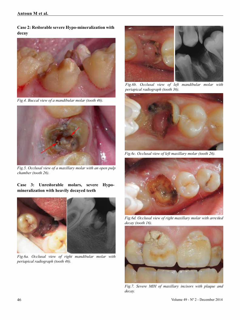
46 Volume 49 - Nº 2 - December 2014
Fig.4. Buccal view of a mandibular molar (tooth 46).
Fig.6c. Occlusal view of left maxillary molar (tooth 26).
Fig.6d. Occlusal view of right maxillary molar with arrested decay (tooth 16).
Fig.7. Severe MIH of maxillary incisors with plaque and decay.
Fig.6a. Occlusal view of right mandibular molar with periapical radiograph (tooth 46).
Fig.6b. Occlusal view of left mandibular molar with periapical radiograph (tooth 36).
Fig.5. Occlusal view of a maxillary molar with an open pulp chamber (tooth 26).
Case 2: Restorable severe Hypo-mineralization with decay
Case 3: Unrestorable molars, severe Hypo-mineralization with heavily decayed teeth
Antoun M et al.

47Journal of the Lebanese Dental Association
C- In case of severe hypo-mineralization, advanced treatment is required with the pedodontic preformed caps (PPC)11,16, or inlays/onlays and prosthetic rehabilitation for the final extraction of affected tooth16. These extractions must be done before the age of 8-10 years, always in collaboration with the pediatric dentist: they allow a proper repositioning of the unaffected second permanent molar. The choice of therapy, however, depends not only on the extent of hypo-mineralization of incisors and molars, but also on Angle's Class and space available14. Severely affected molars can receive endodontic treatment, instead of extraction18.
Figure 1a displays a moderate MIH of a mandibular molar (tooth 36); while Figure 1b exhibits a moderate MIH of a maxillary molar (tooth 16); both were treated with resin composite while keeping the slightly hypo-mineralized cusp untouched. A follow-up with fluorinated gel, combined with patient's good motivation for dental hygiene, can delay the progression of the disease. Similar treatment can be achieved for hypo-mineralized incisors (displayed in figure 2). Composite restorations can preserve healthy hard tissue for long time and reduce the psychological impact of MIH unaesthetic brown spots on anterior incisors (Figure 3).
Figures 4 and 5 display mandibular and maxillary molars with severe hypo-mineralization involvement (endodontic therapy, followed by a crown restoration, are needed).
Figures 6a-b-c and d exhibit unrestorable first molars of a 12-year old patient, indicating the amount of tooth damage and the rapidity of the disease, while incisors can still be conserved using composite restorations (in figure 7).
CONCLUSIONEarly recognition of MIH is essential since damage
of dental hard tissues can be fast. Impairment should be anticipated with the appropriate treatment. Patients in Lebanon aren’t safeguarded against MIH and dentists should be aware and ready to implement a diagnosis and a treatment in order to stop disease progression and limit hard tissue's damage.
Our paper is only a preliminary clinical observation. We have no comparison group: consequently, we cannot draw conclusions about MIH and its pathophysiology. However, cohort studies and randomized controlled trials (RCTs) on MIH are warranted in order to further elucidate various aspects of this dental disease.
REFERENCES1. Angelillo IF, Romano F, Fortunato L, Montanaro D. Prevalence of dental caries and enamel defects in children living in areas with different water fluoride concentrations. Community Dent Health 1990;7:229-236.2. Arrow P. Prevalence of developmental enamel defects of the first permanent molars among school children in Western Australia. Aust Dent J. 2008;53:250-259. 3. Beslot A., Villette F. Prise en charge précoce de l’amélogenèse imparfaite. L’information dentaire 2010:4-2;12-19.4. Bimboire J. La première molaire permanente immature : Mise à jour des connaissances actuelles, Université Bordeaux Segalen 2013.5. Cameron A., Widmer R. Dental Anomalies.Handbook of pediatric dentistry. Parkinson M., TaylorA., Laing A., third edition. Edinburgh, London, New York, 2008. 6. Fteita D, Ali A, Alaluusua S. Molar-incisor hypomineralization (MIH) in a group of school-aged children in Benghazi, Libya. Eur Arch Paediatr Dent 2006;7:92-95.7. Gündüz K, Zengin Z, Celenk P, Ozden B, Kurt M, Gunhan O. Regional odontodysplasia of the deciduous and permanent teeth associated with eruption disorders: A case report. Med Oral Patol Oral Cir Bucal 2008;13:E563-566.8. Hébert S. L’hypominéralisation des molaires et des incisives. 12es Journées de Santé Dentaire Publique du Québec, St-Jean-sur-Richelieu, 14 juin 2013.9. Jasulaityte L, Veerkamp JS, Weerheijm KL. Molar incisor hypomineralization: review and prevalence data from the study of primary school children in Kaunas/Lithuania. Eur Arch Paediatr Dent 2007;8:87-94.10. Jasulaityte L, Weerheijm KL, Veerkamp JS. Prevalence of molar-incisor-hypomineralisation among children participating in the Dutch National Epidemiological Survey (2003). Eur Arch Paediatr Dent 2008;9:218-223.11. Kellerhoff N.M., Lussi A. L’hypominélarisation des molaires-incisives. Mens Suisse Odontostomatol 2004;114:250-253.12. Kukleva MP, Petrova SG, Kondeva VK, Nihtyanova TI. Molar incisor hypomineralisation in 7-to-14-year old children in Plovdiv, Bulgaria-an epidemiologic study. Folia Med (Plovdiv) 2008;50:71-75.13. Kusku OO, Caglar E, Sandalli N. The prevalence and aetiology of molar-incisor hypomineralisation in a group of children in Istanbul. Eur J Paediatr Dent 2008;9:139-144.
Antoun M et al.

48
14. Muratbegovic A., Markovic N., Ganibegovic Selimovic M. Molar incisor hypomineralisation in Bosnia and Herzegovina: aetiology and clinical consequences in medium caries activity population. Eur Arch Paediatr Dent 2007;8:189-194.15. Purohit D, Bhat M, Satish V. Sequential Approach in the Management of Molar Incisor Hypomineralization: A Case Report. Indian J Stomatol 2011;2:63–6716. Rouas P.,Bandon D., Vaysse F. Les hypominéralisations molaires-incisives Diagnostic et prise en charge. L’Information Dentaire 2010;9:13-19.17. Shubha AB., Sapna HEDGE. Molar-Incisor Hypomineralization: Review of its prevalence, etiology, clinical appearance and management. Int J Oral Maxillofac Pathol 2013:4:26-33.18. Soviero V, Haubek D, Trindade C, DA Matta T, Poulsen S. Prevalence and distribution of demarcated opacities and their sequelae in permanent 1st molars and incisors in 7 to 13-year-old Brazilian children. Acta Odontol Scand 2009;67:170-175.19. Subramaniam P, Mathew S, Sugnani SN. Dentinogenesis imperfecta: a case report. J Indian Soc Pedod Prev Dent 2008;26:85-87. 20. Taiani H. MIH (Molar-Incisivor Hypomineralisation) : Sémiologie et hypothèses étiologiques. Thèse pour le diplôme d’état de Docteur en Chirurgie-Dentaire, Université d’Auvergne Clermont Ferrand I, Unité de formation et de recherche d’odontologie, 2008.21. US Department of Health and Human Services. Recommendations for Using Fluoride to Prevent and Control Dental Caries in the United States 2001;50:1-4222. William V, Messer LB, Burrow MF. Molar incisor hypomineralization: review and recommendations for clinical management. Pediatr Dent 2006;28:224-232.23. Weerheijm KL and Mejàre I. Molar incisor hypomineralisation: a questionnaire inventory of its occurence in member countries of the European Academy of Paediatric Dentistry (EAPD). Int J Pediatr Dent 2003 Nov;13(6):411-416.24. Weerheijm KL. Molar Incisor Hypomineralisation (MIH). Euro J Pediatr Dent 2003 Sept;4(3):114-120.25. Gotler M, Ratson T. Molar Incisor Hypomineralisation (MIH): a literature review. Refuat Hapeh Vehashinayim (1993). 2010 Apr;27(2):10-8,60 (Article in Hebrew).
Correspond with:Paul [email protected]
Antoun M et al.
Journal of the Lebanese Dental AssociationVolume 49 - Nº 2 - December 2014

49
INTRODUCTION Patients with inherited bleeding disorders may
often present complications in their daily life, such as recurrent nose bleeding, easy bruising, and longer-lasting hemorrhages associated with trauma, menses, or childbirth. Such complications may often lead to the diagnosis of the bleeding disorder; however, considering the oral cavity’s highly vascular environment, dentists could actually be the ones to first suspect hematologic pathologies, especially after a tooth extraction1. Patients with hematologic conditions are not exempt from oral pathologies, and their management in the dental setting can be challenging. Pediatric dentists, in particular, can find themselves dealing with very young, anxious, or agitated children affected with a bleeding disorder, which raises the risk of injury, trauma, and their subsequent complications1. One type of inherited blood disorder a dentist may have to deal with is von Willbrand’s disease -vWD-, and the purpose of this paper is to report on dental management of two brothers affected with this particular pathology.
VON WILLEBRAND’S DISEASE -vWD-Von Willebrand factor (vWF) is a high-molecular
weight glycoprotein that plays an essential role in early phases of hemostasis by promoting platelet adhesion to the subendothelium and platelet aggregation under high shear stress conditions, and also participates in the proper functioning of factor VIII (FVIII)2. von Willebrand’s disease (vWD), named after Finnish physician Erik Adolf von Willebrand (1870-1949) who first described it in the 1920s, is characterized by a deficiency in vWF (which circulates in a non-covalent complex with factor VIII).
vWD is classified into three subtypes. Type 1, the most frequent, is characterized by a partial quantitative vWF deficiency, resulting in a reduced quantity of circulating vWF (the clotting factor activity level is decreased to between 5 and 35% of normal activity). Type 2 is characterized by defect in function rather than amount of vWF (it is a a qualitative defect, also leading to a decreased clotting factor activity level). Type 3, the least frequent, is characterized by the absence of vWF with clotting factor activity level below 1%; types 2 and 3 are the most symptomatic3. The disease generally exhibits an autosomal dominant pattern of inheritance (types 1 and 2) with sometimes a recessive pattern (types 2 and 3) and is presently thought to be the most prevalent amongst inherited bleeding disorders3. A fourth type, acquired vWD, is not hereditary, “a syndrome of multiple etiologies and
Pediatric dental management of patients with von Willebrand disease: reporting the cases of two brothers.Nada R. Chedid1, DCD, CAGS, M.Sc.D., DEA, Dr. Univ., FICD, Jean-Claude Abou Chedid2, DCD, CES Ped. Dent., DEA, Dr. Univ., Zeina Akil3, DCD
Abstractvon Willebrand disease -vWD- is a genetic hematologic condition characterized by an impaired function of the
von Willebrand factor of coagulation. It is the most common hereditary blood-clotting disorder in humans. Dental management of this type of disease includes thorough cooperation with the patient’s hematologist and rigorous medical and dental treatment protocols. The former aspect includes pre-operative, intra-operative, and post-operative hemostasis controls, often with factor replacement, and the latter may dictate radical decisions in the interest of patient’s general health status. This clinical paper addresses the dental management of two brothers affected with von Willebrand disease and discusses the rationale for treatment choices.
Department of Pediatric and Community Dentistry, Saint- Joseph University Faculty of Dental Medicine, Beirut, Lebanon 1. Professor and Co-Director of Postgraduate Program, 2. Associate Professor and Department Chairperson3. Postgraduate Student
Pediatric Dentistry / Oral Medicine
Journal of the Lebanese Dental AssociationVolume 49 - Nº 2 - December 2014

50 Volume 49 - Nº 2 - December 2014
diverse pathophysiology, but it has a common clinical presentation” (Kumar, Pruthi, and Nichols, 2002)4. The severity and clinical manifestations of vWD depend on the level of factor present and vary from mild (showing muco-cutaneous bleeding and menorrhagia), to moderate (presenting frequent hematomas and bleeds associated with trauma), to severe (with spontaneous bleeding, particularly in joints).
When vWD is suspected, its diagnosis requires a full investigation by a hematologist, including a review of the patient’s and family’s medical histories and specific blood tests. In vWD, bleeding time and activated partial thromboplastin time are prolonged, while partial thromboplastin time is normal3. The most specific tests for vWD are measurements of plasma vWF antigen and ristocetin cofactor activity, which provides a functional assessment of vWF. Once diagnosed, the disease is managed either pharmalogically with desmopressin*, which helps release vWF from the endothelial cells4,or with factor replacement, prescribed usually for severe deficiency, severe trauma, or surgery, in accordance with each individual patient’s case severity and needs. Replacement factors used in vWD are usually purified human plasma-derived factor VIII concentrates5.
Treatment-wise, prevention is the key factor to avoid complications during daily life activities and dental treatments. However, the fear of bleeding while brushing often results in vWD patients neglecting oral care and avoiding dental care, which may turn the need for simple, bleeding-free operative procedures into a need for complex, potentially bloody treatments2. Dental treatment in vWD involves three phases: a pre-operative phase including intravenous factor replacement according to disease type and severity, followed by intra- operative and post-operative phases involving maximum hemostatic control procedures.
REPORTING THE CASES OF 2 BROTHERS
A 5 year, 7-month-old boy (patient A) and his 9 year-old brother (patient B) presented to the Pediatric Dentistry Department at Saint-Joseph University, Beirut, Lebanon, for dental treatment. Both patients
had severe von Willebrand’s disease and were under the regular care of a hematologist (figure 1). They had no other medical disorders and no history of surgeries. Their parents (who were not blood related) and a third sibling, a 4 year-old girl, did not present clinically with the disease (figure 2) and a younger, 2 year-old brother had not been tested yet. Intra-oral examinations were done as gently as possible, without dental probes: they revealed high caries counts associated with inappropriate dietary habits and lack of oral hygiene (figures 3 to 6). In fact, the fear of bleeding had led the parents to avoid advocating their children to brush their teeth, and concern for their children’s medical condition had not included an awareness of the need for regular dental care. Dental panoramic radiographs (figures 7 and 8) were obtained to complete dental examinations and help elaborate treatment plans.
Patient A’s examination showed the presence of all primary teeth with no permanent teeth yet. Carious lesions were present in all the primary molars (teeth A, B, I, J, K, L, S, T) as well as the maxillary right primary lateral and both central incisors (teeth D, E, F). There were pulp exposures in teeth B and I, and extensive crown destructions with pulp necrosis in teeth E and F. As for patient B, all his permanent first molars and incisors (teeth 16, 26, 36, 46 and 12, 11, 21, 31, 32, 41, 42) except the maxillary left lateral (tooth 22) were present, as well as all the primary canines, first and second molars (teeth A, B, C, H, I, J, K, M, R, S, T) except the mandibular left first primary molar (tooth L). All teeth except the incisors showed deep carious lesions; teeth K and M were reduced to root tips.
Patient A’s behavior and cooperation were rated 3 on the Frankl's behavior rating scale, corresponding to a positive behavior. He accepted our approach, but with caution, and was willing to comply with the team, but with reservation. Taking into consideration his age, his unstable state of cooperation, the travel commute between his home and the dental treatment center and the extensive dental procedures needed, decision was made to perform these procedures, by quadrant, under nitrous oxide and oxygen mixture inhalation sedation. Patient B was older, and therefore theoretically more capable of cooperation in the dental treatment setting than his younger sibling. However, given the same considerations regarding commute and treatment needs
* Desmopressin is a synthetic replacement for vasopressin (trade name is DDAVP or Deamino-D-Arginine-Vasopressin). Desmopressin raises plasma levels of FVIII.
Chedid N et al.

51Journal of the Lebanese Dental Association
Chedid N et al.
(specifically the extraction of two permanent molars), his dental procedures were also to be performed, by quadrant, under inhalation sedation.
A written, full description of each patient’s medical condition, as well as approval for the planned dental procedures under local analgesia and nitrous oxide-oxygen mix inhalation sedation were obtained from the treating hematologist, who also specified, in writing, the protocol for pre-operative factor replacement and postoperative precautions. Parental informed consent for dental treatment under the specified conditions was obtained as per Saint Joseph University’s dental treatment center policy requirements.
DENTAL MANAGEMENT14,15,16,17,18,19,20
Pre-operative phaseAs indicated by their hematologist, the children
arrived 30 minutes before each appointment at the University Center for Family and Community Health (UCFCH) at the School of Medicine, Saint-Joseph University, Beirut, and were administered each an intra-venous injection of 500 UI/dl of Haemate® (CSL Behring, Marburg, Germany), a pasteurized plasma-derived vFVIII concentrate, to allow for a six hours window of hemostatic control (figures 9 and 10).
Upon arrival at the dental treatment center, located at the same medical campus, each child was warmly greeted and led to a dental chair where he sat. A nitrous oxide/oxygen gas mask was placed on each patient’s nose with instructions to breathe from the nose and avoid oral breathing. The mother was previously asked to inform the dental team in case of nasal obstruction due to any type of upper respiratory tract infection, the presence of which would have necessitated rescheduling the treatment session. After initial inhalation of 100% oxygen, and progressive introduction of nitrous oxide, a 50/50 mix concentration level was deemed appropriate for patient’s A comfort and cooperation, and maintained during the session at a flow rate of 4 liters per minute (L/min). Patient B was sedated at a similar flow rate of 4 L/min; however his comfort level was estimated and provided at a 40 percent nitrous oxide and 60 percent oxygen mix (figure 11).
Intra-operative phaseDental treatment was initiated once each child was
calm and appropriately relaxed. Precautions were taken
to prevent injuries, trauma and bleeding: each patient was seated in a comfortable position avoiding internal bleeding of joints and muscles, the mask was placed gently on the nose, and the suction ejector wrapped in sterile gauze in order to circumvent injuring lips, cheeks, and oral mucosae. All analgesia procedures consisted of periapical injections using a local anesthetic containing 2% lidocaine and a vasoconstrictor diluted at a 1:100,000 concentration (Septanest®, Septodont, France), preceded by the application at injection sites of a 20% benzocaine topical anesthetic (Gumnumb®, Crosstex, USA) for two minutes before infiltrations. Treatment sequences were followed as planned, over four sessions for each child. Tables 1 and 2 summarize dental work performed in each session for patients A and B, respectively.
Before any operative or surgical treatments were initiated, a dental cleaning was performed, for each child, using a fine ultrasonic dental scaler, followed by polishing with a medium grit 1,23% acidulated phosphate fluoride (APF) prophylactic paste (Gelato® Deepak Products Inc, USA). At the end of the first treatment session, topical 1,23% APF gel (Ionite® Dharma Resaerch Inc, USA) was applied for four minutes in a preformed silicone tray and patients were asked to refrain from eating and drinking for one hour afterwards.
A rubber dam was used with the clamp placed in a non-traumatic position for all restorative and endodontic treatments. The restorative treatments were completed prior to any procedure with a potential to induce bleeding, to ensure good isolation from blood and saliva during restorative materials placement. Restorative technique included acid etching, rinsing, bonding placement and curing, composite placement, curing and polishing. An additional interdental papillae injection of the anesthetic (cited previously) was used prior to the insertion of matrix bands and wedges when restoring Cl II cavities, to control papillary bleeding.
During pulpotomy procedures, pulpal bleeding was controlled by local pressure with sterile cotton pellets then formocresol (Tricresol and formalin, Produits Dentaires, Vevey, Switzerland) was used as a fixating agent, after which the cavity was filled with a fast setting reinforced zinc oxide and eugenol paste (IRM®**, Dentsply, Caulk, USA).
Teeth preparation for the stainless steel crowns
** IRM = Intermediate Restorative Material

52 Volume 49 - Nº 2 - December 2014
Von Willebrand Disease Pediatric Dental Management: reporting on the case of two brothers
Table 1- Dental treatment sessions for patient A Tooth number and
Treatment Session
Composite
resin restoration
Pulpotomy
Stainless steel
crowns
Extraction
Dental cleaning
and topical fluoride
application
1 A (OM) D (B) B B _ 1
2 J (OM) I I _ _
3 _ _ L K _ _
4 _ _ S T
E F
Table 2- Dental treatment sessions for patient B Tooth number and
Treatment Session
Composite
resin restoration
Sealant
Space maintainer
Extraction
Dental cleaning
and topical fluoride
application
1 16 (OP) 11 53, 54, 55 1
2 26 (OM)
63, 64, 65
3
16, 26 band fitting Upper arch impression 73, 75, 36
4 83 (M, D)
Upper bilateral fixed space maintainer
cementation 84, 85, 46
Chedid N et al.

53Journal of the Lebanese Dental Association
Chedid N et al.
required a sub gingival reduction; to avoid excessive bleeding during this procedure, a local interdental papillae infiltration (of the previously cited anesthetic) was performed. The stainless steel crowns were left intact to prevent trauma to gingival tissues that might result from improper polishing of reduced crown gingival limits. After crown cementation, excess cement was carefully removed to preclude any further gingival irritation. The same was done for the bands of patient B’s Nance-type upper bilateral fixed space maintainer during appliance cementation. The appliance’s soldering and acrylic parts had been thoroughly polished during its manufacturing to preclude soft tissue irritation after cementation.
The major concern in the treatment of vWD patients resides in extractions. In patient A’s case, the extraction of the two maxillary primary centrals was performed with care being taken to avoid excessive trauma. An absorbable gelatin sponge (Surigispon Aegis ®, Lifesciences, India) was placed in the sockets to manage post-extraction alveolar bone bleeding. Patient B’s extractions followed the same protocol, with the permanent teeth being extracted by a specialist from the oral surgery department at the dental treatment center.
Post-operative phaseAs recommended by the hematologist, oral
administration of half an ampoule (500 mg) of tranexamic acid*** (Exacyl®; Sanofi-Winthrop laboratory, Gentilly, France) was administered to each child every six hours for five days following every session. Exacyl® is a competitive inhibitor of plasminogen activation and a noncompetitive inhibitor of plasmin; in concentrations of 1g per 10ml, it prolongs thrombin time, thus acting as an antifibrinolytic haemostatic. Post-operative pain was controlled with acetaminophen, prescribed according to patient’s age and weight in syrup form (Panadol®; GlaxoSmithKline, Australia).
Post-operative recommendations regarding eating habits and oral hygiene were provided to the children and their parents. The parents were advised to brush the patients’ teeth vertically with a soft toothbrush, three times daily (after each meal), with age-appropriate toothpaste, and as of the second post-operative day.
The parents were notified to make sure their children cleanse the extraction sites after each meal, with saline-soaked gauze, 24 hours after the extractions, and for five more consecutive days. Soft and cool meals were recommended after each session. No sticky food was allowed after cementing the crowns.
Prognosis for the performed dental treatments was positive (figures 12 to 15). A checkup was scheduled for one month after treatment completion, then the patients were put on a four- months recall schedule.
CASES DISCUSSIONNowadays, dentists are facing an ever-increasing
number of conditions associated with abnormal hemostatic function, sometimes associated with the possibility of excessive blood loss, delayed wound healing, and infection. Despite the complexity of the hemostatic process, the means of controlling it have considerably evolved through the last decade3.
Studies carried out to evaluate the aspects of oral health in hemophilic patients have shown that caries risk factors are the same ones encountered in the general population, namely poor oral hygiene, sugar containing diet, low socio-economic status, reduced host-resistance, and infrequent dental visits. People with bleeding disorders may neglect oral health care, due to gingival bleeding after minor trauma (such as tooth brushing); however, there is some controversy surrounding dental caries prevalence in hemophilic children. Žaliuniene and co-workers observed, in a recent study6, better dental health was observed in children with hemophilia as compared to their healthy counterparts and concluded that hemophilia is not an additional risk factor for caries when other caries-related determinants are controlled.
The use of nitrous oxide/oxygen sedation in cases similar to the ones described here has often been reported. As far back as 1982, Kaufman and colleagues described the use of such sedation in a hemophilic patient, safely, and with success7. According to the recommendations of the American Academy of Pediatric Dentistry AAPD- (2013)8, the use of nitrous oxide and oxygen mix as analgesic/anxiolytic is indicated in cases of fearful, anxious patients, patients with special healthcare needs, and those undergoing lengthy dental procedures. The present cases fulfilled *** Tranexamic acid (TXA) = an antifibrinolytic (an
inhibitor of fibrinolysis)

54 Volume 49 - Nº 2 - December 2014
Fig. 1. Patient A: hematologic test results confirming vWF deficiency
Fig. 3. Patient A: preoperative view of maxillary arch.
Fig. 5. Patient B: preoperative view of maxillary arch
Fig. 7. Patient A: preoperative panoramic radiograph Fig. 8. Patient B: preoperative panoramic radiograph
Fig. 2. Patients’ younger sister’s hematologic test results confirming normal vWF
Fig. 4. Patient A: preoperative view of mandibular arch.
Fig. 6. Patient B: preoperative view of mandibular arch
Chedid N et al.

55Journal of the Lebanese Dental Association
Chedid N et al.
these three indications. Child A was a cautious and anxious patient, child B was very young and couldn’t cooperate properly for chairside treatment, and both children had obvious special health problems, needed extensive dental treatment, and travelled long distance for dental care. The option of sedation use was discussed with the parents who agreed. The goal of this therapeutic choice was to establish a trusting relationship between the patients and their (pediatric) dentists, so that patients could cooperate for an optimal treatment.
Local analgesia during dental treatment is crucial, not only to relieve intra-operative pain, but also to minimize postoperative pain and help with hemostasis. Precautionary measures are to be followed: slow injection allows time for the local anesthetic to diffuse through the tissues and minimizes bruising, and an anesthetic with vasoconstrictor should be used for additional local haemostasis. However, there is a controversy regarding regional nerve blocks infiltration: though it is thought to carry an increased risk of hematoma, internal bleeding and possible subsequent suffocation, the use of modern fine-gauge single-use needles may lower that risk9. Nonetheless, in the cases reported here, a choice was made to avoid regional block analgesia and use local periapical and interpapillary injections, assisted by nitrous oxide/oxygen inhalation sedation.
High-speed vacuum aspirators and saliva ejectors can cause hematomas by aspirating soft tissues during use. Therefore, saliva ejectors were wrapped in sterile gauze to prevent them from harming surrounding oral mucosae and soft tissues10. The use of rubber dam is an essential element of dental treatment; as suggested by Powell and Bartle11, it provides perfect isolation of teeth for better success rates. Therefore, rubber dams were used for all restorative endodontic treatments for improved visibility, better management of excessive bleeding, gingival retraction, and avoidance of trauma to oral mucosae and tissues.
Regarding the restoration of patient A’s right and left mandibular primary molars with preformed stainless steel crowns (SSC) while maintaining pulp vitality, the decision was based on several clinical aspects: the carious lesions were arrested with no extension to the pulp; there were no signs or symptoms of pulpal
inflammation or infection; the use of a rubber dam was not possible due to the collapse of the lingual folds of the mentioned teeth; and with no nerve block infiltration, a sub-gingival placement of the clamp would have been both traumatizing and painful. Therefore, a decision was made to keep these teeth vital and restore them with stainless steel crowns for better protection. A minimal preparation was performed mostly in the interdental region, using a variant of the Hall technique, which consists in restoring vital carious primary teeth with SSCs with no crown preparation. Studies12 have shown positive results with very low failure rates compared to the conventional techniques, due to the preformed SSCs isolating carious lesions from the oral environment and procuring full coverage of the remaining tooth structure.
The choice of extracting the two mandibular first permanent molars in patient’s B case was made after thorough discussion with the orthodontic department, as well as the endodontic and prosthodontic departments. Rationale for the decision to extract was that even though an endodontic treatment may be carried out and completed with a good prognosis, subsequent restoration of the teeth may be more problematic, considering the amount of tooth structure lost (caries) and the need for a restoration and material that could prevent crown fractures while resisting micro-leakage over a period of several years, until the patient was old enough for regular recommended prosthodontic treatment. Therefore, it was deemed best for the patient to extract both mandibular first permanent molars and allow for the second permanent molars to erupt. The patient was at the recommended age for such an extraction13, which is at the beginning of the furcation calcification of the second permanent molars. Another reason for the extraction decision was the uncertainty regarding patient’s capacity for rigorous oral hygiene and possibility of returning for checkups on a potential post endodontic restoration.
Good oral health care and regular preventive measures are essential in the management of bleeding disorders, because they help reduce the need for treatment and emergency therapies, thus decreasing stress for patient and dentist, as well as treatment costs and number of visits. In this case, parents were advised to commit to daily gentle tooth brushing, interdental

56 Volume 49 - Nº 2 - December 2014
Fig. 9. Patient A: vWF injection prior to dental treatment
Fig. 11. Patient B ready for dental treatment
Fig. 13. Patient A: postoperative view of mandibular arch
Fig. 15. Patient B: postoperative view of mandibular arch
Fig. 10. Patient A: report from the University’s Family and Community Health Center confirming vWF injection and dosage, and time given (prior to dental treatment).
Fig. 12. Patient A: postoperative view of maxillary arch
Fig. 14. Patient B: postoperative view of maxillary arch
Chedid N et al.

57
Chedid N et al.
cleaning with floss, using topical fluoride in mouthrinse form, and adhering to regular dental check-ups. The children were instructed to limit their consumption of high-sugar or acid containing foods and drinks.
CONCLUSIONThe key to successful dental management in a
child patient with an inherited bleeding disorder is good coordination between the pediatric dentist, the hematologist and a third, most important party, the patient/parents team. Oral care advice should be given to the patient directly after the hematologic condition’s diagnosis, therefore the hematologist should be aware of dental health preventive measures, and informed about any necessary dental treatments so that he / she can in turn inform dentist about the precautions and protocol for hemostatic control. The dentist should then specify the oral health care measures to both patient and parents. Finally, a most important responsibility of the pediatric dentist in particular is to procure a safe, reassuring, and comfortable environment for the child patient’s care.
REFERENCES1. El Batawi H. Minimizing the Risk of Perioperative Bleeding in a Child with Hemophilia A during Dental Rehabilitation under General Anesthesia: A Case Report. IJCPD. 2013; 6(3):217-222.2. Sumner M, Williams J. Type 3 von Willebrand disease: assessment of complications and approaches to treatment. Results of a patient and Hemophilia Treatment Center Survey in the United States. Haemophilia. 2004; 10(4):360-366.3. Israels S, Schwetz N, Boyar R, McNicol A. Bleeding Disorders: Characterization, Dental Considerations and Management. J Can Dent Assoc. 2006; 72(9):827.4. Kumar S, Pruthi RK, Nichols WL, Acquired von Willebrand Disease, Mayo Clin Proc. 2002;77: 181-1875. Lethagen S, Carlson M, Hillarp A. A comparative in vitro evaluation of six von Willebrand factor concentrates. Haemophilia. 2004; 10(4): 243-249.6. Zaliuniene R et al. Do hemophiliacs have a higher risk for dental caries than the general population. Medicina (Kaunas). (Epub, ahead of print). 7. Kaufman E, Kadari A, Galili D, Garfunkel A. Nitrous Oxide Analgesia in Selected Dental Patients. Anesthesia Progress 1982 May-Jun;29(3):78-80.8. Guidelines on use of nitrous oxide for pdiatric dental patients. AAPD reference manual. 2013.
9. Hewson ID, Daly J, Hallett KB, Liberali SA, Scott CLM, Spaile
G, Widmer R et al. Consensus statement by hospital based dentists providing dental treatment for patients with inherited bleeding disorders. Australian Dental Journal. 2011; 56: 221–226.10. Evans BE, Aledort LM. Hemophilia and dental treatment. J Am Dent Assoc. 1978; 96:827-834.11. Powell D, Bartle J. The hemophiliac: Prevention is the key. Dent Hyg. 1974; 48:209-214.12. Innes NP, Evans DJ. Modern approaches to caries management of the primary dentition, Br Dent J. 2013; 214(11):559-566.13. Cobourne M, Williams A, Harrison M. National clinical guidelines for the extraction of first permanent molars in children, Br Dent J 2014;217:643-64814. O'Brien SH. Common management issues in pediatric patients with mild bleeding disorders. Semin Thromb Hemost 2012 Oct;38(7):720-726.15. Dellinger TM et al. Glycogen storage and von Willebrand's Disease implications for dental treatment: dental management of a pediatric patient. Spec Care Dent 1998 Nov-Dec;18(6):243-246.16. Vierrou AM, de la Fuente B, Poole AE, Hoyer LW. DDAVP (desmopressin) in the dental management of patients with mild or moderate hemophilia and von Willibrand's disease. Pediat Dent 1985 Dec;7(4):297-30117. Neff AT, Sidonio RF Jr. Management of vWD. Hematology Am Soc Hematol Educ Program 2014 Dec 5;(1):536-541.18. El Sherif NH, Narouz MF, ElKerdany TA, El Habashy SA. von Willbrand Factor and factor VIII levels in Egyptian children with newly diagnosed acute lymphoblastic leukemia in relation to peripheral blast cells and steroid therapy. J Pediatr Hematol Oncol 2014 Oct;36(7):518-523.19. Bonduel M et al. von Willebrand disease in children: diagnosis and management of a pediatric cohort in one single center in Argentina. Semin Thromb Hemost 2011 Jul;37(5):560-567.20. Bornert F et al. Hemostatic management in pediatric patients with type I von Willebrand disease undergoing oral surgery: case report and literature review. J Oral Maxillofac Surg 2011 Aug;69(8):2086-2091.
Corresponding author: Nada R. [email protected]
Journal of the Lebanese Dental AssociationVolume 49 - Nº 2 - December 2014

58
INTRODUCTIONIRR is a loss of dental hard tissues as a consequence
of odontoclastic (dentinoclastic) activity, starting from inner root canal wall. IRR is an ongoing inflammatory process that requires the presence of a source of infection -granulation tissue- and the loss or alteration of the odontoblastic layer-predentin1. This lesion starts when pulp chamber becomes inflamed due to an infected coronal necrotic tissue located in any part of the pulp. Among mechanical, chemical or thermal injuries, the most common etiologic factors implicated in IRR are trauma and chronic pulpal inflammation2. In fact, when a tooth is traumatized, infected with bacterial products or overheated by cutting on dentin without an adequate water spray irrigation, vascular changes occur, causing hyperemia and a changing in the metabolism of the pulp. This phenomenon attracts osteoclasts and leads to an inflammatory process, in conjunction with resorbing giant cells in the vital pulp adjacent to the
internal denuded root surface: the loss of dentin begins. For internal resorption to be active, at least part of the pulp must be vital, so that a positive response to pulp sensitivity testing is possible. It is believed that coronal portion of the pulp is often necrotic and provides a stimulus for pulpal inflammation in the more apical parts of the pulp3. Thus, both necrotic infected pulp and inflamed pulp contribute to this type of root resorption3. Therefore, a negative sensitivity test does not rule out active internal resorption. It is also possible that pulp becomes non-vital after a period of active resorption, giving a negative sensitivity test, and giving radiographic signs of internal resorption and apical inflammation. In this case, the inflamed connective tissue exposed to the oral fluids, degenerates and causes pulp necrosis4. The treatment will be more complicated and may negatively influence the long-term endodontic outcome5. In internal inflammatory root resorption, inflamed pulp is the tissue involved in resorbing the root structure. Pathogenesis of internal root resorption is not completely understood. If in‘‘rare’ cases, the inflamed pulp is adjacent to a root surface that has lost its predentin protection, internal root resorption will follow.
Internal root resorptions: current state of understanding.
Valérie Batrouni1, DCD, DESS Endo., Chadi Torbay2, DCD, DESS Endo., MS Cell Therapy/Regen. Med., Jad Azzi3, DCD, DESS Endo., Hassan El-Husseini4, BDS, DESS Endo.
AbstractInternal root resorption (IRR) is a resorption of dentin canal wall due to the action of clastic cells. This rare
endodontic disease starts in pulp chamber and is usually an asymptomatic lesion diagnosed coincidentally through routine radiographs. However, sometimes it may manifest clinically as a “pink spot” and pain can be associated to IRR when it becomes external. IRR usually occurs in anterior teeth and mandibular molars. The treatment of IRR has always been a conventional one, now improved by the use of plenty of armamentarium such as rotary files, bioceramic materials, combination of activated irrigants with ultrasonic tips, LASERs, and the old school temporization with Ca(OH)2. The purpose of this article is to address the current approaches for the diagnosis and management of IRR.
Department of Endodontics, Lebanese University School of Dentistry, Beirut, Lebanon1. Clinical Instructor2. Clinical Instructor3. Associate Clinical Instructor4. Associate Clinical Instructor
Endodontics
Journal of the Lebanese Dental AssociationVolume 49 - Nº 2 - December 2014

59Journal of the Lebanese Dental Association
DIAGNOSIS IRR is a real "endodontic challenge"33: it is usually
asymptomatic with a relatively slow progression. Many causative factors were incriminated for IRR, among them, periodontal infections, vital root resection, cracked tooth syndrome (cracks), idiopathic dystrophic changes within normal pulps, orthodontic forces, traumas, insufficient remaining thickness of dentin after preparation, marginal leakages of crowns, and excessive heat generated during crown preparation on vital teeth (Nilsson et al., 2013 - Mohammadi et al., 2012).
IRR is diagnosed, either clinically through the presence of a pink spot (Fig. 1) located in the coronal part of the tooth, due to the presence of granulation tissue in the coronal dentin6 undermining crown's enamel, or radiographically, with the appearance of an oval shaped enlargement of the root canal space and the original outline of the root canal distorted. Because IRR is initiated in the root canal, the resorptive defect includes some part of the root canal space, and only on rare occasions, when the internal resorptive defect penetrates the root and impacts the periodontal ligament, adjacent alveolar bone will show radiographic changes (Fig 2)3.
IRR is usually painless and the presence of pain depends on pulpal condition or the extension of lesion to periodontal space. Thermal and electric vitality tests are usually positive unless the internal lesion grows and perforates the root to create an external periodontal lesion7. The severity of IRR cannot be precisely judged from 2-dimensional conventional radiographs alone. C. Gabor and co-workers (2012, in J Endo) proved that, contrary to previous beliefs, IPR is very common in teeth with pulpitis. However, development of pulpitis into pulp necrosis stops the resorption process. Most resorptions at this stage are so small that they are not detected on the radiographs. Much more rarely does the resorption have time to develop into a clinically detectable lesion that can be seen on a radiograph of the affected tooth. Cone Beam Computed Tomography (CBCT) is recommended to evaluate the nature and the severity of the lesion because it provides a three-dimensional radiographic appreciation6 to accurately visualize the internal anatomy.
MANAGEMENT16,17,32,33
When the tooth is deemed restorable and with a reasonable prognosis, root-canal therapy is the treatment of choice2. It allows us, at early stages, the removal of granulation tissue and blood supply, and a three-dimensional obturation of the root canal system1. Treatment of internal root resorption is conceptually very easy. Since the resorptive defect is the result of the inflamed pulp and the blood supply to the tissue is through the apical foramina, our endodontic goal is to obtain a blood-free and dry canal with paper points3.
Shaping and cleaning of the lesionIRR lesions present unique difficulties when
preparing and obturating the affected tooth1. The most challenging steps of root canal treatment are
Fig 1. Pink spot appearance on tooth 11 due to internal resorption.
Fig 2. Internal resorption extending towards the periodontal ligament; the tooth is still responsive to vitality tests.
Batrouni V et al.

60 Volume 49 - Nº 2 - December 2014
the removing of granulation tissue and obturation of the newly developed irregular pulp space (Figs. 3-A and 3-B)2. The shape of the resorption defect usually renders it inaccessible to direct mechanical instrumentation1. While our cleaning and shaping can be done with either hand instruments or NiTi rotary files, several studies have demonstrated that large areas of the main root-canal wall will remain untouched by the instruments especially in IRR8. Therefore, with the advantage of multiple irrigation techniques, we will enhance the action of chemicals to optimize the cleaning of these irregularities.
Irrigation
The main goal of instrumentation is to facilitate an efficient irrigation in order to disinfect root canal system. Nowadays, many irrigation solutions compounds have been chemically modified and several mechanical devices have been developed in order to improve the effectiveness of irrigation. Optimal irrigating solutions must have an antimicrobial activity and facilitate removal of microorganisms, organic and inorganic tissue from root canal to predictably obtain a safe and effective treatment9.
Irrigating solutionsSodium hypochlorite (NaOCl) is the most popular
irrigating solution that is used in concentrations between 0,5% and 6%. It is an antimicrobial agent that kills most bacteria and it is the only irrigating solution that dissolves necrotic and vital organic tissue. The problem with NaOCl is that it does not remove the smear layer,it is also used in combination with EDTA* or “citric acid” (CA). Besides its inability to dissolve inorganic material, NaOCl is toxic and has an unpleasant taste9.
To complete chemical irrigation, EDTA and CA should be used to effectively dissolve inorganic material, including hydroxyapatite10-11.
Citric acid marketed at 10% solution, is a chelating agent that reacts with metal to form nonionic soluble chelate. EDTA is used as a 17% neutralized solution.
Di Lenarda and associates12 reported that 1mol L-1 of a CA solution was as effective in removing smear layer as EDTA.
MTAD is a new root-canal irrigant (Singla et al.,
2011) that was introduced by Torabinejad and co-workers13: it is a mixture of doxycycline, a citric acid, and a detergent that removes the smear layer from the instrumented root-canal walls. It has been reported to remove the smear layer (due to the action of citric acid) and provide an antimicrobial effect14,15.
Irrigant activation31
Thorough cleaning of isthmi, lateral canals, curved canals, and complex root canal anatomies (including root-canal systems presenting IRR) is very difficult. The purpose of manual, sonic, ultrasonic, and LASER devices is to agitate the irrigant and facilitate its penetration to all these areas16 in order to improve removal of necrotic debris and biofilms from inaccessible areas of the root canal17.
For many years, manual activation was performed with the aid of syringe/ endodontic needle, the guttapercha master cone/ cone pumping, and brushes (endobrush, navitips). Machine-assisted activation is performed with rotary brushes ( Ruddle brushes, canal brush), continous irrigation during preparation (Quantec-E, SAF), sonic instruments (EndoActivator, Vibringe, Rispi-Sonic, EDDY), ultrasonic apparatus such as PUI/UAI, the pressure alternation devices (Endovac, Rinsendo) and finally, LASERs** (Er:YAG, Er,Cr:YSGG). Taking into consideration the pros and cons of each device, all of them are indicated to achieve a complete chemo-mechanical debridement of the root-canal especially in IRR cases in comparison to passive insertion of hypochlorite with needle. With sonic and ultra-sonic devices, the increase of irrigant effectiveness is due to a three dimensional movement of the instruments that triggers two physical effects: cavitation and acoustic streaming. Cavitation and agitation in fluid-filled root canals create fluid movement and shear stresses along root canal walls, enhancing removal of smear layer and biofilm. Acoustic Streaming refers to the fact that high frequency acoustic oscillation creates small, intense, circular fluid movement around the instrument, swirling in the irrigant which strengthens the cleaning effect and removes more dentin debris and pulp tissue from the root-canal system than syringe irrigation18.
LASERs are an effective tool that has the ability to kill bacteria and remove debris and smear layers from
* EDTA = Ethylene Diamine Tetraacetic Acid ** LASERs: Er:YAG = erbium-doped yttrium aluminium garnet and Er.Cr:YSGG = erbium, chromium:yttrium-scandium-gallium-garnet.
Batrouni V et al.
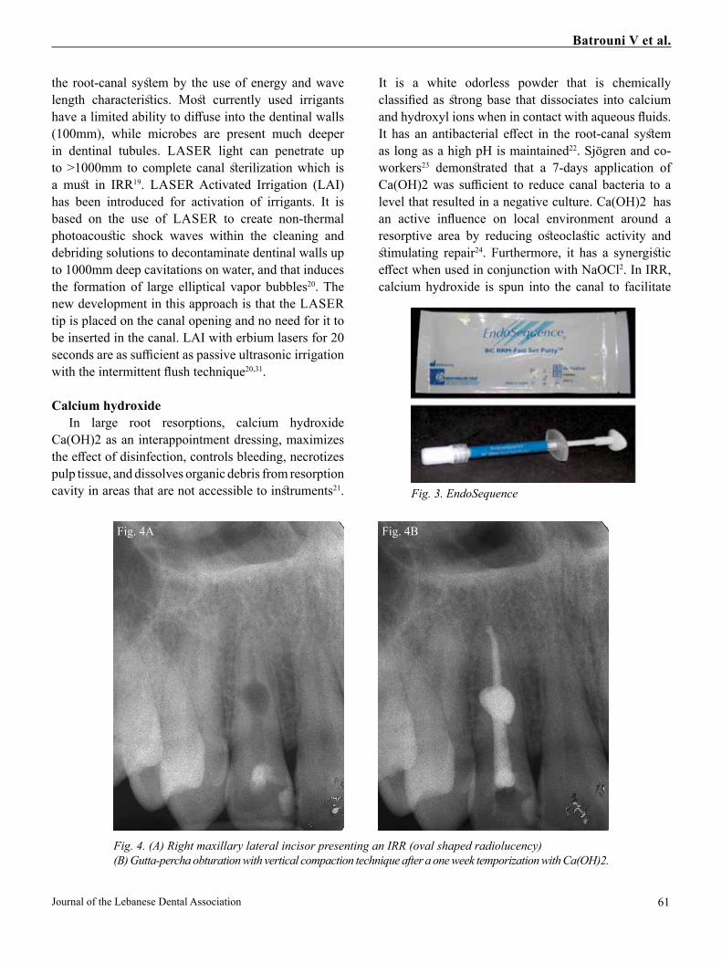
61Journal of the Lebanese Dental Association
the root-canal system by the use of energy and wave length characteristics. Most currently used irrigants have a limited ability to diffuse into the dentinal walls (100mm), while microbes are present much deeper in dentinal tubules. LASER light can penetrate up to >1000mm to complete canal sterilization which is a must in IRR19. LASER Activated Irrigation (LAI) has been introduced for activation of irrigants. It is based on the use of LASER to create non-thermal photoacoustic shock waves within the cleaning and debriding solutions to decontaminate dentinal walls up to 1000mm deep cavitations on water, and that induces the formation of large elliptical vapor bubbles20. The new development in this approach is that the LASER tip is placed on the canal opening and no need for it to be inserted in the canal. LAI with erbium lasers for 20 seconds are as sufficient as passive ultrasonic irrigation with the intermittent flush technique20,31.
Calcium hydroxide In large root resorptions, calcium hydroxide
Ca(OH)2 as an interappointment dressing, maximizes the effect of disinfection, controls bleeding, necrotizes pulp tissue, and dissolves organic debris from resorption cavity in areas that are not accessible to instruments21.
It is a white odorless powder that is chemically classified as strong base that dissociates into calcium and hydroxyl ions when in contact with aqueous fluids. It has an antibacterial effect in the root-canal system as long as a high pH is maintained22. Sjögren and co-workers23 demonstrated that a 7-days application of Ca(OH)2 was sufficient to reduce canal bacteria to a level that resulted in a negative culture. Ca(OH)2 has an active influence on local environment around a resorptive area by reducing osteoclastic activity and stimulating repair24. Furthermore, it has a synergistic effect when used in conjunction with NaOCl2. In IRR, calcium hydroxide is spun into the canal to facilitate
Fig. 3. EndoSequence
Fig. 4. (A) Right maxillary lateral incisor presenting an IRR (oval shaped radiolucency) (B) Gutta-percha obturation with vertical compaction technique after a one week temporization with Ca(OH)2.
Fig. 4A Fig. 4B
Batrouni V et al.

62 Volume 49 - Nº 2 - December 2014
removal of necrotic tissue in the irregular defect, and by that, it will tend to necrotize the remaining tissue in the lacuna and enhance chemomechanical debridement7. At second visit, remnants are removed by a flush of sodium hypochlorite (Chivian, 1991)25 with the aid of mechanical activation3.
Obturation of IRR (Figs 4, A and B)Our aim in obturating IRRs is to completely fill the
root canal system in three dimensions and to have a dense radiopaque filling without overextending into periapical tissues. Therefore, an impermeable tight seal with a soft and warm root filling technique will be provided to prevent oral and apical micro leakage26.
The material needed to completely seal the resorptive defect must be flowable and warm vertical compaction of gutta-percha is recommended in case of IRR17. Thermoplastic gutta-percha techniques seem to give the best results. Gençoğlu and co-workers27 examined the quality of root fillings in teeth with artificially created internal resorptive cavities. They found that Obtura II (Spartan, Fenton, MO, USA) thermoplastic gutta-percha techniques were significantly better in filling artificial resorptive cavities than Thermafil (Dentsply, York, PA, USA), Soft-Core systems (CMS Dental, Copenhagen, Denmark), and cold lateral condensation. Many studies have shown the successful repair of perforating internal resorptions with MTA*, the first in bioceramic materials (Biodentine, EndoSequence® BC RRMTM)** (Fig. 3).
Recently introduced bioceramics are almost similar in chemical composition: alumina, zirconia, bioactive glass, glass ceramics, coatings, and composites, hydroxyapatites, and resorbable calcium phosphates28-29. They are present in three different forms sealer, paste, and putty. The advantages of BC cements are that they tolerate (and require) moisture, tolerate small amounts of blood, provide an excellent seal to dentin, have antimicrobial activity, and have mechanical characteristics close to those of dentin (Fig 3).
Cements are applied directly to resorption area or the pulp chamber using an MTA “gun”. If the resorption is deeper in the root canal, cement is tapped down the canal by inverted paper points in small increments. The filling material for the root canal coronal to the perforation, is chosen based on the need to secure a tight seal and strengthen the tooth. Large internal resorptions, in the
coronal third of the canal in particular, may increase the risk of tooth fracture. The BC used with gutta-percha is the sealer because it is more flowable than the two others.They are proposed as an endodontic sealer with high pH (12.8) during the initial 24 hours of the setting process, hydrophilic not hydrophobic, characterized by an enhanced biocompatibility, they do not shrink or resorb, they set quickly (3 to 4 hours), and they are easy to use. The particle size of BC sealer is so fine (less than 2 microns) that it can actually be delivered with a 0.012 capillary tip. BC sealer has a bioactive characteristic: it uses the water of dentinal tubules to start the hydration reaction of the material. In fact, dentin is composed of approximately 20% of water that initiate the setting of the material and this results in the formation of hydroxyapatite and creation of a bond between dentin and the filling material30. Its major improvement over previous sealers is that if any residual moisture remains in the canal after drying, it will not affect the seal established by the bioceramic cement. That’s why its hydrophilic property and its great bond to dentinal walls make its usage very important in IRRs.
Nevertheless this approach precluded the need for surgical intervention, the latter is sometimes needed when it is not possible to get access to the lesion through the canal. Getting direct access to the lesion and performing a mechanical cleaning of the resorbed defect must be performed after orthograde treatment. If the resorptive defect extends to external root surface, there may be a destruction of adjacent periodontal tissues and this will complicate the treatment and induce periradicular periodontitis. The treatment of choice depends on location and size of the perforation. Sites apical to junctional epithelium can be filled with BC cements carefully and gently tapped against the tissue: large, inverted paper points are excellent tools for the task. The rest of the chamber is filled with some other material depending on the overall restorative plan21.
CONCLUSIONWith modern dental techniques, the prognosis for
treatment of IRR is shifting to be very good. However, if the tooth structure is greatly weakened and large perforation has occurred, clinicians often face the dilemma of whether to treat or extract the diseased tooth. Smaller perforation sizes always lead to a more
* MTA = Mineral Trioxide Aggregate ** RRMTM = Root Repair Material
Batrouni V et al.

63
predictable prognosis of the tooth16. It is important to keep in mind that endodontically treated IRRs should benefit from adequate post-operative restoration with a post, core, and crown.
REFERENCES1. Altundasar E, Demir B. Management of a perforating internal resorptive defect with mineral trioxide aggregate: a case report. J Endod 2009;3:1441–4. 2. Patel D, Ricucci C, DuCak C, Tay F. “Internal root resorption: a review,” J.Endod 2010, vol. 36, no. 7, pp. 1107–11213. Ingle's Endodontics 6 (John Ingle et al.). Published December 31, 2007. Chap. 36: p1352-13544. L. M. Sassone, R. Fidel, M. Faveri, S. Fidel, L. Figueiredo, and M. Feres, “Microbiological evaluation of primary endodontic infections in teeth with and without sinus tract,” Int Endod J 2008 vol. 41, no. 6, pp. 508–515.5. Setzer FC, Boyer KR, Jeppson JR, et al. Long-term prognosis of endodontically treated teeth: a retrospective analysis of preoperative factors in molars. J Endod 2011;37:21–5.6. Lyroudia KM, Dourou VI, Pantelidou OC, Labrianidis T, Pitas IK. Internal root resorption studied by radiography, stereomicroscope, scanning electron microscope, and computerized 3D reconstructive method. Dent Traumatol 2002: 18: 148–1527. Meire M, De Moor R. Mineral trioxide aggregate repair of a perforating internal resorption in a mandibular molar. J Endod 2008;34:220–3. 8. Peters OA, Schonenberger K, Laib A. Effects of four Ni-Ti preparation techniques on root canal geometry assessed by micro computed tomography. Int Endod J 2001;34:221–300.9. Haapasalo M et al. “Irrigation in Endodontics”, Dent Clin North Am 2010;54:291–31210. Czonstkowsky M, Wilson EG, Holstein FA. The smear layer in endodontics. Dent Clin North Am 1990; 34:13–25.11. Loel DA. Use of acid cleanser in endodontic therapy. J Am Dent Assoc 1975; 90: 148–51.12. Di Lenarda R, Cadenaro M, Sbaizero O. Effectiveness of 1 mol L-1 citric acid and 15% EDTA irrigation on smear layer removal. Int Endod J 2000; 33:46 –52.13. Torabinejad M, Khademi AA, Babagoli J, Cho Y, Johnson WB, Bozhilov K. A new solution for removal of the smear layer. J Endod 2003;29:170 –5.14. Shabahang S, Torabinejad M. Effect of MTAD on Enterococcus faecalis-contaminated root canals of extracted human teeth. J Endod. 2003;29(9):576–915. Mohammadzadeh Akhlaghi N, Behrooz E, Saghiri MA. Efficacy of MTAD, Glyde and EDTA in debridement of curved root canals. Iran Endod J 2009;4(2):58–6216. Haapasalo M, Endal U. “Internal inflammatory root resorption: the unknown resorption of the tooth,” Endodontic Topics 2006, vol. 14, pp. 60–79.17. Ozer SY. Diagnosis and treatment modalities of internal and external cervical root resorptions: review of the literature with case
reports. Int Dent Res 2011;1:32–7.18. Huque J, Kota K, Yamaga M, Iwaku M, Hoshino E. Bacterial eradication from root dentine by ultrasonic irrigation with sodium hypochlorite. Int Endod J 1998;31:242–250.19. Moritz A, Schoop U. Lasers in endodontics chapter 6 in: A Mortiz Oral laser application: QuintessenzVerlags-Gmbh. 2006;241-313.20. Roeland JG et al., “Efficacy of Ultrasonic versus Laser-activated Irrigation to Remove Artificially Placed Dentin Debris Plugs”, J.Endod 2010 Sept;36(9):1580-158321. Cohen's Pathways of the pulp. 10th edition:chap10, p375-379:Obturation of the cleaned and shaped root canal system. 22. Siqueira J F. Jr & Lopes HP. «Mechanisms of antimicrobial activity of calcium hydroxide: a critical review”, Int Endod J 1999;32:361-369.23. Sjögren U, Fidgor D, Spångberg L, Sundqvist G (1991) The antimicrobial effect of calcium hydroxide as short-term intracanal dressing. Int Endod J 1991;24, 119-12524. Tronstad L, Andreasen JO, Hasselgren G, Kristerson L, Riis I. pH changes in dental tissues after root canal filling with calcium hydroxide. J Endod 1981;7:17-2125. Bago I et al. “Antimicrobial efficacy of a high-power diode laser, photo-activated disinfection, conventional and sonic activated irrigation during root canal treatment”. Int Endod J 2013;46(4):339-347.26. Delivanis PD, Mattison GD, Mendel SW, “The survivability of F43 strain of Streptococus sanguis in most canals filled with gutta-percha and protocol cement”, J Endod 1983;9:407-41027. Gençoğlu N, Yıldırım T, Garip Y, Karagenç B, Yılmaz H. Effectiveness of different gutta-percha techniques when filling experimental internal resorptive cavities. Int Endod J 2008;41:836-842.28. Best SM et al. Bioceramics: Past, Present and for the Future, Journal of the European Ceramic Society 2008;28(7):1319–1327.29. Hench L., Bioceramics: From Concept to Clinic. Journal Amer. Ceram. Soc. 1991;74(7):1487-1510.30. Koch KA, Brave DG, Nasseh AA, Bioceramic Technology: closing the endo-restorative circle, Part I, Dent. Today, 2010;29:100–105. 31. De Moor RJ et al. Efficacy of ultrasonic versus LASER-activated irrigation to remove artificially placed dentin debris plugs. J Endo 2010;36(9):1580-1583.32. Nillsson E et al. Management of internal root resorption on permanent teeth. Int J Dentistry 2013; Article ID 929486 - 7 pages.http://dx.doi.org/10.1155/2013/929486.33. Mittal S et al. "Internal root resorption: an endodontic challenge". A case series. J Conserv Dent 2014 Nov;17(6):590-593.
Correspond with:Valérie [email protected]
Journal of the Lebanese Dental AssociationVolume 49 - Nº 2 - December 2014
Batrouni V et al.

64
INTRODUCTIONDuring the 1930’s, silver points were introduced by
Jasper and were used as an endodontic filling material because of their ease of handling and placement, their ductility, radiopacity and some antibacterial property1. However, over the past few decades, their use has diminished. In fact, silver points do not seal three dimensionally the root canal system and corrode over time due to the contact with tissue fluids2. Also, it is common to encounter endodontically treated teeth that contain a post3. So, when a periapical disease occurs, and when a treatment is insufficient or in case of insufficient coronal seal (coronal leakage), an endodontic retreatment is indicated. Then, it becomes necessary to remove materials from root canal space in order to reshape and clean the root canal system. Silver points and posts must then be removed to facilitate successful nonsurgical retreatment. Currently, a large number of techniques exist to remove silver points and posts, such as the use of appropriate burs, specialized forceps, ultrasonic instruments in direct or indirect contact, peripheral filing techniques in presence of solvents, chelators, irrigants, and microtube delivery using mechanical adhesion techniques4. The following case report addresses the nonsurgical endodontic retreatment of a permanent mandibular
right first molar (46) with a silver point in the mesio-buccal canal and with a post in the mesio-lingual canal.
CASE REPORTA 59 year-old female was referred to the Endodontic
Department of Saint-Joseph University (Beirut) for an endodontic retreatment of her mandibular right first molar. Clinical examination revealed the presence of a temporary restoration. After removing the restoration, tooth was tender on percussion. Preoperative periapical radiograph revealed the presence of a silver point in the mesio-buccal canal and a post in the mesio-lingual canal (fig. 1).
After completion of regional analgesia, all the cement of temporary restoration was removed with an 802 bur (Maillefer, Switzerland) and the distal wall was adjusted with an Endo-ZTM bur (Maillefer, Switzerland). Then, the tooth was isolated using a rubber dam. Xylol* and K-files (Maillefer, Switzerland) were used to clean the distal canal. Distal canal was then reshaped, cleaned, and sealed without any difficulty (fig. 2).
Then, the entry of distal canal was covered with cement glass ionomer (3M™ ESPE™ Ketac™, Germany) to prevent xylol from entering it. Removal of the post from mesio-lingual canal was then initiated: both a powerful piezoelectric ultrasonic device: Satelec (Marignac, France) and the ultrasonic tip (SO7) were used to release the head of the post.
Endodontic non-surgical retreatment: how to remove a silver point and a post? A case report.Cynthia Antoine Kamel1, DCD, Carla Zogheib Moubarak2, DCD, DES Endo., MS (Oral Biomat. Res.), Dr. Univ.
AbstractRetreatment is a common procedure in endodontics. It is indicated when a periapical disease is present, when
the treatment is insufficient, and in presence of coronal leakage. The goal of an endodontic retreatment is to access pulp chamber and remove materials from root canal space in order to reshape and clean the root canal system. The aim of this case report is to show how a clinician can remove a silver point and a post in order to facilitate successful endodontic nonsurgical retreatment.
Department of Endodontics, Saint-Joseph University Faculty of Dental Medicine, Beirut, Lebanon1. Postgraduate Student2. Assistant Professor
* Xylol is a solvent used to clean up certain hardened cement.
Endodontics
Journal of the Lebanese Dental AssociationVolume 49 - Nº 2 - December 2014

65Journal of the Lebanese Dental Association
The ultrasonic tip was moved around the post circumferentially, after which we implemented "push and pull" motions along its exposed length. Frequent flushing with water was mandatory to cool the tip and the tooth, in order to decrease the potential risk of dangerous heat transfer to periodontal space.
After removing the post in the mesio-lingual canal, removal of silver point was initiated. Xylol and acrylic liquid were used alternately during all the procedure, in order to dissolve the cement that was used to seal the silver point. Piezoelectric ultrasonic device [Satelec (Marignac, France)] was used along with the ultrasonic tip (SO7) to release the head of the post (fig. 3) without eliminating it. K-files 08-10-15 (Dentsply Maillefer, Switzerland) were used to create a prehension zone. When the silver point became loose in the canal, Steiglitz pliers (fig. 4) were used to remove the silver point (fig. 5).
After the removal of the silver point (fig. 5), the 2 mesial canals were reshaped, cleaned, and then sealed (fig. 6).
Fig. 1. Pre-operative periapical radiograph
Fig. 3. The removed post Fig. 5. The removed silver point
Fig. 4. Steiglitz pliers (45o curved)
Fig. 6. Post-operative radiograph
Fig. 2. Obturation of the distal canal
Kamel C, Zogheib Moubarak C

66 Volume 49 - Nº 2 - December 2014
DISCUSSIONAccording to the American Association of
Endodontics -AAE-, a tooth that have had an adequate root canal treatment can last a lifetime. But, sometimes, a treated tooth doesn't heal properly and becomes diseased and/or painful, months or even years after treatment. Indeed, an endodontically treated tooth may cause discomfort and pain for many reasons:
1- Narrow or curved canals that were not treated during the initial endodontic treatment.
2- Complicated root canal anatomy (undetected during the first treatment).
3- Placement of the crown or other restoration was delayed following the endodontic treatment.
4- Post-endodontic restoration did not prevent salivary contamination, exposing -consequently- the root canal filling material to bacteria.
5- A loose, cracked, or broken crown or filling (all these are likely to expose an endodontically treated tooth to a re-infection).
In order to perform a successful nonsurgical endodontic retreatment13, clinician should remove all obstructions that block the root access. Posts and silver points can be present and should then be removed. Many techniques have been developed to remove posts and silver points9,10. Regarding posts, one straight-line access into pulp chamber has to be accomplished and all core materials are to be eliminated and the post, fully exposed. Then, clinician can decide which technique is to be used, knowing that no one particular method always produces a successful result3. However, the use of piezoelectric ultrasonic energy has provided clinicians with a useful adjunct to facilitate post removal, and with minimal loss of tooth structure and root damage such as fractures or roots perforations5. Indeed, an ultrasonic generator in conjunction with the adequate insert will transfer energy, powerfully vibrate, and dislodge posts. Usually, maximum power level is set in order to break the seal between post and tooth structure. The ultrasonic tip has to move around the post circumferentially and up and down along its exposed length. During all the procedure, water spray must be used in order to avoid transferring heat and consequently, damaging the periodontal ligament. The majority of posts can be safely and successfully removed with ultrasonics in about 10 minutes6. For all
those above mentioned reasons, we used this technique to remove the post in the mesio-lingual canal.
Silver points have been utilized for root canal fillings since the 1930’s, often for obturation of narrow canals. In fact, they are easily placed but they are unable to fill the irregularly shaped root canal system permitted leakage. When silver points contact tissue fluids or saliva, they corrode7. Corrosion products have been found to be cytotoxic and produce pathosis or impeded periapical healing2. Actually, their use for root canal obturation is becoming increasingly rare because they are considered to be below the standard of care. Modern techniques and improved materials provided clinicians with better therapeutic options8. During an endodontic retreatment, clinicians must not try to remove immediately a silver point, even if it appears to be poorly adapted to the coronal two-thirds of the canal. In fact, it may fit well in the apical third with a risk of fracturing it. Also, silver points are often affected by corrosion and can be fragile, so clinicians have to be attentive and careful to avoid their fracture during removal. No standardized procedure for successful removal of silver point exists, rather there are various techniques and devices that are established according to the situation9,10. The first step of all techniques is to establish proper access. Frequently, coronal portion of the silver point is embedded in the core material. This material must be carefully removed with burs and ultrasonics. Clinical experience showed that ultrasonics work best when used lightly circumferential to the silver point (the ultrasonic instrument trephines around the silver point, breaking up cements, until the coronal part is free10). Nonetheless, clinicians must be careful not to apply the trephines directly to the silver point, because the portion in contact may be shredded, leaving a smaller segment to work with. Once proper access is established, clinicians should flood the access preparation with a solvent to soften or dissolve the cement, enabling easier removal. An endodontic explorer or small file may be used to carry the solvent down, along the silver point, in order to dissolve as much of the cement as possible11. In this case we used K-files 08-10-15 (Dentsply Maillefer, Switzerland) to carry the solvent down and to create a prehension zone. At this point, the easiest and most predictable technique, is to grasp the exposed end of the silver
Kamel C, Zogheib Moubarak C

67
point with Steiglitz pliers (fig. 4) and gently pull it out of the access cavity preparation (fig. 5). Slow force application is advised because, if too much extraction force is needed, the point may separate. However, when a high force is needed, the ultrasonic tip can be applied on the pliers in order to directly transmit energy that will synergistically enhance retrieval effort10. Clinicians can also place a fine Hedstrom file down into the canal (alongside the point) and the file is then activated by ultrasonics. It will loosen the obstruction and thus the point can be retrieved9. Finally, clinicians should not forget what Clifford J. Ruddle once wrote: “In endodontics, and in particular, retreatment procedures, patience usually pays off.”
CONCLUSIONThe purpose of nonsurgical endodontic retreatment
is to remove materials from root canal system and repair pathologic and/or iatrogenic defects and deficiencies (CJ Ruddle, 2002). Endodontic failures are attributable to inadequancies in shaping, cleaning and obturation, re-infection of root canal system (after loss of coronal seal), and iatrogenic events. Bacterial contamination and leakage are considered to be the sum of all causes (CJ Ruddle, 1991, 1997).
Chronic leakage that reduces the seal of a silver point explains the relative ease of removing the point; ultrasonic instruments are the mainstay of this removal: they brush-cut away remaining restorative materials, fully exposing the silver point14,15.
REFERENCES1. Ingle JI, Bakland LK, Endodontics 5th Edition, Hamilton, London: BC Decker Inc 2002, 645-649.2. Seltzer S., Green DB, Weiner N., DeRenzis F., A Scanning Electron Microscope Examination Of Silver Cones Removed From Endodontically Treated Teeth, Journal of Endodontics, 2004; 30(7): 463-474.3. Ruddle CJ, Nonsurgical Retreatment, Journal of Endodontics, 2004; 30(12): 827-845. (www.endoruddle.com).4. Nehme WB, Elimination of intracanal metallic obstructions by abrasion using an operational microscope and ultrasonics, Journal of Endodontics, 2001; 27(5): 365-367.5. Plotino G., Pameijer C., Grande NM, Somma F, Ultrasonics in Endodontics: A review of the Literature, Journal of Endodontics, 2007; 33(2): 81-95.6. Buoncristiani J., Seto BG, Caputo AA, Evaluation of ultrasonic and sonic instruments for intraradicular post removal. Journal of Endodontics, 1994; 20(10): 486-489.
7. Johnson W., Kulild J., Chapter 10: Obturation of the Cleaned and Shaped Root Canal System, in Cohen Stephen, Hargreaves Kenneth, Pathways of the pulp (tenth edition), Mosby Elsevier, 2011, p.349-388.8. American Association of Endodontists, Use of silver points, 2013, www.aae.org/guidelines9. Gulati S., Sumanthini MV, Shenoy V., Retreatment of Silver Point Obturation: A Case Report and an Overview, Journal of Contemporary Dentistry, 2012; 2(3): 114-118.10. Ruddle CJ. Microendodontic nonsurgical retreatment: silver point removal, Dentistry Today, 1997 February;1-4 (www.endoruddle.com).11. Roda R., Gettleman B., Chapter 25: Nonsurgical Retreatment, in Cohen Stephen, Hargreaves Kenneth, in Pathways of the pulp (tenth edition), Mosby Elsevier, 2011, p.890-952.12. Machtou P, Sarfati P, Cohen AG. Post removal prior to retreatment. J Endo 1989;15(11):552-554.13. Scianamblo MJ. Endodontic failures: the retreatment of previously endodontically treated teeth. Revue d'Odonto-Stomatologie 1988;17(5):409-423.14. Ruddle CJ. Endodontic failures: the rationale and application of surgical retreatment. Revue d'Odonto-Stomatologie 1988;17(6):511 (www.endoruddle.com).15. Ruddle CJ. Nonsurgical endodontic retreatment. J Calif Dent Assoc 2004 Jun;32(6):474-484.
Correspond with:Carla Zogheib [email protected]@gmail.com
Kamel C, Zogheib Moubarak C
Journal of the Lebanese Dental AssociationVolume 49 - Nº 2 - December 2014

Publishing in 2015
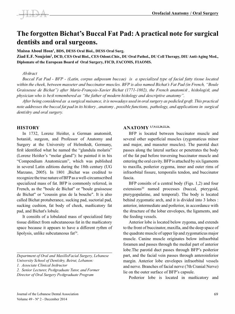
69
HISTORY In 1732, Lorenz Heister, a German anatomist,
botanist, surgeon, and Professor of Anatomy and Surgery at the University of Helmstedt, Germany, first identified what he named the “glandula molaris” (Lorenz Heister’s “molar gland”): he painted it in his “Compendium Anatomicum”, which was published in several Latin editions during the 18th century (UG Marzano, 2005). In 1801 ,Bichat was credited to recognize the true nature of BFP as a well-circumscribed specialized mass of fat. BFP is commonly referred, in French, as the "boule de Bichat" or "boule graisseuse de Bichat" or "coussin gras de la bouche". It is also called Bichat protuberance, sucking pad, suctorial pad, sucking cushion, fat body of cheek, masticatory fat pad, and Bichat's lobule.
It consists of a lobulated mass of specialized fatty tissue distinct from subcutaneous fat in the masticatory space because it appears to have a different rythm of lipolysis, unlike subcutaneous fat24.
ANATOMY 3,7,9,12,20,22,26,
BFP is located between buccinator muscle and several other superficial muscles (zygomaticus minor and major, and masseter muscles). The parotid duct passes along the lateral surface or penetrates the body of the fat pad before traversing buccinator muscle and entering the oral cavity. BFP is attached by six ligaments to maxilla, posterior zygoma, inner and outer rims of infraorbital fissure, temporalis tendon, and buccinator fascia.
BFP consists of a central body (Figs. 1,2) and four extensions20 named processes (buccal, pterygoid, pterygopalatine, and temporal). The body is located behind zygomatic arch, and it is divided into 3 lobes : anterior, intermediate and posterior, in accordance with the structure of the lobar envelopes, the ligaments, and the feeding vessels.
Anterior lobe is located below zygoma, and extends to the front of buccinator, maxilla, and the deep space of the quadrate muscle of upper lip and zygomaticus major muscle. Canine muscle originates below infraorbital foramen and passes through the medial part of anterior lobe.The parotid duct passes through BFP’s posterior part, and the facial vein passes through anteroinferior margin. Anterior lobe envelopes infraorbital vessels and nerve. Branches of facial nerve (7th Cranial Nerve) lie on the outer surface of BFP’s capsule.
Posterior lobe is located in masticatory and
The forgotten Bichat’s Buccal Fat Pad: A practical note for surgical dentists and oral surgeons.Maissa Aboul Hosn1, BDS, DESS Oral Biol., DESS Oral Surg.Ziad E.F. Noujeim2, DCD, CES Oral Biol., CES Odont.Chir., DU Oral Pathol., DU Cell Therapy, DIU Anti-Aging Med., Diplomate of the European Board of Oral Surgery, FICD, FACOMS, FIAOMS.
AbstractBuccal Fat Pad - BFP - (Latin, corpus adiposum buccae) is a specialized type of facial fatty tissue located
within the cheek, between masseter and buccinator muscles. BFP is also named Bichat’s Fat Pad (in French, “Boule Graisseuse de Bichat”) after Marie-François-Xavier Bichat (1771-1802), the French anatomist , histologist, and physician who is best remembered as “the father of modern histology and descriptive anatomy”.
After being considered as a surgical nuisance, it is nowadays used in oral surgery as pedicled graft. This practical note addresses the buccal fat pad in its history , anatomy , possible functions, pathology, and applications in surgical dentistry and oral surgery.
Department of Oral and MaxilloFacial Surgery, Lebanese University School of Dentistry, Beirut, Lebanon:1. Associate Clinical Instructor2. Senior Lecturer, Postgraduate Tutor, and Former Director of Oral Surgery Postgraduate Program
Journal of the Lebanese Dental AssociationVolume 49 - Nº 2 - December 2014
Orofacial Anatomy / Oral Surgery

70 Volume 49 - Nº 2 - December 2014
neighbouring spaces. It extends up to inferior orbital fissure and surrounds temporalis muscle.
Four processes extend from BFP’s body into surrounding spaces (such as infratemporal fossa). Buccal extension, which toghether with the body accounts for about half of BFP’s total weight, lies superficially within the cheek and is largely responsible for its contour: it descends towards mandibular retromolar region, and extends along masseter’s anterior border. Buccal extension is encapsulated by a parotido-masseteric fascia and enters the cheek below the parotid duct.
Pterygoid extension extends in a more downward and backward directions to lateral part of sphenoid’s pterygoid plates. Superficial and deep temporal extensions pass upwards, separating temporal muscle from zygomatic arch. The whole BFP’s body is covered by an envelope of fascia type, preventing spontaneous herniation during surgery.
In neonates, BFP is particularly prominent and herniates more easily. In infants, BFP, is relatively large and referred to as the “sucking pad” (Matarasso,1997):consequently, a limited tear of buccinator muscle will favor its herniation into oral cavity.BFP’s traumatic herniation occurs mainly in infants and young children (Brooke,1978) between ages of 1 to 4 years (Clawson et al.,1968 - Muroki et al.,1996 - Horie et al., 2001).
Blood supply9,18 of BFP originates mainly from three sources: buccal and deep temporal branches of maxillary artery, transverse facial branch of superficial temporal artery, and small branches from facial artery. They all enter the fat pad in order to form subcapsular vascular plexus which permits its use as an axial-pattern pedicled flap (El-Hakim and El-Fakharany, 1999 --- Chao et al., 2004 --- Grőbe et al., 2011--- Amin et al., 2004 --- Horie et al., 2001).
Loukas and co-workers12 analyzed BFP with special emphasis on volumetric variations, and for that used gross anatomy, CT, and MRI analyses of the pad: the mean pad’s volume in males was 10.2 ml and ranged 7.8 -11.2 ml, while in females, it was 8.9 ml and ranged 7.2-10.8 ml. Additionally, the mean thickness was 6mm, with a mean weight of 9.7 g.
POSSIBLE FUNCTIONS OF BFPPhysiologically, BFP is a specialized type of fat
that enhances intermuscular motion21. Zhang and co-workers22 described it as "gliding pads" that facilitate the action of masticatory muscles.
It is well known that BFP has several functions, related to chewing and sucking, especially in infants and children (in infants, BFP prevents the collapse of the cheek during sucking). Another suggested function includes the BFP as a cushion protecting sensitive facial muscles from injuries (Zhang et al., 2002). BFP fills masticatory spaces, increases intermuscular movements, and contributes to the soft tissue contours of the face.
BFP contains a rich venous network with valve-like structures involved in exo- endocranial blood flow through the pterygoid plexus9,20.
PATHOLOGY OF BFPMany pahologies can involve BFP, among them,
lipoma (Dubin et al.,1989), hemangioma (Ikegami and Nishijima, 1984), arteriovenous malformation (Dubin et al., 1989), and intraoral herniation during surgical removal of impacted maxillary third molars (Horie et al., 2001).
Most of BFP herniation cases are reported after a fall with a foreign object (pen, pencil, chopstick, toothbrush,...), thus causing a mouth trauma (Brooke,1978). "Chipmunk cheek" or pseudoherniation of BFP, is an outward prolapse of lower portions of BFP, with a resulting facial mass (Matarasso, 1997): it occurs mainly after surgical trauma (liposuction and facial plasty). Differential diagnosis of BFP intraoral herniation should include salivary neoplasms (adenomas), lipomas, pyogenic granulomas, hemangiomas, inflammatory pseudotumors, traumatic neuromas, foreign body granulomas, and post-traumatic spindle cell nodule (Ide et al., 2000 --- Zipfel et al., 1996 --- Shimizu and Yokobayashi, 1993 --- Brooke and Macgregor, 1969).
HEALING OF BFPBFP flap is characterized by its quick epithelialization.
The layer above the originally uncovered BFP consists of stratified squamous epithelium migrating from adjacent mucosal regions. Histological nature of the
Aboul Hosn M, Noujeim Z

71Journal of the Lebanese Dental Association
healing process of buccal fat pad was first reported by Samman and co-workers24, and Cheung and associates25. These authors concluded that no fat cells were observed in sections taken from healed sites, indicating fibrosis of fat tissue. Reconstructed areas were covered by parakeratotic stratified squamous epithelium migrating from adjacent mucosa.
Singh and co-workers21 reviewed published literature (2004 to 2009) on BFP and its application in oral and maxillofacial surgery: this review was performed to study the frequency and preference of usage of BFP in oral and maxillofacial reconstruction and it was found that BFP has been used most commonly for closure of oroantral communications, followed by reconstruction of maxillary defects, closure of primary clefts, and coverage of oral mucosal defects. These authors reported that clinically and in the typical course, the healing of BFP presented as a surface of orally exposed fat becoming yellowish - white within 3 days, and then gradually becoming red within 1 week, which is most likely due to the formation of young granulation tissue : and this turns into a firmer granulation tissue during the second week, and becomes completely epithelialized with a slight contraction of the wound by 3 weeks after the surgical operation (Poeschl et al., 2009 --- Dolanmaz et al., 2004).
Prashanth and associates17 evaluated the versatility and effectiveness of pedicled BFP used in the reconstruction of intra oral defects: their study comprised 8 patients with oral submucous fibrosis, 1 patient with oroantral fistula, and 1 patient with oral verrucous hyperplasia. All acquired oral defects following resection of these oral pathologies were reconstructed with pedicled BFP and the postoperative follow-up was done at the intervals of 1st, 7th, and 15th day, followed by 1st monh, 2nd month, and 3rd month. The surgical procedure was successful in all operated patients and healing was satisfactory with no breakdown or liquefaction necrosis post-operatively. All patients had definitive colour change at the end of the first post-operative month, owing to the epithelialization.
HARVESTING BFPStuzin and co-workers26 used fresh cadaver
dissections to delineate BFP and determine its relationship to the masticatory space, facial nerve, and parotid duct: they concluded that in properly selected
individuals, judicious harvesting of buccal fat can produce dramatic changes in facial appearance by reducing the fullness of cheek and highlighting the malar eminences. They also described in their paper an intraoral approach for buccal fat harvesting, based on their anatomic findings.
BFP is harvested either under local analgesia, or IV sedation, or general anesthesia: an upper mucosal incision posterior to the area of zygomatic buttress is made21 (2cm horizontal buccal incision extending backwards, above maxillary second molar) in the superior buccal sulcus just lateral to maxillary tuberosity9, followed by a simple incision through periosteum and fascial envelope of BFP. The fat pad is pulled into the mouth by pushing the cheek’s skin under zygomatic arch. Gentle blunt dissection with a fine curved artery forceps anterior and medial to coronoid process exposes the yellowish-coloured buccal pad. Further blunt dissection with 2 vascular clamps is necessary, one to gently pull out the emergent part and the other to dissect the tissues surrounding the pad.
Mechanical suction must be avoided once BFP is exposed: it easily herniates into the defect with little teasing and it should be gently pulled out from its bed with a vascular clamp. External pressure in the temporal and lateral orbital regions can be applied at this time in order to facilitate removal of BFP’s temporal process. Depending on the amount of fat required, various processes of the pad can be manipulated and used as either a pedicled or a random flap16.
When flap is pulled with tissue forceps, it is rotated or transferred onto the defect and sutured with no tension. Use of suction during the delivery of BFP should be avoided. Excessive stretching in the flap invariably impairs vascularity,and consequently, closure of larger defects cannot be guaranteed without producing flap necrosis or creating new fistula.
ORAL APPLICATIONS OF BFP13 to 26
BFP obviously offers many clinical applications in oral and maxillofacial surgery (Niada et al., 2013 --- Singh et al., 2010 --- Bradley, 2011 --- Adeyemo et al., 2004 --- Prashanth et al., 2013 --- Cheung et al., 1994 --- Tideman et al., 1986 --- Samman et al., 1993 --- Stuzin et al.,1990).
The use of BFD as a pedicled graft for the closure of oro-antral and oro- nasal communications was first
Aboul Hosn M, Noujeim Z
72 Volume 49 - Nº 2 - December 2014
reported, in 1997, by Egyedi6. Later, the buccal fat pad was used to reconstruct defects in hard palate, soft palate (up to midline), retromolar trigone, buccal mucosa, anterior tonsillar pillar, superior alveolar ridge (up to canine region), and superior buccal sulcus. It was used alone or in combination with other flaps such as the pedicled temporalis muscle myocutaneous flap (Samman et al.,1993) or the pectoralis major myocutaneous flap (Dean et al., 2001) where the posterior portion of the defects (palatal region and tonsillar pillar) were reconstructed by BFP, leaving anterior and inferior portions to be covered by myocutaneous flaps.
BFP becomes an ideal choice for medium to even larger intraoral defects, because local flaps (such as the buccal advancement flap, palatal pedicled flap, double layered closure flaps using buccal and palatal tissues), and other such procedures, disappointingly produce large denuded areas and are, consequently, unsuitable for surgical closure of large defects14.
In 1964, Gutman and co-workers published a master paper on the management of maxillary surgical defects, in the Journal of Oral Surgery, Anesthesia, and Hospital Dental Service. In 1983, Neder published a report on the use of BFP for grafts. Little has been published about the use of BFP in cleft palate surgery9 since Hudson and colleagues reported ,in 1995,the use of BFP as an adjunct procedure in surgical closure of cleft palate defects .
Various surgical techniques have been suggested for the closure of oral defects such as primary closure, buccal mucosal graft, split thickness skin graft, allogenic graft, regional rotational flap, and the distant flap. Type and size of the defect determine the technique to be used. The use of BFP as a grafting source in the closure of intra oral defects has gained popularity in the last quarter of the 20th century.
Pedicled BFP’s flap was used with success, for the closure of intraoral malignant defects, in a series of 29 cases5: authors concluded that judicious use of BFP reconstruction offers a simple, convenient, and reliable way to reconstruct small to medium oral defects, with low morbidity.
Among local flaps, BFP has no cosmetic hindrance (no extra oral scar) ,it is rich in vasculature, pliable , and can be easily adapted to defects17.
The use of BFP as a pedicled graft in cleft palate surgery3,9,19.
Patients receive pedicled BFP as an additional reconstructive procedure, after determining whether an ordinary or conventional surgical cleft palate repair would not have been sufficient, due to insufficient amount of tissue that could cause an important scarring tension. The main objective of the use BFP is to relieve scarring tension. The concept aims to prevent or treat type III palatal fistulas by transferring fatty tissue in an area with a lack of tissue.
In all cleft palate patients, the recipient area fully epithelializes within 4 weeks or less . And although skin graft closure is recommended, epithelialization usually takes place. There is no consensus on the age of the patient or technique for surgery.
BFP and closure of oro-antral fistulas1,2,6,15,16
The common operation of choice for closure of post-extractional oroantral fistula is the buccal advancement flap. However, the use of BFP is recommended in large fistulas (> 5mm in diameter) and in cases where buccal and/or palatine alveolar periosteum are/is very damaged.
Alonso-González2 and co-workers investigated the level of patient satisfaction after closure of oroantral fistulas with BFP: complications and success related to BFP surgery were evaluated and patient satisfaction was assessed, 6 months after surgery. Nine patients with a mean age of 50.5 years and eleven oroantral fistulas were treated with BFP. The average widest diameter of the fistula was 7.1 mm. The success rate with Bichat’s fat pad was 90.9% : mean patient overall satisfaction was 9.1 out of 10. Patients were satisfied with phonetics (9.4), aesthetics (9) , and chewing (9).
Prolapse of BFP during maxillary third molar surgery
This complication occurs, but rarely, during the surgical extraction of impacted maxillary third molar. It is annoying and embarrassing to the surgeon but when BFP inadvertently appears in the wound, it means that the incision was carried in the cheek. In this case, there should be no surgical procedure: this situation only necessitates the pushing of the prolapsed BFP into the cheek, after which a suture should be done. If the surgeon attempts to pull out the prolapsed BFP, further quantity of fat will appear in the mouth and this will complicate the surgical operation.
Aboul Hosn M, Noujeim Z
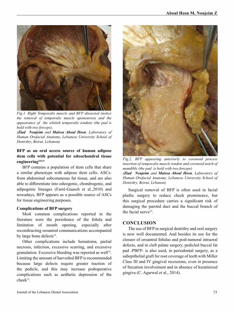
73Journal of the Lebanese Dental Association
BFP as an oral access source of human adipose stem cells with potential for osteochondral tissue engineering4,8,11
BFP contains a population of stem cells that share a similar phenotype with adipose stem cells- ASCs- from abdominal subcutaneous fat tissue, and are also able to differentiate into osteogenic, chondrogenic, and adipogenic lineages (Farré-Guasch et al.,2010) and nowadays, BFP appears as a possible source of ASCs for tissue engineering purposes.
Complications of BFP surgeryMost common complications reported in the
literature were the persistence of the fistula and limitation of mouth opening, especially after reconstructing oroantral communications accompanied by large bone defects15.
Other complications include hematoma, partial necrosis, infection, excessive scarring, and excessive granulation. Excessive bleeding was reported as well10. Limiting the amount of harvested BFP is recommended because large defects require greater traction of the pedicle, and this may increase postoperative complications such as aesthetic depression of the cheek15.
Surgical removal of BFP is often used in facial plastic surgery to reduce cheek prominence, but this surgical procedure carries a significant risk of damaging the parotid duct and the buccal branch of the facial nerve23.
CONCLUSIONThe use of BFP in surgical dentistry and oral surgery
is now well documented. And besides its use for the closure of oroantral fistulas and post-tumoral intraoral defects, and in cleft palate surgery, pedicled buccal fat pad -PBFP- is also used, in periodontal surgery, as a subepithelial graft for root coverage of teeth with Miller Class III and IV gingival recessions, even in presence of furcation involvement and in absence of keratinized gingiva (C. Agarwal et al., 2014).
Fig.1. Right Temporalis muscle and BFP dissected (notice the removal of temporalis muscle aponeurosis and the appearance of the whitish temporalis tendon) (the pad is hold with two forceps).(Ziad Noujeim and Maissa Aboul Hosn, Laboratory of Human Orofacial Anatomy, Lebanese University School of Dentistry, Beirut, Lebanon)
Fig.2. BFP appearing anteriorly to coronoid process insertion of temporalis muscle tendon and coronoid notch of mandible (the pad is hold with two forceps)(Ziad Noujeim and Maissa Aboul Hosn, Laboratory of Human Orofacial Anatomy, Lebanese University School of Dentistry, Beirut, Lebanon)
Aboul Hosn M, Noujeim Z

74
Ongoing preliminary research and clinical experimentations, worldwide, consider BFP as an autologous osteogenic graft material and/or biologic membrane capable of achieving high success rates in maxillary sinus floor elevation surgery. Also, enhanced bone formation is observed during the regeneration of jaw’s odontogenic tumors defects, using autologous dental pulp stem cells and the stromal vascular fraction (SVF) of BPF (K. Manimaran et al., Tamil Nadu, India).
Due to its haversting easiness and pedicled vascularity, PBFP is nowadays considered as a reliable treatment modality in surgical dentistry and oral surgery.
REFERENCES1. Abad-Gallegos M, Figueiredo R, Rodríguez-Baeza A, Gay-Escoda C. “Use of Bichat’s Buccal Fat Pad for the Sealing of Orosinusal Communications. A Presentation of 8 Cases.” Med Oral Patol Oral Cir Bucal . 2011 Mar 1;16 (2): e215–19.2. Alonso-González R,et al.Closure of oroantral communications with Bichat´s buccal fat pad. Level of patient satisfaction. J Clin Exp Dent. 2015 epub.3. Bither S, Halli R, Kini Y. “Buccal Fat Pad in Intraoral Defect Reconstruction.” Journal of Maxillofacial & Oral Surgery 2013;12(4 ) :451–55. 4. Broccaioli E, Niada S, Rasperini G, Ferreira LM, Arrigoni E, Yenagi V, Brini A. “Mesenchymal Stem Cells from Bichat’s Fat Pad: In Vitro Comparison with Adipose-Derived Stem Cells from Subcutaneous Tissue.” BioResearch Open Access Apr 2013:2(2): 107–117. 5. Chakrabarti J, Tekriwal R, Ganguli A, Ghosh S, Mishra PK. “Pedicled Buccal Fat Pad Flap for Intraoral Malignant Defects: A Series of 29 Cases.” Indian J Plast Surg: Official Publication of the Association of Plastic Surgeons of India ( 2009) 42 : 36–42. 6. Egyedi P. Utilization of the buccal fat pad for closure of oro-antral and/or oro- nasal communications. J Maxillofac Surg 1977: 5: 241–2447. Fagan Johan (Author and Editor). Open Access Atlas Of Otolaryngology, Head and Neck Operative Surgery. University of Cape Town. South Africa ([email protected]) (www.entdev.uct.ac.za).8. Farré-Guasch E, Martí-Pagè C, Hernádez-Alfaro F, Klein-Nulend J, Casals N. “Buccal Fat Pad, an Oral Access Source of Human Adipose Stem Cells with Potential for Osteochondral Tissue Engineering: An in Vitro Study.”Tissue Eng Part C Methods. 2010 Oct;16(5):1083-949. Gröbe, A., Eichhorn W, Hanken H, Precht C, Schmelzle R, Heiland M, Blessmann M. “The Use of Buccal Fat Pad (BFP) as a Pedicled Graft in Cleft Palate Surgery.” Int. J.Oral and Maxillofacial. Surg 2011;40: 685–89. 10. Kademani D, Tiwana P.Atlas of Oral and Maxillo-facial Surgery. Elsevier. (Under press).
11. Kishimoto N, Momota Y, Hashimoto Y, Tatsumi S, Ando K, Omasa T, Kotani J. The osteoblastic differentiation ability of human dedifferentiated fat cells is higher than that of adipose stem cells from the buccal fat pad. Clin Oral Investig. 2014;18:1893–190112. Loukas M, Kapos T, Louis Jr RG, Wartman C, Jones A, Hallner B. “Gross Anatomical, CT and MRI Analyses of the Buccal Fat Pad with Special Emphasis on Volumetric Variations.” Surg Radiol Anat 2006;28:254–60. 13. Meyer E, Liebenberg S J R,Fagan J. Buccal fat pad - a simple, underutilised flap. South African Journal of Surgery 2012;v. 50, n. 2, p. 47-49.14. Mohan S, Kankariya H, Harjani B. The use of the buccal fat pad for reconstruction of oral defects: review of the literature and report of cases. J Maxillofac Oral Surg. 2012;11:128–131.15. Peñarrocha-Oltra D, Alonso-González R, Pellicer-Chover H, Aloy-Prósper A, Peñarrocha-Diago M. Closure of oroantral communication with buccal fat pad after removing bilateral failed zygomatic implants: A case report and 6-month follow-up. J Clin Exp Dent. (Epub, ahead of print).16. Poeschl PW, Baumann A, Russmueller G, Poeschl E, Klug C, Ewers R. Closure of oroantral communications with Bichat’s buccal fat pad. J Oral Maxillofac Surg 2009;67:1460-6.17. Prashanth R, Nandini GD, Balakrishna R. Evaluation of versatility and effectiveness of pedicled buccal fat pad used in the reconstruction of intra oral defects. J Maxillofac Oral Surg. 2013;12:152–159.18. Saravanan K, Narayanan V. The use of buccal fat pad in the treatment of oral submucous fibrosis: a newer method. Int J Dent 2012:93513519. Shah G,Mistry M,Pandit J.The use of buccal fat pad (BFP) as a pedicled graft for cleft palate repair.J Med Sci 2012, Vol 3 no 1. 20. Shrivastava G, Padhiary S, Pathak H, Panda S, Lenka S. Buccal Fat Pad to Repair Intraoral Defects . International Journal of Scientific and Research Publications, Volume 3, Issue 2, February 20131ISSN2250-3153. (www.ijsrp.org).21. Singh J, Prasad K, Lalitha R.M., Ranganath K. Buccal pad of fat and its applications in oral and maxillofacial surgery: a review of published literature (February) 2004 to (July) 2009 .Oral Surg Oral Med Oral Pathol Oral Radiol Endod 2010;110:698-705.22. Zhang YP et al. Anatomical structure of the buccal fat pad and its clinical adaptations. Plast Reconstr Surg 2002;109(7):2509-2518.23. Hwang HJ et al. Interrelated buccal fat pad with facial buccal branches and parotid duct. J Craniofac Surg 2005;16(4):658-660.24. Samman N, Cheung LK, Tideman H. The buccal fat pad in oral reconstruction. Int J Oral Maxillofac Surg 1993 Feb;22(1):2-6.25. Cheung LK, Samman N, Tideman H. Reconstructive options for maxillary defects. Ann R Australas Coll Dent Surg 1994 Apr;12:244-251.26. Stuzin JM et al. The anatomy and clinical applications of the buccal fat pad. Plast Reconstr Surg 1990 Jan;85(1):29-37.
Correspond with :Ziad [email protected]
Aboul Hosn M, Noujeim Z
Journal of the Lebanese Dental AssociationVolume 49 - Nº 2 - December 2014

75
INTRODUCTIONOne of the most important aspects in contemporary
endodontics is a thorough knowledge of internal root morphology. This aspect, together with an accurate diagnosis and appropriate cleaning and shaping of the root canal system, will usually lead to a successful outcome. Root canal morphology is limitless in its variability and clinicians must be aware that anatomic variations constitute an impressive challenge to endodontic success1,2. Undetected extra roots or extra root canals may contain microorganisms and their byproducts are a major reason for failure of root canal treatment.
Human molars show considerable anatomic variations and abnormalities with respect to number of roots and root canals. In dental literature, mandibular second molar is typically described as a two rooted tooth with mesial and distal roots2. Unusual tooth anatomy associated with mandibular molars has been investigated in several studies. Manning23,24,25 has studied root canal anatomy of 149 extracted mandibular second molars using clearing technique: he found that 22% have single roots, 76% two roots, and 2% three roots. Rocha26 and associates studied external and internal anatomy of 628 extracted mandibular first and second molars: analysis of mandibular second molars roots showed that 84.1% have two separate roots, 15.9% fused roots, and 1.5% three roots.
Technological advances have given the clinician an opportunity to identify and successfully treat these aberrations1,2,3,4,5. It has been shown that chances of detection of additional canals increased to 93% with an experienced clinician working under an operating
microscope. Matherne and co-workers27 reported the superiority of CBCT over conventional radiological diagnostic methods and suggested the simultaneous use of the operating microscope and CBCT. With the availability of CBCT, new insights were displayed in the non-invasive evaluation of root-canal morphology and also for post-treatment assessment and prognosis14-15. CBCT has the ability of overcoming the limitations of conventional radiography such as three-dimensional evaluation of complex root canal anatomy during endodontic treatment. An important benefit of CBCT is in diagnosis of extra roots or canals11.
To the best of our knowledge, only few cases of mandibular second molars with two mesial roots* have been reported in the literature, to date1,2,3,4,8 (HMA Ahmed, 2011 and 2012 - S Demirbuga et al., 2013). While earlier descriptions were based on endodontic treatment using conventional radiographs and on examination of extracted teeth, no clinical reports have attempted to identify or define the degree and level of separation of the two mesial roots using cone-beam computed tomography (CBCT) imaging7.
This case report addresses the usefulness of CBCT in the management of a mandibular second molar with a separated K-file in the second mesiolingual root canal.
CASE REPORTA 33-year-old Lebanese male was referred to us for
the removal of a stainless steel fragment that had been separated from a K file within the mesial root of the mandibular (permanent) left second molar. His treating
Usefulness of Cone Beam Computed Tomography (CBCT) in the removal of a separated endodontic file in an aberrant root canal morphology: an unusual case report.
Maysoun Ahmad, BDS, PG Diploma (Endo.)
AbstractRoot canal treatment of a mandibular molar with aberrant canal configuration can be diagnostically and
technically challenging. To date, an anatomical variation that has received particular attention is the three-rooted mandibular molar. This case report addresses the clinical management of a broken K-file in a second mandibular molar with unusual anatomy (2 mesial roots). Exact location of the instrument and the hidden canal orifice were revealed, and the fragment removed using ultrasonic tips, under a dental operating microscope. This case report highlights the usefulness of cone beam computed tomography (CBCT) for a better diagnosis and management of an unusual root canal morphology.
* An accessory root = known as extra root, supernumerary root, supplementary root, or additional root
Journal of the Lebanese Dental AssociationVolume 49 - Nº 2 - December 2014
Endodontics

76 Volume 49 - Nº 2 - December 2014
dentist already obturated all the canals of 37, with the exception of the second ML canal.
During oral examination, tooth 37 was sensitive to percussion. A periapical radiograph revealed a broken metallic instrument in the mesial root which was already obturated (Fig.1), which allowed us to think of an abnormal root morphology. The referring dentist stated that the file used to go in two different ways inside the lingual canal. A CBCT scan for mandibular left quadrant was taken and it revealed that tooth 37 has two mesial roots with 3 mesial canals, MB, ML1, and ML2, and that the separated k-file was embedded within the second mesiolingual canal (Fig. 2). After local analgesia, tooth was isolated with a rubber dam.Access cavity preparation was left opened, and the straight-line access to the canals was already achieved by his dentist. Under a microscopic field (8X), the stainless steel fragment was clearly visible within the middle third of the second mesiolingual root canal (ML2).
Multiple attempts for bypassing the obstructed canal with k files no 8 and 10 were implemented, but the instrument was already engaged in dentinal walls of root canal. After re-reading the CBCT, the path of entry of ultrasonic tips was determined, and the patient was recalled for a second appointment during which dislodging of the broken instrument was done under 0,5% sodium hypochlorite copious irrigation, under the operating microscope.
The instrument’s head was luxated so the files no. 10 and 15 could bypass it and consequently increase its luxation, with ‘push and pull’ motions, after which the K-file was aspirated with the suction tip. The working length of the canal was established with an apex locator (Root ZX®) and confirmed by periapical radiography (Fig.3).
The root canal was shaped with stainless steel K-files, using a step back method. The canals were cleaned alternately with RC-Prep® and 5.25% sodium hypochlorite solution, followed by packing with a sterilized cotton pellet and IRM®. At the following appointment, the canal was irrigated and dried (with paper points), and a no .30 master cone was fitted in it. Then, a post-operative CBCT scan was done (Fig.4), and after 3months, 6 months, and one year follow-ups, patient was free of symptoms.
DISCUSSIONDiagnosis and treatment of extra roots or canals
in mandibular second molars can be challenging in endodontic practice. A comprehensive understanding of the most common root canal configuration and its variations is essential to achieve long-term success of endodontic treatment. Hoen and Pink 17 reported a 42% incidence of missed root or canals in the teeth
that needed endodontic retreatment. Thus, complete debridement and obturation of the root canal system is of utmost importance in endodontics 11-1.Presence of extra roots is easily determined using periapical radiographs: however, it is somehow difficult to assess the root canal system completely. Digital radiography at different angles with subsequent image analysis can be used effectively. Computed tomography (CT) imaging has been widely used in medicine since the 1970s and was introduced in the endodontic field in the 1990s. Recently, CBCT imaging has been shown to provide comparable images at reduced dose and costs, and it is considered as an alternative to multi-detector CT imaging in endodontics1-2. So if the outlines of the roots are unclear and the root canal shows sharp density changes or the apices cannot be well defined, then extra roots can be suspected4.
The present case report addresses the removal of a broken instrument in the third mesial canal of a second mandibular molar. A systematic review conducted by de Pablo and associates28 reported that the overall incidence of "3 canals" in mesial root of permanent first mandibular molar was 2.3%.
When an instrument fracture occurs during root canal preparation procedures, the clinician has to evaluate the treatment options with consideration for pulp status, root canal infection, root canal anatomy, position and type of fractured instrument, and amount of damage that would be caused to the remaining tooth structure. Removal of the fractured segment (by passing and sealing the fragment within the root canal space or true blockage) is a possible approach. The prognosis of leaving broken instruments versus removing them from the canal have been discussed in the literature9. Instruments located in the straight portions of the canal can usually be easily removed. If separated instruments lie partially around canal curvatures and straight line access is prepared to the coronal of the fractured instrument segments, they can be removed. Removal of the broken instrument segments that are apically located to the curvature of the canal is usually not possible9. Nowadays, removal of broken instruments is performed using ultrasonics and operating microscopes9. To improve the potential of safety and success of removal procedures, special ultrasonic tips have been developed. These tips vibrate to loosen the obstruction, causing minimal damage to canal walls. Enhanced vision with magnification and illumination from a microscope allows clinicians to observe the most coronal aspects of broken instruments - RC-Prep®= Root Canal Preparation Cream- IRM® = is a reinforced Zinc Oxide Eugenol (ZOE) composition for intermediate restorations. It is an Intermediate Restorative Material (IRM).
Ahmad M

77Journal of the Lebanese Dental Association
and to remove them without perforations9-18. However, with advanced diagnostic aids such as CBCT, these challenges may be overcome11. An advantage of computed tomography (CT) scanning over conventional radiograph is that CT permits the operator to look at multiple sections of the roots and their canals19. Nance and associates20 reported that detection of canals increased significantly by CT, compared with conventional radiography11. Additionally, CBCT scanners use simpler, less complicated, and therefore, less expensive hardware (X-ray source and detector), making it quite popular as an office diagnostic tool16. CBCT, as an aid, can greatly enhance detection and mapping of root canal systems with the potential to improve quality of root canal treatment16.
The knowledge of both "normal" (usual) and "abnormal" (unusual) anatomy of molars shows the parameters under which root canal therapy is to be performed and this can directly modify the probability of success. This is the reason why clinicians should be aware that variations in tooth morphology may often occur. Failure to treat a canal is an obvious reason for root canal treatment failure. Therefore, all clinicians must make every effort in order to locate and treat all existing canals during endodontic treatment.
The present case report is a rare one that showed two mesial roots in a mandibular second molar. It addresses the importance of adequate knowledge of aberrant root canal morphology for an appropriate and complete endodontic treatment.
CONCLUSIONConventional periapical radiographs provide a high
definition image, at low dose, but with certain limitations. CBCT can overcome some of the shortcomings of conventional radiographs in endodontics21: it is known that endodontics fail to identify at least one root canal in almost 40% of teeth, even with parallax radiographs,
Fig. 1. Peri-apical radiograph displaying the broken instrument at middle third of “mesial root”(Later, CBCT revealed the second mesial root).
Fig.2a. 3D reconstruction of the canal system.
Fig. 2b. CBCT revealing the 2 mesial roots and the broken instrument left at the middle third of the second mesiolingual canal of ML root.
Fig. 3. The working length file after the removal of the broken instrument.
Fig. 4. Post-operative CBCT displaying the four obturated root canals.
Ahmad M

78
and with three-dimensional CBCT, decision-making is improved with an incomparable assessment of teeth with unusual anatomy21,22 (such as teeth with unusual number of roots, dilacerated teeth, and dens in dente). Besides all its advantages, CBCT scans offer a significant radiation dose reduction, as compared to medical CT21.
Failure to locate and clean extra canals decreases the long-term prognosis of endodontic treatment. Adequate knowledge of root canal aberrancies, the will to search for them, combined with magnification and modern imaging techniques, will lead us to a better outcome. This case report described the usefulness of CBCT for a successful management of an aberrant morphology in a mandibular second molar with two mesial roots.
Anatomic variations in the number of roots may occur in any tooth and root canal treatment will certainly not succeed if accessory roots are not detected and endodontically treated.
REFERENCES1- Ravanshad Sh, Nabavizade. M. Endodontic Treatment of a Mandibular Second Molar with Two Mesial Roots: case report. Iran Endod J. 2008 Fall; 3(4):137-40. Epub 2008 Oct 12- Karunakaran JV, Shobana R et al. Management of middle mesial canal in mandibular second molar. J Pharm Bioallied Sci. 2012 Aug; 4(Suppl 2):S161-4. doi: 10.4103/0975-7406.1002593- Ragavendran N, Bhat GT, Hegde MN. Mandibular second molar with 3 mesial canals and a radix paramolaris. J Pharm Bioallied Sci. 2014 Jul; 6(Suppl 1):S182-4. doi: 10.4103/0975-7406.1374614- Nayak G, Shetty S, Shekhar R. Asymmetry in mesial root number and morphology in mandibular second molars: a case report. Restor Dent Endod. 2014 Feb;39(1):45-50. 5- de Paula AF, Brito-Júnior M et al. Three Independent Mesial Canals in a Mandibular Molar: Four-Year Follow-up of a Case Using Cone Beam Computed Tomography. Case Rep Dent. 2013;2013:891849. doi: 10.1155/2013/891849. 6- Subbiya A, Kumar KS et al. Management of mandibular first molar with four canals in mesial root. J Conserv Dent. 2013 Sep;16(5):471-3. doi: 10.4103/0972-0707.1174957- Deepalakshmi M1, et al. Independent and Confluent Middle Mesial Root Canals in Mandibular First Molars: A Report of Four Cases. Case Rep Dent. 2012;2012:103125. doi: 10.1155/2012/103125. Epub 2012 Jun 258- Aminsobhani M, Bolhari B et al. Mandibular first and second molars with three mesial canals: a case series. Iran Endod J. 2010 Winter;5(1):36-9.9- McGuigan MB, Louca C, Duncan HF. Clinical decision-making after endodontic instrument fracture. Br Dent J. 2013 Apr;214(8):395-400. doi: 10.1038/sj.bdj.2013.379.10- Gencoglu N, Helvacioglu D. Comparison of the Different Techniques to Remove Fractured Endodontic Instruments from Root Canal Systems. Eur J Dent. 2009 Apr;3(2):90-511- Baziar H, Daneshvar F et al. Endodontic Management of a Mandibular First Molar with Four Canals in a Distal Root by Using Cone-Beam Computed Tomography: a Case Report. J Oral Maxillofac Res. 2014 Apr 1;5(1):e5.
12- Chhabra S, Yadav S, Talwar S. Analysis of C-shaped canal systems in mandibular second molars using surgical operating microscope and cone beam computed tomography: A clinical approach. J Conserv Dent. 2014 May;17(3):238-43. 13- Nur BG, et al. Evaluation of the root and canal morphology of mandibular permanent molars in a south-eastern Turkish population using cone-beam computed tomography. Eur J Dent. 2014 Apr;8(2):154-9. 14- Rajasekhara S, Sharath Chandra S, Parthasarathy LB. Cone beam computed tomography evaluation and endodontic management of permanent mndibular second molar with four roots: A rare case report and literature review. J Conserv Dent. 2014 Jul;17(4):385-8. doi: 10.4103/0972-0707.136518.15- Shetty A, Hegde MN et al. A Three-Dimensional Study of Variations in Root Canal Morphology Using Cone-Beam Computed Tomography of Mandibular Premolars in a South Indian Population. J Clin Diagn Res. 2014 Aug;8(8):ZC224.doi:10.7860/JCDR/2014/8674.4707. Epub 2014 Aug 2.16- Sharma S, Sharma V et al. CBCT diagnosis and endodontic management of a maxillary first molar with unusual anatomy of two palatal canals: A case report. J Conserv Dent. 2014 Jul;17(4):396-9. doi: 10.4103/0972-0707.13652217- Hoen MM, Pink FE. Contemporary endodontic retreatments: an analysis based on clinical treatment findings. J Endod. 2002 Dec;28(12):834-618- Parashos P, Messer H H. Rotary NiTi instrument fracture and its consequences.J Endo 2006; 32: 1031–104319- Patel S, Dawood A, Whaites E, Pitt Ford T. New dimensions in endodontic imaging: part 1. Conventional and alternative radiographic systems. Int Endod J. 2009 Jun;42(6):447-62. 20- Nance R et al. Identification of root canals in molars by tuned-aperture computed tomography. Int Endod J. 2000 Jul;33(4):392-6.21. Patel S, Kanagasingam S, Mannocci F. Cone Beam Computed Tomography (CBCT) in Endodontics. Dental Update 2010 July/August;37:373-379.22. Ahmed HMA and Luddin N. Accessory mesial roots and root canals in mandibular molar teeth: Case Reports, SEM analysis and literature review. ENDO (London, England) 2012;6(3):195-205.23. Manning SA. Variations in root canal anatomy. N Z Dental J 1991 Apr;87(388):52-56.24. Manning SA. Root canal anatomy of mandibular second molars. Part I. Int Endod J Jan;23(1):34-39.25. Manning SA. Root canal anatomy of mandibular second molars. Part II. C-shaped canals. Int Endod J 1990 Jan;23(1):40-45.26. Rocha LF et al. External and internal anatomy of mandibular molars. Braz Dent J 1996;7(1):33-40.27. Matherne RP et al. Use of CBCT to identify root canal systems in vitro. J Endod 2008 Jan;34(1):87-89.28. de Pablo OV et al. Root anatomy and canal configuration of the permanent mandibular first molar: a systematic review. J Endod 2010 Dec;36(12):1919-1931.
Correspond with :Maysoun Ahmad [email protected]
Ahmad M
Journal of the Lebanese Dental AssociationVolume 49 - Nº 2 - December 2014
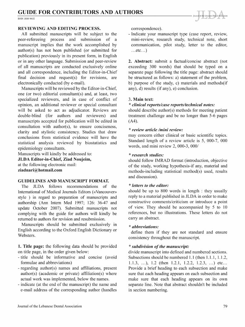
79Journal of the Lebanese Dental Association

80 Volume 49 - Nº 2 - December 2014