the journal of biological chemistry vol. 267, … · date following the procedure of schneider et...
TRANSCRIPT

THE JOURNAL OF BIOLOGICAL CHEMISTRY 8 1992 by The American Society for Biochemistry and Molecular Biology, Inc.
Vol. 267, No. 34, Issue of December 5, pp. 24223-24229,1992 Printed in U.S.A.
Purification and Ligand Binding of a Soluble Class I Major Histocompatibility Complex Molecule Consisting of the First Three Domains of H-2Kd Fused to ,&-Microglobulin Expressed in the Baculovirus-Insect Cell System*
(Received for publication, May 8, 1992)
Francois GodeauS, Immanuel F. LuescherQ, David M. Ojcius, Cecile Saucier, Estelle Mottez, Lucien Cabanien, and Philippe Kourilsky From the Unit6 de Biologie Moleculaire du Gene, Institut National de la Sante et de la Recherche Medicale U 277 and TUnite de Technologie Cellulaire, Institut Pasteur, 28, rue du Docteur Roux, 75724 Paris Cedex 15, France, and the §Ludwig Institute for Cancer Research, Chemin des Boueresses, Epalinges CH 1066, Switzerland
A recombinant baculovirus encoding a single-chain murine major histocompatibility complex class I mol- ecule in which the first three domains of H-2Kd are fused to &-microglobulin (&m) via a 15-amino acid linker has been isolated and used to infect lepidopteran cells. A soluble, 391-amino acid single-chain H-2Kd (SC-Kd) molecule of 48 kDa was synthesized and gly- cosylated in insect cells and could be purified in the absence of detergents by affinity chromatography using the anti-H-2Kd monoclonal antibody SF1.1.1.1. We tested the ability of SC-Kd to bind antigenic pep- tides using a direct binding assay based on photo- affinity labeling. The photoreactive derivative was prepared from the H-2Kd-restricted Plasmodium berghei circumsporozoite protein (P.b. CS) peptide 253-260 (YIPSAEKI), a probe that we had previously shown to be unable to bind to the H-2Kd heavy chain in infected cells in the absence of co-expressed f12-
microglobulin. SC-Kd expressed in insect cells did not require additional mouse 02-m to bind the photoprobe, indicating that the covalently attached Bz-m could sub- stitute for the free molecule. Similarly, binding of the P.b. CS photoaffinity probe to the purified SC-Kd mol- ecule was unaffected by the addition of exogenous &- m. This is in contrast to H-2KdQlo, a soluble H-2Kd molecule in which &-m is noncovalently bound to the soluble heavy chain, whose ability to bind the photo- affinity probe is greatly enhanced in the presence of a n excess of exogenous B2-m. The binding of the probe to SC-Kd was allele-specific, since labeling was selec- tively inhibited only by antigenic peptides known to be presented by the H-2Kd molecule.
Class I major histocompatibility complex (MHC)’ mole- cules are highly polymorphic 45-kDa integral membrane gly- coproteins associated with the 12-kDa soluble Pz-microglob- ulin (Pn-m), and are involved in the presentation of endoge-
* The costs of publication of this article were defrayed in part by the payment of page charges. This article must therefore be hereby marked “advertisement” in accordance with 18 U.S.C. Section 1734 solely to indicate this fact.
$ To whom correspondence should be addressed. ’ The abbreviations used are: MHC, major histocompatibility com-
plex; AcNPV, A . californica nuclear polyhedrosis virus; &-m, p2- microglobulin; mAb, monoclonal antibody; PAGE, polyacrylamide gel; SC-H-2Kd, single-chain H-2Kd; IASA, iodo-4-azido salicylic acid; HPLC, high performance liquid chromatography; PVDF, polyvinyl- idene difluoride; P.6. CS, P. berghei circumsporozoite protein.
nously derived antigenic peptides to CD8-positive cytotoxic T lymphocytes (Townsend and Bodmer, 1989; Rothbard and Gefter, 1991). The general features of class I molecule biosyn- thesis have been known for some time both i n vitro (Dobber- stein et al., 1979) and in vivo (Krangel et al., 1979). N-Linked core glycosylation occurs during biosynthesis of MHC class I heavy chains in the endoplasmic reticulum (see Kornfeld and Kornfeld (1985) for review), and after &-m association (Sege et al., 1981), the heterodimer is transported along the secretory pathway to the cell surface (Owen et al., 1980). The mecha- nism by which endogenous antigenic peptides associate with class I molecules is still incompletely understood, but current evidence suggests that it takes place in the endoplasmic reticulum (Nutchern et al., 1989; Yewdell and Bennink, 1989; Kvist and Hammann, 1990).
The three-dimensional structure of HLA-A2 has revealed that the a1 and a2 domains of the heavy chain fold in a manner constituting an extended, groove-like peptide binding site (Bjorkman et al., 1987a, 1987b). Class I molecules on cell surfaces contain short peptides (8-10 amino acids) with dis- tinct allele-specific binding motifs (Falk et al., 1991; Jardetzky et al., 1991).The binding of antigenic peptides to MHC class I molecules is closely interrelated to the binding of Pz-m to the heavy chain. This is explained, on the one hand, by the intimate interactions of P2-m not only with the a3 domain but also with the a1 and a2 domains, which is likely to be important for the conformation of the peptide binding site (Saper et al., 1991). On the other hand, stable assembly of the class I heterodimer depends on occupancy of the heavy chain peptide binding site (Townsend et al., 1989). Indeed, this hypothesis is supported by recent work with class I assembly- defective mutant cell lines (Townsend et al., 1989; Cerundolo et al., 1990; Townsend et al., 1990) and with purified human class I molecules (Silver et al., 1991), which has shown that suitable antigenic peptides promote class I molecule assembly. Furthermore, there is evidence that peptide epitopes induce a conformational change of the heavy chain, resulting in in- creased affinity for Pz-m (Elliot et al., 1991), and that free heavy chains present on the cell surface are functionally inactive (Rock et al., 1991a, 1991b).
Clearly, further understanding of these interactions will depend on the availability of reconstituted systems using purified molecules. In this respect, the insect-baculovirus expression system has proved particularly useful in recent years for producing, in relatively large quantities, recombinant proteins that are authentically folded, proteolytically proc- essed, post-translationally modified, and biologically active.
24223
24224 A Functional Soluble Single-chain H-2Kd in Insect Cells
In this system, the foreign gene is cloned downstream of a strong very late promoter and introduced via homologous recombination back into the genome of Autographa californica nuclear polyhedrosis virus (AcNPV) (Miller, 1988; Luckow and Summers, 1989; Maeda, 1989). We have previously used this approach to produce biologically active mouse &ma (Dargemont et al., 1989; Godeau et al., 1991) and a H-2Kd heavy chain which is able to associate with &m in an anti- genic peptide-dependent manner and form a functional het- erodimer able to bind peptides (Godeau et al., 1992). However, in doubly infected cells, the extent of association between the heavy and light chain is low and hence precludes isolation of larger amounts of assembled molecules. To circumvent this difficulty, we chose to express a fusion protein comprising the first three domains of H-2Kd heavy chain fused to mouse p2- microglobulin. We describe here some characteristics of this purified soluble protein and demonstrate that its interaction with antigenic peptides is identical to that observed with natural H-2Kd molecules.
MATERIALS AND METHODS
Cell Culture and Transfection-Suspension or monolayer cultures of the Sf9 cell line from Spodoptera frugiperda were maintained a t 27 "C in TC 100 medium supplemented with 0.33% Yeastolate (Difco) and containing 4% heat-inactivated fetal bovine serum, 50 pg/ml penicillin, and 50 pg/ml streptomycin. Cell densities were routinely kept between 2.5 X lo5 and 1.5 X 10⁶/ml by passaging the cells in fresh medium. DNAs from recombinant plasmids (20 pg) and viral DNA (10 pg) were co-transfected by electroporation using a standard cuvette with a 0.4-cm gap containing 1 ml of Sf9 cell suspension (0.3- 1.0 X lo7 cells/ml) in BNP (25 mM 2-[bis(2-hydroxyethyl)amino] ethanesulfonic acid, pH 6.95, 140 mM NaC1, 1.5 mM NaH2P04, and 1 mM glucose) in the presence of 500 pg/ml herring sperm DNA and exposed to the exponential discharge of a 1030 pF capacitance charged under 220 V (Godeau, 1990; Godeau et al., 1992). For large scale recombinant protein preparations, viral stock was prepared and used to infect a t a multiplicity of infection >lo. Sf9 cells were grown in aerated spinner flasks and fed, after infection, with serum-free TC 100 medium. Seventy-two hours postinfection, the suspension was collected and infected cells were removed by centrifugation.
Isolation of AcNPV SC-H-2Kd Recombinant Baculouirus-All plas- mids were constructed and purified according to standard recombi- nant DNA techniques (Sambrook et al., 1989) except that transfor- mation of Escherichia coli hosts was performed by electroporation (Dover et al., 1988). Conditions for restriction endonuclease digestions were those suggested by the manufacturer. DNA fragments were isolated from agarose gels by electroelution. The transfer vector used in this study was the nonfusion pAcYMl (Matsuura et al., 1987), kindly provided by Dr. D. Bishop, which was cleaved by digestion with BamHI restriction endonuclease and filled in with E. coli DNA polymerase large fragment. The structure of SC-Kd chimeric cDNA has already been described (Mottez et al., 1991). Briefly, the entire coding sequence comprises the four H-2Kd exons encoding H-2Kd signal sequence, al, a2, and a3 domains fused to the mouse Ps-rn'
with the sequence: 5'GGG GGG ATC GGA TCC GGA GGC GGT coding sequence lacking its signal sequence via a 45-base pair linker
GGA TCC GGT GGC GGC GGT TCG3'. This DNA sequence, which encodes a 15-amino acid protein sequence predominantly composed of glycine and serine residues, was chosen for its expected flexibility and is represented in Fig. 1. To generate a recombinant baculovirus, SC-Kd chimeric cDNA was excised from pSC-Kd-15 (Mottez et al., 1991) by digestion with Hind111 restriction endonuclease, filled in with E. coli DNA polymerase large fragment, and subsequently blunt- end ligated into the filled in BamHI site of pAcYMl transfer vector. Transformants were selected for proper orientation with respect to the polyhedrin gene transcriptional start site. Plasmid DNA was isolated and transfected together with viral DNA by electroporation, as already described (Chu et al., 1987; Godeau, 1990). Five to seven days after transfection, the viral progeny containing a mixture of wild type and recombinant viruses was submitted to a purification proce- dure based on limiting dilution in 96-well microtiter plates followed by hybridization to a H-2Kd probe. Five to seven days after infection, the virus-containing, adhering cell monolayer was directly dissolved in 200 p1 of 0.4 M NaOH and transferred to a nylon filter (Gene-
Screen Plus, Du Pont) via a dot-blot manifold (Reed and Mann, 1985). The filter was hybridized (Church and Gilbert, 1984) to a 32P- labeled (Feinberg and Vogelstein, 1983) H-2Kd probe, and the recom- binant virus-containing medium from positive wells was further pu- rified by a subsequent repeat of this dilution-hybridization procedure leading to the isolation of AcNPV-SC-Kd. The viruses encoding murine P2-mn, AcNPV-&-m (Godeau et al., 1991), and H-2Kd heavy chain, AcNPV-Kd (Godeau et al., 1992), have already been described.
Purification of SC-Kd-Immunoaffinity matrices were prepared by using anti-H-2Kd monoclonal antibodies SF1.l.l.l from ATCC and 34-1-2 (Ozato et al., 1980). Ten mg of pure IgG2. mAb were covalently coupled to protein A-Sepharose (Pharmacia) via dimethylpimelimi- date following the procedure of Schneider et al. (1982). After saturat- ing unreacted amino groups with ethanolamine and washing the column extensivsly, 1 liter of medium conditioned by AcNPV-SC- Kd-infected Sf9 cells was passed over the column during a 6-h period, after which the column was washed with 20 column volumes of phosphate-buffered saline. Bound material was eluted with 50 mM diethylamine, pH 11.5, and the material was neutralized with 200 mM triethanolamine/HCl, pH 7.5, and concentrated. For affinity chro- matography with concanavalin A, the cell-free conditioned medium was first ultracentrifuged (100,000 X g for 1 h), concentrated to one- tenth of the original volume, and dialyzed against 10 mM Tris, pH 7.5, 100 mM NaCl. The dialysate was passed through an immobilized concanavalin A column (IBF, France), eluted with a-methylmanno- side, and analyzed by immunoblot using an anti-bz-m antiserum.
Biochemical Characterization of Purified SC-Kd-N-terminal se- quence analysis of purified soluble SC-Kd from insect cells was performed by automated Edman degradation after electrophoresis on SDS-PAGE and transfer to PVDF, as described (Matsudaira 1987), on an Applied Biosystems 470 gas-phase peptide sequanator. Phenyl- thiohydantoin amino acids were detected with an on-line Applied Biosystems 120 A analyzer. For internal peptide sequence determi- nation, the purified protein was reduced and alkylated, transfered on PVDF membranes as described above and digested in 100 mM Tris- HCl, pH 8.5, with 1 pg of porcine trypsin (Sigma) in a 1:50 enzyme to substrate ratio for 4 h a t 37 "C. The digest was separated by HPLC on a C18 Vydac column (250 X 2.1 mm) using a 0-60% acetonitrile gradient in 0.1% trifluoroacetic acid. A well resolved peak was chosen for sequence determination.
Antibodies and Immunoblots-The class I H-2d-specific monoclonal antibodies used were 34-1-2 (IgG 2a) (anti H-2Kd and H-2Dd) (Ozato et al., 1980) and SF1.l.l.l (IgG 2a) from ATCC. All antibodies were purified from ascites fluid according to established procedures (Ey et al., 1978; Harlow and Lane, 1988). The rabbit serum raised against a fusion protein comprising the second half of the a2 and all of the a3 domain of H-2Kd will be described,* and the rabbit antiserum anti- mouse &m was from Dr. N. Tanigaki (Natori et al., 1976).
Immunoblots were performed by dissolving and boiling protein samples in SDS-PAGE sample buffer followed by electrophoresis on 12.5% polyacrylamide gels (Laemmli, 1970). Electrotransfer was per- formed using the semidry method (Kyhse-Andersen, 1984) with PVDF membranes (Immobilon Millipore) in 50 mM Tris-borate buffer, pH 8.3, after the gel had been equilibrated for 5 min in the same buffer containing 0.1% SDS. Membranes were blocked in TBS (50 mM Tris, pH 7.5, 200 mM NaC1,0.05% Tween 20) containing 3% gelatin and incubated with a 1:500 dilution of the antiserum in TBS 1% gelatin, washed, and developed using alkaline phosphatase-labeled secondary antibody (Blake et al., 1984).
Peptide Synthesis-Peptides were synthesized using an Applied Biosystems model 430A peptide synthesizer and the standard t- butoxycarbonyl strategy (Merrifield, 1986). The deprotected peptides were purified on a C-18 reverse phase HPLC column, dissolved in phosphate-buffered saline, and stored frozen.
Photoaffinity Labeling with the ["'I]IASA-YIPSEAK(Biotin)l Peptide Probe-The photoprobe was prepared by reacting freshly iodinated [1251]IASA-ONSu with the orthogonal biotinylated octapep- tide from mouse malaria Plasmodium berghei circumsporozoite pro- tein YIPSAEKI(biotin)I and purified by C-18 reversed phase HPLC as previously described (Luescher et al., 1991, 1992). Handling of photoreactive materials was performed under dimmed light. For label- ing whole cells, infected Sf9 cells were harvested after 48 h, washed in phosphate-buffered saline, resuspended (2 X lo6 cells/ml) in serum- free TC 100 medium in the presence of [1s51]IASA-YIPSEAK(biotin)I (1 X lo7 cpm) for 6 h at 27 "C, and UV-irradiated at 4 "C for 10 min (15 watt lamp with an emission maximum a t 312 nm). After labeling,
F. Godeau and H. Ploegh, manuscript in preparation.

A Functional Soluble Single-chain H-2Kd in Insect Cells 24225
the cells were washed twice in cold medium and once in phosphate- buffered saline, detergent solubilized in 0.7% Nonidet P-40, and immunoprecipitated using SF1.l.l.l. mAb. After 1 h the lysate was centrifuged (15,000 X g for 3 min), and the supernatant was filtered through disposahle membrane filters (Millipore Nihon Kogyo, Yone- zawa, Japan). Immunoprecipitation was performed on nitrocellulose membrane filters in a dot-hlot manifold as described (Luescher, 1987). For each immunoprecipitation, 30 pg of pure SFl.I.l.l mAh was used. The immunoprecipitates were subjected to SDS-PAGE (10% acryl- amide) under reducing conditions and the dried gels were exposed to XAR 5 (Kodak) films.
Photoaffinity labeling of purified SC-K' protein was performed using a similar procedure. One pg of purified protein was incubated with ["'IIIASA-YIPSEAK(biotin)I (1 X 10' cpm) in 10 pl of phos- phate-huffered saline. Aliquots were incubated in polypropylene tubes (500 ml Eppendorf, Hamburg, Germany) a t 24 "C for 3 h. The tubes were UV-irradiated in the reflector of the described UV lamp for 3 min. The samples were diluted with 450 pl of phosphate-buffered saline containing 0.5% Nonidet P-40 and the labeled protein was immunoprecipitated as described above. For competition experi- ments, 1 pg of purified protein was incubated with the photoprobe in the absence or presence of graded amounts of competitor peptides in :<-fold dilution steps up to 1000-fold molar excess with respect to the photoprobe. The competitor was added 15 min before addition of the photoprobe.
The K'Q,,, plasmid construct leading to the synthesis of a soluble chimeric heavy chain between the first two domains of H-2K' and the third domain and the 3' sequences of the Q,,," gene and its introduction into mouse L cells has been described (Luescher et a/., 1992). Culture medium conditioned by clone NR37.15.2. was used to purify the K'Q,,, protein by immunoaffinity on an SF 1.1.1.1 column as described above.
RESULTS
Biochemical Characterization of SC-Kd Expressed in Bacu- lovirus-infected Insect Cells-The SC-K' construct, which comprises the signal sequence and the a l , a2, and a3 domains of the H-2K' heavy chain cDNA fused to the coding sequence of murine &mR via a 15-amino acid flexible linker, is repre- sented in Fig. 1. Assuming proper cleavage of its signal se- quence, the mature chimeric polypeptide should be 391 amino acids long. Thus the molecular mass of the core polypeptide (without taking into account possible glycosylation) should be approximately 43 kDa . This chimeric construct has already been shown to lead to the synthesis of a soluble, glycosylated
SC-H-2Ka a N l I
I
c
FK:. 1. St ruc tu re of the class I molecules used in this study. I.($, the memhrane-associated class I H-2K' heterodimer with non- covalently attached µglohulin. Middle, recombinant chimeric soluble molecule H-2K"QQ,,,. The portion in boldface indicates the sequence originating from the Q,,,b class I gene. Although this mole- cule can associate with &m, it has heen represented without associ- ated light chain. Right, recombinant chimeric soluble protein SC-H- 2K". The sequence of the 15-amino acid flexihle linker. inserted between amino acid '276 of the mature H-2K' polypeptide and residue 1 of mature &-m, has heen represented in the single letter code.
fusion protein after transfection in COS-1 cells (Mottez et al., 1991). To express this molecule in the higher amounts re- quired for biochemical studies, we chose the recombinant baculovirus system. Thus, the use of the selection procedure described under "Materials and Methods" led to the isolation of a recombinant baculovirus containing the SC-K' coding sequence under the transcriptional control of the polyhedrin promoter (AcNPV-SC-K"). Preliminary experiments showed that a 48-kDa polypeptide (the expected apparent molecular mass for SC-K' if one assumes that the two N-glycosylation sites are used) was present in the supernatant of metabolically labeled insect cells infected with this virus. This band could be immunoprecipitated by an anti-mouse P2-m antiserum as well as by the 34-1-2 mAb recognizing the native conforma- tion of the H-2K' heavy chain (Godeau et al., 1992) (results not shown). Taken together, these results indicate that the SC-K' polypeptide is exported as a soluble protein in insect cells. In this fusion protein, the first three domains of the H- 2K' heavy chain associate with mouse &-m in a manner that mimics the native conformation of the heterodimer.
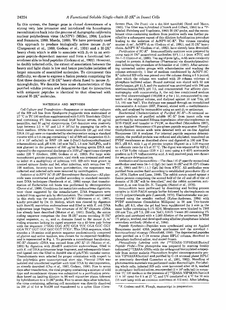
To purify this soluble class I fusion protein, immunoaffinity chromatography with an immobilized SF1.l.l.l. mAb column was carried out using crude cell-free conditioned medium from infected cells as starting material. Whereas no prominent Coomassie Blue stainable band could be detected in an SDS- PAGE analysis of the crude supernatant, the eluate from the affinity column showed a single major band of approximately 48 kDa (Fig. 2). This procedure routinely yielded approxi- mately 500 pg of SC-Kd per liter of conditioned medium. Immunoblot analysis was carried out to further characterize the purified protein. In addition to reacting with the H-2K'- specific rabbit antiserum raised against a fusion protein con- taining half of the a2 and the entire a3 domain of H-2K' (Fig. 3, panel A, lane a), this polypeptide also reacted with the anti- mouse P2-m antibody (Fig. 3, panel A, lane b ) , indicating that the 48-kDa polypeptide contained both MHC Class I heavy and light chain sequences. We then analyzed the glycosylation of this molecule. Conditioned medium from serum-free cul- tures was thus passed over a concanavalin A column, and the a-methylmannoside eluate was analyzed by immunoblot using
a b MW (kDa)
200
-- 94
68
". 46
30
21
14
FIG. 2. One-step purification of SC-Kd f rom the supernatant of Sf9 cells using an immunoaffinity column. One liter of serum- lree culture medium conditioned for 72 h after infection hy Sf9 cells infected with AcNPV-X-K ' was passed through an Sfl.l.1. mAb immunoaffinity column, eluted at pH 11.5. and freeze-dried. Five pg of protein were loaded on an SDS-PAGE which was stained with Coomassie Blue. Lane n, eluate lrom the immunoaffinity column; /rrnP b, molecular weight standards.
24226 A Functional Soluble Single-chain H-2Kd in Insect Cells
the anti-&m antiserum. As shown in Fig. 3, panel R, a prominent 48-kDa and &m immunoreactive band could be visualized in medium conditioned by AcNPV-SC-Kd-infected cells which was absent from that of the wild type control, demonstrating that SC-Kd contains mannose-rich carbohy- drates.
To further confirm the identity of the purified protein, the 48-kDa polypeptide was sequenced following transfer onto PVDF membrane (thus further purifying the input protein). As shown in Table I, N-terminal sequencing yielded the sequence Gly-Pro-His, the expected N-terminal sequence of the mature H-2K' protein. This proves that the signal se- quence of this polypeptide was cleaved in this expression system, as expected for a soluble protein recovered from a cell-free supernatant. Internal sequences were also obtained after tryptic digestion and separation of the digest by HPLC. One well separated peak was sequenced and shown to contain two distinct sequences in molar ratios differing by no more than a factor of 2. The first sequence, Tyr-Tyr-(Asp)-Gln, belongs to the H-2K' heavy chain and encompasses an N- glycosylation site. As expected for a glycosylated protein, the Asn residue is lacking in this sequence and replaced by a minute quantity of Asp. The second sequence, Ile-Pro-Lys, belongs to the mouse &m coding sequence, further indicating that the P2-m polypeptide was present in the SC-K" 48-kDa polypeptide. In view of the purity of the 48-kDa protein, no other peak was sequenced. Thus, among the three sequences obtained, all of them were derived from the expected se- quences. Taken together, these results demonstrate that a soluble, 48-kDa, glycosylated SC-Kd fusion protein with a correct N terminus is expressed efficiently in the baculovirus- insect cell system and can be isolated by a simple one-step procedure. In this polypeptide, the first three domains of H-
A B
a b a b " -
48 kDa C
12kDa + .
FIG. 3. Immunoblot analysis of the purified SC-Kd. A, im- munopurified SC-K' was loaded on SDS-PAGE and analyzed by immunohlot using either the anti-H-2K' 02 and 03 domain antiserum ( lone a) or the anti-mouse &m antiserum (lane b). R, serum-free conditioned medium from SC-K"-infected Sf9 cells was semipurified by concanavalin A affinity chromatography and analyzed by immu- nohlot using the anti-mouse &-m antiserum. Conditioned medium from Sf9 cells infected with wild type AcNPV (lane a) ; or with AcNPV-SC-Kd (lane b). All blots were developed with an alkaline phosphatase second antibody.
2K' are physically linked to mouse Pr-m and both sequences seem to fold to achieve a native conformation.
The Binding of IASA-YIPSAEKfiiotin)I to SC-Kd Is Not Affected by Soluble p2-m-We then determined whether the single-chain class I molecule retained the ability of the paren- tal molecule to bind antigenic peptide ligands. We thus used the recently described H-2Kd-specific photoprobe derived from the octapeptide 253-260 of the mouse malaria P. berghei circumsporozoite protein IASA-YIPSAEK(biotin)I. In as much as photo-cross-linking is several orders of magnitude faster than the interaction of peptides with class I molecules (De Graff et al., 1974; Luescher et al., 1992), it directly reflects the binding of the peptide to class I molecules or any other cell component at the time of UV irradiation. We had previ- ously shown that the binding of this photoprobe to the H-2Kd produced in the baculovirus-insect cell system is dependent on the presence of &-m (Godeau et dl., 1992). We first examined the ability of the probe to label living insect cells infected with AcNPV-SC-Kd. Thus, insect cells infected with various recombinant viruses were incubated with a 3 nM concentration of [""IIIASA-YIPSAEK(biotin)I and UV-irra- diated, and following detergent solubilization, the various Kd constructs were immunoprecipitated and analyzed by SDS- PAGE and autoradiography. As shown in Fig. 4 (panel A ) , a strong labeling of the 48 kDa band was observed in extracts of cells synthesizing the SC-Kd polypeptide (lane b). Similarly, the H-2Kd heavy chain synthesized in the presence of &-m, in Sf9 cells co-infected with AcNPV-K'and AcNPV-&m, strongly labeled as a 45 kDa band (lane a), but no labeling of the heavy chain could be detected in AcNPV-Kd singly in- fected cells (lane c), nor was any band detectable in AcNPV- &-m-infected cells (lane d ) or wild type AcNPV-infected control cells (lane e). These results demonstrate that the SC- Kd produced in insect cells is functional in terms of binding the H-2Kd-restricted photoprobe and that the &-m molecule coupled to the heavy chain via the flexible 15-amino acid linker can replace free mouse &m, which is required for significant binding of the photoprobe (Godeau et al., 1992).
We then wished to examine the binding properties of the SC-Kd protein by incubation of the purified protein with the photoprobe. As a control, we used the soluble class I molecule H-2K'Qlt,, purified in the same manner, in which the heavy and light chains are produced as individual polypeptides and are therefore noncovalently associated. As shown in Fig. 4, panel B , this molecule was able to bind the photoprobe (lune c), but maximal labeling intensity required the addition of an excess of exogenous human Pn-m (lune d). In contrast, purified SC-Kd bound the photoprobe equally well in the absence or presence of an excess of human &m. Taken together, these results indicate that the SC-Kd fusion protein can bind pep- tides in a manner similar to that of the parental class I heterodimer.
Inhibition of Photoreactive Peptide Binding to SC-Kd by Unlabeled H-2Kd-restricted Peptides-We next examined the specificity of the observed labeling of the SC-Kd molecule, by competition in the presence of various concentrations of unlabeled peptides, some of which were bona fide Kd-restricted peptides while others were peptide epitopes restricted by
TABLE I Purified SC-K" N terminus and internal tryptic peptides
H-2K' N terminus H-2K" internal peptide &m internal peptide Observed sequence NH,-Gly-Pro-His NH2-Tyr-Tyr-(Asp)-GIn- NH,-Ile-Pro-Lys-COOH Expected sequence NH,-Gly-Pro-His NH,-Tyr-Tyr-Asn-Gln-Ser NH,-Ile-Pro-Lys-COOH

A Functional Soluble Single-chain H-2Kd in Insect Cells
A
E A + Y E B c u
s : s $ s MW kDa
MWkDa a b c d e a b C - 68
46
30
68
16 HC
d
24227
HC
FIG. 4. B2-m i s no t requi red for binding of the photoreac t ive pept ide to SC-Kd. A, Sf9 cells were infected with wild type AcNPV (rut) (lane e ) , AcNPV-&m (lane a'), AcNPV-K" alone (lane c) , AcNPV-SC-K' (lane b) , or simultaneously with AcNPV-K" and AcNPV-&m (lane a). Two days aft.er infection, cells were harvested, washed, and incubated in the presence of a 3 nM concentration of' ["'IIIASA- YIPSEAK(biotin)l. After UV irradiation, the cells were washed twice in cold medium and once in phosphate-buffered saline and directly dissolved in SDS-PAGE sample buffer prior to electrophoresis and autoradiography. H , 1 pg of purified soluble H-2K" molecule was incubated with a 3 nM concentrat.ion of [""IIIASA-YIPSEAK(biotin)I, immunoprecipitated, and analyzed by SDS-PAGE and autoradiography. Immunopurified SC-K' (lanes a and b ) or KClQl1, (lanes c and a ' ) were incubated either in the absence (lnnes a and c) or presence ( lane h and d ) of 'a 3 pg/ml concentration of human &m. HC (heavy chain) indicates the relative mobility of the baculovirus-encoded unmodified H-2K" polypeptide.
FIG. 5 . Inhibit ion of photoreac- t ive pept ide b inding to SC-Kd by un- labeled H-2Kd-restricted peptides. One pg of purified SC-K" was incubated with a 3 nM concentration of [",-T]IASA- YIPSEAK(biotin)I in the presence of unlabeled peptides in the indicated mo- lar excess, UV-irradiated, immunopre- cipitated, and dissolved in SDS-PAGE sample buffer prior to electrophoresis and autoradiography. The following un- labeled peptide competitors were used: A , P. herghei circumsporozoite protein 253-260; H , P. bprghei circumsporozoite protein 249-260 (Romero et al., 1989); C, 1'198- 14-24 (Sibille et al., 1990); I) , ad- enovirus 5 E1A gene product 234-243 (Kast et al., 1989); E, influenza virus nucleoprotein 366-374 (Townsend et al., 1986); and F', P91A-12-24 (Lurquin et al., 1989). H-2K'-restricted peptides were used in pnnels A, H , and C, while H-2D'"restricted peptides were used in panels I) and I; and an H-2L'"restricted peptide in panel F'. Sequence alignments with the minimal H-2K"consensus motif described by Falk et a/. (1991) are shown in boldface.
A P. b. CS 253-260: YIPSAEKI
I I
0 10 30 100 300 1000
B P. b. CS 249-260: NDDSYIPSAEKI
D Ad5 E1A 234-243: SGPNTPPEI
" " - 0
I
0 10 30 100 300 1000
E NP 366-374: ASNENMETM
0 10 30 100 300 1000
C P 19%. 14-24: KYQAVTTTLEE
I 0 10 30 100 300 1000
either Dl' or E'. Photoaffinity labeling of the SC-K' molecule by radiolabeled IASA-YIPSAEK(biotin)I was completely in- hibited using a 10-fold molar excess of the parental octamer P. b. CS (YIPSAEKI) (Fig. 5, panel A ) . Similarly, a dose- dependent inhibition was observed when the 12-mer P.b. CS 249-260 was used as a competitor, but the molar excess needed for complete inhibition was higher, as expected for a peptide that is suboptimal with respect to the minimal consensus motif for KI-restricted epitopes (Falk et al., 1991; Romero et al., 1991) (Fig. 5, panel R) . The K'-restricted P 198- peptide
~ ~
0 10 30 100 300 1000
F P91A-.12-24: ISTONHRALDLVA
0 10 30 100 300 1000
(Sibille et al., 1990) was also a good competitor, although not as potent as P. b. CS (YIPSAEKI) (Fig. 5, panel C). In contrast, the H-2D'"restricted peptides Ad5 E1A 234-243 (Kast et al., 1989; Luescher et al., 1992) and NP 366-374 (Townsend et al., 1986) had no inhibitory activity (Fig. 5, panels D and E). Finally, P91A-12-24, a E'-restricted peptide (Lurquin et al., 1989), did not show any inhibitory activity. Thus, the specificity of P. b. CS (YIPSAEKI) photoprobe binding to the single-chain Kd molecule correlated with the expected restriction pattern of the K' molecule, indicating
24228 A Functional Soluble Single-chain H-2Kd in Insect Cells
that the observed labeling represents authentic binding to a functional peptide binding site.
DISCUSSION
In the insect cell-baculovirus system, recombinant proteins often retain’ their biological activity (Miller, 1988; Luckow and Summers, 1989; Maeda 1989) and are produced in con- siderable amounts, thus permitting their purification and detailed analysis. Indeed, our previous work using this system has provided evidence for a significant production of biolog- ically active mouse &ma (Godeau et al., 1991) and of a functional H-2Kd heavy chain which was able to associate with &ma in a peptide-dependent manner, so as to give rise t o a functional heterodimer (Godeau et al., 1992). Since our ultimate goal is to study a reconstituted system with purified molecules, the lack of complete heterodimerization observed in doubly infected cells prompted us to seek a more efficient system to produce a soluble molecule. The only studies re- porting the successful production of soluble class I molecules have used a truncated heavy chain associated with the sub- stitution of the transmembrane domain of the Blob molecule, leading to the production of a soluble class I molecule upon association with &-m (Margulies et al., 1986; Schneck et al., 1989). We have used another approach consisting of the creation of a fusion molecule of a truncated heavy chain linked t o the light chain via a flexible 15-amino acid linker (Mottez et al., 1991). Although the transiently transfected COS-1 cells do not constitute an expression system suitable for prepara- tive purposes, we have previously shown that the single-chain H-2Kd molecule produced in COS-1 cells bears some resem- blance to the natural class I molecule (Mottez et al., 1991), based on the presence of a conformational epitope recognized by the 34-1-2 mAb, which we have shown to be conformation- specific (Godeau et al., 1992). However, the peptide binding assay (Bouillot et al., 1989) used to initially assess the func- tionality of the fusion protein was subsequently reported to be of degenerate allele specificity (Chen et al., 1990; Frelinger et al., 1990).
We have expressed the truncated mouse class I major histocompatibility glycoprotein H-2Kd in baculovirus-infected insect cells as a soluble, secreted fusion protein with mouse &-ma. Here we provide direct evidence that SC-Kd, produced in the baculovirus expression system, is able to bind H-2Kd- restricted peptide epitopes, since photoaffinity labeling of the purified soluble SC-Kd by a H-2Kd-restricted peptide deriva- tive was specifically inhibited in the presence of antigenic peptides known to be presented by the H-2Kd molecule. The competitor activity exhibited by a given peptide in the direct binding assay correlated with the activity determined in func- tional competition experiments (Luescher et al., 1992) and with the relatedness of its respective sequence to the allele- specific consensus motifs described by Falk et al. (1991). Other peptides presented in the context of other class I molecules competed weakly if at all with the photoprobe. Thus, the binding properties of this fusion protein seem qualitatively indistinguishible from those of the cell-associated H-2Kd mol- ecule. Because we have shown that &-m is absolutely required for the binding of this ligand to the H-2Kd heavy chain under the present experimental conditions (Godeau et al., 1992), we can infer that the P2-m linked covalently to the heavy chain plays a role identical to that of native Pz-m in stabilizing the native conformation of the heavy chain. Furthermore, in contrast to the soluble version of H-2Kd (KdQlo), in which p2- m is not covalently linked to the heavy chain, maximal peptide binding to the single-chain molecule can be achieved without addition of &-m, suggesting that the covalently linked murine
Pz-m is effective at providing the heavy chain conformation necessary for peptide binding (Elliot et al., 1991).
Other heterodimeric molecules of immunological interest have been produced as fusion proteins between the two chains of the original heterodimer and expressed in E. coli mostly as insoluble molecules. After denaturation and renaturation, however, it has been possible to recover a single-chain anti- body Fv fragment retaining some of the affinity of the original immunoglobulin for its antigen (Huston et al., 1988). Simi- larly, a variable fragment of a T-cell receptor, rendered soluble by site-directed mutagenesis, showed some antigen-combining properties after renaturation (Novotny et al., 1991). Thus, this molecular design can lead to the synthesis of functional heterodimers and could possibly be used for other heterodi- meric molecules.
As demonstrated in the present study, the baculovirus- insect cell system yields directly soluble molecules which are functional. Interestingly, the human class I1 MHC protein HLA-DR1 has recently been expressed in the baculovirus- insect cell system as a soluble heterodimer produced in an “empty” conformation able to bind peptide antigens (Stern and Wiley, 1992). It would be of interest to determine whether the soluble class I molecule herein described is also empty. Peptide binding and kinetic studies are presently in progress to settle this issue.
In conclusion, expression of the mouse class I histocompat- ibility antigen H-2Kd as a fusion protein in insect cells is a convenient source of soluble mouse MHC class I heterodimer, which behaves as the native protein and should be particularly useful for peptide binding studies aimed a t a biochemical and biophysical characterization of peptide-class I molecule com- plex formation.
Acknowledgments-We thank Dr. Tanigaki for the generous gift of antibodies, Dr. Bishop for the generous supply of vector, and Dr. G. Devauchelle for viruses and cell lines. We thank J. d’Alayer for protein sequence determinations. We are also greatly indebted to Drs. G.P. Corradin, P. Romero, and J. Maryanski for the generous supply of peptides.
REFERENCES Bjorkman, P. J., Saper, M. A,, Samraoui, B., Bennett, W. S., Strominger, J. L.,
Bjorkman, P. J., Saper, M. A,, Samraoui, B., Bennett, W. S., Strominger, J. L.,
Blake, M. S., Johnston, K. H., Russel-Jones, G. J., and Gotschlich, E. C. (1984)
Bouillot, M., Choppin, J., Cornille, F., Martinon, F., Papo, T., Gomard, E.,
Cerundolo, V., Alexander, J., Anderson, K., Lamb, C., Cresswell, P., McMichael,
Chen, B. P., Rothbard, J. , and Parham, P. (1990) J. Exp. Med. 172,931-937 Chu, G., Hayakawa, H., and Berg, P. (1987) Nucleic Acids Res. 15,1311-1327 Church, G. M., and Gilbert, W. (1984) Proc. Natl. Acad. Sci. U. S. A . 81,1991-
1995 Dargemont, C., Dunon, D., Deugnier, M. A., Denoyelle, M., Girault, J. M.,
Scrence 246,803-805 Lederer, F., Le, K. H. D., Godeau, F., Thiery, J. P., and Imhof, B. A. (1989)
De Graff, B. A,, Gillepsie, D. V., and Sundberg, R. J. (1974) J. Am. Chem. SOC.
Dobberstein, B. H. G., Warren, C., and Robinson, P. J. (1979) Cell 17, 759- 9 6 , 7491-7498
Dover, W. J., Miller, J. F., and Ragsdale, C. W. (1988) Nucleic Acids Res. 16 , 769
Elliot, T., Cerundolo, V., Elvin, J., and Townsend, A. (1991) Nature 351 , 402- 6127-6145
Ey, P. L., Prowse, S. J., and Jenkin, C. R. (1978) Immunochemistry 15 , 429- 406
and Wiley, D. C. (1987a) Nature 329,506-512
and Wiley, D. C. (1987b) Nature 329,512-518
Anal. Biochem. 3 6 , 175-179
Fournie-Zalaski, M.-C., and LBvy, J:P. (1989) Nature 339,473-475
A,, Gotch, F., and Townsend, A. (1990) Nature 345,449-452
Falk, K., Rotzschke, O., Stevanovic, S., Jung, G., andRammensee, H:G. (1991) 436
Nature 351.290-296 ~ ~ . . . . ~~~
Feinberg, A. P., and Vogelstein, B. (1983) Anal. Biochem 132 , 6-13 Frelinger, J. A,, Gotch, F. M., Zweerink, H., Wain, E., and McMichael, A. J.
I ~ . ~ ~~
(1990) J. EXD. Med. 172.827-835 Godeau; J.-F. -(1990) in Transfert et expression de genes duns les cellules de
mammijeres (Malissen, B., ed) pp. 33-48, Paris, Les Editions INSERM Godeau, J.-F., Casanova, J.-L., Fairchild, K. D., Saucier, C., Delarbre, C.,
Gachelin, G., and Kourilsky, P. (1991) Res. Immunol. 142,409-416 Godeau, J.-F., Casanova, J.-L., Luescher, I. F., Faircbild, K. D., Delarbre, C.,
Saucier, C., Gachelin, G., and Kourilsky, P. (1992) Int. Immunol. 4,265-275 Harlow, E., and Lane, D. (1988) Antibodies: A Laboratory Manual, Cold Spring
Harbor Laboratory, Cold Spring Harbor, NY

A Functional Soluble Single-chain H-2Kd in Insect Cells 24229 Huston, J. S., Lev.inson, D., Mudgett-Hunter, M., Tai, M.-S., Novotny, J.,
Margolies, M. N., Ridge, R. J., Bruccoleri, R. E., Haber, E., Crea, R., and
Jardetzky, T. S., Lane, W. S., Robinson, R. A,, Madden, D. R., and Wiley, D. Oppermann, H. (1988) Proc. Natl. Acad. Sci. U. S. A. 85,5879-5883
Kast, W. M., Offringa, R., Peters, P. J., Voordouw, A. C., Meloen, R. H., A. J., C. (1991) Nature 353 , 326-329
Kornfeld, R., and Kornfeld, S. (1985) Annu. Reu. Biochem. 54,631-664 v.d. E., and Melief, C. J. M. (1989) Cell 5 9 , 603-614
Krangel, M. S., Orr, H. T., and Strominger, J. L. (1979) Cell 18,979-991 Kvist, S., and Hammann, U. (1990) Nature 348,446-448 Kyhse-Andersen, J. (1984) J. Biochem. Biophys. Methods 10 , 203-209 Laemmli, U. K. (1970) Nature 227,680-685 Luckow, V. A,, and Summers, M. D. (1989) Virology 170 , 31-39 Luescher, I. F. (1987) Electrophoresis 8,508-513 Luescher, I. F., Loez, J. A,, Malissen, B., and Cerottini, J.-C. (1992) J. Imrnunol.
Luescher, I. F., Romero, P., Cerrotini, J.-C., and Maryanski, J. L. (1991) Nature
Lurquin, C., Van Pel, A,, Mariame, B., De Plaen, E., Szikora, J. P., Janssens,
Maeda, S. (1989) Annu. Reu. Entomol. 34,351-372 Margulies, D. H., Ramsey, A. L., Boyd, L. F., and McCluskey, J. (1986) Proc.
Matsudaira, P. (1987) J. Biol. Chem. 2 6 2 , 10035-10038 Matsuura, Y., Posee, R. D., Overton, H. A,, and Bishop, D. H. L. (1987) J. Gen.
Merrifield, R. B. (1986) Science 232,341-347 Miller, L. K. (1988) Annu. Reu. Microbiol. 4 2 , 177-199 Mottez, E., Jaulin, C., Godeau, J.-F., Choppin, J., LBvy, J.-P., and Kourilsky,
Natori, T., Tanigaki, N., and Pressman, D. (1976) J. Imrnunogenet. 3,123-134 Novotny, J., Ganju, R. K., Smiley, S. T., Hussey, R. E., Luther, M. A,, Recny,
M. A., Siliciano, R. F., and Reinhertz, E. L. (1991) Proc. Nalt. Acad. Sci. U. S. A. 88,8646-8650
Nutchern, J. G., Bonifacio, J. S., Biddison, W. E., and Klausner, R. D. (1989) Nature 339 , 223-226
148 , 1003-1011
351 , 72-74
C., Reddehase, M. J., Lejeune, J., and Boon, T. (1989) Cell 58, 293-303
Natl. Acad. Sci. U. S. A. 83,5252-5256
Virol. 68,1233-1250
P. (1991) Eur. J. Imrnunol. 21,467-471
Owen, M. J., Kissonerghis, A,"., and Lodish, H. F. (198O)J. Biol. Chern. 255 ,
Ozato, K., Hansen, T. H., and Sachs, D. H. (1980) J. Irnrnunol. 125 , 2473- 9678-9684
3411 Reed, C. R., and Mann, D. A. (1985) Nucleic Acids Res. 13, 7207-7220 Rock, K. L., Gamble, S., Rothstein, L., Gramm, C., and Benacerraf, B. (1991a)
Rock. K. L.. Gamble. S.. Rotbstein. L. E.. and Benacerraf. B. (1991b) Proc. Cell 65,611-620
Ndtl. A c k . Sci. U. 8. A. 88, 301-305 '
Med. 174,603-612
and Zavala. F. (1989) Nature 341.323-326
. .
Romero, P., Corradin, G., Luescher, I. F., and Maryanski, J. L. (1991) J. Exp.
Romero, P., Maryanski, J. L., Corradin, G., Nussenzweig, R., Nussenzweig, V.,
Rothbard, J. B . , and Gefter, M. L. (1991) Annu. Reu. frnrnunol. 9 , 527-563 Sambrook, J., Fritsch, E. F., and Maniatis, T. (1989) Molecular Cloning: A
Laboratory Manual, 2nd Ed., Cold Spring Harbor Laboratory, Cold Spring Harbor, NY
Saper, M. R., Bjorkman, P. J., and Wiley, D. C. (1991) J. Mol. Biol. 219,277- 319
Schneck, J., Maloy, W. L., Coligan, J. E., and Margulies, D. H. (1989) Cell 5 6 , 47-55
Schneider, C., Newman, R. A., Sutherland, R., Asser, U., and Greaves, M. F. (1982) J. Biol. Chem. 2 5 7 , 10766-10769
Sege, K., Rask, L., and Peterson, P. A. (1981) Biochemistry 20,4523-4530 Sibille, C., Chomez, P., Wildmann, C., Van Pel, A., De Plaen, E., Maryanski,
J. L., de Bergeyck, V., and Boon, T. (1990) J. Exp. Med. 172,35-45 Silver, M. L., Parker, K. C., and Wiley, D. C. (1991) Nature 350,619-622 Stern, L. J., and Wiley, D. C. (1992) Cell 6 8 , 465-477 Townsend, A,, and Bodmer, H. A. (1989) Annu. Reu. Zmmunol. 7,601-624 Townsend, A,, Elliott, T., Cerundolo, V., Foster, L., Barber, B., and Tse, A.
Townsend, A,, OhlBn, C., Bastin, J., Ljunggren, H. G., Foster, L., and Karre,
Townsend, A. R. M., Rothbard, J., Gotch, F. M., Bahadur, G., Wraith, D., and
Yewdell, J. W., and Bennink, J. R. (1989) Science 244, 1072-1075
(1990) Cell 62., 285-295
K. (1989) Nature 340,443-448
McMichael, A. J. (1986) Cell 44,959-965