the journal of biological c © 2004 by the … · kingdom biotechnology and biological sciences ......
TRANSCRIPT

Structural Analysis of the Complement Control Protein (CCP)Modules of GABAB Receptor 1aONLY ONE OF THE TWO CCP MODULES IS COMPACTLY FOLDED*□S
Received for publication, June 11, 2004, and in revised form, July 30, 2004Published, JBC Papers in Press, August 9, 2004, DOI 10.1074/jbc.M406540200
Stanislas Blein‡§, Rachel Ginham¶�, Dusan Uhrin‡, Brian O. Smith‡**, Dinesh C. Soares‡,Stefvan Veltel‡, R. A. Jeffrey McIlhinney¶, Julia H. White‡‡, and Paul N. Barlow‡§§
From the ‡Edinburgh Protein Interaction Centre, University of Edinburgh, West Mains Road, Edinburgh EH9 3JJ,Scotland, United Kingdom, the ¶Anatomical Neuropharmacology Unit, Medical Research Council, Mansfield Road,Oxford OX1 3TH, United Kingdom, and ‡‡Pathway Discovery, Genomics and Proteomic Sciences, GlaxoSmithKlineMedicines Research Centre, Gunnels Wood Road, Stevenage SG1 2NY, United Kingdom
The �-aminobutyric acid type B (GABAB) receptor is aheterodimeric G-protein-coupled receptor. In humans,three splice variants of the GABAB receptor 1 (R1) sub-unit differ in having one, both, or neither of two puta-tive complement control protein (CCP) modules at theextracellular N terminus, prior to the GABA-binding do-main. The in vivo function of these predicted modulesremains to be discovered, but a likely association withextracellular matrix proteins is intriguing. The portionof the GABAB R1a variant encompassing both of its CCPmodule-like sequences has been expressed, as have thesequences corresponding to each individual module.Each putative CCP module exhibits the expected pat-tern of disulfide formation. However, the second module(CCP2) is more compactly folded than the first, and thethree-dimensional structure of this more C-terminal mod-ule (expressed alone) was solved on the basis of NMR-derived nuclear Overhauser effects. This revealed astrong similarity to previously determined CCP modulestructures in the regulators of complement activation.The N-terminal module (CCP1) displayed conformationalheterogeneity under a wide range of conditions whetherexpressed alone or together with CCP2. Several lines ofevidence indicated the presence of native disorder inCCP1, despite the fact that recombinant CCP1 contrib-utes to binding to the extracellular matrix protein fibu-lin-2. Thus, we have shown that the two CCP modules ofGABAB R1a have strikingly different structural proper-ties, reflecting their different functions.
�-Aminobutyric acid (GABA)1 is the principal inhibitory neu-rotransmitter of the vertebrate central nervous system. It isthe ligand for both ionotropic GABA type A receptors andmetabotropic GABA type B (GABAB) receptors. GABAB recep-tors belong to G-protein-coupled receptor class III, which in-cludes metabotropic glutamate receptors, Ca2�-sensing recep-tors, and some pheromone and taste receptors (1). Agonist andantagonists of GABAB receptors have been shown to be effec-tive in clinical cases or animal models of nociception, depres-sion, addiction, epilepsy, and cognitive impairment. SelectiveGABAB receptor ligands could also be useful in the treatmentof peripheral nervous system disorders (2, 3).
The GABAB receptor is composed of subunits termed GABAB
R1 and GABAB R2, both of which are needed for receptorfunction (4–7). The two subunits share a similar moleculararchitecture, common to all class III G-protein-coupled recep-tors, consisting of a large extracellular N-terminal domainencompassing a ligand-binding site, followed by a transmem-brane heptahelical domain and an intracellular C-terminaltail. Coupling to G-proteins is mediated by the intracellularloops connecting the transmembrane helices and the C-termi-nal region.
In class III G-protein-coupled receptors, the extracellulardomain of each subunit is proposed to have a dynamic bilobatestructure, where the two globular lobes form a “clamshell”-likeshape. The current model for the function of these dimerssuggests that upon ligand binding, closure of the two lobes inonly one of the two domains is sufficient to create a relativechange in orientation between the two subunits, which in turnresults in the activation of G-proteins (8). In the case of GABAB
receptors, this model is consistent with the observations thatonly the GABAB R1 subunit is able to bind GABAB agonists orantagonists with measurable potency (5, 6), that the GABAB
R2 subunit is important for coupling to G-proteins (9, 10), and
* The costs of publication of this article were defrayed in part by thepayment of page charges. This article must therefore be hereby marked“advertisement” in accordance with 18 U.S.C. Section 1734 solely toindicate this fact.
□S The on-line version of this article (available at http://www.jbc.org)contains “Supplemental Results.”
The atomic coordinates and structure factors (codes 1SRZ and 1SS2)have been deposited in the Protein Data Bank, Research Collaboratoryfor Structural Bioinformatics, Rutgers University, New Brunswick, NJ(http://www.rcsb.org/).
The list of 1H, 15N, and 13C resonance assignments of GABAB R1aCCP2 has been submitted to BioMagResBank with accession numbersBMRB 6171 and BMRB 6166.
§ Supported by an industrial partnership grant from the UnitedKingdom Biotechnology and Biological Sciences Research Counciland GlaxoSmithKline.
� Recipient of a GlaxoSmithKline Ph.D. studentship.** Present address: Div. of Biochemistry and Molecular Biology, Inst.
of Biomedical and Life Sciences, Joseph Black Bldg., University ofGlasgow, Glasgow G12 8QQ, Scotland, UK.
§§ To whom correspondence should be addressed: Edinburgh ProteinInteraction Centre, Joseph Black Chemistry Bldg., University of Edin-burgh, West Mains Rd., Edinburgh EH9 3JJ, Scotland, UK. Tel.: 44-131-650-4727; Fax: 44-131-650-7055; E-mail: [email protected].
1 The abbreviations used are: GABA, �-aminobutyric acid; GABAB,�-aminobutyric acid type B; GABAB R, �-aminobutyric acid type Breceptor; CCP, complement control protein; CCP1, first complementcontrol protein module of GABAB R1a (Gly17–Ile98); CCP2, second com-plement control protein module of GABAB R1a (Val96–Asn159); CCP12,complement control protein module pair of GABAB R1a (Gly17–Asn159);DAF, decay-accelerating factor; BMG, buffered minimal glycerol; YNB,yeast nitrogen base; DTT, dithiothreitol; Ab, antibody; F2C, fibulin-2C-terminal residues 1069–1184; GST, glutathione S-transferase; HA,hemagglutinin; CHO, Chinese hamster ovary; Trx, thioredoxin;GdnHCl, guanidine hydrochloride; DSC, differential scanning calorim-etry; ANS, 1-anilinonaphthalene-8-sulfonic acid; HSQC, heteronuclearsingle quantum coherence; TOCSY, total correlation spectroscopy;NOESY, nuclear Overhauser effect spectroscopy; NOE, nuclear Over-hauser effect; r.m.s.d., root mean square deviation.
THE JOURNAL OF BIOLOGICAL CHEMISTRY Vol. 279, No. 46, Issue of November 12, pp. 48292–48306, 2004© 2004 by The American Society for Biochemistry and Molecular Biology, Inc. Printed in U.S.A.
This paper is available on line at http://www.jbc.org48292

that the heptahelical region of the GABAB R1 subunit alsoinfluences coupling efficacy (11). Further evidence that theGABAB R1 and GABAB R2 subunit extracellular domains arein direct contact is provided by time-resolved fluorescence res-onance energy transfer experiments (12).
With regard to the GABAB R1 subunit, three splice variantsare defined by the presence of one (human GABAB R1c), two(human/rat GABAB R1a), or neither (human/rat GABAB R1b)of the putative complement control protein (CCP) modules atthe N terminus, prior to the GABA-binding domain (13, 14).Two other variations of the GABAB R1 subunit occur: in thefifth extracellular loop (rat GABAB R1c) and at the C terminus(human GABAB R1d) (13, 15). Finally, GABAB R1e is a solubletruncated version of GABAB R1a (16). The human GABAB R1csplice variant is unrelated to rat GABAB R1c and differs fromGABAB R1a in that it has a 62-amino acids deletion thatremoves the second CCP module-like sequence. The function ofGABAB R1c has not been reported, but reverse transcription-PCR studies showed up-regulation of this variant in fetal brain(13). Thus, it is the presence of the putative N-terminal CCPmodules that is the differentiating factor between three of theprincipal variants discovered so far. Significant differences inexpression levels within tissues and during development occurbetween GABAB heterodimers containing GABAB R1a versusthose containing GABAB R1b (13). There are, however, noknown differences in the pharmacological profiles of the two(17). In this respect, observations that the putative CCP mod-ules of GABAB R1a interact with the extracellular matrix areintriguing (17–21).
The 143 residues distinguishing GABAB R1a from GABAB
R1b are thought to form a tandemly arranged pair of CCPmodules (22, 23), the only examples of CCP modules suspectedto occur in any seven-transmembrane domain receptor. CCPmodules are the predominant module type within several sol-uble and cell-surface regulators of complement activation (24),but another example of a central nervous system protein thatcontains CCP modules is the 87.6-kDa human equivalent tomouse SEZ-6 (25, 26). SEZ-6 is a single transmembrane do-main mouse protein of unknown function whose expression isenhanced by perfusion of brain slices with convulsant drugs.Although some examples of CCP modules act merely as struc-tural or spacer units in bigger proteins, wherever they occurtoward the N terminus of a well studied cell-surface protein,they have been shown to participate in specific protein-proteininteractions (22).
The three-dimensional structure of a typical CCP modulehas a compact hydrophobic core containing conserved residuessandwiched between small antiparallel �-sheets. Four con-served cysteines are disulfide-linked Cys-I–Cys-III and Cys-II–Cys-IV. The sequence of the first putative CCP module ofGABAB R1a (CCP1) is a less typical example of a CCP modulethan the second CCP module-like sequence (CCP2) and has aninsertion of 12 residues (Arg43–Asn54; numbering refers to therat GABAB R1a amino acid sequence and includes the signalsequence) that would be expected to be part of the “hypervari-able” loop (22). There is also an N-terminal extension of sevenresidues (Gly17–Asn23) in CCP1 that is not part of the CCPmodule consensus. N-Glycosylation sites found in the GABAB
R1a CCPs are exclusively located in the CCP1 amino acidsequence (at Asn23 and Asn83).
We now show that the two CCP modules of GABAB R1a havestriking structural differences. The first module is not com-pactly folded, whereas CCP2 is a regular CCP module withparticularly high structural similarity to the third module ofthe decay-accelerating factor (DAF) of complement. Despitebeing poorly structured, CCP1 exhibits the standard disulfide
pattern and is able, like the intact GABAB R1a subunit, to bindthe extracellular matrix protein fibulin-2.
EXPERIMENTAL PROCEDURES
Construction of Expression Vectors encoding CCP1, CCP2, andCCP12—DNA fragments encoding rat CCP1 (Gly17–Ile98), CCP2(Val96–Asn159), and CCP12 (Gly17–Asn159) were amplified by PCR usinga DNA template prepared by Dr. Edward Hawrot (Brown University,Providence, RI). The sense primer used to amplify the CCP1 and CCP12DNA fragments was designed to include the first seven residues of themature rat GABAB R1a gene product so that the first residue of therecombinant protein corresponds to Gly17, which immediately followsthe putative signal peptide sequence. The sense primer for the CCP2DNA fragment was designed to include the three “linker” residues(Val96–Ile98) between the fourth consensus Cys of CCP1 and the firstconsensus Cys of CCP2. The antisense primer used to amplify the CCP2and CCP12 DNA fragments was designed to include the three residuesfollowing the fourth consensus Cys of CCP2; the antisense primer usedto amplify CCP1 was designed to include Val96–Ile98. All sense primersincorporated an EcoRI restriction site, and all antisense primers incor-porated a NotI restriction site as well as two stop codons. Each resultingPCR product was gel-purified, digested with EcoRI/NotI, and ligatedinto the same sites of the pPICZ�A yeast expression vector (Invitrogen,Paisley, UK). All plasmids were sequenced to confirm the desired DNAsequence and the correct reading frame. DNA midi-preparations of theexpression constructs were prepared using the Wizard Plus midi-prep-aration DNA purification kit (Promega, Southampton, UK). For eachCCP construct, 10 �g of SacI-linearized plasmid were electroporatedinto Pichia pastoris strain KM71H(MutS) according to the recommen-dations of Invitrogen. Zeocin-resistant colonies were then tested forprotein expression; colonies expressing the most recombinant proteinwere picked for large-scale protein preparations.
Expression and Isotope Labeling—For each protein construct, a sin-gle colony was used to inoculate a starter culture (10 ml) of bufferedminimal glycerol (BMG; 100 mM potassium phosphate (pH 6 or 3),1.34% (w/v) yeast nitrogen base (YNB; with (NH4)2SO4 and withoutamino acids), 1% (v/v) glycerol, and 0.00004% (w/v) biotin). After 2 daysof incubation at 30 °C (250 rpm), this starter culture was diluted to 1liter with BMG; equal portions were transferred into four 1-liter baffledflasks; and the flasks were shaken at 30 °C for an additional 2 days(A600 nm � 13–15). Cells were then pelleted at 1500 � g for 5 min atroom temperature and transferred to 200 ml of buffered minimal meth-anol medium (100 mM potassium phosphate (pH 6 or 3), 1.34% (w/v)YNB (with (NH4)2SO4 and without amino acids), 0.5% (v/v) methanol,and 0.00004% (w/v) biotin) for induction. Inductions were performedover 5 days at 25 °C. Cells were resuspended in fresh induction mediumdaily, and supernatants were stored at �20 °C with protease inhibitors(1 mM phenylmethanesulfonyl fluoride and 5 mM EDTA) while awaitingpurification.
For 15N isotope labeling, cultures were grown in modified BMG inwhich 1.34% (w/v) YNB was replaced with 0.34% (w/v) YNB (without(NH4)2SO4 and without amino acids) supplemented with 0.2% (w/v)(15NH4)2SO4 (15N/YNB). For 13C/15N isotope labeling, a 13C/15N bufferedminimal dextrose medium (100 mM potassium phosphate (pH 6 or 3),15N/YNB, 0.5% (w/v) [13C]glucose, and 0.00004% (w/v) biotin) was usedinstead of BMG. Cells were then harvested by centrifugation as de-scribed above and resuspended in 100 mM potassium phosphate (pH 3),15N/YNB, 0.1% (w/v) [13C]glycerol, and 0.00004% (w/v) biotin for 2 hprior to induction. This stage allows a smooth transition between glu-cose and methanol as the sole carbon source for the cells and was foundto reduce cell death. Induction for both 15N and 13C/15N isotope labelingwas carried out using buffered minimal methanol medium containing15N/YNB instead of 14N/YNB. For 13C/15N isotope labeling, bufferedminimal methanol medium was prepared with [13C]methanol. All iso-topically labeled compounds were purchased from Cambridge IsotopeLtd. (Cambridge, MA).
Purification—Supernatants were filtered through a 0.2-�m filter andconcentrated 50-fold using a combination of a preparative scale spiralwound filter module (Millipore, Watford, UK) linked to a peristalticpump and an N2-pressurized stirred cell (Millipore) at 4 °C. The con-centrated proteins were purified by cation exchange chromatography(Mono S HR 5/5, Amersham Biosciences, Little Chalfont, UK) with a0–1 M NaCl gradient over 25 column volumes; CCP1 and CCP12 werebuffered in 12.5 mM sodium acetate (pH 5.3), and CCP2 was buffered in50 mM sodium acetate (pH 4.6). Following this initial cation exchangestep, CCP1 and CCP12 were deglycosylated with endoglycosidase Hf
(3000 units/mg of recombinant protein) for 6–8 h at 37 °C. Endoglyco-
Structure of GABAB R1a CCP Modules 48293
sidase Hf was removed by reloading the cleavage mixture onto the MonoS column; traces of glycosylated material were removed by concanava-lin A-Sepharose chromatography (Amersham Biosciences). All recom-binant fragments were further purified by reverse-phase chromatogra-phy (RP2 column, Applied Biosystems, Warrington, UK) with a 10–60% acetonitrile and 0.1% (v/v) trifluoroacetic acid gradient over 34column volumes. Protein yields were on the order of 8 mg/preparation.The N-terminal sequence of each protein construct was confirmed byamino acid sequencing (Dr. A. Cronshaw, University of Edinburgh,Edinburgh, UK). Protein concentrations were calculated using absorb-ance at 280 nm and a theoretical extinction coefficient based on theprotein sequence (ProtParam Tool, available at www.expasy.org).
Mass Spectrometry—All GABAB R1a CCP fragments were examinedby mass spectrometry using positive electrospray ionization on a Mi-cromass Platform-II instrument. The number of disulfide bonds presentin CCP1 and CCP12 was deduced from a series of alkylation/reductionexperiments using dithiothreitol (DTT) and neutralized iodoacetic acidtreatment (27), followed by a combination of reverse-phase chromatog-raphy and electrospray ionization mass spectrometry. The disulfidebond pattern in a “truncated” version of CCP1 (lacking the seven-residue N-terminal extension) was analyzed by trypsin digestion andsequencing of the resulting fragments on a Micromass hybrid quadru-pole time-of-flight mass spectrometer.
Binding Experiments—For antibody (Ab) production, a fragment offibulin-2 containing the C-terminal residues 1069–1184 (F2C) wasamplified using the appropriate primers. The product was cloned intothe EcoRI/XhoI sites of the vector pGEX-4T-2 (Amersham Bio-sciences), and the protein was expressed in Escherichia coli strainBL21(DE3) pLysS. The resulting fusion protein, GST-F2C, was puri-fied by affinity chromatography on glutathione-Sepharose and sub-sequently used to immunize New Zealand White rabbits. To producean Ab specific to F2C, the rabbit serum was first absorbed ontoSepharose coupled to GST before purification with Sepharose coupledto GST-F2C. Both affinity resins were produced by coupling GST andGST-F2C to CNBr-activated Sepharose 4B (Sigma, Gillingham, UK)according to the manufacturer’s recommendations.
To generate a stable cell line expressing fibulin, a DNA fragmentencoding full-length fibulin-2 tagged with the hemagglutinin (HA)epitope at the N terminus was cloned in the pcIn6 vector (28) andtransfected into Chinese hamster ovary (CHO) cells. Clones secretingthe protein were primarily selected using 1 mg/ml Geneticin (Sigma). Astable cell line was subsequently produced by the limiting dilutionmethod and maintained with 0.5 mg/ml Geneticin. The secreted fibulincould be detected in the cell medium as an immunoreactive band at�200 kDa with the anti-F2C Ab or as a pair of bands at �200 and 180kDa using an anti-HA epitope monoclonal Ab (Santa Cruz Biotechnol-ogy, Santa Cruz, CA) (see Fig. 2a). The 180-kDa band probably repre-sents a C-terminal cleavage of the protein, whereas the larger band isconsistent with full-length fibulin-2 (29).
For production of larger quantities of an F2C-Sepharose resin, aDNA construct encoding the F2C domain was inserted in the pET-32a(�) vector using the NcoI and XhoI sites (Merck Biosciences, Not-tingham, UK). The resulting plasmid allowed expression of the F2Cdomain as a fusion protein with thioredoxin (Trx) and various affinitypurification tags between the two domains, including a polyhistidinetag (Trx-F2C). BL21(DE3) pLysS cells transformed with the Trx-F2Cvector were grown to large-scale in LB selective broth (37 °C), andprotein expression was induced by the addition of 1 mM isopropyl-�-D-thiogalactopyranoside for 3 h at 37 °C. Protein purification was per-formed using the Talon metal affinity resin (Clontech), and the purifiedprotein was subsequently coupled to CNBr-activated Sepharose 4B.Similarly, we produced CCP12-, CCP1-, and CCP2-Sepharose by cou-pling purified CCP fragments (see above) to CNBr-activated Sepharose4B.
Native GABAB receptors were prepared from rat brain synapticmembranes by sucrose gradient centrifugation using 6–8-week-oldSprague-Dawley rats (30). Solubilization of the membranes wasachieved by incubating them at a detergent/protein ratio of 5:1 in 1%(w/v) sodium deoxycholate in 50 mM Tris-HCl (pH 8) containing prote-ase inhibitors (Complete protease inhibitor mixture, Roche Diagnostics,Lewes, UK), 20 mM iodoacetamide, and 0.1 M NaCl for 15 min on ice.The lysate was adjusted to 0.2% (w/v) sodium deoxycholate with 50 mM
Tris-HCl (pH 8) containing 1% (v/v) Triton X-100 and centrifuged at67,000 � g for 1 h at 4 °C before aliquots were used in pull-downexperiments with Trx-F2C-Sepharose.
Recombinant GABAB receptors were prepared from cells perma-nently expressing GABAB R1a or GABAB R1b with GABAB R2 subunitsas described previously (31). Cells were grown to confluence in 25-cm2
flasks, harvested, and then lysed in 1 ml of lysis buffer (50 mM Tris-HCl(pH 7.4), 1% (v/v) Triton X-100, 0.1 M NaCl, 1 mM CaCl2, and 10 mM
iodoacetamide supplemented with protease inhibitors (Complete prote-ase inhibitor mixture)) for 15 min at 4 °C. The lysate was centrifuged at14,000 rpm for 15 min at 4 °C, and aliquots of the supernatant wereused in pull-down experiments with Trx-F2C-Sepharose.
Pull-down experiments were performed overnight at 4 °C using100-�l volumes of the appropriate affinity supports and GST-Sepharoseas a control resin. The beads were washed three times with lysis bufferand once with 50 mM Tris-HCl (pH 7.5) before the adsorbed proteinswere eluted with SDS-PAGE sample loading buffer (62.5 mM Tris-HCl(pH 6.8), containing 1% (w/v) SDS, 5% (v/v) �-mercaptoethanol or 20 mM
DTT, 0.005% (w/v) bromphenol blue, and 10% (v/v) glycerol) for 15 minat 60 °C. Eluted proteins were analyzed on either 7.5 or 6% SDS-polyacrylamide gels and immunoblotted with an anti-sheep GABAB
R1 subunit Ab (4, 31) or the anti-HA epitope monoclonal Ab. Immuno-blots were developed using the Supersignal substrate (Perbio Science,Tattenhall, UK) on a Bio-Rad Fluor-Smax imaging system.
Circular Dichroism Spectroscopy—Measurements were performedon a Jasco Model 810 spectropolarimeter. Measurements were recordedat 20 °C in 20 mM sodium phosphate (pH 7.5). The protein concentra-tions used were 12 and 36 �M in far- and near-UV experiments, respec-tively. Each CD spectrum represents the average of five scans (far-UV)or seven scans (near-UV) and was corrected by subtraction of a spec-trum obtained for a solution lacking the protein but otherwise identical.The resulting spectrum was further smoothed using the Means-Move-ment method included in the Spectra-Manager software (Jasco Ltd.,Great Dunmow, UK).
Plots of percentage fraction unfolded versus concentration of guani-dine hydrochloride (GdnHCl) were derived from the average of fivepoints, the maximal ellipticity and readings at �1.2 and �2.4 nm, tosmooth experimental errors in the curves. Measurements were per-formed in triplicate, and pH was verified to be 7.5 in all GdnHCl-containing samples. The concentrations of GdnHCl stock solutions wereconfirmed using an Abbe Model B refractometer (Zeiss, Oberkochen,Germany) and applying the formula [M] � 57.147(�N) � 38.68(�N)2 �91.6(�N)3, where �N is the difference between the refraction coefficientof the buffer and that of the GdnHCl solution (made with the samebuffer) being assayed (32).
Reduction of the disulfide bonds was performed by adding DTT to afinal concentration of 10–20 mM to the different protein fragmentsusing a 1.25 M stock solution. Samples were then left to equilibrateovernight at 20 °C under N2.
Differential Scanning Calorimetry (DSC)—Calorimetric measure-ments were carried out on a VP-DSC differential scanning microcalo-rimeter (MicroCal, Northampton, UK) at the Microcalorimetry Facilityof the University of Glasgow by Prof. Alan Cooper. The cell volume was0.5 ml; the heating rate was 1 °C/min; and the excess pressure was keptat 25 p.s.i. All protein fragments were used at a concentration of 40 �M
in 20 mM sodium phosphate (pH 7.5). The molar heat capacity of eachprotein was estimated by comparison with duplicate samples contain-ing identical buffer from which the protein had been omitted. Thepartial molar heat capacities and melting curves were analyzed usingstandard procedures (33).
1-Anilinonaphthalene-8-sulfonic Acid (ANS) Fluorescence Measure-ments—Fluorescence emission spectra were recorded with a Fluoro-max-3 spectrometer (Jobin-Yvon Ltd., Middlesex, UK). Protein samples(3.6 �M) in 20 mM sodium phosphate (pH 7.5) were mixed with 20 �M
ANS (Molecular Probes Europe, Leiden, The Netherlands) prepared inthe same buffer and left to equilibrate for 20 min at room temperature.Excitation was at 370 nm, and emission was recorded between 400 and700 nm using 1.50-nm band-pass excitation and emission slits. Meas-urements were performed at 20 °C. The fluorescence signal obtainedfrom the buffer alone with 20 �M ANS was subtracted. Measurementswere confirmed with samples incubated for longer periods of time (2–3h) at room temperature or at physiological salt concentrations (phos-phate-buffered saline).
NMR Spectroscopy and Analysis—All 1H-15N heteronuclear singlequantum coherence (HSQC) spectra were recorded on a Varian INOVAspectrometer at 600 MHz in 20 mM �3-sodium acetate (pH 4.0) (90%H2O and 10% D2O) at 25 °C. Sample concentrations were 0.6 mM
(CCP1) and 0.7 mM (CCP12 and CCP2). The spectral widths were 5000Hz in the 1H dimension and 2000 Hz in the 15N dimension for allspectra. The CCP1 spectrum was recorded with 64 scans and 512increments in the 15N dimension; the CCP2 spectrum was recordedwith 8 scans and 128 increments in the 15N dimension; and the CCP12spectrum was recorded with 16 scans and 96 increments in the 15Ndimension. After processing, data matrices were 1024 points in 1H and
Structure of GABAB R1a CCP Modules48294
256 points in 15N. For each spectrum, a sine-bell square window func-tion (with a shift of 90°) was applied in both dimensions.
Unlabeled, 15N-labeled, and 13C/15N-labeled CCP2 samples at a con-centration of �1 mM were prepared in 20 mM �3-sodium acetate (pH 4.0)(90% H2O and 10% D2O). Spectra were collected at 600 MHz (37 °C)except where stated. For resonance assignments, 1H-15N HSQC (34, 35)and 1H-13C HSQC (36) spectra were used along with the followingthree-dimensional heteronuclear experiments: CBCA(CO)NH andHNCACB (37), HBHA(CO)NH (38), HBHANH (39), (H)C(CO)NH-totalcorrelation spectroscopy (TOCSY) and H(C)(CO)NH-TOCSY (40),HCCH-TOCSY (41), and (HB)CB(CGCD)HD and (HB)CB(CGCD-CE)HE (42). 15N- and 13C- (at 800 MHz) edited NOESY experimentswere collected with mixing times of 146 and 100 ms, respectively.
Hydrogen bond donors were identified from relatively slowly ex-changing (�20 min) amides in a series of H2O/D2O exchange 1H-15NHSQC spectra, and proton acceptors were inferred from supportingnuclear Overhauser effect (NOE) data. A semiconstant time hetero-nuclear multiple/single quantum coherence was used to derive JHNH�
values (44). NMR data were processed within AZARA (available atwww.bio.cam.ac.uk/azara) making use of maximal entropy methods toprocess the indirectly detected dimensions in three-dimensional exper-iments; spectra were viewed and assigned within ANSIG (45).
For 15N T1 measurements, delays of 6.75, 12.2, 328.3, 655.3, and873.3 ms were used; for of 15N T2 measurements, delays of 17.02, 33.02,65.02, 145.02, 161.02, and 177.02 ms were used (46). For each residue,a single exponential decay was fitted to the extracted peak heights, andrelaxation times were calculated by nonlinear fitting. 1H-15N NOEswere calculated from the ratio of the intensities of the cross-peaks in thereference spectrum to those recorded in the spectrum in which the 1Hsignals were saturated (47).
Structure Calculation and Comparisons—A second backbone 15N-1HN resonance was observed for each of 27 residues in the 1H-15NHSQC spectrum of CCP2, most of which were found in the regionspanning Val111 to Phe128. This second set of resonances was assignedto a minor form of CCP2, in which Pro119 is in the cis-conformation.Resolved 15N- and 13C-edited NOESY cross-peaks for both specieswere picked and assigned unambiguously whenever possible. A ratioof trans- to cis-conformer populations of 3:2 was estimated from1H-15N HSQC cross-peak intensities, and this was used to scale theintensities of conformer-specific cross-peaks (48). The intensity ofeach such cross-peak was multiplied by the inverse of the proportionof the appropriate conformer (i.e. 5/3 for the trans-conformer and 5/2for the cis-conformer). This compensates for the ratio of the popula-tions of the two conformers present in the sample when convertingNOEs to distance restraints.
The Connect program within AZARA was used to convert appropri-ately scaled cross-peak intensities into four distance categories withupper bounds of 2.7, 3.3, 5, and 6 Å. Unambiguous and ambiguous NOErestraint lists were generated with 0.05 and 0.3 ppm margins of error inthe 1H and 13C/15N dimensions, respectively. These restraint files alongwith the hydrogen bond restraints and JHNH� values were input into acrystallography and NMR system-based structure calculation (49) us-ing a simulated annealing protocol (50). Three (for the trans-conformer)and two (for the cis-conformer) “filtering” steps were performed on theambiguous NOE list to remove assignment possibilities that contrib-uted 1% to the total NOE intensity. Redundant NOE restraints werediscarded, and structures were selected on the basis of having thelowest NOE-derived energies, no violations of distance restraints �0.5Å, and no violations of coupling constants �1 Hz.
Combinatorial extension (51) was used to compare all 25 solved CCPmodules individually against the cis- and trans-forms of the GABAB
R1a CCP2 module. Multiprot (52) was used to calculate the multiplestructure superposition of all solved CCP modules (including bothGABAB R1a CCP2 forms).
RESULTS
Expression and Purification—P. pastoris is an appropriateorganism for expression of CCP modules because it possessesthe ability to promote disulfide bond formation and glycosyla-tion (53, 54). In this study, P. pastoris was used to express theputative CCP modules of GABAB R1a as a module pair (CCP12)and as single modules (CCP1 and CCP2). All three proteinfragments were secreted at useful levels (8–10 mg of protein/liter of BMGM) when the recombinant organisms were grownin shaking flasks. Lower induction temperature (25 °C instead
of 30 °C as generally used for culturing P. pastoris), low pH (pH3), and cycles of daily harvests, followed by cell resuspension infresh induction medium, helped to maximize yield. (Note thatsamples from P. pastoris cultured at pH 3 were identical froma biophysical standpoint to protein produced by growth at pH 6(data not shown).)
All fragments were purified by cation exchange chromatog-raphy, followed by reverse-phase chromatography. Note thatalternative methods of purification, not incorporating a re-verse-phase step, produced proteins with identical spectra(data not shown). Consensus N-glycosylation sites occur atAsn23 and Asn83; and in this work, CCP1 and CCP12 weretreated with endoglycosidase Hf. Following purification, eachproduct showed one band upon SDS-PAGE (Fig. 1). Their iden-tities were confirmed by N-terminal sequencing, which showedthe presence of EF (from the DNA cloning strategy) or EAEF(the EA is left over from cleavage of the yeast �-factor signalsequence by Ste13) prior to the native sequence.
Mass Spectrometry—Electrospray ionization mass spectrom-etry of the GABAB CCP modules expressed in P. pastoris con-firmed that their molecular masses match those expected if allcysteines are involved in disulphide formation (see “Supple-mental Results”). In addition to a major species bearing oneresidual GlcNAc unit, minor components with no or two resid-ual GlcNAc units were observed for CCP1 and CCP12. Oxida-tion of Met89 was also observed in most but not all CCP1 andCCP12 preparations and had no significant effect as judged byNMR spectroscopy. Trypsin digestion and quadrupole time-of-flight mass spectrometry confirmed the expected CCP modulepattern of disulfide formation in CCP1.
Binding Experiments—Preliminary experiments carried outusing the yeast two-hybrid system with a human brain cDNAlibrary as prey and DNA fragments encoding the CCP modulesof human GABAB receptors as bait indicated that CCP1 inter-acts with the C-terminal domain of fibulin-2. To confirm thatthe GABAB receptor CCP modules interact with fibulin-2, Trx-F2C was immobilized on Sepharose and used to affinity isolatethe receptor from solubilized rat synaptic plasma membranes.The results shown in Fig. 2b indicate that both GABAB R1aand GABAB R1b were isolated from the solubilized synapticmembranes, but that the GABAB R1a subunit was substan-tially enriched in the isolate compared with the GABAB R1bsubunit. To determine which of the GABAB R1 isoforms inter-acts with F2C, a similar affinity isolation was performed usinglysates of cells permanently expressing the GABAB R1a orGABAB R1b subunit plus the GABAB R2 subunit. The GABAB
R1a subunit was selectively isolated by incubation with theTrx-F2C fusion protein, whereas the GABAB R1b subunit ap-peared to be nonspecifically isolated under these conditions(Fig. 2c, Con lanes). In the case of the GABAB R1a subunit, bothfully and non-glycosylated forms of the receptor were isolated,whereas with the GABAB R1b subunit, only the non-glycosyl-ated intracellular pool of the protein was pulled down in boththe specific and nonspecific incubations. There appeared to bea larger intracellular pool of the unglycosylated GABAB R1bsubunit in the cell lysates (Fig. 2c, Lys lanes, compare the lowerbands), and some of this material could be poorly folded andconsequently give rise to nonspecific interactions with the af-finity supports. Consistently, however, the GABAB R1a sub-unit was pulled down specifically in these experiments, sug-gesting that one or both of the CCP modules are important forthe interaction with fibulin-2.
To confirm that the GABAB R1a CCP modules, as expressedin P. pastoris, retain the ability of the native receptor to bindfibulin-2, CCP12 was immobilized on Sepharose and then in-cubated with medium from cells secreting fibulin-2. Under
Structure of GABAB R1a CCP Modules 48295

these conditions, it was clear that fibulin-2 was indeed en-riched in the eluates from immobilized CCP12-Sepharose, butnot in those from control GST-Sepharose (Fig. 2d, upper panel).In all of these experiments, even when the C-terminally de-graded form of fibulin-2 was present, the only form that wasisolated by CCP12-Sepharose was full-length fibulin-2. Thissuggests that the C-terminal domain of fibulin-2 is necessaryfor the interaction. To determine which of the two CCP modulesinteracts with fibulin-2, the individual modules were immobi-lized on Sepharose. As shown in Fig. 2d (lower panel), CCP1(but not CCP2) pulled down full-length fibulin-2. Thus, byaffinity isolation of the native or recombinantly expressedGABAB R1a-GABAB R2 heterodimer with fibulin-2 and by iso-lation of fibulin-2 with the CCP modules expressed in P. pas-toris, we have confirmed results based on yeast two-hybridstudies showing that these modules can interact with fibulin-2.
NMR Spectroscopy—The double module fragment (CCP12)was investigated by NMR spectroscopy prior to expression ofthe individual modules. Its 1H-15N HSQC spectrum containsan appropriate number of cross-peaks (Fig. 3a), but a propor-tion of these have relatively low intensity. Furthermore, a lackof transfer in TOCSY-type experiments was apparent (data notshown). Conformational heterogeneity was inferred from in-spection of the region of the 1H-15N HSQC spectrum wherethree tryptophan NH�1 cross-peaks would be expected (�125–130 ppm 15N and �10–10.5 ppm 1H). This inference was sub-sequently confirmed by analysis of individual modules (Fig. 3,b and c).
The 1H-15N HSQC spectrum of CCP1 is characterized by amixture of sharp and broad resonances. Many of these signalshave poor 1H dispersion, implying that there is a substantialpart of CCP1 that is not compactly structured. On the otherhand, the presence of a number of dispersed non-glycine reso-nances in its 1H-15N HSQC spectrum indicates some structurewithin CCP1. The number of CCP1 cross-peaks and their dis-persion in 1H-15N HSQC experiments showed no improvementover a wide range of pH values and under other conditions
(including near-physiological conditions); pH 4 was chosen formost of the spectra to reduce solvent exchange of HN reso-nances. Neither the glycosylation state of CCP1, nor the ab-sence (in the truncated version) of the N-terminal extension ofseven residues (Gly17–Asn23) prior to the first consensus Cys,resulted in any significant improvement in the quality of theNMR spectra. The three-dimensional 15N-edited NOESY spec-tra of CCP1 showed similarly poor overall dispersion, whereaseven cross-peaks that are dispersed in the 1H-15N HSQC spec-tra showed few inter-residue NOEs. Only a small number ofNOEs in the NH-NH region were present, and no NOEs couldbe observed between the high field-shifted methyl groups andHN resonances.
In contrast to the 1H-15N HSQC spectrum of CCP1, the CCP2spectrum is characteristic of a well folded protein domain. Thepresence of some doubled cross-peaks is a consequence of cis/trans-isomerization of an X–Pro bond (see below). Standardsets of NMR experiments were collected on a 13C/15N-labeledsample of CCP2, analyzed, and used as a basis for three-dimensional structure calculations (below).
When 1H-15N HSQC spectra (Fig. 3, d and e) and three-dimensional 15N-edited NOESY spectra (data not shown) re-corded on CCP1, CCP2, and CCP12 were overlaid, they showedthat the spectra of the module pair are simply additions, withonly subtle differences, of the individual module spectra. Forexample, in regions where tryptophan, glycine, and serine/threonine peaks occur often (�108–113 ppm 15N and �8.3–8.7ppm 1H), most of the resolved spin systems observed in theCCP1 spectra are clearly identifiable in the CCP12 fragment,with no significant change in NOE patterns or chemical shifts.Likewise, most cross-peaks attributable to CCP2 are present inthe CCP12 spectra. Few amide protons could be detected in the1H-15N HSQC spectrum of CCP12 1 h after redissolving freeze-dried material in D2O at 25 °C (data not shown). All of thesewere attributable to CCP2 and none to CCP1. Glycosylation ofCCP12 did not significantly affect spectral quality (datanot shown).
FIG. 1. SDS-PAGE analysis of puri-fied GABAB R1a CCP fragments.GABAB R1a CCP fragments were sepa-rated by SDS-10–20% PAGE under re-ducing conditions and visualized usingCoomassie Blue. The schematics depictthe nature and quantity of the proteinfragment in each lane. CCP modules arerepresented by ellipses, and N-glycosyla-tion sites are indicated with asterisks.The non-consensus extension of sevenresidues at the beginning of CCP1 is in-dicated by a thick line.
Structure of GABAB R1a CCP Modules48296

Differential Scanning Calorimetry—The thermal unfoldingof the GABAB fragments was studied to probe their thermo-dynamic stability and tertiary structure. The DSC data forCCP12 and CCP2 (Fig. 4a) are consistent with a single en-dothermic transition in each case, with very similar mid-points (64.72 � 0.03 and 66.59 � 0.04 °C). In both cases,there was a nearly perfect agreement between calorimetricand van’t Hoff enthalpies, which indicates that unfolding canbe adequately approximated by a two-state mechanism andthat no intermediates are present at equilibrium. No sign ofvisible precipitation was observed after the recordings. Incontrast, the CCP1 DSC profile (Fig. 4b) has no heat absorp-tion peak over the temperature range employed (22–100 °C),although a weak negative transition can be observed at�52 °C prior to a substantial decline in heat capacity above60 °C. The latter is probably due to the irreversible exother-
mic aggregation of the protein, but no evidence of precipita-tion was seen after the recordings. This result strongly sug-gests that recombinant CCP1 has little rigid tertiary structure,in contrast to CCP2. Moreover, the similarity of the calorimet-ric profiles obtained for CCP2 and CCP12 points to CCP1having a lack of stable tertiary structure in the context of themodule pair.
Circular Dichroism Spectroscopy—To complement NMR andDSC studies, far- and near-UV CD spectra were recorded onindividual modules. Far-UV CD spectra of proteins are used toestimate their secondary structure content, whereas near-UVCD spectra are sensitive to the environments of aromatic sidechains and have contributions from disulfide bonds. It has beenpreviously reported that the far-UV CD spectra of compactlyfolded CCP modules are characterized by an unusual positiveellipticity in the 220–240 nm region (55); furthermore, it has
FIG. 2. Interaction between fibulin-2 and GABAB R1a CCP modules. a, characterization of HA-tagged fibulin-2 secreted from CHO cellspermanently expressing the fibulin. Medium from the cells (F2 lanes) or from untreated normal CHO cells (U lanes) was analyzed on a 6%SDS-polyacrylamide gel and immunoblotted using either the anti-F2C Ab (raised against the C-terminal region of fibulin-2) or the anti-HA Ab.Both antibodies reacted with a band at �200 kDa, but the anti-HA Ab also showed a band at 180 kDa, which probably represents a C-terminallydegraded form of fibulin. b, affinity isolation of rat synaptic GABAB receptors with the fibulin-2 interaction domain. Rat synaptic plasmamembranes were solubilized with 1% (w/v) sodium deoxycholate, and the lysates were diluted 1:5 with 1% (v/v) Triton X-100 as described under“Experimental Procedures.” The lysates (Lys lane) were incubated with either Trx-F2C-Sepharose (F2C lane) or control GST-Sepharose (Con lane),and the bound proteins were analyzed on a 7.5% SDS-polyacrylamide gel and probed for the presence of GABAB R1 using an antibody that reactswith both the GABAB R1a and GABAB R1b subunits. Similar results were obtained in five different experiments. c, affinity isolation of CHOcell-expressed GABAB receptors with the fibulin-2 interaction domain. CHO cells expressing either GABAB R1a or GABAB R1b with GABAB R2were lysed in 1% (v/v) Triton X-100 as described under “Experimental Procedures.” Lysates (Lys lanes) were incubated with either Trx-F2C-Sepharose (F2C lanes) or control GST-Sepharose (Con lanes), and the bound proteins were analyzed on a 7.5% SDS-polyacrylamide gel and probedfor the presence of GABAB R1. Similar results were obtained in three separate experiments. d, upper panel, affinity isolation of fibulin-2 withCCP12-Sepharose. The CHO supernatant containing fibulin-2 (Start lane) was incubated with CCP12-Sepharose (CCP12 lane) or controlGST-Sepharose (Con lane), and the bound proteins were subsequently eluted and analyzed on a 6% SDS-polyacrylamide gel and probed for thepresence of HA-tagged fibulin. Similar results were obtained in three separate experiments. Lower panel, only CCP1-Sepharose (CCP1 lane) allowsaffinity isolation of full-length fibulin-2. The positions of Bio-Rad Precision-Plus prestained protein molecular mass markers are indicated inkilodaltons.
Structure of GABAB R1a CCP Modules 48297

been suggested that this characteristic could arise from thepresence of tryptophan residues in �-strands (55). The near-UVCD spectra of CCP modules are dominated by a large negativeband centered at 280 nm (55).
CCP1 showed a well defined positive ellipticity in the far-UVregion centered at 228 nm (Fig. 5a), which was noteworthy inview of the low extent of folding implied by NMR spectroscopyand the lack of thermal stability inferred from the DSC studies.Moreover, CCP1 has a negative band centered at 280 nm (Fig.5b). A positive ellipticity in the far-UV region (centered at 237nm) and a very large negative band centered at 280 nm in thenear-UV region were observed for CCP2 (Fig. 5, c and d),consistent with its compactly folded nature as determined byNMR and DSC.
CCP modules have two disulfide bonds and a conservedtryptophan that is found within the hydrophobic core of themodule, close to the Cys-I–Cys-III disulfide bond. It is thereforepossible that the negative ellipticity at 280 nm has contribu-tions from disulfide bonds as well as contributions from aro-matic amino acid side chains packed within the core. Eitherpossibility is consistent with the effects of 10 mM DTT, whichabolished the negative ellipticity in CCP1 (Fig. 5b). On the
other hand, in the case of CCP2, 10 mM DTT diminished the280 nm signal, but 20 mM DTT was required to abolish itcompletely (Fig. 5d). This result suggests that the CCP1 disul-fide bonds are more accessible to DTT compared with CCP2.Whether the loss of signal in these cases is due to thermalmelting of the hydrophobic cores or arises directly from reduc-tion of the disulfide linkages (or a combination of the two)remains unknown.
Upon reduction with 10 mM DTT, the CCP1 far-UV positiveellipticity was abolished to a degree equivalent to that of thedenatured (6 M GdnHCl) protein (Fig. 5a). On the other hand,CCP2 retained almost all its far-UV positive ellipticity at 20mM DTT (Fig. 5d). Disulfide bond contributions to CD spectraare normally seen in the near-UV region, but they can alsocontribute to the intensity at �225 nm, depending on theirdihedral angle (56). The fact that 20 mM DTT (which, based onthe evidence of the near-UV spectra, caused reduction of thedisulfides in both CCP1 and CCP2) did not abolish the elliptic-ity of CCP2 at 237 nm implies that, in this case, the signal doesnot arise from the disulfides. Furthermore, this result indicatesthat a significant degree of secondary structure remains, evenin the absence of disulfide bonds. On the other hand, in the case
FIG. 3. 1H-15N HSQC spectra of GABAB R1a CCP modules. a, 1H-15N HSQC spectrum of CCP12. b, 1H-15N HSQC spectrum of CCP1. c,1H-15N HSQC spectrum of GABAB R1a CCP2. d, overlay of the 1H-15N HSQC spectra of CCP1 (red) and CCP12 (blue). e, overlay of the 1H-15NHSQC spectra of CCP2 (black) and CCP12 (blue).
Structure of GABAB R1a CCP Modules48298

of CCP1, no detectable secondary structure remains once thedisulfides are reduced.
The relative stability of each module was further investi-gated by recording far-UV CD spectra as a function of GdnHCl(Fig. 6a). CCP1 showed an almost complete loss of signal above3.5 M GdnHCl, with a midpoint value of 1.8 M GdnHCl. This isa notably low value for a CCP module and implies that thesecondary structural elements that give rise to this negativeellipticity are only marginally stable. By contrast, CCP2showed little loss of secondary structure below 3 M GdnHCl anda total loss only at 7 M GdnHCl, with a midpoint value ofbetween 5 and 5.5 M GdnHCl. CCP2 is therefore a particularlystable example of a CCP module, with a midpoint value higherthan any of the previously reported CCP unfolding transitions.For example, the 16th CCP module of complement receptortype 1 has a midpoint of 4–4.5 M GdnHCl (55).
ANS Fluorescence Experiments—A well established and sen-sitive test to probe partial folding in globular proteins is thebinding of the fluorophore ANS. Binding of a protein to ANSinduces an increase in the fluorescence of ANS at 470 nm uponexcitation at 370 nm. ANS binds to solvent-accessible hydro-
phobic regions in proteins. Although ANS has been shown tobind to surface-exposed patches of non-polar groups in com-pactly folded proteins, binding to a partially folded state ismuch stronger in general compared with the native or fullydenatured state (57). ANS was found to bind only weakly toCCP2 (Fig. 6b). This weak binding is attributable to the pres-ence of a hydrophobic patch seen in the NMR-derived structure(see Fig. 9). By contrast, ANS bound strongly to CCP1 (Fig. 6b).This is evidence of the presence of a substantial number ofsolvent-accessible non-polar groups in CCP1 and impliesstrongly that CCP1 is not compactly folded.
The double module (CCP12) at the same concentrations asCCP1 and CCP2 in the experiments described above exhibiteda substantially higher degree of ANS binding compared withCCP2 alone. This is consistent with the failure of CCP2 toinduce a compactly folded conformation of CCP1 in the doublemodule. On the other hand, ANS binding was lower than thatobserved for CCP1 alone. This suggests that there is somedegree of stabilization of CCP1 in the presence of CCP2.
Three-dimensional Structure of CCP2—Two ensembles eachof 24 three-dimensional structures were calculated on the basisof experimental data: for a major form of CCP2 with the trans-configuration at the Leu118–Pro119 peptide bond and for a sim-ilar (backbone root mean square deviation (r.m.s.d.) � 1.26 Åover all 61 residues) minor form with the cis-configuration atthe Leu118–Pro119 bond (Fig. 7, a and b). Structures represent-ing the trans-form converge slightly better (Table I), reflectingthe greater number of inter-residue NOEs available for thecalculation. Taken together, the number of experimental re-straints (an average of 10 unique and unambiguous inter-residue NOEs/residue in the trans-form), the low number ofviolations exhibited by the final structures, the good conver-gence, and the Ramachandran plot (�91% of residues in themost “favored” or “additional allowed” regions) indicate that arelatively good quality three-dimensional structure has beenobtained for trans-CCP2. This is the first atomic resolutionstructural data for any part of the GABAB receptor. The cis-and trans-forms of the structure differ significantly only in thevicinity of the cis/trans-peptide bond (Fig. 7c). Because thecis-form is less populated in the NMR sample, the descriptionbelow concentrates on the trans-form.
CCP2 has a three-dimensional structure (Fig. 7c) that resem-bles that of other CCP modules (24). Extended segments ofpolypeptide, some regions of which may be classified as�-strands, are connected by loops and turns. The extendedsegments run antiparallel to each other and form five staves ofa barrel-like structure with N and C termini at opposite ends.Up to eight stretches of �-strand (strands A–H) occur in CCPmodules (58), but in the case of trans-CCP2, only strands B andD–H (accounting for 23 residues in total) feature consistentlyamong the 24 members of the ensemble of NMR-derived CCP2structures. Residues corresponding to strands A and C do notsatisfy the criteria of Kabsch and Sander (59) for �-strands inmost cases. With six residues, strand D is the longest �-strandand forms part of a four-stranded antiparallel �-sheet withstrands B and F (on either side of strand D) and strand G (onthe other side of strand F); strands E and H form a separate,small two-stranded antiparallel �-sheet.
As in several other known examples of CCP modules, withinthe fourth stave of the barrel, a looped out section occursbetween strands E and F (Fig. 7c). In most CCP modules, thereis a region of high sequence and structural variation in thesecond stave, after strand B, termed the hypervariable loop orregion (24). This frequently forms a lateral projection and hasoften been suggested as a likely interaction site. In CCP2, thehypervariable region does not project laterally; instead, it and
FIG. 4. DSC studies on GABAB R1a CCP modules. a, overlay ofCCP12 and CCP2 DSC profiles; b, CCP1 DSC profile. Shown are con-centration-normalized heat capacity (Cp) data with the control baseline subtracted.
Structure of GABAB R1a CCP Modules 48299

the region corresponding to strand C in the classical CCPmodule structure form a prominent longitudinal projection.Among the family of 25 solved structures of CCP modules, thisprojection is unique. The sequence here is LPAL (where theLeu118–Pro119 peptide bond is the source of the cis- and trans-forms of CCP2). In the trans-form, there is little evidence thatthis loop undergoes mobility on the nanosecond-to-picosecondtime scale since heteronuclear NOEs in this region are notnoticeably lower than average (Fig. 8a). On the other hand, inthe minor cis-form, residues 117 and 118 do have lowered1H-15N NOEs consistent with mobility on this rapid time scale.In both forms, several residues in the loop have shorter T2
values and elevated T1/T2 ratios, suggestive of motion on themillisecond-to-microsecond time scale (Fig. 8, b and c). Thishypervariable loop lies close to the N terminus of the CCP2module, and therefore, in the intact receptor, it might be ex-pected to interact with CCP1 and may have different dynamicproperties.
In terms of overall structure, trans-CCP2 is most similar(Fig. 7d) to the third module of DAF (r.m.s.d. � 1.49 Å over 52residues), the second CCP module of �2-glycoprotein I (r.m.s.d. �1.57 Å over 57 residues), and the first module of complementreceptor type 2 (r.m.s.d. � 1.60 Å over 53 residues). It is leastsimilar to the fifth module of factor H (r.m.s.d. � 3.86 Å over 57residues). A very unusual feature of the CCP2 sequence is thelack of proline residues in the stretch following the first of the
consensus cysteines. This is unique among CCP modules withsolved three-dimensional structures, and the average numberof proline residues here is two. This segment of CCP2 has ahelical appearance (a short helical turn appears in 37.5% of thestructure ensemble, including the closest-to-mean structure,with hydrogen bonds from the Ser100 oxygen to Tyr103 HN, theLys101 oxygen to Leu104 HN, and the Ser102 oxygen to Thr105
HN), but nonetheless traces a similar overall path to the equiv-alent segments of conventional CCP modules. Compared withthe third module of DAF, the second CCP module of �2-glyco-protein I, and the first module of complement receptor type 2,CCP2 has a longer hypervariable region, which is accommo-dated in the unique longitudinal projection described above.Compared with the third module of DAF, CCP2 lacks an inser-tion in the EF loop. Both of these features are near the Nterminus. Thus, when CCP2 is overlaid with its closest struc-tural relative, the third module of DAF (Fig. 7e), the two struc-tures are seen to be very similar indeed at their C-terminalends, but diverge at their N-terminal ends.
CCP2 does not possess any outstanding electrostatic features(Fig. 9a) apart from a negatively charged patch close to its Cterminus. The same face of CCP2 also carries several exposedhydrophobic side chains, including Phe112 and Leu113, whichare non-conserved (Fig. 9b), whereas the opposite face lacksany notable lipophilic characteristics.
FIG. 5. CD spectra of GABAB R1a CCP modules. a and b, far- and near-UV CD spectra for CCP1, respectively; c and d, far- and near-UV forCCP2, respectively. CD spectra were collected under near physiological conditions (solid lines) and after incubation with 10 mM DTT (dashed lines),20 mM DTT (dotted lines), or 6 M GdnHCl (dashed and dotted lines). deg, degrees.
Structure of GABAB R1a CCP Modules48300

DISCUSSION
Preparation of CCP Modules from GABAB R1a—The workrepresents an additional example of the successful use ofP. pastoris to express CCP modules in useful yields. Dailyharvesting was required because CCP1 and CCP12 are suscep-tible to rapid cleavage (after Lys51) (data not shown). Thepresence of oxidized methionine was previously reported inrecombinant CCP modules from complement receptor type 1
(60) and, as in the present case, had no significant effect uponthree-dimensional structure. The variable levels of N-glycosy-lation at Asn83 were anticipated from previous studies (22), butthe presence or absence of glycosylation had no effect on thestructure as judged by NMR or its stability according to DSC(data not shown). Asn23 (in the seven-residue N-terminal ex-tension; not present in the previously reported construct) wasfound to be N-glycosylated in this study. The presence or ab-sence of this extension (with or without its glycan) had nosignificant effect upon structure or stability (data not shown).Nonetheless, large glycans are undesirable in NMR studies, sothe carbohydrates were trimmed with endoglycosidase Hf toleave single GlcNAc residues. The high yield of protein expres-sion and the formation of disulfides in the expected Cys-I–Cys-III and Cys-II–Cys-IV pattern are both consistent with properprocessing by the secretory pathway. In previous studies (50),other CCP modules (e.g. from complement receptor type 1) weremutated to remove N-glycosylation sites prior to successfulexpression and structure determination. The possibility thatthe addition to the core sugars of more complex, branchedsaccharides (lacking in P. pastoris) is a specific structural re-quirement in CCP1 seems very unlikely.
Recombinant CCP Fragments from the GABAB Receptor AreAble to Bind to Fibulin-2—A useful criterion of the authenticityof a recombinant protein is its functional activity. Based on apreliminary yeast two-hybrid study, the putative CCP modulesof the GABAB R1a subunit were reported to interact with theextracellular matrix protein fibulin-2 (17–20). In this study,this interaction was shown using pull down-type assays for thefirst time, thus confirming an intriguing interaction betweenthe GABAB receptor and an extracellular matrix protein. Thatthe CCP12 fragment expressed in P. pastoris could be used inaffinity purification of fibulin-2 is also highly significant be-cause this confirms that the material expressed in yeast retainsone of the functional properties of the intact receptor. Thepreviously reported yeast two-hybrid experiments were nega-tive for CCP2 binding to fibulin-2 (18, 19). The results pre-sented here confirm that CCP1, in both the native and recom-binant forms, makes the major contribution to the interactionwith fibulin-2.
The functional relevance of CCP1 interaction with fibulin-2is still unknown. Fibulins are present in the central nervoussystem and are known to have roles in development (61). TheGABAB R1a (two CCP modules) and GABAB R1c (possessingonly CCP1) isoforms are more highly expressed in fetal braincompared with GABAB R1b (13), and this suggests a possiblerole for the interaction between CCP and fibulin-2 in humanbrain development. An interaction of the GABAB receptor (al-though not involving the CCP modules) with another extracel-lular matrix protein, tenascin-R, has also been reported (21).Tenascin-R was shown to modify GABAB receptor activity.Efforts to detect modulation of GABAB receptor activity byfibulin-2 are under way.
Unlike CCP2, CCP1 Is Not Compactly Folded—CCP1 andCCP2 are both expressed as soluble proteins in good yield, haveappropriately paired disulfides, and yield similar far-UV CDprofiles. On the other hand, they have contrasting biophysicalproperties. CCP2, which has a sequence more typical of CCPmodules, is compactly folded. It is stable even at relatively hightemperature or denaturant concentration, and its disulfidebridges and hydrophobic side chains are largely buried andgenerally not accessible to solvent (only a small level of ANSbinding was detected), giving rise to a very strong near-UVnegative ellipticity. Its three-dimensional structure was solved,yielding the first structural information for any part of theGABAB receptor. The structure of CCP2 is similar to the struc-
FIG. 6. Structural integrity of CCP1 and CCP2. a, denaturation.Protein fragments were incubated in increasing GdnHCl concentra-tions, and CD spectra were recorded. The observed change in ellipticityat 228 nm (CCP1) or 237 nm (CCP2) compared with the native proteinat each concentration is expressed as a percentage of the total change inellipticity induced by 6 M (CCP1; Œ) or 7 M (CCP2; f) GdnHCl. b,fluorescence emission of ANS. CCP1 (solid line), CCP12 (dashed line),and CCP2 (dotted line) fragments (3.6 �M) were incubated for 20 minwith ANS (20 �M) in 20 mM sodium phosphate (pH 7.5) at 20 °C.Excitation was at 370 nm, and emission was measured between 400 and700 nm.
Structure of GABAB R1a CCP Modules 48301
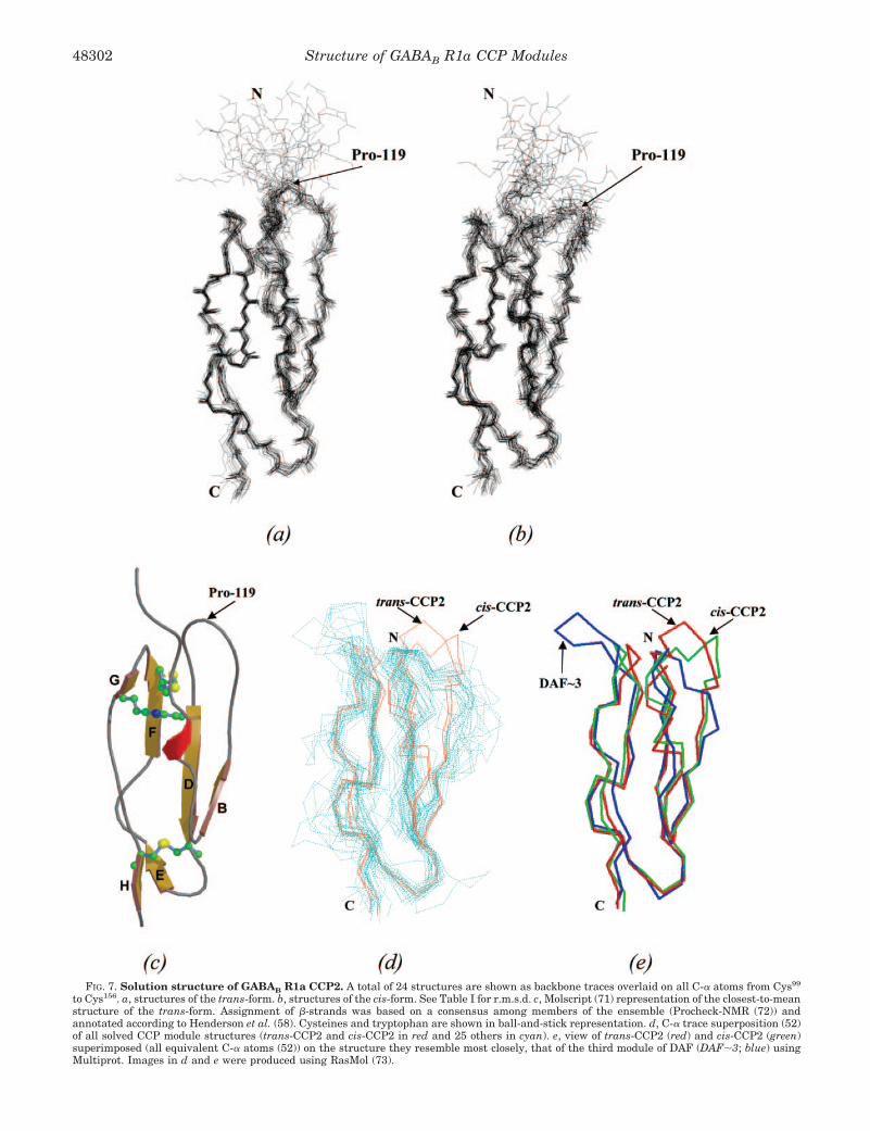
FIG. 7. Solution structure of GABAB R1a CCP2. A total of 24 structures are shown as backbone traces overlaid on all C-� atoms from Cys99
to Cys156. a, structures of the trans-form. b, structures of the cis-form. See Table I for r.m.s.d. c, Molscript (71) representation of the closest-to-meanstructure of the trans-form. Assignment of �-strands was based on a consensus among members of the ensemble (Procheck-NMR (72)) andannotated according to Henderson et al. (58). Cysteines and tryptophan are shown in ball-and-stick representation. d, C-� trace superposition (52)of all solved CCP module structures (trans-CCP2 and cis-CCP2 in red and 25 others in cyan). e, view of trans-CCP2 (red) and cis-CCP2 (green)superimposed (all equivalent C-� atoms (52)) on the structure they resemble most closely, that of the third module of DAF (DAF�3; blue) usingMultiprot. Images in d and e were produced using RasMol (73).
Structure of GABAB R1a CCP Modules48302

tures of CCP modules in complement regulators except for theregion near the N terminus. The hypervariable loop of CCP2extends toward the N terminus and may participate in thejunction with CCP1.
In contrast, CCP1 has marginal stability. It exposes many ofits hydrophobic side chains to solvent (as inferred from ANSbinding), implying a poorly structured hydrophobic core, albeitone that generates a weak near-UV ellipticity. The results froma range of biophysical methods are all consistent with the poorquality of NMR spectra, which precluded any attempts at back-bone assignment. Taken together, the biophysical data suggestthat CCP1 contains a mixture of ordered and molten globularparts. The presence of the ordered parts would explain thelimited number of proteolytically sensitive sites observed in themass spectrometric investigations and explains the cooperativeunfolding of CCP1 with increasing GdnHCl concentrations asmonitored by far-UV CD. That no such cooperative transitionwas seen by DSC may reflect aggregation competing with theunfolding event during the heating process. The presence ofpartially ordered structure is also consistent with ANS binding.
Although it could be argued that the poorly structured na-ture of CCP1 is an artifact of expression, the compact folding ofCCP2 and the successful expression in P. pastoris of manyother CCP modules suggest otherwise (50, 58, 62–64). Further-more, when the two modules are expressed together, the second(but not the first) module is compactly folded, and there is onlya small degree of stabilization imparted to CCP1 by CCP2despite the potential for a relatively extensive intermodularjunction involving the hypervariable loop of CCP2. Most impor-tant, the recombinant material is “functional” in the sense thatit is able to bind to an extracellular matrix protein. The possi-bility that CCP1 has different biophysical properties in thecontext of the intact receptor (perhaps through intrasubunitcontacts or through contacts with the GABAB R2 subunit)cannot of course be ruled out.
Disorder Is Important for Function?—Numerous examples offunctionally competent proteins with a high content of nativedisorder exist intracellularly, and several extracellular exam-
ples are now recognized (65, 66). The common occurrence ofnative disorder has been recognized in the availability of thesoftware tool PONDR (Predictor of Natural Disordered Re-gions) (67–69). The CCP1 and CCP2 sequences were submittedto PONDR. The region from Arg67 to Val77 of CCP1 was pre-dicted as disordered. This corresponds (according to a multiplesequence alignment (22)) to strands E and F in CCP modules ofknown structure. PONDR did not predict disorder in CCP2,and no or very little disorder was predicted in the CCP modulesof Vaccinia virus complement control protein and membranecofactor protein. Disorder might also be expected in the regionof CCP1 corresponding to the hypervariable loop in other CCPmodules, which is uncommonly long. In this respect, it is in-teresting to note that Lys51, where CCP1 proteolytic cleavageoccurs, is predicted to be part of the hypervariable loop. How-ever, in the absence of a backbone assignment, it is difficult toinfer from the experimental data the precise extent of disorderin CCP1.
Despite the relative lack of precedent for a disulfide-stabi-lized extracellular protein domain with a high degree of nativedisorder, we do know from studies of dynamics in other exam-ples that CCP modules are relatively flexible on a range of timescales. For example, in no CCP module studied so far are thereany amide protons that persist for more than a few hours afterdissolving a protonated CCP module in D2O. Furthermore,some CCP modules display a wide range of 1H-15N NOEs andbackbone 15N order parameters throughout their sequence,indicative of motion on a fast time scale. They also contain ahigh proportion of residues undergoing slow (millisecond-to-microsecond) conformational rearrangements. Another N-ter-minal CCP module (the first CCP module of membrane cofactorprotein) with several binding partners was found to have moremobility than an internal module (the 16th CCP module ofcomplement receptor type 1) (70). Therefore, it is conceivablethat a spectrum of mobilities exists among CCP modules andthat CCP1 of the GABAB receptor lies at one extreme. Proteinswith high levels of native disorder are thought of in terms of theadvantage such conformational plasticity affords in terms of
TABLE IStructural statistics for GABAB R1a CCP2
trans cis
Restraints used for structure calculationTotal unambiguous NOEsa 1363 1268Total ambiguous NOEs 100 79Total 1463 1347For unambiguous NOEs
Intraresidue (i 3 i) 636 632Sequential (i3 i � 1) 303 273Short-range (i 3 i � (2–4)) 85 75Long-range (i 3 i � (�4)) 339 288
J coupling (HN 3 HA) 20 23Hydrogen bonds 11 11
r.m.s.d. from experimental restraintsNOEs (Å) 0.021 � 0.0016 0.020 � 0.0018J restraints 0.99 � 0.09° 0.98 � 0.08°
r.m.s.d.from idealized geometryBond lengths (Å) 0.00164 � 0.00011 0.00163 � 0.000095Bond angles 0.331 � 0.0157° 0.330 � 0.0159°Dihedrals 16.34 � 0.54° 16.06 � 0.46°
� vs. � (%)Residues in most favored regions 67.3 70.2Residues in additional allowed regions 24.4 22.4Residues in generously allowed regions 5.2 5.1Residues in disallowed regions 3.1 2.4
Coordinate r.m.s.d. (Å) from average module structureBackbone atoms C-�, N, CO (99–159)b 0.60 0.73C-� only (99–159)b 0.62 0.76All heavy atoms (99–159)b 1.38 1.46
a All statistics are for 24 structures from 120 calculated. 975 restraints were the same for both structure calculations.b Where residues that have heteronuclear NOE values 1 S.D. lower than the mean are excluded from the r.m.s.d. calculation.
Structure of GABAB R1a CCP Modules 48303

displaying a range of affinities for a variety of ligands. In thisrespect, it is interesting to note that our yeast two-hybridscreens identified the C-terminal domain of all five members of
the fibulin family. The exact purpose of the N-terminal CCPmodules that differentiate GABAB R1a from GABAB R1b isunknown, but the results presented here suggest that they
FIG. 8. NMR relaxation data for GABAB R1a CCP2. a, heteronuclear NOEs for trans-CCP2 (�) and cis-CCP2 (● ); b, 15N T2 (upper) and 15NT1 (lower) values for trans-CCP2 (�) and cis-CCP2 (● ); c, 15N T1/15N T2 ratio for trans-CCP2 (�) and cis-CCP2 (● ).
Structure of GABAB R1a CCP Modules48304

could participate in protein-protein interactions with severalextracellular matrix proteins.
Acknowledgements—We thank Prof. Alan Cooper and MargaretNutley (University of Glasgow) for expert assistance with the calorim-etry studies. We are also grateful to Prof. Lindsay Sawyer and Dr.
Andreas Hofmann (University of Edinburgh) for help with recordingand analyzing the CD and fluorescence data.
REFERENCES
1. Blein, S., Hawrot, E., and Barlow, P. (2000) Cell. Mol. Life Sci. 57, 635–6502. Bowery, N. G., Bettler, B., Froestl, W., Gallagher, J. P., Marshall, F., Raiteri,
FIG. 9. Surface features of GABAB R1a CCP2. a, two side views rotated by 180° along the y axis depicting the GRASP (74) electrostaticsurface rendition of trans-CCP2. Negatively charged (acidic) residues are colored red, and positively charged (basic) residues are colored blue (rangeof �5 to �5 kT where k � Boltzman’s constant and T � temperature in Kelvins). b, lipophilic surface: two views rotated by 180° along the y axisdepicting the MOLCAD (75) lipophilic surface rendition of trans-CCP2. The regions of high hydrophilicity are colored blue, and the regions of highlipophilicity (hydrophobicity) are colored brown.
Structure of GABAB R1a CCP Modules 48305

M., Bonner, T. I., and Enna, S. J. (2002) Pharmacol. Rev. 54, 247–2643. Couve, A., Moss, S. J., and Pangalos, M. N. (2000) Mol. Cell. Neurosci. 16,
296–3124. White, J. H., Wise, A., Main, M. J., Green, A., Fraser, N. J., and Disney, G. H.
(1998) Nature 396, 679–6825. Kaupmann, K., Malitschek, B., Schuler, V., Heid, J., Froestl, W., and Beck, P.
(1998) Nature 396, 683–6876. Jones, K. A., Borowsky, B., Tamm, J. A., Craig, D. A., Durkin, M. M., and Dai,
M. (1998) Nature 396, 674–6797. Kuner, R., Kohr, G., Grunewald, S., Eisenhardt, G., Bach, A., and Kornau,
H. C. (1999) Science 283, 74–778. Pin, J.-P., Galvez, T., and Prezeau, L. (2003) Pharmacol. Ther. 98, 325–3549. Robbins, M. J., Calver, A. R., Filippov, A. K., Hirst, W. D., Russell, R. B., Wood,
M. D., Nasir, S., Couve, A., Brown, D. A., Moss, S. J., and Pangalos, M. N.(2001) J. Neurosci. 21, 8043–8052
10. Duthey, B., Caudron, S., Perroy, J., Bettler, B., Fagni, L., Pin, J.-P., andPrezeau, L. (2002) J. Biol. Chem. 277, 3236–3241
11. Galvez, T., Duthey, B., Kniazeff, J., Blahos, J., Rovelli, G., Bettler, B., Prezeau,L., and Pin, J.-P. (2001) EMBO J. 20, 2152–2159
12. Liu, J., Maurel, D., Etzol, S., Brabet, I., Ansanay, H., Pin, J.-P., and Rondard,P. (2004) J. Biol. Chem. 279, 15824–15830
13. Calver, A. R., Medhurst, A. D., Robbins, M. J., Charles, K. J., Evans, M. L.,Harrison, D. C., Stammers, M., Hughes, S. A., Hervieu, G., Couve, A., Moss,S. J., Middlemiss, D. N., and Pangalos, M. N. (2000) Neuroscience 100,155–170
14. Martin, S. C., Russek, S. J., and Farb, D. H. (2001) Gene (Amst.) 278, 63–7915. Isomoto, S., Kaibara, M., Sakurai-Yamashita, Y., Nagayama, Y., Uezono, Y.,
and Yano, K. (1998) Biochem. Biophys. Res. Commun. 253, 10–1516. Schwarz, D. A., Barry, G., Eliasof, S. D., Petroski, R. E., Conlon, P. J., and
Maki, R. A. (2000) J. Biol. Chem. 275, 32174–3218117. Calver, A. R., Davies, C. H., and Pangalos, M. (2002) Neurosignals 11, 299–31418. White, J. H., Ginham, R. L., Pontier, S., Wise, A., Blein, S., Barlow, P. N.,
Bouvier, M., and McIlhinney, R. A. J. (2002) FENS Abstr. 1, 062.119. Ginham, R. L., Blein, S., Barlow, P. N., White, J. H., and McIlhinney, R. A. J.
(2002) FENS Abstr. 1, 144.620. Milligan, G., and White, J. H. (2001) Trends Pharmacol. Sci. 22, 513–51821. Saghatelyan, A. K., Snapyan, M., Gorissen, S., Meigel, I., Mosbacher, J.,
Kaupmann, K., Bettler, B., Kornilov, A. V., Nifantiev, N. E., and Sakanyan,V. (2003) Mol. Cell. Neurosci. 24, 271–282
22. Hawrot, E., Xiao, Y., Shi, Q. L., Norman, D., Kirkitadze, M., and Barlow, P. N.(1998) FEBS Lett. 432, 103–108
23. Bettler, B., Kaupmann, K., and N. G. Bowery, (1998) Curr. Opin. Neurobiol. 8,345–350
24. Kirkitadze, M. D., and Barlow, P. N. (2001) Immunol. Rev. 180, 146–16125. Herbst, R., and Nicklin, M. J. (1997) Brain Res. Mol. Brain Res. 44, 309–32226. Shimizu-Nishikawa, K., Kajiwara, K., and Sugaya, E. (1995) Biochem. Bio-
phys. Res. Commun. 216, 382–38927. Imoto, T., and Yamada, H. (1997) in Protein Function: A Practical Approach
(Creigthon, T. E., ed) pp. 279–316, IRL Press at Oxford University Press,Oxford
28. Rees, S., Coote, J., Stables, J., Goodson, S., Harris, S., and Lee, M. G. (1996)BioTechniques 20, 102–110
29. Pan, T.-C., Sasaki, T., Lang, R. Z., Fassler, R., Timpl, R., and Chu, M. L. (1993)J. Cell Biol. 123, 1269–1277
30. Molnar, E., McIlhinney, R. A. J., Baude, A., Nusser, Z., and Somogyi, P. (1994)J. Neurochem. 63, 683–693
31. White, J. H., McIllhinney, R. A., Wise, A., Ciruela, F., Chan, W. Y., Emson,P. C., Billinton, A., and Marshall, F. H. (2000) Proc. Natl. Acad. Sci. U. S. A.97, 13967–13972
32. Pace, C. N., Shirley, B. A., and Thomson, J. A. (1990) in Protein Structure: APractical Approach (Creigthon, T. E., ed) 2nd Ed., pp. 311–331, IRL Pressat Oxford University Press, Oxford
33. Privalov, P. L., and Potekhin, S. A. (1986) Methods Enzymol. 131, 4–5134. Mori, S., Abeygunawardana, C., Johnson, M. O., and van Zijl, P. C. M. (1995)
J. Magn. Reson. 108, 94–9835. Sklenar, V., Piotto, M., and Leppik, R. (1993) J. Magn. Reson. 102, 241–24536. Vuister, G. W., and Bax, A. (1992) J. Magn. Reson. 98, 428–43537. Muhandiram, D. R., and Kay, L. E. (1994) J. Magn. Reson. 103, 203–21638. Grzesiek, S., and Bax, A. (1993) J. Biomol. NMR 3, 185–204
39. Wang, A. C., Lodi, P. J., and Qin, J. (1994) J. Magn. Reson. 105, 196–19840. Grzesiek, S., Anglister, J., and Bax, A. (1993) J. Magn. Reson. 101, 114–11941. Kay, L. E., Xu, G. Y., Singer, A. U., Muhandiram, D. R., and Forman-Kay, J. D.
(1993) J. Magn. Reson. 101, 333–33742. Yamazaki, T., Forman-Kay, J. D., and Kay, L. E. (1993) J. Am. Chem. Soc. 115,
11054–1105543. Pascal, S. M., Muhandiram, D. R., Yamazaki, T., Forman-Kay, J. D., and Kay,
L. E. (1994) J. Magn. Reson. 103, 197–20144. Aitio, H., and Permi, P. (2000) J. Magn. Reson. 143, 391–39645. Kraulis, P. J. (1989) J. Magn. Reson. 24, 627–63346. Kay, L. E., Nicholson, L. K., Delagio, F., Bax, A., and Torchia, D. A. (1992) J.
Magn. Reson. 97, 359–37547. Grzesiek, S., and Bax, A. (1993) J. Am. Chem. Soc. 115, 12593–1259448. Yuan, X., Werner, J. M., Knott, V., Handford, P. A., Campbell, I. D., and
Downing, K. (1998) Protein Sci. 7, 2127–213549. Brunger, A. T., Adams, P. D., Clore, G. M., DeLano, W. L., Gros, P., Grosse-
Kunstleve, R. W., Jiang, J. S., Kuszewski, J., Nilges, M., Pannu, N. S., Read,R. J., Rice, L. M., Simonson, T., and Warren, G. L. (1998) Acta Crystallogr.D Biol. Crystallogr. 54, 905–921
50. Smith, B. O., Mallin, R., Krych-Goldberg, M., Wang, X., Hauhart, R. E.,Bromek, K., Uhrin, D., Atkinson, J. P., and Barlow, P. N. (2002) Cell 108,769–780
51. Shindyalov, I., and Bourne, P. (1998) Protein Eng. 11, 739–74752. Shatsky, M., Nussinov, R., and Wolfson, H. J. (2002) Proc. Lect. Notes Comput.
Sci. 2452, 235–25053. White, C. E., Kempi, N. M., and Komives, E. A. (1994) Structure 2, 1003–100554. Cereghino, J. L., and Cregg, J. M. (2000) FEMS Microbiol. Rev. 24, 45–6655. Kirkitadze, M. D., Krych, M., Uhrin, D., Dryden, D. T., Smith, B. O., Cooper,
A., Wang, X., Hauhart, R., Atkinson, J. P., and Barlow, P. N. (1999)Biochemistry 38, 7019–7031
56. Kelly, S. M., and Price, N. C. (1997) Biochim. Biophys. Acta 1338, 161–18557. Kirkitadze, M. D., Barlow, P. N., Price, N. C., Kelly, S. M., Boutell, C. J., Rixon,
F. J., and McClelland, D. A. (1998) J. Virol. 72, 10066–1007258. Henderson, C. E., Bromek, K., Mullin, N. P., Smith, B. O., Uhrin, D., and
Barlow, P. N. (2001) J. Mol. Biol. 307, 323–33959. Kabsch, W., and Sander, C. (1983) Biopolymers 22, 2577–263760. Mallin, R. L. (2003) A Structural Study of the C3b-binding Site of Complement
Receptor Type 1 (CD 35). Ph.D. thesis, University of Edinburgh61. Timpl, R., Sasaki, T., Kostka, G., and Chu, M. L. (2003) Nat. Rev. Mol. Cell.
Biol. 4, 479–48962. Wiles, A. P., Shaw, G., Bright, J., Perczel, A., Campbell, I. D., and Barlow, P. N.
(1997) J. Mol. Biol. 272, 253–26563. Murthy, K. H., Smith, S. A., Ganesh, V. K., Judge, K. W., Mullin, N., Barlow,
P. N., Ogata, C. M., and Kotwal, G. J. (2001) Cell 104, 301–31164. Uhrinova, S., Lin, F., Ball, G., Bromek, K., Uhrin, D., Medof, M. E., and
Barlow, P. N. (2003) Proc. Natl. Acad. Sci. U. S. A. 100, 4718–472365. Dunker, A. K., Brown, C. J., Lawson, J. D., Iakoucheva, L. M., and Obradovic,
Z. (2002) Biochemistry 41, 6573–658266. Dunker, A. K., Lawson, J. D., Brown, C. J., Williams, R. M., Romero, P., Oh,
J. S., Oldfield, C. J., Campen, A. M., Ratliff, C. M., Hipps, K. W., Ausio, J.,Nissen, M. S., Reeves, R., Kang, C., Kissinger, C. R., Bailey, R. W., Gris-wold, M. D., Chiu, W., Garner, E. C., and Obradovic, Z. (2001) J. Mol.Graph. Model. 19, 26–59
67. Romero, P., Obradovic, Z., Li, X., Garner, E., Brown, C., and Dunker, A. K.(2001) Proteins 42, 38–48
68. Romero, P., Obradovic, Z., and Dunker, A. K. (1997) Genome Inform. Ser.Workshop Genome Inform. 8, 110–124
69. Li, X., Romero, P., Rani, M., Dunker, A. K., and Obradovic, Z. (1999) GenomeInform. Ser. Workshop Genome Inform. 10, 30–40
70. O’Leary, J. M., Bromek, K., Black, G. M., Uhrinova, S., Schmitz, C., Wang, X.,Krych, M., Atkinson, J. P., Uhrin, D., and Barlow, P. N. (2004) Protein Sci.13, 1238–1250
71. Kraulis, P. J. (1991) J. Appl. Crystallogr. 24, 946–95072. Laskowski, R. A., Rullmann, J. A., MacArthur, M. W., Kaptein, R., and Thorn-
ton, J. M. (1996) J. Biomol. NMR 8, 477–48673. Sayle, R. A., and Milner-White, E. J. (1995) Trends Biochem. Sci. 20, 374–37674. Nicholls, A., Sharp, K. A., and Honig, B. (1991) Proteins 11, 281–29675. Heiden, W., Goetze, T., and Brickmann, J. (1993) J. Comput. Chem. 14,
246–250
Structure of GABAB R1a CCP Modules48306