the journal of biol~~ical chemistry vol. … · and by camp and dexamethasone in fetal lung in...
TRANSCRIPT

T H E JOURNAL OF BIOL~~ICAL CHEMISTRY Q 1994 by The American Society for Biochemistry and Molecular Biology, Inc
Vol. 269, No. 44, Issue of November 4, pp. 27761-27772, 1994 Printed in U.S.A.
Developmental and Hormonal Regulation of Surfactant Protein C (SP-C) Gene Expression in Fetal Lung ROLE OF TRANSCRIPTION AND mRNA STABILITY*
Viiayakumar BoggaramS and Ramgopal K. Margana From the Department of Molecular Biology, University of Texas Health Science Center, Tyler, Texas 75710
Pulmonary surfactant protein C (SP-C) gene expres- sion is developmentally and hormonally regulated in fe- tal lung. In the present study, we investigated the role of transcriptional and posttranscriptional mechanisms in the developmental, CAMP, and dexamethasone induc- tion of SP-C mRNA. We found that developmental induc- tion of SP-C mRNA was not coincident with induction of SP-C gene transcription. SP-C mRNA levels reached -90% of levels in adult lung on day 24 of gestation, whereas SP-C gene transcription was only -4% of level in adult lung and did not increase until day 28 of gesta- tion (term in rabbit = 31 days). Treatment of fetal lung tissues in vitro with dibutyryl cyclic AMP (Bt,cAMP) and dexamethasone increased SP-C mRNA accumulation by different mechanisms. Increase in SP-C mRNA accumu- lation by Bt,cAMP was the result of increased SP-C gene transcription, whereas increased SP-C mRNA accumu- lation by dexamethasone was due to stabilization of RNA. In control tissues the SP-C mRNAhalf-life (tm) was 11.2 h, and after dexamethasone treatment it increased to 30 h. These data show that both transcriptional and mRNA stabilization mechanisms regulate induction of SP-C gene expression during fetal lung development and by CAMP and dexamethasone in fetal lung in vitro.
Pulmonary surfactant prevents collapse of alveoli during ex- piration by reducing net contractile forces at the alveolar sur- face (1). Surfactant is synthesized and secreted by alveolar type I1 epithelial cells of lung and is composed of lipids (90%) and proteins (5-10%) (2). Deficient production of surfactant, as found in premature infants, is associated with the development of respiratory distress syndrome of the newborn (3), the lead- ing cause of neonatal morbidity and mortality in developed countries.
Four lung-specific surfactant-associated proteins have been isolated to date: surfactant protein A (SP)'-A, SP-B, SP-C, and SP-D (4,5). SP-B and SP-C are extremely hydrophobic proteins that appear to be essential for maintenance of biophysical prop- erties and physiological activity of surfactant (6). Although SP-C in concert with SP-B has been implicated in maintaining surface active properties of surfactant and SP-C alone en- hances the adsorption rate of phospholipids in vitro, its precise
* This research was supported in part by Grant 92G-569 from the American Heart Association, Texas Affiliate Inc.
of publication of this article were defrayed in part by the payment of We dedicate this article to the memory of Mauricio X. Zuber. The costs
page charges. This article must therefore be hereby marked "aduertise-
fact. rnent" in accordance with 18 U.S.C. Section 1734 solely to indicate this
Biology, University of Texas Health Science Center, P. 0. Box 2003, $ To whom correspondence should be addressed: Dept. of Molecular
Tyler, TX 75710. Tel.: 903-877-7780; Fax: 903-877-7558. The abbreviations used are: SP, surfactant protein; Bt,cAMP,
kb, kilobase(s). dibutyryl cyclic AMP; MOPS, 3-(N-morpholino)propanesulfonic acid;
role in surfactant function remains to be defined. During fetal lung development SP-C mRNA can be detected prior to the appearance of morphologically identifiable alveolar type I1 epi- thelial cells (7), and in adult lung its expression is confined to type I1 epithelial cells (7-9). These data suggest that SP-C, in addition to enhancing surface activity of surfactant, also serves some undefined function in development and differentiation of the lung.
We previously isolated and characterized SP-C cDNAs in the rabbit and found that SP-C mRNA accumulation is induced during fetal lung development (10). We also found that cyclic AMP analogs and dexamethasone added in. vitro to 21-day ges- tational age fetal lung tissues increased accumulation of SP-C mRNA in a time-dependent manner. Molecular mechanisms that mediate developmental, CAMP, and glucocorticoid regula- tion of SP-C mRNA accumulation have not been defined. In the present study we used transcription run-on assays and North- ern blot analysis to investigate the role of transcriptional and posttranscriptional mechanisms in the developmental, CAMP, and glucocorticoid induction of SP-C mRNA accumulation. We found that increases in SP-C mRNA accumulation during de- velopment were not coincident with increases in SP-C gene transcription; SP-C mRNAlevels reached 90% of levels found in adult lung by day 24 of gestation but an increase in SP-C gene transcription was not found until day 28 of gestation suggest- ing significant stabilization of SP-C mRNA. In fetal lung in vitro, the effect of dexamethasone to increase SP-C mRNA ac- cumulation was found to be due solely to an increase in the half-life of SP-C mRNA, whereas the effect of CAMP was found to be due to an increase in SP-C gene transcription. These findings have been reported in preliminary form (11).
MATERIALS AND METHODS Organ Culture of Fetal Lung Essues-Lung tissues of 21-day gesta-
tional age fetal rabbits were maintained in organ culture in serum-free Waymouth's medium MB 752/1 (Life Technologies, Inc.) according to Snyder et al. (12). Lung explants were maintained in culture for up to 3 days in either control medium or medium containing Bt,cAMP (1 mM) or dexamethasone M) or a combination of the two agents. Dexa- methasone was dissolved in ethanol before addition to the culture me- dium. The concentration of ethanol in culture medium was 0.01%; at this concentration ethanol had no affect on SP-C mRNA levels. Actino- mycin D (actinomycin D-mannitol, water-soluble, Sigma) (5 pg/ml) was added in experiments designed to measure the rate of SP-C mRNA degradation. Culture medium was changed every 24 h.
Isolation of RNA and Northern Blot Analysis-Total RNA from fetal lung tissues was extracted with TRI reagent (Molecular Research Cen- ter, Inc., Cincinnati, OH) according to the manufacturer's instructions. RNA(5-10 pg) was electrophoresed in agarose gels (1.2%) containing 20 mM MOPS and 1.1% formaldehyde (13). RNAs were transferred to Hy- Bond N' (Amersham Corp.) membrane by capillary action using 50 mM sodium hydroxide. Membranes were hybridized to 32P-labeled rabbit SP-C cDNA(-0.9 kb) and human p actin cDNA (-1.7 kb) at 60 "C, and final washes were done in 1 x SSC at 60 "C. Membranes were also hybridized to a ',P-labeled antisense oligonucleotide (30-mer) of riboso- mal 18 S RNA to assess the integrity of RNA preparations, loading of equivalent amounts of RNA, and quantitative transfer of RNA. Auto- radiographic signals were quantified by scanning densitometry (Milli-
27767

27768 Regulation of Surfactant Protein C Gene Expression pore BioImage Analyzer). Densities of SP-C and actin mRNA signals were normalized to the density of 18 S rRNA signal to correct for the amount of RNA loaded on the gels.
Isolation of Nuclei and ZYanscription Run-on Assay-Methods for isolation of nuclei, transcription run-on assay, and hybridization of RNA have been described previously (14). RNA from nuclei was extracted with TRI reagent by modification of the method for extraction of RNA from cells/tissue. Nuclei (20 x lofi in 200 pl) were lysed by adding 1.4 ml of TRI reagent to the reaction mixture, vigorously vortexed for 30-60 s, and stored a t room temperature for 10 min. Chloroform (0.28 ml) was added to the lysed nuclei, and the samples were shaken vigorously for 15 s and stored a t room temperature for 5 min. Samples were centri- fuged for 15 min a t 12,000 x g a t 4 "C. The upper aqueous phase was mixed with 100 pg of yeast or E. coli tRNA that served as a carrier, and total RNA was precipitated with an equal volume of isopropanol for 10 min a t room temperature. RNA was collected by centrifugation a t 12,000 x g for 10 min, rinsed with 1.0 ml of 70% ethanol, dissolved in water, and reprecipitated by addition of 0.3 M NaOAc, pH 5.2, and 2.5 volumes of ethanol a t -20 "C. The final RNA precipitate was dissolved in 200 p1 of hybridization buffer, and radioactivity of RNA was deter- mined by liquid scintillation spectrometry. The final RNA preparations were checked for presence of unincorporated I"2PlUTP.
This method for isolation of RNA does not involve many steps, and the entire procedure can be completed in a relatively short time. It is especially convenient for extraction of RNAs from a large number of samples. RNA extracted by our method produced hybridization results similar to those RNAs obtained by digestions with DNase I, proteinase K, and multiple phenol and chloroform extractions. A similar method for isolation of RNA from labeled nuclei was described recently (15).
Equal amounts of radioactive RNAs were hybridized to 10 pg of linearized plasmid DNA containing rabbit SP-C cDNAor human p actin cDNA that had been bound to nitrocellulose membranes. In some ex- periments RNAs were also hybridized to membranes containing anti- sense SP-C cDNA (0.9 kb) or antisense SP-C gene fragments in M13. Linearized plasmid DNAs were denatured by treatment with 0.3 M NaOH for 30 min a t 37 "C and neutralized with 1 M NH,OAc. M13 DNA and denatured plasmid DNAs were applied to nitrocellulose mem- branes using a dot blot apparatus, and the DNAs were fixed to the membrane by baking a t 80 "C for 2 h under vacuum. Hybridizations were carried out in a final volume of 200 pl at 45 "C for 3 days. Washed membranes were subjected to autoradiography using an intensifying screen, and signals were quantified by scanning densitometry (Milli- pore BioImage Analyzer). After autoradiography membranes were counted in a liquid scintillation counter to determine the amount of bound radioactivity. Usually we detected no degradation of RNA during hybridization. Transcription rates determined by either quantification of autoradiographic signals or by quantification of bound radioactivity were similar. Signalhadioactivity bound to linearized pUC DNA/M13 DNA (background) was subtracted from radioactivity obtained with plasmids containing SP-C/actin cDNAs or M13 containing antisense SP-C cDNA/antisense SP-C gene fragments.
Measurement of Half-life of SP-C and Actin mRNAs-Half-lives of SP-C and actin mRNAs were determined by measuring their rate of degradation after inhibition of RNA synthesis with actinomycin D (5 pg/ml). Actinomycin D at this concentration inhibited total RNA syn- thesis by -80% after 3 h of incubation. Tissues were incubated in the presence or absence of dexamethasone M) for 24 h, and incubation was continued for an additional 24 h in the presence or absence of dexamethasone in medium containing actinomycin D ( 5 pg/ml). Tissues were collected a t 0, 6, 9, 12, 18, and 24 h after addition of actinomycin D, and SP-C and actin mRNA levels were determined by Northern blot analysis. Half-life of RNA was calculated from the equation t,, = ln2/k (t,,, = half-life; degradation rate constant k = 42.303) (slope)) (16).
RESULTS
Developmental Regulation of SP-C Gene Danscription and SP-C mRNA Levels-We found previously that SP-C mRNA accumulation is induced during fetal lung development (10). To determine if the induction of SP-C mRNA levels is regulated a t the transcriptional level, we analyzed SP-C gene transcription and SP-C mRNA levels in fetal lung tissues (19-30-day gesta- tional ages) and adult rabbit lung (Fig. LA). We found a marked lag between the temporal induction of SP-C gene transcription and elevation of SP-C mRNA levels. The increases in SP-C mRNA accumulation were not associated with similar in- creases in SP-C gene transcription. SP-C mRNAlevel in 24-day
Gestational age (days)
Actin - 1.8 kb ic 300- -
a ,$z E ;i 200-
K *
Transcription rate RNA
-.3
._ 0 .I!
'C P E g 100- Y F &
Y 0- 4 . . I ln 21 24 26 28 30 A
Gestational age (days)
and SP-C mRNA accumulation in fetal rabbit lung. A, nuclei from FIG. 1. Developmental regulation of SP-C gene transcription
lung tissues of fetal rabbits of gestational age 19-30 days and of adult animals were isolated and SP-C gene transcription was analyzed by transcription run-on assay. Approximately 6 x 10' cpm were hybridized to filters containing rabbit SP-C cDNAin pUC (panel a). RNAs (5.0 pg) isolated from lung tissues of fetal and adult rabbits were analyzed by Northern blotting with "P-labeled rabbit SP-C cDNA (panel b ) or hu- man actin cDNA(panel c). B , autoradiographic signals obtained after transcription run-on assay and Northern blotting were quantified by scanning densitometry. In transcription run-on assays, signals obtained with filters containing pUC were subtracted from signals obtained with filters containing SP-C cDNA in pUC. SP-C mRNA levels were normal- ized to 18 S rRNA levels. Data of transcription analysis represent mean -c S.D. of analysis of transcription rates in lungs from two separate litters of animals. Data of Northern blotting represent mean = S.E. of analysis of RNA levels in lungs from four separate litters of animals.
fetal lung was -90% of the level in adult lung, whereas SP-C gene transcription level was only -4% of the level in adult lung (Fig. 1B). An increase in SP-C gene transcription was not ob- served until day 28 of gestation, and after day 28 of gestation it increased to reach peak levels in adult lung tissue. Total RNA transcription in nuclei of different gestational age fetal lungs did not change significantly, but a decrease of -50% was con- sistently found in nuclei from adult lungs. SP-C gene transcrip- tion rates were similar or identical whether measured with double-stranded SP-C cDNA or single-stranded antisense SP-C cDNA probes.
CAMP and Glucocorticoid Regulation of SP-C Gene Dan- scription and SP-C mRNA Levels-In previous studies we found that cyclic AMP analogs and dexamethasone increases the SP-C mRNA accumulation in 21-day fetal lung tissues in vitro in a time-dependent manner. Induction by CAMP analogs occurred consistently after only 48 h of incubation, whereas induction by dexamethasone was found after 24 h of incuba- tion. In tissues treated with a combination of Bt,cAMP and dexamethasone, the effects on SP-C mRNA levels were less than additive after 24 h of incubation, whereas after 48-72 h of incubation they were additive. To determine if the changes in SP-C mRNA accumulation are due to changes in SP-C gene transcription, we analyzed SP-C gene transcription and SP-C mRNA levels in tissues treated with Bt,cAMP or dexametha- sone or a combination of the two agents (Fig. 2). Total RNA transcription rate was unaffected by treatment with hormonal agents.
A
SP-c 0.9 kb
Regulation of Surfactant Protein C Gene Expression
24 h 48 h 72 h C B D B D C B D B D C B D B D
- 1.8 kb J I I C
B - Y 5 , H Tra scription rate
T 0 mR A level
c B D B D c B D B D c B . D B D ' 24 h 48 h 72 h
Treatment FIG. 2. Effects of Bt,cAMP ( B ) and dexamethasone ( D ) on SP-C
gene transcription and SP-C mRNAaccumulation in fetal rabbit lung in vitro. A, 21-day gestational age fetal rabbit lung tissues were maintained in the absence (C) or presence of Bt,cAMP (1 ms5) or dexa- methasone M) or a combination of the two agents for 24-72 h. Nuclei were isolated from fetal lung tissues in culture and SP-C gene transcription rates were determined by run-on assay as described under "Materials and Methods." -5.0 x 10' cpm were hybridized to nitrocel- lulose filters containing SP-C cDNA in pUC, and an autoradiogram was obtained (panel a). RNA (5.0 pg) isolated from lung tissues in culture was analyzed by Northern blotting with "'P-labeled SP-C cDNA (panel b ) or "P-labeled human p actin cDNA (panel c). B, autoradiographic signals obtained after transcription run-on assay and Northern blot analysis were quantified by scanning densitometry. SP-C mRNA levels were normalized to 18 S rRNA levels to correct for loading equal amounts of RNA. Data represent mean * S.E. of eight separate experi- ments. In transcription run-on assays, signals obtained with filters containing pUC was considered as background and subtracted from signals obtained with filters containing SP-C cDNA in pUC. Data rep- resent mean * S.E. of four separate experiments.
We found that Rt,cAMP increased SP-C gene transcription and SP-C mRNA levels after 48-72 h of incubation. The in- creases in SP-C mRNA accumulation were associated with sim- ilar or higher levels of SP-C gene transcription (Fig. 2B). In tissues incubated with dexamethasone, induction of SP-C mRNA accumulation after 24 h was not associated with an increase in SP-C gene transcription (Fig. 2B). After 48 h of incubation with dexamethasone, SP-C gene transcription was reduced compared to levels in tissues maintained in control medium. In tissues treated with a combination of Bt,cAMP and dexamethasone for 48-72 h, SP-C gene transcription was re- duced significantly compared to levels in tissues treated with Bt,cAMP alone. Actin mRNA levels and gene transcription were not significantly affected by treatment with Bt,cAMP, dexamethasone, or a combination of the two agents. These data suggest that the dexamethasone-dependent increase in SP-C mRNA accumulation is mediated at posttranscriptional level, whereas the CAMP-dependent increase is due primarily to an increase in transcription.
We also observed that SP-C gene transcription and SP-C mRNA accumulation increased during culture of fetal lung tissues.
Time (h)
A 0 6 9 12 18 24 0 6 9 12 18 24
- 1.8 kb Actin 1
Control Dexamethasone
B
27769
2 1-1 5 1 1 , I , , 1 v, 0 5 10 15 20 25 2 0 5 10 15 20 25
Time (h) Time (h)
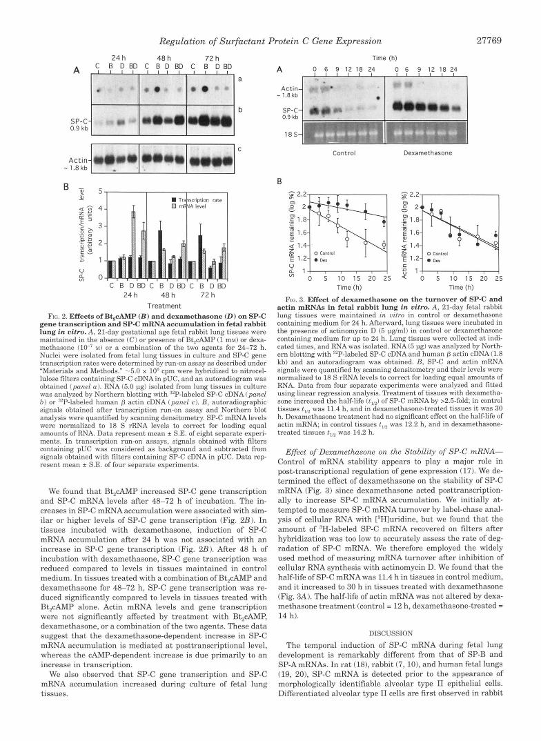
FIG. 3. Effect of dexamethasone on the turnover of SP-C and actin mRNAs in fetal rabbit lung in vitro. A, 21-day fetal rabbit lung tissues were maintained in vitro in control or dexamethasone containing medium for 24 h. Afterward, lung tissues were incubated in the presence of actinomycin D (5 pg/ml) in control or dexamethasone containing medium for up to 24 h. Lung tissues were collected a t indi- cated times, and RNA was isolated. RNA (5 pg) was analyzed by North- ern blotting with ""P-labeled SP-C cDNA and human p actin cDNA (1.8 kb) and an autoradiogram was obtained. R, SP-C and actin mRNA signals were quantified by scanning densitometry and their levels were normalized to 18 S rRNA levels to correct for loading equal amounts of RNA. Data from four separate experiments were analyzed and fitted using linear regression analysis. Treatment of tissues with dexametha- sone increased the half-life (t,,) of SP-C mRNA by >2.5-foId; in control tissues t,, was 11.4 h, and in dexamethasone-treated tissues it was 30 h. Dexamethasone treatment had no significant effect on the half-life of actin mRNA; in control tissues t,, was 12.2 h, and in dexamethasone- treated tissues t,,L was 14.2 h.
Effect of Dexamethasone on the Stability of SP-C mRNA- Control of mRNA stability appears to play a major role in post-transcriptional regulation of gene expression (17). We de- termined the effect of dexamethasone on the stability of SP-C mRNA (Fig. 3) since dexamethasone acted posttranscription- ally to increase SP-C mRNA accumulation. We initially at- tempted to measure SP-C mRNA turnover by label-chase anal- ysis of cellular RNA with ['Hluridine, but we found that the amount of 'H-labeled SP-C mRNA recovered on filters after hybridization was too low to accurately assess the rate of deg- radation of SP-C mRNA. We therefore employed the widely used method of measuring mRNA turnover after inhibition of cellular RNA synthesis with actinomycin D. We found that the half-life of SP-C mRNAwas 11.4 h in tissues in control medium, and it increased to 30 h in tissues treated with dexamethasone (Fig. 3A 1. The half-life of actin mRNA was not altered by dexa- methasone treatment (control = 12 h, dexamethasone-treated = 14 h).
DISCUSSION
The temporal induction of SP-C mRNA during fetal lung development is remarkably different from that of SP-B and SP-AmRNAs. In rat (18), rabbit (7, lo), and human fetal lungs (19, 201, SP-C mRNA is detected prior to the appearance of morphologically identifiable alveolar type I1 epithelial cells. Differentiated alveolar type I1 cells are first observed in rabbit

27770 Regulation of Surfactant Protein C Gene Expression
lung tissue a t approximately day 26 of gestation (21). In rabbit fetal lung, SP-C mRNA is detected at day 19 of gestation, the earliest time point examined (7, 10, 22). SP-C mRNA levels increase as a function of gestation, and in neonatal and adult lung tissues its level decreases significantly compared to levels found in lung tissues of 28-31-day gestational age fetal rabbits.
We found a marked discrepancy between the temporal induc- tion of SP-C gene transcription and SP-C mRNA accumulation. The lag between SP-C gene transcription and SP-C mRNA accumulation suggested that significant stabilization of SP-C mRNA can account for the accumulation of SP-C mRNA during prenatal lung development. In adult lung, however, transcrip- tion appears to regulate SP-C mRNA accumulation. Because of experimental difficulties involved, we did not attempt to measure SP-C mRNA turnover in vivo during fetal lung development.
The temporal induction of SP-C gene transcription and mRNA during fetal rabbit lung development is quite different from the temporal induction of SP-A gene transcription and mRNA. SP-A gene transcription increases as early as day 24 of gestation and continues to increase to reach highest levels in 28-day lung (14). SP-A gene transcription in neonatal lung is decreased compared to levels in 28-day lung. The temporal induction of SP-A gene transcription coincides with temporal induction of SP-A mRNA (14), suggesting that our observed differences between temporal induction of SP-C gene transcrip- tion and SP-C mRNA accumulation represent a specific phe- nomenon, not a pleiotropic effect.
Snyder and co-workers (7) used in situ hybridization analysis to detect SP-C mRNA in epithelial cells of prealveolar region of 19-day rabbit fetal lung. They found that the SP-C mRNA con- tent increased as a function of gestational age, and by day 27 of gestation and thereafter expression of SP-C mRNA was re- stricted to epithelial cells with morphologic characteristics of alveolar type I1 cells (7). Taken together, the data of Snyder and co-workers and our finding that SP-C gene transcription does not increase until day 28 of gestation suggest that an increase in SP-C gene transcription is linked to differentiation of alve- olar type I1 cells.
The number of identifiable type I1 cells increases as a pro- portion of total epithelial cells in lung after day 26 of gestation in the rabbit (21, 23). This makes accurate assessment of in- creases in SP-C gene transcription rate during development difficult. SP-C mRNA content of prealveolar and alveolar epi- thelial cells increase during fetal rabbit lung development to reach highest levels a t term (7). This suggests that an increase in SP-C mRNA content of type 11 epithelial cells during devel- opment must be the result of increases in SP-C gene transcrip- tiodSP-C mRNA stability. These data suggest that increases in SP-C gene transcription rate during development must be due both to an increase in the number of type I1 epithelial cells and to enhanced transcriptiodstability of SP-C mRNA.
The physiological significance of high levels of SP-C mRNA in embryonic lung is unclear. A similar pattern of accumulation was also noted for SP-A and SP-B mRNAs (22). The transition from an aqueous to a gaseous environment that accompanies birth may necessitate an acute requirement for surfactant pro- teins and lipids. Elevated levels of surfactant protein mRNAs may reflect such a need.
Glucocorticoids and CAMP play major roles in promoting fe- tal lung development and enhance surfactant synthesis (24). Glucocorticoids increase SP-C mRNA accumulation in human fetal lung in vitro (19,20) and in rat lung both in vitro (25) and in vivo (26, 27). The effects of maternal administration of glu- cocorticoids on SP-C mRNA levels in fetal lung are not clear. Maternal administration ofglucocorticoids to pregnant rabbits
on day 26 of gestation resulted in a decrease in SP-C mRNA level in lungs of fetal rabbits delivered on day 27 (28). A recent study showed that glucocorticoids administered to pregnant rabbits on days 25 and 26 of gestation increased SP-C mRNA levels 2-fold compared to uninjected control animals (29). In the same study injection of saline to pregnant animals increased SP-C mRNA levels in fetal lung tissues 2-fold, leading the au- thors to suggest that maternal stress related factors alone can increase SP-C mRNA levels. Glucocorticoid regulation of SP-C mRNA levels in vivo might depend on the developmental stage of the fetus when the hormone was administered maternally.
We found that CAMP and dexamethasone regulate SP-C mRNA levels in fetal lung in vitro by distinct mechanisms; the effects of CAMP are mediated at the transcriptional level, whereas the effects of dexamethasone are mediated solely at the posttranscriptional level. We also found that after 48 h of incubation dexamethasone inhibited both basal and Bt,cAMP- dependent increases in SP-C gene transcription. This effect may explain why we did not observe a substantial increase in SP-C mRNA levels in tissues treated with a combination of Bt,cAMP and dexamethasone for 48-72 h, even though dexa- methasone increased the half-life of SP-C mRNA after 24 h and Bt,cAMP increased SP-C gene transcription after 48 h. Mecha- nisms by which dexamethasone inhibited SP-C gene transcrip- tion are not known. Glucocorticoids were found to negatively regulate human glycoprotein a-subunit gene expression by in- terfering with a CAMP responsive element (30). Whether dexa- methasone inhibits CAMP-induced SP-C gene transcription by a similar mechanism remains to be investigated.
We found previously that adenosine 3’,5’-cyclic monophos- phorothioate (Sp-diastereomer) (0.1-0.25 m), an analog of cy- clic AMP, increases SP-C mRNA accumulation in fetal rabbit lung in vitro by a magnitude similar to that of Bt,cAMP (1 m) (10). This suggested that the effects of Bt,cAMP to increase SP-C mRNA accumulation are not due to butyric acid, a by- product of metabolism of Bt,cAMP. Butyric acid modulates glo- bin gene expression in erythroid cells (31,321. Recently, butyric acid was found to modulate surfactant protein gene expression in fetal rat lung in vitro with significant inhibitory effects on SP-A and SP-B mRNA accumulation (33). The effects of butyric acid on SP-C mRNA levels were complex; butyric acid inhibited SP-C mRNA accumulation below control levels after 6 h of incubation but increased to control levels after 24 h of incuba- tion (33). Both transcription and mRNA stability were found to mediate butyric acid-induced changes in SP-C mRNA accumu- lation. The authors have suggested that elevated levels of bu- tyric acid analogs as found in diabetic mothers and fetuses might lead to increased incidence of newborn respiratory dis- tress syndrome. It remains to be determined whether butyric acid has similar effects on surfactant protein gene expression in fetal lungs of other species. Incubation of fetal rabbit lung explants with butyric acid for 5 days did not inhibit levels of immunoreactive SP-A (34).
Glucocorticoids and CAMP influence expression of a number genes in different cell types of fetal lung (35). This might con- found our analysis of the effects of these agents on SP-C gene transcription rate in fetal lung explants. In fetal rabbit lung in vitro SP-A (14), SP-B,2 and SP-C (10) genes are regulated in- dependently by both CAMP and glucocorticoids. By in situ hy- bridization dexamethasone was found to increase SP-B mRNA content of both alveolar and bronchiolar epithelial cells of fetal rat lung explants (36). This suggests that dexamethasone-de- pendent increase in SP-B mRNA levels in fetal rat lung ex- plants must be due to increased accumulation of SP-B mRNA in alveolar and bronchiolar epithelial cells of fetal lung. By dele-
R. K. Margana and V. Boggaram, submitted for publication.

Regulation of Surfactant Protein C Gene Expression 27771
tion mapping and transfection analysis a region of SP-A gene, -378 to +20, was shown to mediate CAMP induction of SP-A- chloramphenicol acetyltransferase fusion gene expression in purified alveolar type I1 epithelial cells (37). This suggests that the inductive effects of CAMP on SP-A gene transcription in fetal rabbit lung i n vitro are indeed due to activation of SP-A gene transcription in type I1 cells. These data suggest that glucocorticoids and CAMP regulate surfactant protein gene ex- pression by altering their expression by specific cell types of lung and that changes in surfactant protein gene expression are not the result of pleiotropic effects of hormones.
We investigated the effects of dexamethasone on SP-C mRNA stability since stabilization of RNA plays a major role in post- transcriptional regulation of gene expression (17). We found that dexamethasone increased the half-life of SP-C mRNA by ~2.5-fold. The action of dexamethasone that results in in- creased SP-C mRNA levels in rabbit fetal lung i n vitro is dif- ferent from that found in rat (25) and human fetal lung (38) tissues. In both rat and human fetal lung tissues i n vitro, glucocorticoids increase SP-C mRNA levels by increasing gene transcription. However, mRNA stabilization and its effect on SP-C mRNA levels in rat and human lung i n vitro cannot be ruled out, because the time course of the effects of glucocorti- coid on SP-C gene transcription and SP-C mRNA accumulation were not investigated. The lack of effect o f dexamethasone on SP-C gene transcription in rabbit fetal lung might be species- specific. SP-A gene expression appears to be regulated differ- ently by glucocorticoids in rabbit (14) and human (39) fetal lung in vitro. In rabbit fetal lung in vitro, glucocorticoids regulate SP-A gene expression predominantly at the transcriptional level (141, whereas in human fetal lung in vitro, they exert dose-dependent effects on transcription and mRNA stability (401.
SP-C gene transcription and SP-C mRNA accumulation in- creased during explant culture in the absence of either serum or hormones. Several other reports indicate increases in mRNAs for rabbit SP-A (141, SP-C (IO), and human SP-B (19) during explant culture of fetal lung tissues in serum-free de- fined medium. Increases in SP-A mRNA in rabbit fetal lung i n vitro appeared to be coincident with increase in gene transcrip- tion (14). Increased expression of surfactant protein mRNAs during explant culture is probably due to an increased number of type I1 epithelial cells as a result of differentiation of fetal tissues (12). The factors and mechanisms that mediate sponta- neous differentiation of fetal lung tissues in vitro are not well understood. Prostaglandins acting through CAMP may promote differentiation of type I1 cells and increase SP-A gene expres- sion in human fetal lung tissues in vitro (41).
In addition to regulating gene expression at the transcrip- tional level, hormonal and developmental signals also modu- late gene expression at the post-transcriptional level by alter- ing RNA stability (42). Changes in mRNA stability occur during maturation of frog and mouse oocytes (43, 44). Both frog and mouse oocytes store a major fraction of maternal mRNA in a stable and translationally inactive form. During development oocyte-specific mRNAs are recruited for translation and later undergo degradation at an increased rate. Developmental changes in mRNA stability also play important roles in regu- lating gene expression in terminally differentiated cells. Dur- ing red cell development, accumulation of globin is attributed to stabilization of globin mRNA and selective destabilization of non-globin mRNAs (45, 46). In cell lines that reflect different stages of B cell development, stabilization of immunoglobin mRNAmediates accumulation of this mRNAin differentiated B cells (47, 48). Our investigation has revealed that changes in both mRNA stability and transcription regulate accumulation
of SP-C mRNA during development and differentiation of fetal lung.
Glucocorticoids regulate gene expression at posttranscrip- tional level by exerting both positive and negative effects on mRNA stability. Glucocorticoids increase stability of growth hormone (49), fibronectin (501, and phosphoenolpyruvate car- boxykinase (51) mRNAs and decrease the stability of interleu- kin-lp (52), interferon (53), and 3-hydroxy-3-methylglutaryl- coenzyme A reductase (54) mRNAs. In human fetal lung in vitro, they stabilize SP-B mRNA (38) and destabilize SPA mRNA (40).
Molecular mechanisms underlying regulation of mRNA sta- bility by hormonal and developmental signals are not well un- derstood. 3”Untranslated regions of mRNAs are required for regulation of mRNA stability, and alterations in RNA stability are mediated by the interaction of discrete protein factors (turnover factors) with specific domains and sequence elements (turnover elements) within mRNAs (42). Aregion located in the 3’ untranslated region of phosphoenolpyruvate carboxykinase mRNA has been shown to confer glucocorticoid stabilization upon a heterologous mRNA when stably transfected into a rat hepatoma cell line (51). A glucocorticoid-inducible protein fac- tor was suggested to interact with the 3”untranslated region. SP-C mRNAs contain a 3’-untranslated region of 240-271 nu- cleotides. Sequence elements within SP-C mRNA and develop- ment- and glucocorticoid-specific putative protein factors that interact with RNA sequence elements to regulate RNA stability remain to be investigated.
Our investigation provides new information on the regula- tion of SP-C gene expression during fetal lung development and by glucocorticoid and CAMP in fetal lung i n vitro. Results of our experiments reveal that, whereas post-transcriptional (mRNA stability) mechanisms play a major role in SP-C gene regula- tion in prenatal lung development, transcriptional mechanisms regulate its expression in adult lung. In fetal lung i n vitro glucocorticoids and CAMP, agents that have profound effects on lung maturation and surfactant synthesis, regulate SP-C gene expression by diverse mechanisms. The effects of CAMP are exerted at the transcriptional level, whereas the effects of glu- cocorticoids occur at the level of mRNA stabilization.
Acknowledgments-We thank Kimberly D. Anderson and Gina M. Carter for skilled technical assistance and Drs. Shaun D. Black and Alice R. Johnson for critical reading of the manuscript.
REFERENCES 1. Goerke, J., and Clements, J. A. (1986) in Handbook of Physiology: The Respi-
ratory System (Macklem, P. T., and Mead, J., eds) Vol. 111, pp. 47-261,
2. King, R. J. (1985)Annu. Rev. Physiol. 47, 775-788 American Physiological Society, Washington, DC
3. Avery, M. E., andMead, J. (1959)Am. J. Dis. Child. 97, 517-523 4. Possmayer, F. (1988) Am. Reu. Respic Dis. 138,990-998 5. Persson, A., Chang, D., Rust, K., Moxley, M., Longmore, W., and Crouch, E.
6. Possmayer, F. (1990) Am. Rev. Respir. Dis. 142, 749-752 7. Wohlford-Lenane, C. L., Durham, P. L., and Snyder, J. M. ( 1992) Am. J. Respic
8. Phelps, D. S., and Floros, J. (1991) Erp. Lung Res. 17, 985-995 9. Kalina, M., Mason, R. J. , and Shannon, J. M. (1992) Am. J. Respir. Cell Mol.
(1989) Biochemistry 28,6361-6367
Cell Mol. Biol. 6, 225-234
Biol. 6 ,594400 10. Boggaram, V., and Margana, R. K. (1992) Am. J. Physiol. 263, L634-L644 11. Boggaram, V., and Margana, R. K. (1994) Am. J. Respir. Crit. Care Med. 149,
12. Snyder, J. M., Mendelson, C. R., and Johnston, J. M. (1981) Deu. Biol. 86,
13. Kroczek, R. A., and Siebert, E. (1989) Anal. Biochem. 184,90-95 14. Boggaram, V., and Mendelaon, C. R. (1988) J. Biol. Chem. 263, 19060-19065 15. Fei, H., and Drake, T. D. (1993) BioTechniques 16, 838
17. Brawerman, G. (1993) in Control ofMessenger RNA Stability (Belasco, J., and 16. Hod, Y., and Hanson, R. W. (1988) J. Biol. Chem. 263, 7747-7752
18. Schellhase, D. E., Emrie, P.A., Fisher, J. H., and Shannon, J. M. (1989)Pediatr
19. Liley, H. G., White, R. T., Warr, R. G., Benson, B. J., Hawgood, S., and Ballard,
20. Whitsett, J. A., Weaver, T. E., Clark, J. C., Sawtell, N.. Glasser, S. W.,
A523 (Abstract)
129-140
Brawerman, G., eds) pp. 149-159, Academic Press, New York
Res. 26,167-174
P. L. (1989) J. Clin. Invest. 83, 1191-1197

27772 Regulation of Surfactant Protein C Gene Expression
21 22.
23. 24. 25.
26.
27.
28.
29.
30.
31.
32.
33.
34.
35.
36.
Korfhagen, T. R., and Hull, W. M. (1987) J. Biol. Chem. 262, 15618-15623 Snyder, J. A,, and Magliato, $. (1991) Anat. Rec. 229, 73-85 Durham, P. L., Nanthakumar, E. J., and Snyder, J. L. (1992) Exp. Lung Res.
Kikkawa, Y., Motoyama, E. K., and Gluck, L. (1968)Am. J. Pathol. 62,177-208 Mendelson, C. R., and Boggaram, V. (1991)Annu. Reu. Physiol. 63, 415-440 Veletza, S., Nichols, K. V., Gross, I., Lu, H., Dynia, D. W., and Floros, J. (1992)
Schellhase, D. E., and Shannon, J. M. (1991) Am. J. Respir. Cell Mol. Biol. 4,
Fisher, J. H., McComack, F., Park, S. S., Stelzner, T., Shannon, J. M., and
Connelly, I. H., Hammond, G. L., Harding, P. G. R., and Possmayer, F. (1991)
Durham, P. L., Wohlford-Lenane, C. L., and Snyder, J. L. (1993)Anat. Rec. 237,
Akerbloom, I. E., Slater, E. P., Beato, M., Baxter, J. D., and Mellon, P. L. (1988)
Ginder, G. D., Whitters, M. J., and Pohlman, J. (1984) Proc. Natl. Acad. Sei.
Glauher, J. G., Wandersee, N. J., Little, J. A,, and Ginder, G. D. (1991) Mol.
Peterec, S. M., Nichols, K. V., Dynia, D. W., Wilson, C. M., and Gross, I. (1994)
Mendelson, C. R., Chen, C., Boggaram, V., Zacharias, and Snyder, J. M. (1986)
Odom, M. W., Ertsey, R., and Ballard, P. L. (1990) Am. J. Physiol. 259, L283-
Floros, J., Gross, I., Nichols, K. V., Veletza, S. V, Dynia, D., Lu, H., Wilson, C.
18,775-793
Am. J. Physiol. 262, L68LL687
304-312
Hofmann, T. (1991) Am. J. Respi,: Cell Mol. Biol. 5, 63-70
Endocrinology 129,2583-2591
365-377
Science (Wash. DC) 241,350-353
U. S. A. 81,3954-3958
Cell. Biol. 11, 4690-4697
Am. J. Physiol. 267, L9-Ll5
J. Biol. Chem. 261,9938-9943
L293
37. Alcorn, J. L., Gao, E., Chen, Q., Smith, M. E., Gerard, R. D., and Mendelson, M., and Peterec, S. M. (1991)Am. J. Respil: Cell Mol. Biol. 4, 449-454
38. Venkatesh, V. C., Ianuzzi, D. M., Ertsey, R., and Ballard, P. L. (1993) Am. J. C. R. (1993) Mol. Endocrinol. 7, 1072-1085
39. Boggaram, V., Smith, M. E., and Mendelson, C. R. (1989) J. Biol. Chem. 264. Respir. Cell Mol. Biol. 8, 22-228
11421-11427 40. Boggaram, V., Smith, M. E., and Mendelson, C. R. (1991) Mol. Endocrinol. 5,
414-423 41. Acarregui, M. J., Snyder, J. M., Mitchell, M. D., and Mendelson, C. R. (1990)
42. Williams, D. L., Sensel, M.. McTigue, M., and Binder, R. (1993) in Control of Endocrinology 127, 1105-1113
Messenger RNA Stability (Belasco, J., and Braweman, G., eds) pp. 161- 197, Academic Press, New York
43. De Leon, V., Johnson, A,, and Bachavarova, R. (1983) Deu. Biol. 98,400-408
45. Lodish, H. F., and Small, B. (1976) Cell 7, 59-65 44. Richter, J. D. (1991) Bioessays 13, 179-183
47. Cox, A., and Emtage, J. S. (1989) Nucleic Acids Res. 17, 10439-10454 46. Aviv, H. Voloch, Z., Bastos, R., and Levy, S. (1976) Cell 8, 495-503
48. Genovese, C., and Milcarek, C. (1990) Mol. Immunol. 27, 733-743 49. Paek, I., and Axel, R. (1987) Mol. Cell. Biol. 7, 1496-1507
51. Petersen, D. D., Koch, S. R., and Granner, D. K. (1989) Proc. Natl. Acad. Sci. 50. Dean, D. C., Newby, R. F, and Bourgeois, S. (1988) J. Cell Biol. 106,2159-2170
U. S. A. 86,7800-7804 52. Lee, S. W., Tsou, A X , Chan, H., Thomas, J., Petrie, K., Eugui, E. M., and
53. Peppel, K., Vinci, J. M., and Baglioni, C. (1991) J. Exp. Med. 173,34%355 Allison, A. C. (1988) Proc. Natl. Acad. Sci. U . S. A. 85, 1204-1208
54. Simonet, W. S., and Ness, G. C. (1989) J. Biol. Chem. 264,569-573