the italian lgmd registry: relative frequency, clinical ... · d’angelo md phd9, giuliano...
TRANSCRIPT
The Italian LGMD registry: relative frequency, clinical features, and differential diagnosis
Authors: Francesca Magri MD1, Vincenzo Nigro MD
2,3,, Corrado Angelini MD
4, Tiziana
Mongini MD5, Marina Mora MD
6, Isabella Moroni MD
7, Antonio Toscano MD
8, Maria Grazia
D’Angelo MD PhD9, Giuliano Tomelleri MD
10, Gabriele Siciliano MD
11, Giulia Ricci MD
11,
Claudio Bruno MD12
, Stefania Corti MD PhD1, Olimpia Musumeci MD
8, Giorgio Tasca MD
PhD13
, Enzo Ricci MD PhD14
, Mauro Monforte MD14
, Monica Sciacco MD16
, Chiara Fiorillo
MD17
, Sandra Gandossini MD9, Carlo Minetti MD
12, Lucia Morandi MD
6, Marco Savarese PhD
2,3,
Giuseppina Di Fruscio PhD2,3
, Claudio Semplicini MD15
, Elena Pegoraro MD15
, Alessandra
Govoni MD1, Roberta Brusa MD
1, Roberto Del Bo
1, Dario Ronchi PhD
1, Maurizio Moggio MD
16,
Nereo Bresolin MD1, Giacomo Pietro Comi MD
1
(1) Dino Ferrari Centre, Department of Pathophysiology and Transplantation, University of
Milan, Neurology Unit, I.R.C.C.S. Foundation Ca’ Granda, Ospedale Maggiore Policlinico,
Milan, Italy
(2) Department of General Pathology, University of Naples, Naples, Italy
(3) Telethon Institute of Genetics and Medicine (TIGEM), Naples, Italy
(4) IRCCS Fondazione "San Camillo" Hospital, Lido di Venezia, Italy
(5) Department of Neurosciences Rita Levi Montalcini, University of Torino, Italy
(6) Neuromuscular Diseases and Neuroimmunology Unit, Fondazione IRCCS Istituto
Neurologico C. Besta, Milan, Italy
(7) Child Neurology Unit, IRCCS Foundation Istituto Neurologico C. Besta, Italy
(8) Department of Neurosciences, Psychiatry and Anaesthesiology, University of Messina,
Messina, Italy
(9) NeuroMuscular Unit-IRCCS E Medea Bosisio Parini, Bosisio Parini (Lecco), Italy
This article has been accepted for publication and undergone full peer review but has not beenthrough the copyediting, typesetting, pagination and proofreading process which may lead todifferences between this version and the Version of Record. Please cite this article as an‘Accepted Article’, doi: 10.1002/mus.25192
This article is protected by copyright. All rights reserved.
(10) Department of Neurological Sciences, Verona
(11) Department of Clinical and Experimental Medicine, University of Pisa
(12) Center of Myology and Neurodegenerative Diseases, Istituto Giannina Gaslini, Genova
(13) Don Carlo Gnocchi ONLUS Foundation, Italy
(14) Department of Neurology, Policlinico Universitario A. Gemelli, University Cattolica del
Sacro Cuore of Rome, Roma
(15) Department of Neurosciences, University of Padua, Padua
(16) Dino Ferrari Centre, Neuromuscular and Rare Diseases Unit, I.R.C.C.S. Foundation Ca’
Granda, Ospedale Maggiore Policlinico, Milan, Italy.
(17) IRCCS Fondazione Stella Maris, Calambrone, Italy
ACKNOWLEDGMENTS: Funding for this research was received from Telethon Grant GUP 10006 and
GUP11006 for NGS. Telethon Genetic Biobanks Network GTB07001E was the source of the DNA used
in this study.
Corresponding author: Prof. Giacomo P. Comi,
Dipartimento di Scienze Neurologiche, Università di Milano, Ospedale Maggiore Policlinico,
Via Francesco Sforza 35, 20122 Milan, Italy.
Telephone: +39-02-55033817, Fax: +39-02-55033800: [email protected]
Running Title LGMD Italian registry
CONFLICTS OF INTEREST STATEMENT: The authors have no conflicts of interest to
declare.
Page 7 of 39
John Wiley & Sons, Inc.
Muscle & Nerve
This article is protected by copyright. All rights reserved.
Abstract
Introduction Limb girdle muscular dystrophies (LGMDs) are characterized by high molecular
heterogeneity, clinical overlap, and a paucity of specific biomarkers. However, their molecular
definition is fundamental for prognostic and therapeutic purposes. Methods We created an Italian
LGMD registry that included 370 molecularly defined patients. We reviewed detailed
retrospective and prospective data and compared each LGMD subtype for differential diagnosis
purposes. Results LGMD2A and 2B are the most frequent forms in Italy. The ages at disease
onset, clinical progression, and cardiac and respiratory involvement can vary greatly between
each LGMD subtype. In a set of extensively studied patients, targeted next-generation sequencing
(NGS) identified mutations in 36.5% of cases. Conclusion: Detailed clinical characterization
combined with muscle tissue analysis is fundamental to guide differential diagnosis and to
address molecular tests. NGS is useful for diagnosing forms without specific biomarkers,
although, at least in our study cohort, several LGMD disease mechanisms remain to be identified.
Key words: Limb girdle muscular dystrophy, differential diagnosis, natural history, next-
generation sequencing, genotype-phenotype correlations
Page 8 of 39
John Wiley & Sons, Inc.
Muscle & Nerve
This article is protected by copyright. All rights reserved.
INTRODUCTION
Limb girdle muscular dystrophies (LGMDs) are heterogeneous genetic disorders characterized by
progressive muscle impairment, predominantly involving proximal and limb girdle muscles with
onset after independent ambulation is achieved, and by histological signs of degeneration and
regeneration in muscles [1-2]. The clinical spectrum of LGMDs can vary from more severe
infantile forms with early loss of independent ambulation to milder adult-onset and slowly
progressive weakness. Cardiac and respiratory involvement can be variably present, and cognitive
impairment is usually absent. The estimated incidence of LGMDs in Northern England is
2.27/100000 [3], but updated epidemiological data in Italy are lacking. The clinical heterogeneity
of LGMDs is generally associated with high molecular variability, and the molecular
classification of LGMDs over the last 2 decades has been revised continuously [4-7].
Historically, the rate of discovery of the first genes related to LGMDs was particularly slow and
was predominantly based on homemade algorithms, candidate gene sequencing, and linkage
analysis. Recently, advanced genetic diagnostic procedures and high-throughput technologies
such as whole-exome sequencing (WES) and next-generation sequencing (NGS) associated with
bioinformatics tools and gene expression pattern analysis [8-9] have facilitated identification of
new causative genes and sped up the diagnostic process. These techniques enabled the diagnosis
of at least 5 new LGMD forms in the last 3 years: DNAJB6, HNRPDL, TNPO3, ISPD, and
GMPPB [10-15].
Canonical categorization of LGMDs divides them into autosomal dominant (AD) forms,
indicated as LGMD type 1, and autosomal recessive (AR) forms, indicated as LGMD type 2.
Among these 2 groups, each LGMD type is indicated with regard to the affected protein and the
corresponding gene with a letter given in the order of gene mapping [16]. To date, many LGMD
forms have been described, and 29 different molecularly proven forms, including 22 AR and 7
AD, have been classified.
Page 9 of 39
John Wiley & Sons, Inc.
Muscle & Nerve
This article is protected by copyright. All rights reserved.
Differential diagnosis between the different forms is difficult, because the initial clinical
presentations and the muscle biopsy patterns can be similar. Moreover, so far, no specific
immunohistochemical/biochemical tests have been defined to identify the specific protein defects
of many of the recently described forms. Some clinical data such as creatine kinase (CK) levels,
the clinical and muscle magnetic resonance imaging pattern of muscle involvement, and the
presence and age of onset of cardiac and respiratory impairment are fundamental clues to guide
the differential diagnosis. Therefore, a detailed knowledge of these aspects, which can only be
obtained through population studies, is needed to improve the rate of detection of mutations.
The collaboration between 12 Italian centers specialized in the diagnosis and treatment of
neuromuscular diseases enabled collection of clinical, morphological, and molecular data of a
large cohort of patients affected by LGMDs. Our aim was to define the spectrum of genetic
heterogeneity of LGMDs and the relative frequency of the different forms in the Italian
population. Furthermore, the aim of the study was to provide a broad clinical characterization of
these forms to define specific clinical features that can guide the differential diagnosis and can
delineate the natural history of each subtype. Interestingly, a subgroup of undiagnosed LGMD
patients was also analyzed by targeted NGS to detect the causative gene. Finally, the molecular
characterization of a large cohort of LGMD patients will aid in the design of natural history
studies and therapeutic trials.
MATERIALS AND METHODS
Patient selection
Patients were selected according to the presence of at least 2 of the following inclusion criteria: 1)
clinical phenotype compatible with a diagnosis of LGMD according to previously published
diagnostic criteria [2]; 2) myopathic or dystrophic muscle biopsy findings; and 3) presence of a
disease-causing mutation in 1 of the LGMD genes.
Page 10 of 39
John Wiley & Sons, Inc.
Muscle & Nerve
This article is protected by copyright. All rights reserved.
Exclusion criteria were as follows: a different clinical presentation from that of LGMDs (e.g.,
distal phenotype onset), as well as genetic and/or histopathological findings suggestive of other
neuromuscular diseases. Original written informed consents were obtained and stored, according
to the declaration of Helsinki, from all subjects or their guardians at the time of the first
evaluation, with explicit consent for future use of their data for research purposes. The protocol
was also approved by the Ethics Committees of each participating institution.
The data were collected in 12 tertiary care Italian neuromuscular centers.
We identified a series of 599 patients belonging to 483 distinct families. Among these patients, a
defined molecular diagnosis was achieved in 370 subjects (187 males and 183 females) belonging
to 285 different families (59% of probands). Most (91%) subjects were Italian, but 31 subjects
live in Italy but are of foreign origin, originating from Albania, Argentina, Australia, Bangladesh,
China, Eritrea, Greece, Macedonia, Morocco, the Middle East, Romania, Slovenia, Sri Lanka,
Switzerland, Tunisia, or Venezuela.
Clinical data collection
Detailed clinical information was collected for each patient. We collected data regarding family
history, consanguinity between parents, age at disease onset, and age at diagnosis. We considered
the appearance of symptoms due to easy fatigability, muscle weakness, inability to run, difficulty
walking, and development of cardiac or respiratory involvement at disease onset. The age at
diagnosis was defined as the age at which the patient came in for medical attention that led to an
LGMD diagnosis.
In all patients, we recorded CK levels at disease onset and during disease progression at least
every year, whenever possible. In the majority of patients, an electromyographic (EMG)
examination performed at the time of the first evaluation was available.
The clinical symptoms and progression of the disease were studied through periodic neurological
visits that included complete neurological examinations and functional tests. Specific features,
Page 11 of 39
John Wiley & Sons, Inc.
Muscle & Nerve
This article is protected by copyright. All rights reserved.
such as selective muscle atrophy, hypertrophy or pseudo-hypertrophy, scoliosis, winged scapulae,
and tendon contractures, were recorded. The mean follow-up period was 17 ± 11.6 years.
The strength of each muscle group was assessed in all patients according to the Medical Research
Council (MRC) Scale, with scores ranging from 0 (no contraction) to 5 (normal strength). Disease
progression was evaluated by the loss of independent ambulation and using the modified scale of
Gardner-Medwin and Walton [17]. In several patients, motor function was assessed using the
motor function measure scale (MFM) and the 6-minute walk test (6MWT).
Cardiac and respiratory functions were assessed at disease onset and annually during follow-up.
The cardiac evaluation included electrocardiogram (ECG), Holter-ECG, and echocardiogram.
The following data were collected: ejection fraction (EF), shortening fraction (SF), and any sign
of cardiac impairment such as hypokinesia, ventricular dilatation, left ventricular hypertrophy,
and/or significant arrhythmias. Also, the presence of non-significant arrhythmias, including right
bundle branch block (RBBB) and supraventricular or ventricular ectopic beats, was recorded. The
onset of cardiac involvement was defined as the onset of cardiac symptoms, reduction of EF <
50%, appearance of hypokinesia, ventricular dilatation, or left ventricular hypertrophy on
echocardiography or the beginning of cardiac therapy.
Respiratory function was investigated by spirometry and overnight oximetry. In particular, forced
vital capacity (FVC), forced expiratory volume (FEV1), and the age at which the patient started
to use non-invasive or invasive ventilation were recorded. The onset of respiratory involvement
was defined as deterioration of spirometry (FVC and FEV1 <70%), appearance of nocturnal
desaturation on overnight oximetry or start of non-invasive or invasive ventilation.
Muscle biopsy: morphological, immunohistochemical, and Western blot analysis
Most of the patients underwent a diagnostic muscle biopsy, usually at the initial phase of the
disease, after written informed consent was obtained. The biopsy was performed mainly in the
Page 12 of 39
John Wiley & Sons, Inc.
Muscle & Nerve
This article is protected by copyright. All rights reserved.
biceps brachii or the quadriceps. All of the laboratories involved in biopsy analyses belonged to
the Italian Association of Myology and shared several standard operating procedures. The
morphological and immunohistochemistry (IHC) analyses were performed in accordance with
standard procedures [18]. In detail, 4 digitized non-overlapping consecutive pictures were taken
in each biopsy from 4 random areas on the same cryostat section using an optical microscope
(model-Leica DC200 equipped with a camera and the image analyzer software IM50) at 20x
magnification. The examination of each picture allowed for evaluation of the parameters for each
biopsy. The presence of fibro-fatty replacement was evaluated using a scale with a short range of
evaluation from + to +++. We considered inflammatory cells as only the cellular infiltrates
present at the interstitial level not related to muscle necrotic fibers. Necrotic fibers were described
using morphological evaluation criteria in agreement with Dubowitz V [18]. The presence of
dystrophinopathy was excluded using 3 monoclonal antibodies against Mid-Rod, NH2, and
COOH dystrophin domains both for IHC and Western blot (WB) analyses (Novocastra,
Newcastle upon Tyne, UK) [19]. The main proteins involved in muscular dystrophies were
analyzed using monoclonal antibodies against α-sarcoglycan, β-sarcoglycan, γ-sarcoglycan, δ-
sarcoglycan (Novocastra), dysferlin, α-dystroglycan (anti-α-Dystroglycan Antibody, clone VIA4-
1 Millipore 05-298), caveolin-3 (Transduction Laboratories, Lexington, KY), telethonin (Santa
Cruz Biotechnology, Santa Cruz, CA), emerin, and merosin [mouse anti-human Merosin (M-
chain) Chemicon MAB1922, reacting with the 80 kDa fragment of the M-chain of human
merosin], as described in the literature [20]. Alterations in the expression of sarcoglycans, α-
dystroglycan, caveolin-3, and merosin detected by IHC were confirmed by WB analysis (WB)
using the same monoclonal antibodies. Furthermore, WB analysis was used to detect the
reduction of calpain-3 and dysferlin [21]. IHC and WB results were classified using 3 grades:
normal, partial deficiency, and absence. The levels of protein reduction were related with clinical
severity (age at onset) and genetic results. In a small proportion of cases, we performed a
Page 13 of 39
John Wiley & Sons, Inc.
Muscle & Nerve
This article is protected by copyright. All rights reserved.
semiquantitative evaluation by comparing the patient samples with control groups and
normalizing the values to the amount of myosin present in the tissue.
Molecular analysis
Direct sequencing of candidate genes
Genetic analysis was performed in all patients and was directed according to the clinical and
pathological characteristics as far as the presence of a specific protein defect. Using a candidate
gene approach, we studied the following genes involved in autosomal dominant and recessive
LGMDs: MYOT, LMNA, CAV3, DNAJB6, TNPO3, CAPN3, DYSF, SGCA, SGCB, SGCG, SGCD,
FKRP, ANO5, FKTN, LARGE, POMT1, POMT2, POMGnT1, ISPD, LAMA2, and GMPPB. Total
DNA was extracted from peripheral blood samples, which were obtained after written informed
consent in accordance with standard procedures approved by the local ethics committee, and
collected in tubes containing EDTA. The samples were then stored at -20°C until the time of
processing. For each gene, all exons and adjacent intronic regions were amplified by polymerase
chain reaction (PCR) using genomic primers and conditions previously described in the literature.
The PCR products were studied by direct sequencing (ABI PRISM 3100 Genetic Analyzer,
Applied Biosystems, Foster City, CA); electropherograms were analyzed using the program
SeqScape Analysis 2.5 (Applied Biosystems) and compared with reference sequences present in
the database. This method allows for detection of small 1-3 nucleotide dup/dels. Furthermore, a
cGH array was performed in patients with single heterozygous mutations in genes causative of
autosomal recessively transmitted LGMD. The mutations identified were named according to the
Leiden Muscular Dystrophy database (http://www.dmd.nl). The nature of the new disease-
causing mutations was confirmed by screening healthy control subjects. The parental origin of
each mutation was demonstrated by analysis of the genomic DNA of the parents, when available.
The effect of putative splicing mutations was investigated by studying the transcript.
Page 14 of 39
John Wiley & Sons, Inc.
Muscle & Nerve
This article is protected by copyright. All rights reserved.
The mutations identified were divided into in-frame mutations and truncating mutations. In-frame
mutations include deletions/duplications that maintain the reading frame and missense mutations
that cause amino acid substitutions. Truncating mutations include nonsense mutations, out-of-
frame deletions/insertions/duplications and splicing alterations that prevent the expression of the
protein or that lead to the synthesis of a truncated protein. Mutations in patients who carried in-
frame and truncating mutations in compound heterozygosity were defined as compound
mutations.
Next-generation Sequencing analysis
A subgroup of patients was studied using an NGS targeted platform (HaloPlex Target Enrichment,
System Agilent Technologies, Santa Clara, CA, US). This platform (Motorplex) allows for the
simultaneous capture of the coding regions of 98 genes that are universally considered to be
genetic causes of non-syndromic myopathies [22]. For each locus, all predicted exons and at least
10 flanking nucleotides were always included in the electronic design by the custom NGS Agilent
SureDesign webtool. Sequencing and bioinformatics analyses were performed at the Telethon
Institute of Genetic and Medicine (TIGEM) in Pozzuoli using an Illumina HiSeq1000 device.
Candidate variants called by Motorplex were confirmed by direct sequencing and by analyzing
segregation within other family members. Furthermore, they were compared with the clinical and
pathological data, and protein IHC analysis was performed in most subjects to confirm the
diagnosis.
For this analysis, we selected a well-defined group of 58 LGMD subjects (52 probands). In all of
these patients, the candidate genes had been previously screened according to the clinical
characteristics and the specific protein defects at muscle biopsy.
Statistical Analysis
Page 15 of 39
John Wiley & Sons, Inc.
Muscle & Nerve
This article is protected by copyright. All rights reserved.

The non-parametric Mann-Whitney test was used to assess the correlations between genotype and
phenotype, particularly between the type of mutation/age at onset, extent of protein defect/age at
onset, and gender/age at onset. Furthermore, a regression analysis was performed to detect
whether some of the proteins or clinical characteristics predicted a specific LGMD form. For all
statistical tests, the threshold of significance was set at P <0.05. The odds of maintaining
independent ambulation in the course of the disease was assessed by Kaplan-Meier analysis.
RESULTS
We collected data from 599 patients (483 probands) from 12 different neuromuscular centers.
The sample is representative of the LGMD population in Italy, because the referral centers cover
the entire nation. The majority of the patients were of Italian nationality (91%).
Patient data are summarized in Table 1. The description of clinical and instrumental features will
be focused on the group of patients with a definitive molecular diagnosis (370 patients; 285
probands), representing 59% of the entire cohort.
Relative frequency of the different LGMD forms
The relative frequency of the LGMD subtypes is shown in Figure 1. Autosomal recessive forms
(LGMD2) were the most frequent forms (84% of patients and 89% of probands), whereas
autosomal dominant forms (LGMD1) affected 16% of patients (11% of probands). The most
frequent forms were LGMD2A (24.7% of patients and 30% of probands) and LGMD2B (23.8%
of patients and 22.6% of probands), followed by the sarcoglycanopathies, which together
represent 20.1% of the genetically diagnosed patients (21.3% of probands). Within the
sarcoglycanopathies, LGMD2D was the predominant form (46.7%). Thirty-six patients (9.7%)
were affected by LGMD2I, whereas 14 were diagnosed with LGMD2L (3.8%). Mutations in
other LGMD genes were detected in only a few patients.
Page 16 of 39
John Wiley & Sons, Inc.
Muscle & Nerve
This article is protected by copyright. All rights reserved.
Among the autosomal dominant forms, the most common was LGMD1C (63.5% of patients with
LGMD1).
Patients carrying mutations in the MYOT, TNPO3, and GMPPB genes were absent in this cohort.
Gender effect
In the majority of LGMD subgroups, both genders were affected equally (Table 1). A higher
prevalence of male patients has been observed in LGMD2L (M:F 11:3; 3.7), LGMD1B (M:F
11:8; 1.4), and LGMD1C (M:F 24:16; 1.5).
Furthermore, males frequently have an earlier onset of symptoms. In LGMD1B, the onset in
women occurs later than in men (47.0 ± 16.2 years vs. 23.7 ± 13.9 years, P = 0.031). This effect
also occurs in patients with LGMD1C (28.1 ± 15.9 years vs 22.7 ± 19.1 years, P = 0.187), and in
subjects suffering from LGMD2C (14.4 ± 4.6 years vs 5.3 ± 2.5 years, P = 0.003) and LGMD2D
(22.1 ± 16.3 years vs 9.3 ± 5.2 years, P = 0.04).
Muscle biopsy findings
Muscle biopsies were performed in all patients, with the exception of family members of
previously diagnosed patients. We collected data from 242 biopsies and analyzed the following
previously described parameters: increase in connective tissue, necrosis, and signs of
inflammation. The majority of the biopsies showed an increase in connective tissue (71%;
173/242 samples) [Supplementary Figure 1A, available online].
Sixty-nine biopsies did not show an increase in connective tissue. This finding occurred mainly in
biopsies from patients suffering from LGMD1B (70% of LGMD1B biopsies), LGMD1C (40%),
LGMD2D (36%), and, to a lesser extent, in LGMD2I (30%), LGMD2B (27%), LGMD2A (22%),
LGMD2E (20%), and LGMD2L (15%) [Supplementary Figure 1A]. The biopsies with normal
connective tissue had often been performed at the beginning of the disease (within 5 years from
Page 17 of 39
John Wiley & Sons, Inc.
Muscle & Nerve
This article is protected by copyright. All rights reserved.
the onset of muscle symptoms) or even in asymptomatic or mildly symptomatic patients (93% of
biopsies with normal connective tissue in LGMD2B patients, 88% in LGMD1C, 83% in
LGMD2I, and all of the biopsies in LGMD2E). Interestingly, in 17 patients (8 LGMD2A, 3
LGMD1B, 1 LGMD2B, 2 LGMD2D, 1 LGMD2I, 1 LGMD due to LAMA2 mutations, and 1
LGMD due to POMT2 mutations), no increase in connective tissue was observed even if biopsies
were collected many years after disease onset (up to 33 years). Furthermore, in LGMD2L, the
morphological picture varied depending on the muscle biopsied, because lower limb muscles
appeared to be more affected than upper limb muscles.
Also, the degree of connective tissue increase varied among the different forms: LGMD2A,
LGMD2C, and LGMD2E had more severe connective tissue infiltration; LGMD2B, LGMD2D,
LGMD2I, and LGMD2L had an intermediate pattern; and LGMD1B and LGMD1C showed only
mild connective tissue replacement.
Some LGMDs had a high number of necrotic fibers. This aspect was most evident in the forms
with severe connective tissue replacement, such as LGMD2B (75%), LGMD2A (65%), and
sarcoglycanopathies (62%), but was also present in LGMD2I (60%) and LGMD2L (58%). By
contrast, LGMD1B (11%) and LGMD1C (32%) were the forms with less prominent myofiber
necrosis [Supplementary Figure 1B].
Inflammatory cells and interstitial infiltrates were particularly frequent in subjects affected by
LGMD2A (25%), LGMD2I (33%), LGMD2B (38%), and LGMD2E (50%), and they were
completely absent in patients with LGMD1B, LGMD1C, and LGMD2L (except for 1 patient)
[Supplementary Figure 1C].
Protein analysis by IHC and WB generally allowed detection of the reduction of specific
protein(s). Interestingly, primary calpainopathy generally presents with a reduction of the full
length 94 kDa band and a variable decrease of the 30 kDa band observed with the Novocastra
monoclonal antibody. Secondary calpain-3 deficiency was found in 41% (16/39) of biopsies from
LGMD2B patients and was also found less frequently in biopsies of patients with LGMD2I (n=2),
Page 18 of 39
John Wiley & Sons, Inc.
Muscle & Nerve
This article is protected by copyright. All rights reserved.
LGMD2L (n=3), LGMD2D (n=1), LGMD2E (n=1), and LGMD due to merosin deficiency (n=1).
Mild dystrophin deficiency was detected by IHC and/or WB analysis in 32 patients (1 LGMD2A,
1 LGMD2B, 5 LGMD2C, 12 LGMD2D, 6 LGMD2E, 2 LGMD2F, 2 LGMD2I, and 3 LGMD2L).
In the remaining patients, proteins not directly involved in the pathogenesis showed infrequent
and mild reduction.
Molecular aspects
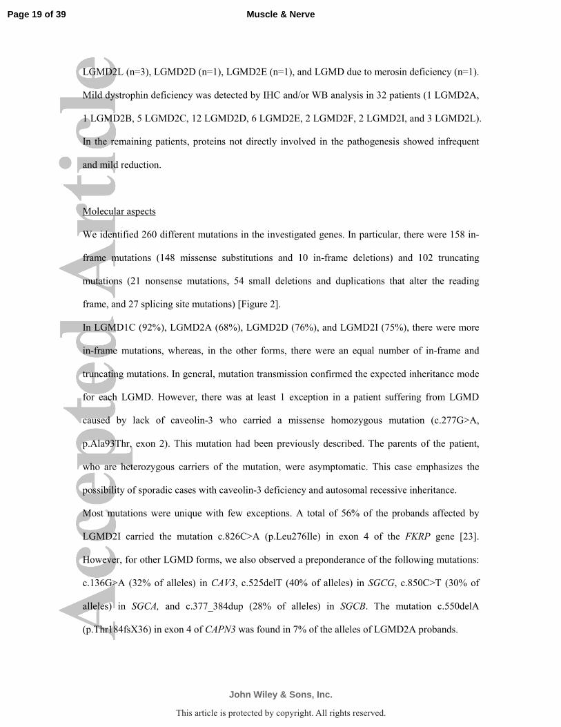
We identified 260 different mutations in the investigated genes. In particular, there were 158 in-
frame mutations (148 missense substitutions and 10 in-frame deletions) and 102 truncating
mutations (21 nonsense mutations, 54 small deletions and duplications that alter the reading
frame, and 27 splicing site mutations) [Figure 2].
In LGMD1C (92%), LGMD2A (68%), LGMD2D (76%), and LGMD2I (75%), there were more
in-frame mutations, whereas, in the other forms, there were an equal number of in-frame and
truncating mutations. In general, mutation transmission confirmed the expected inheritance mode
for each LGMD. However, there was at least 1 exception in a patient suffering from LGMD
caused by lack of caveolin-3 who carried a missense homozygous mutation (c.277G>A,
p.Ala93Thr, exon 2). This mutation had been previously described. The parents of the patient,
who are heterozygous carriers of the mutation, were asymptomatic. This case emphasizes the
possibility of sporadic cases with caveolin-3 deficiency and autosomal recessive inheritance.
Most mutations were unique with few exceptions. A total of 56% of the probands affected by
LGMD2I carried the mutation c.826C>A (p.Leu276Ile) in exon 4 of the FKRP gene [23].
However, for other LGMD forms, we also observed a preponderance of the following mutations:
c.136G>A (32% of alleles) in CAV3, c.525delT (40% of alleles) in SGCG, c.850C>T (30% of
alleles) in SGCA, and c.377_384dup (28% of alleles) in SGCB. The mutation c.550delA
(p.Thr184fsX36) in exon 4 of CAPN3 was found in 7% of the alleles of LGMD2A probands.
Page 19 of 39
John Wiley & Sons, Inc.
Muscle & Nerve
This article is protected by copyright. All rights reserved.
Approximately 70% of CAPN3 mutations are located in 8 of the 24 exons of the gene (exons 11,
4, 10, 13, 1, 21, 5, and 6) [24]. Furthermore, 91% of SGCG mutations affect 3 exons (exons 6, 3,
and 8), 88% of SGCA mutations involve exons 7 and 6, and 70% of SGCB mutations are located
in exon 3.
Next-generation sequencing results
The DNA of a group of 52 LGMD probands (58 patients) without molecular diagnosis was
analyzed using an NGS targeted platform (Motorplex). All of these patients had a clinical
diagnosis of LGMD. In all cases, their muscle biopsies showed a dystrophic pattern with an
increase in connective tissue. The analysis revealed causative gene mutations in 19 probands (22
patients) as follows: CAPN3 (n=5), DYSF (n=3), ANO5 (n=2), FKRP (n=1), SGCA (n=1),
POMT2 (n=1), TTN (n=1), LAMA2 (n=1), and TCAP (n=1). Furthermore, in 3 probands, we
detected mutations in DMD (n=1), COL6A3 (n=1), and GAA (n=1), and these patients were
excluded from the LGMD cohort. The 5 LGMD2A patients had been previously studied by direct
sequencing of the CAPN3 gene, but the analysis revealed only 1 heterozygous mutation. All
patients had a dystrophic muscle biopsy with an absence or severe reduction of calpain-3
observed by WB analysis, and NGS allowed detection of the second mutation in the CAPN3 gene.
NGS also revealed the second mutation in the 3 LGMD2B subjects. In the other patients (patients
harboring ANO5, TTN, or TCAP mutations), no specific protein test was available at our
laboratory or no muscle tissue was available for further tests (FKRP, SGCA, and POMT2).
However, previous studies in these patients showed a dystrophic pattern. Overall, the NGS
approach was shown to be useful and resulted in a diagnosis in 36.5% of the patients analyzed.
Clinical aspects
Age of onset
Page 20 of 39
John Wiley & Sons, Inc.
Muscle & Nerve
This article is protected by copyright. All rights reserved.
The age of onset was highly variable and ranged from the first to the fifth decades of life (range
1-65 years) [Figure 3 A]. The forms with earlier onset were LGMD2E (mean onset age 6.4 ± 5.2
years), LGMD2C (10.1 ± 5.9 years), and some dystroglycanopathies (onset at ages 2, 4, and 23
years for LGMD due to POMT2 mutations and onset at age 5 years for LGMD due to ISPD
mutations). The forms with later onset were LGMD1B (mean onset age 35.3 ± 18.8 years) and
LGMD2L (36.0 ± 13.2 years). Between these 2 extremes, the other forms sorted from the earliest
onset to the latest onset are as follows: LGMD2D (mean onset age 15.5 ± 13.4 years), LGMD2A
(16.7 ± 11.1 years), LGMD2I (18.1 ± 15.7 years), LGMD1C (24.4 ± 18.0 years), and LGMD2B
(25.1 ± 11.3 years) [Figure 3 B]. Some cases of LGMD2A (n=3), LGMD2B (n=1), LGMD2D
(n=1), and LGMD2I (n=1) had onset during early infancy (ages 1-2 years).
Clinical presentation and muscle involvement.
Patients came to medical attention for the following reasons: onset of mild proximal weakness
(199 patients), hyperCKemia (79 patients), cardiac involvement (4 patients), or cramps and
myalgias (10 patients). Weakness, when present, most frequently involved the lower limbs and
extended during disease progression to the upper limbs. It is important to note that 31 patients did
not have muscle weakness. These patients were family members of other affected individuals
(n=12) or presented for clinical attention with isolated hyperCKemia (3) or clinical features such
as rippling and calf hypertrophy (3). Thirteen patients were presymptomatic children assessed at
an earlier age than the mean age of onset of the subgroup of LGMD defined by their mutational
status. Distal weakness was more frequently detected, also earlier than expected, in some forms,
in particular LGMD2B (n=18), LGMD2L (n=3), LGMD1B (n=2), LGMD2A (n=3), LGMD2D
(n=1), and LGMD2C (n=1); however, in advanced stages, all patients developed involvement of
the distal extremities to varying degrees.
Muscle atrophy/hypertrophy occurred in a different pattern in each LGMD. Calf
pseudohypertrophy was frequently found in patients suffering from sarcoglycanopathies
Page 21 of 39
John Wiley & Sons, Inc.
Muscle & Nerve
This article is protected by copyright. All rights reserved.
(LGMD2C 5/10, LGMD2D 17/28, and LGMD2E 15/18) and in patients with LGMD1C (21/28),
LGMD2I (14/30), and LGMD2L (4/12), sometimes with asymmetric involvement. Hypertrophy
of the quadriceps femoris muscle was observed in a few patients with LGMD1C (n=7), and
macroglossia was observed in some patients affected with LGMD2I (n=2) and LGMD2E (n=3).
Subjects suffering from dysferlinopathy frequently had distal lower limb atrophy (17 patients in
our sample).
Tendon contractures were common and appeared at early stages in patients with LGMD2E (15/20,
75%), LGMD2A (42/66; 64%), and LGMD2C (6/10, 60%) but were even found in other forms:
LGMD2B (23/53; 43%), LGMD2D (12/29, 41%), LGMD2I (5/18, 28%), LGMD1C (6/23; 26%),
LGMD1B (2/10, 20%), LGMD2N (1/3, 33%), and LGMD2L (1/11; 9%). Contractures mainly
involved the ankles, elbow flexors, and knees. Scoliosis was more frequent in wheelchair-
dependent patients with LGMD2E (9/17, 53%) and LGMD2C (4/10; 40%) and in ambulant
patients with LGMD2B (17/47; 36%) and LGMD2A (16/58, 28%). Winged scapulae were
observed in 14 patients with LGMD2A and occurred sporadically in the other forms.
Patients affected by LGMD1C typically exhibited cramps, myalgias, and rippling, especially
localized to the quadriceps muscles. Three patients with LGMD2L and 3 patients with
sarcoglycanopathies had episodes of myoglobinuria.
The EMG examination showed a myopathic pattern in the majority of patients (148/191), which
was sometimes associated with neurogenic signs (21/191). In some LGMD patients (11/191), an
isolated neurogenic pattern was occasionally observed (LGMD1B, LGMD1C, LGMD2A,
LGMD2B, LGMD2D, and LGMD2E). The patients did not show signs of neuropathy, and this
pattern was considered part of the LGMD phenotype. Interestingly, in 11 patients, EMG
examination was normal. These data are reported in Table 1-2.
Muscle weakness progression.
Page 22 of 39
John Wiley & Sons, Inc.
Muscle & Nerve
This article is protected by copyright. All rights reserved.

The majority of LGMD patients had a progressive course, but the evolution was different among
each form [Suppl Fig. 2A-C]. LGMD1B, LGMD1C and LGMD2L showed a more benign course:
all patients at the time of the last evaluation (mean age 51.1 ± 9.2 years old in LGMD1B; 35.9 ±
19.8 years old in LGMD1C; and 47.7 ± 11.1 years old in LGMD2L) had independent walking
function [Suppl Fig 2A]. A more rapid evolution was observed in LGMD2C and LGMD2E, in
which 73% and 53% of patients lost independent ambulation during follow-up at 26.0 ± 16.0
years of age (17.2 ± 17.4 years from onset) and 12.4 ± 2.3 years of age (8.5 ± 3.2 years from
onset), respectively [Suppl Fig 2B]. Difficulties in postural changes (Walton >5) were found
mainly in patients with LGMD2A (65% of patients), LGMD2B (71%) and sarcoglycanopathies
(39% of LGMD2D, 61% of LGMD2E and 80% of LGMD2C patients).
Creatine kinase levels.
Serum CK levels at disease onset were elevated in all patients. Higher levels at disease onset were
found in sarcoglycanopathies (mean value 7000 U/L) and in dysferlinopathies (mean value 5000
U/L, although some cases had values above 25000 U/L). Conversely, other forms showed very
low levels; for example, in LGMD1B patients, serum CK levels never exceeded 1000 U/L [Suppl
Fig 3].
Reflecting disease progression, the values greatly decreased over years, and almost all subjects
older than 50 years of age had CK values lower than 2000 U/L.
Generally, during disease progression, the higher values were found in LGMD2B (1.5-125x),
LGMD2D (1-150x), LGMD2E (1-111x), LGMD2C (2-128x) and LGMD2A (1-108x), whereas
the forms with lower values of CK were LGMD2I (1-57x), LGMD2L (2-50x), LGMD1C (1-42x)
and LGMD1B (1-4x).
Cardiac involvement
Page 23 of 39
John Wiley & Sons, Inc.
Muscle & Nerve
This article is protected by copyright. All rights reserved.
According to our database, the forms that were most frequently associated with heart involvement
were LGMD1B (13/13, 100%), LGMD2E (7/20; 35%) and LGMD2I (12/26, 46%). In the
remaining forms, cardiomyopathy was observed only in isolated cases [Suppl. Fig. S4A; Table
2A].
Cardiac involvement in LGMD1B was more severe than in other forms and was always present.
Seven patients had cardiac arrhythmia, which required the implantation of a cardiac defibrillator,
and 3 subjects underwent cardiac transplantation (at the ages of 42, 50, and 58 years). Patients
suffering from LGMD2I mainly had ventricular dilatation and/or hypokinesia wall, whereas
hypokinetic heart disease was predominant in LGMD2E subjects.
The age at onset of heart involvement varied in the different forms, with a relatively earlier onset
in LGMD2E (20.3 ± 4.2 years old) and a later onset in LGMD1B (36.4 ± 10.2 years old) and
LGMD2I (31.0 ± 21.7 years old) [Suppl Fig.S4 C; Table 2A].
In patients with LGMD1B, cardiac impairment often preceded skeletal muscle involvement (7/11
patients), whereas cardiac impairment generally followed skeletal muscle involvement in the
other forms. In LGMD2E and LGMD2I, cardiac function deteriorated 16.6 ± 9.6 and 2.7 ± 2.1
years after disease onset, respectively.
Respiratory involvement
Respiratory involvement was uncommon in LGMDs and was generally characterized by a
restrictive pattern. The deterioration of respiratory function appeared at an average age of 15.6 ±
8.5 years after symptoms onset. This occurred more often in LGMD2I (12/24, 50%) and
sarcoglycanopathies (LGMD2C 7/14, 50%; LGMD2D 6/25, 24%; and LGMD2E 8/20, 40%) and
less frequently in patients with calpainopathy (11/53, 21%) and dysferlinopathy (7/40; 18%)
(Suppl. Fig. 4B, 4D). In some cases, respiratory impairment was very severe and required assisted
ventilation, as observed in 3 LGMD2D, 3 LGMD2E, 2 LGMD2B, 1 LGMD2I and 1 LGMD2C
Page 24 of 39
John Wiley & Sons, Inc.
Muscle & Nerve
This article is protected by copyright. All rights reserved.
patients. Respiratory dysfunction was very rare in the remaining forms [Suppl. Fig S4 B; Table
2B].
Genotype-phenotype correlations
We identified a correlation between the type of mutation, protein expression and clinical
progression. We used the patient’s age at disease onset as an indicator of clinical severity, and we
observed that truncating mutations were generally associated with more severe protein reduction
and clinical presentation. Mutations that preserve the reading frame were usually responsible for
milder phenotypes.
In fact, in-frame mutations were generally associated with a later onset than truncating mutations.
In particular, this difference was statistically significant in LGMD2A (19.7 ± 11.5 years vs. 12.2
± 11.2 years, p = 0.02), LGMD2B (30.5 ± 10.3 years vs. 17.7 ± 5.5 years, p <0.0001) and
LGMD2E (10.8 ± 5.4 years vs. 4.1 ± 3.7 years, p = 0.007).
LGMD2A, LGMD2B, LGMD2D and LGMD2E patients with protein absence observed by IHC
and/or WB assays had a lower age of disease onset than patients with partial deficiency. The
difference was statistically significant in LGMD2D (absence of α-sarcoglycan 5.4 ± 2.3 years vs.
partial defect 20.0 ± 14.5 years, p = 0.0001) and LGMD2E (absence of β-sarcoglycan 3.1 ± 1.4
years vs. partial defect 13.7 ± 5.7 years, p = 0.015). We also evaluated the predictive value of
specific protein and clinical data to unequivocally address the differential diagnosis between the
different LGMD forms. According to this analysis, the presence of rippling was a specific
indicator of LGMD1C with 100% specificity and 40% sensitivity (p = 0.002). The other clinical
and muscle biopsy findings, even if they were particular to certain LGMD forms, did not reach
statistical significance.
Discussion
Page 25 of 39
John Wiley & Sons, Inc.
Muscle & Nerve
This article is protected by copyright. All rights reserved.
In this study, we present an overview of the clinical and molecular characteristics of a large
cohort of LGMD patients who are representative of the Italian LGMD population.
The data collected allowed us to better define the epidemiology, the phenotype and the natural
history of each LGMD and to detect the most important parameters that can help the differential
diagnosis.
This project was carried out with the collaboration of 12 different Italian centers involved in the
diagnosis and cure of neuromuscular diseases. We collected data from 599 patients, and the
clinical histories of 370 molecularly confirmed patients were analyzed in detail.
Relative frequency of LGMD in Italy
The collection of a large cohort of patients allowed us to define the relative frequency of
genetically diagnosed patients with LGMD in Italy (Fig. 1). According to the data reported in the
literature, in our cohort, AR LGMDs were more frequent then AD LGMDs (84% of patients -
89% of probands). Even after the discovery of several new genes, mutations in CAPN3 and DYSF,
which cause LGMD2A and LGMD2B, remain responsible for half of the cases in our cohort
(30% and 22.6.2% of probands, respectively). Also, sarcoglycanopathies were frequent and
affected 21.3% of probands. LGMD2A appears to be the most frequently diagnosed form not
only in Italy, as previously reported in the literature [9, 25], but also in the other countries
because it has been described as the most common form in populations from Brazil, Japan, the
Netherlands, Turkey, Russia and Basque regions [26-31].
By contrast, LGMD2I, common in Denmark and Northern England [16; 32], and LGMD2L,
common in Northern Europe [33], affected only 9.8% and 3.8% of patients, respectively, in our
sample. In Denmark, LGMD2I is responsible for 42% of patients with AR LGMD (12% of the
patients in our sample), followed by LGMD2L (11%), which represents, with LGMD2A, the
second most common form of AR LGMD (5% of LGMD2 patients in our sample) [33]. This
difference may be partially due to a North-South gradient and to a founder effect for some
Page 26 of 39
John Wiley & Sons, Inc.
Muscle & Nerve
This article is protected by copyright. All rights reserved.
mutations in Northern Europe populations. The other dystroglycanopathies were poorly
represented in our cohort and affected only 1 ISPD subject and 3 POMT2 patients (now 15, 24,
42 and 48 years old), with childhood onset.
Interestingly, we described 2 families (5 patients) who presented with adult-onset LGMD and
carried mutations in the LAMA2 gene, which is generally associated with Congenital Muscular
Dystrophies [34]. Patients affected with AD forms in our cohort carried mutations in the CAV3,
DNAJB6 and LMNA genes. We did not find mutations in the other LGMD1 genes, which have
presently only been reported in a few families worldwide [10-12].
Targeted NGS increased the detection rate of LGMD genetic causative mutations in this cohort,
and 36.5% of genetic diagnoses were identified using this technique. This figure does not fully
address the potential of targeted NGS in LGMD because the sample analyzed by NGS had been
previously extensively studied with other techniques. Therefore, the rate of detection might be
higher in a “naïve” LGMD sample. Meanwhile, the rate of detection using 98 genes involved in
muscle diseases as a reference does not address the cause of LGMD in 63.5% of patients,
confirming the still unexplored genetic causes of this group of disorders.
LGMD differential diagnosis
The clinical findings showed wide variability, both between different forms and within the same
LGMD groups. We focused the analysis on the aspects that were specific for each form and that
should be evaluated to aid in differential diagnosis.
The study of the natural history and the detailed knowledge of the specific characteristics of each
LGMD subtype allowed us to detect the most important clues that could help the clinician in the
diagnosis (Figure 4).
From this perspective, the most important parameters were the muscle pathology findings and the
clinical presentation, in particular CK levels, age of onset, presence of contractures, and cardiac
Page 27 of 39
John Wiley & Sons, Inc.
Muscle & Nerve
This article is protected by copyright. All rights reserved.
and respiratory involvement. Taken together, all of these data can help one choose the molecular
tests.
Muscle biopsy can provide important clues for the differential diagnosis. The presence of a
dystrophic or myopathic pattern combined with the clinical presentation of limb girdle weakness
is highly suggestive of LGMD. However, it should be noted that the absence of an increase in
connective tissue does not exclude the diagnosis (Suppl Figure S1A, 29% of biopsies), especially
when the biopsy was performed at early stages. LGMD1C and LGMD1B often showed isolated
mild myopathic signs. Furthermore, the different degree of connective tissue substitution, the
prevalence of necrotic and degenerating fibers and the presence or absence of inflammatory
infiltrates can aid in the differential diagnosis (Suppl Figure S1). For example, inflammation was
more frequent in dysferlinopathies and calpainopathies (Suppl Fig. S1B).
IHC and WB studies performed on muscle samples allowed us to detect reductions in specific
proteins. In some forms, for example sarcoglycanopathies, reduction of the specific protein
complex strongly correlated with mutations (100% of our patients with mutations in sarcoglycan
genes had corresponding protein deficiency measured by IHC or WB assays). However,
sometimes this analysis can be misleading, as demonstrated in by the presence of 3 LGMD2A
subjects with normal calpain-3 expression. Furthermore, some proteins can be secondarily
reduced. In this cohort, calpain-3 deficiency was found in patients with LGMD2B,
sarcoglycanopathies, LGMD2I, LGMD2L, and LGMD due to merosin deficiency, whereas
dystrophin deficiency was detected in LGMD2A, sarcoglycanopathies, LGMD2I, and LGMD2L.
When a deficiency of glycosylation of α-dystroglycan is detected, the molecular diagnosis is
difficult, because in recent years the discovery of genes responsible for these α-
dystroglycanopathies has increased exponentially. However, according to our experience and to
the epidemiological data, the LGMD2I form is more frequent than the others, and the FKRP gene
should be investigated first. Unfortunately, for some subtypes, for example LGMD2L, no protein
Page 28 of 39
John Wiley & Sons, Inc.
Muscle & Nerve
This article is protected by copyright. All rights reserved.
test is available. In these cases, the clinical characteristics are fundamental to aid in the molecular
analysis.
Sarcoglycanopathies, with the exception of LGMD2D, are generally the most severe forms, with
early onset and rapid progression, leading to loss of independent ambulation in the first 2 decades
of life in a high proportion of patients.
According to the age of onset, we can subdivide the LGMD population into different groups
(Figure 3). Forms with onset before age 10 years are mainly sarcoglycanopathies,
dystroglycanopathies, and some cases of calpainopathies. Generally, a significant proportion of
LGMD2A patients have an age of onset <20 years, whereas the onset of symptoms for patients
with dysferlinopathy occurs between ages 11 and 40 years. LGMD1C and LGMD2I may
manifest in all age groups, but more frequently occur before age 20 years. Conversely, patients
with LGMD1B and LGMD2L develop their first symptoms primarily after age 20 years (Figure
3). Furthermore, in some forms with later onset, few cases of unusually early onset (ages 1-2
years) were observed.
Earlier age of onset often correlated with severe prognosis. The most severe evolution was
observed in LGMD2C and LGMD2E patients, who lost ambulation in 73% and 53% of cases and
at an average age of 26.0 ± 16.0 and 12.4 ± 2.3 years, respectively (Suppl Figure S2).
Additionally, CK levels reflect the severity of the disease and can contribute to the differential
diagnosis. Only CK at onset can be used for this purpose, because, during disease progression,
CK values decrease in all LGMD forms. The presence of high CK levels at onset suggests the
molecular diagnosis of sarcoglycanopathies and dysferlinopathies, whereas very low levels are
frequently found in LGMD1B and LGMD1C [Suppl Figure S3].
Cardiac and respiratory involvement are variably present in LGMD patients and can point toward
specific subtypes. Cardiac involvement is constant in LGMD1B and frequent in LGMD2E and
LGMD2I [Supplementary Figure S4A]. Given the early onset of heart disease and its severity,
Page 29 of 39
John Wiley & Sons, Inc.
Muscle & Nerve
This article is protected by copyright. All rights reserved.
molecular analysis of the LMNA gene and a regular cardiologic follow-up should be performed in
all family members of patients with LGMD1B. A restrictive respiratory pattern is present in
LGMD2I, sarcoglycanopathies and rarely in calpainopathy and dysferlinopathy [Suppl Figure
S4B].
The definition of the frequency, characteristics and severity of cardiomyopathy and pulmonary
involvement in each form is fundamental to develop guidelines for follow-up and to develop
programs for proper cardiac and respiratory monitoring, which allows for establishing timely and
adequate medical treatment or surgery [6].
We highlight the contributions of other peculiar clinical characteristics for the differential
diagnosis (Tables 1, 2). Sarcoglycanopthies and patients affected by LGMD1C, LGMD2I, and
LGMD2L frequently presented with calf pseudohypertrophy. Some subjects carrying mutations
in the dysferlin gene developed distal lower limb atrophy, whereas a few patients suffering from
LGMD1C and LGMD2E had quadriceps femoris hypertrophy. The early appearance of tendon
contractures is mainly found in LGMD2A, LGMD2E, and LGMD2C patients, whereas scoliosis
is found in LGMD2E, LGMD2C, LGMD2B, and LGMD2A patients in particular. Episodes of
myoglobinuria can be observed in patients with LGMD2L and rippling in CAV3 forms (P=0.002).
Key clinical and paraclinical findings that may aid in addressing a specific diagnosis during
clinical work-up are summarized in Tables 1 and 2 and in Figure 4.
Molecular analysis and contribution of NGS
The majority of patients were studied by direct sequencing of candidate genes. Generally, we did
not find mutational hotspots, although there were some useful exceptions, as described in the
Results section. NGS was found to be a cost-effective method that can be used to study patients
for whom the candidate gene approach was unable to reveal mutations. NGS allowed for
detection of the mutation on the second allele in AR LGMD and the study of subjects without an
Page 30 of 39
John Wiley & Sons, Inc.
Muscle & Nerve
This article is protected by copyright. All rights reserved.
available muscle protein test. Overall, in our sample, NGS allowed detection of mutations in 19
of the 52 probands studied (36.5%).
A detailed knowledge of the clinical characteristics of a large sample of LGMD patients allowed
us to define important clues that can be used in the differential diagnosis of LGMD patients and
for designing clinical studies of each individual form. Gene product expression and function in
skeletal muscle and clinical data should still be combined to choose the molecular tests. New
molecular techniques, such as NGS, must be considered in patients for whom the candidate gene
approach failed to detect mutations and in forms still lacking a tissue-specific marker.
Page 31 of 39
John Wiley & Sons, Inc.
Muscle & Nerve
This article is protected by copyright. All rights reserved.

Abbreviations list
AD autosomal dominant
AR autosomal recessive
CK creatine kinase
ECG electrocardiogram
EF ejection fraction
EMG electromyographic
FVC forced vital capacity
FEV1 forced expiratory volume
IHC immunohistochemistry
LGMD Limb girdle muscular dystrophies
MFM Motor function measure scale
MRC Medical Research Council
6MWT Six-minute walking test
NGS next-generation sequencing
PCR polymerase chain reaction
RBBB right bundle branch blocks
SF shortening fraction
TIGEM Telethon Institute of Genetic and Medicine
WES whole exome sequencing
WB Western blot
Page 32 of 39
John Wiley & Sons, Inc.
Muscle & Nerve
This article is protected by copyright. All rights reserved.
References
[1] Walton JN, Nattrass FJ. On the classification, natural history and treatment of the myopathies.
Brain 1954;77:170-231
[2] Beckmann JS. Disease taxonomy--monogenic muscular dystrophy. Br Med Bull. 1999;55
(2):340-57.
[3] Norwood FL, Harling C, Chinnery PF, Eagle M, Bushby K, Straub V. Prevalence of genetic
muscle disease in Northern England: in-depth analysis of a muscle clinic population. Brain. 2009
Nov;132(Pt 11):3175-86.
[4] Magri F, Brajkovic S, Govoni A, Brusa R, Comi GP. Revised genetic classification of Limb
Girdle Muscular Dystrophies. Curr Mol Med. 2014 Oct 10.
[5] Nigro V, Savarese M. Genetic basis of limb-girdle muscular dystrophies: the 2014 update.
Acta Myol. 2014; 33(1):1-12.
[6] Narayanaswami P, Weiss M, Selcen D, David W, Raynor E, Carter G, et al. Guideline
Development Subcommittee of the American Academy of Neurology; Practice Issues Review
Panel of the American Association of Neuromuscular & Electrodiagnostic Medicine. Evidence-
based guideline summary: diagnosis and treatment of limb-girdle and distal dystrophies: report of
the guideline development subcommittee of the American Academy of Neurology and the
practice issues review panel of the American Association of Neuromuscular & Electrodiagnostic
Medicine. Neurology. 2014; 83(16):1453-63.
[7] Murphy AP, Straub V. The classification, Natural history and Treatment of Limb Girdle
Muscular Dystrophies. Journal of Neuromuscular Diseases 2015; S7-S19
Page 33 of 39
John Wiley & Sons, Inc.
Muscle & Nerve
This article is protected by copyright. All rights reserved.
[8] Norwood F1, de Visser M, Eymard B, Lochmüller H, Bushby K; EFNS Guideline Task Force.
EFNS guideline on diagnosis and management of limb girdle muscular dystrophies. Eur J Neurol.
2007; (12):1305-12.
[9] Guglieri M, Straub V, Bushby K, Lochmüller H. Limb-girdle muscular dystrophies. Curr
Opin Neurol. 2008; (5):576-84.
[10] Sarparanta J, Jonson PH, Golzio C, Sandell S, Luque H, Screen M, et al. Mutations affecting
the cytoplasmic functions of the co-chaperone DNAJB6 cause limb-girdle muscular dystrophy.
Nat Genet. 2012;44(4):450-5, S1-2.
[11] Vieira NM, Naslavsky MS, Licinio L, Kok F, Schlesinger D, Vainzof M, et al. A defect in
the RNA-processing protein HNRPDL causes limb-girdle muscular dystrophy 1G (LGMD1G).
Hum Mol Genet. 2014; 23(15):4103-10.
[12] Torella A, Fanin M, Mutarelli M, Peterle E, Del Vecchio Blanco F, Rispoli R, et al. Next-
generation sequencing identifies transportin 3 as the causative gene for LGMD1F. PLoS One.
2013;8(5):e63536.
[13] Nectoux J, de Cid R, Baulande S, Leturcq F, Urtizberea JA, Penisson-Besnier I, et al.
Detection of TRIM32 deletions in LGMD patients analyzed by a combined strategy of CGH array
and massively parallel sequencing. Eur J Hum Genet. 2015;23(7):929-34.
[14] Carss KJ, Stevens E, Foley AR, Cirak S, Riemersma M, Torelli S, et al. Mutations in GDP-
mannose pyrophosphorylase B cause congenital and limb-girdle muscular dystrophies associated
with hypoglycosylation of α-dystroglycan. Am J Hum Genet. 2013;93(1):29-41.
Page 34 of 39
John Wiley & Sons, Inc.
Muscle & Nerve
This article is protected by copyright. All rights reserved.
[15] Tasca G, Moro F, Aiello C, Cassandrini D, Fiorillo C, Bertini E, et al. Limb-girdle muscular
dystrophy with α-dystroglycan deficiency and mutations in the ISPD gene. Neurology. 2013; 80:
963-5.
[16] Bushby K. Diagnosis and management of the limb girdle muscular dystrophies. Pract Neurol.
2009; 9(6):314-23.
[17] Walton JN, Gardner-Medwin D. Progressive Muscular Dystrophy and the myotonic
disorders. In: Walton JN editor, Disorders of voluntary muscle. 4th ed. Edinburgh: Churchill
Livingstone; 1981 p. 481-524
[18] Dubowitz V, Sewry CA, Lane RJM. Muscle Biopsy: APractical Approach, 3rd ed.
Philadelphia: Saunders Elsevier;2007).
[19] Nicholson LV, Davison K, Johnson MA, Slater CR, Young C, Bhattacharya S, et al.
Dystrophin in skeletal muscle. Immunoreactivity in patients with Xp21 muscular dystrophy. J
Neurol Sci 1989; 94:137–146.
[20] Prelle A, Comi GP, Tancredi L, Rigoletto C, Ciscato P, Fortunato F, et al. Sarcoglycan
deficiency in a large Italian population of myopathic patients. Acta Neuropathol. 1998;96(5):509-
14.
[21] Anderson LV, Davison K, Moss JA, Richard I, Fardeau M, Tomé FM, et al. Characterization
of monoclonal antibodies to calpain 3 and protein expression in muscle from patients with limb-
girdle muscular dystrophy type 2A. Am J Pathol. 1998;153(4):1169-79.
[22] Savarese M, Di Fruscio G, Mutarelli M, Torella A, Magri F, Santorelli FM, et al. MotorPlex
provides accurate variant detection across large muscle genes both in single myopathic patients
and in pools of DNA samples. Acta Neuropathol Commun. 2014;2:100.
Page 35 of 39
John Wiley & Sons, Inc.
Muscle & Nerve
This article is protected by copyright. All rights reserved.
[23] Frosk P, Greenberg CR, Tennese AA, Lamont R, Nylen E, Hirst C, et al. The most
common mutation in FKRP causing limb girdle muscular dystrophy type 2I (LGMD2I) may
have occurred only once and is present in Hutterites and other populations. Hum Mutat.
2005;25(1):38-44.
[24] Zatz M, Starling A. Calpains and disease. N Engl J Med. 2005 Jun 9;352(23):2413-23.
[25] Fanin M, Nascimbeni AC, Aurino S, Tasca E, Pegoraro E, Nigro V, et al. Frequency of
LGMD gene mutations in Italian patients with distinct clinical phenotypes. Neurology.
2009;72(16):1432-5.
[26] Van der Kooi AJ, Barth PG, Busch HF, de Haan R, Ginjaar HB, van Essen AJ, et al. The
clinical spectrum of limb girdle muscular dystrophy. A survey in The Netherlands. Brain.
1996;119 ( Pt 5):1471-80.
[27] Topaloğlu H, Dinçer P, Richard I, Akçören Z, Alehan D, Ozme S, et al. Calpain-3 deficiency
causes a mild muscular dystrophy in childhood. Neuropediatrics. 1997;28(4):212-6.
[28] Urtasun M, Sáenz A, Roudaut C, Poza JJ, Urtizberea JA, Cobo AM, et al. Limb-girdle
muscular dystrophy in Guipúzcoa (Basque Country, Spain). Brain. 1998;121 ( Pt 9):1735-47.
[29] Passos-Bueno MR, Vainzof M, Moreira ES, Zatz M. Seven autosomal recessive limb-girdle
muscular dystrophies in the Brazilian population: from LGMD2A to LGMD2G. Am J Med Genet.
1999;82(5):392-8.
[30] Pogoda TV1, Krakhmaleva IN, Lipatova NA, Shakhovskaya NI, Shishkin SS, Limborska SA.
High incidence of 550delA mutation of CAPN3 in LGMD2 patients from Russia. Hum Mutat.
2000;15(3):295.
[31] Chae J, Minami N, Jin Y, Nakagawa M, Murayama K, Igarashi F, et al. Calpain 3 gene
mutations: genetic and clinico-pathologic findings in limb-girdle muscular dystrophy.
Neuromuscul Disord. 2001;11(6-7):547-55.
Page 36 of 39
John Wiley & Sons, Inc.
Muscle & Nerve
This article is protected by copyright. All rights reserved.

[32] Sveen ML, Schwartz M, Vissing J. High prevalence and phenotype-genotype correlations of
limb girdle muscular dystrophy type 2I in Denmark. Ann Neurol. 2006;59(5):808-15.
[33] Witting N, Duno M, Petri H, Krag T, Bundgaard H, Kober L, et al. Anoctamin 5 muscular
dystrophy in Denmark: prevalence, genotypes, phenotypes, cardiac findings, and muscle protein
expression. J Neurol. 2013;260(8):2084-93.
[34] Tan E, Topaloglu H, Sewry C, Zorlu Y, Naom I, Erdem S, et al. Late onset muscular
dystrophy with cerebral white matter changes due to partial merosin deficiency. Neuromuscul
Disord 1997;7:85–89.
Page 37 of 39
John Wiley & Sons, Inc.
Muscle & Nerve
This article is protected by copyright. All rights reserved.

Table 1 Clinical characteristics of different forms of LGMD
Probands/pa
tients, n
Male/
Female
Last evaluation
(years)
Onset
(years)
CK at
onset
(UI/L)
CK range
(follow-up)
LGMD1B 9/19 11/8 52.0 ± 10.6 35.3 ± 18.8 394 ± 197 1-4x
LGMD1C 21/40 24/16 36.5 ± 20.0 24.4 ± 18.0 1890 ±
1907 1-42x
LGMD1D 1/4 1/3 52.2 ± 11.6 28 1600 4-8x
LGMD2A 86/92 39/53 36.7 ± 15.4 16.7 ± 11.1 4025 ±
3393 1-108x
LGMD2B 65/85 41/44 39.3 ± 11.8 25.1 ± 11.3 5147 ±
5299 1.5-125x
LGMD2C 15/17 8/9 25.1 ± 15.7 10.1 ± 5.9 6388 ±
7640 2-128x
LGMD2D 28/35 18/17 28.9 ± 18.3 15.5 ± 13.4 8011 ±
8651 1-150x
LGMD2E 16/21 10/11 21.5 ± 10.2 6.4 ± 5.2 7922 ±
6797 1-111x
LGMD2F 2/2 2/0 14.0 4.0 20000 100x
LGMD2I 25/36 19/17 38.3 ± 21.1 18.1 ± 15.7 3162 ±
2682 1-57x
LGMD2L 12/14 11/3 47.7 ± 11.1 36.0 ± 13.2 3925 ±
2771 2-50x
LGMD
POMT2 3/3 1/2 24.3 ± 11.7 9.7 ± 11.6
5217 ±
1624 17-32x
LGMD
LAMA2 2/5 1/4 55± 13.5 17.0 ± 7.5 670 ± 239 2-3x
LGMD ISPD 1/1 1/0 42.0 5.0 ND 3-6x
Page 38 of 39
John Wiley & Sons, Inc.
Muscle & Nerve
This article is protected by copyright. All rights reserved.
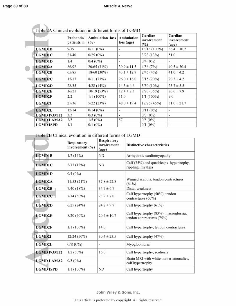
Table 2A Clinical evolution in different forms of LGMD
Probands/
patients, n
Ambulation loss
(%)
Ambulation
loss (age)
Cardiac
involvement
(%)
Cardiac
involvement
(age)
LGMD1B 9/19 0/11 (0%) - 13/13 (100%) 36.4 ± 10.2
LGMD1C 21/40 0/25 (0%) - 3/23 (13%) 51.0
LGMD1D 1/4 0/4 (0%) - 0/4 (0%) -
LGMD2A 86/92 20/65 (31%) 39.9 ± 11.5 4/56 (7%) 40.5 ± 30.4
LGMD2B 65/85 18/60 (30%) 43.1 ± 12.7 2/45 (4%) 41.0 ± 4.2
LGMD2C 15/17 8/11 (73%) 26.0 ± 16.0 3/15 (20%) 20.3 ± 4.2
LGMD2D 28/35 4/28 (14%) 14.3 ± 4.6 3/30 (10%) 25.7 ± 5.5
LGMD2E 16/21 10/19 (53%) 12.4 ± 2.3 7/20 (35%) 20.6 ± 7.9
LGMD2F 2/2 1/1 (100%) 11,0 1/1 (100%) 9.0
LGMD2I 25/36 5/22 (23%) 48.0 ± 19.4 12/26 (46%) 31.0 ± 21.7
LGMD2L 12/14 0/14 (0%) - 0/11 (0%) -
LGMD POMT2 3/3 0/3 (0%) - 0/3 (0%) -
LGMD LAMA2 2/5 1/5 (0%) 57 0/5 (0%) -
LGMD ISPD 1/1 0/1 (0%) - 0/1 (0%) -
Table 2B Clinical evolution in different forms of LGMD
Respiratory
involvement (%)
Respiratory
involvement
(age)
Distinctive characteristics
LGMD1B 1/7 (14%) ND Arrhythmic cardiomyopathy
LGMD1C 2/17 (12%) ND Calf (75%) and quadriceps hypertrophy,
rippling, myalgia
LGMD1D 0/4 (0%) - -
LGMD2A 11/53 (21%) 37.8 ± 22.8 Winged scapula, tendon contractures
(64%)
LGMD2B 7/40 (18%) 34.7 ± 6.7 Distal weakness
LGMD2C 7/14 (50%) 23.2 ± 7.0 Calf hypertrophy (50%), tendon
contractures (60%)
LGMD2D 6/25 (24%) 24.8 ± 9.7 Calf hypertrophy (61%)
LGMD2E 8/20 (40%) 20.4 ± 10.7 Calf hypertrophy (83%), macroglossia,
tendon contractures (75%)
LGMD2F 1/1 (100%) 14.0 Calf hypertrophy, tendon contractures
LGMD2I 12/24 (50%) 30.4 ± 23.5 Calf hypertrophy (47%)
LGMD2L 0/8 (0%) - Myoglobinuria
LGMD POMT2 1/2 (50%) 16.0 Calf hypertrophy, scoliosis
LGMD LAMA2 0/5 (0%) - Brain MRI with white matter anomalies,
calf hypertrophy
LGMD ISPD 1/1 (100%) ND Calf hypertrophy
Page 39 of 39
John Wiley & Sons, Inc.
Muscle & Nerve
This article is protected by copyright. All rights reserved.
Figure legends
Figure 1. Relative frequency of LGMDs in the Italian sample. (A) Relative frequency of
different LGMD forms among probands. (n = 285) (B) Relative frequency of different
LGMD forms among all patients. (n = 370). LGMDs due to mutations in CAPN3 and
DYSF are the most frequent forms.
Figure 2. Mutation distribution in different LGMD forms. The relative proportion of in-
frame (in green) and out-of-frame (red and yellow) mutations are detailed for
each LGMD form. In the majority of LGMD subtypes. in-frame mutations are more
frequent than mutations causing protein loss.
Figure 3. Age of onset in different LGMDs. (A) Differential diagnosis of LGMD
according to age of onset. The LGMD sample was divided into 4 groups according to the
age of onset. For each group. the figure shows the number of patients with each LGMD
form. The age of onset can be used to help in the differential diagnosis. (B) Mean age at
onset for each LGMD form. Each line shows the range of age of onset for each group; in
bold the mean age of onset is expressed in years.
Figure 4. Differential diagnosis in LGMDs. The diagram illustrates the main muscle
histology and clinical aspects that should be taken into consideration for LGMD
differential diagnosis.
Page 40 of 39
John Wiley & Sons, Inc.
Muscle & Nerve
This article is protected by copyright. All rights reserved.
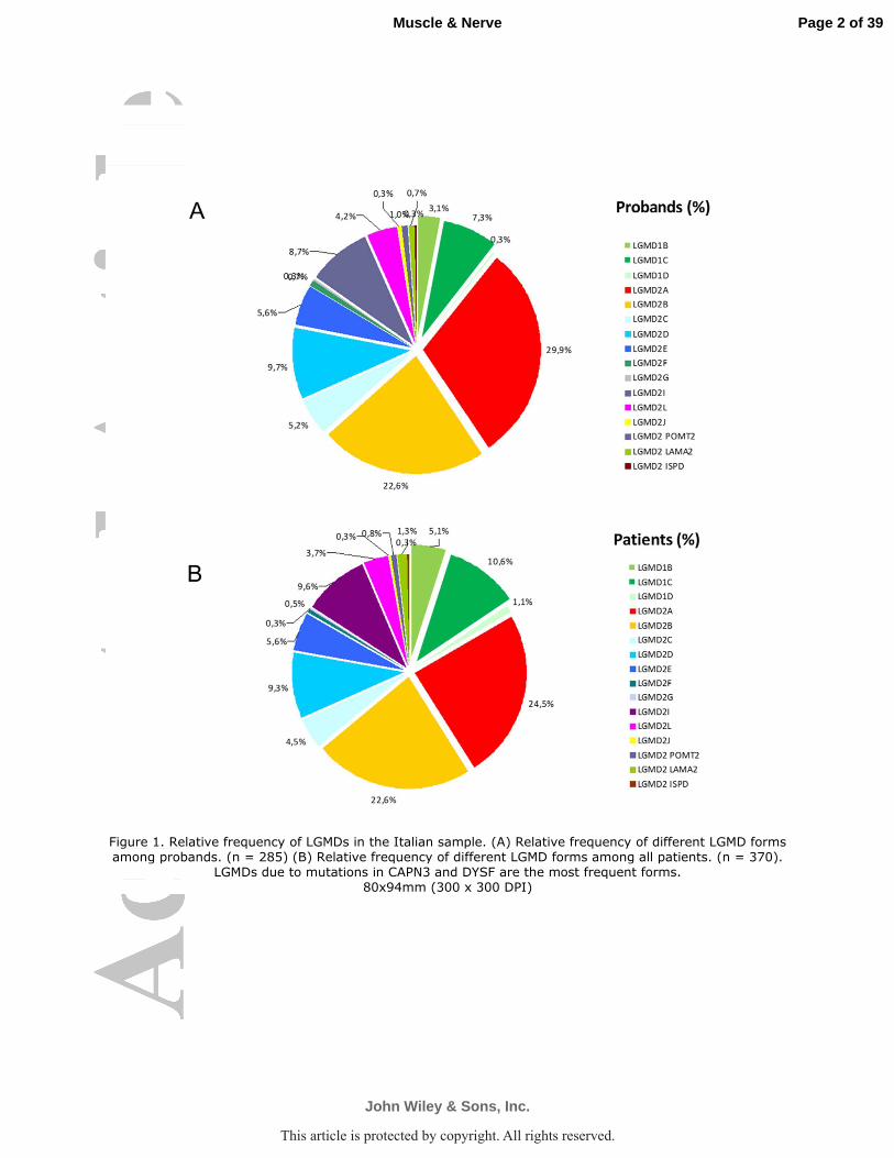
Figure 1. Relative frequency of LGMDs in the Italian sample. (A) Relative frequency of different LGMD forms among probands. (n = 285) (B) Relative frequency of different LGMD forms among all patients. (n = 370).
LGMDs due to mutations in CAPN3 and DYSF are the most frequent forms. 80x94mm (300 x 300 DPI)
Page 2 of 39
John Wiley & Sons, Inc.
Muscle & Nerve
This article is protected by copyright. All rights reserved.
Fig 2. Mutation distribution in different LGMD forms The relative proportion of in-frame (in green) and out-of-frame (red and yellow) mutations are detailed for each LGMD form. In the majority of LGMD subtypes, in-frame mutations are more frequent than mutations
causing protein loss.
90x39mm (300 x 300 DPI)
Page 3 of 39
John Wiley & Sons, Inc.
Muscle & Nerve
This article is protected by copyright. All rights reserved.
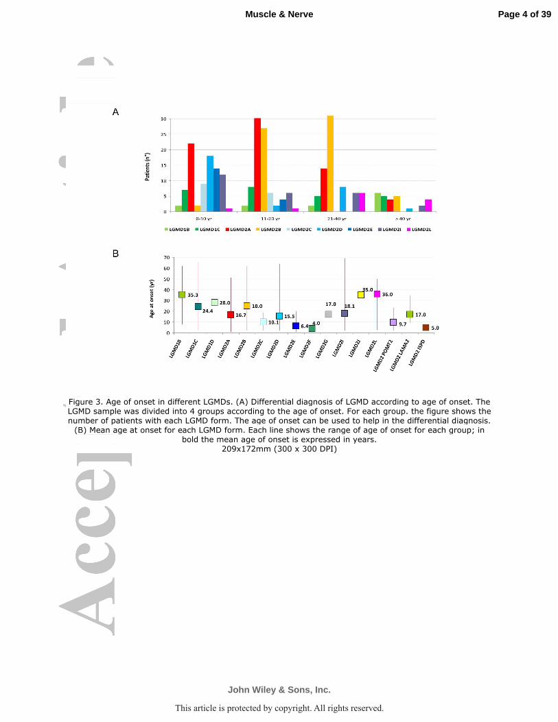
Figure 3. Age of onset in different LGMDs. (A) Differential diagnosis of LGMD according to age of onset. The LGMD sample was divided into 4 groups according to the age of onset. For each group. the figure shows the number of patients with each LGMD form. The age of onset can be used to help in the differential diagnosis. (B) Mean age at onset for each LGMD form. Each line shows the range of age of onset for each group; in
bold the mean age of onset is expressed in years. 209x172mm (300 x 300 DPI)
Page 4 of 39
John Wiley & Sons, Inc.
Muscle & Nerve
This article is protected by copyright. All rights reserved.
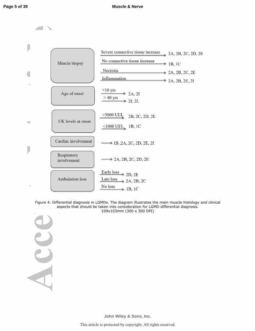
Figure 4. Differential diagnosis in LGMDs. The diagram illustrates the main muscle histology and clinical aspects that should be taken into consideration for LGMD differential diagnosis.
109x103mm (300 x 300 DPI)
Page 5 of 39
John Wiley & Sons, Inc.
Muscle & Nerve
This article is protected by copyright. All rights reserved.