the innate immune response chapters 15 and 19 nester 4th. ed. anjum odhwani, md mph public health mp...
TRANSCRIPT
The Innate Immune Response
Chapters 15 and 19
Nester 4th. Ed.
Anjum Odhwani, MD MPH
Public Health MP 3304
Definition of Immunology
The branch of science that is devoted to the study of many mechanisms the body uses to defend itself against invading organisms or microbes.

Types of Defense Mechanisms
Non-specific (Innate immunity) Specific (adaptive immunity)- Immune
Response

One branch of immunity
Non-specific immunity– found in most of the humans and animals – inborn, innate, natural – already in place before the organism
enters the host– directed against any organism that tries to
invade a host

Mechanisms involved in Non-specific immunity Tissue barriers
– Mechanical – Chemical
Non-specific antimicrobial substances Acute inflammation Phagocytosis Fever Changes in iron metabolism
First – line defense
Physical barriers Normal flora Antimicrobial substances

Innate Immunity Mechanical Barriers that prevent entry of
microorganisms (Fig 15.1-15.3)– Skin
• Most difficult barrier• Physically prevents microbes from
accessing the tissues• Skin is tough and durable• Outer layer Keratin constantly slough
off• Arid environment• Sweat high in salt and lysozyme• Sebum (fatty acid)
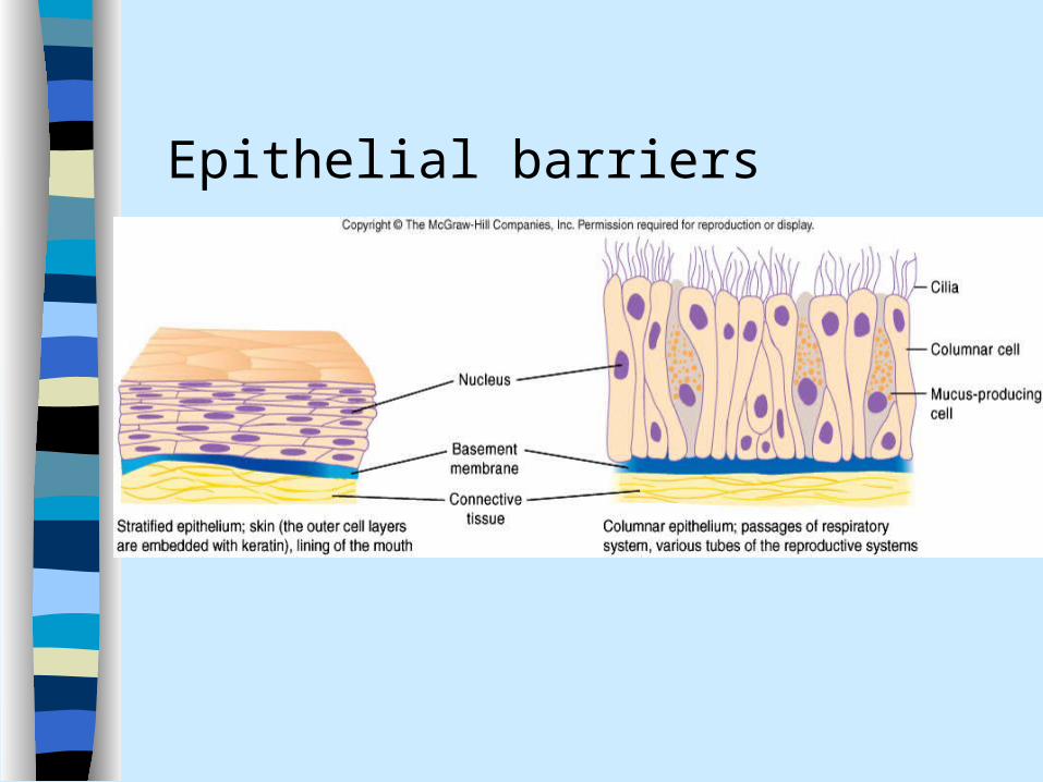
Epithelial barriers
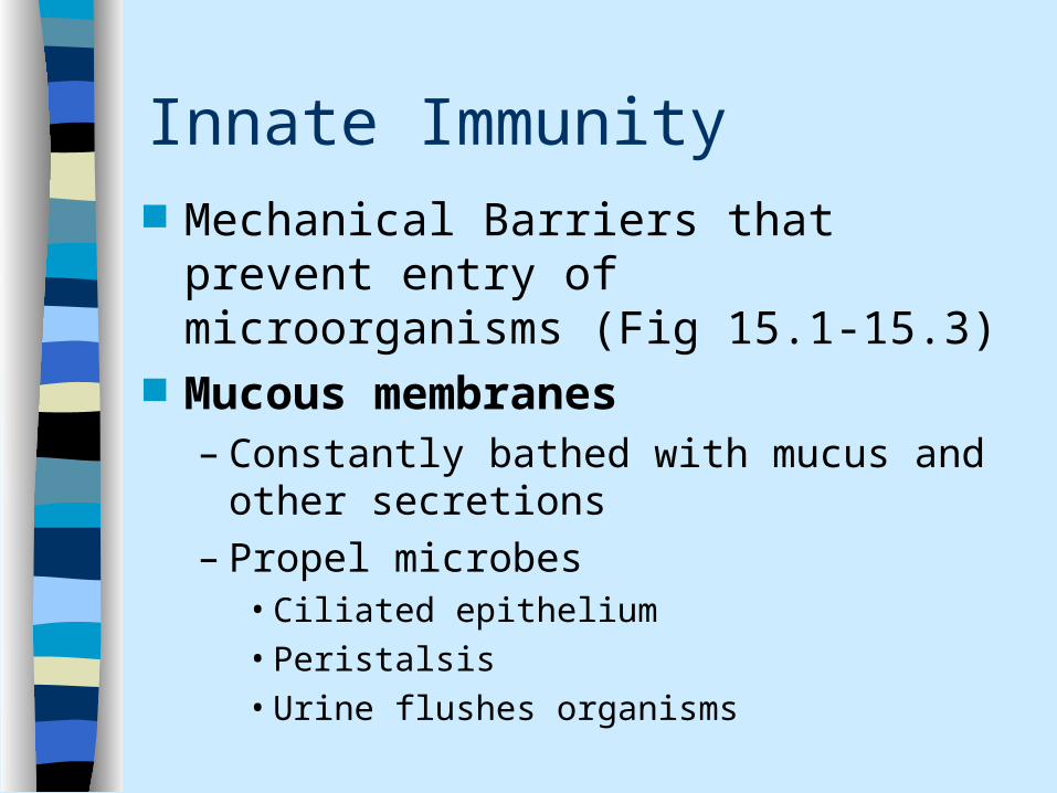
Innate Immunity Mechanical Barriers that prevent entry
of microorganisms (Fig 15.1-15.3) Mucous membranes
– Constantly bathed with mucus and other secretions
– Propel microbes • Ciliated epithelium• Peristalsis• Urine flushes organisms
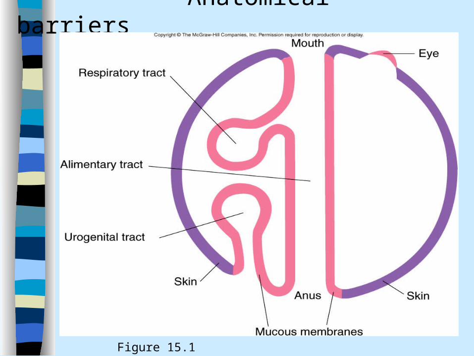
Anatomical barriers
Figure 15.1
Innate Immunity
Chemical Barriers (Figure 15.3)– Acid mantle
• urine, stomach acid and vaginal pH

First-line defense mechanisms in humans

Innate Immunity
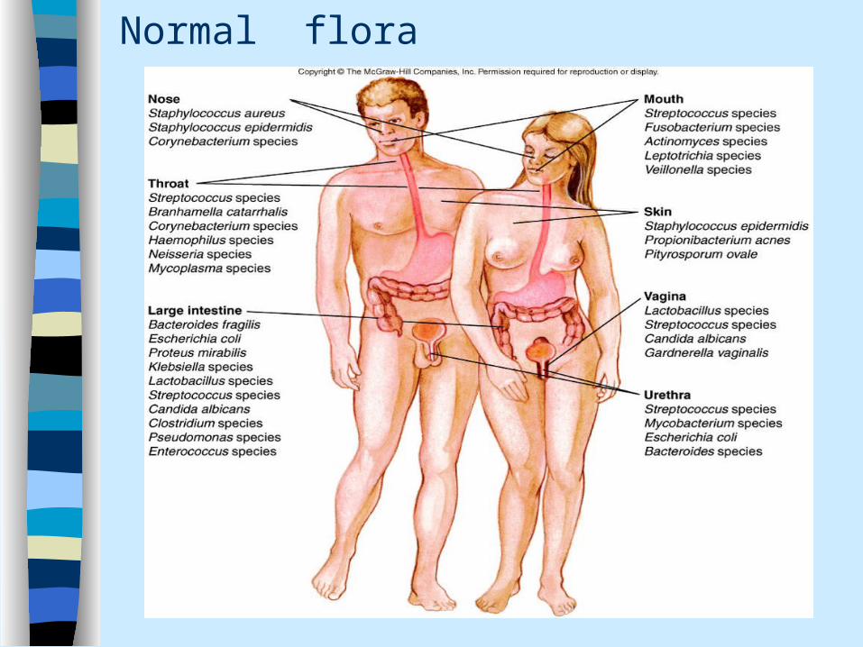
Normal flora (resident flora)– Covers binding sites– Compete– Consume resources– Produce toxins e.g. Propionibacterium,
Lactobacillus species in vagina– Stimulate host defense mechanism
Normal flora
Establishment– fetus has no normal flora– acquire some at birth– acquire some in food– acquire some from other people– colonization
Normal flora

Importance of Normal Flora
Prevent growth of other organisms– by taking up space - commensals– by competing for nutrients– by preventing attachment

Importance of Normal Flora Prevent growth of other organisms
– by making products that are toxic to other organisms• lipid catabolic by-products e. g.
Propionibacterium (sebaceous secretion to fatty acid)
• Colicins (E. coli synthesis protein toxic to other organisms)
• Lactic acids (lactobacillus in vagina acidic environment)
Importance of Normal Flora
Prevent the growth of other organisms– by stimulating immunity
• an immune response to one organism may help against a similar organism
Providing a useful function– degrading cellulose for nutrients
– making vitamins (biotin, panthothenic acid (B5), folic acid and Vitamin K)

Normal flora
– Types of symbiotic relationships• Mutualism
– An association in which both partners benefit e.g. intestinal bacteria synthesize vit K and vit B
• Commensalism– An association in which one partner benefits
but other remains unharm• Parasitism
– An association in which one organism, the parasite derives benefit at the expense of the other organism, the host

Normal Flora Can Change
How?– increased perspiration– acidity of the stomach (Alteration of the acid barrier
of the stomach by disease, surgery, drugs or antacids)
– ingestion of antibiotics– meat diet vs. vegetarian diet (People living on
the high carbohydrate diet have significantly fewer Bacteroides and more Enterococci in their faeces)
– changes in peristalsis (e.g. diarrhea)

Innate Immunity Non-specific antimicrobial factors Antimicrobial substances found in
saliva, tears, mucus and skin– Lysozyme– Peroxidase– Lactoferrin– Interferons

Innate Immunity
Non-specific antimicrobial factors– Lysozyme
• Found in tears, saliva, mucus, in phagocytic cells, blood and fluid that bathes tissues
• Enzyme that degrades peptidoglycan• Gram-positive bacteria peptidoglycan is
exposed• Gram-negative peptidoglycan not
exposed
Innate Immunity Non-specific antimicrobial factors
– Lactoferrin and transferrin• Found in saliva, mucus, milk, blood and
tissue fluids• Iron binding protein• Major element for growth of the
organism• Sequester iron from the organisms

Innate Immunity Non-specific antimicrobial factors
Peroxidase• Found in saliva, milk, body tissues and
phagocytes• Peroxidases are a group of enzymes that
catalyze oxidation-reduction reactions
• H2O2 + peroxidase + Cl = yields chlorine and hypochlorite (bleach)
• H2O2 + catalase yields H2O and O2
• Catalase (+) organisms are more resistant to peroxidase
• Catalase –negative organisms are sensitive to perxidase killing

Innate Immunity Non-specific antimicrobial factors
– Defensins• Short antimicrobial peptides• Found on mucus membranes and
within phagocytic cells (macrophages and neutrophils)
• Insert themselves into bacterial membranes and form pores
• Distrupt the integrity of bacterial membrane

Interferon

Innate Immunity Non-specific antimicrobial factors
– Interferons• Group of glycoproteins (control viral
infection) See Figure 15.11• Inhibits protein synthesis in cells near
virally infected cells• Prevent viral replication• Are species specific with regards to host

Cells and Tissues Involved in Defense Mechanisms
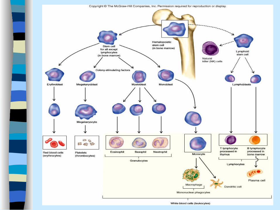
Cells are mainly the leukocytes– Figure 15.4– Table 15.2– Non-specific are the granulocytes,
monocyte/macrophages and null cells (natural killer cells)
– Specific are the lymphocytes

Cells and Tissues Involved in Defense Mechanisms
Tissues – lymphoid tissues (dense accumulation of
lymphocytes e.g. Peyer’s
patches)
– Lymphoid organ (spleen, lymph nodes, tonsils, bone marrow, adenoids and appendix)

Professional phagocytes
Professional phagocytes

Cells and Tissues Involved in Defense Mechanisms Acute bacterial infection Neutrophils ↑ Inflammation and allergic reaction Basophils ↑ Eosinophils ↑ Allergic reaction and parasitic
infestation Eosinophils ↑

Cell Communication
Trauma or invasion Communicate to immediate
environment and to other cells How do they communicate?
– Surface Receptors (eyes and ears)– Cytokines (Voice)– Adhesion molecules (hands)

Cell Communication
Surface receptors– Are integral membrane proteins– Bind specific compound or compounds– Molecule binds to a particular receptor is
called a ligand for that receptor– Internal portion of the receptor becomes
modified– Elicit response (chemotaxis)

Cell Communication Cytokines
– Low molecular weight proteins– Made by certain cells
• lymphokine (cytokines made by lymphocytes)
• monokine (cytokines made by monocytes)
• chemokine (cytokines with chemotactic activities)
• interleukin (cytokines made by one leukocyte and acting on other leukocytes).

Cell Communication Cytokines
– Communicate with other cells (chemical messengers)
– Short-lived– Very powerful– Act at extremely low concentration– Act locally, regionally or systemically

Cell Communication Cytokines
– Cytokines binds to cytokine receptors• Induce a change such as growth
(different kind of leukocytes, precursors of blood cell and mast cells)
• Differentiation (different leukocytes)
• Movement
• Cell death

Cell Communication Cytokines
• See Table 15.3• Chemokines• Colony stimulating factors• Interferons• Interleukins• Tumor necrosis factor

Direct immature leukocytes into the appropriate maturation pathway
Cell Communication
Cytokines: Groups of cytokines work together– Chemokines
• 50 different varieties• Chemotaxis of immune cells
– Colony-stimulating factors (CSFs)• multiplication and differentiation of
leukocytes

Cell Communication Cytokines
– Interferons (IFNs)• Glycoproteins• Control viral infections• Gamma-interferon helps regulate the
function of the cells involved in inflammatory response (phagocytes)
• Modulates certain responses of adaptive immunity

Cell Communication
Cytokines– Interleukins (ILs)
• Produced by leukocytes• At least 18 interleukins been studied• Innate and adaptive immunity
– Tumor necrosis factors (TNFs)• TNF- alpha produced by macrophages
plays an instrumental role in initiating the inflammatory response
• Programmed cell death or appoptosis (destroy self-cells without eliciting inflammation)

Cell Communication
Cytokines– Groups of cytokines work together
• Pro-inflammatory cytokines contributes to inflammation (TNF-alpha, IL-1, IL-6)
• Promotion of antibody responses (IL-4, IL-5, IL-10 and IL-14)
• Promotion of T cell activity (IL-2, and INF-gamma)

Cell Communication
Adhesion molecules– Allow cells to adhere to other cells– Ex. Endothelial cells bind to phagocytic
cells grab cells– Slow down phagocytic cell movement– Allow cells to adhere to other cells and
deliver cytokines or other compounds

Cell Communication Sensor Systems
– Present within blood and tissues– Senses tissue damage and microbial
invasion– Either directly destroy the microbes – Or recruit other components of the host
defenses E.g. Toll-like receptors Complement system
Sensor systems
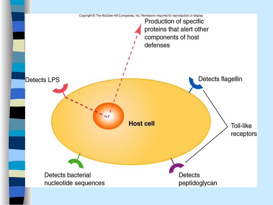
Toll-like Receptors (TLRs) Figure 15-6 At least 10 TLRs identified Each recognizes a distinct compound or
groups of compounds E.g. TLR-2 recognizes peptidoglycan TLR-4 is triggered by lipopolysacchride

Sensor systems
Toll-like Receptors (TLRs) Other bacterial structures or compounds that
activate these receptor Flagellin Bacterial nucleotides What do they do?

Sensor systems
Toll-like Receptors (TLRs)– What do they do?– Transmit signals to the nucleus of the
host cells to alter the expression of certain genes
– E.g. – Lipopolysaccharide → triggers a TLRs of
monocytes and macrophages → chemokines attracts additional phagocytes

Sensor Systems• The Complement System (Figure 15.7 and
15.8)– Series of about 20 proteins
• Circulate in the blood and tissue fluids• C1 through C9 are the major components of
this system• Routinely circulate in an inactive form• once activated a cascade of events occurs• one event triggers the next event• Activated forms have specialized functions to
quickly remove and destroy the offending material

Compliment system
Regulatory proteins halt the process inactivate C3b
Regulatory proteins are not associated with microbial surfaces
C3a and C5a→induce changes in endothelial cells and mast cells →↑ vascular permeability
C5a attracts phagocytes
C3b binds to foreign material called opsonized, C3b called opsonins
Membrane attack complex

Sensor Systems Complement (continued)
– Two pathways of activation• Classical pathway part of specific immunity• Alternate pathway part of innate immunity
– Final common pathway• Ultimate function is lysis of a bacteria by the
membrane attack complex• C3a and C5a are the anaphylatoxins
Sensor Systems Complement System
– Classical pathway inflammation• Antigen-antibody complexes red
flag portion of the antibody interact with complement component in turn C3a and C5a induce changes in endothelial cells increase permeability associated with inflammation
• C5a is a potent chemoattractant

Sensor Systems Complement System
– Alternative pathway • C3b binds to foreign material is said opsonized
(prepared for eating)
• Phagocytes more easily destroy C3b coated cells as they have C3b receptors
• C3a and C3b cause phagocytes to produce more receptors for C3b

Sensor Systems Complement System
– Lactin pathway Lysis of foreign cells
• Mannan-binding lectins (MBLs), a polymer of mannose found on microbial cells MBL binds to a surface and interact with compliment component initiating classical pathway C5b, C6, C7, C8 and C9 forms doughnut shaped structure called membrane attack complex (MAC) creates pores in membrane, disrupting the integrity of the cell

Complement System C3a and C5a→induce changes in endothelial
cells and mast cells →↑ vascular permeability C5a attracts phagocytes C3b binds to foreign material called
opsonized C3b called opsonins Regulatory proteins halt the process
inactivate C3b Regulatory proteins are not associated with
microbial surfaces

Phagocytosis

Innate Immunity Phagocytosis (Figure 15.9)
– Chemotaxis- phagocytic cells are recruited
– Recognition and adhesion• Direct: mannose sugar found on the surface
of certain bacteria and yeast• Indirect: binding opsonized (C3b)
– Engulfment• phagocyte engulf the invader forming a
membrane-bound vacuole called phagosome

Innate Immunity
• Phagocytosis • Fusion of the phagosome with lysosome• Phagosome transported towards lysosome
forming phagolysosome
• Lysosome a membrane bound body filled with
various digestive enzymes

Innate Immunity• Phagocytosis • Destruction and digestion
• Oxygen dependent mechanisms oxidized sugars via TCA cycle
• Highly toxic oxygen by-products such as superoxide, hydrogen peroxide and hydroxyl radicals are produced
• Once oxygen is depleted fermentation anaerobic metabolism starts
• Metabolic pathway switches to lactic acid production lowering pH
• Enzymes degrade bacterial cell wall and other components of cells
Innate Immunity
Phagocytosis Exocytosis
– Digested material is expelled to external environment

Innate Immunity
Neutrophils – First to arrive during an immune response– Involved in inflammation– Inherently have more killing power than
macrophages

Innate Immunity Macrophages
– Located throughout the body (Kupffer cells, alveolar macrophages, etc.)
– Produce cytokines– Interact with T helper cells – activated
macrophages– Help form granulomas (Macrophages, Giant
cells and T-helper cells)


Innate Immunity
Inflammation-A coordinated response to invasion or damage– Types of inflammation– Cardinal signs of acute inflammation– Factors that initiate the inflammatory
response– Process of acute inflammation– Outcomes of inflammation

Innate Immunity
Inflammation– Definition
• When a tissue have been damaged, such as when an object penetrates the skin or when microbes produce toxic compound, a coordinated response called the inflammatory response, or inflammation occurs
– Types of inflammation• Acute
– Immediate and short lived response (neutrophils)• Chronic
– Delayed and long lived response (macrophages, giant cells, T cells forms granulomas)

Innate Immunity
Signs of inflammation– Swelling– Redness– Heat– Pain– Loss of function (sometimes present)
Intension of the inflammatory process– limit damage and restore function

Innate immunity Inflammation
– Factors that initiate the inflammatory response
• Microbial products– Lipopolysaccharide, flagellin, bacterial
DNA trigger toll-like receptors

Innate immunity Factors that initiate the inflammatory
• Microbial cell surface– Trigger complement cascasde
» leading to production of C3a and C5a» Stimulate changes associated with
inflammation– Complement component also induces
mast cells to release various proinflammatory cytokines

Innate immunity Factors that initiate the
inflammatory• Tissue damage
– Activate two enzymatic cascades
»Coagulation cascade results in blood clotting and catch microbe in a clot too
»Bradykinin increases vascular permeability, dilates blood vessels, contracts non-vascular smooth muscle, and causes pain

The inflammatory process

The inflammatory process
Innate Immunity The acute inflammatory response
(Figure 15.10)– Two components
• Vascular component–Vasodilation- produces redness and
heat–Increased vascular permeability -
produces swelling
Innate Immunity
The acute inflammatory response (Figure 15.10)– Two components
• Cellular component–Cell recruitment–Phagocytosis

Innate immunity
Inflammation– Outcomes of acute inflammation
• Resolution• Abscess formation• Scarring• Chronic inflammation

Innate immunity
Inflammation– Systemic response is life threatening– Enzymes and toxic products within
phagocytic cells can damage healthy tissues
– Inflammation in brain and spinal cord can be life threatening

Innate immunity Septic shock: Gram-negative bacteria → endotoxins→
proinflammatory cytokines →activate complement cascade and clotting cascade →results in rapid decrease in blood pressure → shock
Clot plugs the capillaries of vital organs e.g. liver, lung, brain etc cutting blood supply

Innate immunity Apoptosis
– Mechanism of eliminating dead self-cells without evoking an inflammatory response
– Cell suicide– Dying cells undergo changes
• Shape changes• Enzyme cut the DNA• Portions of the cells bud off• Cell shrink

Innate Immunity
Fever– one of the strongest indications of
infectious disease– the hypothalamus regulates our normal
body temperature in a narrow range• 98.6ºF or 37 ºC
– central temperature receptors and peripheral temperature receptors
Innate Immunity
Fever – Cytokines and other fever-inducing
substances are called pyrogens
– Fever-inducing cytokines body makes it e.g. IL-1 and TNF (endogenous pyrogens)
– Bacterial endotoxins, cell wall Lipoplysaccharide are (exogenous pyrogens)

Innate Immunity Fever
– microorganisms can cause the body to respond by changing the “set point”• components of microorganism attach to
phagocytic cells• phagocytic cells release interleukin-1• interleukin-1 travels via the blood to the
hypothalamus• the hypothalamus responds by raising
body temperature

Innate Immunity Fever
– inhibits pathogens by:• increasing body temperature above the
optimal temperature for growth of microorganisms
–enzymes are inactivated• activating and speeding a number of
body defenses–there are many examples

Innate Immunity
Fever– Examples of beneficial effects of fever
• enhance inflammation• increase killing by phagocytes• release of chemo-attractants of
neutrophils• activation and replication of lymphocytes• increase production of antibody and
interferon• decrease host’s ability to absorb iron