the importance of mineral elements for humans, domestic
TRANSCRIPT

African Journal of Food Science Vol. 4(5) pp. 200-222, May 2010 Available online http://www.academicjournals.org/ajfs ISSN 1996-0794 ©2010 Academic Journals Review Paper
The importance of mineral elements for humans, domestic animals and plants: A review
K. O. Soetan1*, C. O. Olaiya2 and O. E. Oyewole3
1Department of Veterinary Physiology, Biochemistry and Pharmacology, University of Ibadan, Ibadan, Oyo state,
Nigeria. 2Department of Biochemistry, University of Ibadan, Ibadan, Oyo state, Nigeria.
3Department of Health Promotion and Education, University of Ibadan, Ibadan, Oyo state, Nigeria.
Accepted 12 February, 2010
Minerals are inorganic nutrients, usually required in small amounts from less than 1 to 2500 mg per day, depending on the mineral. As with vitamins and other essential food nutrients, mineral requirements vary with animal species. For example, humans and other vertebrates need large amounts of calcium for construction and maintenance of bone and normal function of nerves and muscles. Phosphorus is an important constituent of adenosine triphosphate (ATP) and nucleic acid and is also essential for acid-base balance, bone and tooth formation. Red blood cells can not function properly without iron in haemoglobin, the oxygen-carrying pigment of red blood cells. Iron is also an important component of the cytochromes that function in cellular respiration. Magnesium, copper, selenium, zinc, iron, manganese and molybdenum are important co-factors found in the structure of certain enzymes and are indispensable in numerous biochemical pathways. Vertebrates need iodine to make thyroid hormones. Sodium, potassium and chlorine are important in the maintenance of osmotic balance between cells and the interstitial fluid. Magnesium is an important component of chlorophyll in plants. The interactions between nutrition and diseases, nutrition and drug metabolism have been reported. Excessive intake of some minerals can upset homeostatic balance and cause toxic side effects. For example, excess sodium intake is associated with high blood pressure and excess iron can cause liver damage. Also, severe shortages or self-prescribed minerals can alter the delicate balance in body functions that promotes health. The knowledge of the biochemistry of the mineral elements is also essential because individuals suffering from a chronic illness or taking medications that affect the body’s use of specific nutrients need to be enlightened. The aim of this paper is to review the biochemical functions and the importance of the mineral elements in health and disease conditions of humans, animals and plants as this will assist in the prevention of nutrition-related diseases and maintenance of good health for humans and animals that depend on plants for food. This paper could also serve as a ready source of literature review for researchers involved in nutritional sciences. Key words: Mineral elements, humans, animals, plants, nutrition.
INTRODUCTION Minerals are inorganic substances, present in all body tissues and fluids and their presence is necessary for the *Corresponding author. E-mail: [email protected].
maintenance of certain physicochemical processes which are essential to life. Minerals are chemical constituents used by the body in many ways. Although they yield no energy, they have important roles to play in many activities in the body (Malhotra, 1998; Eruvbetine, 2003). Every form of living matter requires these inorganic

elements or minerals for their normal life processes (Hays and Swenson, 1985; Ozcan, 2003). Minerals may be broadly classified as macro (major) or micro (trace) elements. The third category is the ultra trace elements. The macro-minerals include calcium, phosphorus, sodium and chloride, while the micro-elements include iron, copper, cobalt, potassium, magnesium, iodine, zinc, manganese, molybdenum, fluoride, chromium, selenium and sulfur (Eruvbetine, 2003). The macro-minerals are required in amounts greater than 100 mg/dl and the micro-minerals are required in amounts less than 100 mg/dl (Murray et al., 2000). The ultra trace elements include boron, silicon, arsenic and nickel which have been found in animals and are believed to be essential for these animals. Evidence for requirements and essentialness of others like cadmium, lead, tin, lithium and vanadium is weak (Albion Research Notes, 1996).
The mineral elements are separate entities from the other essential nutrients like proteins, fats, carbohy-drates, and vitamins. Animal husbandry had demon-strated the need for minerals in the diet (Hegsted et al., 1976). In this century, biological assay methods clarified the significance and importance of mineral elements for human and animal nutrition and modern analytical techniques led to the detection of trace elements as essential nutrients and this is still an active area of current research.
Micronutrient deficiencies are a major public health problem in many developing countries, with infants and pregnant women especially at risk (Batra and Seth, 2002). Infants deserve extra concern because they need adequate micronutrients to maintain normal growth and development (Rush, 2000). The micronutrient deficien-cies which are of greatest public health significance are iron deficiency, causing varying degrees of impairment in cognitive performance, lowered work capacity, lowered immunity to infections, pregnancy complications e.g. babies with low birth weight, poor learning capacity and reduced psychomotor skills (Batra and Seth, 2002). Medical reports show that very severe anaemia is a direct cause of maternal and child mortality (Chakravarty and Ghosh, 2000). There have been suggestions that more than anything else, lack of adequate information about the composition of varied feed resources in some regions have been the major drawback to their utilization, rather than real shortage (Aletor and Omodara, 1994). For instance, there is very limited information on the mineral elements in some plants used as human food and animal feeds consumed in Nigeria, especially the newly-introduced varieties of diets and the lesser known legumes. Some of the earlier information on mineral elements was based on analysis employing less sensitive methods, which may not be reliable. The aim of this review is to re-visit the provision of information on the importance of mineral elements to humans, animals and plants and also to emphasize on the need for their levels to be ascertained in water and commonly consumed plant foods using modern analytical techniques. This will assist
Soetan et al. 201 greatly in providing relevant information on the importance of minerals to health and this would subsequently assist in prevention and management of mineral-associated deficiency diseases. Data on mineral contents of human foods and animal feeds are essential for formulation of feeding regimes and food processing techniques. THE IMPORTANCE OF THE MINERAL ELEMENTS The importance of mineral elements in human, animal and plant nutrition has been well recognized (Underwood, 1971; Darby, 1976). Deficiencies or disturbances in the nutrition of an animal cause a variety of diseases and can arise in several ways (Gordon, 1977). When a trace element is deficient, a characteristic syndrome is pro-duced which reflects the specific functions of the nutrient in the metabolism of the animal. The trace elements are essential components of enzyme systems. Simple or conditioned deficiencies of mineral elements therefore have profound effects on metabolism and tissue structure. To assess the dietary intake and adequacy of minerals, information needs to be collected on mineral element content of foods, diets and water (Rao and Rao, 1981; Simsek and Aykut, 2007). There is limited information on the trace element content of water and numerous plant foods consumed in some less developed countries.
The significance of the mineral elements in humans, animals and plants nutrition can not be overemphasized. The presence of mineral elements in animal feed is vital for the animal’s metabolic processes. Grazing livestock from tropical countries often do not receive mineral supplementation except for common salt and must depend almost exclusively upon forage for their mineral requirements (McDowell et al., 1984). Mineral deficiencies or imbalances in soils and forages account partly for low animal production and reproductive problems. Soil acidity and season are factors affecting mineral uptake by plants. Plants use these minerals as structural components in carbohydrates and proteins; organic molecules in metabolism, such as magnesium in chlorophyll and phosphorus in ATP; enzyme activators like potassium, and for maintaining osmotic balance. Calcium is highly implicated in the maintenance of firmness of fruits (Olaiya, 2006) and its requirements in fruits are related to cell wall stability and membrane integrity (Belakbir et al., 1998). Mineral elements play important roles in health and disease states of humans and domestic animals. For example, iron deficiency anaemia and goitre due to iodine deficiency are reported to be problems of public health importance in some communities (Partwardhan, 1961; Deosthale and Belavady, 1978). Trace elements of significance to people with HIV are zinc and selenium. Selenium is an antioxidant that increases immune function. Zinc, usually taken to stimulate the immune system, has been reported to weaken immune system function and lower calcium

202 Afr. J. Food Sci. levels in HIV – positive men (O’ Connor, 1995; Wood, 2000). METHODS OF ANALYSES FOR THE MINERALS Mineral elements in plant and soil samples are essentially estimated by colorimetric procedure according to the method of Sandell (1959). The wet-ashing procedure of Gorsuch (1959) using nitric acid, perchloric acid and sulphuric acid in the ratio of 3:2:1 is usually employed for the digestion of the food samples. Processing of samples of foods and water for mineral analysis are carried out in a dust-free room to avoid contamination, using distilled deionized water for all dilutions (Rao and Rao, 1981). Appropriate blanks are also included along with the samples and trace elements are estimated in the samples using an atomic absorption spectrophotometer. Internationally approved standards are also run along with the samples to be analysed to check the results obtained. Studying the chemical composition of plants provide clues about their nutritional requirements (Roberts, 1985). Researchers use a method known as hydroponic culture to determine which of the mineral elements are actually essential nutrients. In the technique known as hydroponic culture, a researcher baths the roots of plants in solutions of various minerals dissolved in known concentrations. Aerating the water will provide the roots with oxygen for cellular respiration. A particular mineral, such as potassium, can be ommited from the culture medium to test whether it is essential to the plants. If the element removed from the mineral solution is an essential nutrient, then the incomplete medium will cause plants to become abnormal in appearance compared with controls grown on a complete mineral medium. Analyses of the mineral content of a plant or soil are used to confirm the diagnosis of a specific mineral deficiency. Such studies have helped identify 17 elements that are essential nutrients in all plants and a few other elements that are essential to certain groups of plants as shown in Table 1. FACTORS AFFECTING THE CONCENTRATION AND AVAILABILITY OF THE MINERAL ELEMENTS Several factors directly or indirectly influence the levels of minerals in plants and hence the amounts available for humans and animals that depend on plants for foods and feeds respectively. The amount of a particular nutrient in the diet may be insufficient to meet the requirements. However, the metabolism of the animal may be deranged by the interaction of dietary, environmental and genetic factors (Gordon, 1977).
Location has been reported to influence the mineral and trace element compositions of rice, wheat, oats and barley (Basargin and Peregudora, 1969; Kavanek and Janicek, 1969) and these are mainly attributed to the altered soil conditions. The nature and chemical composition of the soil are also involved in the locational differences observed in the mineral elements present in grain sorghum (Deosthale and Belavady, 1978). Feeds grown on high-selenium soils are good sources of selenium and may be used in ration formulation for poultry in order to supply a source of selenium (Merck, 1986). Uptake of copper, zinc and manganese by plants is affected by the level of phosphate fertilizer (Mongia,
1966; Baser and Deo, 1967), while the chemical form of nitrogen fertilizer has been reported to affect the uptake of copper (Chand, 1969). Environmental factors such as location rather than genotype is reported to have greater influence on the mineral and trace element composition of sorghum grain (Belavady and Deosthale, 1978). Cobalt, copper, iodine and selenium deficiencies in the soil and flora in certain areas of the world have led to deficiencies of these minerals in domestic animals (Hays and Swenson, 1985). Also, the increment of selenium in the soil may lead to high levels in plants which are toxic to animals. Nutritional disorders involving the mineral elements may arise as simple deficiencies or excesses of particular elements but the extent to which the other organic or inorganic nutrients are present in the diet will determine the deficiencies or toxicities. The conditioning factors may be a reflection of the soils on which the plants are grown, or they may be related to the presence of specific plants that are seleniferous or goitogenic (Hays and Swenson, 1985). Mineral deficiencies or imbalances in soils or forages have been implicated, in part, for low animal production, and poor reproductive performance in the developing regions of the world (Aletor and Omodara, 1994).
Antinutritional factors (ANF) present in plants could also affect the absorption and availability of some minerals by humans and animals. Anti-nutritional factors reduce the nutrient utilization and or food intake of plant foods (Osagie, 1998). The need for adequate processing to reduce the antinutritional factors in plants used as human foods and animal feeds have been reviewed by (Soetan and Oyewole, 2009). The levels of these substances in plants vary with the specie of plant, cultivar and post-harvest treatment. Examples of ANF which could reduce the bioavailability of minerals are oxalates and phytates. Oxalalic acid, like phytic acid, has the ability to bind some divalent metals such as calcium and magnesium thereby interfering with their metabolism (Blood and Radostits, 1989). Chelates bind many elements making them nutritionally unavailable, thereby inducing dietary deficiencies (Nelson et al., 1968). Zinc may be complexed with calcium-phytate and lead to inefficient utilization of dietary zinc. Adequate calcium intake will compensate for the reduced availability of calcium, but this will aggravate the zinc deficiency and such a relationship has frequently led to parakeratosis in pigs. Phytic acid reduces the absorption of calcium from the gastro-intestinal tract and consequently implicated in the development of rickets when chicks are fed cereals such as sorghum. Zinc and iron deficiency symptoms have been reported in man and chicken (Lease, 1966) when fed diets high in phytic acid. There is a significant inverse relationship between phytic acid content and the availability of calcium, magnesium, phosphorus and zinc in products like soya bean, palm kernel, rapeseed and cottonseed meals (Nwokolo and Bragg, 1977). Phytic acid may decrease the availability of divalent cations
Soetan et al. 203
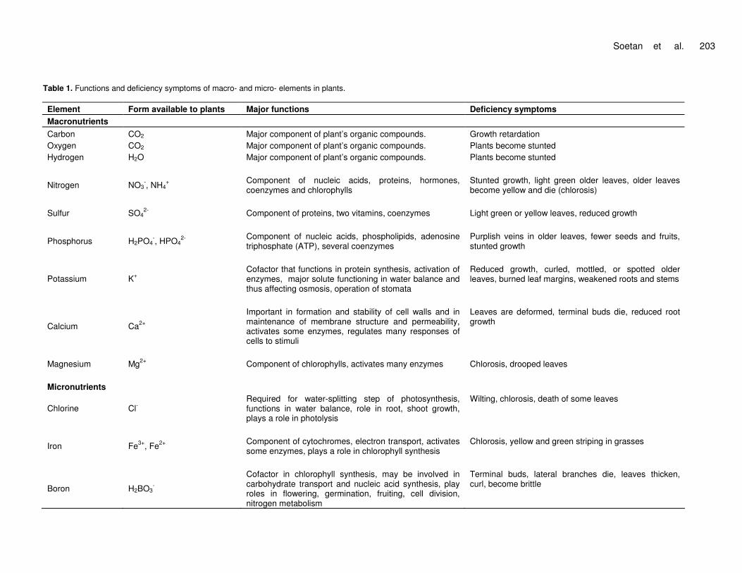
Table 1. Functions and deficiency symptoms of macro- and micro- elements in plants. Element Form available to plants Major functions Deficiency symptoms Macronutrients Carbon CO2 Major component of plant’s organic compounds. Growth retardation Oxygen CO2 Major component of plant’s organic compounds. Plants become stunted Hydrogen H2O Major component of plant’s organic compounds. Plants become stunted
Nitrogen
NO3
-, NH4+
Component of nucleic acids, proteins, hormones, coenzymes and chlorophylls
Stunted growth, light green older leaves, older leaves become yellow and die (chlorosis)
Sulfur
SO4
2- Component of proteins, two vitamins, coenzymes
Light green or yellow leaves, reduced growth
Phosphorus
H2PO4
-, HPO42-
Component of nucleic acids, phospholipids, adenosine triphosphate (ATP), several coenzymes
Purplish veins in older leaves, fewer seeds and fruits, stunted growth
Potassium
K+
Cofactor that functions in protein synthesis, activation of enzymes, major solute functioning in water balance and thus affecting osmosis, operation of stomata
Reduced growth, curled, mottled, or spotted older leaves, burned leaf margins, weakened roots and stems
Calcium
Ca2+
Important in formation and stability of cell walls and in maintenance of membrane structure and permeability, activates some enzymes, regulates many responses of cells to stimuli
Leaves are deformed, terminal buds die, reduced root growth
Magnesium
Mg2+
Component of chlorophylls, activates many enzymes
Chlorosis, drooped leaves
Micronutrients
Chlorine Cl- Required for water-splitting step of photosynthesis, functions in water balance, role in root, shoot growth, plays a role in photolysis
Wilting, chlorosis, death of some leaves
Iron
Fe3+, Fe2+
Component of cytochromes, electron transport, activates some enzymes, plays a role in chlorophyll synthesis
Chlorosis, yellow and green striping in grasses
Boron
H2BO3
-
Cofactor in chlorophyll synthesis, may be involved in carbohydrate transport and nucleic acid synthesis, play roles in flowering, germination, fruiting, cell division, nitrogen metabolism
Terminal buds, lateral branches die, leaves thicken, curl, become brittle

204 Afr. J. Food Sci. Table 1. Contd.
Manganese
Mn2+
Active in formation of amino acids, activates some enzymes, coenzyme activity, required for water-splitting step of photosynthesis, chlorophyll synthesis
Light green leaves with green major veins, leaves whiten and fall off
Zinc
Zn2+
Active in formation of chlorophyll, activates some enzymes, plays a role in formation of auxin, chloroplasts, and starch
Chlorosis, mottled or bronzed leaves, abnormal roots
Copper
Cu+,Cu2+
Component of many redox and lignin-biosynthetic enzymes
Chlorosis, dead spots in leaves, stunted growth, terminal buds die, necrosis in young leaves
Molybdenum
MoO4
2-
Essential for nitrogen fixation, cofactor that functions in nitrate reduction, component of enzyme used in nitrogen metabolism
Possible nitrogen deficiency, pale green, rolled or cupped leaves, mottling and necrosis in old leaves
Nickel
Ni2+
Cofactor for an enzyme functioning in nitrogen metabolism
Source: (Williams, 1992). Note: All mineral elements contribute to the water-solute balance.
such as calcium, zinc and iron, by the formation of an insoluble protein-phytic acid- mineral complex, it has been cited as causing reduced availability of zinc in soybean foods (O’Dell and Savage, 1960; Forbes and Parker, 1977). Sometimes, either as a conse-quence of low availability of iodine from the soil, or the presence in the food of goitrogenic substances, which interfere with the utilization of iodine by the thyroid, the iodine requirements of some animals e.g. the sheep are not met in the natural diet and so iodine supplements must be fed (Merck, 1986). INTERRELATIONSHIPS AND INTERFERENCES AMONG THE MINERAL ELEMENTS There are many metabolic and absorptive
interrelationships among the mineral elements which contribute to variations in the degree of physiological response to deficient or toxic levels. These relationships make it difficult to determine the optimum dietary level for the individual elements required for humans and domestic animals. As a result of this, the recommended dietary level of any element should rarely be considered independent of the level of other essential nutrients (Hays and Swenson, 1985). The functions of minerals in humans and animals are inter-related. Examples are the definite relationship of calcium and phosphorus in the formation of bones and teeth and as the major structural elements of the skeletal tissue. Hypocalcaemia may cause weakness of the heart similar to that caused by hyperkalaemia. A high level of potassium appears to increase the
requirement for sodium and vice versa (Merck, 1986). Potassium deficiency leads to an increase in the basic amino acid concentration of the tissue fluids and some increase in cellular sodium levels as a means of maintaining cation-anion balance. Potassium influences the contractibility of smooth, skeletal, and cardiac muscles and has an effect on muscular irritability that, like that of sodium, tends to antagonize the effect of the calcium ion. Under conditions of salt restriction, calcium appears highly important in helping to maintain the potassium content of tissue (Hays and Swenson, 1985). The plasma phosphorus level is inversely related to the blood calcium level. There are also inter-relationships of iron, copper and cobalt (in vitamin B12) in haemoglobin synthesis and red blood cell formation (Hays and Swenson, 1985). It is reported that mercuric chloride toxicity in

mammals can be overcome by co-administration of sodium selenite (Gailer et al., 2000). It has been repeatedly speculated that a deficiency in copper (Cu) or an excessive provision with dietary manganese (Mn) could play a role in spongiform encephalopathies [bovine (BSE) and others] (Clauss et al., 2007).
Copper functions in the utilization of iron in an early stage of haemopoiesis. Copper deficiency results in an increase in iron in the liver, whereas an excesss of copper results in a decrease in iron content of the liver, thus reflecting the role of copper in iron utilization. Copper is present in blood plasma as a copper-carrying plasma protein called erythrocuprin. Erythrocuprin provides a link between copper and iron metabolism and mediates the release of iron from ferritin and haemosiderin (Hays and Swenson, 1985). The dietary requirement of copper is affected by the level of some other minerals in the diet, and is increased in ruminants by excessive molybdenum. Treatment of copper poi-soning is based on the rationale that excess molybdenum may cause copper deficiency and molybdenum in conjunction with the sulfate ion has been used in treating copper poisoning in ruminants (Pierson and Aenes, 1958). The Cu requirement varies among animal species to some extent but is influenced to a large degree by its relationship with and the intake of other mineral elements such as iron, molybdenum and sulfate.
Sodium, potassium, calcium, phosphorus and chlorine serve individually and collectively in the body fluids. Under stress conditions, a loss of sodium may be compensated for by an increase in potassium; but the animal is limited in its capacity to substitute bases, and major losses of sodium lead to a significant lowering of osmotic pressure, and therefore to a loss of water or dehydration. A close relationship exists between chloride and sodium ions. Chloride ion is accompanied by hydrogen ion in nearly equal amounts. In animals, excess chloride and a constant level of sodium can result in acidosis, whereas an excess sodium and a constant level of chloride can result in alkalosis. Intake of excess dietary manganese had been reported to interfere with phos-phorus retention (Hays and Swenson, 1985). The amount of copper in the diet of animals necessary to prevent a copper deficiency is influenced by the intake of other dietary constituents, notably molybdenum and inorganic sulphate (Merck, 1986). High intake of molybdenum in the presence of adequate sulphate increases the require-ment for copper in animals (sheep) (Merck, 1986).
Cardiac muscle, skeletal muscle and nervous tissue depend on a proper balance between calcium and magnesium ions. A high calcium intake will increase the need for zinc. The symptoms of magnesium deficiency resemble that of low-calcium tetany (Hays and Swenson, 1985). Deficiencies of trace elements like zinc, copper and magnesium have been implicated in various reproductive events like infertility, pregnancy wastage, congenital anomalies, pregnancy-induced hypertension,
Soetan et al. 205 placental abruption, premature rupture of membranes, still births and low birth weight (Pathak and Kapil, 2004). An excess of dietary fat or poor digestion of fat may reduce calcium absorption through the formation of insoluble calcium soaps; however, small amounts of fat may improve calcium absorption. An excess of iron, aluminium or magnesium interferes with phosphorus absorption through the formation of insoluble phosphates. In plants, a high level of fertilization results in high levels of nitrogen and potassium and this tends to reduce the uptake of magnesium by the plant. In turn, the high level of potassium interferes with the absorption of magne-sium, and the high levels of dietary nitrogen provided by the plants increase the excretion of magnesium. These effects lead to a physiological deficiency of magnesium and can result in tetany and death (Hays and Swenson, 1985). A reciprocal antagonism exists between molybde-num and copper. Molybdenum, in the presence of sulfates, limits copper retention in cattle and sheep; however, neither molybdenum or sulfates alone affect copper retention. Chronic copper poisoning associated with high levels of copper in the liver of ruminants had been observed under conditions of moderate intake of copper but very low dietary levels of molybdenum and sulfate.
Conversely, excessive levels of molybdenum and sulfate may cause characteristic symptoms of copper deficiency even with apparently adequate intake of copper (Hays and Swenson, 1985). Trace elements and anti-oxidants have also been shown to influence host cellular and humoral immunological functions. The immune system utilizes these essential minerals and factors to meet the demands of challenges by infectious agents (Spallholz et al., 1990; Sherman, 1990). Trace elements are essential in the maturation, activation and functions of host defence mechanism (Bendich, 1990; Romero, 2003). Cu, Fe, Se, Mn and Zn are integral part of enzymatic anti-oxidants. Arinola et al. (2008b) reported that infection with malaria parasites during pregnancy leads to raised plasma trace elements and total anti-oxidants as a result of free radical generation. Trace metals are known to play important roles in the catalytic activities of major antioxidant enzymes (Arinola et al., 2008a). Trace elements are required in small concentra-tions as essential components of biological enzyme systems or of structural portions of biologically active constituents (Arinola et al., 2008c). Immune cells, like all other types of cells, require an adequate supply of trace elements (Fe, Cu and Zn) to express and preserve the structure and function of key metalloproteins that participate in house keeping processes such as energy production and to protect the cell against highly toxic reactive oxygen species. Also adequate level of Fe and Zn is required for continous generation of immune cells in bone marrow and the clonal expansion of lymphocytes in response to antigenic stimulation (Chandra, 1990). Neurologic disorders and trace elements are closely

206 Afr. J. Food Sci. related (Arinola et al., 2008c). Non-living plant biomass have the ability to bind metals and this has been attributed to the presence of various functional groups, which can attract and sequester the metal ions (Horsfall et al., 2003). Cu and Zn are an integral part of Cu-ZnSOD (superoxide dismutase). Fe is an integral part of catalase, Mn is an integral part of Mn-SOD while Se is an integral part of Se-GPX (glutathione peroxidase) which is a major scavenger of H2O2 (Arinola et al., 2008a). The SOD catalyses the dismutation of of superoxide to H2O2, which must be removed by catalase or glutathione peroxidase. Deficiency of Se, Zn and Cu decreases activities of superoxide dismutase and glutathione peroxidase (El-Behairy et al., 1997; Ellis and Salt, 2003). It has been reported that plants can use rich source of nitrogen, phosphorus and potassium for growth if suitably managed (Sharma et al., 2008). However, some elements serve very specific roles such as iodine in thyroxine and cobalt as an integral part of vitamin B12. Thyroxin and vitamin B12 are also intimately involved in processes related to many other organic and inorganic nutrients. Strontium and calcium have a similar and interrelated physiological behaviour. Rubidium is found in animal tissue and it resembles potassium in its distribu-tion and excretory pattern (Hays and Swenson, 1985). The amelioration of mercury toxicity by selenium shows the importance of interactions of selenium with other elements (Panzek and Ostadalova, 1967). Studies of these interactions on a molecular level indicate a profound effect of selenium on mercury metabolism in animals (Burk et al., 1974). Arsenic has long been known to decrease selenium toxicity under some conditions. Increased biliary excretion of selenium accompanies arsenic administration and is the presumed mechanism (Lexander and Baumann, 1966). Interactions of selenium with several other elements are known and new developments in this area are being expected. THE BIOCHEMISTRY AND FUNCTIONS OF THE INDIVIDUAL MINERAL ELEMENTS The basic functions performed by the minerals are: they are structural components of body tissues, are involved in the maintenance of acid-base balance and in the regulation of body fluids, in transport of gases and in muscle contractions (Malhotra, 1998; Murray et al., 2000).
Knowledge of the biochemistry and functions of the mineral elements in humans, animals and plants will assist plant physiologists and breeders/geneticists, to give priority to mineral elements of importance in health and disease of humans and animals when selecting desirable traits in diets and this will also assist food industries, dieticians, human and animal nutritionists, and veterinarians to be aware of the effects of different processing methods/techniques on these important mineral elements . For example, high dietary sodium is
implicated in cardiovascular and renal disorders. Consequently, high dietary sodium is often discouraged in patients/subjects who suffer from or are prone to hypertension. Also, knowledge of the importance of the mineral elements in plants is essential as the global trend in nutrition and medicine is shifting towards the consumption of plant foods (fruits and vegetables) and medicinal plants (phyto-medicines) respectively, because the plant kingdom is reported to be full of large numbers of beneficial substances to both human and animal health (Igile, 1995; Nwadiaro and Nwachukwu, 2007; Ogbonna et al., 2007; Soetan, 2008).
The symptoms of a mineral deficiency depend on the function and mobility of the element. The metabolism of soil bacteria makes nitrogen available to plants (Roberts, 1985). Carnivorous plants supplement their mineral nutri-tion by digesting animals (Roberts, 1985). Plants need sunlight as the energy source for photosynthesis, but to synthesize organic matter, plants also require raw materials in the form of inorganic substances: carbon dioxide, water, and a variety of minerals present as inorganic ions in the soil. With its ramifying root system and shoot system, a plant is extensively networked with its environment-soil and air, which are the reservoirs of the plant’s inorganic nutrients (Roberts, 1985). A particular chemical element is considered to be an essential nutrient if it is required for a plant to grow from a seed and complete the life cycle, producing another generation of seeds. Biochemical functions of mineral elements in humans and animals Calcium (Ca) Calcium functions as a constituent of bones and teeth, regulation of nerve and muscle function. In blood coagu-lation, calcium activates the conversion of prothrombin to thrombin and also takes part in milk clotting. It plays a vital role in enzyme activation. Calcium activates large number of enzymes such as adenosine triphosphatase (ATPase), succinic dehydrogenase, lipase etc. It is also required for membrane permeability, involved in muscle contraction, normal transmission of nerve impulses and in neuromuscular excitability. A reduced extracellular blood calcium increases the irritability of nerve tissue, and very low levels may cause spontaneous discharges of nerve impulses leading to tetany and convulsions (Hays and Swenson, 1985; Malhotra, 1998; Murray et al., 2000). Calcium absorption requires calcium-binding proteins and is regulated by vitamin D, sunlight, parathyroid hormone and thyrocalcitonin. Thyrocalcitonin decreases plasma calcium and phosphate levels whereas parathyroid hormone increases them. Dietary calcium and phos-phorus are absorbed mainly in the upper small intestine, particularly the duodenum and the amount absorbed is dependent on source, calcium-phosphorus ratio,

intestinal pH, lactose intake and dietary levels of calcium, phosphorus, vitamin D, iron, aluminium, manganese and fat. The greater the need, the more efficient the absorp-tion. Absorption of calcium and phosphorus is facilitated by a low intestinal pH which is necessary for their solubility and thus normal gastric secretion of hydro-chloric acid or H+ is necessary for efficient absorption. Achlorhydria decreases absorption of these minerals. The low pH of the duodenum accounts for the greater absorption in that area. Lactose also enhances the aborption of calcium (Hays and Swenson, 1985).
In plants, calcium is taken up in the ionized form (as Ca2+), the leafy parts are relatively high in calcium and low in phosphorus, whereas, the reverse is true of the seeds. Legumes, in general, have higher calcium content than grasses (Merck, 1986). In children, calcium deficiency causes rickets due to insufficient calcification by calcium phosphate of the bones in growing children. The bones therefore remain soft and deformed by the body weight. In adults, it causes osteomalacia, a genera-lized demineralization of bones. It may also contribute to osteoporosis, a metabolic disorder resulting in decalcifi-cation of bone with a high incidence of fracture, that is, a condition where calcium is withdrawn from the bones and the bones become weak and porous and then breaks (Hays and Swenson, 1985; Malhotra, 1998; Murray et al., 2000). Calcium deficiency also affects the dentition of both children and adult. Toxicity symptoms occur with excess absorption due to hypervitaminosis D or hypercalcaemia due to hyperparathyroidism, or idiopathic hypercalcaemia. Excess calcium depresses cardiac activity and leads to respiratory and cardiac failure; it may cause the heart to stop in systole, although, normally, calcium ions increase the strength and duration of cardiac muscle contraction. Excess calcium and phosphorus are excreted by the kidney. Ca and P excreted in faeces are largely the unabsorbed dietary minerals; some comes from the digestive juices, including bile (Hays and Swenson, 1985). Growing, pregnant and especially lactating humans and animals require liberal amounts of calcium and phosphorus. Parturient paresis, or milk fever, in cows is associated with calcium metabolism. This illness usually occurs with the onset of profuse lactation and the most common abnormality is acute hypocalcaemia with decline in blood calcium level from normal. Serum magnesium levels may be elevated or depressed, low levels being accompanied by tetany and high levels by a flaccid paralysis. Sources of calcium include Beans, lentils, nuts, leafy vegetables, dairy products, small fishes including sardines, bones, etc. Phosphorus (P) Phosphorus is located in every cell of the body and is vitally concerned with many metabolic processes, including those involving the buffers in body fluids (Hays and Swenson, 1985). It functions as a constituent of
Soetan et al. 207 bones, teeth, adenosine triphosphate (ATP), phosphory-lated metabolic intermediates and nucleic acids. It serves buffering action, that is, phosphate buffers, functions in the formation of high energy compounds, that is, adenosine triphosphate (ATP) and is involved in the synthesis of phospholipids and phosphoproteins. Practically, every form of energy exchange inside living cells involve the forming or breaking of high-energy bonds that link oxides of phosphorus to carbon or to carbon-nitrogen com-pounds (Hays and Swenson, 1985; Malhotra, 1998; Murray et al., 2000). Vitamin D is probably involved in the control of phosphorus absorption and serum levels are regulated by kidney reabsorption. Phosphorus is an essential macronutrient for plants and one of the three nutrients generally added to soils in fertilizers because of its vital role of energy transfer in living organisms and in plants. Adequate phosphorus availability stimulates early growth and hastens maturity in plants (Sharma et al., 2008).
Phosphorus is also needed for soil fertility. In plants, as grasses mature, phosphorus is transferred to the grain. Also, the phosphorus content of the plant is influenced markedly by the availability of phosphorus in the soil. As a result of this, low-quality pastures devoid of legumes and range plants tend to be naturally low in phosphorus, as the forage matures and the seeds fall; characteristi-cally, the range soil is also deficient in phosphorus (Merck, 1986). A large percentage (60-80%) of the total phosphorus of cereal grains and oil seeds exists organically bound as phytic acid. Phytic acid, the hexaphosphoric acid ester of inositol, is present in cereal and legume seeds primarily as the Ca-Mg salt called phytin. The organically bound phosphorus, phytin phosphorus, is largely unavailable to monogastric animals, whereas ruminants can utilize it relatively very well. This species difference is explained by the presence of the enzyme phytase from rumen microorganisms, which hydrolyzes the organically bound phosphorus and renders it available for absorption. This partly, coupled with the slower growth rate of ruminants, accounts for the rather large difference in phosphorus requirements of ruminant and nonruminant animals (Hays and Swenson, 1985).
Decrease in serum phosphorus is found in rickets, hyperparathyroidism, De Toni-Fanconi Syndrome. Defi-ciency disease or symptoms in children causes rickets and in adults, it causes osteomalacia. Increase in serum phosphorus is found in chronic nephritis and hypopara-thyroidism. Toxicity disease or symptoms include low serum Ca2+ : P ratio. It may also lead to bone loss (Malhotra, 1998; Murray et al., 2000). Sources of phosphorus include phosphate food additives, green leafy vegetables and fruits, especially banana. Sodium (Na) Sodium is the principal cation in extracellular fluids. It

208 Afr. J. Food Sci. regulates plasma volume and acid-base balance, involved in the maintenance of osmotic pressure of the body fluids, preserves normal irritability of muscles and cell permeability, activates nerve and muscle function and involved in Na+/K+-ATPase, maintenance of membrane potentials, transmission of nerve impulses and the absorptive processes of monosaccharides, amino acids, pyrimidines, and bile salts. The changes in osmotic pressure are largely dependent on sodium concentration (Hays and Swenson, 1985; Malhotra, 1998; Murray et al., 2000). Its metabolism is regulated by aldosterone.
Commonly used vegetable foodstuffs do not contain sufficient quantities of sodium to meet the animal’s dietary need. This inadequacy is compensated for by including sodium chloride, common salt, in their diet or by allowing them to consume salt ad libidum. Sodium is readily absorbed as the sodium ion and circulates throughout the body. Excretion occurs mainly through the kidney as sodium chloride or phosphate. There are appreciable losses in perspiration, and the quantities lost by this route vary rather markedly with the environmental humidity (Hays and Swenson, 1985). Sodium deficiency in young chicks cause growth retardation. Egg production and hatchability in laying chickens are depressed (Merck, 1986).
Increased level of sodium in the serum is called hypernatraemia and this occurs in Cushion’s disease, administration of adrenocorticotropic hormone (ACTH), administration of sex hormones, diabetes insipidous and after active sweating (Malhotra, 1998). Excessive intake of sodium chloride may result in salt toxicity which is mainly caused by sodium ion, since sodium acetate or sodium propionate affects the animals in a manner similar to that of sodium chloride. The amounts required for toxicity vary and are largely dependent on the availability of water to the animals. Toxicity usually occurs when animals are deprived of salt and then have access to a brine solution or loose salt without access to sufficient water (Hays and Swenson, 1985). Low level of sodium in the serum is hyponatraemia and this occurs in acute Addison’s disease, vomiting, diarrhea, nephro-sissevere burns and intestinal obstruction (Malhotra, 1998). Toxicity disease or symptoms may cause hypertension in susceptible individuals. Sources include table salt, salt added to prepared foods and most natural foods contain sodium. Potassium (K) Potassium is the principal cation in intracellular fluid and functions in acid-base balance, regulation of osmotic pressure, conduction of nerve impulse, muscle contrac-tion particularly the cardiac muscle, cell membrane function and Na+/K+-ATPase. Potassium is also required during glycogenesis. It also helps in the transfer of phosphate from ATP to pyruvic acid and probably has a
role in many other basic cellular enzymatic reactions. Its metabolism is regulated by aldosterone. Hyperkalaemia is increased level in serum potassium and this occurs in Addison’s disease, advanced chronic renal failure, shock and dehydration. Toxicity disease or symptoms include dilatation of the heart, cardiac arrest, small bowel ulcers. Hypokalaemia is low level of serum potassium and this occurs in diarrhoea, metabolic alkalosis and familial periodic paralysis. When lactating dairy cows have hypokalaemia, the milk production is markedly lowered.
Deficiency disease or symptoms occurs secondary to illness, functional and structural abnormalities including impaired neuromuscular functions of skeletal, smooth, and cardiac muscle, muscular weakness, paralysis, mental confusion (Hays and Swenson, 1985; Malhotra, 1998; Murray et al., 2000). Others are cardiac arrythmias, impaired carbohydrate tolerance, altered electrocardio-gram in calves. Potassium deficiency affects the collecting tubules of the kidney, resulting in the inability to concentrate urine, and also causes alterations of gastric secretions and intestinal motility (Streeten and Williams, 1952). The rapidly growing animals apparently have a higher requirement for potassium, and increasing the protein level increases the requirement. Plant products contain many times as much potassium as sodium. Sources include vegetables, fruits, nuts. Chlorine (Cl) Chlorine is involved in fluid and electrolyte balance, gastric fluid and chloride shift in HCO3
- transport in erythrocytes. Chloride is the principal anion in extra-cellular fluid. It is involved in the regulation of extracellular osmotic pressure and makes up over 60% of the anions in this fluid compartment and is thus important in acid-base balance. The concentration of chloride ion is subject to more variation than that of sodium, since other anions, especially bicarbonates, can exchange for the chloride. It is the chief anion of the gastric juice and is accompanied by the hydrogen ions in nearly equal amounts. The chloride of the gastric secretions is derived from blood chloride and is normally reabsorbed during the latter stages of digestion in the lower intestine (Hays and Swenson, 1985; Murray et al., 2000).
It is the major negative ion of the extracellular fluid and participates in acid production in the stomach. Deficiency disease or symptoms occur in infants fed salt-free formula. On a chloride-deficient diet, the excretion of chloride in the urine or perspiration is markedly reduced (Hays and Swenson, 1985; Murray et al., 2000). It is also secondary to vomiting, diuretic therapy, renal disease. Excessive depletion of chloride ions through losses in the gastric secretions or by deficiencies in the diet may lead to alkalosis due to an excess of bicarbonate, since the inadequate level of chloride is partially compensated for or replaced by bicarbonate. Chloride is excreted in the faeces, sweat, and urine primarily as sodium or
potassium chloride, although it may be accompanied by ammonium ions when base needs to be conserved (Hays and Swenson, 1985). Sources include table salt and drinking water. Magnesium (Mg) Magnesium is an active component of several enzyme systems in which thymine pyrophosphate is a cofactor. Oxidative phosphorylation is greatly reduced in the absence of magnesium. Mg is also an essential activator for the phosphate-transferring enzymes myokinase, diphophopyridinenucleotide kinase, and creatine kinase. It also activates pyruvic acid carboxylase, pyruvic acid oxidase, and the condensing enzyme for the reactions in the citric acid cycle. It is also a constituent of bones, teeth, enzyme cofactor, (kinases, etc) (Murray et al., (2000). The health status of the digestive system and the kidneys significantly influence magnesium status. Magnesium is absorbed in the intestines and then transported through the blood to cells and tissues. Approximately one-third to one-half of dietary magnesium is absorbed into the body. Gastrointestinal disorders that impair absorption such as Crohn's disease can limit the body's ability to absorb magnesium. These disorders can deplete the body's stores of magnesium and in extreme cases may result in magnesium deficiency. When a magnesium-deficient diet is fed to young chicks, it leads to poor growth and feathering, decreased muscle tone, ataxia, progressive incoordination and convulsions followed by death (Merck, 1986).
Chronic or excessive vomiting and diarrhea may also result in magnesium depletion. Deficiency diseases or symptoms is secondary to malabsorption or diarrhoea, alcoholism. Acute magnesium deficiency results in vasodilation, with erythemia and hyperaemia appearing a few days on the deficient diet. Neuromuscular hyperirritability increases with the continuation of the deficiency, and may be followed eventually by cardiac arrhythmia and generalized tremours. A common form of magenesium-deficiency tetany in ruminants is called grass tetany or wheat wheat-pasture poisoning. This condition occurs in ruminants grazing on rapidly growing young grasses or cereal crops and develops very quickly. The physiological deficiency of magnesium can be prevented by magnesium supplementation of a salt or grain mixture and adequate consumption is also very important (Hays and Swenson, 1985). Toxicity disease or symptoms of magnesium deficiency in humans include depressed deep tendon reflexes and respiration (Murray et al., 2000). Sources include leafy green vegetables (containing chlorophyll). Chromium (Cr) Chromium is an essential element for animals and
Soetan et al. 209 humans (Frieden, 1984). It has been found in nucleoproteins isolated from beef liver and also in RNA preparations (Uppala et al., 2005). It could play a role in maintaining the configuration of the RNA molecule, because Cr has been shown to be particularly effective as a cross-linking agent for collagen (Eastmond et al., 2008). Cr has also been identified as the active ingredient of the glucose tolerant factor (Brown, 2003), a dietary factor required to maintain normal glucose tolerance in the rat. Trivalent chromium is a constituent of ‘‘glucose tolerance factor’’ (GTF), which binds to and activates/potentiates insulin action (Wennberg, 1994; Murray et al., 2000).
Chromium compounds have a wide variety of industrial uses, including production of stainless steel and other alloys, high-melting refractory materials, pigments and mordants for paints and dyes, tanned leather goods and chrome plating (Frieden, 1984). Cr affects the action of insulin in protein metabolism, as indicated by rats fed chromium-deficient diets repleted by chromium (Roginski and Mertz, 1969). Insulin-mediated amino acid transport into tissues was enhanced and incorporation of labeled glycine, serine and methionine into heart protein was greater in chromium-supplemented animals. Evidence of a role for chromium in lipid metabolism and chromium deficiency in the development of atherosclerosis is accumulating from animal and human studies (Frieden, 1984). Cr deficiencies may exist, particularly in children suffering from protein-calorie malnutrition (Mertz, 1974). In experimental animals, Cr deficiency leads to a reduced rate of removal of ingested glucose, due to a low sensitivity of peripheral tissues to insulin. Chromium is needed for growth of rats and its deficiency leads to a reduced life span, corneal lesions and interference with insulin action producing a diabetic state and this causes removal of glucose from the blood at a rate that is one-half that of animals on a chromium-containing diet (Wennberg, 1994; Juturu and Komorowski, 2003). The Cr content of foodstuffs varies widely and is present in combination with a small organic molecule, the glucose tolerant factor. Chromium poisoning in humans is usually limited to accidental ingestion of chromic acid or chromates. Toxicity to kidney, liver, nervous system and blood are the major causes of death (Langard, 1980). Sources include meat, liver, brewer’s yeast, whole grains, nuts, cheese. Cobalt (Co) Cobalt is required as a constituent of vitamin B12 and its metabolism is the same as for vitamin B12. In addition to its role in vitamin B12, cobalt is also a cofactor of enzymes involved in DNA biosynthesis and amino acid metabolism (Arinola et al., 2008c). In cattle and sheep, bacteria in the rumen can use metallic Co to synthesize vitamin B12 and are thus the ultimate source of the vitamin in human diets. Vitamin B12 also plays a role in methylating choline

210 Afr. J. Food Sci. and thamine. The latter is required for the synthesis of DNA, which regulates cell division and growth. Co is readily absorbed into the bloodstream and excreted primarily in the urine. Deficiency disease or symptoms is manifested in vitamin B12 deficiency. Co deficiency in ruminants have been successfully alleviated by the use of cobalt oxide pellets, which remain in the reticulum or rumen fluid (Hays and Swenson, 1985). In humans, toxicity disease or symptoms include goitre, hypothyroidism and heart failure (Murray et al., 2000). In animals, excessive intake results in polycythaemia, apparently due to the inhibition by cobalt of certain respiratory enzyme systems, for example, cytochrome oxidase and succinic dehydrogenase. Deficiencies of cobalt in ruminants cause anorexia, wasting of skeletal muscle, fatty livers, haemosiderosis of the spleen and anaemia (Hays and Swenson, 1985). Dietary sources of cobalt are the same as for vitamin B12, such as foods of animal origin or fermented foods where the bacteria produce the vitamin. Organ meats are the best source of vitamin B12 (liver, kidney, heart, and pancreas), followed by clams, oysters, extra-lean beef, seafood, eggs, milk and yogurt, chicken, cheese, and miso (a fermented soybean product). Copper (Cu) Copper is a constituent of enzymes like cytochrome c oxidase, amine oxidase, catalase, peroxidase, ascorbic acid oxidase, cytochrome oxidase, plasma monoamine oxidase, erythrocuprin (ceruloplasmin), lactase, uricase, tyrosinase, cytosolic superoxide dismutase etc. and it plays a role in iron absorption (Chandra, 1990). Cu is an essential micro-nutrient necessary for the haematologic and neurologic systems (Tan et al., 2006). It is necessary for the growth and formation of bone, formation of myelin sheaths in the nervous systems, helps in the incorporation of iron in haemoglobin, assists in the absorption of iron from the gastrointestinal tract (GIT) and in the transfer of iron from tissues to the plasma (Malhotra, 1998; Murray et al., 2000).
It is transported by albumin; bound to ceruloplasmin. Ceruloplasmin has oxidase activity and thereby facilitates the incorporation of ferric iron into transferrin. The copper-containing protein in red blood cells (rbc) is erythrocuperin, in liver, it is hepatocuperin and in brain, it is cerebrocuperin. In the monogastric animals, copper is absorbed mainly in the upper part of the small intestine, where the pH of the contents is still acidic. In general, Cu is poorly absorbed, and under normal conditions >90% of the ingested copper appears in the faeces. Most of the faecal copper is unabsorbed dietary copper, but some of it comes from the bile, which is the major pathway of Cu excretion. Biliary obstruction increases the excretion of copper through the kidney and intestinal wall (Hays and Swenson, 1985). Increased levels of copper are seen in acute infections and in chronic conditions such as
cirrhosis, rheumatoid arthritis and in post-operative stages. Increased level is also found in malnutrition (Malhotra, 1998). Clinical disorders associated with Cu deficiencies include anaemia, bone disorders, neonatal ataxia, depigmentation and abnormal growth of hair, fur or wool, impaired growth and reproductive performance, heart failure and gastrointestinal disturbances. The incidence of these disorders varies widely among animal species. Cu deficiency has also been associated with cardiac hypertrophy and sudden cardiac failure. Gardea-Torresdey et al. (1990) reported that carboxyl groups found on the cell walls of dead algal biomass are potentially responsible for copper binding. Toxicity disease or symptoms are rare and is secondary to Wilson’s disease (Murray et al., 2000).
In Wilson’s disease, a large amount of copper is deposited in liver, brain, etc. Total copper content in the plasma and ceruloplasmin-bound copper content decreases and there is an increased excretion of copper in the urine. Sometimes, Cu may be deposited in the renal tubules giving rise to renal tubular degeneration and this is manifested as glycosuria and amino aciduria (Malhotra, 1998). Excess dietary Cu causes an accumulation of Cu in the liver with a decrease in blood haemoglobin concentration and packed cell volume. Liver function is adversely affected in copper poisoning. Jaundice results from erythrocyte haemolysis and this may lead to death unless treatment is started. In animals, sheep are more susceptible than cattle to the toxic effects of copper (Merck, 1986). Deficiency disease or symptom include anaemia (hypochromic, microcytic). Sources include liver, whole grains, molasses, legumes, nuts, shell fish and other seafoods. Iodine (I) Iodine is a basic component of the thyroid hormones, thyroxine and mono-, di-, and tri-iodothyronine and it is stored in thyroid as thyroglobulin (Hays and Swenson, 1985; Murray et al., 2000). A few cases of goiter, or enlarged thyroids, have been observed in poultry, probably because it is difficult to observe the glands. Goitre has been produced experimentally in chickens by feeding a diet exceedingly low (about 0.025 ppm) in iodine to laying hens. Iodine ion is freely diffusible, is readily absorbed from the gastrointestinal tract, and is excreted mainly in the urine at a relatively constant rate provided the dietary intake is sufficient. Some older varieties of rapeseed meal, which were high in goitrogenic factors, invariably resulted in birds with enlarged thyroids if large quantities were used in the ration. Goitrogens have been demonstrated in the milk of cows following their ingestion of cruciferous plants in grazing or as fodder. Iodine deficiency was reported to be the most widespread of all mineral deficiencies in grazing livestock. A deficiency of iodine can be prevented by feeding stabilized iodized salt to animals or in poultry

by supplementing the feed with as little as 0.35 mg/kg of iodine (Merck, 1986). Plants are highly variable in iodine content depending on the species, soil type including its iodine content, fertilizer, and climate. In children, deficiency causes cretinism and in adults, goiter and hypothyroidism and myxedema. The toxicity diseases or symptoms of iodine are thyrotoxicosis, goiter. Sources include iodized salt, sea food. Iron (Fe) Iron functions as haemoglobin in the transport of oxygen. In cellular respiration, it functions as essential component of enzymes involved in biological oxidation such as cytochromes c, c1, a1, etc (Malhotra, 1998). Fe is an important constituent of succinate dehydrogenase as well as a part of the haeme of haemoglobin (Hb), myoglobin and the cytochromes (Chandra, 1990). Iron is required for proper myelination of spinal cord and white matter of cerebellar folds in brain and is a cofactor for a number of enzymes involved in neurotransmitter synthesis (Larkin and Rao, 1990). Iron is involved in synthesis and packaging of neurotransmitters, their uptake and degradation into other iron-containing proteins which may directly or indirectly alter brain function (Beard, 2001).
Iron exists in the blood mainly as haemoglobin in the erythrocytes and as transferrin in the plasma. It is transported as transferrin; stored as ferritin or haemosiderin and it is lost in sloughed cells and by bleeding (Murray et al., 2000). Fe is required for making Hb and it is a prooxidant which is also needed by microorganisms for proliferation (Galan et al., 2005). Biologically important compounds of iron are haemoglo-bin, myoglobin, cytochromes, catalases and peroxidase (Malhotra, 1998). Factors effecting the absorption of iron are: low phosphate diet which increases iron absorption, whereas high phosphate diet decreases iron absorption by forming insoluble iron phosphates. Adrenocortical hormones (glucocorticoids) play a role in regulating the level of plasma iron. During stress, when the hypothalamus, adenohypophysis, and adrenal cortex are activated, regardless of the source, the plasma iron decreases (Hays and Swenson, 1985). Iron in ferrous form is more soluble and is readily absorbed than the ferric form. Phytic acid and oxalic acid decreases iron absorption by forming iron phytate and iron oxalate. The absorption of iron is inhibited by profuse diarrhoea, malabsorption syndrome, achlorohydria, dissertion of small intestine and partial or total gastrectomy (Malhotra, 1998).
The plasma iron content is determined by the extent of blood losses, role of erythropoeisis, rate of apoferritin synthesis, rate of iron absorption from intestines and rate of red blood cell destruction. Deficiency disease or symptoms include anaemia, (hypochromic, microcytic). Fe deficiency has been reported to have a role in brain development and in the pathophysiology of restless legs
Soetan et al. 211 syndrome (Tan et al., 2006). Also, Fe deficiency is associated with alterations in many metabolic processes that may impact brain functioning, among whom are neurotransmitter metabolism, protein synthesis, organo-genesis etc (Beard, 1999). Early iron deficiency has also been reported to affect GABA metabolism in adult rats (Youdim et al., 1989; Taneja et al., 1986). Fe accumu-lation has been related to some neurologic disorders such as Alzheimer disease, Parkinson disease, type-1 neuro-degeneration with brain iron accumulation and other disorders (Sadrzadeh and Saffari, 2004). Brain is quite sensitive to dietary iron depletion and uses a host of mechanisms to regulate iron flux homostatically (Batra and Seth, 2002). The pig is born with low iron stores and develops an iron deficiency anaemia if not provided with supplementary iron. The factors causing the onset of anaemia in piglets are its relatively low iron stores at birth, its high growth rate early in life, and the low level of iron in sow milk. If the pig is given iron supplements at birth, the total red cell mass or volume per unit of body weight increases from birth to three weeks of age (Hays and Swenson, 1985).
Iron deficiency anaemia also occurs at birth in other animals like dogs, cats, cattle, etc but it is more pronounced in pigs. Excessive accumulation of iron in the liver, pancreas, heart, lungs and other tissues cause haemosiderosis and when this is accompanied by bronze pigmentation of the skin, the condition is called haemochromatosis (Malhotra, 1998; Murray et al., 2000). Sources include red meat, spleen, heart, liver, kidney, fish, egg yolk, nuts, legumes, molasses, iron cooking ware, dark green leafy vegetables. Manganese (Mn) Manganese is a cofactor of hydrolase, decarboxylase, and transferase enzymes (Murray et al., 2000). It is involved in glycoprotein and proteoglycan synthesis and is a component of mitochondrial superoxide dismutase. Manganese is a co-factor in phosphohydrolases and phosphotransferases involved in the synthesis of proteoglycans in cartilage. Mn is a part of enzymes involved in urea formation, pyruvate metabolism and the galactotransferase of connective tissue biosynthesis (Chandra, 1990). Mn activates several important enzyme systems and in this capacity it is required for the synthesis of acid mucopolysaccharides, such as chondroitin sulphate, to form the matrices of bones and egg shells. Consequently skeletal deformities and defects in shell quality occur when the manganese intake is inadequate (Gordon, 1977). The fact that Mn is concentrated in the mitochondria has led to the suggestion that, in vivo, manganese is involved in the partial regulation of oxidative phosphorylation. Absorption of Mn is inhibited by the presence of excessive amounts of calcium and phosphorus in the diet.
The absorption and retention of manganese from foods
212 Afr. J. Food Sci. low in iron, such as milk, are relatively high. If milk is supplemented with iron, the percentage of manganese absorbed is reduced (Gruden, 1977). Increased absorp-tion of manganese has been reported during pregnancy in sows (Kirchgessner et al., 1981) and with coccidiosis infection in chickens (Southern and Baker, 1983). Mn deficiency has been demonstrated in several animal species including laboratory animals, pigs, poultry, and possibly in cattle. Its severity depends greatly on the degree and duration of the deficiency and on the maturity of the animal (Hays and Swenson, 1985). Manganese deficiency presents with the following signs; in pigs, lameness, enlarged hock joints, and shortened legs, in cattle, leg deformities with overknuckling, in chicks, poults and ducklings, perosis or slipped tendon; and in chick embryos, nutritional chondrodystrophy. In laboratory animals, effects of deficiency include deformities of bone, poor growth, impaired reproduction, egg shell formation, and blood clotting. Some of these defects are related to the role of the manganese ion as the most effective activator of glycosyl transferase enzymes in the synthesis of mucopolysaccharides and glycoproteins (Leach, 1974).
Other deficiency disease symptoms are ataxia and abnormal formation of otoliths in the inner hear. In other species, congenital defects in embryonic bone develop-ment result from Mn deficiency. Birds are much more susceptible to manganese deficiency than mammals because their requirements for this element are considerably higher and this is attributable partly to relatively poor absorption from the intestine (Gordon, 1977). Deficiency disease or symptoms is unknown in humans. Mn overexposure reportedly may have an adverse effect on central nervous system (CNS) function and mood (Tan et al., 2006). Toxicity disease or symptoms by inhalation poisoning produces psychotic symptoms and parkinsonism. Corn is extremely low in manganese (4-12 ppm) and so animals fed high-corn diets especially if supplemented with animal by-products, which are also low in manganese content, may receive inadequate amounts. The high requirement of poultry and the low levels of Mn in many of the ingredients of poultry diets make Mn supplementation highly important (Hays and Swenson, 1985). Sources include whole grains, tea, legumes, nuts and seeds. Molybdenum (Mo) Molybdenum is a component of several metalloenzyme including xanthine oxidase, aldehyde oxidase, nitrate reductase, and hydrogenase. Xanthine oxidase and aldehyde oxidase play a role in iron utilization as well as in cellular metabolism in electron transport. Xanthine oxidase is actively involved in the uptake and release of iron from ferritin in the intestinal mucosa and in the release of iron from ferritin in the liver, placenta, and erythropoietic tissues to the ferrous form (Hays and
Swenson, 1985; Murray et al., 2000). It is readily absorbed from foods. Molybdenum is an important micronutrient both in animal and plant nutrition (Deosthale, 1980). In plants, it plays a role in nitrogen fixation and nitrate assimilation through nitrate reductase which is a key enzyme in the metabolic process in leguminous plants.
Dietary molybdenum affects copper metabolism in man (Deosthale and Gopalan, 1974). High intake of molybde-num can apparently precipitate copper deficiency in cattle and sheep (Underwood, 1971). The amount in the body is regulated by excretion in the urine and bile. Molybdenum is a cofactor for enzymes necessary for the metabolism of sulphur-containing amino acid and nitrogen-containing compounds present in DNA and RNA, the production of uric acid, and the oxidation and detoxification of various other compounds. Sardesai, (1993) reported that low molybdenum intake is a predisposing cause of renal xanthine calculi. Deficiency disease or symptoms are secondary to parenteral nutrition. It also causes gout. Sources include whole grains, dairy products, organ meats and legumes. Selenium (Se) Selenium is a constituent of glutathione peroxidase (Murray et al., 2000). It is a constituent element of the entire defence system that protects the living organism from the harmful action of free radicals. Organic selenium is more thoroughly resorbed and more efficiently metabolized than its inorganic equivalent, which is poorly resorbed and acts more as a prooxidant provoking glutathione oxidation and oxidative damage to the DNA (Levander, 1983; Schrauzer, 2000; Wycherly et al., 2004). Se is a synergistic antioxidant with vitamin E. Its activity appears to be closely related to the antioxidative properties of �-tocopherol (vitamin E) and coenzyme Q (ubiquinone). It enhances the overall activity of the �-ketoglutarate oxidase system, probably by affecting the decarboxylation reaction. Organic and inorganic selenium compounds function in preventing certain disease conditions that have in the past been associated with vitamin E deficiency. Se prevents liver necrosis in rats, white muscle disease in lambs, and exhudative diathesis in chicks. Se protects the organism from oxidative damage to cell membranes by destroying H2O2, whereas vitamin E protects against damage by preventing the formation of the lipid hydroperoxides (Hays and Swenson, 1985).
The most important metabolic role of selenium is manifested in the activities of the selenoenzymes glutathione peroxidase (GSH-Px) and thioredoxin reductase. According to the reports by Surai, (2000), over the first few days after hatching, chicks mainly depend on selenium supplies stored in the liver. The activity of selenium and an adequate supply of vitamin E and some enzymes like GSH-Px, superoxide dismutase (SOD),

catalase (CAT) protect chicks from numerous diseases like encephalomalacia, exudative diathesis and muscular dystrophy (Combs Jr. 1981; Hassan et al., 1990). The activity of SOD in the cells and in the extracellular fluid is very important in the prevention of diseases closely associated with oxidative stress, for example, cardiovas-cular diseases, Alzheimer’s disease, Parkinson’s disease and many other diseases (Pollack and Leeuwenburgh, 1999).
Se deficiency results in white muscle disease, an illness that cause high mortality in young calves and lambs. The clinical signs are myopathy that affects both the heart and skeletal muscle, frequently accompanied by abnormal calcification. White muscle disease is not a serious problem in areas where the soil is high in selenium. In the mild acute form, Se deficiency interferes with the normal growth processes of sheep and cattle. Se deficiency also disrupts the normal reproductive process, apparently affecting ovulation and fertilization, resulting in a higher incidence of embryonic mortality. Se deficiency has also been associated with a high incidence of retained placenta, resulting in delayed onset of oestrus and impaired conception (Hays and Swenson, 1985). Se deficiency in growing chickens cause exudative diathesis, signs like unthriftiness, ruffled feathers appear early at the growing stage. Egg production and feed conversion are adversely affected (Merck, 1986). In plants, Se is incorporated primarily by replacement of sulfur in the amino acids cystine and methionine. Toxic levels in plants result in blind staggers in horses and in the sloughing of hair and hoofs in horses and cattle.
The animals become lame, and death in such cases is mainly caused by starvation resulting from movement impedement. In pigs, Se deficiency results in liver necrosis, hepatosis dietetica, resulting in high mortality. Mulberry heart disease in swine has been attributed to Se deficiency and is complicated by other factors other than Se deficiency (Hays and Swenson, 1985). In humans, toxic levels in some soils and megadose supplementation induces hair loss, dermatitis, and irritability (Murray et al., 2000). Deficiency disease or symptoms are also secondary to parenteral nutrition, protein-energy malnutrition. There is a considerable variation in the availability of selenium in different feedstuffs. Even in the presence of adequate levels of vitamin E, poultry rations still require an adequate level of selenium in the feed but care should be taken to prevent selenium toxicity. Sources include fishmeal, seafoods, dried brewer’s yeast, kidney, liver, eggs and grains. Silicon (Si) Silicon is one of the most abundant elements in plant and animal tissue. Silicon was confirmed as an essential element in 1972, when rats and chickens fed on a silicon-depleted diet showed reduced weight gains and pathological changes in the formation and structures of
Soetan et al. 213 collagenous connective tissues and bone (Birchall et al., 1996). Si is an essential component of certain mucopoly-saccharides, hyaluronic acid and chondroitin-4-sulfate, which are important constituents of connective tissue. It appears to function as a biological cross-linking agent contributing to the structure and resiliency of connective tissue (Carlisle, 1974; Hays and Swenson, 1985). It plays a role in calcification of bone and in glycosaminoglycan metabolism in cartilage and connective tissue (Murray et al., 2000). Studies have shown that silicic acid, Si(OH)4, the form in which silicon exists in physiology, interacts with aqueous aluminium (Al) species to form hydroxyl aluminosilicates that can have low bio-availability and toxicity and it is also established that the gastrointestinal absorption of aluminium is greatly reduced in the presence of silicic acid and also that the intake of dietary silicic acid also influences the excretion of aluminium via the kidneys so that Si appears to be profoundly involved in aluminium homesostasis (Birchall et al., 1996).
There are also strong indications that silicic acid pro-motes copper utilization and that the observed effects of silicon deficiency on collagen and osteogenesis may be due to low copper utilization (Birchall et al., 1996). It is however uncertain whether reduced copper utilization is caused by aluminium so that it is enhanced by silicic acid via the latters interaction with aluminium. Silicon and its salt are poorly soluble in water and only trace amounts are present in tissues. Herbivorous animals consume relatively large quantities of silica (silicon dioxide) daily, and most of the insoluble silica passes unabsorbed through the alimentary tract, but appreciable amounts are absorbed and excreted in the urine. Although, the silica is eliminated without side-effects, in some animals a portion is deposited as granules in the bladder, kidney, or urethra to form uroliths or calculi and these can block urine passages, which can result in death. These calculi may be composed of various minerals, especially Mg, P and Si. Silicon is essential for the normal growth and skeletal development in rats and chicks (Schwarz, 1974). Deficiency disease or symptoms implies impairment of normal growth. Silicosis due to long-term inhalation of silica dust arises from silicon toxicity. Sources include whole grain products, root vegetables. Zinc (Zn) Zinc is distributed widely in plant and animal tissues and occurs in all living cells. It functions as a cofactor and is a constituent of many enzymes like lactate dehydrogenase, alcohol dehydrogenase, glutamic dehydrogenase, alkaline phosphatase, carbonic anhydrase, carboxypep-tidase, superoxide dismutase, retinene reductase, DNA and RNA polymerase. Zn dependent enzymes are involved in macronutrient metabolism and cell replication (Hays and Swenson, 1985; Arinola, 2008). Carbonic anhydrase is present in erythrocytes, kidney tubules, gastrointestinal mucosa and glandular epithelium. The

214 Afr. J. Food Sci. primary roles of zinc appear to be in cell replication and gene expression and in nucleic acid and amino acid metabolism. Vitamins A and E metabolism and bioavaila-bility are dependent on zinc status (Szabo et al., 1999). It is necessary for fertility of mice. It is also required for normal testicular development (Merck, 1986) and for functions of the taste buds. It is needed for tissue repair and wound healing, plays a vital role in protein synthesis and digestion, and is necessary for optimum insulin action as zinc is an integral constituent of insulin. It is an important constituent of plasma (Malhotra, 1998; Murray et al., 2000).
In birds, zinc is required primarily for the growth and development of the skeleton, the formation and mainte-nance of epithelial tissue and for egg production (Gordon, 1977). Elemental zinc prevents and cures parakeratosis (thickening or hyperkeratinization of the epithelial cells of the skin and oesophagus) in swine and it prevents a similar disease in chicks. Excess calcium in diet however hastens the onset of parakeratosis. Formation of zinc fingers in nuclear receptors for steroid-thyroid, calcitriol receptors, gene expression, essential in protein synthesis, involves in the storage and release of insulin, growth and repair of tissues, development of sex organs, needed in the enzymes required for the synthesis of DNA and RNA, mobilization of vitamin A from the liver and stabilization of cell membranes. It is present in meat and other protein foodstuffs, but intestinal absorption is affected by other dietary constituents. Absorbed zinc enters the liver where it is incorporated into zinc metalloenzymes and exported to peripheral tissue in plasma, bound to albumin. A large percentage, about 90%, of the total plasma zinc concentration is associated with albumin, <10% with alpha-2 macroglobulin, and a small amount, <1%, complexed to amino acids and other low molecular weight species. A high dietary iron intake can decrease zinc absorption.
Zinc homeostasis is achieved by regulation of enterohepatic re-circulation. An amount of zinc equivalent to the total absorbed zinc is re-excreted into the gut in intestinal fluids. In normal health zinc output by the gut is equal to the total dietary intake. Urinary excretion of zinc is low and does not vary markedly with dietary supply. It is increased in catabolic states, by certain drugs and/or chelating agents. Galvanized cages and troughs provide poultry with appreciable amounts of zinc and to a certain extent, this have been responsible for the infrequent occurrence of zinc deficiency in the field (Gordon, 1977). However, with the increasing use of plastics instead of galvanized steel, it is now important to ensure that poultry diets contain adequate amounts of this element and some are now supplemented with zinc oxide or carbonate. In humans, deficiency disease or symptoms include hypogonadism, growth failure, impaired wound healing, decreased taste and smell acuity, secondary to acrodermatitis enteropathica, parenteral nutrition (Murray et al., 2000). In poultry, Zn deficiency causes growth retardation and poor feather development. The hock
joints may become enlarged and the long bones shortened and thickened. The skin on the footpads may become dry and thickened with fissures and hyperkeratosis develops (Merck, 1986). In mature hens, Zn deficiency reduces egg production and hatchability. Embryos show a wide range of skeletal abnormalities, including micromelia, curvature of the spine and shortened, fused thoracic and lumbar vertebrae (Merck, 1986).
Zinc deficiency in pigs causes a marked depression of appetite, growth rate and parakeratosis. In the young birds a deficiency of zinc is characterized by poor growth, severe dermatitis, especially of the feet and poor feathering, abnormal respiration, skeletal abnormalities causing leg weakness and ataxia. The long bones are shortened and thickened and are sometimes crooked and the joints are enlarged and rigid (Gordon, 1977). A necrotic dermatitis appears, particularly on the legs and feet, and feather development is impaired by hyperkerati-nization of the epidermis. In severely deficient embryos, there are gross faults in the development of the skeleton and entire limbs may be absent. The biochemical lesions causing this syndrome in the osteogenic processes are probably due partly from a reduction in the activity of alkaline phosphatase which is a zinc enzyme (Gordon, 1977). Toxicity disease or symptoms of zinc in humans include gastrointestinal irritation, vomiting, decreased immune function and a reduction in high density lipoprotein (HDL) cholesterol. Higher dietary levels of Zn are required in the presence of phytic acid to prevent parakeratosis and allow for normal growth (Sidhu et al., 2004).
The oxide, carbonate, and sulfate forms of zinc are efficiently utilized, whereas the sulfide form is poorly utilized. Zn, either ingested or injected, is primarily excreted in the faeces. The Zn found in the faeces consist mainly of unabsorbed dietary Zn, and the balance is from pancreatic excretions. Urinary excretion of Zn and several other metals are increased if chelating agents such as ethylenediaminetetraacetic acid (EDTA) are administered in combination with zinc (Hays and Swenson, 1985). Sources include red meat, fish meals, liver, eggs, dairy products, vegetables and some sea foods. Fluorine (F) Fluorine is widely but unevenly distributed in nature. It increases hardness of bones and gives strength to the enamel tissues of the teeth. It is also essential for rat growth. It plays a role in the prevention and treatment of dental caries. Fluoride in saliva reduces cavities by reducing acid produced by bacteria and by increasing enamel remineralization after acid exposure. Fluoride has great affinity for calcium and so it is associated with calcification of bones and teeth (Malhotra, 1998; Murray et al., 2000). Fluoridation is the addition of fluoride salts
to water. Low fluoride intake had been reported to cause growth retardation, reduces fertility, and results in anaemia (Messer et al., 1974; Cerklewski, 1998). Fluoride is incorporated into the tooth enamel crystals, where it forms the compound fluorhydroxyapatite. This compound is more resistant to acidic materials than the hydroxyapatite, which used to cover the tooth before. Deficiencies of fluorine seem to increase the incidence of dental caries. It is also involved in osteoporosis.
Excessive intake of fluorine gives rise to fluorosis. Excessive concentrations of fluorine in drinking water causes corrosion of the enamel of the teeth, a process called mottling. Fluorine is an enzyme inhibitor and when this happens in odontoblasts and osteoblasts, teeth and bone deformities occur. Exostoses of the jaw and long bones develop and the joints become thickened and ankylosed, resulting in lameness. This could further result in reduction in food consumption, accompanied by reduced growth or weight loss (Hays and Swenson, 1985). No single symptom of fluorosis is a proof of toxicity. The chemical analysis of bone, teeth, and urine, with evidence of chalkiness, mottling, erosion of the enamel, enamel hypoplasia, and excessive wear of the teeth, as well of evidence of anorexia, inanition, and bone changes, are all necessary in diagnosing fluorine toxicosis. Sources include fluorinated drinking water, tea, toothpaste, marine fish consumed with bones, food cooked with Teflon utensils can pick up fluoride, leading to increase in content. Sulfur (S) Sulfur is present in three amino acids which are cystine, cysteine and methionine. Connective tissue, skin, hair and nails are rich in sulfur. Also, thiamine and biotin (member of vitamin B complex) and coenzyme A contain sulfur in their molecules (Malhotra, 1998). Proteins vary widely in sulfur content, depending on their amino acid composition. The preformed amino acid and sulfur deficiencies are reflected as sulfur-containing amino acid deficiencies. Ruminants, that depend largely on nonprotein nitrogen sources, such as urea, biuret, or ammonium phosphate, may need supplemental inorganic sulfur, which is utilized by the microorganisms for synthesis of methionine and cystine. Inadequate sulfur intake reduces the feed intake, digestibility, rate of weight gain, and milk production (Hays and Swenson, 1985).
Taurine and sulfuric acid are the end products of sulfur metablosim. The sulfuric acid is either neutralized and excreted as inorganic sulfates in the urine or conjugated with phenol, glucuronic acid, or indoxyl. The taurine is conjugated with cholic acid and excreted in the bile. The other sulfur compounds of biological significance are cyanate (SCN) in saliva and other fluids, ergothioneine of the red blood cells, glutathione, present in all cells, and chondroitin sulfate, which serves a structural function in
Soetan et al. 215 cartilage, bone, tendons, blood vessel walls etc. The primary function of sulphur is as the disulfide linkage, -S-S-, of such organic compounds (Hays and Swenson, 1985). Dietary sources of sulfur are diets which are adequate in protein and these meet the daily requirement of sulfur. Iodine (I) Iodine is a trace element that is essential for the synthesis of thyroid hormones in vertebrates, although iodoproteins are are present in invertebrates (Frieden, 1984). Some regions of the world are naturally deficient in iodine due to the low availability of iodine from their soil or other climatic and environmental factors affecting iodine availability.
Several goiter, cretinism and the other iodine deficiency disorders (IDD) have their main cause in the lack of availability of iodine from the soil which is linked to a severe limitation of food exchanges (Thilly et al., 1992). Severe selenium deficiency have also been associated with IDD in the human population, whereas in animals, it has been proven to play a role in thyroid function either through a thyroidal or extrathyroidal mechanism. The former involves oxidative damages mediated by free radicals, whereas the latter implies an inhibition of the deiodinase responsible for the utilization of T4 and T3 and is then concluded that: (i). goiter has a multifactorial origin (ii). IDD are an important public health problem and (iii). IDD are a good model to study the effects of other trace elements whose actions in many human metabo-lisms have been somewhat underestimated. Not less than 5% of the world’s population have goiters and many of these are associated with other disorders and constitute a major public health problem (Gaitan, 1990). The Biochemical functions of the other Mineral Elements Tin (Sn) Tin is required for normal growth in the rat and there is no specific clinical or pathological changes in the animal (Schwarz, 1974). It also improved the pigmentation of incisors in the rats. Bromine (Br) Bromine increases the growth rate of chicks and mice (Huff et al., 1956). It has been reported that thyroid glands of rabbits concentrate bromine during periods of dietary iodine deficiency. This then suggests that the thyroid gland does not distinguish perfectly between iodine and bromine. However, bromine does not prevent

216 Afr. J. Food Sci. the occurrence of goiter, and it is rapidly replaced by iodine when the latter is restored to the diet. According to Vobecky et al., (1996), in experiments with rats, it has been found that at enhanced intake of bromide, bromine does not replace chlorine in the thyroid, it replaces iodine. They further reported that more than one third of the iodine content in the thyroid was replaced by bromine. Strontium (Sr) The omission of strontium caused an impairment of the calcification of the bones and teeth and a higher incidence of carious teeth. 90Sr is one of the most abundant and potentially hazardous radioactive by-products of nuclear fission and plants are more efficient than animals in the absorption of strontium. Radioactive strontium is absorbed and deposited in tissues especially the bones, and is also readily transmitted to the foetus and secreted in the milk (Hays and Swenson, 1985). Stronium is preferentially excreted, especially in the urine, thereby providing some means of protection against 90Sr. Nickel (Ni) Nickel is an essential element in animals (Nielsen, 1976). It has been speculated that nickel may play a role in the maintenance of membrane structure, control of prolactin, nucleic acid metabolism or as a cofactor in enzymes. It appears that most dietary intakes would provide sufficient amounts of this element (Hurley, 1976). Nickel (Ni) is an in nickel that is, containing <0.05 ppm developed increased skin pigmentation of the legs, swollen hocks and thickening of the legs near the joints. These signs were not apparent in chicks fed the same diet supplemented with 3-5 ppm nickel (Nielsen, 1974). In a trace element-controlled environment, chicks unsupple-mented with nickel have shorter, thicker legs, lower haematocrit and plasma cholesterol levels and and higher liver cholesterol levelsthan control chicks. In rats, there is slower growth rate and post-natal mortality. In swine, there is impaired reproduction, abnormal haircoats, and poor growth of offspring (Anke et al., 1974; Tejada-Jimenez et al., 2009). Arsenic (As) Arsenic stimulates the growth of tissue cultures (Nandi et al., 2006) and the beneficial effects of various organic arsenicals on the health and performance of pigs and poultry had been reported. Its action is similar to that of antibiotics and appears to be mainly related to the control of harmful intestinal micro-organisms (Hays and Swenson, 1985). The fact that cadmium in specific
association with protein macromolecules had been fractionated from the horse kidney cortex suggests a biological role for cadmium (Kagi and Vallee, 1960). Rubidium (Rb) Rubidium is found in animal tissue and it resembles potassium in its distribution and excretory pattern. Relatively high levels can be found in the soft tissue, while the skeletal tissue contains low level. The additions of rubidium or cesium (Cs) to potassium-deficient diets prevent the lesions characteristic of potassium depletion in rats (Follis, 1943) and supports near normal growth for short periods of time (Heppel and Schmidt, 1938). Vanadium (V) Vanadium compounds have been demonstrated to have effects on glucose metabolism in vitro (Frieden, 1984) and also in lipid metabolism (Hopkins and Mohr, 1974). Vanadium also has a wide range of other biological activities like effects on ribonuclease, alkaline phosphatase, adenyl cyclase, NADH oxidase and phosphofructokinase (Ramasarma and Crane, 1981). Vanadium has been shown to be nutritionally essential for both the chick and the rat (Hopkins and Mohr, 1971; Strasia, 1971; Nielsen and Ollerich, 1973; Hopkins and Mohr, 1974). Hopkins and Mohr, (1971) reported reduced growth of wing and tail feathers in chicks fed a diet containing less than 10mg vanadium per gram. Bone development was also abnormal. In rats, reduced growth and increased packed cell volume as well as increased iron in blood and bone were observed (Strasia, 1971).
Reproductive performance was also impaired in rats. However, rats fed purified diets low in vanadium responded to dietary vanadium supplementation with increased weight gain (Strasia, 1971) and with improved reproductive performance (Hopkins and Mohr, 1974).
A precise biochemical or physiological role for vanadium (V) is yet to be determined, but it has long been suspected because of its effects on phospholipids oxidation and inhibition of cholesterol synthesis. It is necessary for normal feather development in chicks and for normal growth in chicks and rats and the required level appears to be very low, < 0.1 to 2 ppm (Hays and Swenson, 1985). Essentiality of vanadium in man has been hypothesized but not demonstrated (Hopkins and Mohr, 1974). Cadmium (Cd) The possible effects of the general population of long term, low level exposure to cadmium have been of concern recently (Asagba, 2009), because cadmium is

readily distributed in tissues after exposure and it inhibits antioxidant enzymes (Gupta et al., 1991; Bagchi et al., 1996; Asagba and Eriyamremu, 2007; Chater et al., 2009) and this inhibition can lead to increased oxidative stress which may result in membrane damage and loss of membrane-bound enzymes like ATPases (Figueiredo-Pereira et al., 1998; Asagba et al., 2004; Asagba and Obi, 2005; Galazyn-Sidorczuk et al., 2009). Liver and kidney are considered as the main target organs in acute and chronic Cd exposure (Sarker et al., 1995; Asagba and Obi, 2000). Other tissues involved in Cd toxicity are the testis, heart, bone, eye and brain (WHO, 1992; Pal et al., 1993; Lall et al., 1997; Asagba et al., 2002; Eryamremu et al., 2005; Asagba 2007). The role of diet in absorption and toxicity of oral cadmium have been reviewed by (Asagba, 2009). Lead (Pb) Lead is an ubiquitous environmental and industrial pollutant that has been detected in every facet of environmental and biological systems. Lead can be found in water pipes, insecticides, lining of equipment where corrosion resistance and pliability are required, in petroleum refining, in construction, gun bullets, x-ray and atomic radiation protection (Ait et al., 2009) and the manipulation of lead for these uses had caused lead contamination of air, dust and soil (Kiran et al., 2008). Reports suggest that there is a close relationship between cases of declining reproductive health and environmental pollutants like lead (Bonde et al., 2002), while human exposure to lead continues to be a serious public health problem (Pirke et al., 1998). According to Satoskar et al. (1999), chronic lead poisoning is commonly seen in young children arising from sucking lead paint or lead toys and is also seen in workers engaged in printing, paint and petroleum industries.
Reproductive dysfunction by lead has distinct morpho-logical and biochemical features such as disorganized epithelia, decreased sperm quality and altered sperm morphology and low androgen levels (Hsu et al., 1997; Alexander et al., 1996). In the animal model, lead has a primary toxic effect on the hypothalamic-pituitary unit, a primary effect on the testes and acts at all levels of the reproductive axis (Sokol et al., 2002). Male rats exposed to lead acetate have been reported to show a significant decrease in the weight of both the testes, epididymis (Wang et al., 2008; Smith et al., 2008) and pituitary (Cooper et al., 1986; Coffigny et al., 1994) and this weight reduction in sex glands and pituitary was accompanied by an alteration of the normal histological structure. There are reports that some metal such as lead, cadmium, arsenic and mercury can affect male reproductive functions including sperm counts (Chia et al., 1992), motility and morphology (Meeker et al., 2008) and spermatogenesis (Telisman et al., 2007).
The coastal zone receives a large amount of metal
Soetan et al. 217 pollution from agricultural and industrial activities (Usero et al., 2005). Pollution by heavy metals is a serious problem due to their toxicity and ability to accumulate in the biodata (Islam and Tanak, 2004). There is a general concern about the impact of metals in the aquatic environment (Grosell and Brix, 2005). Lead (Pb), Cadmium (Cd), copper (Cu), zinc (Zn) are the most present trace heavy metals in the wastewater of the gulf (Semadi and Deruelle, 1993; Abdennour et al., 2000; 2004). Zn and Cu are used in small amounts as fertilizers in some soils deficient in these elements, while Zn, Cu and Cd are constituents of some fungicides. In addition, Cd and Zn occur as contaminants of phosphoric fertilizers (Migon et al., 1991).
According to Beldi et al. (2006), biomonitoring programs based on measures of contaminants in marine organisms are interesting from a human health point of view. Many factors such as season and physiological state or size of animals could affect the bioaccumulation of heavy metals in molluscs (Romeo and Gnassia-Barelli, 19988). It is generally agreed that heavy metal uptake occurs mainly from water, food and sediment (Beldi et al., 2006). However, effectiveness of metal uptake from these sources may differ in relation to ecological needs and metabolism of animals and concentrations of the heavy metals in water, food and sediments as well as some other factors such as salinity, temperature and interacting agents (Roesijiadi and Robinson, 1994). THE BIOCHEMICAL FUNCTIONS AND THE DEFICIENCY SYMPTOMS OF THE MINERAL ELEMENTS IN PLANTS The essential elements that are required for the healthy growth of higher plants are carbon, hydrogen, oxygen and those listed in Table 1 (Williams, 1992). Plants take in carbon in the form of atmospheric carbon dioxide, obtain hydrogen and oxygen from water and take up the remaining elements from the soil. Mineral nutrients are essential chemical elements absorbed from the soil in the form of inorganic ions. More than 50 chemical elements have been identified in plants among the inorganic substances present in plants, although it is unlikely that all these elements are essential. Roots can absorb minerals selectively, thus, enabling the plant to accumu-late essential elements that may be present in the soil in very minute quantities. To a certain extent, however, the minerals in a plant reflect the composition of the soil in which the plant is growing. For example, plants growing on mine tailings may contain gold or silver.
Minerals play essential role in enzyme activity in plants. For instance, the effects of cobalt on enzyme activity in plants have been reported in many publications (Frieden, 1984). Cobalt additions increased the activity of catalase in cotton and of catalase, ascorbic oxidase, polypheno-loxidase and peroxidase in grapevines. Cobalt also enhanced the activity of catalase and dehydro-genase in
218 Afr. J. Food Sci. potatoes and of catalase and peroxidase in tomatoes. In legumes, cobalt was found to exert to exert a positive effect on the enzyme activity of nodules. Cobalt has also been reported to improve the drought-resistance of barley, corn, oats and soybeans, and also increases the rates of synthesis and migration of carbohydrates from the leaves to the fruit-bearing organs (Frieden, 1984). The fact that all soil-grown plants contain silicon and that large quantities may be present in such species as rice, other grasses and horsetails had led to the suggestion that silicon has beneficial effects on plant growth and crop yield. It has also been suggested in some circumstances to promote resistance to insect and fungal attack and this has been reviewed by (Lewin and Reimann, 1969).
Plant uptake of silicon is governed by its concentration in the soil solution and other factors like the nature of the plant (Frieden, 1984). Boron is also believed to be important mineral element for all vascular plants (Lewis, 1980). It is the only element known to be required for higher plants for which there is no role in humans and animals (Mondy and Munshi, 1993). Lee and Aronoff, (1967) have proposed that the primary function of boron is based on its capacity to form stable 6-P-gluconate-borate complexes and hence restrict both the influx of substrate into the pentose-phosphate pathway and the synthesis of phenols resulting in increases in glycolysis and the synthesis of hemicelluloses and other related cell wall materials. In plants, boron is thought to regulate the metabolic pathways leading to phenol synthesis and is also thought to be involved in the pathway leading to the synthesis of lignin. Lewis (1980) proposed that boron’s primary role in plants is in the biosynthesis of lignin and the differentiation of xylem tissue. Compounds like leucoanthocyanins, flavonols, flavonones and flavonol-3-glucosides, have been reported to accumulate in boron-deficient plant tissue (Rajaratnam and Lowry, 1974; Shkol’nik and Abysheva, 1975). The interralationships among boron, lignification, peroxidase enzymes and auxin in plants have been discussed by (Lewis, 1980). In addition, he discussed the similarities by which wounding a vascular plant, microbial infection of the plant and boron deficiency bring about changes in the level of phenolic compounds and in indole acetic acid (IAA) metabolism (Lewis, 1980).
It has been proposed that a study of the evolution of vascular plants may provide a possible key to the role of boron in plant metabolism. Lewis (1980a) had proposed that the development of an essential role for boron was a prerequisite to the evolution of vascular plants. Augsten and Eichhorn (1976) demonstrated that a suboptimal level of boron caused an increase in the photosynthetic rate of Wolffia arrhiza. Leaves usually exhibit the first symptoms of boron toxicity which include yellowing of the leaf tip, with the chlorosis subsequently progressing along the leaf margin and then spreading into the blade.
Necrosis of the chlorotic tissue follows, followed by leaf abscission (Wilcox, 1960). As a result of this, boron
toxicity results in a loss of plant toxicity.
The most common symptoms of mineral deficiency are stunted growth and discoloured leaves. The symptoms of a mineral deficiency depend partly on the function of that nutrient in the plant. For example, a deficiency of magne-sium, an ingredient of chlorophyll, causes yellowing of the leaves or chlorosis and in some cases, the relationship between a mineral deficiency and its symptoms is less distinct. For instance, iron deficiency can cause chlorosis even though chlorophyll contains no iron, because this metal is required as a cofactor in one of the steps of chlorophyll synthesis (Roberts, 1985).
Mineral deficiency symptoms depend not only on the role of the nutrient in the plant but also on its mobility within the plant. If a nutrient moves about freely from one part of the plant to another, symptoms of a deficiency will show up first in older organs. This is because young, growing tissues have more ability/power than old tissues to draw the nutrients that are in short supply. In contrast, the deficiency of a nutrient that is relatively immobile within the plant will first affect young parts of the plant because older tissues may have adequate amounts of the mineral, which they are able to retain during periods of short supply. The symptoms of a mineral deficiency are often distinctive enough for a plant physiologist or farmer to diagnose its cause. Deficiencies of nitrogen, potassium and phosphorus are the most common problems, while shortages of micronutrients are less common and tend to be geographically localized because of deficiencies in soil composition. However, overdoses of some micronutrients can be toxic to plants (Roberts, 1985). Table 1 shows the macronutrients and micronutrients in plants, the form available to plants, their major functions and deficiency symptoms. CONCLUSION In view of the importance of these macro and micro mineral elements in the nutrition of humans, animals and plants and their metabolic inter-relationships which influences other vital factors needed for the survival of living organisms like enzymes, anti-oxidants, vitamins etc, it is important to regularly obtain up-to-date information on the minerals content of water and the variously commonly consumed plant foods used for human and animal foods and feeds respectively e.g. legumes, cereals, fruits and vegetables. This is because of the effects of chemicals like pesticides, herbicides, fertilizers on the mineral contents of the soils where these plants are cultivated. Genetic, location and environmental factors could also influence the levels of the mineral elements in plants. RECOMMENDATION With the advancements in the field of analytical sciences,

involving the use of more sophisticated equipment for the quantitative and qualitative analysis of foods/feeds. There is need to re-visit the studies/investigations /assessments /evaluations, so as to re-validate the data on the mineral elements composition of human and animal diets, especially in the developing countries. The earlier information on these mineral elements were based on analysis employing less sensitive methods which may not be reliable. Further research is required to find out the interactions between the mineral elements, for example, the interaction between silicon, aluminium and copper. There is also the need to evaluate the effects of different processing methods on the minerals content of water and plant foods. This will go a long way in reducing the problem of malnutrition caused by inadequate food supply, consumption of unbalanced diets and inadequate nutrition education. REFERENCES Abdennour C, Khelili K, Boulakoud MS, Rainbow PS (2004). Trace
metals in marine, brackish and fresh water prawns (Crustacea, Decapoda) from NorthEast Algeria. Hydrobiologia 432: 217-227.
Abdennour C, Smith BD, Boulakoud MS, Samraoui B, Rainbow PS (2000). Trace metals in shrimps and sediments from Algerian water. J. Catalog. Mat. Env. 3: 9-12.
Albion Research Notes (1996). A compilation of vital research updates on human nutrition, 5: 2, Albion Laboratories, Inc. May, 1996.
Aletor VA.Omodara OA (1994). Studies on some leguminous browse plants, with particular reference to their proximate, mineral and some endogenous anti-nutritional constituents. Anim. Feed Sci. Technol. 46: 343-348.
Alexander BH, Chechoway H, Van Netten C, Muller CH, Ewers TG, Kaufman JD (1996). Semen quality of men employed at a lead smelter. Occup. Environ. Med. 53(6): 411-416.
Anke M, Grun M, Dittrick G, Groppel B, Hennig A (1974). In: Trace Element Metabolism in Animals-2, WG Hoekstra, JW Shuttle, HE Ganther and W Mertz Editors. University Park Press, Baltimore pp. 715-718.
Arinola OG (2008). Essential trace elements and metal binding proteins in Nigerian consumers of alcoholic beverages. Pak. J. Nutr. 7(6): 763-765.
Arinola OG, Badmos SO, Ademowo OG (2008b). Trace elements and anti-oxidant status in Gravid BALB/c mice infected with Plasmodium yoelii malaria parasites at different gestational periods. Pak. J. Nutr. 7(6): 757-762.
Arinola OG, Nwozo SO, Ajiboye JA, Oniye AH (2008c). Evaluation of trace elements and total antioxidant status in Nigerian cassava processors Pak. J. Nutr. 7(6): 770-772.
Arinola OG, Olaniyi JA, Akiibinu MO (2008a). Evaluation of antioxidant levels and trace element status in Nigerian sickle cell disease patients with Plasmodium parasitaemia. Pak. J. Nutr. 7(6): 766-769.
Asagba SO (2007). Alterations in activities of tissue enzymes in oral cadmium toxicity. Niger. J. Sci. Env. 6: 91-102.
Asagba SO (2009). Role of diet in absorption and toxicity of oral cadmium. Afr. J. Biotechnol. 8(25): 7428-7436.
Asagba SO, Eriyamremu GE (2007). Oral cadmium exposure alters haematological and liver function parameters of rats fed a Nigerian-like diet. J. Nutr. Envt. Med. 16(1-3): 267-274.
Asagba SO, Eriyamremu GE, Adaikpoh MA, Ezeoma A (2004). Levels of lipid peroxidation, superoxide dismutase and Na+/K+- ATPase in some tissues of rats exposed to a Nigerian diet and cadmium. Biol. Trace Elem. Res. 100(1): 075-086.
Asagba SO, Isamah GK, Ossai EK, Ekakitie AO (2002). Effect of oral exposure to cadmium on the levels of vitamin A and lipid peroxidation in the eye. Bull. Environ. Contam. Toxicol. 68: 18-21.
Soetan et al. 219 Asagba SO, Obi FO (2005). A comparative evaluation of the biological
effects of environmental cadmium contaminated control diet and laboratory cadmium supplemented test diet. Biometals 18: 155-161.
Asagba SO, Obi FO (2000). Effect of cadmium on kidney and liver cell membrane integrity and antioxidant enzyme status: implications for Warri River cadmium level. Trop. J. Environ. Sci. Health 3(1): 33-39.
Augsten H, Eichhorn B (1976). Biochemistry and physiology of the effect of boron in plants, Biol. Rundsch. 14: 268-285.
Bagchi D, Bagchi M, Hassoun EA, Stohs SJ (1996). Cadmium-induced excretion of urinary lipid metabolites, DNA damage, glutathione depletion and hepatic lipid peroxidation in Sprague-Dawley rats. Biol. Trace Elements Res. 52(2): 143-154.
Basargin NN, Peregudora LA (1969). Zinc and molybdenum content of different varieties of spring and winter wheat. Vop. Pttan, 28: 65.
Baser BL, Deo R (1967). J. Ind. Soc. Soil Sci. 15: 245. as cited by Kanwar JS and Randhawa NS (1974). Micronutrient Research in Soils and Plants in India (A Review). 2nd Edn, pp. 41-78. Indian Council of Agricultural Research, New Delhi.
Batra J, Seth PK (2002). Effect of iron deficiency on developing rat brain. Indian J. Clin. Biochem. 17(2): 108-114.
Beard JL (1999). Iron deficiency and neural development: an update-Arch. Latinoam Nutr. 49(3 Suppl. 2): 34-39.
Beard JL (2001). Iron biology in immune function, muscle metabolism and neuronal functioning, J. Nutr. 131: 5685-5695.
Belakbir A, Ruiz JM, Romero L (1998). Yield and fruit quality of pepper (Capsicum annum L.) in response to bioregulators. Hort. Sci. 33: 85-87.
Beldi H, Gimbert F, Maas S, Scheifler R, Soltani N (2006). Seasonal variations of Cd, Cu, Pb and Zn in the edible Mollusc Donax trunculus (Mollusca, Bivalvia) from the gulf of Annaba, Algeria. Afr. J. Agric. Res. 1(4): pp. 085-090.
Bendich A (1990). Antioxidant micronutrients and immune responses. Ann. N.Y. Acad. Sci. 587: 180.
Birchall JD, Bellia JP, Roberts NB (1996). On the mechanisms underlying the essentiality of silicon-interactions with aluminium and copper. Coordination Chemistry Reviews (Abstract), 149: 231-240.
Blood DC, Radostits OM (1989). Veterinary Medicine, 7th Ed. Balliere Tindall, London pp. 589-630.
Bonde JP, Joffe M, Apostoli P, Dale A, Kiss P, Spano M (2002). Sperm count and chromatin structure in men exposed to inorganic lead: lowest adverse effect levels. Occup. Environ. Med. 59(4): 234-242.
Brown M (2003). Harnessing chromium in the fight against diabetes. Drug Discovery today 8: 962-963.
Burk RF, Foster KA, Greenfield PM, Kiker KW (1974). Binding of Simultaneously Administered Inorganic Selenium and Mercury to a Rat Plasma Protein. Proc. Soc. Exp. Biol. Med. 145: 782-785.
Carlisle EM (1974). Essentiality and function of silicon. In Hoekstra et al., eds. Trace Elements Metabolism in Animals, 2nd Ed. University Park Press, Baltimore.
Cerklewski FL (1998). Fluoride: Essential or just beneficial. Nutr. Rev. 14: 475-476.
Chand M (1969). Effect of different levels of nitrogen application on the micronutrients removal by Mexican and local wheat varieties. M.Sc. Thesis. Punjab Agricultural University, Ludhiana.
Chandra RK (1990). Micro-nutrients and immune functions: An overview. Annal New York Acad. Sci. 587: 9-16.
Chater S, Douki T, Favier A, Sakly M, Abdelmelek H (2009). Changes in antioxidant status and biochemical parameters after oral cadmium administration in female rats. Acta. Biol. Hung. 60(1): 79-88.
Chakravarty I, Ghosh k (2000). Micronutrient malnutrition-present status and future remedy. J. Indian Med. Assoc. 98(9): 539-542.
Chia SE, Ong CN, Lee ST, Tsakok FH (1992). Blood concentrations of lead, cadmium, mercury, zinc and copper and human semen parameters. Arch. Androl. 29: 177-183.
Clauss M, Sauter-Loius C, Kienzle E (2007). Historical copper and manganese levels in cattle feeds in Bavaria, Germany. J. Nutr. Environ. Med. 16: 69-74.
Coffigny H, Thoreux-Manlay A, Pinon-Lataillade G, Masse R, Soufir JC (1994). Effects of lead poisoning of rats during pregnancy on the reproductive system and fertility of their offspring. Hum. Exp. Toxicol. 13(4): 241-246.
Combs Jr. GF (1981). Influence of dietary vitamin E and selenium on

220 Afr. J. Food Sci. the oxidant defence system of the chick. Poultry Sci. 60: 2098-2105. Cooper RL, Goldman JM, Rehnberg GL (1986). Pituitary function
following treatment with reproductive toxins. Environ. Health Persp. 70: 177-184.
Darby WJ (1976). Trace elements in human health and disease, Prasad AS.and Oberleas D. Eds (Academic Press, New York, San Francisco, London) 1: 17.
Deosthale YG (1990). Molybdenum content of some common Indian Foods. The Ind. J. Nutr. Dietet. 18: 15-19.
Deosthale YG, Belavady B (1978). Mineral and Trace Element Composition of Sorghum grain: Effect of Variety, Location and Application of the Nitrogen Fertilizer. Ind. J. Nutr. Dietet. 15: 302- 308.
Deosthale YG, Gopalan C (1974). The effect of molybdenum levels in sorghum (Sorghum vulgare Pers.) on uric acid and copper excretion in man. Brit. J. Nutr. 31: 451.
Eastmond DA, MacGregor JT, Slesinki RS (2008). Trivalent Chromium: Assessing the genotoxic risk of the essential trace element and widely used human and animal nutritional supplement. Crit. Rev. Toxicol. 38: 173-190.
Ellis DR, Salt DE (2003). Plants, Selenium and Human Health. Current Opinions in Plant Biology 6: 273-279.
El-Behairy F, El-Blassousy E, Diwany KM (1997). Manual of Paediatrics. University Book Centre p. 16.
Eriyamremu GE, Asagba SO, Onyeneke EC, Adaikpoh MA (2005). Changes in carboxypeptidase A, dipeptidase and Na+/K+-ATPase activities in the intestine of rats orally exposed to different doses of cadmium. Biometals 18: 1-6.
Eruvbetine D (2003). Canine Nutrition and Health. A paper presented at the seminar organized by Kensington Pharmaceuticals Nig. Ltd., Lagos on August 21, 2003.
Figueiredo-Pereira ME, Yakushin S, Cohen G (1998). Disruption of the intracellular sulhydryl homeostasis by cadmium-induced oxidative stress leads to protein thiolation and ubiquitination in neuronal cells. J. Biol. Chem. 273: 12703-12709.
Follis RH Jr. (1943). Histological effects in rats resulting from adding rubidium or cesium to a diet deficient in potassium. Am. J. Physiol. 138: 246-250.
Forbes RM, Parker HM (1977). Biological availability of zinc in and as influenced by whole-fat soy flour in rat diets. Nutr. Reports Int. 15: 681- 688.
Frieden E (1984). Biochemistry of the essential ultratrace elements. Plenum press, New York.
Gailer J, George GN, Pickering IJ, Madden S, Prince RC, Yu EY, Denton MB, Younis HS, Aposhian HV (2000). Structural basis of the antagonism between inorganic mercury and selenium in mammals. Chem. Res. Toxicol. 13(11): 1135-1142.
Gaitan E (1990). Goitrogens in food and water. Annual Rev. Nutr. 10: 21-37.
Galan P, Viteri F, Bertrais S, Czernichow S, Faure H (2005). Serum concentrations of beta carotene, vitamins C and E, zinc and selenium are influenced by sex, age, diet, smoking status, alcohol consumption and corpulence in a general French adult population. Eur. J. Clin. Nutr. 59: 1181-1190.
Galazyn-Sidorczuk M, Brzoska MM, Jurczuk M, Moniuszk-Jaconiuk J (2009). Oxidative damage to proteins and DNA in rats exposed to cadmium and or ethanol. Chem. Biol. Interac. 180(1): 31-38.
Gardea-Torresdey JL, Becker-Hapak MK, Hosea JM, Darnall DW (1990). Effect of chemical modification of algae carboxyl groups onmetal ion binding. Environ. Sci. Technol. 24: 1372-1378.
Gordon RF (1977). Poultry Diseases. The English Language Book Society and Bailliere Tindall, London.
Gorsuch TT (1959). Radiochemical investigations on the recovery for analysis of trace elements in organic and biological materials. Analyst 84: 135.
Grashuis J, Lehr JJ, Beuvery LLE, Beuvery-Asman A (1953). ‘‘De Schothorste’’ Inst. Moderne Veevoeding, Netherlands. Cited by EJ Underwood in Trace Elements in Human and Animal Nutrition, 2nd ed. Academic Press, New York, 1962, p. 197.
Grossell M, Brix KV (2005). Introduction to the special issue on mechanisms in metal toxicology. Aquat. Toxicol. 72: 3-4.
Gruden N (1977). Suppression of transduodenal manganese transport
by milk diet supplemented with iron, Nutr. Metab. 21: 305-309. Gupta S, Althar M, Behari JR, Srivastava RC (1991). Cadmium-
mediated induction of cellular defence mechanism: a novel example for the development of adaptive response against a toxicant. Ind. Health 29: 1-9.
Hassan S, Hakkarainen J, Jonson L, Tyopponen J (1990). Histopathological and biochemical changes associated with selenium and vitamin E deficiency in chicks. J. Vet. Med. Assoc. 37: 708-720.
Hays VW, Swenson MJ (1985). Minerals and Bones. In: Dukes’ Physiology of Domestic Animals, Tenth Edition pp. 449-466.
Hegsted DM, Chichester CO, Darby WJ, McNutt KW, Stalvey RM, Stotz EH (1976). In: Present Knowledge in Nutrition (Nutrition Reviews’), Fourth Edition. The Nutrition Foundation, Inc. New York, Washington.
Heppel LA, Schmidt CLA (1938). Studies on the potassium metabolism of the rat during pregnancy, lactation and growth. Univ. Calif. (Berkeley) Pub. Physiol. 8: 189-205.
Hopkins LL Jr, Mohr HE (1971). The biological essentiality of vanadium, In: Newer Trace Elements in Nutrition. W Mertz, WE Cornatzer, Editors, Marcel Dekker, New York pp. 195-213.
Hopkins LL Jr, Mohr HE (1974). Vanadium as an essential nutrient, Fed. Proc. 33: 1773-1775.
Horsfall M Jnr, Abia AA, Spiff AI (2003). Removal of Cu(II) and Zn(II) ions from waste water by cassava (Manihot esculenta Cranz) waste biomass. Afr. J. Biotechnol. 2(10): 360-364.
Hsu PC, Liu MY, Hsu CC, Chen LY, Leon Guo Y (1997). Lead exposure causes generation of reactive oxygen species and functional impairment in rat sperm.Toxicology 122(12): 133-143.
Huff JW, Bosshardt DK, Miller OP, Barnes RH (1956). A nutritional requirement for bromine. Proc. Soc. Exp. Biol. Med. 92: 216-219.
Hurley LS (1976). Manganese and other Trace Elements. In: Present Knowledge in Nutrition (Nutrition Reviews’), Fourth Edition. The Nutrition Foundation, Inc. New York, Washington.
Igile GO (1995). Phytochemical and Biological Studies on some constituents of Vernonia amygdalina (compositae) leaves. Ph.D Thesis, Department of Biochemistry, University of Ibadan, Nigeria.
Islam MD, Tanaka M (2004). Impact of pollution on coastal and marine ecosystems including coastal and marine fisheries and approach for management: A review and synthesis. Mar. Pollut. Bull. 48: 624-649.
Juturu V, Komorowski JR (2003). Chromium Supplements, glucose and insulin responses. Am. J. Clin. Nutr. 78: 190.
Kagi JHR, Vallee BL (1960). Metallothionein: A cadmium- and zinc-containing protein from quine renal cortex. J. Biol. Chem. 235: 3460-3465.
Kavanek M, Janicek G (1969). Effect of locality and variety on the content of some trace elements in oat grains. Shorn. Vys. Skoly Chemtechnol Praze E, 24: 65.
Kiran KB, Erika B, Rashidi M, Prabhakara RY, Sharada R, Rajanna B (2008). Lead-induced increase in antioxidant enzymes and lipid peroxidation products in developing rat brain, Biometals 21: 9-16.
Kirchgessner M, Schwarz FJ, Roth-Maier DA (1981). Changes in the metabolism (retention, absorption, excretion) of copper, zinc, and manganese in gravidity and lactation, in: Trace Element Metabolism in Man and Animals (TEMA-4), McC
Howell J, Gawthorne JM and White CL (eds.), Australian Academy of Sciences, Canberra pp. 85-88.
Lall SB, Das N, Rama R, Peswin SS, Khattar S, Gulatti K, Seth SP (1997). Cadmium- induced nephrotoxicity in rats. Indian J. Exp. Biol. 35(2): 151-154.
Langard S (1980). Chromium, in Metals in the Environment, H.H.Waldron (ed,), Academic Press, London pp. 111-132.
Larkin EC, Rao CA (1990). Importance of fetal and neonatal iron; adequacy for normal development of central nervous system, In: Brain behavior and iron in the infant diet (Dobbing J ed), London, UK pp. 43-63.
Lease JG (1966). The efficiency of autoclaving sesame meat on its phytic acid content and on the availability of its zinc to the chicken. Poult. Sci. 45: 237 - 241.
Leach RM Jr. (1974). Biochemical role of manganese. In: WG Hoekstra et al eds., Trace Element Metabolism in Animals, 2nd ed. University Park Press, Baltimore.
Lee S, Aronoff S (1967). Boron in Plants: A Biochemical role. Science 158(3802): 798-799.

Lexander OA, Baumann CA (1966). Selenium Metabolism VI. Effect of
Arsenic on the Excretion of Selenium in the Bile. Toxicol. Aplied Pharmacol. 9: 106-115.
Levander O (1983). Considerations in the design of selenium bioavailability studies. Fed. Proc. 42: 1721-1725.
Lewin J, Reimann BEF (1969). Silicon and plant growth, Ann. Rev. Plant Physiol. 20: 289-304.
Lewis OH (1980). Boron is an essential micronutrient for higher plants. Plant Physiol. 113: 649-555.
Malhotra VK (1998). Biochemistry for Students. Tenth Edition. Jaypee Brothers Medical Publishers (P) Ltd, New Delhi, India.
McDowell LR, Conrad JH, Ellis GI (1984). Mineral deficiencies and imbalances and their diagnosis. Proc. Symp. on Herbivore nutrition in subtropics and tropics, Pretoria pp. 67-88.
Meeker JD, Rossano MG, Protas B, Diamond MP, Puscheck E, Daly D, Paneth N, Wirth JJ (2008). Cadmium, lead and other metals in relation to semen quality: Human evidence for Molybdenum as a male reproductive toxicant. Environ. Health Perspec. 116: 1473-1479.
Merck VM (1986). The Merck Veterinary Manual. Sixth Edition. A handbook of diagnosis, therapy and disease prevention and control for the veterinarian. Published by Merck and Co., Inc., Rahway, New Jersey, USA.
Mertz W (1974). Chromium as a dietary essential for man. In: WG Hoekstra et al eds., Trace Element Metabolism in Animals, 2nd ed. University Park Press, Baltimore.
Messer HH, Armstrong WD, Singer L (1974). Essentiality and function of fluoride. In WG Hoekstra et al., eds., Trace Element Metabolism in Animals, 2nd ed. University Park Press, Baltimore.
Migon C, Morelli J, Nicolas E, Copin-Motegut G (1991). Evaluation of total atmospheric deposition of Pb, Cd, Cu and Zn to the Ligurian sea. Sci. Total Environ. 105: 135-148.
Mongia AD (1966). Phosphorus-zinc relationship in wheat as affected by sources and rates of applied phosphorus M.Sc. Thesis. Punjab Agricultural University, Hissar.
Murray RK, Granner DK, Mayes PA, Rodwell VW (2000). Harper’s Biochemistry, 25th Edition, McGraw-Hill, Health Profession Division, USA.
Nandi D, Patra RC, Swarap D (2006). Oxidative stress indices and plasma biochemical parameters during oral exposure to arsenic in
rats. Food Chem. Toxicol. 44: 1579-1584. Neher GM, Doyle LP, Thrasher DM, Plumlee MP (1956). Radiographic
and histopathological findings in the bones of swine deficient in manganes. Am. J. Vet. Res. 17: 121-128.
Nelson TS, Ferrara LW, Stover HL (1968). Effect of phytate on the calcium requirement of chicks. Poult. Sci. 47: 185-189.
Nielsen FH (1976). In: Trace Elements in Human Health and Disease. A Prasad, Editor, Academic Press, New York 2: 379-399.
Nielsen FH (1974). Essentiality and function of nickel. In: WG Hoekstra et al eds., Trace Element Metabolism in Animals, 2nd ed. University Park Press, Baltimore.
Nielsen FH, Ollerich DA (1973). Studies on a vanadium deficiency in chicks, Fed. Proc. 32: 929.
Nwadiaro PO, Nwachukwu I (2007). Inhibition of Pathogenic Fungi by Ethnobotanical Extracts of Cymbopogon citrates, Ceiba pentandra and Loranthus bengwelensis. Niger. J. Biotechnol. 18: 61-65.
Nwokolo EN, Bragg DB (1977). Influence of phytic acid and crude fibre on the availability of minerals from four protein supplements in growing chicks. Can. J. Anim. Sci. 57: 475-477.
O’ Connor BB (1995). Vernacular health care response to HIV and AIDS. Alternative Therapies 1(5): 35-52.
Ogbonna AI, Makut MD, Gyar SD, Adamu EU (2007). Antimicrobial Activity of ethanolic extracts of the seeds of Ricinus communis L. Niger. J. Biotechnol. 18(1-2): 40-43.
Olaiya CO (2006). Effects of three plant bioregulators on some biochemical properties of Lycopersicon esculentum (L.) Mill. Ph.D Thesis, Department of Biochemistry, University of Ibadan, Nigeria.
Osagie AU (1998). Nutritional quality of plant foods pp. 221-244. Ozcan M (2003). Mineral Contents of some Plants used as condiments
in Turkey. Food Chemistry 84: 437-440. Pal R, Nath R, Gill DK (1993). Influence of Ethanol on cadmium
accumulation and its impact on lipid peroxidation and membrane-
Soetan et al. 221 bound functional enzymes (Na+/K+ ATPase, acetylcholinesterase) in
various regions of adult rat brain. Neurochem. Int. 235: 451-458. Panzek J, Ostadalova I (1967). The Protective effect of small amounts
of selenite in sublimate intoxication. Experientia, 23: 142-143. Pathak P, Kapil U (2004). Role of trace elements zinc, copper and
magnesium during pregnancy and its outcome. Indian J. Paediatr. 71: 1003-1005.
Patwardhan VN (1961). Nutrition in India. Ind. J. Med. Sci., Bombay. Pierson RE, Aenes WA (1958). Treatment of chronic copper poisoning
in sheep. J. Am. Vet. Med. Ass. 133: 307-311. Pirkle JL, Kaufmann RB, Brody DJ, Hickman T, Gunter EW, Paschal
DC (1998). Exposure of the US population to lead, 1991-1994. Environ. Health Perspec. 106: 745-750.
Pollack M, Leeuwenburgh C (1999). Mollecular mechanisms of oxidative stress in aging: free radicals, aging, antioxidant and disease. In: Handbook of Oxidants and Antioxidants in Exercise (Sen CK, Packer L, Hinninen O. Eds). Elsevier Science, BV, pp. 881-923.
Rajaratnam JA, Lowry JB (1974). The role of boron in the oil palm (Elaeis guineensis), Ann. Bot. 38: 193-2000.
Ramasarma T, Crane FL (1981). Does vanadium play a role in cellular regulation? Current Topics in Cellular Regulation 20: 247-301.
Rao CN, Rao BSN (1981). Trace element content of Indian foods and the dietaries. Indian J. Med. Res. 73: 904-909.
Roberts JV (1985). Textbook of Biology, First Edition, JB Publishers, India.
Roesijiadi G, Robinson WE (1994). Metal regulation in aquatic animals: mechanism of uptake, accumulation and release. In: Malins DC, Ostrander GK (Eds), Aquatic Toxicology (Molecular, Biochemical and Cellular Perspectives), Lewis Publishers,London p. 539.
Roginski EE, Mertz W (1969). Effects of chromium supplementation on glucose and amino acid metabolism in rats fed a low protein diet, J. Nutr. 97: 525-530.
Romeo M, Gnassia-Barelli M (1988). Donax trunculus and Venus verrucusa as bioindicators of trace metal concentrations in Mauritanian coastal waters. Mar. Biol. 99: 223-227.
Romero R (2003). Intra-uterine infection, premature birth and the Fetal Inflammatory Response Syndrome. J. Nutr. 33: 1668-1673.
Rush D (2000). Nutrition and Maternal mortality in the developing world, Am. J. Clin. Nutr. 72(Suppl.): 2125-2405.
SAtoskar RS, Bhandarkar SD, Alnapure SS (1999). Pharmacology and Pharmacotherapeutics, 6th Edition, Popular Prakashan, Mumbai, India
pp. 1012-1013. Sadrzadeh SM, Saffari Y (2004). Iron and brain disorders. Am. J. Clin.
Pathol, pp. 64-70. Sandel EB (1959). Colorimetric Determination of Traces of Metals. 3rd
Edition, Interscience Publishers Inc., New York p. 460. Sardesai OK (1993). Molybdenum: An essential trace element. Nutr.
Clin. Pract. 8: 277-281. Sarker S, Yadar P, Trivedi R, Bansal AK, Bhatnagar D (1995).
Cadmium-induced lipid peroxidation and the status of the antioxidant system in rat tissues. J. Trace Elem. Med. Biol. 9(3): 144-149.
Schrauzer GN (2000). Selenometionine: A role of its nutritional significance, metabolism and toxicity. J. Nutr. 130: 1653-1656.
Schwarz K (1974). New essential trace elements (Sn, V, F, Si): Progress report and outlook. In: WG Hoekstra et al eds., Trace Element Metabolism in Animals, 2nd ed. University Park Press, Baltimore.
Semadi A, Deruelle S (1993). Lead pollution monitoring by transplanted lichens in Annaba area (Algeria). Rev. Poll. Atmos. Oct.-Dec. pp. 86-102.
Sharma S, Mahotra P, Bhattacharyya AK (2008). Effect of electroplating industrial waste on ‘‘available phosphorus’’ of soil in relation to other physico-chemical properties. Afr. J. Environ. Sci. Technol. 2(9): 257-264.
Sherman RA (1990). Influence of iron on immunity and disease resistance. Ann. N.Y. Acad. Sci. 587: 123-139.
Shkol’nik MY, Abysheva LN (1975). Effect of boron deficiency on the level of the growth inhibitor flavonol-3-glycoside and other flavonoids in tomatoes, Fiziol. Biokhim. Kul’t. Rast. 7: 291-297.
Sidhu P, Gorg ML, Morgenstern P, Vogt J, Butz T, Dhawan DK (2004). Role of Zinc in regulating the levels of hepatic elements following nickel toxicity in rats. Biological Trace Elements Res. 102: 161-172.

222 Afr. J. Food Sci. Simsek A, Aykut O (2007). Evaluation of the microelement profile of
Turkish hazelnut (Corylus avellana L) varieties for human nutrition and health. Int. J. Food Sci. Nutr. 58: 677-688.
Smith DM Jr, Mielke HW, Heneghan JB (2008). Subchronic lead feeding study in male rats. Arch. Environ. Contamn. Toxicol. 55(3): 518-528.
Soetan KO (2008). Pharmacological and other Beneficial effects of Antinutritional Factors in Plants- A Review. Afr. J. Biotechnol Vol. 7(25): 4713-4721.
Soetan KO, Oyewole OE (2009). The need for adequate processing to reduce the anti- nutritional factors in plants used for human food and animal feeds- A Review. Afr. J. Food Sci. 3(9): 123-132.
Sokol RZ, Saixi W, Yu-Jui Y, Frank ZS, Elizabeth G, Robert EC (2002). Long-term, low-dose lead exposure alters the gonadotropin-releasing hormone system in the male rat. Environmental Health Perspectives 110: 871-874.
Southern LL, Baker DH (1983). Eimeria acervulina infection in chicks fed deficient or excess levels of manganese, J. Nutr. 113: 172-177.
Spallholz EJ, Boylan ML, Larsen SH (1990). Advances in understanding selenium’s role in the immune system. Ann. N.Y. Acad. Sci. 587: 123-139.
Strasia CA (1971). Vanadium: Essentiality and toxicity in the laboratory rat. Ph.D Thesis, University Microfilms, Ann. Arbor, Mich.
Streeten DHP, Williams EMV (1952). Loss of cellular potassium as a cause of intestinal paralysis in dogs. J. Physiol. 118: 149-170.
Surai PF (2000). Selenium in poultry nutrition 2. Reproduction, egg and meat quality and practical applications. World Poult. Sci. J. 58: 431-450.
Szabo G, Chavan S, Mandrekar P, Catalano D (1999). Acute alcoholic consumption attenuates IL-8 and MCP-1 induction in response to ex vivo stimulation. J. Clin. Immunol. 19: 67-76.
Tan JC, Burns DL, Jones HR (2006). Severe ataxia, myelopathy and peripheral neuropathy due to acquired copper deficiency in a patient with history of gastrectomy. J. Paenteral Nutr. 30: 446-450.
Taneja V, Mishra K, Agarwal KN (1986). Effect of early iron deficiency in the rat on gamma amino butyric acid shunt in brain. J. Neurochem. 46: 1670-1674.
Tejada-Jimenez M., Galvan A., Fernandez E., Liamas A. (2009). Homeostasis of the micronutrients Ni, Mo and Cl with specific biochemical functions. Curr. Opin. Plt. Biol. 12: 358-363.
Telisman S, Colak B, Pizent A, Jurasovic J, Cvitkovic P (2007).
Reproductive toxicity of low-level lead exposure in men. Environ. Res. 105: 256-266.
Thilly CH, Vanderpas JB, Bebe N, Ntambue K, Contempre B, Swennen B, Moreno-Ryes R, Bourdoux P, Delange F (1992). Iodine deficiency, other trace elements and goitrogenic factors in the ethiopathogeny of iodine deficiency disorders (IDD). Biological Trace Elem. Res. 32(1-3): 229-243.
Underwood EJ (1971). Trace Elements in Human and Animal Nutrition, 3rd Edition, Academic Press, New York p. 116.
Uppala RT, Roy SK, Tousson A, Barnes S, Uppala GR, Eastwood Da (2005). Induction of cell proliferation micronuclei and hyperdiploidy /diploidy in the mammary cells of DDT and DMBA-treated pubertal rats.Environmental and Molecular Mutagenesis 46: 43-52.
Usero J, Morillo J, Gracia I (2005). Heavy metal concentrations in molluscs from the Atlantic coast of Southern Spain. Chemosphere 59: 1175-1181.
Vobecky M, Babicky A, Lener J, Svandova E (1996). Interaction of bromine with iodine in the rat thyroid gland at enhanced bromide intake. Biological Trace Element Res. 54(3): 207-212.
Wang L, Xun P, Zhao Y, Wang X, Qian L, Chen F (2008). Effects of lead exposure on sperm concentrations and testes weight in male rats: a meta-regression analysis. Toxicol. Environ. Health. 71(7): 454-463.
Williams US (1992). A Textbook of Biology, Third Edition. Wennberg A (1994). Neurotoxic effects of selected metals. Scand. J.
Work. Environ. Health 20: 65-71. Wilcox LV (1960). Boron injury to plants. Agric. Info. Bull. 211, US. Dept
of Agric., Washington, DC, 1960. Wood RJ (2000). Assessment of marginal zinc status in humans. J.
Nutr. 130: 1350-1354. World Health Organization (WHO) (1992). Environmental Health
Criteria, Cadmium, WHO, Geneva p.134. Wycherly BJ, Moak MA, Christensen MJ (2004). High dietary intake of
sodium selenite induces oxidative DNA damage in rat liver. Nutr. Cancer 48: 78-83.
Youdim MBH, Ben Shacher D, Yehuda S (1989). Putative biological mechanism of the effect of iron deficiency on brain biochemistry and behavior. Am. J. Clin. Nutr. 50: 607-650.